Article

Mutant KRAS and GATA6 Stratify Survival in Patients Treated with Chemotherapy for Pancreatic Adenocarcinoma: A Prospective Cohort Study

1
Center for Liver and Pancreatobiliary Cancer, National Cancer Center, 323 Ilsan-ro, Ilsandong-gu, Goyang-si 10408, Republic of Korea
2
Research Institute, National Cancer Center, 323 Ilsan-ro, Ilsandong-gu, Goyang-si 10408, Republic of Korea
3
Cancer Biomedical Science, National Cancer Center, Graduate School of Cancer Science and Policy, 323 Ilsan-ro, Ilsandong-gu, Goyang-si 10408, Republic of Korea
4
Department of Pathology, National Cancer Center, 323 Ilsan-ro, Ilsandong-gu, Goyang-si 10408, Republic of Korea
5
Targeted Therapy Branch, Center for Rare Cancers, National Cancer Center, 323 Ilsan-ro, Ilsandong-gu, Goyang-si 10408, Republic of Korea
6
Department of Laboratory Medicine, National Cancer Center, 323 Ilsan-ro, Ilsandong-gu, Goyang-si 10408, Republic of Korea
*
Authors to whom correspondence should be addressed.
These authors contributed equally to this work.
Cancers 2025, 17(5), 896; https://doi.org/10.3390/cancers17050896
Submission received: 6 February 2025 / Revised: 26 February 2025 / Accepted: 28 February 2025 / Published: 5 March 2025
(This article belongs to the Section Cancer Biomarkers)

Simple Summary

Comprehensive studies on biomarkers in pancreatic adenocarcinoma (PA) remain scarce. The aim of our prospective cohort study was to assess the potential prognostic value of several biomarkers in real-world practice. Among several blood- and tissue-driven biomarkers, mutant KRAS and GATA6 RNA expression are significant prognostic biomarkers in PA. High mutant KRAS ctDNA concentration correlates with poor survival. Elevated GATA6 RNA expression is associated with longer survival outcomes. These biomarkers, along with CA19-9, can guide therapeutic decisions in PA.

Abstract

Background: Several pancreatic adenocarcinoma (PA) biomarkers beyond the traditional carbohydrate antigen (CA)19-9 have been identified but are lacking large-scale prospective validation. This prospective cohort study evaluated the prognostic impact of potential PA biomarkers. Methods: We enrolled 238 of 288 patients with histologically proven PA. We assessed candidate biomarkers, including CA19-9, germline BRCA1/2, and ATM mutations, as well as mutant KRAS circulating tumor DNA (ctDNA) in blood samples. Additionally, we evaluated the expression of SLC29A1 (hENT1), DCK, CES2, and GATA6. We examined the association of candidate biomarkers with progression-free survival (PFS) and overall survival (OS). Results: We analyzed biomarker efficacy in 200 (median age 65 years; 55% male) of the enrolled patients who received chemotherapy. A high mutant KRAS ctDNA concentration (hazard ratio [HR]: 1.508 and 95% confidence interval [CI]: 1.052–2.161 for PFS; HR: 1.796 and 95% CI: 1.203–2.681 for OS) and high CA19-9 level (HR: 1.647 and 95% CI: 1.177–2.306 for PFS; HR: 1.803 and 95% CI: 1.248–2.605 for OS) were associated with poor prognosis. High GATA6 RNA expression was linked to longer PFS (HR: 0.336 and 95% CI: 0.195–0.582) and OS (HR: 0.304 and 95% CI: 0.165–0.560). Conclusions: Plasma mutant KRAS ctDNA concentrations and GATA6 expression could serve as significant prognostic biomarkers in patients with PA, potentially guiding therapeutic decisions and prognostication.

1. Introduction

Pancreatic adenocarcinoma (PA) is the seventh leading cause of cancer-related deaths worldwide and has the poorest prognosis among cancers [1]. The overall 5-year survival rate is approximately 10% owing to its late diagnosis, with nearly 80% of patients presenting with locally advanced or metastatic disease [2]. Systemic chemotherapy is the mainstay of treatment for unresectable PA, which also plays an important role in resectable or borderline resectable PA as neoadjuvant or adjuvant therapy [3,4,5,6].
A significant challenge in improving PA treatment is the lack of precise predictive and prognostic biomarkers. Carbohydrate antigen 19-9 (CA19-9) is the most well-known serum biomarker for detecting PA and predicting chemotherapy response and survival [7,8]. However, this biomarker has limitations, including poor sensitivity, false positives in the presence of obstructive jaundice or other benign hepatic and pulmonary diseases, and false negatives in individuals with a Lewis-negative phenotype [9]. Therefore, there is a critical need for more reliable and specific biomarkers that can accurately predict prognosis and therapeutic response in PA patients.
Recent advances in molecular technology have enabled the discovery of novel biomarkers. Genetic alterations, such as the KRAS proto-oncogene, GTPase (KRAS) mutations (predominantly observed in PA), neuregulin 1 fusions, B-Raf proto-oncogene, serine/threonine kinase, and BRCA1 DNA repair-associated/BRCA2 DNA repair-associated (BRCA1/2) or ATM serine/threonine kinase (ATM) mutations, show promise for targeted therapies [10,11,12]. Additionally, microRNAs, circulating tumor DNA (ctDNA) and tumor cells, and epigenomic alterations may be potential biomarkers [13]. Among these, mutant KRAS (mKRAS) ctDNA has shown prognostic effects and is considered a highly promising biomarker [10].
Several proteins have also been evaluated as potential prognostic and predictive biomarkers. GATA binding protein 6 (GATA6), a transcription factor, plays a significant role in PA development and progression, and its expression level has a prognostic impact [14,15]. Human equilibrative nucleoside transporter 1 (hENT1) encoded by the SLC29A1, the primary gemcitabine transporter into cells, has been shown to predict the response to gemcitabine [16,17]. High levels of deoxycytidine kinase (DCK), which phosphorylates gemcitabine into its active form, are associated with improved survival in gemcitabine-treated patients [17]. Carboxylesterase 2 (CES2) and SMAD family member 4 have also been studied as protein biomarkers in PA [18,19].
Given the limitations of CA19-9 and the potential of these novel biomarkers, identifying reliable biomarkers that are adequately validated for real-world practice is warranted. This prospective cohort study aimed to evaluate the clinical significance of previously reported potential biomarkers in patients receiving chemotherapy for PA.

2. Methods

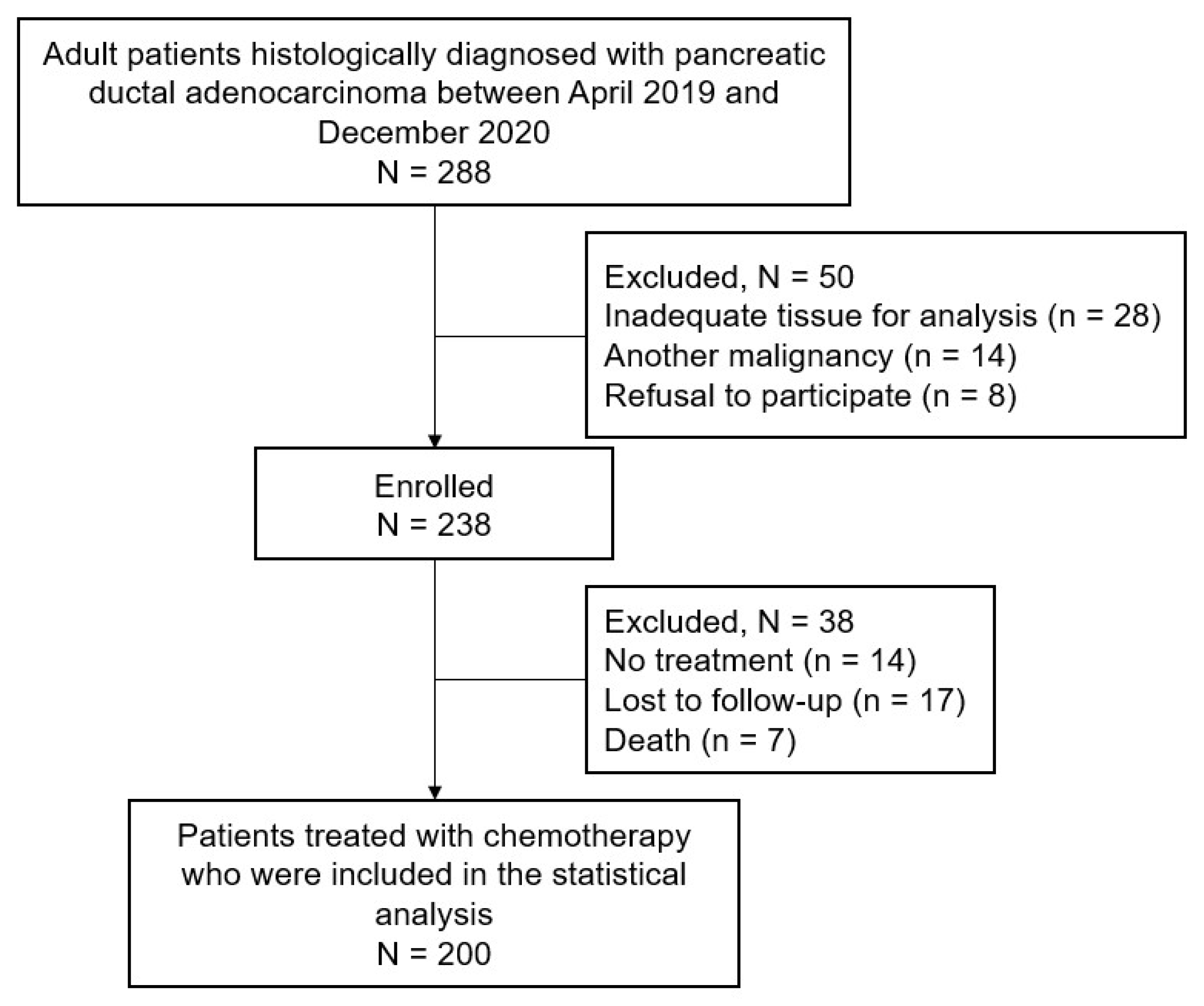
2.1. Patient Population

This study was approved by the institutional review board of the National Cancer Center, Republic of Korea (IRB number: NCC2019-0034) and was registered at ClinicalTrials.gov (identifier: NCT04281511). All participants provided written informed consent. Patients diagnosed with histologically proven pancreatic ductal adenocarcinoma between April 2019 and December 2020 were eligible for enrollment. We excluded patients with insufficient tissue for biomarker analysis, those who had been treated for another current malignancy, and those who did not provide informed consent to participate in the study. After enrollment, tissue and blood samples were collected to identify potential biomarkers before treatment. Tumor tissue samples were obtained from the surgical specimens of patients with resectable PA or the biopsy specimens of those with unresectable cancer before chemo- or radiotherapy. Primary resection specimens were used to evaluate tissue biomarkers in patients with recurrence after curative-intent surgery. Blood samples were collected to detect KRAS, BRCA1/2, and ATM mutations.

2.2. Treatment Strategy

The optimal treatment strategy was determined using a multidisciplinary team approach. After curative-intent surgery, patients who were fit underwent adjuvant chemotherapy with FOLFIRINOX (oxaliplatin, irinotecan, leucovorin, and 5-fluorouracil), gemcitabine plus capecitabine, or gemcitabine monotherapy, depending on their performance status. The neoadjuvant treatment consisted of chemotherapy with FOLFIRINOX and/or radiotherapy. Palliative chemotherapy was considered for patients with unresectable PA, and the regimen was determined at the discretion of the treating physician. Computed tomography and/or magnetic resonance imaging were performed every 8–12 weeks to evaluate treatment response or tumor recurrence. The tumors were evaluated using the Response Evaluation Criteria in Solid Tumors version 1.1.

2.3. Blood Biomarkers

Several blood biomarkers, including CA19-9, mKRAS ctDNA, and germline BRCA1/2 and ATM mutations, were evaluated. Serum CA19-9 levels were measured using an immunoradiometric assay (Riakey CA19-9 kit; Shinjin Medics Inc., Goyang, Republic of Korea). The mKRAS ctDNA concentration was measured using a KRAS screening multiplex droplet digital polymerase chain reaction (ddPCR) kit (Bio-Rad, #1863506), which covers seven different KRAS mutations (G12A, G12C, G12D, G12R, G12S, G12V, and G13D) [11]. Detection of germline mutations in BRCA1/2 and ATM was performed using a customized hereditary cancer panel (Celemics, Seoul, Republic of Korea) that targets coding sequences and intron–exon boundaries in more than 28 cancer predisposition genes, as previously reported [20].

2.4. Tissue Biomarkers

Several tissue biomarkers associated with prediction and prognosis in PA were evaluated. Hematoxylin and eosin-stained slides containing specimens from each tumor sample were reviewed, and representative tumor regions and corresponding formalin-fixed paraffin-embedded tissue blocks were selected for immunohistochemistry (IHC). IHC staining was performed using a BenchMark XT automated slide stainer (Ventana Medical Systems, Inc., Tucson, AZ, USA) for one to three paraffin-embedded blocks (median of two blocks) of each specimen. The primary antibodies used were rabbit polyclonal antibodies against human DCK (LS-B1825, LifeSpan Bioscience, Inc., Seattle, WA, USA), human hENT1 (11337-1-AP, ProteinTech Group, Inc., Chicago, IL, USA), human CES2 (HPA018897, Atlas Antibodies Inc., Stockholm, Sweden), and anti-GATA6 polyclonal antibody (HPA066629, Atlas Antibodies Inc.)
Representative images of the tissue biomarkers are shown in Supplementary Figure S1. Protein expression in tumor cells was scored by immunostaining intensity as follows: grade 0: not stained; grade 1: faintly positive; grade 2: weakly to moderately positive; and grade 3: strongly positive. IHC labeling was defined as positive when hENT1 and DCK were 1+ or higher, and CES2 was classified as low (negative, 1+) or high (2+, 3+). The expression of GATA6 was assessed using digital assistance as follows: GATA6 slides were scanned using an Aperio AT2 scanner (Leica Biosystems Inc., Vista, CA, USA) at 20× magnification, and open-source quantitative pathology and bioimage analysis software (QuPath v0.4.0; https://qupath.readthedocs.io) was used to view and analyze whole-slide images [21]. The proportion of GATA6-positive cells was determined by the ratio of positive cells to the total number of cells and divided into two groups: GATA6 high and low expression (Supplementary Figure S1).
GATA6 RNA expression was adopted as a potential tissue biomarker because GATA6 expression was associated with the classical subtype, which had a better outcome and favorable response to 5-fluorouracil-based treatment [22]. Total RNA extractions were performed from FFPE samples using a Maxwell RSC RNA FFPE kit (Promega, Madison, WI, USA) on a Maxwell RSC 48 Instrument (Promega), according to the manufacturer’s protocol. The RNA was eluted in 40 µL of nuclease-free water. RNA concentrations, as well as the purity and contamination ratio values, were measured on a NanoDrop 8000 spectrophotometer (Thermo Scientific, Waltham, MA, USA), and 400 ng was used to synthesize cDNA using a cDNA EcoDry Premix kit (Takara Bio, Shiga, Japan) with random hexamer primer premix. The levels were assessed through polymerase chain reaction using the QX200TM Droplet DigitalTM PCR System (Bio-Rad, Hercules, CA, USA). The mixture of the GATA6 FAM target probe and GUSB HEX reference probe, 2× ddPCR supermix (no dUTP), and cDNA template was adjusted to a final volume of 20.0 μL with diethylpyrocarbonate-treated water. Caco-2, U-2 OS, and HEK293T cells were used as positive controls. Relative GATA6 RNA expression in each sample was normalized to GUSB using QuantaSoft Software version 1.6.6 (Bio-rad). Patients were divided into GATA6 high and low expression groups using the median value of gene expression (see the Supplementary Data File).

2.5. Statistical Analyses

This single-arm prospective observational study compared registry data, using overall survival (OS) as the primary endpoint. For biomarker-positive patients, an interim analysis was planned based on the O’Brien–Fleming alpha spending method, with a hazard ratio (HR) of 0.67, alpha assumed to be a two-sided type I error of 5%, and power assumed to be 80%. Considering a recruitment period of 36 months, a follow-up period of 24 months, and a 10% dropout rate, 119 patients were enrolled in the study. As 50% of the patients with PA were expected to have at least one positive biomarker, 238 study participants were enrolled in the study.
OS was defined as the time from patient enrollment to death and progression-free survival (PFS) as the time from patient enrollment until tumor progression or death. For patients who underwent curative-intent surgery, recurrence-free survival (RFS) was defined as the time from surgical resection to the onset of tumor recurrence or death. Survival curves were estimated using the Kaplan–Meier method and compared using log-rank tests. To identify biomarkers with prognostic significance, a Cox proportional hazards model was used, adjusting for clinical variables with prognostic significance at baseline. CA19-9 levels were dichotomized with a known cutoff concentration of 37 U/mL, and mKRAS ctDNA concentration was dichotomized with a previously described cutoff value of 160 copies/mL [11]. All analyses were performed using SAS version 9.4 (SAS Institute Inc., Cary, NC, USA) and R version 4.3.3 (R Foundation for Statistical Computing, Vienna, Austria).

3. Results

3.1. Patient Characteristics

Of the 288 patients with histologically confirmed pancreatic ductal adenocarcinoma who were screened, 238 were enrolled. Of these, 200 patients who received at least one cycle of chemotherapy were included in the final analysis (Figure 1). The baseline characteristics are shown in Table 1. The median age was 65 years (range: 45–90 years) and 110 patients (55%) were male. Most patients had an Eastern Cooperative Oncology Group performance status score of 0. At enrollment, 46 (23%) had resectable PA, 58 (29%) had borderline resectable or locally advanced PA, and 96 (48%) had metastatic PA. Most patients received palliative chemotherapy, with FOLFIRINOX being the most common regimen.

3.2. Baseline Distribution of Candidate Biomarkers

The distribution of candidate biomarkers is summarized in Supplementary Table S1. The mKRAS ctDNA concentration was significantly higher in patients with metastatic disease than those with localized disease (resectable, borderline resectable, and locally advanced) (Supplementary Figure S2A). Germline mutations in BRCA1/2 were observed in 22 patients (3 pathogenic variants and 19 of uncertain significance) and ATM mutations in 15 patients (4 pathogenic variants and 11 of uncertain significance). Serum CA19-9 levels were elevated in 65% of patients.
Among the tissue biomarkers, GATA6 expression was elevated in 53 patients at the RNA level and in 77 patients according to IHC measurements (Supplementary Table S1). GATA6 RNA and tissue (IHC) expression levels were correlated with each other and associated with the cancer stage (Supplementary Figure S2B–D). Positivity for hENT1 and DCK was observed in 88 (44%) and 80 (40%) patients, respectively. High CES2 expression was observed in 32 (16%) patients (Supplementary Table S1).

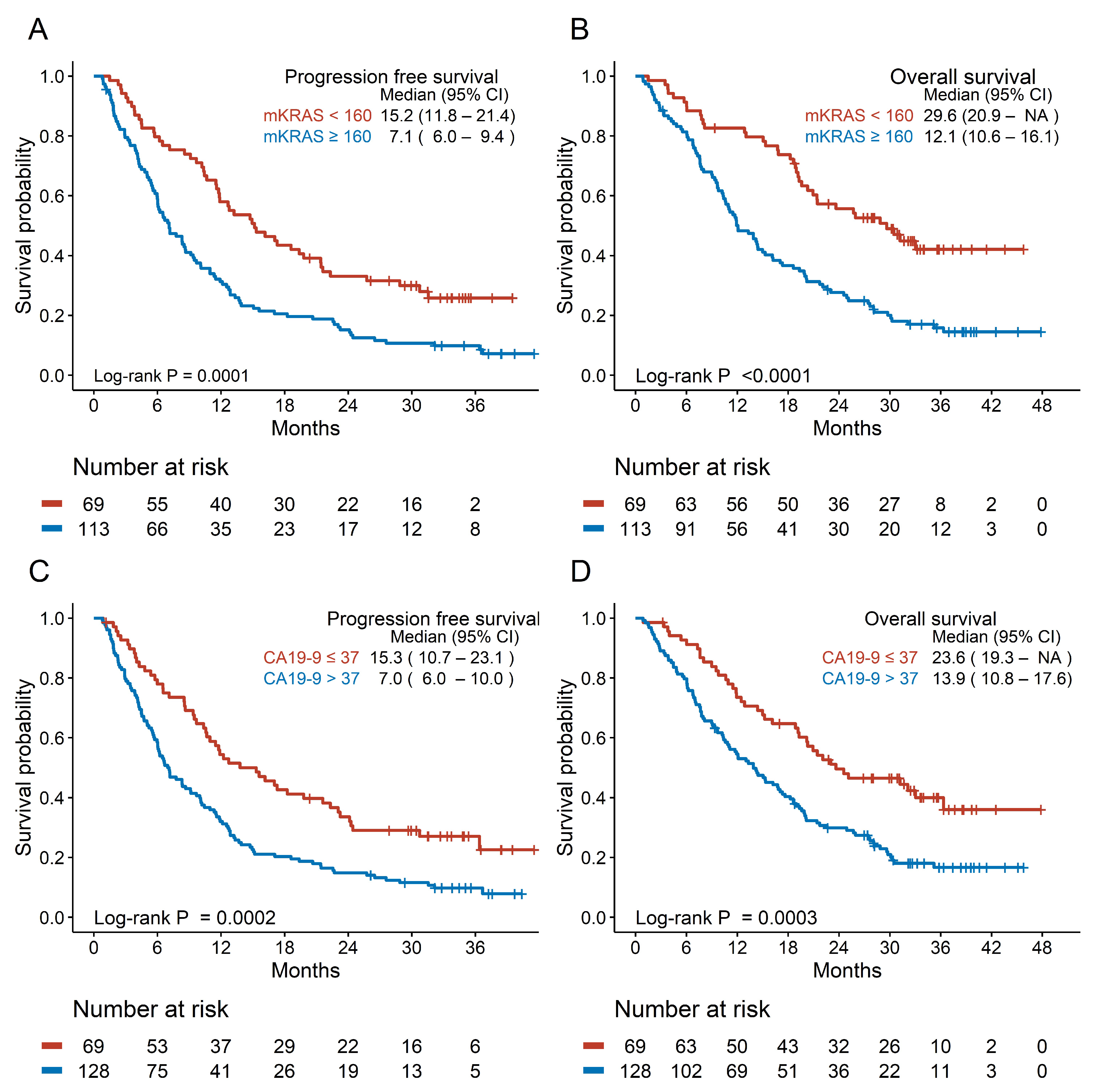
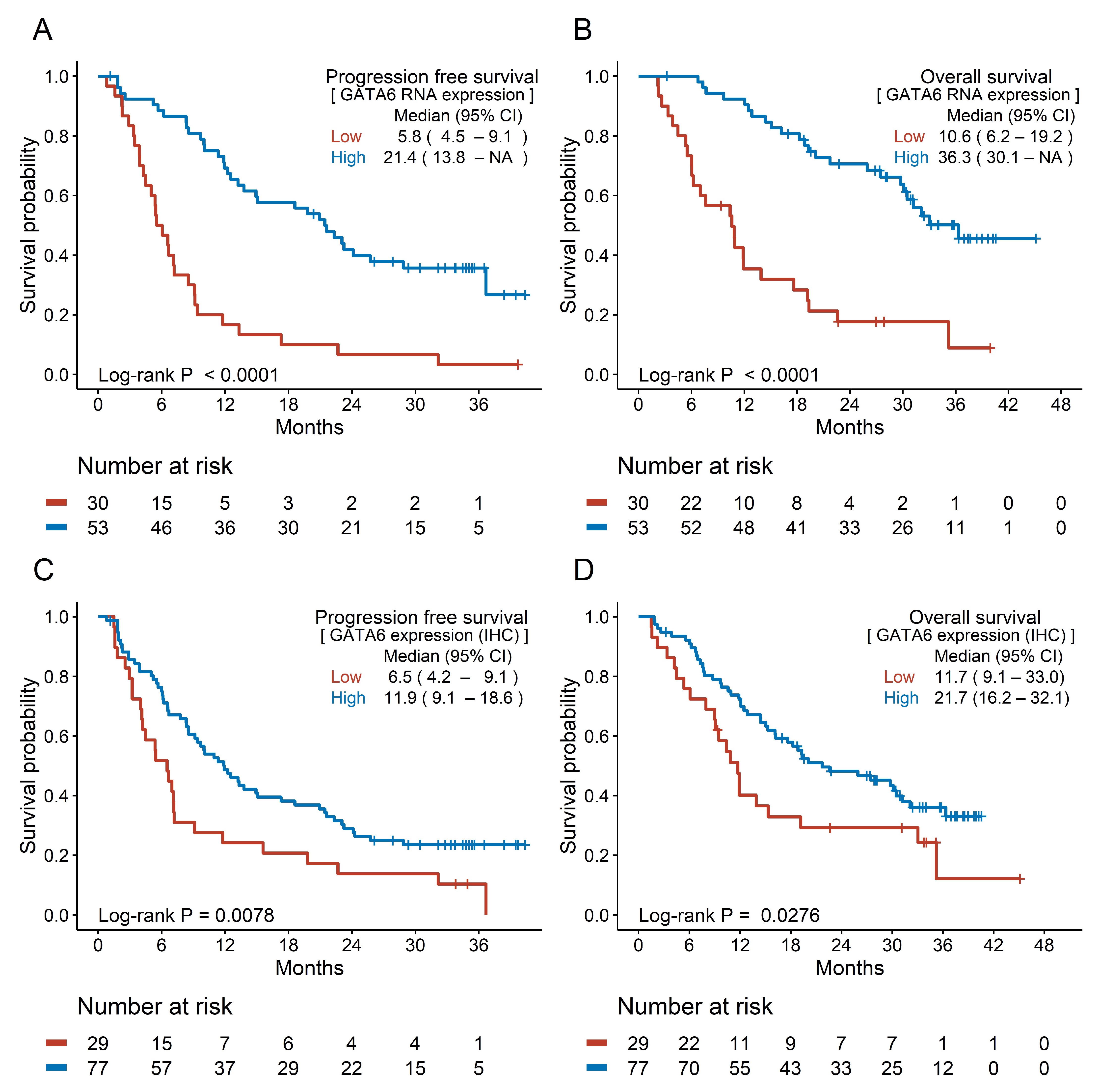
3.3. Prognostic Efficacy of Candidate Biomarkers

The median duration of follow-up was 35.5 months (range: 3.2–47.8 months). In total, 145 (72.5%) patients died, and 169 (84.5%) experienced disease progression. The median OS was 9.3 months (95% confidence interval [CI]: 7.7–11.9) for metastatic disease and 27.7 months (95% CI: 21.4–not applicable) for localized disease. The median PFS was 5.7 months (95% CI: 4.7–6.6) for metastatic disease and 13.9 months (95% CI: 12.1–21.4) for localized disease (Supplementary Figure S3).
Patients with mKRAS ctDNA concentrations >160 copies/mL had significantly shorter PFS (7.1 vs. 15.2 months) and OS (12.1 vs. 29.6 months) than those with concentrations <160 copies/mL (Figure 2A,B). Patients with elevated CA19-9 levels had shorter PFS (7.0 vs. 15.3 months, respectively) and OS (13.9 vs. 23.6 months, respectively) than those with normal levels (Figure 2C,D). Patients with pathogenic germline variants showed a trend toward longer PFS and OS, although this was not statistically significant (Supplementary Figure S4).
Among tissue biomarkers, patients with high GATA6 expression showed better survival outcomes than those with low GATA6 expression. The median PFS and OS in patients with high GATA6 RNA expression were 21.4 months and 36.3 months compared with 5.8 months and 10.6 months in those with low RNA expression, respectively (Figure 3A,B). High GATA6 expression was associated with longer survival (median PFS: 11.9 vs. 6.5 months; median OS: 21.7 vs. 11.7 months) (Figure 3C,D). No significant association was found between hENT1 and CES2 expression and survival outcomes, whereas DCK positivity was associated with longer OS (20.1 vs. 12.0 months) (Supplementary Figure S5).

3.4. Cox Regression Analysis of Survival

Cancer stage was significantly associated with both PFS and OS among clinical factors (Supplementary Table S2). Among biomarkers, mKRAS ctDNA concentration, CA19-9 level, and GATA6 RNA expression were significantly associated with PFS in the univariable Cox model, and DCK was significantly associated with OS (Table 2). After adjusting for cancer stage, high mKRAS ctDNA concentration (HR: 1.508 and 95% CI: 1.052–2.161 for PFS; HR: 1.796 and 95% CI: 1.203–2.681 for OS) and CA19-9 level (HR: 1.647 and 95% CI: 1.177–2.306 for PFS; HR: 1.803 and 95% CI: 1.248–2.605 for OS) remained significantly associated with poor survival. High GATA6 RNA expression was associated with better survival (HR: 0.336 and 95% CI: 0.195–0.582 for PFS; HR: 0.304 and 95% CI: 0.165–0.560 for OS). DCK expression was not significantly associated with OS after adjusting for cancer stage.

3.5. Subgroup Analysis Based on the Treatment Setting

We further evaluated the prognostic value of mKRAS ctDNA and GATA6 according to treatment modality owing to the heterogeneity of cancer stages among patients. In patients who underwent curative-intent surgery before/after chemotherapy, a high mKRAS ctDNA concentration was associated with shorter RFS (14.3 vs. 25.5 months, p = 0.068) (Supplementary Figure S6A). Conversely, high GATA6 RNA expression indicated a trend toward longer RFS in patients with resected PA (23.9 vs. 13.2 months, p = 0.056) (Supplementary Figure S6B). Among clinical factors, the pathological T and N stages were significant prognostic factors; however, they were not significantly associated with the KRAS level or GATA6 RNA expression (Supplementary Table S3 and Supplementary Figure S7). In patients who received palliative chemotherapy, a high mKRAS ctDNA concentration was associated with poor survival outcomes (6.0 vs. 10.4 months for PFS; 10.6 vs. 19.6 months for OS) (Supplementary Figure S6C,D).

4. Discussion

Our findings demonstrate that among several candidate biomarkers, mKRAS ctDNA concentration and GATA6 RNA expression were meaningful prognostic markers in PA, alongside the CA19-9 level. A high mKRAS ctDNA concentration and elevated CA19-9 level were associated with poor PFS and OS, whereas high GATA6 RNA expression correlated with better survival outcomes. In resected PA, these markers were associated with RFS after surgery, and in palliative chemotherapy, a high mKRAS ctDNA concentration was significantly associated with poor OS.
ctDNA is increasingly recognized as a promising cancer biomarker for diagnosis, treatment, and prognosis because it can identify tumor-specific abnormalities. KRAS mutations are key oncogenic drivers of PA and are found in almost all patients with this disease. mKRAS ctDNA showed significant associations with survival in a series of studies and meta-analyses [23,24]. Guven et al. demonstrated that positive ctDNA was associated with lower RFS/PFS and OS in both localized and advanced PA in a pooled analysis, and the KRAS mutation was a target for ctDNA analyses [23].
Although most previous studies focused on the detection of mKRAS ctDNA, several have used ctDNA as a continuous biomarker with mutant allele fraction (MAF) values [8,25,26]. Quantification of the biomarker may help generalize the clinical use of mKRAS ctDNA for real-world applications, similar to serum CA19-9. Our previous research indicated that a high mKRAS ctDNA concentration was associated with shorter PFS and OS, and this study confirms its prognostic value and clinical applicability, aligning with other studies [11]. Further study into the prognostic value of the mKRAS subtype may help inform treatment strategies given the recent advances in KRAS inhibitors [27].
Regarding GATA6 expression, our findings align with previous research, highlighting its role as a favorable prognostic marker. Duan et al. demonstrated that high GATA6 IHC values are associated with improved clinical outcomes in advanced PA [28]. GATA6 IHC values are correlated with GATA6 RNA gene expression, which is consistent with our results. O’Kane et al. showed that GATA6 expression, measured using both RNA sequencing and in situ hybridization, was a prognostic factor in multivariate analysis [29]. Our results further support the prognostic value of GATA6 by showing significant associations between high GATA6 RNA expression and longer PFS and OS, reinforcing its potential as a prognostic biomarker for PA.
The strengths of our study include the comprehensive assessment of potential biomarkers and its prospective cohort design, which allowed for a rigorous evaluation of the prognostic impact. Among the candidate biomarkers, mKRAS ctDNA and GATA6 were identified as significant prognostic factors, along with CA19-9, consistent with previous studies, providing a strong rationale for their clinical implications. The use of advanced molecular techniques, such as ddPCR for qualitative mKRAS detection and digital quantification for GATA6 assessment, further enhanced the robustness and reliability of our findings.
However, this study has some limitations. First, although we performed a comprehensive assessment of multiple biomarkers in a large number of patients, some tissue biomarkers could not be assessed simultaneously owing to limited tissue volume. Second, for germline BRCA1/2 or ATM mutations, significance was not achieved owing to their low prevalence. Germline mutation carriers of homologous recombination repair genes, such as ATM, BRCA1/2, and PALB2, are considered predictive and prognostic biomarkers, especially in patients receiving platinum-based chemotherapy [30,31]. Finally, although our study confirms the clinical significance of the biomarkers, it does not elucidate a causal relationship based on biological mechanisms. KRAS is a representative oncogenic gene driver of PA, whereas GATA6, a transcription factor for pancreatic development, correlates with the molecular subtypes of PA and plays an inhibitory role in epithelial–mesenchymal transition, which may explain the opposite trends in PA prognosis [22,29].

5. Conclusions

Our findings highlight the significant prognostic value of mKRAS ctDNA and GATA6 RNA expression in patients with PA undergoing chemotherapy. These biomarkers provide valuable insights into patient prognosis and guide therapeutic decision-making, ultimately improving personalized treatment strategies and outcomes in patients with PA.

Supplementary Materials

The following supporting information can be downloaded at: https://www.mdpi.com/article/10.3390/cancers17050896/s1, Figure S1: Representative images of the tissue biomarkers; Figure S2: Distribution of mutant KRAS status and GATA6 expression; Figure S3: Survival analysis according to cancer stage; Figure S4: Survival analysis according to the blood biomarkers. Figure S5: Survival analysis according to the tissue biomarkers. Figure S6: Analysis of subgroups according to treatment type; Figure S7: Distribution of mutant KRAS status and GATA6 RNA expression according to pathological T and N stage in surgical patients; Table S1: Biomarker distributions; Table S2: Cox hazard model of baseline characteristics; Table S3: Cox Hazard Model of Baseline Characteristics in Surgical Patients.

Author Contributions

Concept and design: S.M.W., S.-Y.K. and Y.-H.K. Acquisition, analysis, and interpretation of data: J.W.C., D.-e.L., N.H., S.H., H.K. and M.R.L. Drafting of the manuscript: J.W.C., D.-e.L., N.H. and S.-Y.K. Critical review of the manuscript for important intellectual content: S.M.W., S.-Y.K., Y.-H.K., T.H.K. and W.J.L. Statistical analysis: D.-e.L. and J.W.C. Obtained funding: S.M.W., S.-Y.K. and J.W.C. Administrative, technical, and material support: S.M.W., S.-Y.K., H.M.P., S.-S.H., S.-J.P. and W.J.L. Supervision: S.M.W. and S.-Y.K. All authors have read and agreed to the published version of the manuscript.

Funding

This study was supported by grants from the National Cancer Center (grant numbers: 1910191, 1910192, 2212470, and 2310280). The funders of the study had no role in study design; in the collection, analysis, and interpretation of data; in the writing of the report; or in the decision to submit the paper for publication.

Institutional Review Board Statement

This study was approved by the institutional review board of the National Cancer Center, Republic of Korea (IRB number: NCC2019-0034; approval date: 18 February 2019).

Informed Consent Statement

Informed consent was obtained from all subjects involved in the study.

Data Availability Statement

All data used and analyzed during the current study are available from the corresponding author upon reasonable request.

Acknowledgments

The authors thank Young Hwa Kang, Jihye Yu, and Mee Young Lee for their assistance in collecting patient specimens and clinical data.

Conflicts of Interest

The authors declare no conflicts of interest.

Abbreviations

PA: pancreatic adenocarcinoma; CA19-9: carbohydrate antigen 19-9; KRAS: KRAS proto-oncogene, GTPase; ctDNA: circulating tumor DNA; mKRAS: mutant KRAS; BRCA1/2: BRCA1 DNA repair-associated and BRCA2 DNA repair-associated; ATM: ATM serine/threonine kinase; GATA6: GATA binding protein 6; SLC29A1: solute carrier family 29 member 1; hENT1: human equilibrative nucleoside transporter 1; DCK: deoxycytidine kinase; ddPCR: droplet digital polymerase chain reaction; IHC: immunohistochemistry; OS: overall survival; PFS: progression-free survival; RFS: recurrence-free survival; HR: hazard ratio; CI: confidence interval.

References

  1. Pourshams, A.; Sepanlou, S.G.; Ikuta, K.S.; Bisignano, C.; Safiri, S.; Roshandel, G.; Sharif, M.; Khatibian, M.; Fitzmaurice, C.; Nixon, M.R.; et al. The global, regional, and national burden of pancreatic cancer and its attributable risk factors in 195 countries and territories, 1990–2017: A systematic analysis for the Global Burden of Disease Study 2017. Lancet Gastroenterol. Hepatol. 2019, 4, 934–947. [Google Scholar] [CrossRef] [PubMed]
  2. Huang, J.; Lok, V.; Ngai, C.H.; Zhang, L.; Yuan, J.; Lao, X.Q.; Ng, K.; Chong, C.; Zheng, Z.J.; Wong, M.C.S. Worldwide Burden of, Risk Factors for, and Trends in Pancreatic Cancer. Gastroenterology 2021, 160, 744–754. [Google Scholar] [CrossRef]
  3. Conroy, T.; Hammel, P.; Hebbar, M.; Ben Abdelghani, M.; Wei, A.C.; Raoul, J.L.; Chone, L.; Francois, E.; Artru, P.; Biagi, J.J.; et al. FOLFIRINOX or Gemcitabine as Adjuvant Therapy for Pancreatic Cancer. N. Engl. J. Med. 2018, 379, 2395–2406. [Google Scholar] [CrossRef]
  4. Miyasaka, Y.; Ohtsuka, T.; Kimura, R.; Matsuda, R.; Mori, Y.; Nakata, K.; Kakihara, D.; Fujimori, N.; Ohno, T.; Oda, Y.; et al. Neoadjuvant Chemotherapy with Gemcitabine Plus Nab-Paclitaxel for Borderline Resectable Pancreatic Cancer Potentially Improves Survival and Facilitates Surgery. Ann. Surg. Oncol. 2019, 26, 1528–1534. [Google Scholar] [CrossRef]
  5. Murphy, J.E.; Wo, J.Y.; Ryan, D.P.; Jiang, W.; Yeap, B.Y.; Drapek, L.C.; Blaszkowsky, L.S.; Kwak, E.L.; Allen, J.N.; Clark, J.W.; et al. Total Neoadjuvant Therapy With FOLFIRINOX Followed by Individualized Chemoradiotherapy for Borderline Resectable Pancreatic Adenocarcinoma: A Phase 2 Clinical Trial. JAMA Oncol. 2018, 4, 963–969. [Google Scholar] [CrossRef]
  6. Tempero, M.; O'Reilly, E.; Van Cutsem, E.; Berlin, J.; Philip, P.; Goldstein, D.; Tabernero, J.; Borad, M.; Bachet, J.; Parner, V.; et al. LBA-1 Phase 3 APACT trial of adjuvant nab-paclitaxel plus gemcitabine (nab-P + Gem) vs gemcitabine (Gem) alone in patients with resected pancreatic cancer (PC): Updated 5-year overall survival. Ann. Oncol. 2021, 32, S226. [Google Scholar] [CrossRef]
  7. Berger, A.C.; Garcia, M., Jr.; Hoffman, J.P.; Regine, W.F.; Abrams, R.A.; Safran, H.; Konski, A.; Benson, A.B., 3rd; MacDonald, J.; Willett, C.G. Postresection CA 19-9 predicts overall survival in patients with pancreatic cancer treated with adjuvant chemoradiation: A prospective validation by RTOG 9704. J. Clin. Oncol. 2008, 26, 5918–5922. [Google Scholar] [CrossRef] [PubMed]
  8. Bernard, V.; Kim, D.U.; San Lucas, F.A.; Castillo, J.; Allenson, K.; Mulu, F.C.; Stephens, B.M.; Huang, J.; Semaan, A.; Guerrero, P.A.; et al. Circulating Nucleic Acids Are Associated with Outcomes of Patients with Pancreatic Cancer. Gastroenterology 2019, 156, 108–118.e104. [Google Scholar] [CrossRef] [PubMed]
  9. Kim, S.; Park, B.K.; Seo, J.H.; Choi, J.; Choi, J.W.; Lee, C.K.; Chung, J.B.; Park, Y.; Kim, D.W. Carbohydrate antigen 19-9 elevation without evidence of malignant or pancreatobiliary diseases. Sci. Rep. 2020, 10, 8820. [Google Scholar] [CrossRef]
  10. Haas, M.; Ormanns, S.; Baechmann, S.; Remold, A.; Kruger, S.; Westphalen, C.B.; Siveke, J.T.; Wenzel, P.; Schlitter, A.M.; Esposito, I.; et al. Extended RAS analysis and correlation with overall survival in advanced pancreatic cancer. Br. J. Cancer 2017, 116, 1462–1469. [Google Scholar] [CrossRef]
  11. Kim, M.K.; Woo, S.M.; Park, B.; Yoon, K.A.; Kim, Y.H.; Joo, J.; Lee, W.J.; Han, S.S.; Park, S.J.; Kong, S.Y. Prognostic Implications of Multiplex Detection of KRAS Mutations in Cell-Free DNA from Patients with Pancreatic Ductal Adenocarcinoma. Clin. Chem. 2018, 64, 726–734. [Google Scholar] [CrossRef] [PubMed]
  12. Perkhofer, L.; Gout, J.; Roger, E.; Kude de Almeida, F.; Baptista Simões, C.; Wiesmüller, L.; Seufferlein, T.; Kleger, A. DNA damage repair as a target in pancreatic cancer: State-of-the-art and future perspectives. Gut 2021, 70, 606. [Google Scholar] [CrossRef] [PubMed]
  13. Woo, S.M.; Kim, M.K.; Park, B.; Cho, E.H.; Lee, T.R.; Ki, C.S.; Yoon, K.A.; Kim, Y.H.; Choi, W.; Kim, D.Y.; et al. Genomic Instability of Circulating Tumor DNA as a Prognostic Marker for Pancreatic Cancer Survival: A Prospective Cohort Study. Cancers 2021, 13, 5466. [Google Scholar] [CrossRef]
  14. Bailey, P.; Chang, D.K.; Nones, K.; Johns, A.L.; Patch, A.M.; Gingras, M.C.; Miller, D.K.; Christ, A.N.; Bruxner, T.J.; Quinn, M.C.; et al. Genomic analyses identify molecular subtypes of pancreatic cancer. Nature 2016, 531, 47–52. [Google Scholar] [CrossRef] [PubMed]
  15. Collisson, E.A.; Sadanandam, A.; Olson, P.; Gibb, W.J.; Truitt, M.; Gu, S.; Cooc, J.; Weinkle, J.; Kim, G.E.; Jakkula, L.; et al. Subtypes of pancreatic ductal adenocarcinoma and their differing responses to therapy. Nat. Med. 2011, 17, 500–503. [Google Scholar] [CrossRef]
  16. Farrell, J.J.; Elsaleh, H.; Garcia, M.; Lai, R.; Ammar, A.; Regine, W.F.; Abrams, R.; Benson, A.B.; Macdonald, J.; Cass, C.E.; et al. Human equilibrative nucleoside transporter 1 levels predict response to gemcitabine in patients with pancreatic cancer. Gastroenterology 2009, 136, 187–195. [Google Scholar] [CrossRef]
  17. Wei, C.H.; Gorgan, T.R.; Elashoff, D.A.; Hines, O.J.; Farrell, J.J.; Donahue, T.R. A meta-analysis of gemcitabine biomarkers in patients with pancreaticobiliary cancers. Pancreas 2013, 42, 1303–1310. [Google Scholar] [CrossRef]
  18. Capello, M.; Fahrmann, J.F.; Perez, M.V.R.; Vykoukal, J.V.; Irajizad, E.; Tripathi, S.C.; Roife, D.; Bantis, L.E.; Kang, Y.a.; Kundnani, D.L.; et al. CES2 Expression in Pancreatic Adenocarcinoma Is Predictive of Response to Irinotecan and Is Associated With Type 2 Diabetes. JCO Precis. Oncol. 2020, 4, 426–436. [Google Scholar] [CrossRef]
  19. Wang, F.; Xia, X.; Yang, C.; Shen, J.; Mai, J.; Kim, H.C.; Kirui, D.; Kang, Y.; Fleming, J.B.; Koay, E.J.; et al. SMAD4 Gene Mutation Renders Pancreatic Cancer Resistance to Radiotherapy through Promotion of Autophagy. Clin. Cancer Res. 2018, 24, 3176–3185. [Google Scholar] [CrossRef]
  20. Ryu, K.H.; Park, S.; Chun, J.W.; Cho, E.; Choi, J.; Lee, D.E.; Shim, H.; Kim, Y.H.; Han, S.S.; Park, S.J.; et al. Prevalence and Risk Factors of Germline Pathogenic Variants in Pancreatic Ductal Adenocarcinoma. Cancer Res. Treat. 2023, 55, 1303–1312. [Google Scholar] [CrossRef]
  21. Bankhead, P.; Loughrey, M.B.; Fernández, J.A.; Dombrowski, Y.; McArt, D.G.; Dunne, P.D.; McQuaid, S.; Gray, R.T.; Murray, L.J.; Coleman, H.G.; et al. QuPath: Open source software for digital pathology image analysis. Sci. Rep. 2017, 7, 16878. [Google Scholar] [CrossRef] [PubMed]
  22. Chan-Seng-Yue, M.; Kim, J.C.; Wilson, G.W.; Ng, K.; Figueroa, E.F.; O’Kane, G.M.; Connor, A.A.; Denroche, R.E.; Grant, R.C.; McLeod, J.; et al. Transcription phenotypes of pancreatic cancer are driven by genomic events during tumor evolution. Nat. Genet. 2020, 52, 231–240. [Google Scholar] [CrossRef] [PubMed]
  23. Guven, D.C.; Sahin, T.K.; Yildirim, H.C.; Aktepe, O.H.; Dizdar, O.; Yalcin, S. A systematic review and meta-analysis of the association between circulating tumor DNA (ctDNA) and prognosis in pancreatic cancer. Crit. Rev. Oncol./Hematol. 2021, 168, 103528. [Google Scholar] [CrossRef]
  24. Sivapalan, L.; Kocher, H.M.; Ross-Adams, H.; Chelala, C. Molecular profiling of ctDNA in pancreatic cancer: Opportunities and challenges for clinical application. Pancreatology 2021, 21, 363–378. [Google Scholar] [CrossRef]
  25. Pietrasz, D.; Pécuchet, N.; Garlan, F.; Didelot, A.; Dubreuil, O.; Doat, S.; Imbert-Bismut, F.; Karoui, M.; Vaillant, J.-C.; Taly, V.; et al. Plasma Circulating Tumor DNA in Pancreatic Cancer Patients Is a Prognostic Marker. Clin. Cancer Res. 2017, 23, 116–123. [Google Scholar] [CrossRef] [PubMed]
  26. Wei, T.; Zhang, Q.; Li, X.; Su, W.; Li, G.; Ma, T.; Gao, S.; Lou, J.; Que, R.; Zheng, L.; et al. Monitoring Tumor Burden in Response to FOLFIRINOX Chemotherapy Via Profiling Circulating Cell-Free DNA in Pancreatic Cancer. Mol. Cancer Ther. 2019, 18, 196–203. [Google Scholar] [CrossRef] [PubMed]
  27. Norton, C.; Shaw, M.S.; Rubnitz, Z.; Smith, J.; Soares, H.P.; Nevala-Plagemann, C.D.; Garrido-Laguna, I.; Florou, V. KRAS Mutation Status and Treatment Outcomes in Patients With Metastatic Pancreatic Adenocarcinoma. JAMA Netw. Open 2025, 8, e2453588. [Google Scholar] [CrossRef]
  28. Duan, K.; Jang, G.H.; Grant, R.C.; Wilson, J.M.; Notta, F.; O’Kane, G.M.; Knox, J.J.; Gallinger, S.; Fischer, S. The value of GATA6 immunohistochemistry and computer-assisted diagnosis to predict clinical outcome in advanced pancreatic cancer. Sci. Rep. 2021, 11, 14951. [Google Scholar] [CrossRef]
  29. O’Kane, G.M.; Grunwald, B.T.; Jang, G.H.; Masoomian, M.; Picardo, S.; Grant, R.C.; Denroche, R.E.; Zhang, A.; Wang, Y.; Lam, B.; et al. GATA6 Expression Distinguishes Classical and Basal-like Subtypes in Advanced Pancreatic Cancer. Clin. Cancer Res. 2020, 26, 4901–4910. [Google Scholar] [CrossRef]
  30. Kasi, P.M.; Couch, F.; Bamlet, W.R.; Hu, C.; Hart, S.; Polley, E.; Petersen, G.M.; McWilliams, R.R. Germline BRCA1/2, PALB2, and ATM mutations in 3030 patients with pancreatic adenocarcinoma: Survival analysis of carriers and noncarriers. J. Clin. Oncol. 2018, 36, 280. [Google Scholar] [CrossRef]
  31. Yadav, S.; Kasi, P.M.; Bamlet, W.R.; Ho, T.P.; Polley, E.C.; Hu, C.; Hart, S.N.; Rabe, K.G.; Boddicker, N.J.; Gnanaolivu, R.D.; et al. Effect of Germline Mutations in Homologous Recombination Repair Genes on Overall Survival of Patients with Pancreatic Adenocarcinoma. Clin. Cancer Res. 2020, 26, 6505–6512. [Google Scholar] [CrossRef] [PubMed]
Figure 1. Participant enrolment flow diagram. Adult patients with a histologic diagnosis of pancreatic ductal adenocarcinoma were screened, and those who received chemotherapy were included in the final analysis.
Cancers 17 00896 g001
Figure 2. Survival analysis according to the blood biomarkers. (A) Progression-free survival and (B) overall survival according to mutant KRAS status. (C) Progression-free survival and (D) overall survival according to CA19-9 level. CA19-9: carbohydrate antigen 19-9; CI: confidence interval; KRAS: KRAS proto-oncogene, GTPase; mKRAS: mutant KRAS; NA: not assessed.
Cancers 17 00896 g002
Figure 3. Survival analysis according to the tissue biomarkers. (A) Progression-free survival and (B) overall survival according to GATA6 RNA expression. (C) Progression-free survival and (D) overall survival according to GATA6 tissue expression (IHC). CI: confidence interval; GATA6: GATA binding protein 6; IHC: immunohistochemistry; NA: not assessed.
Cancers 17 00896 g003
Table 1. Baseline patient characteristics.
VariableN = 200
Age (years)Median (range)65 (45–90)
SexFemale90 (45.0%)
Male110 (55.0%)
ECOG-PS0128 (64.0%)
1 or more 72 (36.0%)
Tumor locationHead or neck or uncinate process92 (46.0%)
body or tail108 (54.0%)
StageResectable 46 (23.0%)
Borderline resectable/locally advanced58 (29.0%)
Metastatic/recurrent96 (48.0%)
SurgeryNo125 (62.5%)
Yes75 (37.5%)
ChemotherapyNeoadjuvant21 (10.5%)
Adjuvant45 (22.5%)
Palliative 134 (67.0%)
RegimenFOLFIRINOX131 (65.5%)
Gemcitabine + nab-paclitaxel40 (20.0%)
Other 5-fluorouracil-based12 (6.0%)
Other gemcitabine-based 17 (8.5%)
ECOG-PS: European Cooperative Oncology Group Performance Status; FOLFIRINOX: oxaliplatin, irinotecan, leucovorin, and 5-fluorouracil.
Table 2. Cox proportional hazards model showing associations between biomarkers and survival.
VariableNProgression-Free SurvivalOverall Survival
EventUnivariableAdjusted for StageEventUnivariableAdjusted for StageAdjusted for Sex and Stage
HR (95% CI)p-ValueHR (95% CI)p-ValueHR (95% CI)p-ValueHR (95% CI)p-ValueHR (95% CI)p-Value
CA19-9 level
(U/mL)
≤37 69501 (ref) 1 (ref) 401 (ref) 1 (ref) 1 (ref)
>37 1281161.866 (1.336–2.605)<0.0011.647 (1.177–2.306)0.0036 1031.942 (1.345–2.802)<0.0011.803 (1.248–2.605)0.0017 1.764 (1.22–2.551)0.0026
mKRAS ctDNA concentration<160 69501 (ref) 1 (ref) 371 (ref) 1 (ref) 1 (ref)
(copies/mL)≥1601131031.965 (1.398–2.763)<0.0011.508 (1.052–2.161)0.0253 942.344 (1.598–3.437)<0.0011.796 (1.203–2.681)0.0042 1.813 (1.215–2.704)0.0036
BRCA mutationNot detected1751511 (ref) 1 (ref) 1311 (ref) 1 (ref) 1 (ref)
VUS/PV22150.717 (0.421–1.219)0.2190.724 (0.424–1.235)0.235120.650 (0.360–1.175)0.1530.640 (0.354–1.158)0.1400.694 (0.382–1.261)0.230
ATM mutationND1821551 (ref) 1 (ref) 1331 (ref) 1 (ref) 1 (ref)
VUS/PV15110.707 (0.384–1.305)0.2670.739 (0.401–1.365)0.334100.778 (0.409–1.479)0.4430.796 (0.418–1.516)0.4870.823 (0.432–1.568)0.553
hENT1Negative40331 (ref) 1 (ref) 311 (ref) 1 (ref) 1 (ref)
Positive88720.905 (0.598–1.368)0.63540.904 (0.598–1.367)0.6321 600.748 (0.484–1.156)0.19090.682 (0.440–1.055)0.0853 0.706 (0.455–1.093)0.1184
DCKNegative48431 (ref) 1 (ref) 411 (ref) 1 (ref) 1 (ref)
Positive80620.724 (0.490–1.070)0.10561.045 (0.692–1.579)0.8327 500.575 (0.380–0.870)0.00880.788 (0.513–1.210)0.2767 0.788 (0.513–1.21)0.2767
CES2Low 93761 (ref) 1 (ref) 641 (ref) 1 (ref) 1 (ref)
High32230.772 (0.484–1.231)0.27740.850 (0.532–1.358)0.4966 190.769 (0.461–1.284)0.31540.838 (0.501–1.400)0.4990 0.978 (0.579–1.652)0.9328
GATA6 (RNA)Low 30291 (ref) 1 (ref) 251 (ref) 1 (ref) 1 (ref)
High53340.266 (0.159–0.447)<0.00010.336 (0.195–0.582)<0.0001 240.225 (0.126–0.402)<0.00010.304 (0.165–0.560)0.0001 0.269 (0.144–0.502)<0.0001
GATA6 (Tissue)Low 29271 (ref) 1 (ref) 221 (ref) 1 (ref) 1 (ref)
High77580.541 (0.341–0.857)0.00880.968 (0.587–1.596) 0.8970 470.568 (0.341–0.945)0.02941.301 (0.749–2.260)0.3507 1.302 (0.755–2.243)0.3423
ATM: ATM serine/threonine kinase; BRCA: breast cancer gene; CA19-9: carbohydrate antigen 19-9; CES2: carboxylesterase 2; CI: confidence interval; DCK: deoxycytidine kinase; GATA6: GATA binding protein 6; hENT1: human equilibrative nucleoside transporter 1; HR: hazard ratio; IHC: immunohistochemistry; KRAS: KRAS proto-oncogene, GTPase; PV: pathogenic variant; VUS: variants of unknown significance.
Disclaimer/Publisher’s Note: The statements, opinions and data contained in all publications are solely those of the individual author(s) and contributor(s) and not of MDPI and/or the editor(s). MDPI and/or the editor(s) disclaim responsibility for any injury to people or property resulting from any ideas, methods, instructions or products referred to in the content.

