Article

Exploring the Expression of CD73 in Lung Adenocarcinoma with EGFR Genomic Alterations

by
Elodie Long-Mira
1,2,
Christophe Bontoux
1,
Guylène Rignol
1,2,
Véronique Hofman
1,2,
Sandra Lassalle
1,
Jonathan Benzaquen
3,
Jacques Boutros
3,
Salomé Lalvée-Moret
1,
Katia Zahaf
1,
Virginie Lespinet-Fabre
1,
Olivier Bordone
1,
Sophia Maistre
1,
Christelle Bonnetaud
1,
Charlotte Cohen
4,
Jean-Philippe Berthet
4,
Charles-Hugo Marquette
3,
Valerie Vouret-Craviari
2,
Marius Ilié
1,2 and
Paul Hofman
1,2,*
1
Laboratory of Clinical and Experimental Pathology, IHU RespirERA, Biobank Côte d’Azur BB-0033-00025, FHU OncoAge, Centre Hospitalier Universitaire de Nice, 06000 Nice, France
2
Institute for Research on Cancer and Aging, Team 4, Inserm U1081, CNRS UMR 7413, Université Côte d’Azur, 06000 Nice, France
3
Department of Thoracic Oncology, IHU RespirERA Hôpital Pasteur, Centre Hospitalier Universitaire de Nice, Université Côte d’Azur, 06100 Nice, France
4
Department of Thoracic Surgery, Hôpital Pasteur, Centre Hospitalier Universitaire de Nice, Université Côte d’Azur, 06100 Nice, France
*
Author to whom correspondence should be addressed.
Cancers 2025, 17(6), 1034; https://doi.org/10.3390/cancers17061034
Submission received: 2 January 2025 / Revised: 16 March 2025 / Accepted: 18 March 2025 / Published: 20 March 2025

Simple Summary

Lung cancer patients with EGFR mutations experience limited benefits from immune checkpoint inhibitors due to a tumour microenvironment that hinders the immune response. CD73, a protein involved in immune evasion, has emerged as a new promising therapeutic target. This study evaluates CD73 expression in tumour and stromal cells of lung adenocarcinoma with EGFR genomic alteration and explores its association with clinical and molecular features. Our findings show that high CD73 expression in tumour cells correlates with longer event-free survival and a unique immune environment characterised by low PD-L1 expression. These results suggest that CD73 could serve as both a predictive biomarker and a therapeutic target, potentially guiding the development of new treatments for EGFR-mutated lung cancer.

Abstract

Background/Objectives: Immune checkpoint inhibitors (ICIs) benefit some lung cancer patients, but their efficacy is limited in advanced lung adenocarcinoma (LUAD) with EGFR mutations (EGFRm), largely due to a non-immunogenic tumour microenvironment (TME). Furthermore, EGFRm LUAD patients often experience increased toxicity with ICIs. CD73, an ectonucleotidase involved in adenosine production, promotes tumour immune evasion and could represent a novel therapeutic target. This study investigates CD73 expression in LUAD with EGFR alterations and its clinico-pathological correlations. Methods: CD73 expression in tumour (CD73TC) and stromal (CD73SC) cells was assessed in 76 treatment-naive LUAD patients using immunohistochemistry (IHC) (D7F9A clone) alongside IHC PD-L1 (22C3 clone). EGFR alterations were identified by molecular sequencing and FISH. Event-free survival (EFS) was analysed based on CD73TC expression. Results: CD73TC expression was observed in 66% of cases, with high expression (Tumour Proportion Score > 50%) correlating with improved EFS (p = 0.045). CD73TC and PD-L1 expression were not significantly correlated (p = 0.44), although a weak inverse trend was observed. CD73SC expression was detected in 18% of cases, predominantly in early-stage (p = 0.037), PD-L1-negative (p = 0.030), and non-EGFR-amplified (p = 0.0018) tumours. No significant associations were found with disease stage, histological subtype, EGFR mutation type, and amplification. Conclusions: CD73 expression in EGFRm LUAD is heterogeneous and associated with diverse TME profiles. These findings support the potential of CD73 as a predictive biomarker and therapeutic target, highlighting its clinical relevance in EGFRm LUAD.

1. Introduction

Non-small cell lung carcinoma (NSCLC) remains one of the most challenging forms of cancer to treat, particularly in the context of EGFR mutations. The advent of third-generation tyrosine kinase inhibitors (TKIs) has significantly improved the management of EGFR-mutated (EGFRm) lung adenocarcinoma (LUAD), establishing these inhibitors as the standard of care for first-line treatment [1,2,3]. However, various mechanisms of resistance to EGFR TKIs poses significant clinical challenges. Primary resistance occurs in 10 to 30% of cases [4,5] and is influenced by factors such as the type of EGFR mutation [6,7], mutant allele frequency [8], co-occurring genetic alterations [9,10,11,12], and the immunosuppressive tumour microenvironment [13]. Moreover, all patients with EGFR mutations will experience acquired resistance to TKIs, limiting the long-term effectiveness of these treatments [14,15,16]. One of the mechanisms contributing to resistance to EGFR TKIs, notably at diagnosis, is the amplification of the EGFR gene (EGFR-amp), leading to increased protein expression and activation of downstream signalling pathways that promote tumour growth and survival [17]. An increased EGFR gene copy number has been reported in 50–75% of LUAD cases with EGFR mutations either before or after treatment [18,19,20].
While immune checkpoint inhibitors (ICIs), targeting PD-1 (programmed cell death protein 1), PD-L1 (Programmed Death-Ligand 1), and CTLA-4 (cytotoxic T-lymphocyte antigen-4), have become standard care for advanced NSCLC without actionable mutations [21], their effectiveness in EGFRm NSCLC is limited, with low response rates [22,23,24,25], which are associated with toxicity [26,27]. PD-L1 expression, quantified as the Tumour Proportion Score (TPS), is the only approved biomarker used in daily practice for the stratification of ICI therapies for NSCLC [3,22]. However, its relevance in EGFRm tumours is less straightforward [23,28]. Notably, studies have shown that increased PD-L1 expression in EGFRm NSCLC predicts worse outcomes with first-line TKIs but may improve the outcome with ICI therapy in later lines of treatment [29,30,31,32]. TKIs can also modulate the immune landscape of cancer, increasing the therapeutic potential of ICIs by boosting the cytotoxic CD8+ T cell population, increasing dendritic cell counts, and depleting Foxp3+ Tregs [33]. However, these beneficial changes are temporary and decrease with continued TKI treatment, counteracting the therapeutic benefit [33]. Therefore, combining EGFR-TKIs with ICIs can enhance the response of a subpopulation of NSCLC patients with EGFRm tumours resistant to first-line TKIs [4,34] despite the risk of additional adverse effects [26,35].
Recent studies highlighted the critical role of the tumour microenvironment (TME) in modulating the response to ICIs, particularly in EGFRm NSCLC [36,37]. These tumours frequently exhibit poor infiltration of inflammatory cells and a low level of immunogenicity, characterised by low PD-L1 expression, reduced tumour mutational burden, a low level of infiltration of CD8+ tumour-infiltrating lymphocytes, and activation of the immunosuppressive CD73/adenosine axis [38,39]. CD73, an ecto-5′-nucleotidase, works in concert with CD39 to regulate the immune response [40]. CD39, primarily expressed by regulatory T cells (Tregs), dendritic cells, macrophages, and endothelial cells, catalyses the conversion of ATP to AMP, a key step in the adenosine pathway. CD73, expressed by tumour cells, endothelial cells, fibroblasts, and immune T lymphocytes and Tregs, converts AMP to adenosine [36,37]. The resulting extracellular adenosine inhibits the immune cell function and promotes the formation of Tregs and myeloid-derived suppressor cells (MDSCs), enhancing their suppressive capabilities. This mechanism allows tumours to evade immune surveillance by inhibiting the anti-tumour functions of T cells and inducing T cell apoptosis [33,41,42].
Beyond its enzymatic role, the intracellular signalling pathway of CD73 has an impact on various events within cancer cells, including epithelial–mesenchymal transition (EMT) [43,44,45], cell cycle progression [46,47], and chemotherapy resistance via the Src and AKT pathway [48,49]. The co-expression of CD39 and CD73 on immune and tumour cells creates a highly immunosuppressive environment, facilitating tumour progression and resistance to therapies [50,51,52,53,54]. Given the elevated expression of CD73 in many LUAD cases [55] and its role in immune suppression, we aimed to investigate its expression using IHC in the context of EGFRm LUAD. Few studies have investigated CD73 expression in NSCLC using IHC, and only two have specifically assessed homogeneous cohorts of EGFRm LUAD [52,56]. Our study provides an original contribution to the literature by analysing a larger cohort of patients that are exclusively EGFRm- and treatment-naive. Findings across previous studies have been generally inconsistent [34,53,54,55,56,57,58,59,60], likely due to differences in cohort composition, sample types, histological and molecular subtypes, CD73 antibody used clones, and variability in IHC assessment and scoring methods. A detailed comparison of these studies is provided in Table 1. We conducted our study within the context of the development of anti-CD73 therapies as a potential approach to enhance TKI efficacy and overcome the resistance of EGFRm tumours. Combining anti-CD73 therapy, with or without anti-PD-L1 therapy, together with an EGFR-TKI may offer a promising strategy to improve the outcomes of patients with resistant EGFRm LUAD while reducing the associated toxicity [61,62]. As the prognostic value of expression of CD73 in NSCLC remains debatable [55], there is an urgent need to standardise methodologies for CD73 IHC evaluation by using larger, well-characterised patient cohorts to clarify the potential of CD73 as a predictive or prognostic marker, which could provide valuable insights into its suitability in guiding anti-CD73 therapeutic strategies.
Our objective was to characterise the expression of CD73 in a unique cohort of 76 EGFRm LUAD patients who were treatment-naive and to correlate its expression with the level of PD-L1, clinicopathological and molecular features, as well as event-free survival (EFS).

2. Materials and Methods

2.1. Patients and Samples

Between May 2005 and June 2019, we retrospectively collected (Laboratory of Clinical and Experimental Pathology, Nice, France) data from 90 EGFRm LUAD patients encompassing all stages, which were obtained at diagnosis via bronchial or transthoracic biopsies, transbronchial needle aspiration (TBNA), and surgical resection. The study was conducted according to the Helsinki guidelines. Histological growth patterns in surgical resected samples were classified according to the WHO classification of tumours (5th edition) [64]. A high-grade pattern was identified in biopsy samples based on the presence of solid, micropapillary, or cribriform growth patterns. Clinicopathological data, including patient demographics, pathological tumour-node-metastasis (pTNM), stage (according to the 8th American Joint Committee on Cancer (AJCC) staging system and International Association for the Study of Lung Cancer (IASLC)) [65], PD-L1 expression, histological patterns, and molecular status, were available for all cases.
Formalin-fixed paraffin-embedded (FFPE) tissues were processed for CD73 protein expression and EGFR amplification. Overall, 76 (76/90; 84%) cases were included for the IHC analysis, and 83 (83/90; 92%) cases were dedicated to the FISH analysis (Supplementary Figure S1).

2.2. Immunohistochemistry

IHC was performed on 3 μm thick formalin-fixed paraffin-embedded (FFPE) tissue sections placed on positively charged slides and using an automated staining system (BenchMark ULTRA; Ventana Medical Systems, Tucson, AZ, USA) with antibodies against CD73 (NT5E/CD73; clone D7F9A, dilution 1:150; Cell Signalling Technology) [57] and PD-L1 (clone 22C3, dilution 1:50; Dako Inc., Carpinteria, CA, USA) [66,67]. Expression of these proteins was detected using an OptiView Detection kit (Ventana) for PD-L1 and an Ultraview Detection kit (Ventana) for CD73 with a diaminobenzidine reaction to detect antibody labelling and haematoxylin counterstaining. PD-L1 expression was quantified routinely on tumour cells as the Tumour Proportion Score (TPS), as previously reported [68].
The expression level of CD73 was evaluated in tumour cells (CD73TC) in a semi-quantitative manner [53,57]. The percentage of positive tumour cells with any membrane staining (luminal, lateral, and complete), including the intensity of the staining (graded as follows: 0, non-staining; 1+, weak; 2+, median; or 3+, strong) was assessed. A minimum of 50 tumour cells were counted to ensure accuracy in the evaluation. The H-score was calculated to quantify the expression level of CD73 using the following formula: H-score = (% of cells at 1 + intensity × 1) + (% of cells at 2 + intensity × 2) + (% of cells at 3 + intensity × 3). This score ranges from 0 to 300, where a high score indicates high expression. A mean cut-off value of 150 was applied to categorise expression levels, with scores ≥ 150 considered as high and those < 150 considered as low.
The Tumour Proportion Score (TPS) was calculated to determine the proportion of tumour cells expressing CD73. The TPS is defined as the percentage of viable tumour cells showing partial or complete membrane staining at any intensity and is calculated as follows: TPS = (Number of CD73 positive tumour cells/Number of total viable tumour cells) × 100. Expression levels with TPS values above 50% were classified as high, while those at or below 50% were considered low. Expression in stromal cells (CD73SC) was recorded as present or absent for lymphocytes, cancer-associated fibroblasts, and macrophages.

2.3. EGFR In Situ Fluorescent Hybridisation

EGFR FISH analysis was carried out using the LSI EGFR SpectrumOrange/CEP 7 SpectrumGreen probe (Vysis, Abbott Molecular, IL, USA) that recognised the EGFR gene and centromere 7. Tissue sections, 3 μm thick, were prepared for FISH staining; the process was performed according to the manufacturer’s instructions (with overnight hybridisation at 37 °C). For analyses, at least 60 nuclei were scored for signals using a Nikon Eclipse 80i microscope equipped with a triple-pass filter (DAPI/Green/Orange, Vysis) at a final magnification of 600×. Difficult cases were also analysed with the digital scanner (PathScan® FISH, Excilone, Elancourt, France). The EGFR gene status was classified into six categories according to Capuzzo et al. [69,70]. Amplification was defined as one of the following aspects: (1) ≥15 copies of EGFR per cell in ≥10% of cells; (2) a ratio of the EGFR gene to chromosome 7 of ≥2; or (3) high polysomy ≥ 4 copies of EGFR per cell in ≥40% of analysed cell.

2.4. EGFR Mutation Analysis

In this retrospective study, molecular analysis of the EGFR gene was performed using one of three ISO 15189-certified methods at the LPCE, as previously described: (1) pyrosequencing using the therascreen EGFR Pyro Kit (Qiagen, Hilden, Germany); (2) EGFR mutation assay on the Idylla platform (Biocartis, Mechelen, Belgium); or (3) next-generation sequencing with the Ion PGM NGS platform (Thermo Fisher Scientific, Waltham, MA, USA) [71,72,73]. To ensure consistency and comparability of the results, we focused exclusively on mutations that were detectable by all three methods.

2.5. Statistical Design and Analysis

Categorical variables are expressed as n (%) and compared with the Chi-squared test or its non-parametric alternative Fisher’s test with simulated p-values. Numerical variables are expressed as the mean (standard deviation) or median [Interquartile Range] and compared to Student’s t-test or its non-parametric alternative Wilcoxon’s rank-sum test where appropriate. p-values are not adjusted. For survival analysis, Event-free survival (EFS) was defined as the time from diagnosis to an event that may include disease progression, recurrence of the disease, or cancer-related death. The Kaplan–Meier method with the log-rank test was used to compare survival curves in two different groups. p-values < 0.05 were considered significant. Statistical tests and representations of the data were performed using GraphPad PRISM (version 10.1.2) and STATAID software (https://github.com/VincentAlcazer/StatAid, accessed date 1 September 2023) [74].

3. Results

3.1. Patients and Characteristics of Samples

The clinical and pathological characteristics of the 76 LUAD patients are shown in Table 2.

3.2. Analysis of Expression of CD73 and Correlation with Clinicopathological Features

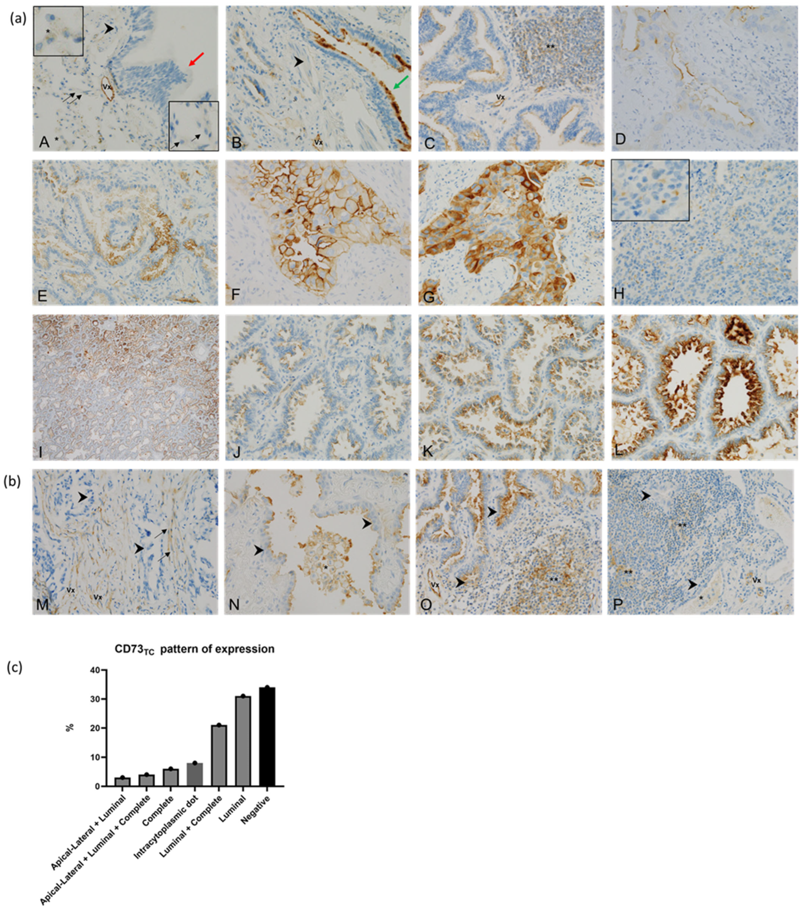
3.2.1. CD73 in Adjacent Non-Tumour Lung Tissue

In normal adjacent lung tissue, staining for CD73 was not observed in pneumocytes, smooth muscle, or intra alveolar macrophages. However, positive staining for CD73 was observed in the apical part of ciliated epithelial cells of the respiratory bronchial epithelium and in the endothelial cells of blood vessels (Figure 1a).

3.2.2. CD73 in Tumour Cells (CD73TC)

The tumour cells exhibited four patterns of expression of CD73: (i) complete membrane staining, (ii) apical–lateral staining, (iii) luminal staining (in glandular structures), and (iv) intracytoplasmic dot staining (Figure 1a). These patterns coexisted within the same tumour sample, observed in 28 out of 76 cases (37%) (Figure 1c). Among the evaluated cases, 50/76 (66%) exhibited positive membranous expression of CD73, with 24/50 (48%) showing complete membrane staining. The TPS ranged from 5% to 100% with a median of 10%, while the H-score varied between 0 and 280 with a median of 20. A high expression of CD73TC was detected in 12/76 samples (16%) based on the TPS and 11/76 samples (14%) based on the H-score. Both scoring methods yielded comparable results, with only one patient displaying discrepant values (TPS = 80%, H-score = 120) (Supplementary Figure S2). Given this consistency, all subsequent results are reported using the TPS.
No statistically significant correlations were found between the pattern of CD73TC expression and clinicopathological parameters. CD73TC expression, quantified using the TPS, showed no significant association with histological subtype, grade, vascular invasion, mitotic index, necrosis, TTF-1 expression, stage, smoking history, age, or sex. Similarly, no significant difference in CD73 expression was observed between primary tumours and metastatic sites. Additionally, no correlation was identified between CD73TC expression and molecular biomarkers, including sensitive or resistant EGFR mutations and EGFR amplification. However, tumours with high TPS CD73TC expression were significantly more likely to exhibit a complete membranous staining pattern (9/12, 75%; p = 0.015) (Table 3).
Table 3. Descriptive clinicopathological data related to TPS CD73TC expression level.
Variable (n = 76, Unless Stated Otherwise)TypeHIGH CD73TC (TPS > 50%)LOW CD73TC (TPS ≤ 5 0%)p-Value
Clinical and follow-up data
Age at diagnosisMean (sd)66 (7)66.48 (11.07)0.9525
Median [IQR]67 [60–71]67 [63–74]
SexeFemale9 (75) 43 (67.19) 0.8447
Male3 (25) 21 (32.81)
Smoking (n = 72)Smoker5 (41.67) 24 (40) 1
Non-smoker7 (58.33) 36 (60)
Stage categoryAdvanced5 (42) 37 (58) 0.4740
Early7 (58) 27 (42)
Brain_metastasis (n = 53)No6 (75) 32 (71) 1.0000
Yes2 (25) 13 (29)
OS_Month
Mean (sd)75 (50)41 (36)0.0868
Median [IQR]65 [29–79]28 [14–60]
Pathological data
Origin of samplesMetastasis3 (25) 20 (31) 0.9282
Primary9 (75) 44 (695)
Histology subtype (n = 39)Acinar2 (22) 13 (43) 0.35
Cribriform1 (11) 0 (0)
Lepidic0 (0) 1 (3)
Papillary5 (56) 12 (40)
Solid0 (0) 2 (7)
Micropapillary1 (11) 2 (7)
High-grade component (n = 71)No6 (50) 36 (61) 0.6998
Yes6 (50) 23 (39)
Emboli (n = 46)No3 (27) 14 (29) 1.0000
Yes8 (73) 34 (71)
Immunohistochemistry data
PD-L1 expression category (n = 73)High 0 (0) 10 (17) 0.2467
Moderate2 (17) 5 (8)
Negative 10 (83) 46 (75)
PDL1 % TCMean (sd)1 (3)13 (30)0.44
Median [IQR]0 [0–0]0 [0–0]
CD73TC pattern (n = 50)Apical lateral1 (8) 0 (0) 0.0152
Complete9 (75) 16 (42)
Luminal2 (17) 22 (58)
CD73TC dot staining (n = 50)Yes3 (25)5 (8)0.11
No9 (75)59 (92)
CD73SC in lymphocyte Negative12 (100) 57 (89) 0.5103
Positive0 (0) 7 (11)
CD73SC in macrophageNegative11 (92) 60 (94) 1.0000
Positive1 (8) 4 (6)
Molecular data
EGFR amplification status (n = 71)Yes9 (75) 38 (64) 0.7096
No3 (25) 21 (36)
EGFR mutation at baselineEx18_G7191 (8) 2 (3) 0.7366
Ex20_S768I0 (0) 2 (3)
L858R5 (42) 23 (36)
del196 (50) 37 (58)
EGFR mutation at baseline classification (Common vs. Uncommon)Common11 (92)60 (94)1.0000
Uncommon1 (8)4 (6)
T790M during follow-up (n = 21)No2 (100) 9 (47) 0.5007
Yes0 (0) 10 (53)
CD73TC and PD-L1 were co-expressed in 11/73 (15%) of cases. Among PD-L1-negative cases, 62% (37/60) were positive for CD73TC. The high expression of both PD-L1 and CD73TC was mutually exclusive. Notably, a higher proportion of tumours in the high CD73 category were PD-L1-negative (83%; 10/12), although this trend did not reach statistical significance (p = 0.44) (Figure 2). To further investigate this relationship, we performed Pearson correlation analyses, which revealed a weak inverse correlation between CD73 and PD-L1 expression (CD73 H-score: r = −0.1639, p = 0.1657; CD73 TPS: r = −0.2, p = 0.1646), though this was not statistically significant (Supplementary Figure S3).
The scatter plots illustrate this distribution and suggest a potential inverse trend. However, this association remains inconclusive due to high inter-sample variability and a lack of statistical significance.

3.2.3. CD73 in Stromal Cells (CD73 SC)

CD73SC was expressed in cancer-associated fibroblasts in 2/76 cases (3%), while macrophages or infiltrating lymphocytes were positive in 12/76 (16%) of cases (Figure 1b). The expression of CD73SC was more frequently observed in an early tumour stage (p = 0.037), PD-L1-negative tumours (p = 0.030), non-amplified EGFR tumours (p = 0.0018), and EGFR L858R-mutated tumours (p = 0.067) (Table 4).

3.3. EGFR Amplification and Correlation with Clinicopathological Features and Expression of CD73

The amplification of EGFR was observed in 70% (58/83) of cases (Supplementary Figure S4). Testing was uninformative for FISH in 8% (7/90) of the samples due to an inadequate amount of material or lack of hybridisation. Given that amplified tumours exhibit heterogeneous clinical features, we analysed their association with key clinicopathological parameters. No significant correlations were found between EGFR-amp and stage, smoking history, PD-L1, and EGFR mutation type. However, EGFR-amp was significantly associated with a high-grade pattern (p = 0.016) (Supplementary Table S1).

3.4. Event-Free Survival and Follow-Up

Shorter event-free survival (EFS) was significantly associated with being male (p = 0.019), advanced stage (p < 0.001), high-grade component (p = 0.016), and high PD-L1 (p = 0.025) (Table 5). High CD73TC expression was correlated with longer EFS in the univariate analysis, as shown by the median survival not being reached after 34 months of follow-up (p = 0.045) (Figure 3). However, in the multivariate analysis, which included CD73 expression, PD-L1 expression, stage, and age, only disease stage remained statistically significant (p = 0.0078) (Supplementary Figure S5). To further explore the impact of tumour stage, we performed an exploratory subgroup analysis, stratifying patients into early-stage (Stages I–IIIA) and advanced-stage (Stages IIIB–IV) disease (Supplementary Figure S6). In early-stage patients, a trend towards improved EFS in the high CD73TC expression group was observed, but this did not reach statistical significance (p = 0.11). In advanced-stage patients, no significant association was observed between CD73 expression and EFS (p = 0.91).
EGFR-amp demonstrated no significant association with EFS. In patients treated with EGFR TKIs, follow-up was available in 32/44 (73%) patients with a median of 29 months (range 17–54.5), showing no statistical correlation between EFS and the expression of CD73TC (p = 0.92) (Supplementary Table S2; Figure S7).

4. Discussion

Recent evidence highlights the emerging role of CD73 inhibitors in treatment decision-making for LUAD [34,63]. Preclinical studies have demonstrated the therapeutic potential of targeting the CD73/adenosine pathway in EGFRm lung cancer. In an immunocompetent mouse model, CD73 was found to be upregulated, and its blockade significantly inhibited tumour growth [36,37]. Furthermore, in an independent xenograft mouse model, the combination of anti-PD-L1 and anti-CD73 therapy enhanced T cell-mediated killing of EGFRm NSCLC, whereas monotherapy with either antibody was ineffective [54]. These findings suggest that the dual inhibition of CD73 and PD-L1 may enhance immune responses and could be a promising therapeutic strategy for LUAD.
In human EGFRm tumours, targeting CD73 shows promising results, as demonstrated by ongoing clinical trials testing adenosine pathway inhibitors, either as monotherapy or in combination with ICIs and/or TKIs [56,61,63,75,76]. CD73 inhibition may sensitise tumours to treatment by reducing adenosine production, thereby enhancing T cell and dendritic cell activity while suppressing the immunosuppressive functions of regulatory T cells and [63]. Additionally, the inhibition of CD73 improves the efficacy of anti-PD-1 and anti-CTLA-4 monoclonal antibodies [50], inhibits tumour growth, and reverses the exhausted T cell phenotype in mouse models [77].
Among CD73-targeting agents, the human monoclonal antibody Oleclumab (MEDI9447) exhibits high specificity towards CD73, inhibiting its enzymatic activity by sterically blocking and cross-linking CD73 dimers. Additionally, it reduces CD73 expression through internalisation, preventing the production of immunosuppressive extracellular adenosine [51].
While CD73 inhibition may enhance ICI efficacy, it could also exacerbate inflammatory side effects, particularly in EGFRm patients, who are known to be at a higher risk of immune-related toxicities when treated with ICIs. Clinical data on anti-CD73 therapy in EGFRm patients remain limited; however, early findings suggest a favourable safety profile with a low incidence of severe adverse effects and a low rate of treatment discontinuation [34,56]. Furthermore, no additional toxicities have been reported beyond those already known with durvalumab monotherapy [63].
In this context, we explored the feasibility of using IHC to establish a correlation between the expression of CD73 and clinicopathological data, thus identifying a potential predictive biomarker. We performed two methods for the quantitative evaluation of CD73TC. Both the TPS and H-score produced similar results within the context of EGFRm LUAD. Statistical analyses underscored the consistency of these two scoring systems when considering the clinicopathological data. Yet, the increased complexity of the H-score does not augment precision within our limited patient cohort and may hamper reproducibility among pathologists. We tested the cut-offs published in the literature but did not observe significant results in our cohort. Therefore, we opted for a mean cut-off based on the average score of the total range: 150 out of 300 for the H-score and 50 out of 100 for the TPS.
Consistent with the existing literature, we demonstrate the absence of expression of CD73 in normal alveolar tissue and expression in tumour cells, reflecting 66% CD73TC positivity, in line with prior data linking the expression of CD73 to EGFR mutations [54,57,58]. Endothelial cells and the respiratory bronchial epithelium could serve as internal controls. This is further supported by the literature, which documents similar observations in the function of endothelial cells [78] and in human oviducts, where CD73 regulates ciliary beat frequency and mucociliary clearance [79]. We also report for the first-time intracytoplasmic dot staining of CD73TC in lung cancer tissue; however, we did not observe any correlation between this staining pattern and clinicopathological parameters. Interestingly, Alcedo et al. previously documented para-nuclear expression in hepatocellular carcinoma cells, with co-localisation observed at the Golgi complex. This discovery highlights the abnormal glycosylation of CD73 in tumour hepatocytes, leading to the mislocalisation and functional suppression of CD73. This insight suggests a potential new therapeutic target for improving the treatment outcome by addressing this specific regulatory mechanism [80].
While the role of CD73 in tumour progression has been well documented [42,81,82,83,84], our investigations into the correlation between CD73TC and tumour emboli, metastasis, high grade, and stage did not yield any significant associations.
Regarding immune checkpoints, the co-expression of CD73TC and PD-L1 was rare, occurring in only 15% of cases. Moreover, the high expression of both PD-L1 and CD73TC was mutually exclusive, with a trend towards an inverse association between the high expression of CD73TC and reduced PD-L1 expression in tumour cells. However, a correlation analysis between CD73 and PD-L1 expression did not reach statistical significance (r = −0.2, p = 0.16), indicating that while an inverse trend may exist, these markers are largely independent in our cohort. Previous studies [53,57,58] identified a correlation between high CD73 and increased PD-L1 expression, either in smaller cohorts of 15 to 25 EGFR tumours or in cohorts analysed after TKI treatment [52] (Table 1). Unlike these studies, our research focused on a more homogeneous and larger cohort, providing insights into a broader patient population, albeit under different treatment conditions. These results suggest the presence of distinct immune evasion mechanisms in EGFRm LUAD. The inverse relationship could indicate that tumours with a high expression of CD73 may rely on the adenosine pathway for immune suppression\ rather than on PD-L1-mediated immune checkpoint inhibition. Moreover, these findings highlight the complexity of the tumour immune microenvironment, suggesting that tumours may employ diverse strategies to escape immune surveillance. This variability could have significant implications for the development of targeted therapies, highlighting the need for personalised approaches that address these distinct immune evasion mechanisms. It could be of strong interest to further investigate CD73’s interactions with immune cells. However, given the limitations of our cohort—notably the low proportion of TILs+ cases and limited CD73 stromal expression—we prioritised a tumour-centred approach.
Of interest, we observed a correlation between a high CD73TC level and improved EFS at baseline, suggesting potential prognostic significance. To further investigate this association, we performed a multivariate Cox proportional hazards model incorporating CD73TC expression, PD-L1 expression, disease stage, and age. While a univariate analysis identified a significant correlation between high CD73TC expression and improved EFS, this association was no longer significant in the multivariate model, likely due to the limited sample size (60 patients with available EFS data). In this adjusted model, only the disease stage remained statistically significant, confirming its well-established prognostic impact. CD73 expression was not correlated with tumour stage (Table 3), indicating that the observed association with EFS is not solely driven by stage distribution. These findings suggest that while CD73 may play a role in tumour progression, larger studies are needed to fully elucidate its independent prognostic value in EGFRm LUAD. This association has previously been reported only once by Ishii et al., who demonstrated that in a cohort of 25 treated EGFRm LUAD patients, a high expression of CD73TC was linked to improved progression-free survival and overall survival [53]. However, the prognostic implications of the expression of CD73 have been inconsistently reported, with some studies showing no correlation [34,57,58] and others suggesting an association with worse prognosis [55,56,60] (Table 1). Several factors may explain these discrepancies. One key distinction is that our cohort includes a higher proportion of early-stage tumours (43%), whereas most previous studies focused on advanced-stage disease. Our exploratory subgroup analysis (Supplementary Figure S6) indicated that the association between high CD73TC expression and improved EFS is more apparent in early-stage tumours, though it does not reach statistical significance. These findings suggest that CD73 may have a more prominent role in early tumour progression, where immune evasion mechanisms could differ from those in advanced disease. However, the absence of statistical significance in early-stage tumours and the limited sample size of our cohort warrant cautious interpretation. Further validation in larger independent cohorts is necessary to determine whether CD73 has stage-dependent prognostic implications and to clarify its potential role in tumour progression.
In contrast to some studies reporting an association between CD73 expression and poorer survival in EGFRm patients treated with TKIs [56], we did not observe a significant correlation between baseline CD73TC expression and survival in our TKI-treated subgroup. Differences in sample size, treatment heterogeneity, and patient selection criteria may explain these discrepancies. Additionally, Ishii et al. previously reported that high CD73TC expression was linked to improved progression-free and overall survival in a small cohort of 25 treated EGFRm LUAD patients, highlighting the inconsistency in CD73’s prognostic impact across studies. Previous reports have suggested that TKI treatment may modulate the tumour immune microenvironment, potentially influencing CD73 expression patterns and their prognostic significance. Specifically, studies have indicated that CD73 expression may increase upon TKI resistance, particularly in tumours with acquired PD-L1 positivity [52]. However, our study focused exclusively on CD73 expression at baseline, before TKI exposure, allowing us to assess its potential prognostic role independently of treatment-induced changes. This distinction may explain why our findings differ from those observed in cohorts enriched with TKI-resistant patients.
Beyond cohort composition and treatment variability, technical factors may also contribute to the observed differences. Variations in IHC interpretation and scoring criteria could influence the reported correlations. Additionally, the inverse expression pattern between CD73 and PD-L1, a biomarker known to impact prognosis, may play a role. Another important limitation is the reduced follow-up duration for some patients, which may affect the assessment of long-term outcomes.
Although the analysis of the expression of CD73SC is limited in our cohort, we observed a trend towards better prognosis, as shown in Table 4. The association of the expression of CD73SC in early-stage tumours and PD-L1 negativity indicates that these tumours may have a less aggressive phenotype and a better response to therapy. Additionally, the lack of EGFR amplification and the presence of the common EGFR L858R mutation, both of which are linked to better responses to EGFR-TKIs, further support the idea that the expression of CD73SC may be indicative of a more favourable clinical outcome. This is consistent with previous reports suggesting that CD73 in the stroma plays an anti-tumour role by reducing NF-κB signalling in tumour cells [85,86].
Despite the known influence of EGFR signalling on the expression of CD73, our investigation did not establish a direct relationship between EGFR-amp and the expression of CD73. While FISH is commonly used to detect an EGFR amplification, the results often reflect chromosome 7 polysomy rather than a true gene amplification. Amplification and polysomy, although distinct genetic events, both lead to EGFR protein overexpression, activating downstream signalling pathways that promote tumour growth and survival [87,88]. Consequently, they are often considered to be equivalent from a biological perspective. This complexity poses challenges in accurately classifying cases as amplification or high polysomy [89]. Consistent with previously published data [90,91], the proportion of EGFR-amp was high (70%, n = 58) when scored with the method described by Capuzzo et al. [69]. EGFR-amp is often associated with poor outcomes, invasion, and metastasis [91,92]. However, the literature presents conflicting findings [17,87,90,93,94,95,96], and a recent study by Chmielecki et al. concerning the FLAURA study found no significant association between baseline EGFR-amp and a suboptimal treatment response to first-line osimertinib [16]. Our study likewise did not establish a significant association between EGFR-amp, disease stage, and EFS.
While IHC remains a widely used technique to assess the expression of CD73, its accuracy in reflecting the enzyme’s true biological activity is questionable. Protein expression, detected with IHC, does not always correlate with enzymatic activity. Emerging techniques to detect the activity of CD73 include liquid biopsy, which offers a less invasive and potentially more accurate assessment of the enzymatic function [97,98]. Exosomes are small extracellular vesicles that carry proteins, lipids, and nucleic acids, reflecting the molecular composition of their cell of origin. Studies have shown that soluble CD73 in serum and exosomal CD73 can be a reliable indicator of enzymatic activity, providing a real-time snapshot of the tumour biology. These biomarkers may serve as indicators for disease progression and response to therapy, as demonstrated in melanoma [99] and prostate cancer [100], suggesting a similar potential in lung cancer.
Ongoing research aimed at characterising the unique features of tumour biology and the TME in EGFRm NSCLC, as well as identifying patient subgroups with enhanced responses to ICI therapy, is promising. Considering the prevalence of EGFR mutations and amplifications in LUAD and their association with restricted responses to ICIs, it is imperative to conduct large-scale prospective studies to comprehensively investigate the expression of CD73 as a potential prognostic factor and predictor of the response to immunotherapy and establish standardised methodologies to assess its expression. Moreover, future research should focus on validating liquid biopsy techniques in clinical settings and on exploring their utility in conjunction with traditional IHC.

5. Conclusions

The present study is the first comprehensive investigation to explore the expression of CD73 in LUAD with different EGFR genomic alterations. The observed upregulation of expression of CD73TC at baseline, along with its association with a longer EFS and lower expression of PD-L1, highlights its potential importance in the evolving landscape of immunotherapy and targeted treatments for EGFR-mutated NSCLC. This suggests the presence of a distinct immune TME profile in EGFRm-LUAD, characterised by distinct mechanisms of immune evasion driven by specific molecular pathways. Understanding these unique profiles could have significant therapeutic implications, particularly in the context of combined therapies, including CD73 blockers and ADORA receptor inhibitors to maximise CD73 blockade, along with conventional ICIs.
Such insights may lead to the development of more effective treatment strategies, enhancing the efficacy of immunotherapies by targeting the specific pathways involved in immune resistance within this subset of lung cancer.

Supplementary Materials

The following supporting information can be downloaded at https://www.mdpi.com/article/10.3390/cancers17061034/s1, Figure S1. Flowchart of sample selection and analysis; Figure S2. Dot plot illustrating correlation between expression of CD73 measured by TPS and H-score; Figure S3. Scatter dot plot illustrating correlation between expression of CD73 measured by TPS (and H-score); Figure S4. EGFR FISH assay; Figure S5. Multivariate Cox Regression Model adjusted for CD73TC expression, PD-L1, stage, and age; Figure S6. Event-free survival (EFS) in early-stage (6a) and advanced-stage (6b) patients based on CD73TC expression.; Figure S7. Kaplan–Meier curves illustrating event-free survival in patients treated with ITK categorised by high and low expression of CD73; Table S1. Comparison of clinicopathological features and molecular markers between patients with and without EGFR amplification; Table S2. Characteristics of patients treated with EGFR ITK.

Author Contributions

Conceptualisation: P.H. and M.I.; Methodology: E.L.-M., C.B. (Christophe Bontoux), S.L.-M., K.Z., V.L.-F., O.B., M.I. and P.H.; Formal Analysis and Investigation: E.L.-M. and C.B. (Christophe Bontoux); Data Curation: C.B. (Christophe Bontoux) and G.R.; Writing—Original Draft Preparation: E.L.-M.; Writing—Review and Editing: C.B. (Christophe Bontoux), G.R., V.H., S.L., J.B. (Jonathan Benzaquen), J.B. (Jacques Boutros), S.M., C.B. (Christelle Bonnetaud), C.C., J.-P.B., C.-H.M. and V.V.-C.; Funding Acquisition: P.H.; Supervision: M.I. and P.H. All authors have read and agreed to the published version of the manuscript.

Funding

This research was funded by a grant from the French government managed by the Agence Nationale de la Recherche under the France 2030 programme, reference ANR-23-IAHU-0007, by Fondation ARC within the TRANSCAN-3 ERA-NET and by the FHU OncoAge.

Institutional Review Board Statement

This study was conducted in accordance with the Declaration of Helsinki and approved by the Institutional Review Board of Centre Hospitalier Universitaire de Nice (N°2022-0038).

Informed Consent Statement

Informed consent was obtained from all subjects involved in this study.

Data Availability Statement

The data presented in this study are available upon request from the corresponding author.

Acknowledgments

This publication benefits from discussions with PRESTO COST action CA21130 supported by COST (European Cooperation in Science and Technology).

Conflicts of Interest

M.I. received research grants from Amgen and speaker bureau fees from MSD and AstraZeneca. P.H. received research grants from Thermo-Fisher Scientific, Amgen, Biocartis, and BMS and participated on the advisory boards of AstraZeneca, Janssen, Abbvie, Roche, BMS, Thermo-Fisher Scientific, Biocartis, Pfizer, Amgen, Qiagen, Sanofi, Eli Lilly, Qiagen, Novartis, and Bayer. The remaining authors declare no conflicts of interest.

Abbreviations

The following abbreviations are used in this manuscript:
EFSEvent-Free Survival
EGFREpidermal Growth Factor Receptor
EGFR-ampEGFR-amp
EGFRmEGFR-Mutated
EMTEpithelial–Mesenchymal Transition
FFPEFormalin-Fixed Paraffin-Embedded
FISHIn Situ Fluorescent Hybridisation
IHCImmunohistochemistry
LUADLung Adenocarcinoma
MDSCsMyeloid-Derived Suppressor Cells
NSCLCNon-Small Cell Lung Cancer
SCStromal Cells
TCTumour Cells
TKIsTyrosine Kinase Inhibitors
TMETumour Microenvironment
TPSTumour Proportion Score
HIF1αHypoxia-Inducible Factor 1-alpha
LDH5Lactate Dehydrogenase 5

References

  1. Remon, J.; Soria, J.-C.; Peters, S. Early and Locally Advanced Non-Small-Cell Lung Cancer: An Update of the ESMO Clinical Practice Guidelines Focusing on Diagnosis, Staging, Systemic and Local Therapy. Ann. Oncol. 2021, 32, 1637–1642. [Google Scholar] [CrossRef] [PubMed]
  2. Soria, J.-C.; Ohe, Y.; Vansteenkiste, J.; Reungwetwattana, T.; Chewaskulyong, B.; Lee, K.H.; Dechaphunkul, A.; Imamura, F.; Nogami, N.; Kurata, T.; et al. Osimertinib in Untreated EGFR-Mutated Advanced Non-Small-Cell Lung Cancer. N. Engl. J. Med. 2018, 378, 113–125. [Google Scholar] [CrossRef] [PubMed]
  3. Ettinger, D.S.; Wood, D.E.; Aisner, D.L.; Akerley, W.; Bauman, J.R.; Bharat, A.; Bruno, D.S.; Chang, J.Y.; Chirieac, L.R.; D’Amico, T.A.; et al. Non-Small Cell Lung Cancer, Version 3.2022, NCCN Clinical Practice Guidelines in Oncology. J. Natl. Compr. Cancer Netw. 2022, 20, 497–530. [Google Scholar] [CrossRef] [PubMed]
  4. Zeng, Y.; Yu, D.; Tian, W.; Wu, F. Resistance Mechanisms to Osimertinib and Emerging Therapeutic Strategies in Nonsmall Cell Lung Cancer. Curr. Opin. Oncol. 2022, 34, 54–65. [Google Scholar] [CrossRef]
  5. Lee, J.K.; Shin, J.-Y.; Kim, S.; Lee, S.; Park, C.; Kim, J.-Y.; Koh, Y.; Keam, B.; Min, H.S.; Kim, T.M.; et al. Primary Resistance to Epidermal Growth Factor Receptor (EGFR) Tyrosine Kinase Inhibitors (TKIs) in Patients with Non-Small-Cell Lung Cancer Harboring TKI-Sensitive EGFR Mutations: An Exploratory Study. Ann. Oncol. 2013, 24, 2080–2087. [Google Scholar] [CrossRef]
  6. Jackman, D.M.; Yeap, B.Y.; Sequist, L.V.; Lindeman, N.; Holmes, A.J.; Joshi, V.A.; Bell, D.W.; Huberman, M.S.; Halmos, B.; Rabin, M.S.; et al. Exon 19 Deletion Mutations of Epidermal Growth Factor Receptor Are Associated with Prolonged Survival in Non–Small Cell Lung Cancer Patients Treated with Gefitinib or Erlotinib. Clin. Cancer Res. 2006, 12, 3908–3914. [Google Scholar] [CrossRef]
  7. Riely, G.J.; Pao, W.; Pham, D.; Li, A.R.; Rizvi, N.; Venkatraman, E.S.; Zakowski, M.F.; Kris, M.G.; Ladanyi, M.; Miller, V.A. Clinical Course of Patients with Non-Small Cell Lung Cancer and Epidermal Growth Factor Receptor Exon 19 and Exon 21 Mutations Treated with Gefitinib or Erlotinib. Clin. Cancer Res. 2006, 12, 839–844. [Google Scholar] [CrossRef]
  8. Zhou, Q.; Zhang, X.-C.; Chen, Z.-H.; Yin, X.-L.; Yang, J.-J.; Xu, C.-R.; Yan, H.-H.; Chen, H.-J.; Su, J.; Zhong, W.-Z.; et al. Relative Abundance of EGFR Mutations Predicts Benefit from Gefitinib Treatment for Advanced Non-Small-Cell Lung Cancer. J. Clin. Oncol. 2011, 29, 3316–3321. [Google Scholar] [CrossRef]
  9. Takeda, M.; Okamoto, I.; Fujita, Y.; Arao, T.; Ito, H.; Fukuoka, M.; Nishio, K.; Nakagawa, K. De Novo Resistance to Epidermal Growth Factor Receptor-Tyrosine Kinase Inhibitors in EGFR Mutation-Positive Patients with Non-Small Cell Lung Cancer. J. Thorac. Oncol. 2010, 5, 399–400. [Google Scholar] [CrossRef]
  10. Jin, Y.; Shi, X.; Zhao, J.; He, Q.; Chen, M.; Yan, J.; Ou, Q.; Wu, X.; Shao, Y.W.; Yu, X. Mechanisms of Primary Resistance to EGFR Targeted Therapy in Advanced Lung Adenocarcinomas. Lung Cancer 2018, 124, 110–116. [Google Scholar] [CrossRef]
  11. Lim, S.M.; Kim, H.R.; Cho, E.K.; Min, Y.J.; Ahn, J.S.; Ahn, M.-J.; Park, K.; Cho, B.C.; Lee, J.-H.; Jeong, H.C.; et al. Targeted Sequencing Identifies Genetic Alterations That Confer Primary Resistance to EGFR Tyrosine Kinase Inhibitor (Korean Lung Cancer Consortium). Oncotarget 2016, 7, 36311–36320. [Google Scholar] [CrossRef] [PubMed]
  12. Wang, J.; Wang, B.; Chu, H.; Yao, Y. Intrinsic Resistance to EGFR Tyrosine Kinase Inhibitors in Advanced Non-Small-Cell Lung Cancer with Activating EGFR Mutations. Onco Targets Ther. 2016, 9, 3711–3726. [Google Scholar] [CrossRef] [PubMed]
  13. Hsu, K.-H.; Huang, Y.-H.; Tseng, J.-S.; Chen, K.-C.; Ku, W.-H.; Su, K.-Y.; Chen, J.J.W.; Chen, H.-W.; Yu, S.-L.; Yang, T.-Y.; et al. High PD-L1 Expression Correlates with Primary Resistance to EGFR-TKIs in Treatment Naïve Advanced EGFR-Mutant Lung Adenocarcinoma Patients. Lung Cancer 2019, 127, 37–43. [Google Scholar] [CrossRef] [PubMed]
  14. Passaro, A.; Jänne, P.A.; Mok, T.; Peters, S. Overcoming Therapy Resistance in EGFR-Mutant Lung Cancer. Nat. Cancer 2021, 2, 377–391. [Google Scholar] [CrossRef]
  15. Yu, H.A.; Arcila, M.E.; Rekhtman, N.; Sima, C.S.; Zakowski, M.F.; Pao, W.; Kris, M.G.; Miller, V.A.; Ladanyi, M.; Riely, G.J. Analysis of Tumor Specimens at the Time of Acquired Resistance to EGFR-TKI Therapy in 155 Patients with EGFR-Mutant Lung Cancers. Clin. Cancer Res. 2013, 19, 2240–2247. [Google Scholar] [CrossRef]
  16. Chmielecki, J.; Gray, J.E.; Cheng, Y.; Ohe, Y.; Imamura, F.; Cho, B.C.; Lin, M.-C.; Majem, M.; Shah, R.; Rukazenkov, Y.; et al. Candidate Mechanisms of Acquired Resistance to First-Line Osimertinib in EGFR-Mutated Advanced Non-Small Cell Lung Cancer. Nat. Commun. 2023, 14, 1070. [Google Scholar] [CrossRef]
  17. Tamiya, A.; Osuga, M.; Harada, D.; Isa, S.; Taniguchi, Y.; Nakamura, K.; Mizumori, Y.; Shinohara, T.; Yanai, H.; Nakatomi, K.; et al. Mechanisms of Resistance and Correlation between Pre-Treatment Co-Alterations and p-Prognosis to Osimertinib in Chemo-Naïve Advanced Non-Small Cell Lung Cancer. Lung Cancer 2024, 195, 107917. [Google Scholar] [CrossRef]
  18. Yokoyama, T.; Kondo, M.; Goto, Y.; Fukui, T.; Yoshioka, H.; Yokoi, K.; Osada, H.; Imaizumi, K.; Hasegawa, Y.; Shimokata, K.; et al. EGFR Point Mutation in Non-Small Cell Lung Cancer Is Occasionally Accompanied by a Second Mutation or Amplification. Cancer Sci. 2006, 97, 753–759. [Google Scholar] [CrossRef]
  19. Liang, Z.; Zhang, J.; Zeng, X.; Gao, J.; Wu, S.; Liu, T. Relationship between EGFR Expression, Copy Number and Mutation in Lung Adenocarcinomas. BMC Cancer 2010, 10, 376. [Google Scholar] [CrossRef]
  20. Grob, T.J.; Hoenig, T.; Clauditz, T.S.; Atanackovic, D.; Koenig, A.M.; Vashist, Y.K.; Klose, H.; Simon, R.; Pantel, K.; Izbicki, J.R.; et al. Frequent Intratumoral Heterogeneity of EGFR Gene Copy Gain in Non-Small Cell Lung Cancer. Lung Cancer 2013, 79, 221–227. [Google Scholar] [CrossRef]
  21. Shields, M.D.; Marin-Acevedo, J.A.; Pellini, B. Immunotherapy for Advanced Non-Small Cell Lung Cancer: A Decade of Progress. Am. Soc. Clin. Oncol. Educ. Book 2021, 41, e105–e127. [Google Scholar] [CrossRef] [PubMed]
  22. Lee, C.K.; Man, J.; Lord, S.; Links, M.; Gebski, V.; Mok, T.; Yang, J.C.-H. Checkpoint Inhibitors in Metastatic EGFR-Mutated Non–Small Cell Lung Cancer—A Meta-Analysis. J. Thorac. Oncol. 2017, 12, 403–407. [Google Scholar] [CrossRef] [PubMed]
  23. Gainor, J.F.; Shaw, A.T.; Sequist, L.V.; Fu, X.; Azzoli, C.G.; Piotrowska, Z.; Huynh, T.G.; Zhao, L.; Fulton, L.; Schultz, K.R.; et al. EGFR Mutations and ALK Rearrangements Are Associated with Low Response Rates to PD-1 Pathway Blockade in Non-Small Cell Lung Cancer: A Retrospective Analysis. Clin. Cancer Res. 2016, 22, 4585–4593. [Google Scholar] [CrossRef] [PubMed]
  24. Garon, E.B.; Rizvi, N.A.; Hui, R.; Leighl, N.; Balmanoukian, A.S.; Eder, J.P.; Patnaik, A.; Aggarwal, C.; Gubens, M.; Horn, L.; et al. Pembrolizumab for the Treatment of Non–Small-Cell Lung Cancer. N. Engl. J. Med. 2015, 372, 2018–2028. [Google Scholar] [CrossRef] [PubMed]
  25. Lisberg, A.; Cummings, A.; Goldman, J.W.; Bornazyan, K.; Reese, N.; Wang, T.; Coluzzi, P.; Ledezma, B.; Mendenhall, M.; Hunt, J.; et al. A Phase II Study of Pembrolizumab in EGFR-Mutant, PD-L1+, Tyrosine Kinase Inhibitor Naïve Patients with Advanced NSCLC. J. Thorac. Oncol. 2018, 13, 1138–1145. [Google Scholar] [CrossRef] [PubMed]
  26. Yang, J.C.-H.; Gadgeel, S.M.; Sequist, L.V.; Wu, C.-L.; Papadimitrakopoulou, V.A.; Su, W.-C.; Fiore, J.; Saraf, S.; Raftopoulos, H.; Patnaik, A. Pembrolizumab in Combination with Erlotinib or Gefitinib as First-Line Therapy for Advanced NSCLC with Sensitizing EGFR Mutation. J. Thorac. Oncol. 2019, 14, 553–559. [Google Scholar] [CrossRef]
  27. Creelan, B.C.; Yeh, T.C.; Kim, S.-W.; Nogami, N.; Kim, D.-W.; Chow, L.Q.M.; Kanda, S.; Taylor, R.; Tang, W.; Tang, M.; et al. A Phase 1 Study of Gefitinib Combined with Durvalumab in EGFR TKI-Naive Patients with EGFR Mutation-Positive Locally Advanced/Metastatic Non-Small-Cell Lung Cancer. Br. J. Cancer 2021, 124, 383–390. [Google Scholar] [CrossRef]
  28. Hui, R.; Gandhi, L.; Carcereny Costa, E.; Felip, E.; Ahn, M.-J.; Eder, J.P.; Balmanoukian, A.S.; Leighl, N.B.; Aggarwal, C.; Horn, L.; et al. Long-Term OS for Patients with Advanced NSCLC Enrolled in the KEYNOTE-001 Study of Pembrolizumab (Pembro). J. Clin. Oncol. 2016, 34, 9026. [Google Scholar] [CrossRef]
  29. Garassino, M.C.; Cho, B.-C.; Kim, J.-H.; Mazières, J.; Vansteenkiste, J.; Lena, H.; Jaime, J.C.; Gray, J.E.; Powderly, J.; Chouaid, C.; et al. Final Overall Survival and Safety Update for Durvalumab in Third- or Later-Line Advanced NSCLC: The Phase II ATLANTIC Study. Lung Cancer 2020, 147, 137–142. [Google Scholar] [CrossRef]
  30. Chen, K.; Cheng, G.; Zhang, F.; Zhu, G.; Xu, Y.; Yu, X.; Huang, Z.; Fan, Y. PD-L1 Expression and T Cells Infiltration in Patients with Uncommon EGFR-Mutant Non-Small Cell Lung Cancer and the Response to Immunotherapy. Lung Cancer 2020, 142, 98–105. [Google Scholar] [CrossRef]
  31. Yoneshima, Y.; Ijichi, K.; Anai, S.; Ota, K.; Otsubo, K.; Iwama, E.; Tanaka, K.; Oda, Y.; Nakanishi, Y.; Okamoto, I. PD-L1 Expression in Lung Adenocarcinoma Harboring EGFR Mutations or ALK Rearrangements. Lung Cancer 2018, 118, 36–40. [Google Scholar] [CrossRef] [PubMed]
  32. Gettinger, S.; Hellmann, M.D.; Chow, L.Q.M.; Borghaei, H.; Antonia, S.; Brahmer, J.R.; Goldman, J.W.; Gerber, D.E.; Juergens, R.A.; Shepherd, F.A.; et al. Nivolumab Plus Erlotinib in Patients with EGFR-Mutant Advanced NSCLC. J. Thorac. Oncol. 2018, 13, 1363–1372. [Google Scholar] [CrossRef]
  33. Jia, Y.; Li, X.; Jiang, T.; Zhao, S.; Zhao, C.; Zhang, L.; Liu, X.; Shi, J.; Qiao, M.; Luo, J.; et al. EGFR-Targeted Therapy Alters the Tumor Microenvironment in EGFR-Driven Lung Tumors: Implications for Combination Therapies. Int. J. Cancer 2019, 145, 1432–1444. [Google Scholar] [CrossRef] [PubMed]
  34. Herbst, R.S.; Majem, M.; Barlesi, F.; Carcereny, E.; Chu, Q.; Monnet, I.; Sanchez-Hernandez, A.; Dakhil, S.; Camidge, D.R.; Winzer, L.; et al. COAST: An Open-Label, Phase II, Multidrug Platform Study of Durvalumab Alone or in Combination with Oleclumab or Monalizumab in Patients with Unresectable, Stage III Non–Small-Cell Lung Cancer. J. Clin. Oncol. 2022, 40, 3383–3393. [Google Scholar] [CrossRef] [PubMed]
  35. O’Reilly, D.; O’Leary, C.L.; Reilly, A.; Teo, M.Y.; O’Kane, G.; Hendriks, L.; Bennett, K.; Naidoo, J. Toxicity of Immune Checkpoint Inhibitors and Tyrosine Kinase Inhibitor Combinations in Solid Tumours: A Systematic Review and Meta-Analysis. Front. Oncol. 2024, 14, 1380453. [Google Scholar] [CrossRef]
  36. Wiest, N.; Majeed, U.; Seegobin, K.; Zhao, Y.; Lou, Y.; Manochakian, R. Role of Immune Checkpoint Inhibitor Therapy in Advanced EGFR-Mutant Non-Small Cell Lung Cancer. Front. Oncol. 2021, 11, 751209. [Google Scholar] [CrossRef]
  37. Le, X.; Negrao, M.V.; Reuben, A.; Federico, L.; Diao, L.; McGrail, D.; Nilsson, M.; Robichaux, J.; Munoz, I.G.; Patel, S.; et al. Characterization of the Immune Landscape of EGFR-Mutant NSCLC Identifies CD73/Adenosine Pathway as a Potential Therapeutic Target. J. Thorac. Oncol. 2021, 16, 583–600. [Google Scholar] [CrossRef]
  38. Dong, Z.-Y.; Zhang, J.-T.; Liu, S.-Y.; Su, J.; Zhang, C.; Xie, Z.; Zhou, Q.; Tu, H.-Y.; Xu, C.-R.; Yan, L.-X.; et al. EGFR Mutation Correlates with Uninflamed Phenotype and Weak Immunogenicity, Causing Impaired Response to PD-1 Blockade in Non-Small Cell Lung Cancer. Oncoimmunology 2017, 6, e1356145. [Google Scholar] [CrossRef]
  39. Zhao, C.; Su, C.; Li, X.; Zhou, C. Association of CD8 T Cell Apoptosis and EGFR Mutation in Non-Small Lung Cancer Patients. Thorac. Cancer 2020, 11, 2130–2136. [Google Scholar] [CrossRef]
  40. Yegutkin, G.G. Nucleotide- and Nucleoside-Converting Ectoenzymes: Important Modulators of Purinergic Signalling Cascade. Biochim. Biophys. Acta 2008, 1783, 673–694. [Google Scholar] [CrossRef]
  41. Ohta, A.; Gorelik, E.; Prasad, S.J.; Ronchese, F.; Lukashev, D.; Wong, M.K.K.; Huang, X.; Caldwell, S.; Liu, K.; Smith, P.; et al. A2A Adenosine Receptor Protects Tumors from Antitumor T Cells. Proc. Natl. Acad. Sci. USA 2006, 103, 13132–13137. [Google Scholar] [CrossRef] [PubMed]
  42. Stagg, J.; Divisekera, U.; McLaughlin, N.; Sharkey, J.; Pommey, S.; Denoyer, D.; Dwyer, K.M.; Smyth, M.J. Anti-CD73 Antibody Therapy Inhibits Breast Tumor Growth and Metastasis. Proc. Natl. Acad. Sci. USA 2010, 107, 1547–1552. [Google Scholar] [CrossRef] [PubMed]
  43. Gao, Z.-W.; Liu, C.; Yang, L.; Chen, H.-C.; Yang, L.-F.; Zhang, H.-Z.; Dong, K. CD73 Severed as a Potential Prognostic Marker and Promote Lung Cancer Cells Migration via Enhancing EMT Progression. Front. Genet. 2021, 12, 728200. [Google Scholar] [CrossRef] [PubMed]
  44. Nam, M.-W.; Kim, C.-W.; Choi, K.-C. Epithelial-Mesenchymal Transition-Inducing Factors Involved in the Progression of Lung Cancers. Biomol. Ther. 2022, 30, 213–220. [Google Scholar] [CrossRef]
  45. Zarek, P.E.; Huang, C.-T.; Lutz, E.R.; Kowalski, J.; Horton, M.R.; Linden, J.; Drake, C.G.; Powell, J.D. A2A Receptor Signaling Promotes Peripheral Tolerance by Inducing T-Cell Anergy and the Generation of Adaptive Regulatory T Cells. Blood 2008, 111, 251–259. [Google Scholar] [CrossRef]
  46. Wu, R.; Chen, Y.; Li, F.; Li, W.; Zhou, H.; Yang, Y.; Pei, Z. Effects of CD73 on Human Colorectal Cancer Cell Growth in Vivo and in Vitro. Oncol. Rep. 2016, 35, 1750–1756. [Google Scholar] [CrossRef]
  47. Zhou, L.; Jia, S.; Chen, Y.; Wang, W.; Wu, Z.; Yu, W.; Zhang, M.; Ding, G.; Cao, L. The Distinct Role of CD73 in the Progression of Pancreatic Cancer. J. Mol. Med. 2019, 97, 803–815. [Google Scholar] [CrossRef]
  48. Yu, X.; Liu, W.; Wang, Z.; Wang, H.; Liu, J.; Huang, C.; Zhao, T.; Wang, X.; Gao, S.; Ma, Y.; et al. CD73 Induces Gemcitabine Resistance in Pancreatic Ductal Adenocarcinoma: A Promising Target with Non-Canonical Mechanisms. Cancer Lett. 2021, 519, 289–303. [Google Scholar] [CrossRef]
  49. Snider, N.T.; Altshuler, P.J.; Wan, S.; Welling, T.H.; Cavalcoli, J.; Omary, M.B. Alternative Splicing of Human NT5E in Cirrhosis and Hepatocellular Carcinoma Produces a Negative Regulator of Ecto-5′-Nucleotidase (CD73). Mol. Biol. Cell 2014, 25, 4024–4033. [Google Scholar] [CrossRef]
  50. Allard, B.; Allard, D.; Buisseret, L.; Stagg, J. The Adenosine Pathway in Immuno-Oncology. Nat. Rev. Clin. Oncol. 2020, 17, 611–629. [Google Scholar] [CrossRef]
  51. Hay, C.M.; Sult, E.; Huang, Q.; Mulgrew, K.; Fuhrmann, S.R.; McGlinchey, K.A.; Hammond, S.A.; Rothstein, R.; Rios-Doria, J.; Poon, E.; et al. Targeting CD73 in the Tumor Microenvironment with MEDI9447. Oncoimmunology 2016, 5, e1208875. [Google Scholar] [CrossRef] [PubMed]
  52. Isomoto, K.; Haratani, K.; Hayashi, H.; Shimizu, S.; Tomida, S.; Niwa, T.; Yokoyama, T.; Fukuda, Y.; Chiba, Y.; Kato, R.; et al. Impact of EGFR-TKI Treatment on the Tumor Immune Microenvironment in EGFR Mutation–Positive Non–Small Cell Lung Cancer. Clin. Cancer Res. 2020, 26, 2037–2046. [Google Scholar] [CrossRef] [PubMed]
  53. Ishii, H.; Azuma, K.; Kawahara, A.; Kinoshita, T.; Matsuo, N.; Naito, Y.; Tokito, T.; Yamada, K.; Akiba, J.; Hoshino, T. Predictive Value of CD73 Expression for the Efficacy of Immune Checkpoint Inhibitors in NSCLC. Thorac. Cancer 2020, 11, 950–955. [Google Scholar] [CrossRef] [PubMed]
  54. Tu, E.; McGlinchey, K.; Wang, J.; Martin, P.; Ching, S.L.K.; Floc’h, N.; Kurasawa, J.; Starrett, J.H.; Lazdun, Y.; Wetzel, L.; et al. Anti–PD-L1 and Anti-CD73 Combination Therapy Promotes T Cell Response to EGFR-Mutated NSCLC. JCI Insight 2022, 7, e142843. [Google Scholar] [CrossRef]
  55. Inoue, Y.; Yoshimura, K.; Kurabe, N.; Kahyo, T.; Kawase, A.; Tanahashi, M.; Ogawa, H.; Inui, N.; Funai, K.; Shinmura, K.; et al. Prognostic Impact of CD73 and A2A Adenosine Receptor Expression in Non-Small-Cell Lung Cancer. Oncotarget 2017, 8, 8738–8751. [Google Scholar] [CrossRef]
  56. Kim, M.; Kim, S.; Yim, J.; Keam, B.; Kim, T.M.; Jeon, Y.K.; Kim, D.-W.; Heo, D.S. Targeting CD73 to Overcomes Resistance to First-Generation EGFR Tyrosine Kinase Inhibitors in Non-Small Cell Lung Cancer. Cancer Res. Treat. 2023, 55, 1134–1143. [Google Scholar] [CrossRef]
  57. Rocha, P.; Salazar, R.; Zhang, J.; Ledesma, D.; Solorzano, J.L.; Mino, B.; Villalobos, P.; Dejima, H.; Douse, D.Y.; Diao, L.; et al. CD73 Expression Defines Immune, Molecular, and Clinicopathological Subgroups of Lung Adenocarcinoma. Cancer Immunol. Immunother. 2021, 70, 1965–1976. [Google Scholar] [CrossRef]
  58. Giatromanolaki, A.; Kouroupi, M.; Pouliliou, S.; Mitrakas, A.; Hasan, F.; Pappa, A.; Koukourakis, M.I. Ectonucleotidase CD73 and CD39 Expression in Non-Small Cell Lung Cancer Relates to Hypoxia and Immunosuppressive Pathways. Life Sci. 2020, 259, 118389. [Google Scholar] [CrossRef]
  59. Ramdani, H.O.; Falk, M.; Heukamp, L.C.; Schatz, S.; Tiemann, M.; Wesseler, C.; Diehl, L.; Schuuring, E.; Groen, H.J.M.; Griesinger, F. Immune Related Endonucleases and GTPases Are Not Associated with Tumor Response in Patients with Advanced Non-Small Cell Lung Cancer Treated with Checkpoint Inhibitors. Pathol.–Res. Pract. 2021, 227, 153651. [Google Scholar] [CrossRef]
  60. Haratani, K.; Nakamura, A.; Mamesaya, N.; Mitsuoka, S.; Yoneshima, Y.; Saito, R.; Tanizaki, J.; Fujisaka, Y.; Hata, A.; Tsuruno, K.; et al. Tumor Microenvironment Landscape of Non-Small Cell Lung Cancer Reveals Resistance Mechanisms for PD-L1 Blockade Following Chemoradiotherapy: A Multi-Center Prospective Biomarker Study (WJOG11518L/SUBMARINE). J. Thorac. Oncol. 2023, 8, 1334–1350. [Google Scholar] [CrossRef]
  61. Kowash, R.R.; Akbay, E.A. Tumor Intrinsic and Extrinsic Functions of CD73 and the Adenosine Pathway in Lung Cancer. Front. Immunol. 2023, 14, 1130358. [Google Scholar] [CrossRef] [PubMed]
  62. Janho Dit Hreich, S.; Benzaquen, J.; Hofman, P.; Vouret-Craviari, V. The Purinergic Landscape of Non-Small Cell Lung Cancer. Cancers 2022, 14, 1926. [Google Scholar] [CrossRef] [PubMed]
  63. WHO Classification of Tumours Online. Available online: https://tumourclassification.iarc.who.int/welcome/ (accessed on 11 October 2023).
  64. Goldstraw, P.; Chansky, K.; Crowley, J.; Rami-Porta, R.; Asamura, H.; Eberhardt, W.E.E.; Nicholson, A.G.; Groome, P.; Mitchell, A.; Bolejack, V.; et al. The IASLC Lung Cancer Staging Project: Proposals for Revision of the TNM Stage Groupings in the Forthcoming (Eighth) Edition of the TNM Classification for Lung Cancer. J. Thorac. Oncol. 2016, 11, 39–51. [Google Scholar] [CrossRef] [PubMed]
  65. Ilie, M.; Hofman, V.; Dietel, M.; Soria, J.; Hofman, P. Assessment of the PD-L1 Status by Immunohistochemistry: Challenges and Perspectives for Therapeutic Strategies in Lung Cancer Patients. Virchows Arch. 2016, 468, 511–525. [Google Scholar] [CrossRef]
  66. Falk, A.; Yazbeck, N.; Guibert, N.; Chamorey, E.; Paquet, A.; Ribeyre, L.; Bence, C.; Zahaf, K.; Leroy, S.; Marquette, C.; et al. Effect of Mutant Variants of the KRAS Gene on PD-L1 Expression and on the Immune Microenvironment and Association with Clinical Outcome in Lung Adenocarcinoma Patients. Lung Cancer 2018, 121, 70–75. [Google Scholar] [CrossRef]
  67. Lantuejoul, S.; Damotte, D.; Hofman, V.; Adam, J. Programmed Death Ligand 1 Immunohistochemistry in Non-Small Cell Lung Carcinoma. J. Thorac. Dis. 2019, 11, S89–S101. [Google Scholar] [CrossRef]
  68. Cappuzzo, F.; Hirsch, F.R.; Rossi, E.; Bartolini, S.; Ceresoli, G.L.; Bemis, L.; Haney, J.; Witta, S.; Danenberg, K.; Domenichini, I.; et al. Epidermal Growth Factor Receptor Gene and Protein and Gefitinib Sensitivity in Non–Small-Cell Lung Cancer. JNCI J. Natl. Cancer Inst. 2005, 97, 643–655. [Google Scholar] [CrossRef]
  69. Varella-Garcia, M.; Diebold, J.; Eberhard, D.A.; Geenen, K.; Hirschmann, A.; Kockx, M.; Nagelmeier, I.; Ruschoff, J.; Schmitt, M.; Arbogast, S.; et al. EGFR Fluorescence in Situ Hybridisation Assay: Guidelines for Application to Non-Small-Cell Lung Cancer. J. Clin. Pathol. 2009, 62, 970–977. [Google Scholar] [CrossRef]
  70. Ilie, M.; Butori, C.; Lassalle, S.; Heeke, S.; Piton, N.; Sabourin, J.; Tanga, V.; Washetine, K.; Long-Mira, E.; Maitre, P.; et al. Optimization of EGFR Mutation Detection by the Fully-Automated QPCR-Based Idylla System on Tumor Tissue from Patients with Non-Small Cell Lung Cancer. Oncotarget 2017, 8, 103055–103062. [Google Scholar] [CrossRef]
  71. Lassalle, S.; Hofman, V.; Heeke, S.; Benzaquen, J.; Long, E.; Poudenx, M.; Lantéri, E.; Boutros, J.; Tanga, V.; Zahaf, K.; et al. Targeted Assessment of the EGFR Status as Reflex Testing in Treatment-Naive Non-Squamous Cell Lung Carcinoma Patients: A Single Laboratory Experience (LPCE, Nice, France). Cancers 2020, 12, 955. [Google Scholar] [CrossRef]
  72. Heeke, S.; Benzaquen, J.; Long-Mira, E.; Audelan, B.; Lespinet, V.; Bordone, O.; Lalvée, S.; Zahaf, K.; Poudenx, M.; Humbert, O.; et al. In-House Implementation of Tumor Mutational Burden Testing to Predict Durable Clinical Benefit in Non-Small Cell Lung Cancer and Melanoma Patients. Cancers 2019, 11, 1271. [Google Scholar] [CrossRef] [PubMed]
  73. Alcazer, V. StatAid: An R Package with a Graphical User Interface for Data Analysis. J. Open Source Softw. 2020, 5, 2630. [Google Scholar] [CrossRef]
  74. Bendell, J.; LoRusso, P.; Overman, M.; Noonan, A.M.; Kim, D.-W.; Strickler, J.H.; Kim, S.-W.; Clarke, S.; George, T.J.; Grimison, P.S.; et al. First-in-Human Study of Oleclumab, a Potent, Selective Anti-CD73 Monoclonal Antibody, Alone or in Combination with Durvalumab in Patients with Advanced Solid Tumors. Cancer Immunol. Immunother. 2023, 72, 2443–2458. [Google Scholar] [CrossRef]
  75. Horinouchi, H. Another Pirate in the Red Ocean? CD73-Targeted Therapy in EGFR-Mutated NSCLC. J. Thorac. Oncol. 2023, 18, 552–555. [Google Scholar] [CrossRef] [PubMed]
  76. Spira, A.I.; Conkling, P.R.; Johnson, M.L.; Gardner, O.; Gilbert, H.N.; Scharville, M.; Yin, F.; Krishnan, K.; Paoloni, M.C.; Chaudhry, A. ARC-4 Study: Efficacy and Safety of AB928 plus Carboplatin, Pemetrexed and a PD-1 Antibody in Participants with Metastatic Non-Small Cell Lung Cancer (MNSCLC). J. Clin. Oncol. 2020, 38, e21659. [Google Scholar] [CrossRef]
  77. Deng, W.-W.; Li, Y.-C.; Ma, S.-R.; Mao, L.; Yu, G.-T.; Bu, L.-L.; Kulkarni, A.B.; Zhang, W.-F.; Sun, Z.-J. Specific Blockade CD73 Alters the “Exhausted” Phenotype of T Cells in Head and Neck Squamous Cell Carcinoma. Int. J. Cancer 2018, 143, 1494–1504. [Google Scholar] [CrossRef]
  78. Allard, B.; Turcotte, M.; Stagg, J. CD73-Generated Adenosine: Orchestrating the Tumor-Stroma Interplay to Promote Cancer Growth. J. Biomed. Biotechnol. 2012, 2012, 485156. [Google Scholar] [CrossRef]
  79. Villamonte, M.L.; Torrejón-Escribano, B.; Rodríguez-Martínez, A.; Trapero, C.; Vidal, A.; Gómez de Aranda, I.; Sévigny, J.; Matías-Guiu, X.; Martín-Satué, M. Characterization of Ecto-Nucleotidases in Human Oviducts with an Improved Approach Simultaneously Identifying Protein Expression and in Situ Enzyme Activity. Histochem. Cell Biol. 2018, 149, 269–276. [Google Scholar] [CrossRef]
  80. Alcedo, K.P.; Guerrero, A.; Basrur, V.; Fu, D.; Richardson, M.L.; McLane, J.S.; Tsou, C.; Nesvizhskii, A.I.; Welling, T.H.; Lebrilla, C.B.; et al. Tumor-Selective Altered Glycosylation and Functional Attenuation of CD73 in Human Hepatocellular Carcinoma. Hepatol. Commun. 2019, 3, 1400–1414. [Google Scholar] [CrossRef]
  81. Zhi, X.; Chen, S.; Zhou, P.; Shao, Z.; Wang, L.; Ou, Z.; Yin, L. RNA Interference of Ecto-5′-Nucleotidase (CD73) Inhibits Human Breast Cancer Cell Growth and Invasion. Clin. Exp. Metastasis 2007, 24, 439–448. [Google Scholar] [CrossRef]
  82. Zhi, X.; Wang, Y.; Zhou, X.; Yu, J.; Jian, R.; Tang, S.; Yin, L.; Zhou, P. RNAi-Mediated CD73 Suppression Induces Apoptosis and Cell-Cycle Arrest in Human Breast Cancer Cells. Cancer Sci. 2010, 101, 2561–2569. [Google Scholar] [CrossRef] [PubMed]
  83. Terp, M.G.; Olesen, K.A.; Arnspang, E.C.; Lund, R.R.; Lagerholm, B.C.; Ditzel, H.J.; Leth-Larsen, R. Anti-Human CD73 Monoclonal Antibody Inhibits Metastasis Formation in Human Breast Cancer by Inducing Clustering and Internalization of CD73 Expressed on the Surface of Cancer Cells. J. Immunol. 2013, 191, 4165–4173. [Google Scholar] [CrossRef] [PubMed]
  84. Koszałka, P.; Gołuńska, M.; Stanisławowski, M.; Urban, A.; Stasiłojć, G.; Majewski, M.; Wierzbicki, P.; Składanowski, A.C.; Bigda, J. CD73 on B16F10 Melanoma Cells in CD73-Deficient Mice Promotes Tumor Growth, Angiogenesis, Neovascularization, Macrophage Infiltration and Metastasis. Int. J. Biochem. Cell Biol. 2015, 69, 1–10. [Google Scholar] [CrossRef] [PubMed]
  85. Zeng, Z.; Yang, F.; Wang, Y.; Zhao, H.; Wei, F.; Zhang, P.; Zhang, X.; Ren, X. Significantly Different Immunological Score in Lung Adenocarcinoma and Squamous Cell Carcinoma and a Proposal for a New Immune Staging System. Oncoimmunology 2020, 9, 1828538. [Google Scholar] [CrossRef]
  86. Leclerc, B.G.; Charlebois, R.; Chouinard, G.; Allard, B.; Pommey, S.; Saad, F.; Stagg, J. CD73 Expression Is an Independent Prognostic Factor in Prostate Cancer. Clin. Cancer Res. 2016, 22, 158–166. [Google Scholar] [CrossRef]
  87. Ruiz-Patiño, A.; Castro, C.D.; Ricaurte, L.M.; Cardona, A.F.; Rojas, L.; Zatarain-Barrón, Z.L.; Wills, B.; Reguart, N.; Carranza, H.; Vargas, C.; et al. EGFR Amplification and Sensitizing Mutations Correlate with Survival in Lung Adenocarcinoma Patients Treated with Erlotinib (MutP-CLICaP). Target. Oncol. 2018, 13, 621–629. [Google Scholar] [CrossRef]
  88. Zhang, X.; Zhang, Y.; Tang, H.; He, J. EGFR Gene Copy Number as a Predictive/Biomarker for Patients with Non-Small-Cell Lung Cancer Receiving Tyrosine Kinase Inhibitor Treatment: A Systematic Review and Meta-Analysis. J. Investig. Med. 2017, 65, 72–81. [Google Scholar] [CrossRef]
  89. Casorzo, L.; Corigliano, M.; Ferrero, P.; Venesio, T.; Risio, M. Evaluation of 7q31 Region Improves the Accuracy of EGFR FISH Assay in Non Small Cell Lung Cancer. Diagn. Pathol. 2009, 4, 36. [Google Scholar] [CrossRef]
  90. Dacic, S.; Flanagan, M.; Cieply, K.; Ramalingam, S.; Luketich, J.; Belani, C.; Yousem, S.A. Significance of EGFR Protein Expression and Gene Amplification in Non-Small Cell Lung Carcinoma. Am. J. Clin. Pathol. 2006, 125, 860–865. [Google Scholar] [CrossRef]
  91. Jia, X.F.; Li, J.; Zhao, H.B.; Liu, J.; Liu, J.J. Correlation of EGFR Gene Amplification with Invasion and Metastasis of Non-Small Cell Lung Cancer. Genet. Mol. Res. 2015, 14, 11006–11012. [Google Scholar] [CrossRef]
  92. Gao, X.; Wei, X.-W.; Zheng, M.-Y.; Chen, Z.-H.; Zhang, X.-C.; Zhong, W.-Z.; Yang, J.-J.; Wu, Y.-L.; Zhou, Q. Impact of EGFR Amplification on Survival of Patients with EGFR Exon 20 Insertion-Positive Non-Small Cell Lung Cancer. J. Thorac. Dis. 2020, 12, 5822–5832. [Google Scholar] [CrossRef] [PubMed]
  93. Shan, L.; Wang, Z.; Guo, L.; Sun, H.; Qiu, T.; Ling, Y.; Li, W.; Li, L.; Liu, X.; Zheng, B.; et al. Concurrence of EGFR Amplification and Sensitizing Mutations Indicate a Better Survival Benefit from EGFR-TKI Therapy in Lung Adenocarcinoma Patients. Lung Cancer 2015, 89, 337–342. [Google Scholar] [CrossRef] [PubMed]
  94. Fukuoka, M.; Wu, Y.-L.; Thongprasert, S.; Sunpaweravong, P.; Leong, S.-S.; Sriuranpong, V.; Chao, T.-Y.; Nakagawa, K.; Chu, D.-T.; Saijo, N.; et al. Biomarker Analyses and Final Overall Survival Results from a Phase III, Randomized, Open-Label, First-Line Study of Gefitinib versus Carboplatin/Paclitaxel in Clinically Selected Patients with Advanced Non-Small-Cell Lung Cancer in Asia (IPASS). J. Clin. Oncol. 2011, 29, 2866–2874. [Google Scholar] [CrossRef] [PubMed]
  95. Fiala, O.; Pesek, M.; Finek, J.; Minarik, M.; Benesova, L.; Sorejs, O.; Svaton, M.; Bortlicek, Z.; Kucera, R.; Topolcan, O. Epidermal Growth Factor Receptor Gene Amplification in Patients with Advanced-Stage NSCLC. Anticancer. Res. 2016, 36, 455–460. [Google Scholar]
  96. Kelly, K.; Altorki, N.K.; Eberhardt, W.E.E.; O’Brien, M.E.R.; Spigel, D.R.; Crinò, L.; Tsai, C.-M.; Kim, J.-H.; Cho, E.K.; Hoffman, P.C.; et al. Adjuvant Erlotinib Versus Placebo in Patients with Stage IB-IIIA Non-Small-Cell Lung Cancer (RADIANT): A Randomized, Double-Blind, Phase III Trial. J. Clin. Oncol. 2015, 33, 4007–4014. [Google Scholar] [CrossRef]
  97. Kalluri, R.; LeBleu, V.S. The Biology, Function, and Biomedical Applications of Exosomes. Science 2020, 367, eaau6977. [Google Scholar] [CrossRef]
  98. Marrugo-Ramírez, J.; Mir, M.; Samitier, J. Blood-Based Cancer Biomarkers in Liquid Biopsy: A Promising Non-Invasive Alternative to Tissue Biopsy. Int. J. Mol. Sci. 2018, 19, 2877. [Google Scholar] [CrossRef]
  99. Turiello, R.; Capone, M.; Morretta, E.; Monti, M.C.; Madonna, G.; Azzaro, R.; Gaudio, P.D.; Simeone, E.; Sorrentino, A.; Ascierto, P.A.; et al. Exosomal CD73 from Serum of Patients with Melanoma Suppresses Lymphocyte Functions and Is Associated with Therapy Resistance to Anti-PD-1 Agents. J. Immunother. Cancer 2022, 10, e004043. [Google Scholar] [CrossRef]
  100. Gardani, C.F.F.; Cappellari, A.R.; de Souza, J.B.; da Silva, B.T.; Engroff, P.; Moritz, C.E.J.; Scholl, J.N.; Battastini, A.M.O.; Figueiró, F.; Morrone, F.B. Hydrolysis of ATP, ADP, and AMP Is Increased in Blood Plasma of Prostate Cancer Patients. Purinergic Signal. 2019, 15, 95–105. [Google Scholar] [CrossRef]
Figure 1. Expression of CD73. (a) Representative image of expression of CD73 in adjacent normal lung tissue and tumour cells. Analysis of adjacent normal lung tissue showed consistent negative staining for CD73 in pneumocytes (black arrows and lower right insert), macrophages (black asterisk and upper left insert), and smooth muscle (black arrowheads) (A,B, ×40). Normal bronchial epithelium was mostly negative (red arrows) (A) but occasionally showed positive staining in apical ciliated cells (green arrow) (B). Blood vessels (Vx) were positive and served as internal control (A,B, ×40). In LUAD, CD73 was expressed on cancer cell membranes with luminal (C,D, ×40), apico-lateral (E, ×40), or complete membrane staining (F,G, ×40) with infrequent cytoplasmic reinforcement (G, ×40). Intracytoplasmic dot staining in tumour cells (H, ×40) and positive tumour-infiltrating lymphocytes ** (C, ×40) were also observed. Intensity of CD73 staining varied among LUAD, and within same adenocarcinoma (I, ×4), with weak (1+) (J, ×40), medium (2+) (K, ×40), and strong (3+) (L, ×40) intensity staining, highlighting complexity of calculated H-score. (b) Representative image of expression of CD73 in stromal cells. Cancer-associated fibroblasts (black arrow) exhibited CD73 overexpression in 3% of cases, even when LUAD cells (black arrowheads) were negative (M, ×40). Stromal fibroblasts can be difficult to differentiate from small vessels (VX) in neovascularised tumours (M, ×40). In addition, CD73 expression was observed in macrophages (*) and tumour-infiltrating lymphocytes (**) in subset of cases (N,O,P, ×40) regardless of expression of CD73 in cancer cells. (c) Bar graph illustrating different patterns of expression of CD73 in tumour cells. Patterns observed include complete membrane staining, apical–lateral staining, luminal staining, and intracytoplasmic dot staining. These expression patterns often coexisted within same tumour sample.
Cancers 17 01034 g001
Figure 2. Comparison of PD-L1 expression and CD73 expression in tumour cells (CD73TC) using Tumour Proportion Score (TPS). Dot Tumour plot illustrating relationship between expression of PD-L1 and CD73 in tumour cells. High expression of both PD-L1 and CD73TC was mutually exclusive. Plot suggests trend of inverse correlation, where high levels of PD-L1 are generally associated with low levels of expression of CD73. However, this correlation is not statistically significant.
Cancers 17 01034 g002
Figure 3. Kaplan–Meier curves illustrating event-free survival in patients categorised by high and low levels of CD73 expression in tumour cells (CD73TC) using the Tumour Proportion Score (TPS). Patients with a high level of expression of CD73TC (red line) had a significantly extended event-free survival in contrast to patients with a low level of expression of CD73TC (blue line). The median event-free survival for patients with a low level of expression of CD73TC was 24 months, while for those with a high level of expression of CD73, it was yet to be reached (log-rank test, p = 0.045 with TPS). The number of patients at risk at each time point is specified below the x-axis.
Cancers 17 01034 g003
Table 1. Comprehensive review of CD73 immunohistochemical expression: correlations with clinicopathological features and treatment outcomes.
CD73TC Expression and Correlation with
ReferencesType of StudyNo. of CasesSampleStageHistological SubtypeNo. of EGFR MutantTreatment HistoryIHC (Clone and Dilution)InterpretationCut-OffHistological
Pattern
Patients CharacteristicsEGFRmTMBPDL1 SurvivalResponse to ICI
Inoue,
2017 [55]
retrospective642FFPE (TMA)I-IIISCC; LUAD; others119
(protein expression)
before or
after chemo
D7F9A
1:200
H-score low < 162 < highYes
(LUAD;
TTF-1; ALK)
Yes
(female;
never smoked)
Yes
(based on EGFR protein expression)
__Yes (whole cohort; shorter OS and RFS)_
Isomoto,
2020 [52]
retrospective70FFPEAdvancedLUAD70before and
after TKI
D7F9AH-score
TILs
____Yes
(High PD-L1, after TKI)
__
Giatromanolaki, 2020
[58]
retrospective98FFPE I-IIISCC; LUAD; others_treatment-naiveEPR6114
1:200
TPS

CAF
(% stained stroma area)
5% < low < 40%

50% < medium < 70%

80% < high < 100%
Yes
Inverse correlation between CD73TC and CAF

Less CD73SC expression in stage I
No__NoNo_
Ishii,
2020 [53]
retrospective91FFPEAdvancedNSCLC 25after TKID7F9ATPS ≥50%_NoNo_Yes
(high PD-L1)
Yes
(longer
PFS, OS)
Yes
(better ORR in EGFRm)
Ramdani,
2021 [59]
retrospective48FFPEAdvancedSCC; LUAD; others3treatment-naive1D7no or heavy staining for TC and IC______NoNo
Rocha,
2021 [57]
retrospective106FFPE (TMA)I–IIILUAD15treatment-naiveD7F9A
1:200
TPS
luminal; basolateral and total stain
T negative ≤ 1%;
1% > T low < 55%;
T high ≥ 55%
Yes
(solid)
Yes
(smoking; sex)
NoYesYes
(high PD-L1)
No_
Tu,
2022 [54]
retrospective231FFPE _NSCLC 99_EPR6115TPS
luminal or complete stain
___Yes____
Herbst,
2022 [34]
prospective107FFPEIII NSCLC_treatment-naiveD7F9A
1:200
TPSlow < 10 ≤ high_____No No
Bendell,
2023 [63]
retrospective126FFPEAdvancedLUAD; PDAC, CRC42after treatmentEPR6115TPS >10%_______
Kim,
2023 [56]
retrospective26
paired tumour
FFPE_LUAD26before and
after TKI
1st generation
D7F9A
1:100
H-scorelow < 60 ≤ high_____Yes
(shorter PFS with 1st generation TKI)
_
Haratani,
2023 [60]
prospective135FFPEIIINSCLC13treatment naive D7F9A
1:200
H-score
IC
low < 70 < highYes
(NS-NSCLC)
_No__shorter PFS (with ICI)_
Abbreviation: CAF: Cancer-Associated Fibroblasts; Chemo: Chemotherapy; CRC: Colorectal Cancer; FFPE: Formalin-Fixed, Paraffin-Embedded; IC: Immune Cells; ICI: Immune Checkpoint Inhibitor; LUAD: Lung Adenocarcinoma; NSCLC: Non-Small Cell Lung Cancer; OS: Overall Survival; PDAC: Pancreatic Ductal Adenocarcinoma; PD-L1: Programmed Death-Ligand 1; PFS: Progression-Free Survival; RFS: Recurrence-Free Survival; SCC: Squamous Cell Carcinoma; TC: Tumour Cells; TILs: Tumour-Infiltrating Lymphocytes; TKI: Tyrosine Kinase Inhibitors; TMA: Tissue Microarray; TMB: Tumour Mutational Burden; TPS: Tumour Proportion Score. Symbol “-“: Not assessed.
Table 2. Characteristics of 76 enrolled patients with EGFR-mutated LUAD.
Variable (n = 76 Unless Stated Otherwise)TypeWhole Cohort, n (%) Unless Stated Otherwise
Clinical and follow-up data
Age at diagnosisMedian [IQR]67 [62–73]
SexFemale52 (68)
Male24 (32)
Follow-upMedian [IQR]29 [14–63]
Smoking status Former smoker10 (13)
Smoker19 (25)
Non-smoker43 (65)
Stage categoryAdvanced42 (55)
Early34 (45)
Brain metastasis (n = 53)Yes15 (28)
No38 (72)
Treatment type (n = 59)Including TKI44 (75)
Including immunotherapy4 (7)
Chemotherapy alone11 (19)
Radiotherapy alone3 (5)
Number of treatments (n = 76)Mean (sd)1 (1)
Median [IQR]1 [0–2]
Status at end of follow-up (n = 49)Dead23 (47)
Alive26 (53)
Pathological data
Type of sampleBiopsy36 (47)
Cytology1 (2)
Surgical specimen39 (51)
Origin of samplesMetastasis23 (30)
Primitive53 (70)
High-grade component (n = 71)No42 (60)
Yes29 (41)
Type of high-grade component (n = 23)Cribriform7 (31)
Micropapillary4 (17)
Solid12 (52)
Necrosis (n = 47)No32 (68)
Yes15 (32)
Emboli (n = 59)No17 (29)
Yes42 (71)
Mitosis (n = 45)No5 (11)
Yes40 (89)
Immunohistochemistry data
TTF1 expression on tumour cells Negative1 (1)
Positive75 (99)
Percentage of PD-L1 expression on tumour cells (n = 73)Mean (sd)11 (28)
Median [IQR]0 [0–5]
PD-L1 expression category (n = 73)Negative (<1%)56 (77)
Moderate (1–49%)7 (9)
High (≥50%)10 (14)
CD73 tumour expression (n = 76)Negative26 (34)
Positive50 (66)
Type of CD73 staining (n = 50)Apical–lateral5 (7)
(co-existing within same tumour)Complete24 (48)
Luminal44 (88)
Dot intracytoplasmic8 (16)
Percentage of CD73 tumour staining (n = 76)Mean (sd)27 (31)
Median [IQR]10 [0-51]
Percentage of CD73 tumour expression 1+ (n = 76)Mean (sd)5 (10)
Median [IQR]0 [0–5]
Percentage of CD73 tumour expression 2+ (n = 76)Mean (sd)22 (28)
Median [IQR]10 [0–40]
Percentage of CD73 tumour expression 3+ (n = 76)Mean (sd)33 (59)
Median [IQR]0 [0–53]
CD73 H-score (n = 76)High 11 (14)
Low 65 (86)
CD73 TPS (n = 76)High12 (16)
Low64 (84)
CD73 expression on lymphocytes (n = 76)Negative69 (91)
Positive7 (9)
CD73 expression on macrophages (n = 76)Negative71 (93)
Positive5 (7)
CD73 expression on CAFs (n = 73)Negative71 (97)
Positive2 (3)
Molecular data
EGFR mutation at baselineEx18_G7193 (4)
Ex20_S768I2 (3)
L858R28 (37)
del1943 (56)
T790M during follow-up (n = 21)No11 (52)
Yes10 (48)
Legends: IQR: Interquartile Range; sd: Standard Deviation; TTF1: Thyroid Transcription Factor 1; PD-L1: Programmed Death-Ligand 1; TPS: Tumour Proportion Score; EGFR: Epidermal Growth Factor Receptor; Ex: Exon; del: Deletion; T790M: specific mutation in EGFR gene that confers resistance to certain TKIs; LUAD: Lung Adenocarcinoma; CAF: Cancer-Associated Fibroblast.
Table 4. Association between CD73 expression in stromal cells (recorded as present (positive) or absent (negative)) and clinic pathological data.
Variable (n = 76, Unless Stated Otherwise)TypeCD73SC Negative, n (%)CD73SC Positive, n (%)p-Value
STAGE Advanced38 (61) 4 (29) 0.0372
Early24 (39) 10 (71)
EGFR mutation at baselineEx18_G7192 (3) 1 (7) 0.0670
Ex20_S768I2 (3) 0 (0)
L858R19 (31) 9 (64)
Del1939 (63) 4 (29)
PD-L1 expression (n = 73)Moderate/high17 (29) 0 (0) 0.0303
Negative42 (71) 14 (100)
PDL1 categoryHigh10 (16.95) 0 (0) 0.0710
Moderate7 (11.86) 0 (0)
Negative42 (71.19) 14 (100)
EGFR amplification (n = 71)Amplified44 (75) 3 (25) 0.0018
Not amplified15 (25) 9 (75)
Table 5. Event-free survival analysis based on clinical and molecular variables.
VariableNo. of Patients with DataMedian Survival, MonthHR (95%CI)p-Value
Sex 2.5 (1.4–4.4)0.019
Male5112
Female2230
Stage 5.4 (3.0–9.9)<0.001
Advanced3614
Early3376
High-grade component 2.6 (1.5–4.7)0.016
Yes2918
No3847
PD-L1 expression 2.5 (1.2–4.9)0.025
High1011
Negative/moderate5827
CD73 TPS 2.2 (1.0–4.9)0.045
Low5224
High8Not Reached
CD73 H-score 2.0 (0.9–4.7)0.092
Low5324
High7Not Reached
Disclaimer/Publisher’s Note: The statements, opinions and data contained in all publications are solely those of the individual author(s) and contributor(s) and not of MDPI and/or the editor(s). MDPI and/or the editor(s) disclaim responsibility for any injury to people or property resulting from any ideas, methods, instructions or products referred to in the content.

