Communication

Synthesis, Characterization and Photocatalytic Activity of MoS2/ZnSe Heterostructures for the Degradation of Levofloxacin

School of Natural Sciences, National University of Sciences and Technology (NUST), Islamabad 44000, Pakistan
*
Authors to whom correspondence should be addressed.
Catalysts 2020, 10(12), 1380; https://doi.org/10.3390/catal10121380
Submission received: 11 October 2020 / Revised: 28 October 2020 / Accepted: 30 October 2020 / Published: 26 November 2020

Abstract

:
Antibiotics have been extensively used over the last few decades. Due to their extensive usage and persistence in the environment, they are considered as emergent pollutants. It is, therefore, important to synthesize new materials for efficient antibiotic degradation. Herein, we report the MoS2/ZnSe heterostructures prepared by a simple ultrasonication method. Heterostructures were prepared with different ratios of MoS2 and ZnSe, i.e., 1:3, 1:1 and 3:1. Characterization of the heterostructures was done by UV-vis diffused reflectance spectroscopy (UV-vis-DRS), X-ray diffraction (XRD), X-ray photoelectron spectroscopy (XPS), scanning electron microscopy (SEM), and photoluminescence (PL) techniques to understand the morphology and surface chemistry. The results show that an efficient interface was formed to harness the visible light and degrade levofloxacin, which was monitored by gradual decreases in the UV-vis absorbance signal of levofloxacin. Among the prepared heterostructures and their pure counter parts, MoS2/ZnSe 3:1 (3:1 MZ) showed a better degradation activity of 73.2% as compared to pure MoS2 (29%) and ZnSe (17.1%) in the presence of visible light in a time span of two hours. The reusability studies showed that the catalytic performance of 3:1 MZ did not decrease significantly after three cycles. Moreover, the morphology and the crystal structure also remained unchanged.

1. Introduction

Antibiotics have become emerging pollutants due to their extensive usage and persistence in the environment [1]. These are used as therapeutic agents for humans and as veterinary medicine. The anthropogenic sources through which antibiotics enter the environment include household drainage systems, hospital waste, pharmaceutical industrial waste, agricultural runoff, the discarding of expired and unused medicines, and livestock [2]. The presence of antibiotics in water bodies is a matter of serious concern due to their potential for inducing resistance in the population and their biological activity. Antibiotics like levofloxacin belong to the fluoroquinolone class, with a broad-spectrum antibacterial activity against skin, respiratory tract, soft tissues and urinary tract infections [3]. Therefore, there is a high need to find a solution for their existence and persistence in the environment [4]. Photocatalysis is the emerging technology for the remediation of hazardous wastes from the environment [5]. It is being applied for degrading volatile organic compounds [6], degrading antibiotics [7], industrial dyes degradation [8] and photoelectrochemical water splitting [9].
Amongst the latest synthesized photocatalysts, molybdenum disulphide is gaining high consideration [10]. This is due to its layered structure, which is analogous to graphene and offers efficient electron transport and absorbance of sunlight [11]. The two-dimensional layered (2D) structure comprises a total of three layers loaded together with the Mo layer in the middle and S atom layers on the top and bottom [12]. As the number of layers in the MoS2 structure decreases, its band gap increases, showing the quantum confinement effect [13]. MoS2 has a direct band gap of 1.8 eV, making it an active photocatalyst in visible light [14]. It has many qualities, such as high stiffness, reliability, strong oxidizing property [15], non-toxicity, a tunable band gap, and it is cost efficient [16]. All these properties make it a very promising photocatalyst. However, the rate of recombination of photoexcited holes and electrons is high and limits its performance. To overcome this shortcoming, semiconductors can be incorporated with MoS2 to attain the required properties [17]. The morphology and the crystallinity of the photocatalysts are important parameters that affect the photocatalytic process. A recent study showed that increases in the crystallite size increase the decomposition of the substrate [18]. A more specific work on MoS2 showed that surface area has no significant effect on the degradation of organic wastes, while surface morphology plays an important role in this process [19]. Lots of work is being done currently to find the best photocatalyst in the form of heterostructures; MoS2/MoO3/TiO2 [20], MoS2/ZnSnO3 [21], MoS2/g-C3N4/Bi24O31Cl10 [22], CoO@ meso–CN@MoS2 [23], and WO3/MoS2 [24] are a few among many heterostructures synthesized for photodegradation.
Semiconductors that belong to the II–VI groups have fascinated the researchers because of their promising applications. ZnSe is an important group member of this family, having a direct band gap of 2.8 eV [25]. It shows significant applicability in electronic and optoelectronic devices like transistors, non-linear optical devices, lasers and light emitting diodes [26]. The band gap of ZnSe makes it visible light-active, so it has also been studied as a photocatalyst, but it shows low efficiency in terms of degradation time, i.e., 96% in 10 h for methyl orange [27], 92.5% in 6 h for methylene blue [28], 95.1% in 10 h for methyl orange [29], and almost complete degradation in 9 h for methyl orange [30] and 10 h for methyl orange [31].
In this work, MoS2 and ZnSe were synthesized by the using hydrothermal method and were used to prepare heterostructures via the ultrasonic method. Characterization of the heterostructure photocatalysts was performed to understand their morphological structures and properties. Compared to pure MoS2 and ZnSe, their heterostructures showed significant enhancements in photocatalytic activity with reduced degradation time against levofloxacin. The photodegradation of levofloxacin was examined and a possible mechanism is proposed.

2. Results and Discussion

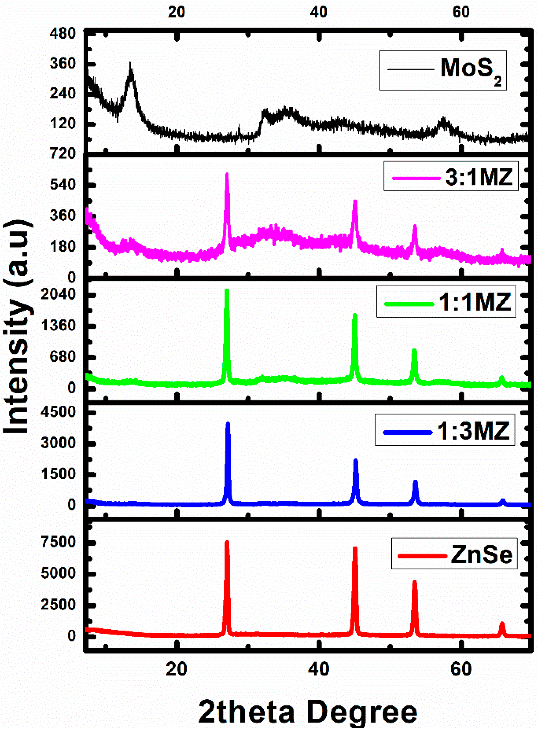
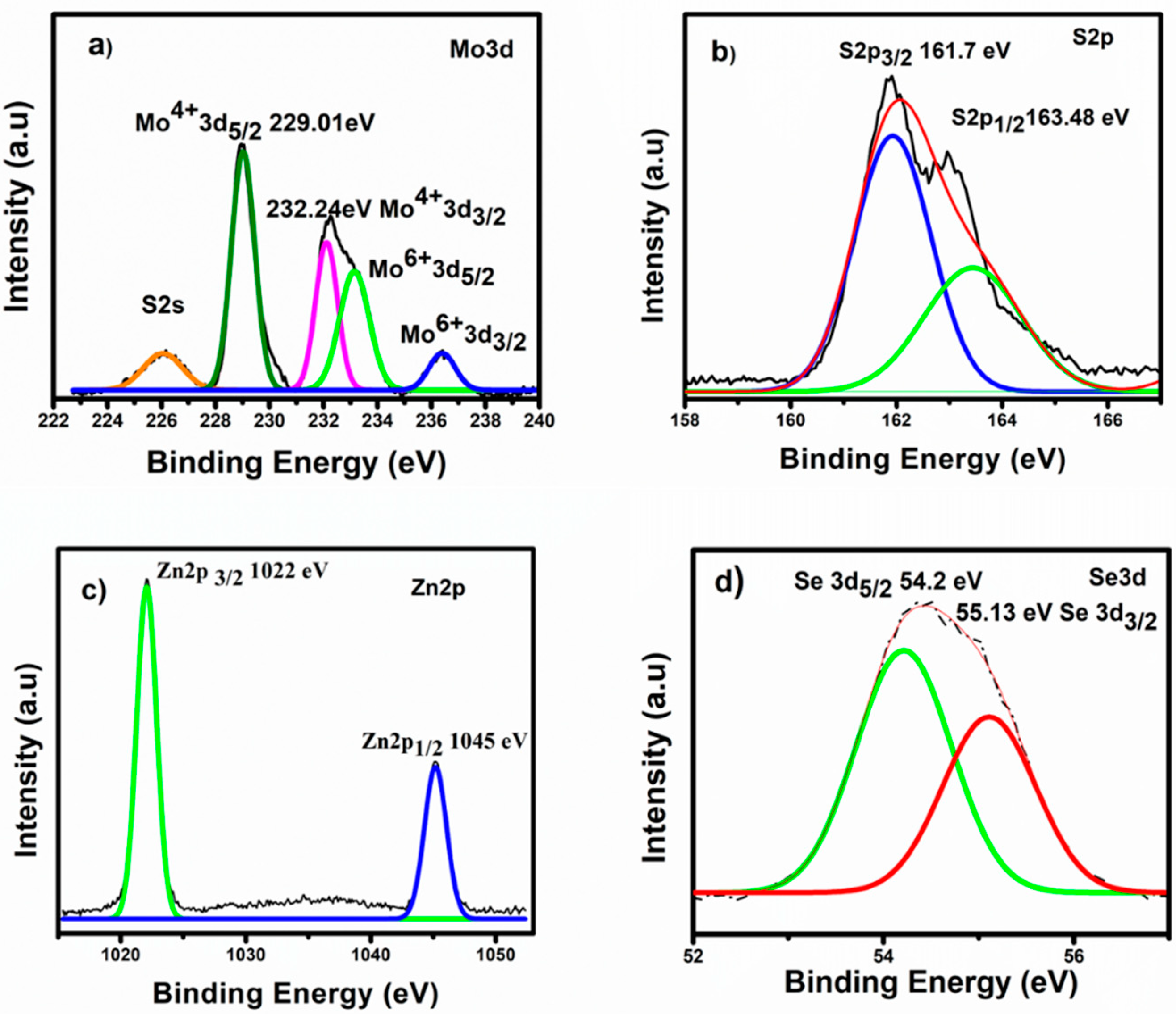
2.1. Phase and Microstructural Analysis

The phase purity and crystalline structure of the synthesized photocatalysts were determined by using XRD. Figure 1 shows the XRD patterns of pure MoS2, 1:3 MoS2/ZnSe (1:3 MZ), 1:1 MoS2/ZnSe (1:1 MZ) and 3:1 MoS2/ZnSe (3:1 MZ) heterostructures, and pure ZnSe. The diffraction peaks of MoS2 agree with the hexagonal phase and match with the JCPDS card no. 37-1492. Diffraction peaks at 13.8°, 28.7°, 34.4°, 42.6° and 57.5° correspond to the (002), (103), (100), (105) and (110) planes of the hexagonal wurtzite structure of MoS2 [32]. The diffraction peaks of ZnSe include peaks at 27.1°, 45.1°, 53.5° and 65.7°, as shown in Figure 1. These peaks correspond to the (111), (220), (311) and (400) planes of cubic ZnSe in accordance with the JCPDS card no. 37-1463 [33]. The sharpness of the peaks indicates the high crystallinity of ZnSe, and no extra peaks show that the prepared ZnSe is phase-pure. The steady increases in the peak intensities of MoS2 in the heterostructures, with increases in wt. % ratio, indicate a brilliant coupling interaction owing to the better photocatalytic activity [34]. Slight shifts in the position of the peaks, i.e., for MoS2 in the heterostructures it was 13.8° to 14°, and a shift of 0.2° was detected in the peak positions of ZnSe in the heterostructures, confirm the formation of the heterostructures.
X-ray photoelectron spectroscopy was employed to confirm the synthesis and oxidation states of the prepared materials. Figure 2a shows the XPS spectra of the Mo three-dimensional orbitals of MoS2. The peaks of the binding energies of Mo3d5/2 and Mo 3d3/2 at 229.01 eV and 232.24 eV show the oxidation state of Mo+4. The peaks centered at 233.1 eV and 236.2 eV can be ascribed to the Mo+6 3d3/2 and 3d1/2 oxidation states [35], which appear due to the slight oxidation of the edges during MoS2 synthesis [36]. The peaks at 161.7 eV for S2p3/2 and at 163.48 eV for S2p1/2 (Figure 2b) also confirm the presence of sulphur [37]. The XPS spectra of ZnSe display the presence of two peaks, Zn 2p3/2 at 1022 eV and Zn 2p1/2 at 1046 eV, showing the successful synthesis of ZnSe. The Se three-dimensional spectrum shows a peak at 54.4 eV, which is exactly on point for 3d5/2, and 55.13 eV for the 3d3/2 orbital of selenium [38]. These results indicate that catalysts are clearly formed with no impurities.
Figure 3 displays the SEM images (scale 1 µm) of pure MoS2, ZnSe and the 3:1 MZ composite, from which we can determine the morphology of the synthesized materials. Figure 3a shows clearly that large microspheres of flower-like MoS2 nanosheets are formed [39]. The sheets have a thickness range of 32–43 nm. The SEM image of sheet thickness is shown is Figure S7 (Supplementary Information), and Figure 3b shows the flower-like microspheres of cubic ZnSe [30]. The petals of the flower have a diameter range of 54–180 nm. The flower-like ZnSe is distinctly decorated on the nanosheets of MoS2 microspheres, as is clearly seen in Figure 3c. The SEM images of 1:3 MZ and 1:1 MZ are shown in Figure S1 (Supplementary Information), and also show a clear ornamentation of ZnSe microspheres on the MoS2 microspheres.
Energy dispersive X-ray spectroscopy helps to determine the elemental composition and hence the purity of the synthesized materials. The EDS spectra in Figure S2a–e (Supplementary Information) show that there are no impurities present in MoS2, ZnSe, or their composites.

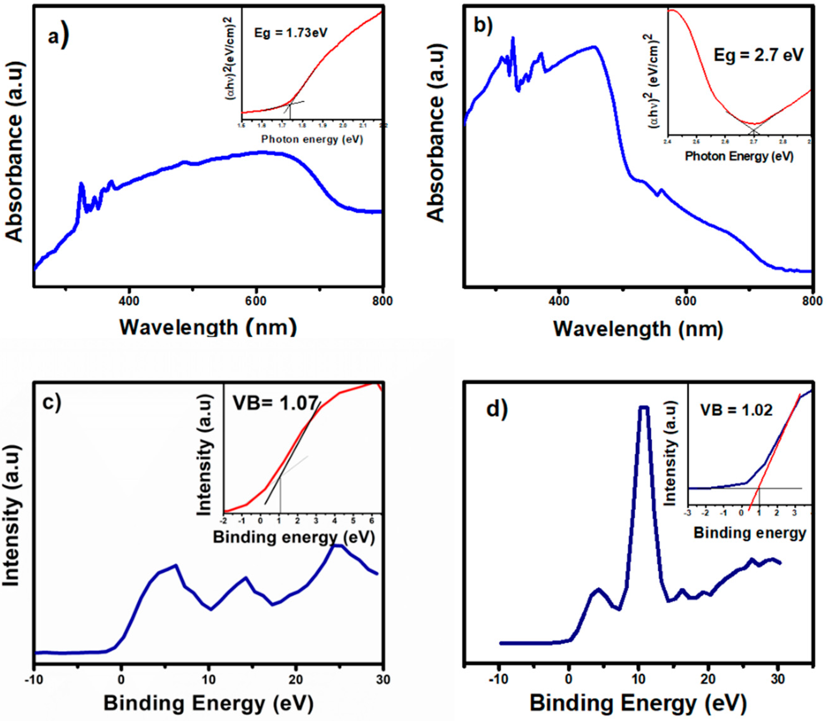
2.2. Alignment of Energy Level

Band gap energy and the position of the valence bands against standard hydrogen electrode (SHE) are the two important key factors used to understand the mechanism of the photocatalytic degradation, and help to draw the alignment of the energy levels [40]. UV-vis-DRS was employed to determine the band gap of the prepared material. The band gaps of both MoS2 and ZnSe were calculated by using the Kubelka Munk equation and by the Tauc plots [41], as shown in Figure 4a. Molybdenum sulfide showed a narrow band gap of 1.73 eV, and the band gap of ZnSe was calculated to be 2.70 eV, as shown in Figure 4b. UV-vis-DRS spectra of all the composites are shown in Figure S3 (Supplementary Information). The XPS spectra of these compounds show a valence band position of 1.07 eV for MoS2 in Figure 4c and 1.02 eV for ZnSe, as shown in Figure 4d. Based on this information (Figure 4), the energy levels are drawn and aligned.

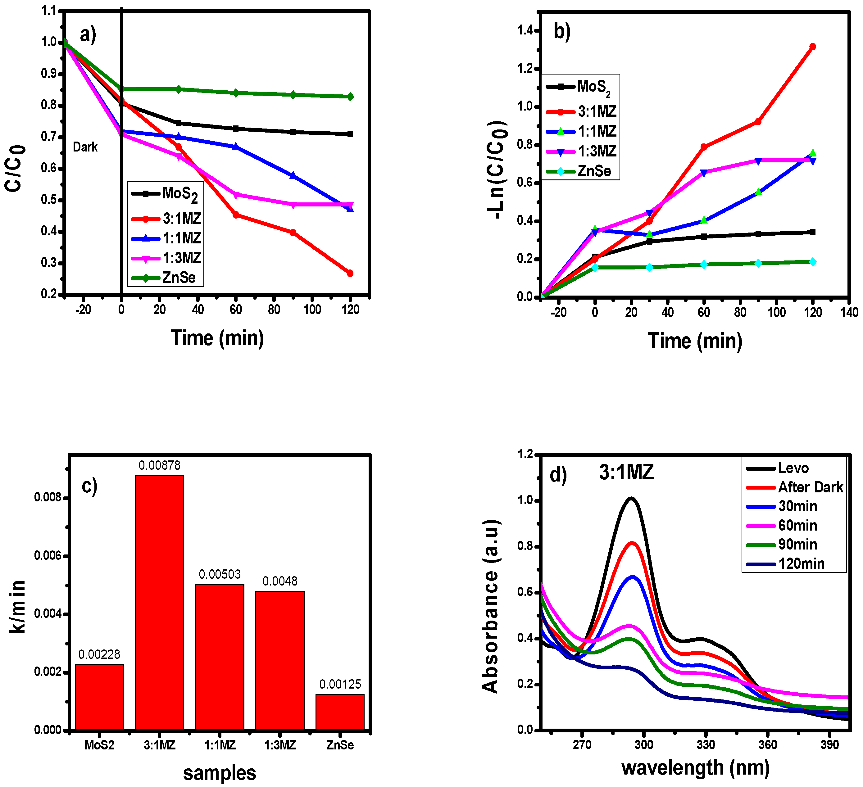
2.3. Photocatalytic Degradation of Levofloxacin

The photocatalytic activity of the prepared samples was measured as a function of the degradation of the antibiotic levofloxacin. All measurements were performed under the same conditions. Detailed absorption spectra of the degradation process are shown in Figure S2 (Supplementary Information). The heterostructures showed efficient degradation as compared to the pure semiconductors. The rate of degradation, i.e., C/C0, is plotted against time, as shown in Figure 5a. The heterostructure of 3:1 MZ showed the best result, with a 73.2% degradation of levofloxacin in 2 h. On the other hand, MoS2, ZnSe, 1:1 MZ and 1:3 MZ showed 29%, 17.1%, 53% and 52% degradations, respectively. The degradation rates are shown in Figure 5b,c. These show that the composite 3:1 MZ caused the highest degradation rate, i.e., 0.00878 k/min. The absorption spectra of the 3:1 MZ heterostructure for the degradation of levofloxacin are shown in Figure 5d. This also implies that a good interface has been developed between MoS2 and ZnSe in the heterostructures.
The photocatalytic activity is examined by the electron transfer through the heterojunction [42]. So, the PL studies were performed. The PL spectra in Figure 6 show the emission intensity of the different photocatalysts. A high-intensity peak in the PL spectrum shows rapid recombination, and vice versa [43]. The results show that the 1:3 MZ composite manifested a 28.6% decrease in the PL intensity, and 1:1 MZ an 85.7% decrease in the PL intensity. The 3:1 MZ composite manifested a 96.2% decrease in the PL intensity as compared to the pure counterpart. It can be inferred from the results that the increase in the concentration of MoS2 in the photocatalyst decreases the electron/hole recombination.
The results of the reusability studies of the 3:1 MZ photocatalyst for the degradation of levofloxacin, over three cycles, are shown in Figure 7. The concentration of the antibiotic was adjusted to the initial value after the degradation studies. The photocatalyst 3:1 MZ was studied for three cycles under visible light irradiation. The degradation values after the first, second and third run were found to be 73.2%, 72.3% and 71.9%, respectively, indicating that the performance efficiency of the photocatalyst was not reduced significantly. The photocatalyst was centrifuged and collected from the reaction mixture. The recovered photocatalyst was analyzed with XRD, SEM and EDS, and no change in the crystal phase or morphology, and no impurities, were observed, as shown in Figures S5 and S6 (Supplementary Information).
The degradation mechanism is proposed based on the results obtained and schematically drawn in Figure 8. When the photocatalyst is exposed to light, electrons in the valence band get excited and move to the conduction band of ZnSe, and holes are produced [44]. These photoexcited electrons are transferred to the conduction band of MoS2, which acts as electron receiver [45]. This transfer decreases the electron–hole recombination, as shown in the PL spectra [46]. The transferred electrons on the conduction band of MoS2 react with the adsorbed oxygen, and convert it to superoxide radical (O2), which can instantly decompose the antibiotic [47]. The holes in the valence band are transferred from low-energy MoS2 to high-energy ZnSe. These holes react with water and produce HO [34]. These hydroxyl and superoxide radicals degrade the antibiotic [48]. So MoS2 provides a platform for the separation and effective transport of the photogenerated electrons and holes [49,50]. Therefore, this binary system provides active sites that show the ability to harvest large amounts of light, hence their better degradation efficiency. Many heterostructure systems have been reported before now; for example, rGO-Bi2WO6 degraded 74.3% levofloxacin in 120 min [51], Ag/AgBr/BiOBr degraded 74% levofloxacin in 150 min [52], and CeV-BiV degraded 95.7% levofloxacin in 5 h [53]. Regarding the present study, the 3:1 MZ heterostructure shows a comparable activity of 73.2% in 120 min. However, this was an entirely new study for the ZnSe and MoS2 heterostructures, performed to understand their behavior and photocatalytic activity.

3. Materials and Methods

All the chemicals, viz. sodium molybdate, thiourea, zinc acetate, Se powder, hydrazine hydrate and ethanol, were purchased from Sigma Aldrich (St. Louis, MO, USA). All chemicals were of analytical grade and were used without purification.

3.1. Synthesis of MoS2

Analytical-grade chemicals were used in this study without further purification. MoS2 was synthesized using the hydrothermal method. Totals of 1.451 g sodium molybdate and 2.283 g thiourea were dissolved in 32 mL of water and placed in a 40 mL Teflon-lined stainless steel autoclave. The autoclave was heated in an oven at a temperature of 200 °C for 24 h. The obtained black powder was washed 3 times with water and one time with ethanol and dried in a vacuum oven at 80 °C for 24 h.

3.2. Synthesis of ZnSe

The synthesis of ZnSe was also carried out by the hydrothermal method. Totals of 0.175 g of zinc acetate and 0.016 g of selenium powder were dissolved in 8 mL of 6 M KOH solution and 2 mL of hydrazine hydrate was added in it. The mixture was then stirred for 1 h and transferred into a 40 mL Teflon-lined stainless steel autoclave and heated in an oven at 200 °C for 3 h. The resultant product was washed three times with water and one time with ethanol and vacuum dried at 70 °C for 10 h.

3.3. Synthesis of MoS2/ZnSe Heterostructures

MoS2/ZnSe heterostructures were prepared by the ultrasonic method. The heterostructures were prepared with 1:3, 1:1 and 3:1 wt.% of MoS2 and ZnSe, respectively. Totals of 25 mg of MoS2 and 75 mg of ZnSe were mixed in 25 mL of ethanol under ultrasonic shaking for 3 h. They were then dried at 80 °C for 10 h. These heterostructures were used for photocatalytic degradation.

3.4. Photodegradation of Levofloxacin

The photodegradation was measured by irradiating 11 ppm solution of levofloxacin under visible light using a 500 W Xe lamp with a light intensity of 100 mW/cm2 of AM 1.5 G and a cut-off filter λ > 420 nm. The reaction mixture was placed 10 cm below the lamp. A total of 30 mg of the photocatalyst was added into 100 mL of the antibiotic solution and stirred for 30 min in the dark to achieve the adsorption–desorption equilibrium. The mixture was then irradiated with visible light for two hours, then 3 mL samples were collected at a time interval of 30 min, which were centrifuged, and its concentration in terms of absorbance was measured using a UV-vis spectrophotometer.

3.5. Characterization

XRD analysis was carried out on an X-ray powder diffractometer (STOE Darmstadt, Germany) by using Cu Kα at λ = 1.54 Å. UV-vis and DRS analyses were performed on a Perkin Elmer Lambda 365 spectrometer. A VEGA3 TESCAN scanning electron microscope in combination with an energy dispersive X-ray spectroscope was used to obtain images and elemental compositions of the photocatalysts at an acceleration voltage of 20 KV. Photoluminescence spectra were obtained by using a Perkin Elmer FL 6500/8500 spectrometer. The X-ray photoelectron spectroscopic analyses were recorded on an ESCALAB 250 Xi X-ray photoelectron spectrophotometer.

4. Conclusions

In summary, MoS2/ZnSe heterostructures were successfully prepared for the first time, to the best of our knowledge, with different ratios through a facile sonochemical method. The as-prepared flower-like heterostructures showed excellent photocatalytic activity, as compared to the pure MoS2 and ZnSe counterparts. Levofloxacin was successfully degraded using these photocatalysts. The heterostructure 3:1 MZ showed the best catalytic activity of 73.2% in two hours. The prepared photocatalysts showed no significant change in morphology, crystal structure or photocatalytic activity after three cycles of use. This study shows that the formation of heterostructures led to the efficient light absorption and degradation of the antibiotic.

Supplementary Materials

The following are available online at https://www.mdpi.com/2073-4344/10/12/1380/s1. Figure S1: SEM images of 1:3 MZ (a) at 5µm and (b) 500 nm and 1:1 MZ at (c) 5 µm and (d) at 500 nm, Figure S2: EDS spectra of, (a) MoS2, (b) ZnSe, (c) 1:3 MoS2/ZnSe, (d) 1:1 MoS2/ZnSe, and (e) 3:1 MoS2/ ZnSe photocatalysts, Figure S3: DRS spectra of pure MoS2, ZnSe and their composites, Figure S4: Photocatalytic study for all the photocatalyst, shows the detailed absorption spectra for degradation of Levofloxacin, Figure S5: XRD of 3:1 MZ heterostructure before and after degradation, Figure S6: SEM of 3:1 MZ heterostructure, (a) before and (b) after degradation, and EDS spectra (c) before and (d) after degradation, Figure S7: SEM image of MoS2 nanosheets.

Author Contributions

Conceptualization and writing—reviewing, M.F.E., supervising and writing—review, H.N., methodology E.S., S.I., and S.A.B.B., formal analysis and writing—first draft, E.S. All authors have read and agreed to the published version of the manuscript.

Funding

This project was supported by the National University of Sciences and Technology Islamabad, Pakistan.

Acknowledgments

Authors are grateful to the National University of Sciences and Technology for funding and providing all the required facilities to carry out the project.

Conflicts of Interest

The authors declare no conflict of interest.

References

  1. Meng, F.Q.; Ma, W.; Duan, C.Y.; Liu, X.; Chen, Z.; Wang, M.Y.; Gao, J.; Zhang, Z. High efficient degradation of levofloxacin by edge-selectively Fe@3D-WS2: Self-renewing behavior and Degradation mechanism study. Appl. Catal. B 2019, 252, 187–197. [Google Scholar] [CrossRef]
  2. Klaus, K. Antibiotics in the aquatic environment—A review—Part II. Chemosphere 2009, 75, 435–441. [Google Scholar]
  3. Chow, D.N.F.T. The Clinical Pharmacokinetics of Levofloxacin. Clin. Pharmacokinet. 1997, 32, 101–119. [Google Scholar]
  4. Fujishima, A.; Honda, K. Electrochemical photolysis of water at a semiconductor electrode. Nature 1972, 238, 37–38. [Google Scholar] [CrossRef]
  5. Wang, H.; Zhang, L.; Chen, Z.; Hu, J.; Li, S.; Wang, Z.; Liu, J.; Wang, X. Semiconductor heterojunction photocatalysts: Design, construction, and photocatalytic performances. Chem. Soc. Rev. 2014, 43, 5234–5244. [Google Scholar] [CrossRef]
  6. Ding, C.; Fu, K.; Pan, Y.; Liu, J.; Deng, H.; Shi, J. Comparison of Ag and AgI-Modified ZnO as Heterogeneous Photocatalysts for Simulated Sunlight Driven Photodegradation of Metronidazole. Catalysts 2020, 10, 1097. [Google Scholar] [CrossRef]
  7. Ghosh, M.; Lohrasbi, M.; Chuang, S.S.C.; Jana, S.C. Mesoporous Titanium Dioxide Nanofibers with a Significantly Enhanced Photocatalytic Activity. ChemCatChem 2016, 8, 2525–2535. [Google Scholar] [CrossRef]
  8. Wen, X.-J.; Niu, C.-G.; Guo, H.; Zhang, L.; Liang, C.; Zeng, G.-M. Photocatalytic degradation of levofloxacin by ternary Ag2CO3/CeO2/AgBr photocatalyst under visible-light irradiation: Degradation pathways, mineralization ability, and an accelerated interfacial charge transfer process study. J. Catal. 2018, 358, 211–223. [Google Scholar] [CrossRef]
  9. Brillas, E. A Review on the Degradation of Organic Pollutants in Waters by UV Photoelectro-Fenton and Solar Photoelectro-Fenton. J. Braz. Chem. Soc. 2014, 25, 393–417. [Google Scholar] [CrossRef]
  10. Zhang, T.; Lin, P.; Wei, N.; Wang, D. Enhanced Photoelectrochemical Water-Splitting Property on TiO2 Nanotubes by Surface Chemical Modification and Wettability Control. ACS Appl. Mater. Interfaces 2020, 12, 20110–20118. [Google Scholar] [CrossRef] [PubMed]
  11. Li, M.; Yin, S.; Wu, T.; Di, J.; Ji, M.X.; Wang, B.; Chen, Y.; Xia, J.X.; Li, H.M. Controlled preparation of MoS2/PbBiO2I hybrid microspheres with enhanced visible-light photocatalytic behaviour. J. Colloid Interface Sci. 2018, 517, 278–287. [Google Scholar] [CrossRef] [PubMed]
  12. Theerthagiri, J.; Senthil, R.A.; Senthilkumar, B.; Reddy Polu, A.; Madhavan, J.; Ashokkumar, M. Recent advances in MoS2 nanostructured materials for energy and environmental applications—A review. J. Solid State Chem. 2017, 252, 43–71. [Google Scholar] [CrossRef]
  13. Li, Z.; Meng, X.; Zhang, Z. Recent development on MoS2 -based photocatalysis: A review. J. Photochem. Photobiol. C 2018, 35, 39–55. [Google Scholar] [CrossRef]
  14. Sabarinathan, M.; Harish, S.; Archana, J.; Navaneethan, M.; Ikeda, H.; Hayakawa, Y. Highly efficient visible-light photocatalytic activity of MoS2–TiO2 mixtures hybrid photocatalyst and functional properties. RSC Adv. 2017, 7, 24754–24763. [Google Scholar] [CrossRef] [Green Version]
  15. Bandow, S.; Maruyama, Y.; Bi, X.X.; Ochoa, R.; Holden, J.M.; Lee, W.T.; Eklund, P.C. Electronic and vibrational properties of Rb-intercalated MoS2 nanoparticles. Mat. Sci. Eng. A 1995, 204, 222–226. [Google Scholar] [CrossRef]
  16. Ge, L.; Han, C.; Xiao, X.; Guo, L. Synthesis and characterization of composite visible light active photocatalysts MoS2–g-C3N4 with enhanced hydrogen evolution activity. Int. J. Hydrog. Energy 2013, 38, 6960–6969. [Google Scholar] [CrossRef]
  17. Islam, S.E.; Hang, D.R.; Chen, C.H.; Sharma, K.H. Facile and Cost-Efficient Synthesis of Quasi-0D/2D ZnO/MoS2 Nanocomposites for Highly Enhanced Visible-Light-Driven Photocatalytic Degradation of Organic Pollutants and Antibiotics. Chemistry 2018, 24, 9305–9315. [Google Scholar] [CrossRef]
  18. Wang, C.; Lin, H.; Xu, Z.; Cheng, H.; Zhang, C. One-step hydrothermal synthesis of flowerlike MoS2/CdS heterostructures for enhanced visible-light photocatalytic activities. RSC Adv. 2015, 5, 15621–15626. [Google Scholar] [CrossRef]
  19. Nandiyanto, A.B.D.; Zaen, R.; Oktiani, R. Correlation between crystallite size and photocatalytic performance of micrometer-sized monoclinic WO3 particles. Arab. J. Chem. 2020, 13, 1283–1296. [Google Scholar] [CrossRef]
  20. Li, Y.; Xiang, F.; Lou, W.; Zhang, X. MoS2 with structure tuned photocatalytic ability for degradation of methylene blue. IOP Conf. Ser. 2019, 300, 052021. [Google Scholar] [CrossRef]
  21. Li, Z.; Cao, F.; Wang, L.; Chen, Z.; Ji, X. A novel ternary MoS2/MoO3/TiO2 composite for fast photocatalytic degradation of rhodamine B under visible-light irradiation. New J. Chem. 2020, 44, 537–542. [Google Scholar] [CrossRef]
  22. Guo, F.; Huang, X.; Chen, Z.; Ren, H.; Li, M.; Chen, L. MoS2 nanosheets anchored on porous ZnSnO3 cubes as an efficient visible-light-driven composite photocatalyst for the degradation of tetracycline and mechanism insight. J. Hazard. Mater. 2020, 390, 122158. [Google Scholar] [CrossRef] [PubMed]
  23. Kang, J.; Jin, C.; Li, Z.; Wang, M.; Chen, Z.; Wang, Y. Dual Z-scheme MoS2/g-C3N4/Bi24O31Cl10 ternary heterojunction photocatalysts for enhanced visible-light photodegradation of antibiotic. J. Alloys Compd. 2020, 825, 153975. [Google Scholar] [CrossRef]
  24. Chen, L.; Nguyen, T.B.; Lin, Y.-L.; Wu, C.-H.; Chang, J.-H.; Chen, C.-W.; Dong, C.-D. Enhanced Heterogeneous Photodegradation of Organic Pollutants by a Visible Light Harvesting CoO@meso–CN@MoS2 Nanocomposites. Catalysts 2020, 10, 722. [Google Scholar] [CrossRef]
  25. Li, G.; Hou, J.; Zhang, W.; Li, P.; Liu, G.; Wang, Y.; Wang, K. Graphene-bridged WO3/MoS2 Z-scheme photocatalyst for enhanced photodegradation under visible light irradiation. Mater. Chem. Phys. 2020, 246, 122827. [Google Scholar] [CrossRef]
  26. Patel, J.D.; Mighri, F.; Ajji, A. A facile route towards the preparation of ZnSe nanocrystals. Mater. Lett. 2014, 131, 366–369. [Google Scholar] [CrossRef]
  27. Zhang, Q.; Li, H.Q.; Ma, Y.; Zhai, T.Y. ZnSe nanostructures: Synthesis, properties and applications. Prog. Mater. Sci. 2016, 83, 472–535. [Google Scholar] [CrossRef]
  28. Zhang, Y.; Hu, C.G.; Feng, B.; Wang, X.; Wan, B.Y. Synthesis and photocatalytic property of ZnSe flowerlike hierarchical structure. Appl. Surf. Sci. 2011, 257, 10679–10685. [Google Scholar] [CrossRef]
  29. Liu, H.R.; Hu, Y.C.; He, X.; Jia, H.S.; Liu, X.G.; Xu, B.S. In-situ anion exchange fabrication of porous ZnO/ZnSe heterostructural microspheres with enhanced visible light photocatalytic activity. J. Alloys Compd. 2015, 650, 633–640. [Google Scholar] [CrossRef]
  30. Cao, H.Q.; Xiao, Y.J.; Zhang, S.C. The synthesis and photocatalytic activity of ZnSe microspheres. Nanotechnology 2011, 22, 015604. [Google Scholar] [CrossRef]
  31. Zhang, L.H.; Yang, H.Q.; Xie, X.L.; Zhang, F.H.; Li, L. Preparation and photocatalytic activity of hollow ZnSe microspheres via Ostwald ripening. J. Alloys Compd. 2009, 473, 65–70. [Google Scholar] [CrossRef]
  32. Zhang, L.H.; Yang, H.Q.; Yu, J.; Shao, F.H.; Li, L.; Zhang, F.H.; Zhao, H. Controlled Synthesis and Photocatalytic Activity of ZnSe Nanostructured Assemblies with Different Morphologies and Crystalline Phases. J. Phys. Chem. C 2009, 113, 5434–5443. [Google Scholar] [CrossRef]
  33. Miao, H.; Hu, X.; Sun, Q.; Hao, Y.; Wu, H.; Zhang, D.; Bai, J.; Liu, E.; Fan, J.; Hou, X. Hydrothermal synthesis of MoS2 nanosheets films: Microstructure and formation mechanism research. Mater. Lett. 2015, 166, 121–124. [Google Scholar] [CrossRef]
  34. Feng, B.; Cao, J.; Han, D.L.; Liang, H.T.; Yang, S.; Li, X.Y.; Yang, J.H. ZnSe nanoparticles of different sizes: Optical and photocatalytic properties. Mater. Sci. Semicond. Process. 2014, 27, 865–872. [Google Scholar] [CrossRef]
  35. Nayak, S.; Swain, G.; Parida, K. Enhanced Photocatalytic Activities of RhB Degradation and H2 Evolution from in Situ Formation of the Electrostatic Heterostructure MoS2/NiFe LDH Nanocomposite through the Z-Scheme Mechanism via p–n Heterojunctions. ACS Appl. Mater. Interfaces 2019, 11, 20923–20942. [Google Scholar] [CrossRef] [PubMed]
  36. Chen, J.; Liao, Y.; Wan, X.; Tie, S.; Zhang, B.; Lan, S.; Gao, X. A high performance MoO3@MoS2 porous nanorods for adsorption and photodegradation of dye. J. Solid State Chem. 2020, 291, 121652. [Google Scholar] [CrossRef]
  37. Shi, L.; He, Z.; Liu, S.Q. MoS2 quantum dots embedded in g-C3N4 frameworks: A hybrid 0D–2D heterojunction as an efficient visible-light driven photocatalyst. Appl. Surf. Sci. 2018, 457, 30–40. [Google Scholar] [CrossRef]
  38. Wang, X.; Xiong, W.; Li, X.Y.; Zhao, Q.D.; Fan, S.Y.; Zhang, M.M.; Mu, J.C.; Chen, A.C. Fabrication of MoS2@g-C3N4 core-shell nanospheres for visible light photocatalytic degradation of toluene. J. Nanoparticle Res. 2018, 20, 243. [Google Scholar] [CrossRef]
  39. Lohar, G.M.; Jadhav, S.T.; Takale, M.V.; Patil, R.A.; Ma, Y.R.; Rath, M.C.; Fulari, V.J. Photoelectrochemical cell studies of Fe2+ doped ZnSe nanorods using the potentiostatic mode of electrodeposition. J. Colloid Interface Sci. 2015, 458, 136–146. [Google Scholar] [CrossRef]
  40. Zhang, Y.J.; Zeng, W.; Li, Y.Q. Hydrothermal synthesis and controlled growth of hierarchical 3D flower-like MoS2 nanospheres assisted with CTAB and their NO2 gas sensing properties. Appl. Surf. Sci. 2018, 455, 276–282. [Google Scholar] [CrossRef]
  41. Ehsan, M.F.; Qudoos, S.; Ahmad, Z.; Hamid, S.; Arfan, M.; Zia, A.; Umbreen, K.; Ashiq, M.N.; Tyagi, D. ZnTe/ZnSe heterostructures: In-situ synthesis, characterization and photocatalytic activity for Congo Red degradation. SN Appl. Sci. 2019, 1, 197. [Google Scholar] [CrossRef] [Green Version]
  42. Wu, M.-H.; Li, L.; Liu, N.; Wang, D.-J.; Xue, Y.-C.; Tang, L. Molybdenum disulfide (MoS2) as a co-catalyst for photocatalytic degradation of organic contaminants: A review. Process. Saf. Environ. 2018, 118, 40–58. [Google Scholar] [CrossRef]
  43. Fu, Y.; Liang, W.; Guo, J.; Tang, H.; Liu, S. MoS2 quantum dots decorated g-C3N4/Ag heterostructures for enhanced visible light photocatalytic activity. Appl. Surf. Sci. 2018, 430, 234–242. [Google Scholar] [CrossRef]
  44. Zhang, W.; Xiao, X.; Zheng, L.; Wan, C. Fabrication of TiO2/MoS2Composite Photocatalyst and Its Photocatalytic Mechanism for Degradation of Methyl Orange under Visible Light. Can. J. Chem. Eng. 2015, 93, 1594–1602. [Google Scholar] [CrossRef]
  45. Kuehnel, M.F.; Creissen, C.E.; Sahm, C.D.; Wielend, D.; Schlosser, A.; Orchard, K.L.; Reisner, E. ZnSe Nanorods as Visible-Light Absorbers for Photocatalytic and Photoelectrochemical H2 Evolution in Water. Angew. Chem. 2019, 58, 5059–5063. [Google Scholar] [CrossRef] [PubMed] [Green Version]
  46. Peng, W.-C.; Li, X.-Y. Synthesis of MoS2/g-C3N4 as a solar light-responsive photocatalyst for organic degradation. Catal. Commun. 2014, 49, 63–67. [Google Scholar] [CrossRef] [Green Version]
  47. Ghosh, M.; Liu, J.; Chuang, S.S.C.; Jana, S.C. Fabrication of Hierarchical V2O5 Nanorods on TiO2 Nanofibers and Their Enhanced Photocatalytic Activity under Visible Light. ChemCatChem 2018, 10, 3305–3318. [Google Scholar] [CrossRef]
  48. Li, Q.; Zhang, N.; Yang, Y.; Wang, G.; Ng, D.H. High efficiency photocatalysis for pollutant degradation with MoS2/C3N4 heterostructures. Langmuir 2014, 30, 8965–8972. [Google Scholar] [CrossRef]
  49. Zheng, Z.X.; Qiao, Y.; Cai, Y.H.; He, Y.N.; Tang, Y.M.; Li, L.S. MoS2 decorated CdS hybrid heterojunction for enhanced photoelectrocatalytic performance under visible light irradiation. J. Colloid Interface Sci. 2019, 533, 561–568. [Google Scholar] [CrossRef]
  50. Tahir, M.B. Construction of MoS2/CND-WO3 Ternary Composite for Photocatalytic Hydrogen Evolution. J. Inorg. Organomet. Polym. Mater. 2018, 28, 2160–2168. [Google Scholar] [CrossRef]
  51. Chen, Y.L.; Wang, L.J.; Wang, W.Z.; Cao, M.S. Enhanced photoelectrochemical properties of ZnO/ZnSe/CdSe/Cu2-xSe core-shell nanowire arrays fabricated by ion-replacement method. Appl. Catal. B Environ. 2017, 209, 110–117. [Google Scholar] [CrossRef]
  52. Arya, M.; Kaur, M.; Kaur, A.; Singh, S.; Devi, P.; Kansal, S.K. Hydrothermal synthesis of rGO-Bi2WO6 heterostructure for the photocatalytic degradation of levofloxacin. Opt. Mater. 2020, 107, 110126. [Google Scholar] [CrossRef]
  53. Gupta, G.; Kaur, A.; Sinha, A.S.K.; Kansal, S.K. Photocatalytic degradation of levofloxacin in aqueous phase using Ag/AgBr/BiOBr microplates under visible light. Mater. Res. Bull. 2017, 88, 148–155. [Google Scholar] [CrossRef]
Figure 1. X-ray diffraction spectra of MoS2, ZnSe and their heterostructures.
Catalysts 10 01380 g001
Figure 2. XPS spectra of (a) Mo 3d, (b) S 2p, (c) Zn 2p and (d) Se 3d orbitals.
Catalysts 10 01380 g002
Figure 3. SEM images of (a) MoS2, (b) ZnSe and (c) 3:1 MZ at 1 µm scale.
Catalysts 10 01380 g003
Figure 4. UV-visible DRS spectra of (a) MoS2 and (b) ZnSe with insets showing the respective Tauc plots and band gap of MoS2 and ZnSe. XPS spectra of (c) MoS2 and (d) ZnSe with their respective insets showing valence band positions.
Catalysts 10 01380 g004
Figure 5. (a) Photodegradation rate of MoS2, ZnSe and composites, (b) kinetic plots of the photocatalytic degradation, (c) rate of degradation as a function of time and (d) absorption spectra of photodegradation of levofloxacin by 3:1 MZ.
Catalysts 10 01380 g005
Figure 6. PL spectra of the photocatalysts.
Catalysts 10 01380 g006
Figure 7. Reusability of 3:1 MZ under visible light illumination.
Catalysts 10 01380 g007
Figure 8. Schematic illustration of alignment of the energy level of 3:1 MoS2/ZnSe nanocomposite.
Catalysts 10 01380 g008
Publisher’s Note: MDPI stays neutral with regard to jurisdictional claims in published maps and institutional affiliations.

