Abstract
A large number of studies have examined the origins of high-catalytic activities of nanoparticles, but very few have discussed the lifetime of high-energy electrons in nanoparticles. The lifetime is one of the factors determining electron transfer and thus catalytic activity. Much of the lifetime of electrons reported in the literature is too short for a high transfer-efficiency of photo-excited electrons from a catalyst to the attached molecules. We observed TiO2 nanoparticles using the femtosecond laser two-color pump-probe technique with photoemission electron microscopy having a 40 nm spatial resolution. A lifetime longer than 4 ps was observed together with a fast decay component of 100 fs time constant when excited by a 760 nm laser. The slow decay component was observed only when the electrons in an intermediate state pumped by the fundamental laser pulse were excited by the second harmonic pulse. The electronic structure for the asymmetry of the pump-probe signal and the origin of the two decay components are discussed based on the color center model of the oxygen vacancy.
1. Introduction
In chemical reactions, energetic electrons called “hot electrons” are transported over a potential barrier from one material to another [1,2,3,4,5,6,7]. The chemical reaction rate or electron current, Ie, over the barrier is given by the Richardson–Dushman equation
which was derived for thermionic emission [8,9,10,11,12,13]. Here, kT is the thermal energy of the transferred electrons when the energy of electrons is high, kT is high or the potential barrier, , is low, and transfer current Ie is large. For example, Weik et al. [14] observed that cross-sections for both photo-desorption and photo-dissociation of O2 adsorbed on Pd increased exponentially from the photon energy of 4 to 6 eV. The efficient generation of hot electrons is the most important subject in nanomaterial science. Furthermore, the hot electron has been the focus of many reviews [1,2,5,15,16,17].
Bulk gold (Au) is the highest chemically inert metal, but Au in the form of nanoparticles (NPs) is one of the most important catalysts [18]. Understanding how a chemically inert material changes to a highly active catalyst is a major topic in nanoscience. The high catalytic activity has been attributed to metal–oxide interactions, such as charge transfer between the Au and oxides, and strain in the Au NPs due to lattice mismatches at the interface. However, Nørskov et al. [19] concluded that the low-coordinated Au atom is the main source of the very high catalytic activity. The CO oxidation activity increases with the decrease in diameter d of the NPs. From the activity scaling of 1/d3, they concluded that the corner atom is the site of catalysis. Their conclusion is highly important. However, they did not discuss the change in electronic structure of Au NPs from bulk Au, which should be essentially important in chemical reactions.
We propose that the essence of the high catalysis of NPs originates in the creation of excited states for maintaining hot electrons [20]. When the size of materials approaches the nanometer scale, the electronic states are quantized, and discrete energy levels are created in the conduction band, even in metals.
TiO2 is an important material, particularly in photocatalysis, and its properties have been extensively studied. Numerous studies have revealed that a decisive role in the catalysis is played by defect [21,22], which has been identified as oxygen vacancy, Vo. Because Vo has one atomic size, the electronic states in Vo are quantized and our proposal can explain the reason for the high catalysis of TiO2 NPs, which have a large number of Vos. Although a large number of studies have reported the electronic structure in the bandgap, only a small number of papers have reported excited states in the conduction band. Onda et al. [23] reported that the “wet electrons” state lies at 2.4 eV above the Fermi level on the surface of an H2O/TiO2 (110) rutile. Argondizzo et al. [24] reported the eg state 3.66 eV above the defect level that lies 0.85 eV below the conduction band minimum (CBM). More excited states may exist in the conduction band, and surveying unknown excited states will help to understand the electron dynamics in the photocatalysis of TiO2 NPs.
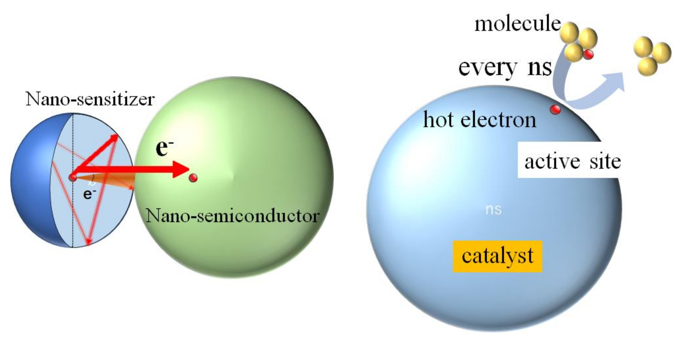
We also propose that the long lifetime of hot electrons is essential for high catalysis [20] from the following consideration. The lifetime of hot electrons required for high catalytic activities can be quantitatively estimated, as illustrated in Figure 1, in which an electron is treated as a small particle. Furube et al. [25] reported that more than 80% of hot electrons generated in 10 nm-diameter gold (Au) NPs as a sensitizer are transferred to TiO2 NPs to supply a photocurrent. To realize such a high transfer efficiency, electrons need to be reflected many times on the wall before most pass through a small window between the Au NP and TiO2 NP. Then, they travel a distance of more than ten times the diameter of the NP. When the diameter of the Au NPs is 10 nm, the traveling distance is longer than 100 nm. An electron with a kinetic energy of 1 eV moves only 6 nm in 10 fs. Then, the photoexcited hot electrons need to stay at a high energy for a time longer than 200 fs so that they can overcome a potential barrier between the Au NP and the TiO2 NP. Another example is the photo-dissociation catalysis. The time interval of molecules arriving at the active site is about 1 ns at atmospheric pressure. For efficient transfer of high-energy electrons generated in a catalyst to molecules reaching the catalyst, electrons are desired to be active for the order of 1 ns until the next molecule arrives at the active site.
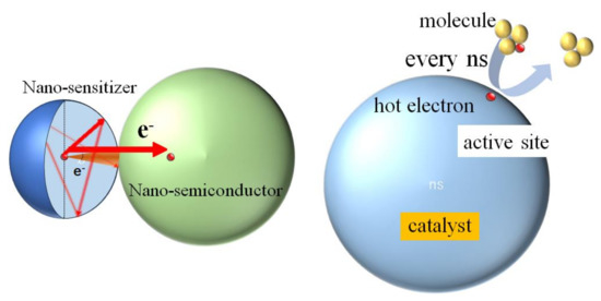
Figure 1.
For high catalytic activities, the lifetime of hot electrons needs to be very long.
In the case of Au NPs, the energy separation between quantized electronic levels will be large, and the energy relaxation time will be long due to the “phonon bottleneck” [26]. Then, it will be possible to realize near 100% transfer efficiency of the hot electron from 10 nm Au NP to TiO2 NP, as reported by Furube et al. [25]. In the case of TiO2, however, the lifetime of hot electrons reported by Onda et al. [23], and Argondizzo et al. [24] in a TiO2 single crystal were only 20 fs, which is too short for high photocatalysis. Even if photogenerated hot electrons in a TiO2 NP are trapped in the excited state discovered by Argondizzo et al. [24], electrons can only stay for several fs inside NPs, and the fraction of only 10−5 of photogenerated electrons contributes to a chemical reaction when molecules arrive at the active site every 1 ns. Because the photocatalytic activity of TiO2 NPs is very high, there should be other excited states with a much longer lifetime. In fact, in a study using photoemission-electron-microscopy (PEEM) with high spatial resolution, we discovered a new excited state located 1.63 eV above the ground state of Vo, which has a lifetime as long as 4 ps [20].
The present paper reports a further detailed analysis of the result of the lifetime of hot electrons in TiO2 nanoparticles. We found two decay components in the delay dependence of the pump-probe signal. The slow decay component was observed only when the electrons excited by the fundamental laser were probed by the second harmonic laser pulse and the lifetime longer than 4 ps was observed when the wavelength of the fundamental pulse was 760 nm. The fast component decayed with a time constant of 100 fs, which is longer than the values reported for hot electrons in the literature. The ratio of the intensity of the slow component to the fast component was about 10%. The electronic structure for observing the asymmetry of the pump-probe signal and the origin of the slow and fast components are discussed.
2. Results
2.1. Three and Four-Photon Slopes
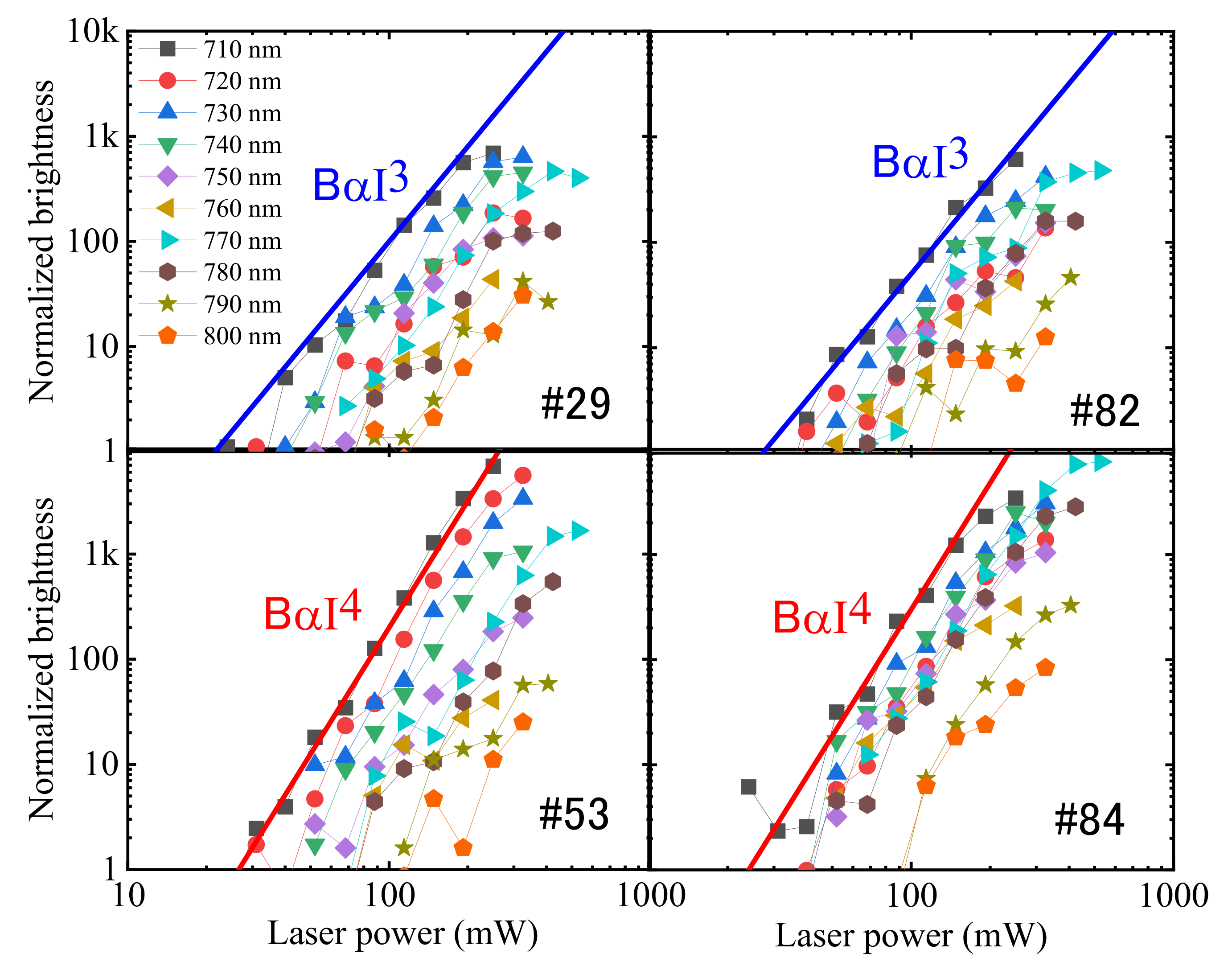
When I represents the laser power, the number of n-photon excited electrons is given by In, and the number of n-photon excitations is given by the slope of the logarithmic representation. Figure 2 shows the electron signals for four typical particles as a function of laser power. The brightness of NPs increased nearly exponentially and there was a difference in the slope; NP #29 and #82 were three-photon-excited and particles #53 and #84 were four-photon-excited at all wavelengths from 710 to 800 nm. At a high laser power, a slight saturation is noticed and the degree of brightness saturation was different among four NPs: clear saturation in #29 and very small in #53.
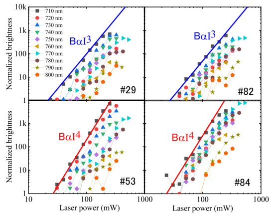
Figure 2.
Some nanoparticles (NPs), such as #29 and #82, showed a three-photon slope, and others, such as #53 and #84, showed a four-photon slope in the laser power dependence of the brightness.
When an electron is excited by n photons of photon energy hν, the energy difference E between the ground state and the excited state is (n − 1) hν < E < n hν. In particles #53 and #84, the energy E is 1.746 × 3 < E < 1.55 × 4 eV, that is, 5.23 < E < 6.2 eV, because they were four-photon excited at all wavelengths from 710 nm (1.746 eV) to 800 nm (1.55 eV). In three-photon excited NPs #29 and #82, 3.5 eV < E < 4.7 eV. Concerning the ionization energy, Ei, one theory predicted 8.3 eV [27], and two experimental observations reported 7.5 eV [28] and 7.25 eV. [29] We assume Ei = 7.5 eV. Therefore, electrons in the valence band cannot be directly ionized either by three-photon excitation or by four-photon excitation by the laser of wavelengths from 710 to 800 nm.
Since the bandgap energy is about 3.2 eV, the energy difference, EA, between CBM and the vacuum level, which is called the electron affinity, is 4.3 eV. If there are electron trap levels from 0.4 to 0.8 eV below the CBM, the electrons at these levels will be three-photon-ionized, and if there are electron trap levels between 0.9 and 1.9 eV below the CBM, the electrons at these levels are four-photon-ionized by the laser of wavelength 710 nm. It is well known that there are many defect levels in TiO2. Ghosh et al. [30] reported eight shallow traps between 0.27 and 0.87 eV below the CBM and deep luminescence centers near 1.5 eV and around 2 eV. The absorption in reduced TiO2 increases with the wavelength and becomes flat between 1.5 and 2 μm [30,31,32]. This infrared absorption is due to the shallow traps reported by Ghosh et al. [10]. Yoshihara et al. [33] reported that the absorption by trapped electrons in TiO2 NP had a broad peak at around 800 nm, which will correspond to the luminescence center reported by Ghosh et al. [10]. From this knowledge, we can explain the observed small photon numbers in Figure 2 as follows. Three-photon NPs #29 and #82 have many shallow traps, and trapped electrons were three-photon-ionized. In four-photon NPs #53 and #84, deep luminescence centers were four-photon-ionized.
Another possibility is that valence band electrons were excited to the electronic states in the conduction band, from where they were thermally ionized as was observed in Au NPs by Li et al. [13]. If this is the case, we can suppose that four-photon NPs have higher density defects whose intermediate energy levels assist the excitation of the valence band electrons to the excited states in the conduction band and that the excitation of the valence band electrons assisted by the intermediate-level is weak in three-photon NPs which have lower density defects.
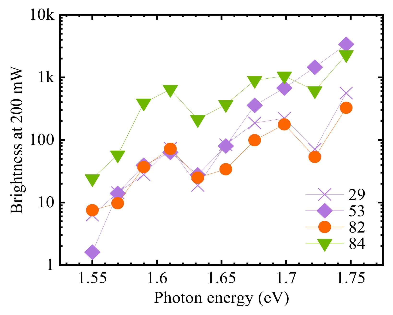
Figure 3 shows the wavelength dependence of the signal intensity at 200 mW of NPs shown in Figure 2. The electron signal was larger at a shorter laser wavelength. This wavelength dependence is contrary to the report by Yoshihara et al. [33], who observed an absorption peak at 800 nm, and to the reports in references [30,31,32] in which the absorption coefficient was larger at longer wavelengths up to 1.5 μm. Therefore, the wavelength dependence of the brightness seen in Figure 3 is difficult to explain by a direct ionization and hence, thermal ionization will be more plausible as was observed in Au NPs [13]. In the one-color excitation spectra shown in Figure 3, there were two dips at 1.63 eV (760 nm) and 1.72 eV (720 nm) in #29, #82, and #84, and the dip at 1.72 eV was missing in #53.
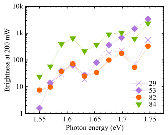
Figure 3.
The brightness of TiO2 NPs #29, #53, #82, and #84 shown in Figure 2 at various photon energies. The laser power was 200 mW. The trend of a larger electron signal at a shorter wavelength is contrary to the wavelength dependence of the absorption coefficient, which suggests that electrons are not directly multi-photon-ionized.
2.2. Measurement of the Electron Lifetime
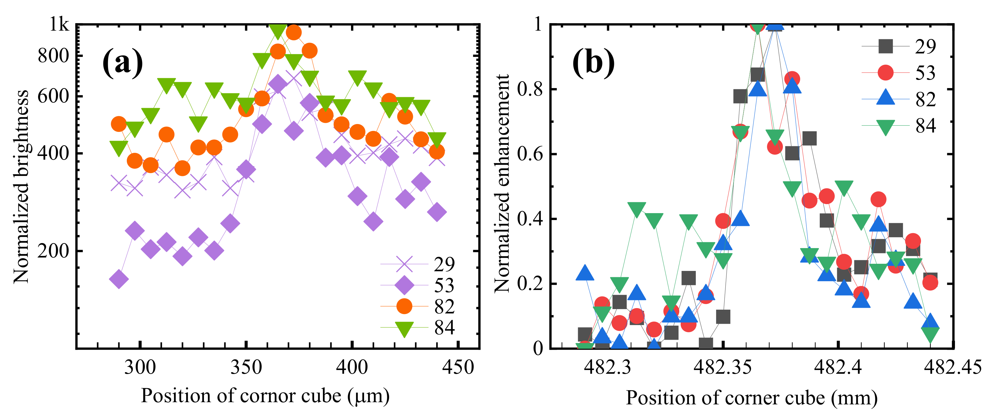
The lifetime of hot electrons was measured by the pump-probe technique using two colors: fundamental wavelength (F) and the second harmonic wavelength (SH). The result is shown in Figure 4a for the four NPs shown in Figure 2 when the wavelength of the F pulse was 800 nm. The laser power was 168 mW for the F pulse and 1.80 mW for the SH pulse. The brightness of NPs was similar when F pulse only or SH pulse only was irradiated. The horizontal axis is the position of the corner cube shown later in Section 4 for varying the delay time. When the number of the position is large, the SH pulse arrives later, i.e., the sample was irradiated first by the F pulse. At around 482.37 mm, the brightness was the maximum, which showed that both F and SH pulses irradiated the sample at the same time.
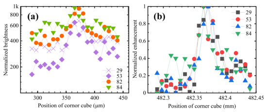
Figure 4.
(a) When the corner cube for reflecting the fundamental (F) beam was placed at 482.37 mm, the brightness of all NPs was enhanced. The wavelengths of F and second harmonic (SH) pulses were 800 and 400 nm, respectively. (b) As explained in the text, the brightness enhancement was normalized for the comparison of the delay time dependence. No clear difference was noticed in the delay dependence among NPs having a difference in brightness.
We refer to the dependence of brightness on delay time as “delay dependence” and the increase in the brightness of images when two pulses arrive at the same time as brightness “enhancement”. In #53, the brightness was enhanced by three times from 200 at 300 μm to 600 at 370 μm. The “enhancement” was about 2 in #82 and 1.5 in #84.
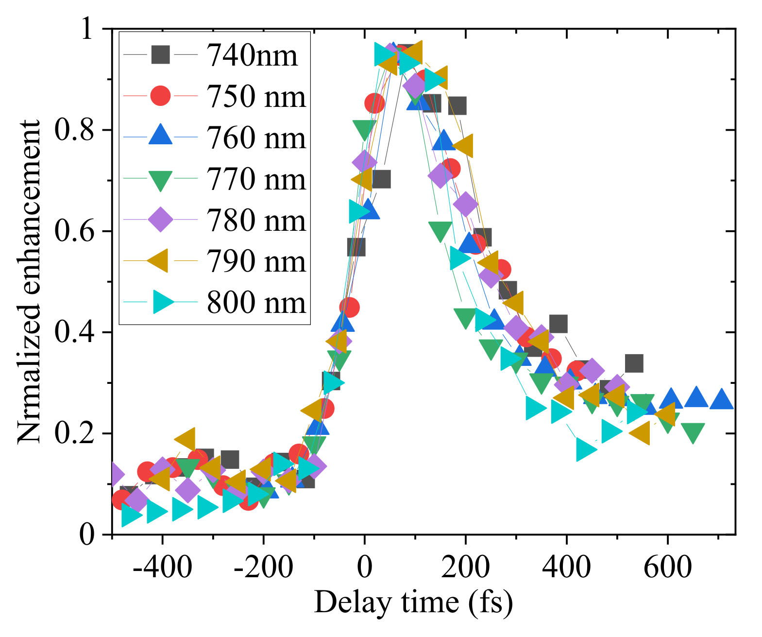
To compare the enhancement among particles with a large difference in brightness, the brightness enhancement was normalized by setting the lowest brightness to 0 and the highest brightness to 1 for each particle. The “normalized enhancements” for particles #29, #53, #82 and #84 are shown in Figure 4b. The signal-to-noise ratio of individual NPs was not as high, and the “normalized enhancement” was averaged over all of the selected 95 particles to study the detail of the delay dependence of enhancement, and the results at various wavelengths are shown in Figure 5.
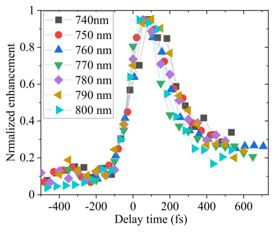
Figure 5.
The normalized enhancement shown in Figure 4b was averaged over all 95 NPs. A long-tail slow decay component was observed only at positive delay time when the second harmonic (SH) irradiated the sample after the irradiation by the fundamental (F) pulse.
Two features are noticed in Figure 5. One is that there are two components: a central peak and a long tail. The other feature is that the delay dependence is asymmetrical: the slow decay of the signal was observed only at the positive position of the corner cube, which means that the lifetime of electrons was long only when the F pulse irradiated the sample first before the SH irradiation.
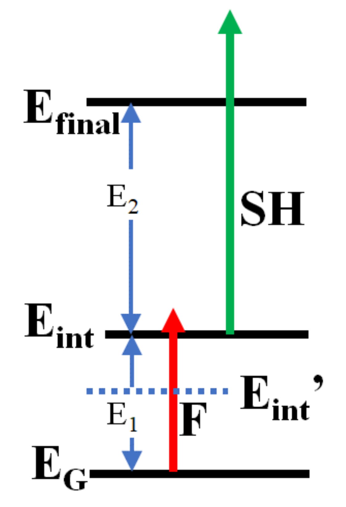
The second feature is of interest. This feature is observable only when two colors are employed. If one color is used, the delay dependence should be symmetrical around t = 0 where two pulses arrive at the same time. Even in a two-color pump-probe, the asymmetry is observable under the following two conditions. We assume electrons are excited by two steps, i.e., electrons in the ground state, EG, are excited to the intermediate state, Eint, by the first pulse, shown by the red arrow F in Figure 6, and the second pulse, shown by the green arrow SH, excites the Eint state to the final state, Efinal. E1 is the energy of the intermediate state, Eint, from the ground state, EG, and E2 is the energy of the final state, Efinal, from the intermediate state, Eint. The first condition for observing an asymmetry in the delay dependence of enhancement is that the intermediate state, Eint, is excited to Efinal more preferably by the SH pulse than by the F pulse. The second condition is that the intermediate state, Eint, can be excited from the EG state by the F pulse. The first condition is satisfied when 1.75 eV < E2 < 3.1 eV. The second condition is satisfied when E1 < 1.55 eV. When E1 > 1.55 eV, the second condition is satisfied if there are other electronic states Eint’ with an energy separation of less than 1.55 eV between the states EG and Ein. In Section 3.2 we discuss candidate electronic states satisfying the two conditions for the asymmetry.
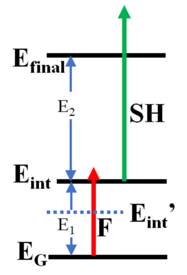
Figure 6.
The asymmetry of the delay dependence of the pump-probe signal seen in Figure 5 is observable when the energy separation, E2, between the final, Efinal, and the intermediate, Eint, state is larger than the one-photon energy of the fundamental (F) and smaller than the one-photon energy of the second harmonic (SH) and when the energy separation, E1, between the Eint state and the initial state, EG, is smaller than one-photon energy of the fundamental (F) pulse.
2.3. Detailed Analysis of the Delay Dependence of the Enhancement in Pump-Probe
Here, the delay dependence of the normalized enhancements shown in Figure 5 is analyzed in detail. There are two components: a relatively narrow peak and a long tail. The profile of the narrow peak is expected to reflect the pulse shape of the laser. In our previous report [20], we subtracted the same Gaussian profile from the delay dependences at all wavelengths and discussed the wavelength dependence of the decay time of the tail part only. However, by careful examination, we notice that the width of the peak is wavelength-dependent, while the rising part is similar. In the present paper, we report a detailed analysis of the delay dependence by performing numerical calculations to re-produce profiles. The detail of the simulation and accuracy of the estimated values are described in the Appendix A.
In the numerical calculation, four parameters are introduced: the laser pulse width tL, the lifetimes τ1 and τ2 of the fast and slow components, respectively, and the relative amplitude α of the slow component to the fast component. The rising part of the peak is determined by tL and the tail part of a large delay time is determined by τ2. The falling side of the peak is determined by all parameters. In the simulation, the laser intensity at t1 “in” the pump laser contributes to the probe signal at t2 “in” the probe signal only when t1 < t2 to reproduce the observed asymmetry.
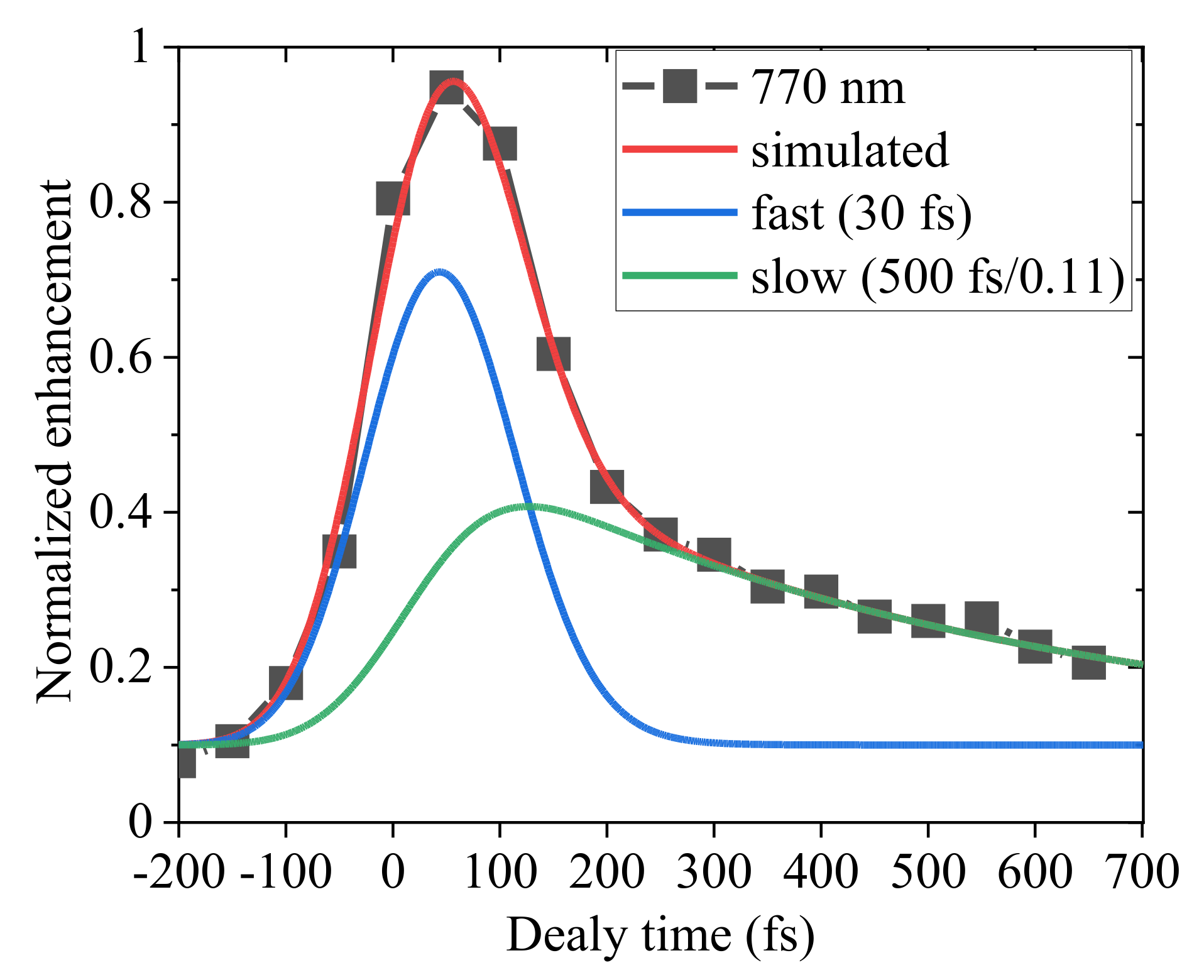
The closed squares in Figure 7 show the delay dependence of the brightness enhancement at 770 nm averaged over the 95 NPs shown in Figure 5. At this wavelength, the peak was the narrowest. From the fitting, we determined that the laser pulse width was 110 fs and τ1 was 30 fs, as explained in detail in the Appendix A. The best fit was obtained by a slow component with a lifetime of 500 fs with an amplitude of 10% of that of the fast component. The profiles of the fast and slow components are displayed in the figure. As seen in the figure, the calculated result reproduces well the observed delay dependence and the fitting parameters are reliable.
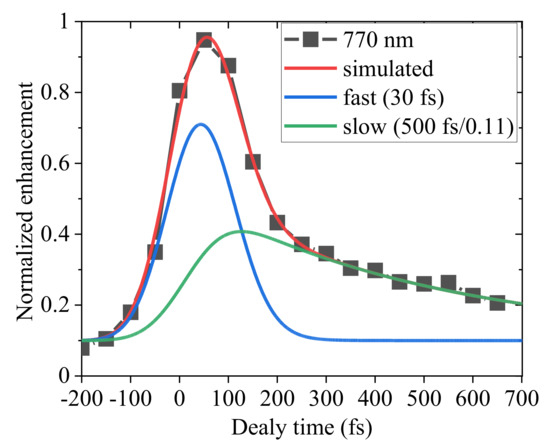
Figure 7.
Reproduction of the delay dependence of the normalized enhancement at 770 nm (closed squares) by a numerical calculation explained in the Appendix A. Fitting parameters are tL = 110 fs, τ1 = 30 and τ2 = 500 fs, and α = 0.11.
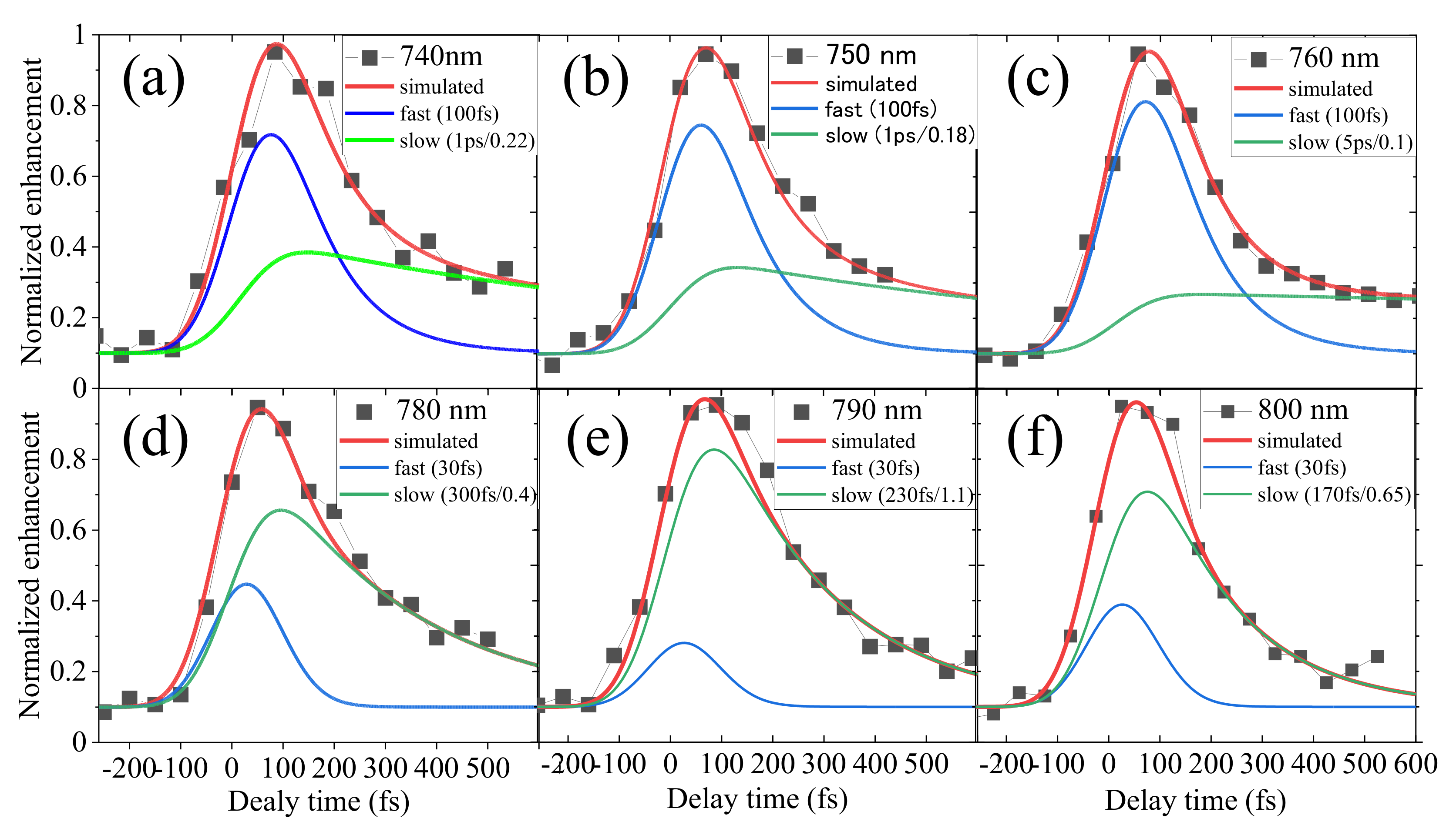
Figure 8 shows the fittings to the delay dependence of the brightness enhancements at other wavelengths. At 760 nm shown in (c), the delay dependence of the enhancement was nearly flat at a large delay time. As explained in the Appendix A, the fitting gave τ1 of 100 fs and τ2 larger than 4 ps. The delay dependence of the enhancement was similar at 740 nm (a) and 750 nm (b), and good fittings were obtained with τ2 of 1 ps and τ1 of 100 fs. The decay of the delay dependence was faster at longer than 780 nm. At these wavelengths, τ1 can be shorter than 30 fs which is the value at 770 nm, but we estimated τ2 by assuming τ1 = 30 fs. The values for τ2 were 300, 250, and 150 fs, at 770 (d), 790 (e), and 800 nm (f), respectively.
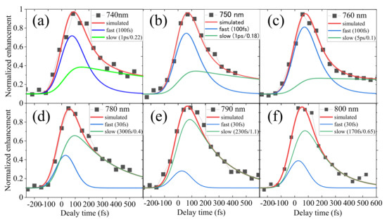
Figure 8.
The delay dependence of the “normalized” brightness enhancement averaged over 95 NPs at (a) 740 nm, (b) 750 nm, (c) 760 nm, (d) 780 nm, (e) 790 nm, and (f) 800 nm. The numerical calculations shown by the red solid curves reproduce well the experimentally observed datapoints shown by the solid squares. The fast and slow decay components are also displayed.
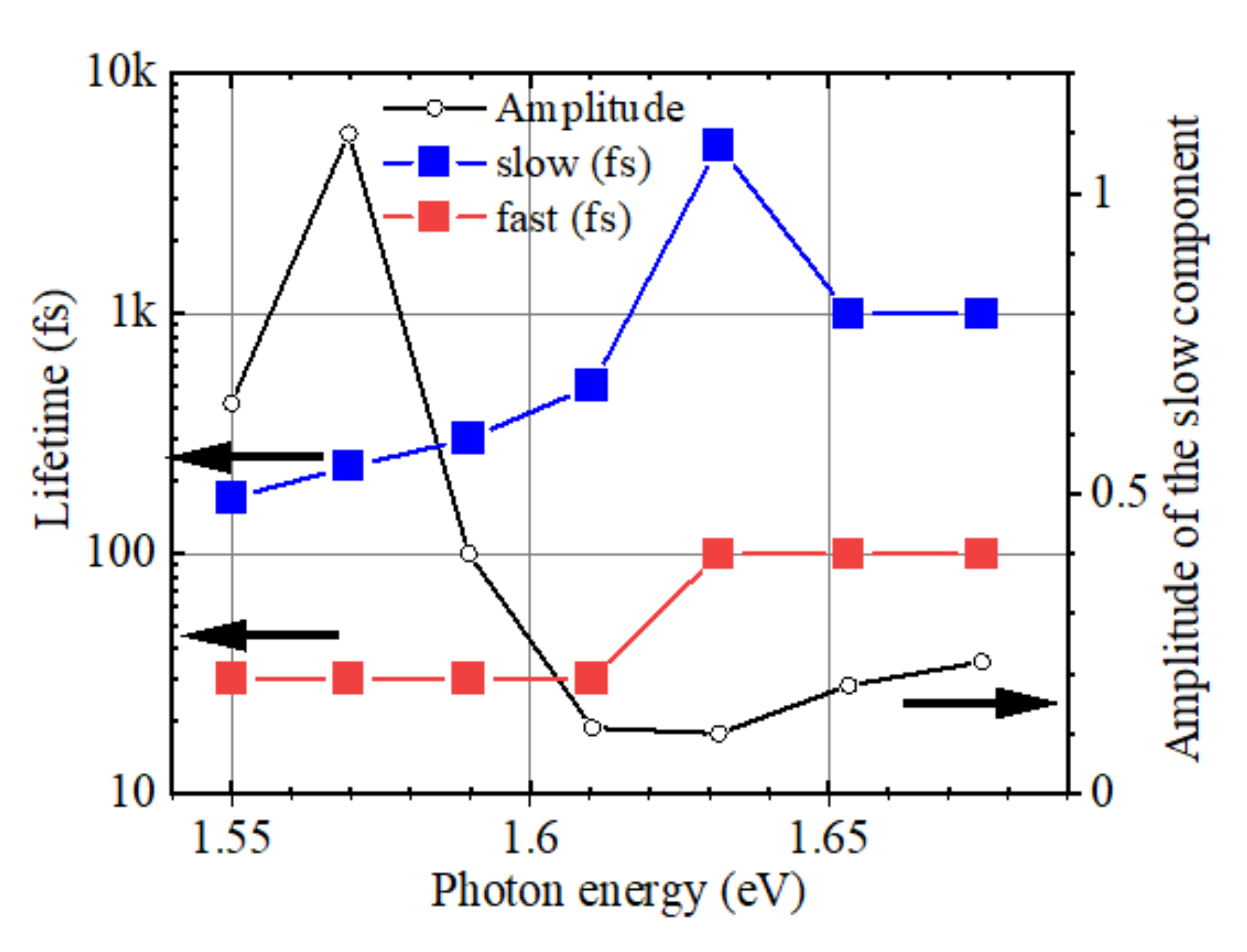
Figure 9 summarizes lifetimes τ1 and τ2 evaluated by the fittings shown in Figure 8. The lifetime of the slow component, τ2, increased with the photon energy from 150 fs to 1 ps with the resonance at 1.63 eV (760 nm), where the lifetime was 4 ps or longer. The lifetime of the fast component, τ1 jumped from 30 to 100 fs at the resonance at 1.63 eV. Figure 9 also shows a spectrum of the amplitude of the slow component. The slow component at the resonance was 10% and larger at higher energy. At the wavelength longer than 780 nm, the amplitude of the fast component was small, as seen in Figure 8.
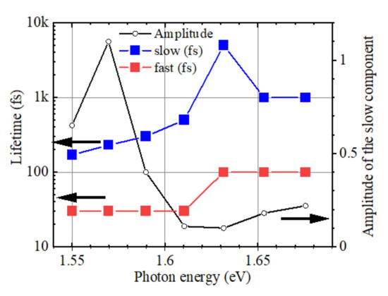
Figure 9.
Spectra of the lifetime and the amplitude of the slow decay component. At the resonance peak at 760 nm (1.63 eV), the lifetime was longer than 4 ps.
3. Discussions
3.1. Picosecond Lifetime Hot Electrons and High Catalytic Activities
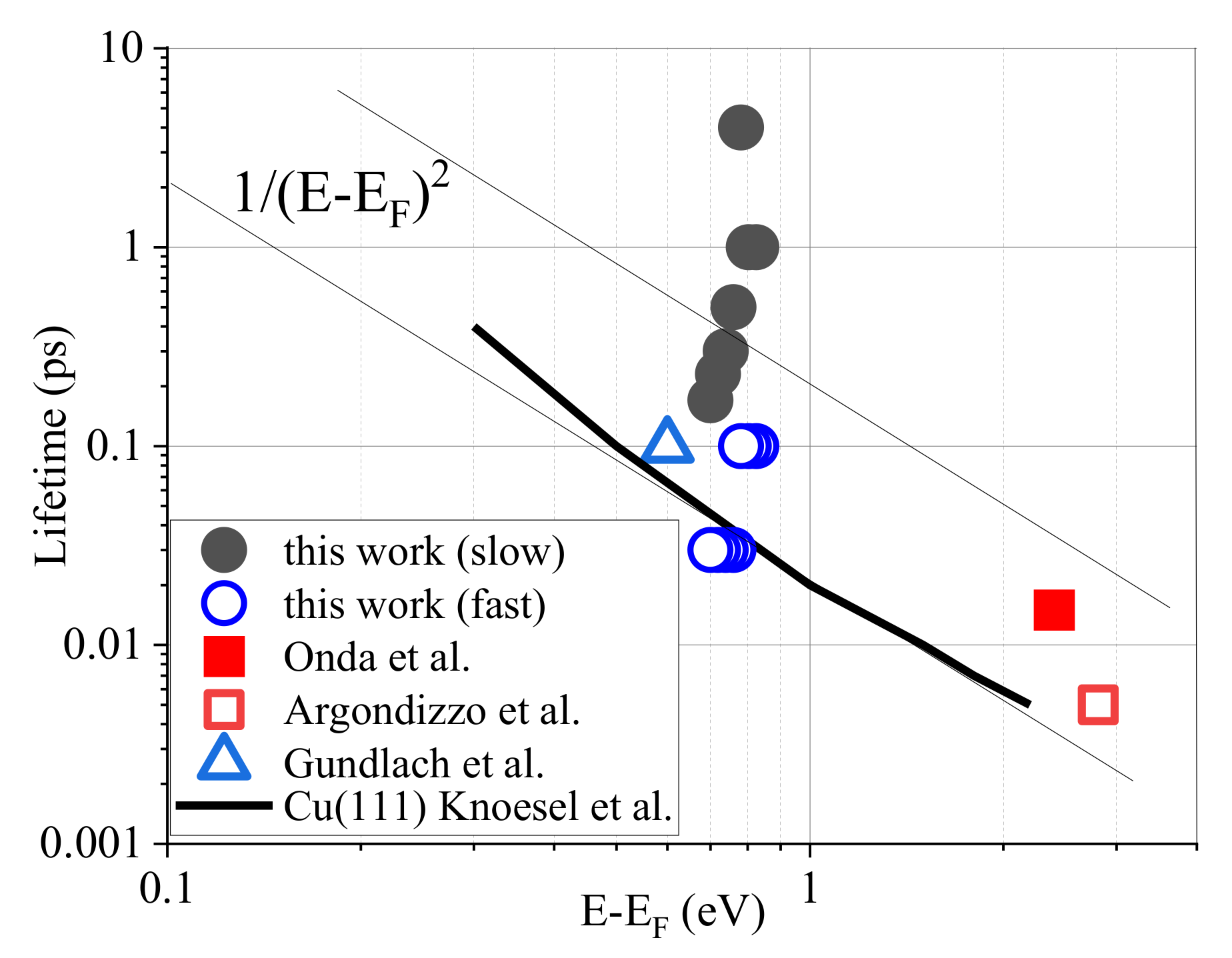
We compare the observed lifetime of this study with those reported in the literature in Figure 10. The values shown in Figure 9 are displayed as filled circles (slow component) and open circles (fast component). The horizontal axis is the kinetic energy of electrons above the Fermi level. We assumed that the initial state of the electrons in our work was located 0.85 eV below the CBM as assumed by Argondizzo et al. [24]. The lifetime in a Cu (111) single crystal reported by Knoesel et al. [34] is shown by the solid curve, which scaled as the inverse square root of the kinetic energy, as expected by theory. The electron lifetime in Ag (110) and Ta (poly) [34] was lower than shown by the curve. Link and El-Sayed [35], Zhukov et al. [36], and Bauer and Aeschlimann [37] reported similar values. The value observed in TiO2 reported by Gundlach et al. [38] is shown by an open triangle and was close to those in metals. In Argondizzo et al. [24], the electron lifetime was significantly shorter than the pulse width of their 20 fs laser. The lifetime of the “wet electrons” discovered by Onda et al. [11] was longer than that in Argondizzo et al. [24]. A lifetime of 15 fs by Tisdale et al. [39] and a few tens of fs by Deinert et al. [40] at E-EF~0.3 eV were reported in ZnO single crystals (not plotted in the figure), both of which are one order-of-magnitude faster than that on the solid curve of Knoesel et al. [34].
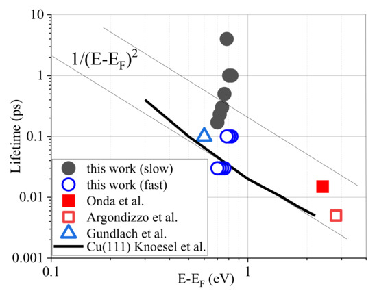
Figure 10.
Comparison of electron lifetime. The values obtained in the present work, are displayed as black solid circles (slow component) and open circles (fast component). The lifetime observed in a Cu (111) single crystal by Knoesel et al. [34] is shown by the solid curve s, which followed the theoretical scaling of 1/(E-EF)2 for Fermi liquid [34]. The vertical axis is the kinetic energy, E-EF. The data from Onda et al. [23] are shown by the closed red square. The open red square is from Argondizzo et al. [24], and the open triangle is from Gundlach et al. [38]. These results were observed for single crystals. The longest lifetime of 4 ps observed in the present work in NPs is more than 10 times longer than previously reported for single crystals.
The lifetime of 4 ps or longer observed at 760 nm in the present study is more than two orders of magnitude longer than those in metals shown by the solid curve. Even the shortest slow component of 170 fs at 800 nm in Figure 9 is longer than that of the “wet electron”. The fast component at shorter than 760 nm is comparable to the wet electron.
In the Introduction, we noted that a lifetime in the order of 1 ns is desired for the highly efficient use of photogenerated hot electrons in photocatalysis when molecules arrive at the active site every 1 ns. The lifetime of hot electrons trapped at the eg state in a single crystal is shorter than 10 fs, but the lifetime of the Eint state in TiO2 NPs discovered in the present study is longer than 4 ps, as reported above. Then, simply speaking, the efficiency in NPs is enhanced more than 400 times at the resonance wavelength of 760 nm. At shorter wavelengths, the lifetime is a little shorter but still as long as 1 ps. At wavelengths longer than 770 nm, the lifetime is shorter but still longer than that in a single crystal. Our experimental result supports our claim that the essence of the high catalytic activity of TiO2 NPs is in the creation of excited states with a very long lifetime in the conduction band.
3.2. Electronic Structure
Here, we consider the electronic states involved in our pump-probe experiment.
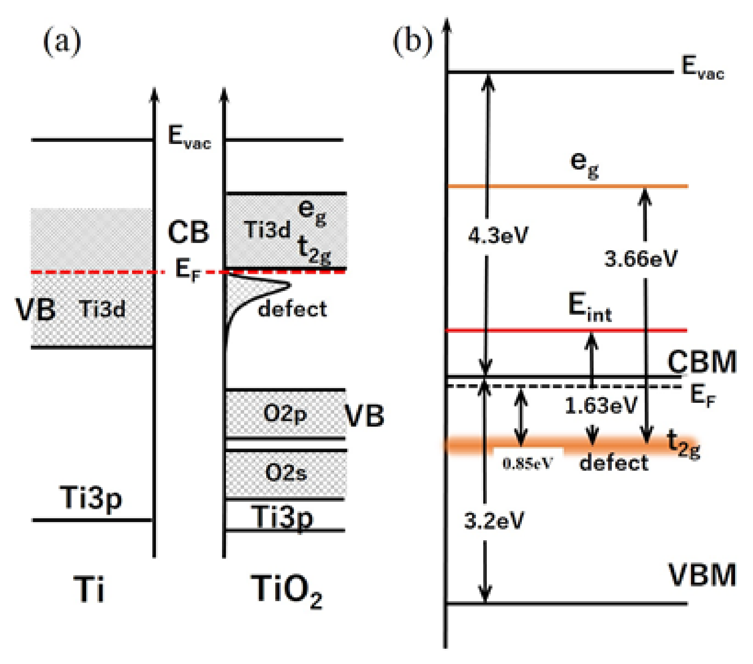
When a metal Ti atom with four 3d electrons in the valence band bonds with oxygen, O, it gives all four 3d electrons to two O atoms. The valence band of TiO2 with a bandgap of 3.0 eV or 3.2 eV is occupied by O2p electrons, and the empty Ti3d level serves as the conduction band. Vos [41] reported that the conduction band of TiO2 comprises two groups: the lower group is the triple-degenerated 2t2g state and the upper group is the doubly degenerated 3eg state, as shown in Figure 4a. The energy separation of the t2g and eg states was measured as 2.1 eV by Fisher [42], estimated from the X-ray absorption spectrum.
In an oxygen vacancy, some electrons transferred to O atoms return to the Ti atom and form defect levels in the bandgap as shown in Figure 11a. The 3eg state in the upper conduction band was observed by Argondizzo et al. [24]. They irradiated a single crystal of rutile TiO2 (111) by 20 fs laser pulses and observed several times greater enhancement of the photoemission for a 3.66 eV laser than at 3.22 and 3.95 eV. In their explanation, the defect state, t2g, at 0.85 eV below the Fermi level was two-photon-ionized, and the 3.66 eV light was in resonance with the transition from the defect state to the eg state which worked as an intermediate state assisting the two-photon ionization. Their claim is shown in Figure 11b. The bandgap energy of rutile TiO2 is 3.0 eV. By assuming the Fermi level is very close to the conduction band bottom, the energy of the eg state from the valence band top is 6.01 eV, which is 1.5 eV below the vacuum level when the ionization energy is 7.5 eV, and the intermediate state in Argondizzo et al. belongs to the upper group (doubly degenerated 3eg) reported by Vos [41].
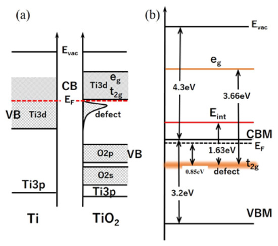
Figure 11.
Energy levels in TiO2. (a) In TiO2, four 3d electrons of Ti atom are transferred to oxygen and O2p is the valence band and the empty 3d level of Ti atom constitutes the conduction band. The conduction band is comprised of two bands, the 3eg and t2g bands, according to Vos [41]. (b) In oxygen vacancy, electrons returned from oxygen to Ti are trapped in the states in the bandgap. The 3eg was observed at 3.66 eV from the defect level by Argondizzo et al. [24]. Our observed localized state, Eint, at 1.63 eV above the defect state in the bandgap may belong to the lower band, the t2g band, in the conduction band.
In Section 2.2, we discussed the conditions for observing the asymmetry of the delay dependence of normalized enhancement seen in Figure 7 and Figure 8. In our pump-probe experiment, the metastable Eint state is located at 1.63 eV, i.e., E1 = 1.63 eV in Figure 6, and the eg state observed by Argondizzo et al. [24], can be the final state, Efinal. When we assume the ground state EG is the t2g state in the bandgap as was assumed in Argondizzo et al., Eint is located at 0.78 eV above CBM, and E2 is calculated as 2.03 eV. Then, the SH of 400 nm (3.1 eV) can excite electrons in the Eint state to the states above the eg state, and electrons instantly relaxed to the eg state, Efinal, and the electrons in the Efinal state are thermally ionized, as observed in gold NPs [13]. If the SH pulse irradiates the sample first, an electron in the t2g state, EG, is excited to the states above the Eint state, but the electrons relaxed to the Eint state can not be excited to the eg state, Efinal, because the photon energy (<1.68 eV) is smaller than E2 of 2.03 eV. Hence, the slow decay component is observable only for positive delay when the SH arrives after the F. For F longer than 770 nm, the EG state should be shallower than 0.85 eV because photon energy is not large enough to reach the Eint state at 0.78 eV above CBM. Ghosh et al. [30] identified eight shallow traps from 0.27 to 0.87 eV below CBM, and one of them can be EG when excited a the wavelength longer than 770 nm.
Because the energy separation of Efinal and Eint, E2, of 2.03 eV is close to the energy separation of the t2g and eg states of 2.1 eV measured by Fisher [42], the Efinal and Eint states in our pump-probe measurement can be closely related with the electronic states observed by Fisher.
3.3. Fast and Slow Decay Components
Finally, we discuss the origin of the fast and slow decay components in the delay dependence of the enhancement of the pump-probe signal.
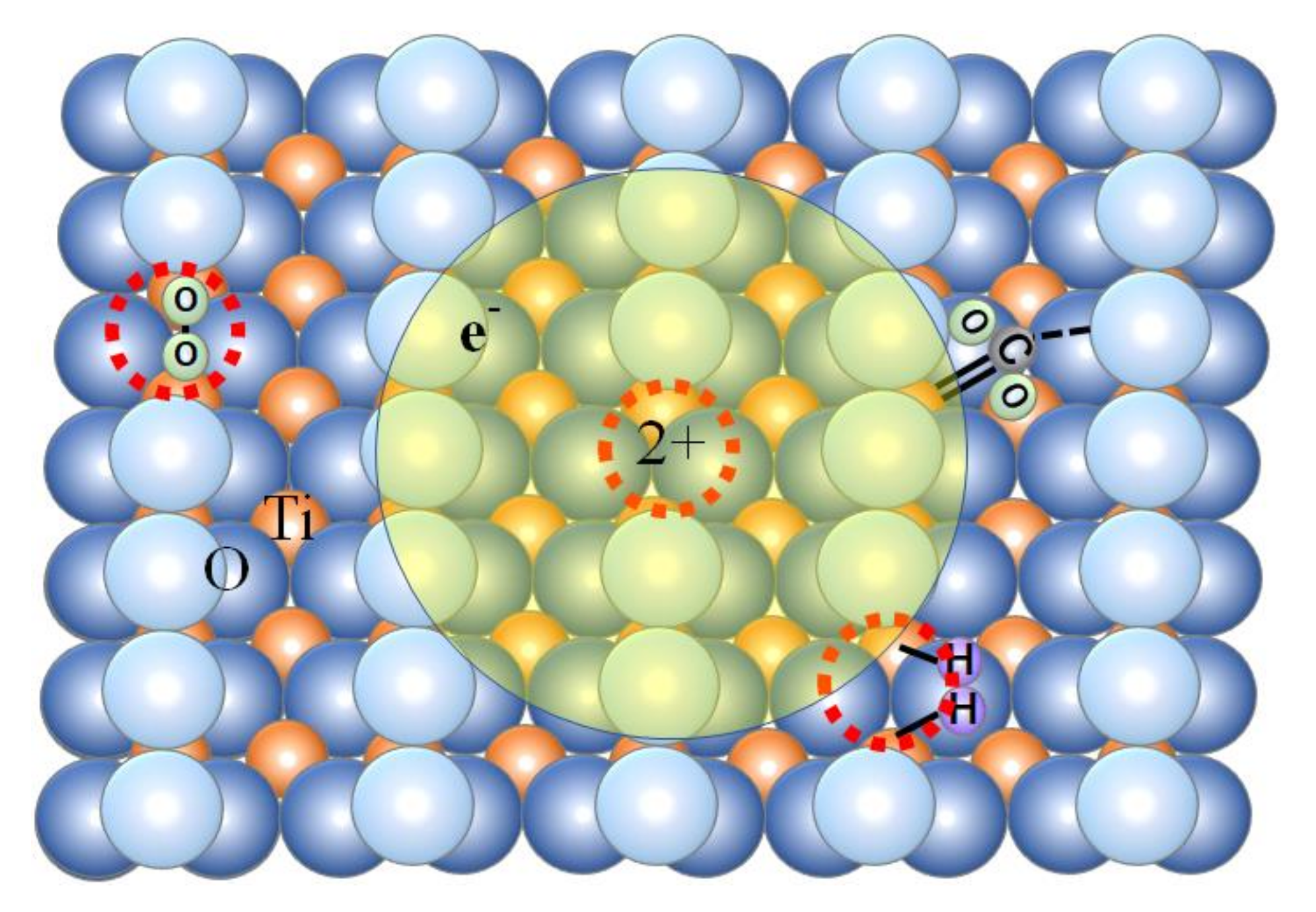
The defect in TiO2 is the oxygen vacancy on the topmost atomic layer, which is schematically shown in Figure 12. Henrich et al. [43] found that a signal about 0.7 eV below the Fermi level in a UPS (ultraviolet–photoelectron–spectroscopy) spectrum increased with Ar ion bombardment and that the signal was greatly decreased by exposure of the sample to 108 Langmuir of oxygen. Their experiment proved that the defects are formed on the topmost atomic layer.
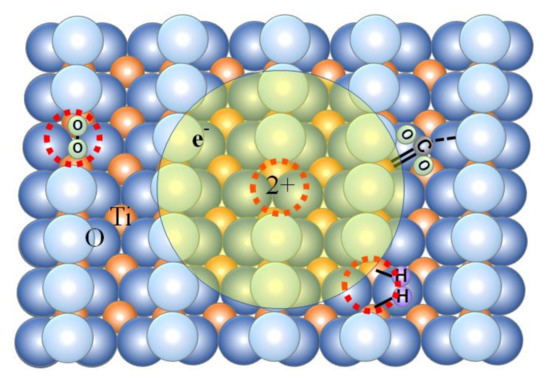
Figure 12.
Localized electron trapped at oxygen vacancy, Vo, on the surface has a lifetime as long as 4 ps when resonantly excited at 760 nm. Electrons generated other than Vo can be attracted by the potential gradient by the positive charge at Vo, and a fraction larger than the concentration of Vo of photo-excited electrons will have a long lifetime.
The oxygen vacancy is the catalytic active site, and the density of defects determines catalytic activities. Therefore, numerous studies have evaluated the density of the defect site [44,45,46,47]. Many have reported several % monolayer (ML) defects on the surface in a single crystal. The defect density increases by ion sputtering as reported by Henrich et al. [43], and can be 0.4 ML according to Pan et al. [44]. However, the volume concentration of the defects is very low because the oxygen vacancy is created mostly on the surface. Hasiguti et al. [48] reported that the volume concentration of defects in reduced rutile was 3~5 × 10¹⁸/cm3 which corresponds to 0.01%.
The oxygen vacancy, Vo, acts as a point defect having +2 charge because O2− ion is removed, and can capture two electrons. According to a calculation treating Vo as a color center by Chen et al. [49], one electron is trapped deep at 1.78 eV from the conduction band and the other electron is shallowly trapped at 0.87 eV. Their calculation agrees with the proposal by Cronemeyer [32]. Cronemeyer proposed trapped states at 0.75 and 1.18 eV based on the absorption spectrum of reduced rutile single crystal TiO2, which explains the signal at 0.7 eV below the Fermi level in UPS observed by Henrich et al. [43]. Because the trap is shallow, the wave function of the trapped electron is significantly extended. According to Hasiguti et al. [48], the radius of the orbital of the shallow-trapped electron is 1.3 nm, which is four times the average separation of the TiO2 molecule. The deep trap in the calculation by Chen et al. [49] may correspond to the absorption peak at 800 nm reported by Yoshihara et al. [31].
From knowledge of the electronic structure of Vo explained above, we interpret that the slow component is the decay of electrons at the Vo center, and the fast component is the decay of electrons other than those at the Vo centers. When an electron is trapped at the intermediate state Eint localized at the Vo center, it stays active for 4 ps or longer.
We interpret that the ratio of the amplitudes of the fast and slow components reflects the fraction of electrons trapped at Vo among the electrons generated in the entire NP. As mentioned above, the volume concentration of defects is only 0.01% based on the report by Hasiguti et al. [48]. The 10% ratio of the slow component to the fast component at 760 nm shown in Figure 9 is three orders of magnitude larger than the Vo volume concentration of 0.01%. We attribute this significant difference to the potential gradient generated by the Vo center. Electrons generated other than the Vo will be attracted toward the Vo center by the potential gradient generated by the charge at Vo. Ten percent of the electrons will be trapped if the potential gradient extends to 1.7 nm from Vo. The mentioned value may not be as accurate but the Coulomb potential by the Vo center will increase the fraction of hot electrons trapped by Vo that stay active for a long time for high catalysis.
The larger amplitude of the slow component at the photon energy higher than 1.63 eV, seen in Figure 9, can be explained as follows. An electron loses some fraction of its energy when traveling to the Vo center, and the probability of reaching the Vo while maintaining its energy at a level higher than Eint is higher for a higher-energy electron.
As discussed in Section 3.2, the initial state in the measurement at wavelengths longer than 780 nm will not be the t2g state. If electrons in a deep trap calculated by Chen et al. [49], which will correspond to the deep traps reported by Ghosh et al. [30] and to the absorption peak at 800 nm reported by Yoshihara et al. [31], are in the EG state in our pump-probe experiment at longer than 780 nm, the positive charge is 2+ and the potential gradient is large. Furthermore, most of the electrons generated other than the Vo can be attracted to the center and the fraction of the fast component that represents the decay of electrons without being trapped by the Vo will be small.
4. Materials and Methods
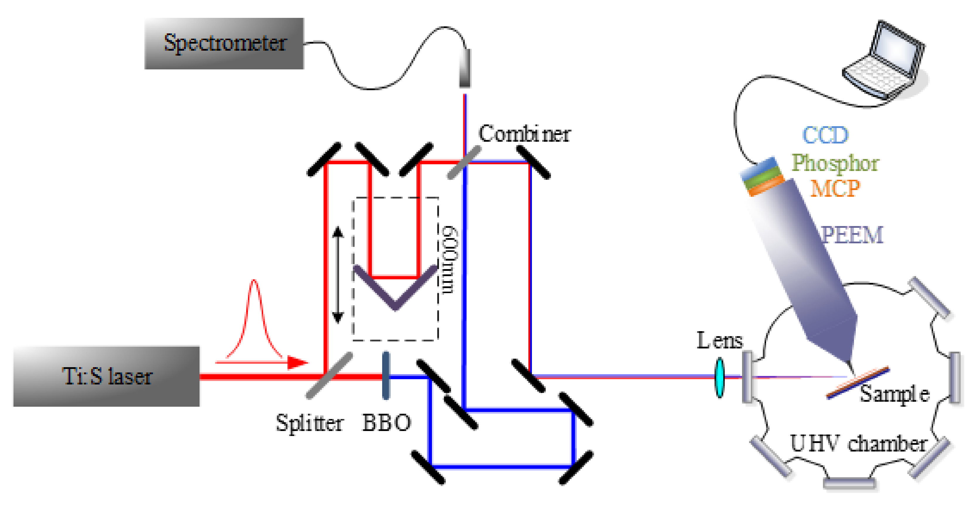
The experimental setup for the lifetime measurement is briefly explained below [20]. As shown in Figure 13, a beam of a femtosecond laser of 150 fs pulse width (80MHz, Mira, Coherent) was separated by a beam splitter and focused on a Beta Barium Borate (BBO) crystal for generating the second harmonic (SH). The optical path length of the fundamental (F) wavelength laser pulse was varied by sending it to a corner cube, which was mounted on a translator. The position of the corner cube was controlled by an electronic controller. The position accuracy was 1.25 μm, which corresponds to an 8-fs accuracy. Two beams were combined by a beam combiner and focused on a sample in the vacuum chamber of PEEM (FOCUS, IS-PEEM). The vacuum pressure was about 10−5 Pa. The power of the F and SH pulses was varied by attenuators at each beam. The laser powers were 160 mW for F and 1.6 mW for SH, respectively. The pulse energies were 2 nJ and 20 pJ, corresponding to a peak power of 13 kW and 130 W, respectively. The focused beam diameter was about 100 μm and, hence, the power density was about 100 and 1 MW/cm2 for the F pulse and the SH pulse, respectively. The laser power was chosen so that the image brightness was similar when F only and SH only irradiated the sample. The laser wavelength was varied from 700 to 900 nm.
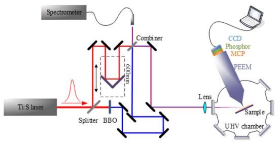
Figure 13.
Experimental setup. The delay time of the fundamental (F) beam was varied using a translator. The second harmonic (SH) and F beams were combined by a beam combiner. The combined beam were focused on a sample by a lens with a focal length of 200 mm at 25 degrees from the surface.
In performing the pump-probe measurement, the optical paths for two beams should be the same. When the laser pulse is 150 fs, in our case, the accuracy should be better than 45 μm. This accuracy can be achieved by observing the interference pattern by overlapping two beams. Two colors do not generate interference but we adjusted the optical paths of F and SH beams as follows. Although the conversion efficiency of the SH generation was as high as 7.5% in our case, most of the F pulse transmits the BBO crystal for SH generation. To eliminate the F wavelength light in the path of the SH beam, the SH beam was reflected by many dichroic mirrors which have a very low reflectivity for the F wavelength. When adjusting optical path lengths for F and SH pulses, some of the dichroic mirrors in the SH beam path were replaced with mirrors for reflecting the F wavelength, and interference between the F pulse in the F beam path and the F pulse traveling in the SH beam path was detected by a photodiode by scanning the position of the corner cube. When replacing mirrors, the optical path was changed slightly, and the final adjustment was needed to observe the enhancement of the electron signal by PEEM, as shown in Figure 4.
The other difficulty in the two-color pump-probe measurement was adjusting the focused beam size on the sample. Two colors were combined by a beam combiner and we focused the combined beam by using a single lens with a focal length of 200 mm. Because of large chromatic aberration, the focal length is largely different for F and SH beams, and the two colors are not best-focused at the same time. The lens position was adjusted so that two beams were similarly defocused to have a similar focused beam size. When changing the focused beam size on the sample, the focal length of the lens should be changed in the two-color pump-probe, while it is easily changed by changing the lens position in the one-color pump-probe. When changing the wavelength, we needed to re-adjust the lens position because the focal length changes with the wavelength. This problem can be solved using an off-axis parabola mirror, but there was no space to put it in our PEEM room.
The sample investigated was anatase type TiO2 NP from Daoking Science and Technology. The average diameter was 100 nm, the purity was 99.9%, and the specific surface area was 20 m2/g. The color of the powder was white, indicating that the sample was not heavily reduced.
For sparsely distributing TiO2 NPs to observe individual NPs, the powder was diluted in distilled water and NPs were ultrasonically separated. One drop of the solution was dropped on an n-type Si wafer. The drop was blown off by an air current for a few seconds, and a thin layer of TiO2 NPs was left on the Si substrate. According to Henderson et al. [50], water molecules molecularly adsorbed and hydrogen-bonded on TiO2 are desorbed at room temperature, and a dissociative water molecule, OH, chemically adsorbed at the oxygen vacancy, is desorbed at 270 °C. For removing OH, the Si wafer on which the TiO2 NPs were sparsely distributed was baked for 1 hour in the air at 500 °C.
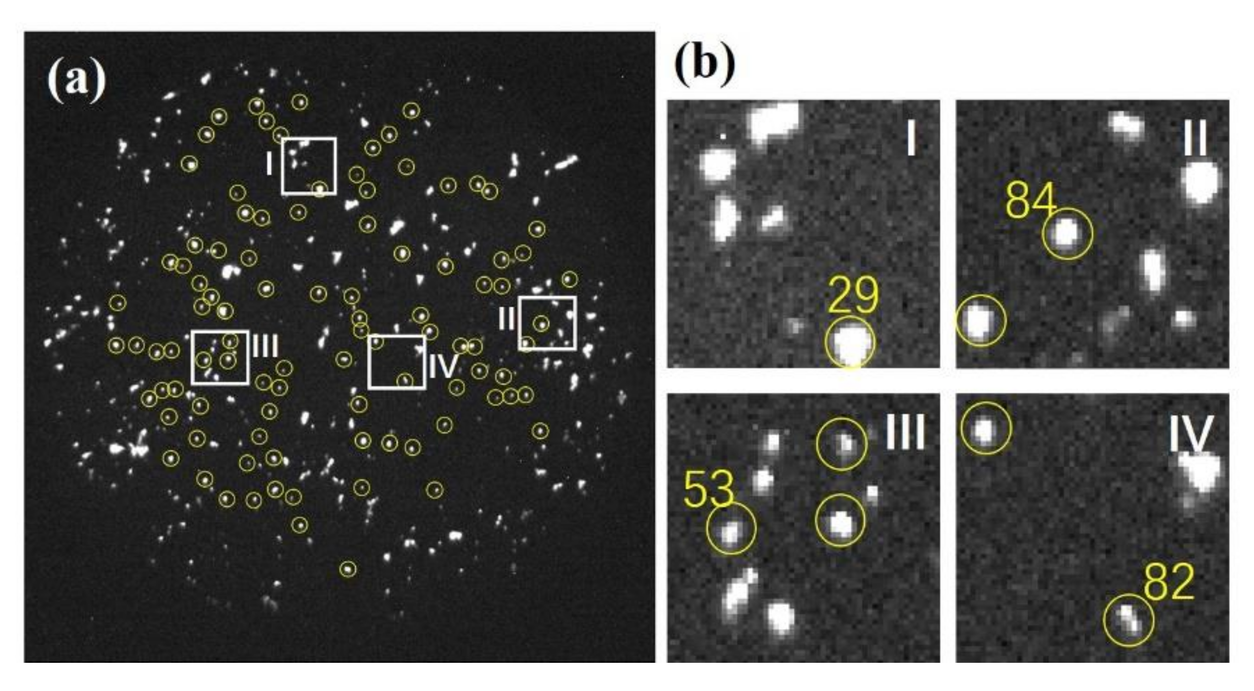
Figure 14 shows a PEEM image. A total of 95 particles shown by the yellow circles in (a) were selected for the analysis in the present paper. Figure 14b shows enlarged displays of the NPs shown in from Figure 2, Figure 3 and Figure 4. To observe many NPs in a single frame, the field of view was 64 μm. The number of pixels was 560 × 560, and the one-pixel size was about 120 nm. Then, the size of the particles was about 400 nm. Therefore, strictly speaking, particles were clusters of NPs. In this paper, we refer to them simply as NPs. In this paper, the brightness of each NP refers to the brightness of the brightest pixel in each NP. Therefore, we expect that the brightness of NPs is determined by the electronic structure of individual NPs, such as electron lifetime and defect density, with a small effect of the size of NPs.
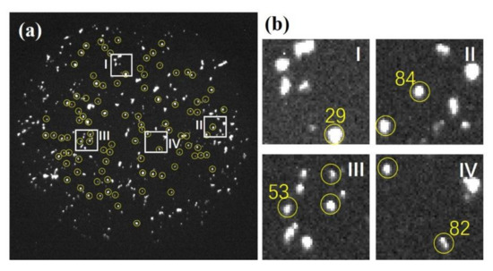
Figure 14.
PEEM (Photoemission-electron-microscopy) image of TiO2 NPs. (a) The field of view of the image was set as 64 μm to observe many particles in one frame. The 95 particles selected for the analysis are marked by yellow circles. (b) Enlarged images of four NPs shown in Figure 2, Figure 3 and Figure 4. The particle sizes were several pixels in height and width, which corresponds to about 400 nm. The positions of four areas in (b) are shown by squares in (a).
5. Summary
TiO2 nanoparticles were observed using the femtosecond laser two-color pump-probe technique with PEEM, which has a 40 nm spatial resolution. When excited by a 760 nm laser, a lifetime longer than 4 ps was observed together with a fast decay component of 100 fs time constant. The slow decay component was observed only when the fundamental laser pulse irradiated the sample before the irradiation by the second harmonic pulse. We conclude that there is an excited state, Eint, in the conduction 1.63 eV above the 2t2g defect state which lies at 0.85 eV below the CBM, and that the electrons excited to the Eint are excited by the SH pulse to the eg state, from which electrons are thermally ionized to be observed by PEEM. The energy separation of the 2t2g and eg sates of about 2 eV explains the asymmetry of the pump-probe signal. The observed 4 ps lifetime is nearly two orders of magnitude longer than those reported in the literature. The result supports our claim that the essence of high catalytic activities of nanoparticles is in the creation of excited states in the conduction band with a long lifetime. The slow component in the delay-time-dependence of the pump-probe signal shows the decay in electrons in the Eint, state, localized at the oxygen vacancy. The fast component is the decay of electrons other than those at the Vo center. A fraction of the slow component which is larger than the volume concentration of Vo can be attributed to the attraction of electrons to Vo by the potential gradient generated by the positive charge at Vo. The electronic structure for the asymmetry of the pump-probe signal and the origin of the two decay components are discussed based on the color center model of the oxygen vacancy. A future detailed study of the fast and slow components will provide deep insight into electron dynamics in the photocatalysis of TiO2.
Author Contributions
Conceptualization, T.T.; B.L., H.L., C.Y. and B.J. performed the experiments; B.L. analyzed the data and prepared original draft; writing, T.T.; B.L. and J.L. edited the manuscript; funding acquisition, J.L. All authors have read and agreed to the published version of the manuscript.
Funding
National Natural Science Foundation of China (NSFC) (91850109, 61775021, 11474040); Education Department of Jilin Province (JJKH20181104KJ, JJKH20190555KJ, JJKH20190549KJ); Changchun University of Science and Technology (XQNJJ-2018-02); “111” Project of China (D17017); Project funded by China Postdoctoral Science Foundation (2019M661183). Authors thank Ministry of Education Key Laboratory for Cross-Scale Micro and Nano Manufacturing, Changchun University of Science and Technology.
Conflicts of Interest
The authors declare no conflict of interest.
Appendix A
- (1)
- Equations
The calculation of the simulation for reproducing the delay time dependence of the pump-probe signal was performed as follows.
The number I(t1) of electrons excited to the excitation level at time t1 decays with a lifetime τ, and becomes I(t1) × exp (−((t2 − t1)/τ)) at time t2. If the pump laser waveform is set as f(t), the number of electrons at time t2 is . When the probe light is g(t), the probe signal is given by
We replace exp (−((t2 − t1)/τ)) with h(t1, t2) to generalize lifetimes, then, the pump-probe signal is given by
Here, t1 < t2.
In our experiment, the pump light is the fundamental wave (600~900 nm), the probe light is the second harmonic (340~450 nm), and it is dichroic excitation. Setting t1 < t2 means that it can be excited to the final excited state only when one color arrives before the other color.
Since two lifetimes were required to reproduce the experimental results, h(t1, t2) is set as
h(t1, t2) = exp(−((t2 − t1)/τ1)) + α exp (−((t2 − t1)/τ2)),
where α is the relative intensity of the second component.
For simplicity, the probe laser and pump laser have the same waveform
f(t) = g(t) = exp(−(t/tL)2),
where tp = tL (ln2)0.5 is the pulse width of the laser.
Four parameters, tL, τ1, τ2 (»τ1), and α, were evaluated by the following procedure.
- (2)
- Determination of the Laser pulse width
The rising part of the pump-probe signal is mainly determined by tL, the slope of at a large delay time is determined by τ2, the intensity ratio to the peak is determined by α, and the falling waveform of the peak is mainly determined by τ1.
- (3)
- The lifetime of the fast component
As seen in Figure 5, in which the measurement results of seven wavelengths are overlapped and drawn, the rising slope was slightly different depending on the wavelength. Since the pulse width of the laser was the same at different wavelengths, the rising waveform of the pump-probe signal was slightly different depending on the wavelength, because τ1 was different depending on the wavelength. The pulse width of the laser was estimated from the waveform of the pump-probe signal at 770 nm, where the rising edge was the steepest. The waveform of the rising part is not significantly affected by τ1, so, regardless of the value set, there is no difference in the estimation of the laser pulse width. The calculation was performed by setting τ1 > 10 fs because it is reported that the lifetime of hot electrons in metals is about 10 fs, At tL > 120 fs and tL < 90 fs, the fitting of the experimental results was very slightly poor on the falling side, and we decided that tL = 110 fs. The nominal pulse width given by the manufacturer was 150 fs. Since the probe light was the second harmonic and the pulse width was shortened to about 106, the tL = 110 fs obtained above is reasonable. As seen in Figure A1, the good fit over the whole delay time at 770 nm was obtained with τ1 = 30 fs, τ2 = 500 fs, and α = 0.115.

Figure A1.
The delay time dependence of the pump-probe signal at 770 nm. (a), (b), and (c) show calculations for 120, 110, and 90 fs, respectively, for tL. The best fit was achieved with tL = 110 fs.
Figure A1.
The delay time dependence of the pump-probe signal at 770 nm. (a), (b), and (c) show calculations for 120, 110, and 90 fs, respectively, for tL. The best fit was achieved with tL = 110 fs.

Figure A2 shows three cases of τ1 for reproducing the delay time dependence of the pump-probe signal at 770 nm. The steepness of the falling side of the peak is mainly determined by τ1. We notice a very slight deviation of the curves from the datapoints for τ1 = 20 fs and 50 fs, and a good fit was obtained by τ1 =30 fs with τ2 = 500 fs, and α = 0.1.

Figure A2.
The delay time dependence of the pump-probe signal at 770 nm. (a), (b), and (c) show calculations for 20, 30, and 50 fs, respectively, for τ1. A good fit was achieved with τ1 = 50 fs.
Figure A2.
The delay time dependence of the pump-probe signal at 770 nm. (a), (b), and (c) show calculations for 20, 30, and 50 fs, respectively, for τ1. A good fit was achieved with τ1 = 50 fs.

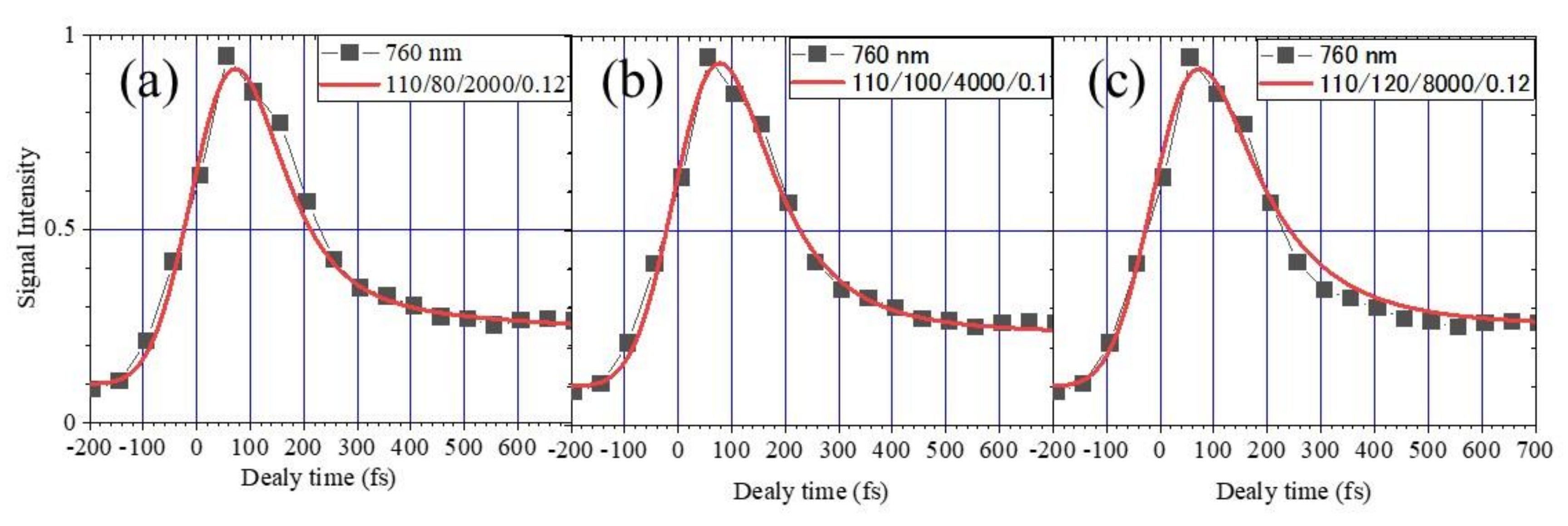
Figure A3 shows three cases of τ1 for reproducing the delay time dependence of the pump-probe signal at 760 nm. We notice a slight deviation of the curves from the datapoints for τ1 = 80 fs and 120 fs, and a good fit was obtained by τ1 =100 fs, τ2 = 4 ps, and α = 0.1.
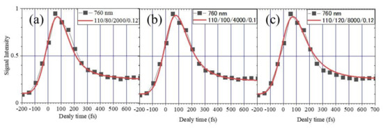
Figure A3.
The delay time dependence of the pump-probe signal at 760 nm. (a), (b), and (c) show calculations for 80, 100, and 120 fs, respectively, for τ1. A good fit was achieved with τ1 = 100 fs.
Figure A3.
The delay time dependence of the pump-probe signal at 760 nm. (a), (b), and (c) show calculations for 80, 100, and 120 fs, respectively, for τ1. A good fit was achieved with τ1 = 100 fs.
- (4)
- The lifetime of the slow component
Figure A4 shows three cases of τ2 for reproducing the delay time dependence of the pump-probe signal at 760 nm. We notice a slight deviation of the curve from the datapoints for τ2 = 2 ps. We would say that τ2 is larger than 4 ps with α = 0.1.

Figure A4.
(a), (b), and (c) show calculations for 3000, 4000, and 5000 fs, respectively, for τ2. A good fit to the delay time dependence at 760 nm was obtained for τ2 > 4 ps.
Figure A4.
(a), (b), and (c) show calculations for 3000, 4000, and 5000 fs, respectively, for τ2. A good fit to the delay time dependence at 760 nm was obtained for τ2 > 4 ps.

References
- Linic, S.; Christopher, P.; Ingram, D.B. Plasmonic-metal nanostructures for efficient conversion of solar to chemical energy. Nat. Mater. 2011, 10, 911–921. [Google Scholar] [CrossRef] [PubMed]
- Mukherjee, S.; Libisch, F.; Large, N.; Neumann, O.; Brown, L.V.; Cheng, J.; Lassiter, J.B.; Carter, E.A.; Nordlander, P.; Halas, N.J. Hot electrons do the impossible: Plasmon-induced dissociation of H2 on Au. Nano Lett. 2013, 13, 240–247. [Google Scholar] [CrossRef] [PubMed]
- Kale, M.J.; Avanesian, T.; Christopher, P. Direct photocatalysis by plasmonic nanostructures. ACS Catal. 2014, 4, 116–128. [Google Scholar] [CrossRef]
- Wu, K.; Chen, J.; McBride, J.R.; Lian, T. Efficient hot-electron transfer by a plasmon-induced interfacial charge-transfer transition. Science 2015, 349, 632–635. [Google Scholar] [CrossRef] [PubMed]
- Zhang, Y.; He, S.; Guo, W.; Hu, Y.; Huang, J.; Mulcahy, J.R.; Wei, W.D. Surface-plasmon-driven hot electron photochemistry. Chem. Rev. 2018, 118, 2927–2954. [Google Scholar] [CrossRef]
- Kodiyath, R.; Manikandan, M.; Lequan, L.; Ramesh, G.V.; Koyasu, S.; Miyauchi, M.; Sakuma, Y.; Tanabe, T.; Gunji, T.; Duy Thang, D.; et al. Visible-light photodecomposition of acetaldehyde by TiO2-coated gold nanocages: Plasmon-mediated hot electron transport via defect states. Chem. Commun. 2014, 50, 15553–15556. [Google Scholar] [CrossRef]
- Kazuma, E.; Jung, J.; Ueba, H.; Trenary, M.; Kim, Y. A direct pathway to molecular photodissociation on metal surfaces using visible light. J. Am. Chem. Soc. 2017, 139, 3115–3121. [Google Scholar] [CrossRef]
- Richardson, O.W. Electron emission from metals as a function of temperature. Phys. Rev. 1924, 23, 153–155. [Google Scholar] [CrossRef]
- Park, J.Y.; Lee, H.; Renzas, J.R.; Zhang, Y.; Somorjai, G.A. Probing hot electron flow generated on Pt nanoparticles with Au/TiO2 schottky diodes during catalytic CO oxidation. Nano Lett. 2008, 8, 2388–2392. [Google Scholar] [CrossRef]
- Akbari, A.; Berini, P. Schottky contact surface-plasmon detector integrated with an asymmetric metal stripe waveguide. Appl. Phys. Lett. 2009, 95, 021104. [Google Scholar] [CrossRef]
- Giugni, A.; Torre, B.; Toma, A.; Francardi, M.; Malerba, M.; Alabastri, A.; Proietti Zaccaria, R.; Stockman, M.I.; Di Fabrizio, E. Hot-electron nanoscopy using adiabatic compression of surface plasmons. Nat. Nanotechnol. 2013, 8, 845–852. [Google Scholar]
- Leenheer, A.J.; Narang, P.; Lewis, N.S.; Atwater, H.A. Solar energy conversion via hot electron internal photoemission in metallic nanostructures: Efficiency estimates. J. Appl. Phys. 2014, 115, 134301. [Google Scholar] [CrossRef]
- Li, B.; Yang, C.; Li, H.; Ji, B.; Lin, J.; Tomie, T. Thermionic emission in gold nanoparticles under femtosecond laser irradiation observed with photoemission electron microscopy. AIP Adv. 2019, 9, 025112. [Google Scholar] [CrossRef]
- Weik, F.; De Meijere, A.; Hasselbrink, E. Wavelength dependence of the photochemistry of O2 on Pd(111) and the role of hot electron cascades. J. Chem. Phys. 1993, 99, 682. [Google Scholar] [CrossRef]
- Clavero, C. Plasmon-induced hot-electron generation at nanoparticle/metal-oxide interfaces for photovoltaic and photocatalytic devices. Nat. Photonics 2014, 8, 95–103. [Google Scholar] [CrossRef]
- Chalabi, H.; Brongersma, M.L. Harvest season for hot electrons. Nat. Nanotechnol. 2013, 8, 229–230. [Google Scholar]
- Furube, A.; Hashimoto, S. Insight into plasmonic hot-electron transfer and plasmon molecular drive: New dimensions in energy conversion and nanofabrication. NPG Asia Mater. 2017, 9, e454. [Google Scholar] [CrossRef]
- Haruta, M.; Kobayashi, T.; Sano, H.; Yamada, N. Novel gold catalysts for the oxidation of carbon monoxide at a temperature far below 0°C. Chem. Lett. 1987, 16, 405–408. [Google Scholar] [CrossRef]
- Lopez, N.; Janssens, T.V.W.; Clausen, B.S.; Xu, Y.; Mavrikakis, M.; Bligaard, T.; Nørskov, J.K. On the origin of the catalytic activity of gold nanoparticles for low-temperature CO oxidation. J. Catal. 2004, 223, 232–235. [Google Scholar] [CrossRef]
- Li, B.; Li, H.; Yang, C.; Ji, B.; Lin, J.; Tomie, T. Excited states in the conduction band and long-lifetime hot electrons in TiO2 nanoparticles observed with photoemission electron microscopy. AIP Adv. 2019, 9, 085321. [Google Scholar] [CrossRef]
- Thompson, T.L.; Yates, J.T., Jr. Surface science studies of the photoactivation of TiO2—New photochemical processes. Chem. Rev. 2006, 106, 4428–4453. [Google Scholar] [CrossRef] [PubMed]
- Henderson, M.A. A surface science perspective on TiO2 photocatalysis. Surf. Sci. Rep. 2011, 66, 185–297. [Google Scholar] [CrossRef]
- Onda, K.; Li, B.; Zhao, J.; Jordan, K.D.; Yang, J.; Petek, H. Wet electrons at the H2O/TiO2(110) surface. Science 2005, 308, 1154–1158. [Google Scholar] [CrossRef]
- Argondizzo, A.; Cui, X.; Wang, C.; Sun, H.; Shang, H.; Zhao, J.; Petek, H. Ultrafast multiphoton pump-probe photoemission excitation pathways in rutile TiO2(110). Phys. Rev. B 2015, 91, 155429. [Google Scholar] [CrossRef]
- Du, L.; Furube, A.; Hara, K.; Katoh, R.; Tachiya, M. Ultrafast plasmon induced electron injection mechanism in gold–TiO2 nanoparticle system. J. Photochem. Photobiol. C Photochem. Rev. 2013, 15, 21–30. [Google Scholar] [CrossRef]
- Ignatiev, I.V.; Kozin, I.E.; Nair, S.; Ren, H.; Sugou, S.; Masumoto, Y. Carrier relaxation dynamics in InP quantum dots studied by artificial control of nonradiative losses. Phys. Rev. B 2000, 61, 15633. [Google Scholar] [CrossRef]
- Scanlon, D.O.; Dunnill, C.W.; Buckeridge, J.; Shevlin, S.A.; Logsdail, A.J.; Woodley, S.M.; Cartlow, C.R.A.; Powell, M.J.; Palgrave, R.G.; Parkin, I.P.; et al. Band alignment of rutile and anatase TiO2. Nat. Mater. 2013, 12, 798–801. [Google Scholar] [CrossRef]
- Toyoda, T.; Yindeesuk, W.; Okuno, T.; Akimoto, M.; Kamiyama, K.; Hayase, S.; Shen, Q. Electronic structures of two types of TiO2 electrodes: Inverse opal and nanoparticulate cases. RSC Adv. 2015, 5, 49623. [Google Scholar] [CrossRef]
- Fujisawa, J.; Eda, T.; Hanaya, M. Comparative study of conduction-band and valence-band edges of TiO2, SrTiO3, and BaTiO3 by ionization potential measurements. Chem. Phys. Lett. 2017, 685, 23–26. [Google Scholar] [CrossRef]
- Ghosh, A.M.; Wakim, F.G.; Addiss, R.R., Jr. Photoelectronic processes in rutile. Phys. Rev. 1969, 184, 979. [Google Scholar] [CrossRef]
- Yoshihara, T.; Katoh, R.; Furube, A.; Tamaki, Y. Identification of reactive species in photoexcited nanocrystalline TiO2 films by wide-wavelength-range (400–2500 nm) transient absorption spectroscopy. J. Phys. Chem. B 2004, 108, 3817–3823. [Google Scholar] [CrossRef]
- Cronemeyer, D.C. Electrical and optical properties of rutile single crystals. Phys. Rev. 1952, 87, 876. [Google Scholar] [CrossRef]
- Breckenridge, R.G.; Hosler, W.R. Electrical properties of titanium dioxide semiconductors. Phys. Rev. 1953, 91, 793. [Google Scholar] [CrossRef]
- Knoesel, E.; Hotzel, A.; Hertel, T.; Wolf, M.; Ertl, G. Dynamics of photoexcited electrons in metals studied with time-resolved two-photon photoemission. Surf. Sci. 1996, 368, 76–81. [Google Scholar] [CrossRef]
- Link, S.; El-Sayed, M.A. Spectral properties and relaxation dynamics of surface plasmon electronic oscillations in gold and silver nanodots and nanorods. J. Phys. Chem. B 1999, 103, 8410–8426. [Google Scholar] [CrossRef]
- Zhukov, V.P.; Andreyev, O.; Hoffmann, D.; Bauer, M.; Aeschlimann, M.; Chulkov, E.V.; Echenique, P.M. Lifetimes of excited electrons in Ta: Experimental time-resolved photoemission data and first-principles GW+T theory. Phys. Rev. B 2004, 70, 233106. [Google Scholar] [CrossRef]
- Bauer, M.; Aeschlimann, M. Dynamics of excited electrons in metals, thin films and nanostructures. J. Electron. Spectros. Relat. Phenom. 2002, 124, 225–243. [Google Scholar] [CrossRef]
- Gundlach, L.; Ernstorfer, R.; Willig, F. Escape dynamics of photoexcited electrons at catechol: TiO2(110). Phys. Rev. B 2006, 74, 035324. [Google Scholar] [CrossRef]
- Tisdale, W.A.; Muntwiler, M.; Norris, D.J.; Aydil, E.S.; Zhu, X.Y. Electron dynamics at the ZnO (101j0) surface. J. Phys. Chem. C 2008, 112, 14682–14692. [Google Scholar] [CrossRef]
- Deinert, J.C.; Wegkamp, D.; Meyer, M.; Richter, C.; Wolf, M.; Stähler, J. Ultrafast exciton formation at the ZnO(1010) surface. Phys. Rev. Lett. 2014, 113, 057602. [Google Scholar] [CrossRef]
- Vos, K. Reflectance and electroreflectance of TiO2 single crystals. II. Assignment to electronic energy levels. J. Phys. C Solid State Phys. 1977, 10, 3917. [Google Scholar] [CrossRef]
- Fisher, D.W. X-ray band spectra and molecular-orbital structure of rutile TiO2. Phys. Rev. B 1972, 5, 4219. [Google Scholar] [CrossRef]
- Henrich, V.E.; Dresselhaus, G.; Zeiger, H.J. Observation of two-dimensional phases associated with defect states on the surface of TiO2. Phys. Rev. Lett. 1976, 36, 1335. [Google Scholar] [CrossRef]
- Pan, J.M.; Maschhoff, B.L.; Diebold, U.; Madey, T.E. Interaction of water, oxygen, and hydrogen with TiO2(110) surfaces having different defect densities. J. Vac. Sci. Tech. A 1992, 10, 2470. [Google Scholar] [CrossRef]
- Brinkley, D.; Engel, T. Active site density and reactivity for the photocatalytic dehydrogenation of 2-propanol on TiO2 (110). Surf. Sci. 1998, 415, L1001–L1006. [Google Scholar] [CrossRef]
- Schaub, R.; Thostrup, P.; Lopez, N.; Lægsgaard, E.; Stensgaard, I.; Nørskov, J.K.; Besenbacher, F. Oxygen vacancies as active sites for water dissociation on rutile TiO2(110). Phys. Rev. Lett. 2001, 87, 266104. [Google Scholar] [CrossRef]
- Wahlström, E.; Lopez, N.; Schaub, R.; Thostrup, P.; Rønnau, A.; Africh, C.; Lægsgaard, E.; Nørskov, J.K.; Besenbacher, F. Bonding of gold nanoclusters to oxygen vacancies on rutile TiO2(110). Phys. Rev. Lett. 2003, 90, 026101. [Google Scholar] [CrossRef]
- Hasiguti, R.R.; Minami, K.; Yonemitsu, H. Electrical resistivity and defect energy levels in reduced titanium dioxide at low temperatures. J. Phys. Soc. Jpn. 1961, 16, 2223–2226. [Google Scholar] [CrossRef]
- Chen, J.; Lin, L.; Jing, F. Theoretical study of F-type color center in rutile TiO2. J. Phys. Chem. Solids 2001, 62, 1257–1262. [Google Scholar] [CrossRef]
- Henderson, M.A.; Epling, W.S.; Peden, C.H.F.; Perkins, C.L. Insights into photoexcited electron scavenging processes on TiO2 obtained from studies of the reaction of O2 with OH groups adsorbed at electronic defects on TiO2(110). J. Phys. Chem. B 2003, 107, 534–545. [Google Scholar] [CrossRef]
© 2020 by the authors. Licensee MDPI, Basel, Switzerland. This article is an open access article distributed under the terms and conditions of the Creative Commons Attribution (CC BY) license (http://creativecommons.org/licenses/by/4.0/).

