Article

Engineering of Microbial Substrate Promiscuous CYP105A5 for Improving the Flavonoid Hydroxylation

1
Department of Life Science and Biochemical Engineering, Sunmoon University, 70 Sunmoon-ro 221, Tangjeong-myeon, Asan-si 31460, Korea
2
Department of Biomedical Science and Research Institute for Bioscience & Biotechnology, Hallym University, 1 Hallymdaehak-gil, Chuncheon 24252, Korea
3
Genome-Based BioIT Convergence Institute, 70 Sunmoon-ro 221, Tangjeong-myeon, Asan-si 31460, Korea
4
Department of BT-Convergent Pharmaceutical Engineering, Sunmoon University, 70 Sunmoon-ro 221, Tangjeong-myeon, Asan-si 31460, Korea
*
Author to whom correspondence should be addressed.
Catalysts 2022, 12(10), 1157; https://doi.org/10.3390/catal12101157
Submission received: 2 September 2022 / Revised: 21 September 2022 / Accepted: 22 September 2022 / Published: 1 October 2022
(This article belongs to the Section Biocatalysis)

Abstract

:
Bacterial cytochrome P450 (CYP) enzymes are versatile biocatalysts that are responsible for the biotransformation of diverse endogenous substances. CYP105A5 from Streptomyces sp. showed substrate flexibility with different flavonoids and was able to catalyze O-demethylation of biochanin A, regioselective C3′-hydroxylation of daidzein, genistein, and naringenin, and additional C8-hydroxylation for daidzein using heterologous redox partners putidaredoxin and putidaredoxin reductase. By rational design of substrate-binding pocket based on experimental data, homology modeling, and molecular docking analysis, we enhanced the product formation rate of flavonoids. The double mutant L100A/I302A and L100A/I408N exhibited greatly enhanced in vivo conversion rates for flavonoid hydroxylation. Particularly, the L100A/I302A mutant’s kcat/Km values and in vivo conversion rate increased by 1.68-fold and 2.57-fold, respectively, for naringenin. Overall, our result might facilitate the potential use of CYP105A5 for future modification and application in whole-cell biocatalysts for the production of valuable polyphenols.

Graphical Abstract

1. Introduction

Cytochrome P450 enzymes (CYP) are monooxygenases present in diverse organisms and catalyze a great number of reactions of xenobiotics such as natural products, drugs, and other chemicals. They catalyze various chemical reactions, such as hydroxylation, epoxidation, demethylation, sulfoxidation, N-oxidation, and carbon–carbon bond cleavage [1,2,3,4,5]. During catalysis, they introduce an oxygen atom into the substrate with the reduction of a second oxygen into water [6]. Due to this ability for integrating molecular oxygen selectively into nonactivated carbon under mild conditions, P450 monooxygenases offer great potential for biocatalysis [7]. Microbial CYP needs an electron transfer component, ferredoxin reductase (FDR) and ferredoxin (FDX), commonly called redox partners, for the delivery of electrons from NAD(P)H to the heme for catalysis. Meanwhile, some CYPs, such as peroxygenase, can function using hydrogen peroxide (H2O2) [8,9].
The CYP105 family is related to a wide range of pathways and processes, from the degradation of xenobiotics to the biosynthesis of natural products [10]. They are also capable of catalyzing diverse reactions with a variety of structurally different compounds. It has been demonstrated that CYP105A1 carries out two successive hydroxylations of vitamin D3 with varying specificities [11]. CYP105A1 and CYP105B1 from Streptomyces griseolus can metabolize sulfonylurea herbicides [12,13]. CYP105D1 is capable of both oxidation and dealkylation reactions [14]. CYP105M1 catalyzes oxidative deamination in the biosynthesis of clavulanic acid [15]. Some CYPs such as CYP105D6, CYP105L1, CYP105P1, CYP105U1, and CYP105V1 are involved in the biosynthesis of macrolide metabolites [16,17,18,19,20]. Moreover, some members of the 105 families have been applied successfully as biocatalysts in the industry through rational mutagenic strategies to improve desired biocatalytic properties. The double mutant (R73V/R84A) of CYP105A1 exhibited a 435-fold higher kcat/Km value for the 25-hydroxylation of vitamin D3 [21]. A mutant with ten amino acid substitutions of CYP105A3 (P450sca-2) increased the biotransformative activity by 29.3 times compared to the wild type for the production of drug pravastatin [22]. Similarly, for CYP105D7, the double mutant (R70A/R190A) increased the in vivo conversion rates of testosterone by almost 9-fold, with high regio- and stereoselectivity [23].
Flavonoids are structurally diverse natural products present in plants and form the most common group of polyphenolic compounds found in the human diet [24]. Flavonoids can display antioxidant, anti-inflammatory, antibacterial, hepatoprotective, and anticancer properties [25,26,27,28]. Due to their poor water solubility and stability, flavonoids have limited pharmaceutical applications [29]. Variations in the type, number, and attachment position of the flavonoid’s functional groups directly influence its biological function. Hydroxylation of the carbon atom of flavonoids displayed increased solubility and improved stability, which significantly enhanced their biological activity. Hydroxylation of flavonoids increased the antioxidant activity, protected against ultraviolet (UV)-B induced cancer, induced apoptosis, and enhanced cognitive function [30,31,32,33]. The hydroxylation of flavonoids in bacteria has not been well-studied. There are few reports of bacterial hydroxylases, and they were able to hydroxylate very few flavonoids. Monooxygenase from Saccharothrix espanaensis exhibited hydroxylation of different flavonoids [34]. The hydroxylase complex of E. coli exhibited 3′ hydroxylation of naringenin to yield eriodyctiol [35]. CYP450 BM3 variants, CYP105D7, and CYP107Y1 were also able to hydroxylate selected flavonoids [36,37,38,39].
In this study, we report on the identification, expression, biochemical characterization, and enzyme engineering of CYP105A5 from Streptomyces sp. This enzyme was capable of oxyfunctionalization of diverse sets of flavonoids. We achieved O-demethylation of biochanin A and regioselective hydroxylation of flavonoids: daidzein, genistein, and naringenin. Moreover, site-directed mutagenesis was applied to alter specific active-site residues that mediate significant enzyme–substrate interactions, and resulting mutants were applied in bioconversions.

2. Results

2.1. Bioinformatics Analysis

Multiple sequence alignment of selected close homologs of CYP105A5 was performed to observe sequence conservation. The characteristic conserved oxygen-binding and -activating I-helix motif, K helix (EXXR) motif, and heme-binding domain for the CYP family were observed (Figure S1). All proteins contained an acid–alcohol pair: glutamate and a threonine residue, which facilitates oxygen activation in the CYPs [40]. The phylogenetic tree constructed using protein sequences of CYP105A5 and their closest homologs (Figure S2) revealed CYP105A5 closer to the previously studied CYP105A1 and A3, having (70 and 74)% identity, respectively.

2.2. Cloning, Over-Expression, Purification, and Spectral Characterization of Proteins

The gene for CYP105A5 was PCR amplified and cloned into the pET-32a(+) expression vector, with better co-expression of target proteins in E. coli BL21(DE3) cells, in soluble form. The sodium dodecyl sulfate-polyacrylamide gel electrophoresis (SDS-PAGE) analysis of the purified soluble fraction of CYP105A5 and redox partners Pdx and PdR showed a single and homogeneous band of purified proteins (Figure S3A). The oxidized forms of CYP105A5 showed spectral properties characteristic of CYP enzymes with absorption at 418 nm. The carbon monoxide-bound, dithionite-reduced form of CYP105A5 displayed an absorption maximum at 449 nm (Figure S3B). For the FeII–CO complex, the Soret shift from (418 to 449) nm is indicative of the native FeII–CO complexes of CYPs [41].

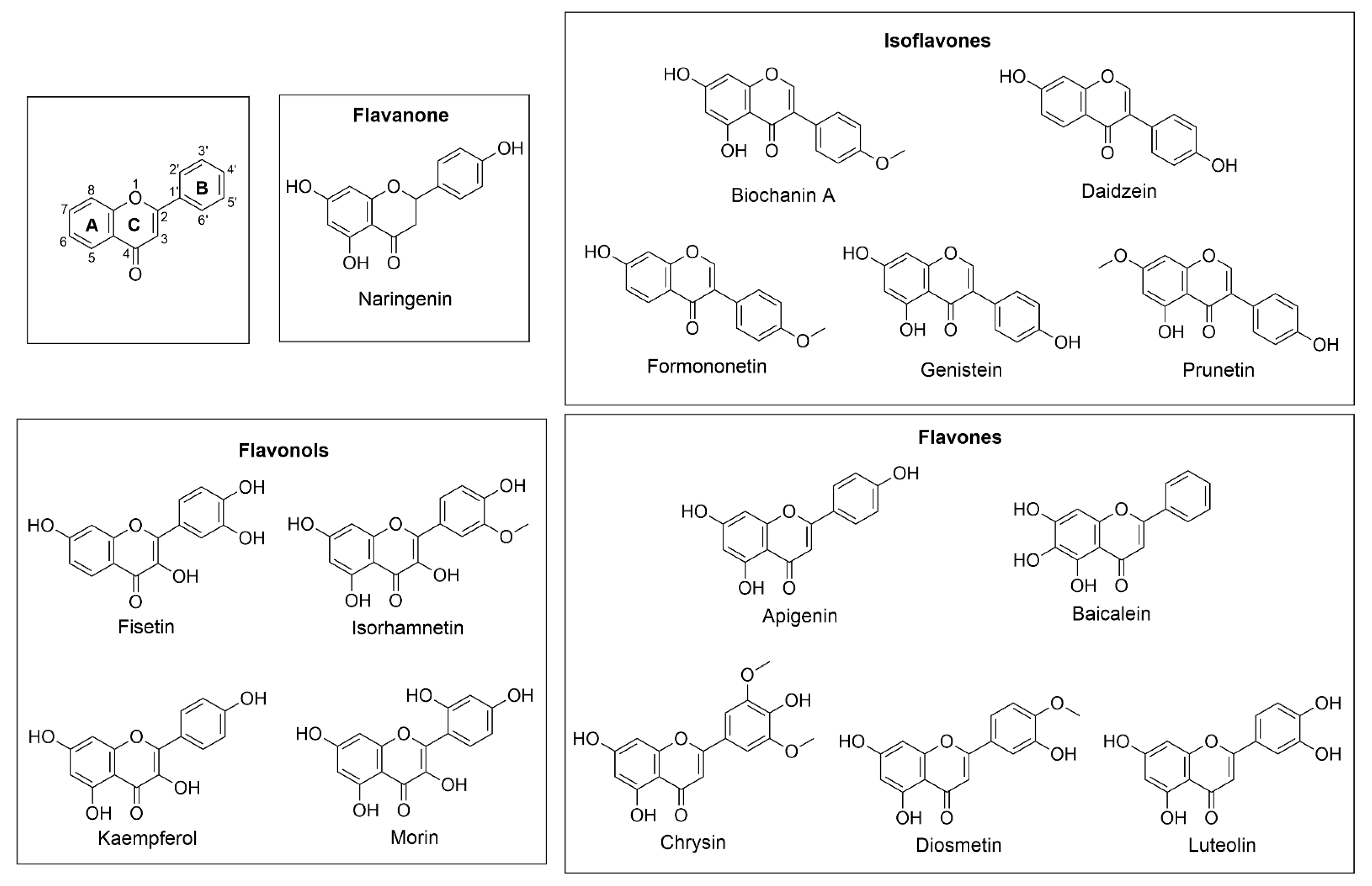
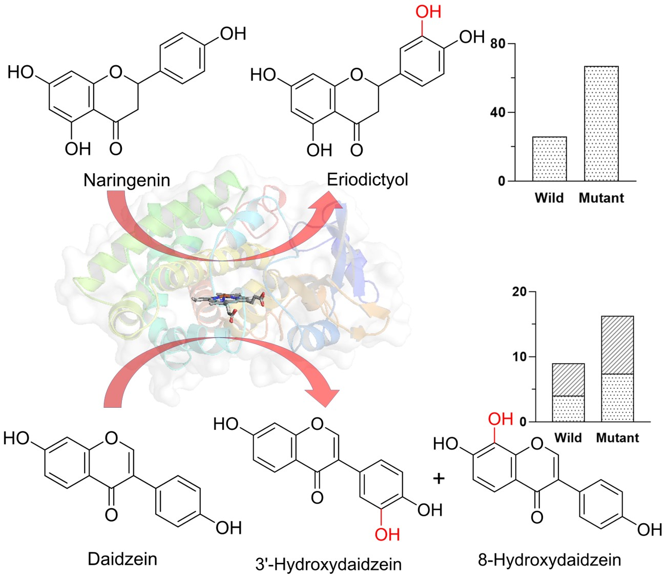
2.3. In Vitro Assay and Product Identification

To investigate the substrate specificity of the enzyme, an in vitro reaction was carried out with fifteen different flavonoids (flavonols, flavones, flavanone, and isoflavones) under identical conditions, as mentioned in the methods section (Figure 1). Initially, the in vitro biotransformation by CYP105A5 was conducted with heterologous redox partners Pdx/Pdr from Pseudomonas putida (purified from P450cam system) and Fdx/FdR from spinach (commercial), along with the chemical redox partner hydrogen peroxide and (Diacetoxyiodo) benzene. CYP105A5 was able to catalyze the hydroxylation of flavonoids using purified heterologous redox partners Pdx–PdR in the presence of cofactor NADH, while other redox partners showed slight or no activity. Out of 15 substrates selected, only flavanone (naringenin) and isoflavones (biochanin A, daidzein, and genistein) showed the conversion. The products were characterized by high-performance liquid chromatography–photodiode array (HPLC-PDA) and liquid chromatography–mass spectroscopy (LC-MS) (Figure S4).
Table S1 summarizes the mass analysis of the products. The analysis of the biochanin A showed two product peaks during the HPLC. The LC-MS analysis confirmed the O-demethylated (peak P2) and subsequently hydroxylated product (peak P1) of biochanin A (Figure S4A). However, LC-MS did not show any hydroxylated product of biochanin A. Since no hydroxylated product for biochanin A was observed, we confirmed that the first O-demethylation of biochanin A occurs, followed by hydroxylation (Scheme 1). The O-demethylated product of biochanin A is genistein, which is successively hydroxylated (Figure S5A). Likewise, the HPLC analysis of the daidzein showed two monohydroxylated products, P1 and P2, representing 43% and 57% of the total product formed, respectively (Figure S4B). Structure elucidation verified two different hydroxylated products of daidzein to be 8-hydroxyaidzein (Figure S5C) and 3′-hydroxydaidzein (Figure S5D). Furthermore, the genistein and naringenin reaction mixture analysis showed one monohydroxylated product. The NMR analysis confirmed the C-3′ position hydroxylation of genistein and naringenin, and the products were known as orobol (Figure S5B) and eriodictyol (Figure S5E), respectively. The bioconversion of naringenin, genistein, and daidzein by CYP105A5 produced a 3′ hydroxylated product. Only in the case of daidzein, 8-hydroxy daidzein was formed as a second product. We also tested other methoxy flavonoids, such as diosmetin, formononetin, prunetin, and iso-rhamnetin, but the HPLC and mass analysis did not show any O-demethylated or hydroxylated product. Despite having a similar structure to naringenin, apigenin did not show any product formation, indicating the unique substrate selectivity of CYP105A5.

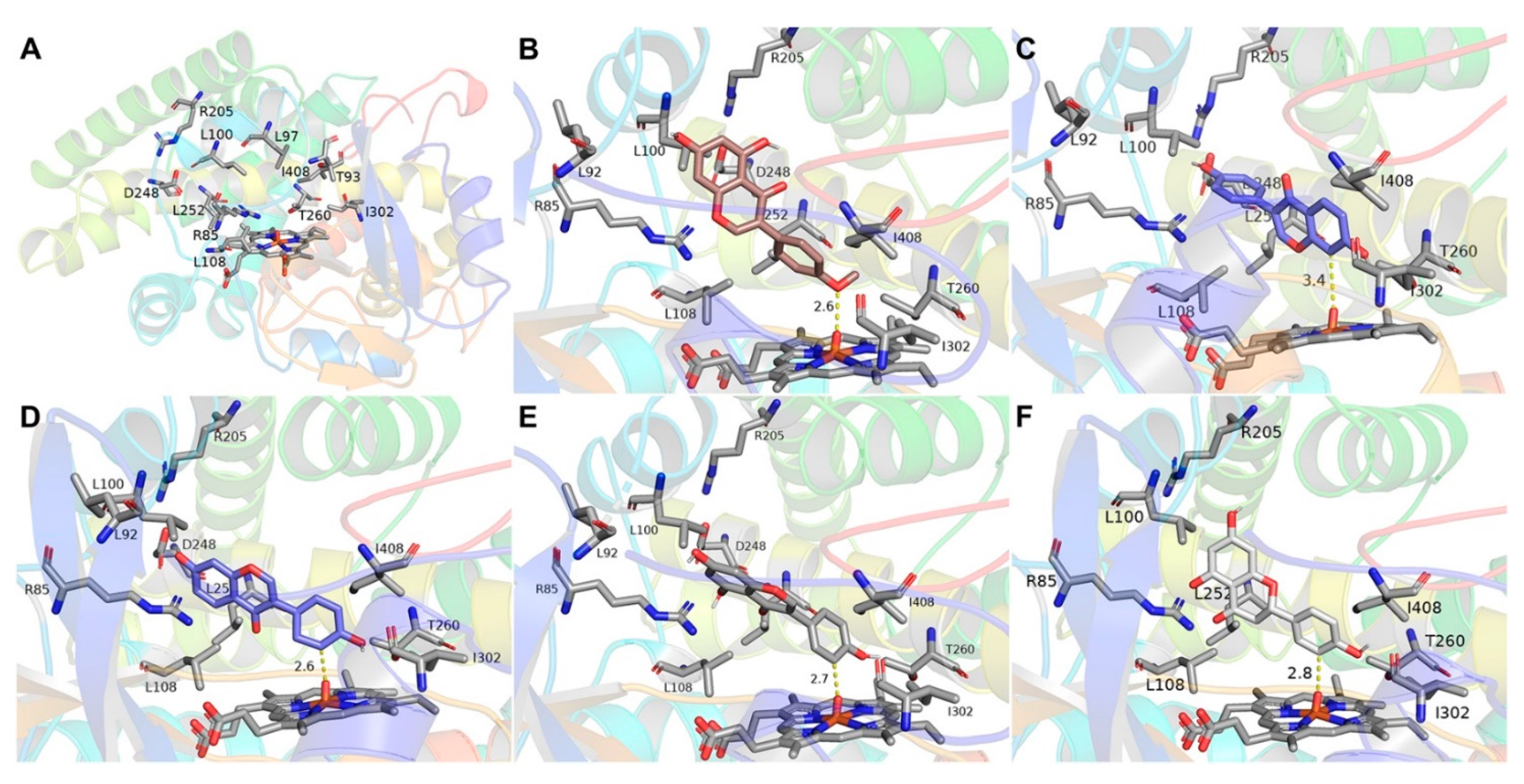
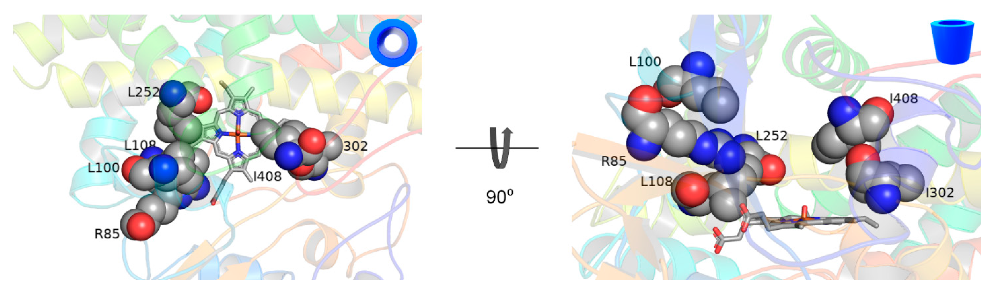
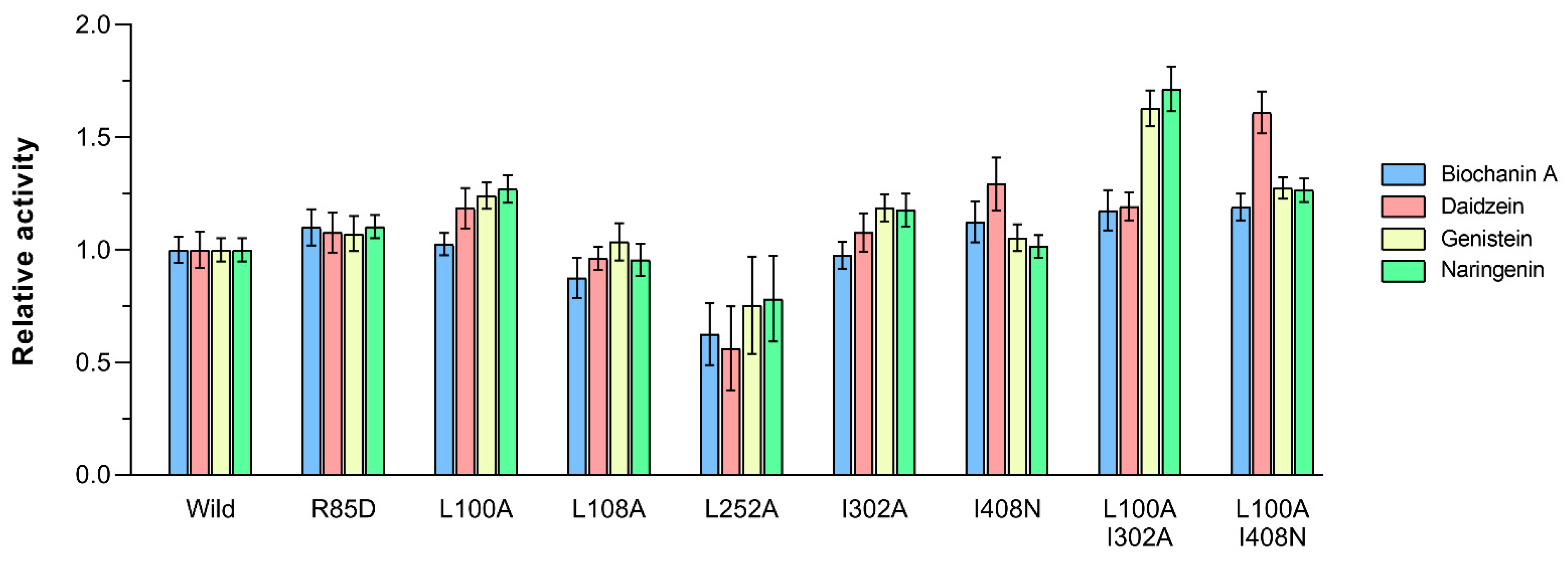
2.4. Docking and Mutagenesis Study

In order to gain detailed structural information about the active site, the three-dimensional structure of the protein was generated by Modeller, using the most homologous template (PDB ID: 2ZBX) with 71.50% sequence identity from Streptomyces griseolus [42,43]. The quality of the model was evaluated by the online programs PROCHECK, VERIFY-3D, and ERRAT (see Supplementary Materials; Figure S6) [44,45,46]. The modeled 3D structure of CYP105A5 showed a large funnel-shaped, substrate-binding pocket just above the heme porphyrin. Hydrophilic and hydrophobic amino acids (Arg 85, Arg96, Leu100, Leu108, Arg205, Asp248, Leu252, Thr260, Ala306, and Ile408) occupied the internal cavity of the protein and were expected to interact with the substrate (Figure 2A). Molecular docking using AutoDock Vina predicted the thermodynamically favorable substrate conformation in the active site [47]. Docked poses were selected based on dominant ligand conformation and analyzed for binding modes with CYP (Figure 2B–F). In almost all docked poses, the substrates were in proximity to the compound I of the reaction center, and two major binding modes were observed: one with the ring A, C3′ position of flavonoid, facing toward the compound I, herein called the A orientation, and another, where ring B, C8 position, faces toward the compound I, the B orientation.
Based on the analysis of docked substrate contact with the amino acids in the active site, mainly hydrophobic amino acids were selected for further study. In addition to BC-loop residue R85, L100, L108, and L252, amino acids close to the catalytically active heme (I302 and I408) were also selected to explore their impact on flavonoid binding and CYP105A5′s selectivity in flavonoid conversions (Figure 3). Mutants R85D, L100A, L108A, L252A, I302A, and I408N were prepared using site-directed mutagenesis, heterologously expressed in E. coli, and purified. We observed that the single mutants (R85D, L100A, I302A, and I408N) displayed an increased rate of conversion compared to wild type (Figure 4). Mutants L100A and I302A showed an increase in activity by 26% and 22% of genistein and by 27% and 17% of naringenin, respectively, compared with the wild type. Similarly, mutant L100A and I408N showed an increase in the activity of daidzein by 18% and 29%, respectively. Next, we constructed the double mutants by combining the most promising variant L100A/I302A and L100A/I408N. Furthermore, double variants exhibited increases in activities by 59%, 63%, and 71% of daidzein, genistein, and naringenin, respectively, compared with the wild type. Mutant L100A/I408N increased the daidzein hydroxylation activity, but it was unable to change the product distribution pattern. Similar product distribution patterns were observed for all mutants (Figure S7). However, some of the single and double variants showed low functional expression (data not shown). None of the single or double mutants showed significant changes in the demethylation activity of the enzyme or conversion of other flavonoids.

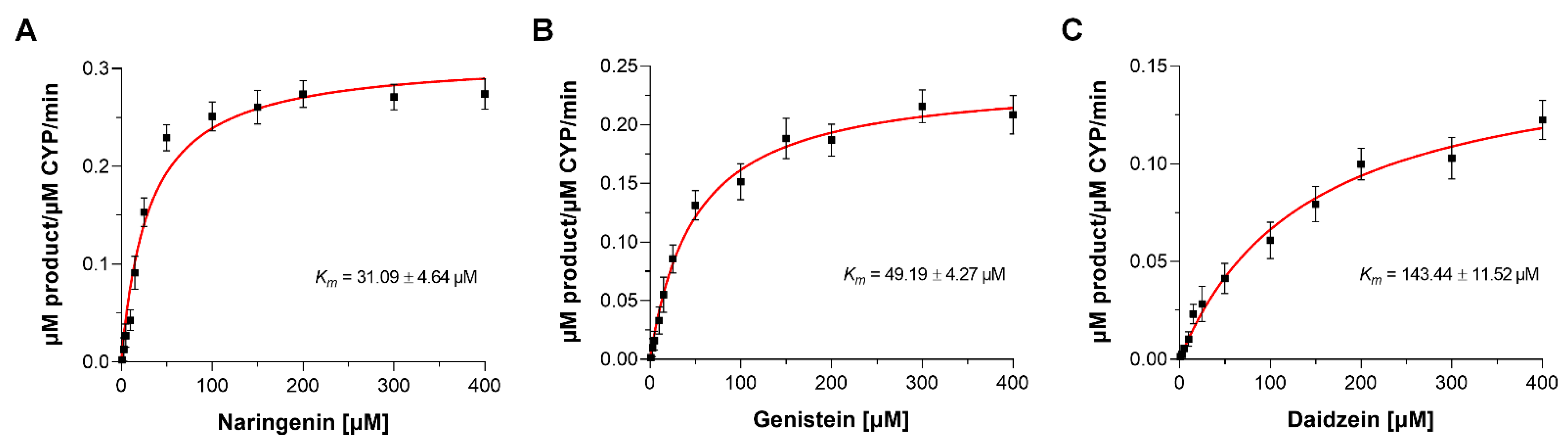
2.5. Enzyme Kinetic Studies

The kinetic parameters of purified CYP105A5 along with the selected mutants were calculated. The Michaelis–Menten constant (Km), the catalytic rate constant (kcat), specificity constants (kcat/Km), and the coupling efficiency for substrates were calculated using redox partners Pdx/Pdr (with a P450/Pdx/Pdr ratio of 1:8:2) and are summarized in Table 1. Wild-type CYP105A5 showed a higher affinity for naringenin, with the Km and kcat values of (31.09 ± 4.64) µM and (0.31 ± 0.09) min−1, respectively (Figure 5). Meanwhile, daidzein had a lower affinity, with a Km value of (143.44 ± 11.52) µM. Although the mutants affected the Km value for substrates positively or negatively, mutants showed an increase in the kcat values compared to wild type. The kcat value for naringenin and genistein was increased by 220% and 180%, respectively, for mutant L100A/I302A and for daidzein by 145% for mutant L100A/I408N. Moreover, the calculated kcat/Km values for naringenin, genistein, and daidzein were increased by 1.68, 1.43, and 1.57-fold using respective mutants. Meanwhile, mutants negatively affected the coupling efficiencies for the bioconversion of all but a few flavonoids. The calculated coupling efficiency of daidzein was the lowest among all. The mutant L100A/I408N increased its coupling efficiency by 1.37-fold.

2.6. Whole-Cell Biotransformation

It has been observed that some CYPs can utilize the native redox partner of E. coli for electron transfer during whole-cell bioconversion [48]. When CYP105A5 was expressed without the redox partner, hydroxylation of flavonoids was not observed, suggesting CYP105A5 could not support endogenous redox partners from E. coli. The comparative in vivo bioconversion of exogenously supplemented flavonoids was conducted by using separate E. coli BL21(DE3) cells harboring pET32a_CYP105A5 and a duet vector containing redox partner pACYC_camAB or pCDF_camAB (Figure 6). The HPLC-PDA chromatograms of extract from the biotransformation reaction utilizing cells harboring pET32a_CYP105A5 and pACYC_camAB system showed the peaks of the hydroxylated product of respective substrates, along with additional peaks. However, when the pCDF_camAB was used, slight improvement in hydroxylation and no additional peaks were observed. Naringenin and genistein showed an improvement in hydroxylation by ~1.2 times. Moreover, the biotransformation using mutants L100A/I302A and L100A/I408N, in a separate assay, along with the pCDF_camAB system, improved the hydroxylation of naringenin, genistein, and daidzein by 2.57-fold, 2.39-fold, and 1.8-fold, respectively.

3. Discussion

Among the cytochromes P450, the CYP105 family belongs to the largest group, comprising at least 17 subfamilies represented in Streptomycetes, and are associated with the biosynthesis of secondary metabolites and biotransformation and biodegradation of xenobiotics [49]. This suggests their functional diversity, represented by catalyzing diverse reactions with a variety of structurally different compounds [10]. For flavonoids hydroxylation, different flavonoid hydroxylases, including flavonoid 3′-hydroxylase (F3′H), flavonoid 5′-hydroxylase (F5′H), flavonoid 6-hydroxylase (F6H), and flavonoid 8-hydroxylase (F8H), have been characterized in mammals and plants [50,51,52]. Similarly, the fungal PcCYP65a2 from Phanerochaete chrysosporium exhibited naringenin hydroxylation at 3′-position to yield eriodictyol [53]. However, flavonoid hydroxylation by bacterial CYPs is rarely reported. Monooxygenase from Saccharothrix espanaensis (Sam5) exhibited regioselective hydroxylation of isoflavones, flavanones, and flavones [34]. CYP450 BM3 variants were also able to hydroxylate selected flavonoids [36,37]. CYP105D7 from S. avermitilis (54% identity to CYP105A5) also functionalized flavonoids in addition to steroids [38,39].
Here, we reported on CYP105A5 from Streptomyces sp. for the efficient biotransformation of flavonoids. CYP105A5 was able to catalyze the demethylation and hydroxylation of selected flavonoids using heterologous redox partners putidaredoxin and putidaredoxin reductase. CYP105A5 catalyzed the 3′-hydroxylation of the B-ring of daidzein, genistein, and naringenin. In addition, it was able to catalyze the 8-position hydroxylation of the A-ring of daidzein. Moreover, the methoxy group present in the B-ring of biochanin A is de-methylated, and the subsequent product formed (genistein) is hydroxylated. The amino acid sequence identity of this enzyme is 70% to CYP105A1 and 74% to CYP105A3. CYP105A1 has been reported to hydroxylate vitamin D3 and diterpenoids, while CYP105A3 has been reported to hydroxylate the compactin molecule [11,22].
For rational enzyme design, a combination of substrate docking, comparison to other hydroxylases, and mutational data from the literature were used. The molecular docking analysis showed that the flavonoids were surrounded by hydrophobic residues, along with hydrophilic residues such as Arg 85, Arg96, Leu100, Leu108, Arg205, Asp248, Leu252, Thr260, Ala306, and Ile408 (Figure 2A). For the P450 catalyzed reaction to occur, the electrophilic oxygen of compound I must abstract hydrogen from the substrate [54]. The particular catalytically competent orientation of flavonoid toward the compound 1 and the interaction of the hydroxyl group with the hydrophilic T260 are crucial for biotransformation [55]. The daidzein with only two hydroxyl groups showed two alternative binding modes with its A- or B-ring exposed toward the compound I, a suitable position for forming two different hydroxylated products (Figure 2C,D). It is also interesting to note that the dihydroxylated product of daidzein was not observed. After hydroxylation of the A-ring of daidzein, the B-ring is still open for hydroxylation or vice versa, but no other product peaks were detected. Meanwhile, the genistein, biochanin A, and naringenin bearing an extra 5-OH group can catalyze only one product, in contrast to daidzein. The presence of the 5-OH group might have provided a steric hindrance to the orientation responsible for A-ring hydroxylation. Moreover, the formononetin with a similar structure to daidzein and an extra methyl group on 4′-OH is neither demethylated nor hydroxylated. Similarly, prunetin with an extra methyl group on 7-OH was not hydroxylated despite having a similar structure to naringenin. These observations indicate the importance of interaction between residues (5-OH, 7-OH, and 4′-OH) and polar amino acids for the orientation of flavonoids in the active site.
In CYPs, substrate recognition sites are defined as six regions that are involved in substrate identification and binding and, consequently, influence substrate specificity [56]. Among the six sites, the highly variable BC-loop plays a crucial role in substrate recognition and stabilization [57]. In this study, the site-directed mutagenesis of three residues in the BC-loop region (R85, L100, and L108) and three other residues in other sites that directly interact with the bound substrate (L252, I302, and I408) were selected. Particularly, the changes at positions L100A, I302A, and I408N had a significant favorable impact on activity and altered the product bioconversion. Moreover, the additive effect of mutants was also observed. Therefore, it may be inferred that these residues are likely to interact with substrates, by changing the activity and selectivity of enzymes. The replacement of the bulky amino acid residue lining the access channel by small amino acids altered the accessibility of substrate toward the active site [58]. It has also been observed that by increasing the volume of the substrate access channel and binding pocket through mutagenesis, the rate of testosterone conversion by CYP105D7 was increased by almost 9-fold [23]. However, these mutants were unable to show a significant difference in the conversion for naringenin.
The calculated coupling efficiencies of CYP105A5 and its mutants for flavonoid bioconversion are lower, and mutations also did not significantly increase them. This lower value indicates the significant loss of electrons from the cofactor NADH. The use of heterologous redox partners during the catalysis may be responsible for this. CYP154C5 mutants also showed reduced coupling efficiency while using Pdx/PdR as a redox partner [59]. They also suggested higher uncoupling was due to the water entering the active site. Meanwhile, the coupling efficiency of 100% was displayed by P450cam using Pdx/PdR for the conversion of the substrate D-camphor [60]. Although the mutants showed reduced coupling efficiency, they greatly improved the bioconversion. For in vivo bioconversion of flavonoids, utilization of cells harboring pET32a_CYP105A5 and pACYC_camAB produced additional peaks along with the hydroxylated product. Plasmid vector pACYCDuet-1 contains a chloramphenicol resistance gene (chloramphenicol acetyltransferase) to detoxify the antibiotic chloramphenicol, which might have acetylated the hydroxylated product, ultimately decreasing its production yield [48]. The use of duet vector pCDFDuet-1 containing redox partner along with CYP105A5 showed improved conversion. By using pCDF_ camAB and double mutants L100A/I302A and L100A/I408N in a separate assay, we achieved almost 2.6-fold and 1.8-fold increases in naringenin and daidzein hydroxylation, respectively, during bioconversion. These results suggest the potential use of CYP105A5 for future rational design and application as a whole-cell biocatalyst to produce polyphenolic compounds.

4. Materials and Methods

4.1. Chemicals and Reagents

All the flavonoids used in this study, ampicillin, δ-aminolevulinic acid (ALA), formate dehydrogenase, and NADH, were purchased from Sigma–Aldrich (St. Louis, MO, USA). Isopropyl-1-thio-β-D-galactopyranoside (IPTG), 1,4-dithiothreitol (DTT), and kanamycin were purchased from Duchefa Bochemie (Haarlem, The Netherlands). Restriction enzymes, T4 DNA ligase, dNTPs, and DNA polymerase were purchased from Takara Bio (Kusatsu, Shiga, Japan). All other high-grade chemicals were purchased from available commercial sources.

4.2. Bioinformatics Analysis

The basic local alignment search tool (BLAST) was used to identify and compare the close homologs. Multiple sequence alignment was accomplished with GeneDoc [61]. An evolutionary study was conducted by Molecular Evolutionary Genetics Analysis MEGA X [62]. The phylogenetic tree was constructed based on the maximum likelihood method with one thousand bootstrap replicates, and the Poisson correction method was used to compute the evolutionary distances [63]. The name for CYP was assigned by Dr. David Nelson (http://drnelson.utmem.edu/CytochromeP450.html) (accessed on 13 October 2017). The nucleotide sequence information of CYP105A5 is provided in the supporting materials (Sequence S1).

4.3. Molecular Cloning and Protein Over-Expression

The CYP105A5 encoding sequence of length 1257 bp was amplified from the genomic DNA of Streptomyces sp. The primers used for polymerase chain reaction (PCR) are listed in Table S2. PCR products were cloned in pMD20-T (Takara, Japan) vector and transformed into E. coli XL1-Blue for gene amplification. After the sequence confirmation, the genes were ligated into the pET-32a(+) vector to create the pET32a_CYP105A5 construct encoding N-terminal His6-tag protein. The construct was transformed into E. coli XL1-Blue and, finally, into over-expression host E. coli C41(DE3). One milliliter of the overnight grown transformed cells was inoculated into 100 mL LB media supplemented with 100 µg/mL ampicillin and incubated at 37 °C (180 rpm), until cell density reached 0.6 at OD600 nm. For protein expression, the cells were induced with 0.5 mM IPTG, along with supplementation of 1.0 mM ALA and 0.5 mM FeCl3. The cells were harvested, after 48 h of incubation at 20 °C, by centrifugation at 3500 rpm at 4 °C for 30 min and washed twice with 50 mM potassium phosphate buffer (pH 7.4) containing 10% glycerol. The cell pellets were resuspended in 20 mL of the same buffer.
For the in vitro system, redox partners putidaredoxin (PDX) and putidaredoxin reductase (PDR) were over-expressed in E. coli BL21(DE3) using pET-28a(+) and pET-32a(+) plasmid construct, respectively, which has previously been described [64]. For whole-cell bioconversion, Pdx and Pdr genes previously cloned into pCDF-duet and pACYC-duet vector were introduced into E. coli cell BL21(DE3) harboring the plasmid (pET32a_CYP105A5) [48].

4.4. Protein Purification and Determination of the Concentration

To obtain the soluble fraction, the cells were lysed with ultra-sonication, and cell debris was removed by centrifugation. The obtained supernatant was mixed with TALON His-tag resin, pre-equilibrated with equilibrium buffer (potassium phosphate buffer pH 7.4), and kept in a shaker for 60 min. Elution buffer (potassium phosphate buffer pH 7.4 with 10% glycerol) containing (20, 100, and 250) mM imidazole was used to elute the protein bound to the resin. Fractions containing proteins of interest were concentrated by ultrafiltration using Amicon centrifugal filters (Millipore, Merck, Germany) with 50 kDa cutoff for CYP105A5 and 30 kDa for PDR, whereas 10 kDa cutoff was used for PDX. The concentration of CYP105A5 was measured based on the CO-difference spectra method [65]. Protein was diluted to 2.0 mL using the potassium phosphate buffer and separated into two cuvettes (reference and sample). Carbon monoxide was bubbled gently into the cuvette sample, at a rate of 1 bubble per sec for 1 min. Spectrum was recorded using Biochrome Libra S35PC UV/Visible Spectrophotometry (Cambridge, UK), after reducing both reference and sample by adding a few grains of sodium dithionite. The concentration of functional CYP105A5 was estimated using the extinction coefficient Ɛ450–490 = 91 mm−1cm−1 [66]. PdR concentration was determined based on average concentration calculated from wavelengths (378, 454, and 480) nm using the extinction coefficient (ε) = (9.7, 10.0, and 8.5) mM−1cm−1, respectively [67]. Pdx concentration was determined based on average concentration calculated from wavelength (415 and 454) nm using the extinction coefficient (ε) = (11.1 and 10.4) mM−1cm−1, respectively [65].

4.5. Enzyme Activity Assay

We carried out 300 µL reactions using the protein CYP105A5 in 50 mM potassium phosphate buffer (pH 7.4). All the substrates were prepared by dissolving in DMSO. The reaction mixture contained CYP105A5 (3 µM), substrate (200 µM), PdR (6 µM), Pdx (24 µM), catalase (100 µg/mL), and a NADH regeneration system comprising formate dehydrogenase (1.0 U), sodium formate (150 mM), and MgCl2 (1.0 mM) in 50 mM potassium phosphate buffer (pH 7.4). The reaction was initiated by the addition of 250 µM NADH and incubated for 1 h at 30 °C with shaking. The reaction was stopped by adding a double volume of chilled ethyl acetate. The ethyl acetate organic layer was collected, dried, dissolved in methanol, and analyzed by high-performance liquid chromatography–photodiode array (HPLC-PDA). After integrating the substrate and product peaks, the conversion percentage of each substrate was determined.

4.6. Site-Directed Mutagenesis

Mutagenesis was performed using EZchangeTM site-directed mutagenesis kit using the nPfu-Frote PCR Mix (Enzynomics, Daejeon, Korea), according to the manufacturer’s instructions. All the primers used in the PCR reactions are listed in Table S2. The thermal cycling reaction consisted of initial denaturation for 1 min at 95 °C, 30 cycles of denaturation at 95 °C for 1 min, annealing at 55 °C for 1 min, and extension at 65 °C for 14 min, followed by a final elongation at 65 °C for 5 min. The PCR mixture was digested with DpnI restriction enzyme for 30 min at 37 °C and transformed into DH5α competent E. coli cells. After selection of the colonies, plasmid isolation, and confirmation of mutants by sequencing (Macrogen, Daejeon, Korea), the confirmed mutants were transformed into E. coli C41(DE3), over-expressed, and purified as described above.

4.7. Determination of Kinetic Parameters

The time-dependent reaction progress curve was created by measuring the amount of product formed over time using different substrates. The reaction mixture contained 1.0 μM CYP105A5 or its mutant, 2.0 μM PdR, 8.0 μM Pdx, and 100 μM substrate in 50 mM potassium phosphate buffer (pH 7.4). Reactions were started by adding 350 μM NADH. The initial velocity condition was established, and the saturation curve was generated using the same reaction condition, except for the varied substrate concentration (0–400) μM. Km and kcat values for each substrate were calculated from the plot of the rate of reaction versus the substrate concentration. The NADH oxidation rate was measured, and the coupling efficiency was determined as the percentage of NADH used for product formation over total NADH consumption [65]. All the kinetic analysis was performed by non-linear regression analysis based on Michaelis–Menten kinetics, using the GraphPad Prism 7.0 software (La Jolla, CA, USA).

4.8. In silico Analysis: Homology Modeling and Docking

We performed a docking simulation with AutoDock Vina-1.1.2 to explore the binding interactions between CYP and four substrates (biochanin A, daidzein, genistein, and naringenin) [47]. The crystal structure of CYP105A1 from Streptomyces griseolus (PDB ID: 2ZBX and 71.50% sequence identity with CYP105A5) was chosen as the template. A three-dimensional (3D) model of CYP105A5 was generated using Modeller 9.9.2 [42]. Homology model was validated by PROCHECK, Verify-3D, and ERRAT [44,45,46]. To generate the PDBQT files of the protein and ligand, as well as grid box, the graphical user interface program AutoDock Tools 1.5.6 was used [68]. The grid map was prepared with a grid box of size 22 Å × 20 Å × 24 Å with 1 Å spacing, which can cover the entire ligand molecule. The exhaustiveness was set to 100, and all the other parameters were set to the default values. Visualization of the result was performed using PyMOL [69]. The dominant ligand conformations were selected and analyzed for binding modes with CYPs.

4.9. Whole-Cell Bioconversion

Whole-cell bioconversion for analytical purpose was performed in E. coli cells at a 1.0 mL scale. E. coli cells harboring genes for CYP105A5, PdR, and Pdx were grown with appropriate antibiotics at 37 °C. When the OD600 reached 0.6, the culture was supplemented with 0.1 mM ALA and 0.5 mM FeCl3 and induced by 0.5 mM final concentration of IPTG. Then, the cultures were incubated for 48 h at 20 °C. The cells were collected by centrifugation (3500 rpm) for 20 min at 4 °C and washed twice with 50 mM potassium phosphate buffer (pH 7.4). The cells were resuspended in the same buffer, supplemented with 1.0 mg/mL of glucose, along with 0.2 mM of the substrate. Bioconversion was performed for 24 h at 30 °C and 200 rpm. The reaction mixture was extracted twice with an equal volume of ethyl acetate, dried, and analyzed by HPLC-PDA. Whole-cell bioconversion was performed in a 10 mL system. We precultured E. coli harboring genes for CYP105A5 and redox partner in 5.0 mL LB broth with appropriate antibiotics at 37 °C for about 6 h. To 10 mL of LB with antibiotic, 100 µL of pre-inoculum was transferred and grown at 37 °C. When the OD600 reached 0.6, similar methods to those described above were followed for the remaining procedure.

4.10. Analytical Methods

Ethyl acetate organic fraction collected by extracting in vitro and in vivo reaction was dried by using a rotatory evaporator. The dried residue was dissolved in HPLC-grade methanol, filtered through a 0.2 μm pore polytetrafluoroethylene filter, and analyzed by HPLC-PDA using a reversed-phase column (Mightysil RP–18 GP 250–4.6, 5 µm, Kanto Chemical, Japan). Gradient mobile phase was composed of solvent A (0.05% trifluoroacetic acid in HPLC-grade water) and solvent B (100% acetonitrile, CH3CN). The percentage of solvent B used was as follows: 10% for (0–5) min, 50% for (5–10) min, 70% for (10–14) min, 90% for (14–17) min, 10% for (17–20) min, and 10% for (20–25) min, with a flow rate of 1.0 mL/min. Detection of the substrate and its products was performed by UV absorbance at their respective wavelength. The purification of compounds was carried out by preparative HPLC (Shimadzu, Tokyo, Japan) with C18 column (YMC–Pack ODS-AQ 150 mm × 20 mm I.D., 10 µm) connected to a UV detector using a 35 min binary program, with CH3CN 10% for (0–3) min, 25% for (3–7) min, 40% for (7–12) min, 45% for (12–15) min, 50% for (15–27) min, 90% for (17–23) min, 90% for (23–25) min, 10% for (25–28) min, and 10% for (28–35) min, at a flow rate of 10.0 mL/min. LC-MS analysis of the flavonoids and their products was performed by HR-QTOF ESI/MS in positive ion mode using an ACQUITY (UPLC, Waters Corp., Billerica, MA, USA) column coupled with an SYNAPT G2-S (Water Corp., Billerica, MA, USA). For structural elucidation, products were dried, lyophilized, and dissolved in dimethyl sulfoxide (DMSO)-d6, and subjected to 700 MHz within a Bruker Biospin instrument (GmbH, Reinstates, Germany). One-dimensional NMRs (1H NMR and 13C NMR), followed by 2D NMRs (rotating-frame overhauser enhancement spectroscopy [ROESY], heteronuclear multiple bonded connectivity [HMBC], and heteronuclear multiple quantum coherence [HMQC]) were performed, as needed, for structure elucidation of the compound. The raw fid files were processed using MestReNOVA version 14.0.1 (Mestrelab Research S.L., Santiago de Compostela, Spain).
3′-hydroxyaidzein:1H NMR (700 MHz, [D6]DMSO): δ = 8.25 (s, 1H), 7.96 (d, J = 8.8, 1H), 7.02 (d, J = 2.1, 1H), 6.93 (dd, J = 8.7, 2.2, 1H), 6.85 (d, J = 2.1, 1H), 6.81 (dd, J = 8.1, 2.1, 1H), and 6.76 ppm (d, J = 8.1, 1H); 13C NMR (176 MHz, [D6]DMSO): δ = 175.13 (C4), 163.19 (C7), 157.86 (C8a), 153.20 (C2), 145.73 (C4′), 145.25 (C3′), 127.74 (C5), 124.07 (C3), 123.48 (C1′), 120.30 (C6′), 117.07 (C2′), 117.00 (C4a), 115.75 (C5′), 115.67 (C6), 102.53 ppm (C8).
8-hydroxydaidzein: 1H NMR (700 MHz, [D6]DMSO): δ = 8.33 (s, 1H), 7.47 (d, J = 8.7, 1H), 7.39 (d, J = 8.6, 2H), 6.95 (d, J = 8.7, 1H), and 6.81 ppm (d, J = 8.6, 2H); 13C NMR (176 MHz, [D6]DMSO): δ = 175.61 (C4), 157.58 (C4′), 153.12 (C2), 150.47 (C7), 147.17 (C8a), 133.38 (C8), 130.60 (C2′, C6′), 123.37 (C1′), 123.16 (C3), 117.88 (C4a), 116.09 (C5), 115.38 (C3′, 5′), 114.62 ppm (C6).
Genistein:1H NMR (700 MHz, [D6]DMSO): δ = 12.96 (s, 1H), 8.31 (d, J = 1.4, 1H), 7.38 (d, J = 8.6, 1H), 6.82 (d, J = 8.6, 1H), 6.38 (s, 1H), and 6.22 ppm (d, J = 2.0, 1H); 13C NMR (176 MHz, [D6]DMSO): δ = 180.60 (C4), 165.15 (C7), 162.45 (C5), 158.10 (C8a), 157.80 (C4′), 154.38 (C2), 130.63 (C2′, C6′), 122.70 (C3), 121.73 (C1′), 115.48 (C3′, C5′), 104.75 (C4a), 99.52 (C6), 94.18 ppm (C8).
Orobol:1H NMR (700 MHz, [D6]DMSO): δ = 13.00 (s, 1H), 8.29 (s, 1H), 7.00 (d, J = 2.0, 1H), 6.81 (dd, J = 8.1, 2.0, 1H), 6.78 (d, J = 8.1, 1H), 6.37 (d, J = 1.6, 1H), and 6.22 ppm (d, J = 1.9, 1H); 13C NMR (176 MHz, [D6]DMSO): δ = 180.66 (C4), 165.00 (C7), 162.48 (C5), 158.03 (C8a), 154.37 (C2), 145.96 (C4′), 145.34 (C3′), 122.85 (C3), 122.13 (C1′), 120.42 (C6′), 117.00 (C2′), 115.83 (C5′), 104.83 (C4a), 99.48 (C6), 94.14 ppm (C8).
Eriodictyol: 1H NMR (700 MHz, [D6]DMSO): δ = 12.15 (s, 1H), 6.88 (s, 1H), 6.76– 6.73 (m, 2H), 5.89–5.85 (m, 2H), 5.38 (dd, J = 12.5, 3.0, 1H), 3.18 (dd, J = 17.1, 12.5, 1H), and 2.67 ppm (dd, J = 17.1, 3.1, 1H); 13C NMR (176 MHz, [D6]DMSO): δ = 196.71 (C4), 167.42 (C7), 163.95 (C5), 163.36 (C8a), 146.17 (C4′), 145.65 (C3′), 129.94 (C1′), 118.40 (C6′), 115.79 (C5′), 114.80 (C2′), 102.15 (C4a), 96.27 (C6), 95.49 (C8), 78.90 (C2), 42.54 ppm (C3).

5. Conclusions

In the present study, we explored the ability of CYP105A5 to achieve the O-demethylation and hydroxylation of various flavonoids. We observed the conversion of flavonoids into 3′ and 8 hydroxy flavonoids. By rational design of substrate-binding pocket based on the experimental data and molecular docking analysis, we enhanced the product formation rate of flavonoids. Here, hydroxylation of flavonoid is particularly interesting, as hydroxylation of flavonoids by bacterial cytochrome P450 monooxygenases has been reported rarely. The study revealed the application of protein in whole-cell biocatalysts by simple fermentation to produce valuable polyphenols that are cheaper and easier to scale-up, compared to other approaches. Overall, our result also demonstrates the future application of protein engineering to expand the substrate flexibility of enzymes towards flavonoids and/or other substrates.

Supplementary Materials

The following supporting information can be downloaded at https://www.mdpi.com/article/10.3390/catal12101157/s1. Result: Homology modeling; Table S1: LC-MS of parent compounds and their hydroxylated products; Table S2: Primers used in this study; Figure S1: Multiple sequence alignment of the CYP105A5 protein sequence with other known CYPs from the CYP105 series; Figure S2: Phylogenetic tree of the CYP105s and their closest homologs; Figure S3: SDS-PAGE and spectroscopic analysis of protein; Figure S4: HPLC-PDA and HR-QTOF ESI/MS analysis of reaction products; Figure S5: Structural elucidation of hydroxylated products. 1H and 13C NMR analysis of the demethylated (A), demethylated and hydroxylated (B) product of biochanin A, daidzein (C and D), and naringenin (E); Figure S6: Homology model validation by PROCHECK plot (A), Verify-3D plot (B), and ERRAT plot (C) for CYP105A5; Figure S7: The product distribution of wild-type and mutants of CYP105A5 catalyzed hydroxylation of daidzein; Sequence S1: Nucleotide sequence of the CYP105A5.

Author Contributions

Conceptualization, P.S. and T.-J.O.; methodology, P.S. and T.-J.O.; validation, P.S., J.K.P. and T.-J.O.; formal analysis, P.S., J.K.P. and T.-J.O.; investigation, P.S.; resources, T.-J.O.; writing—original draft preparation, P.S. and T.-J.O.; writing—review and editing, P.S. and T.-J.O.; supervision, T.-J.O.; funding acquisition, T.-J.O. All authors have read and agreed to the published version of the manuscript.

Funding

This research was supported by the Basic Science Research Program through the National Research Foundation of Korea (NRF) funded by the Ministry of Education (NRF-2019R1D1A3A03103903). In addition, this work was supported by the BioGreen21 Agri-Tech Innovation Program (Project No. PJ015710), Rural Development Administration, Republic of Korea.

Data Availability Statement

Data are contained within the article and its supplementary material.

Acknowledgments

We would like to thank the Division of Magnetic Resonance, Korea Basic Science Institute, Ochang, Chungbuk, Korea, for the NMR analyses. In addition, the authors would like to sincerely thank Tokutaro Yamaguchi for his generous and considerable help regarding NMR analysis.

Conflicts of Interest

The authors declare no conflict of interest.

References

  1. Guengerich, F.P. Cytochrome P450 enzymes in the generation of commercial products. Nat. Rev. Drug Discov. 2002, 1, 359–366. [Google Scholar] [CrossRef] [PubMed]
  2. Isin, E.M.; Guengerich, F.P. Complex reactions catalyzed by cytochrome P450 enzymes. Biochim. Biophys. Acta Gen. Subj. 2007, 1770, 314–329. [Google Scholar] [CrossRef] [PubMed]
  3. Dangi, B.; Oh, T.J. Bacterial CYP 154C8 catalyzes carbon-carbon bond cleavage in steroids. FEBS Lett. 2019, 593, 67–79. [Google Scholar] [CrossRef] [Green Version]
  4. Urlacher, V.; Schmid, R.D. Biotransformations using prokaryotic P450 monooxygenases. Curr. Opin. Biotechnol. 2002, 13, 557–564. [Google Scholar] [CrossRef] [Green Version]
  5. Volz, T.J.; Rock, D.A.; Jones, J.P. Evidence for two different active oxygen species in cytochrome P450 BM3 mediated sulfoxidation and N-dealkylation reactions. J. Am. Chem. Soc. 2002, 124, 9724–9725. [Google Scholar] [CrossRef] [PubMed]
  6. Guengerich, F.P. Mechanisms of cytochrome P450-catalyzed oxidations. ACS Catal. 2018, 8, 10964–10976. [Google Scholar] [CrossRef] [PubMed]
  7. Klenk, J.M.; Dubiel, P.; Sharma, M.; Grogan, G.; Hauer, B. Characterization and structure-guided engineering of the novel versatile terpene monooxygenase CYP 109Q5 from Chondromyces apiculatus DSM 436. Microb. Biotechnol. 2019, 12, 377–391. [Google Scholar] [CrossRef] [Green Version]
  8. Munro, A.W.; McLean, K.J.; Grant, J.L.; Makris, T.M. Structure and function of the cytochrome P450 peroxygenase enzymes. Biochem. Soc. Trans. 2018, 46, 183–196. [Google Scholar]
  9. Rude, M.A.; Baron, T.S.; Brubaker, S.; Alibhai, M.; Del Cardayre, S.B.; Schirmer, A. Terminal olefin (1-alkene) biosynthesis by a novel P450 fatty acid decarboxylase from Jeotgalicoccus species. Appl. Environ. Microbiol. 2011, 77, 1718–1727. [Google Scholar] [CrossRef] [Green Version]
  10. Moody, S.C.; Loveridge, E.J. CYP 105—Diverse structures, functions and roles in an intriguing family of enzymes in Streptomyces. J. Appl. Microbiol. 2014, 117, 1549–1563. [Google Scholar] [CrossRef] [Green Version]
  11. Sawada, N.; Sakaki, T.; Yoneda, S.; Kusudo, T.; Shinkyo, R.; Ohta, M.; Inouye, K. Conversion of vitamin D3 to 1α, 25-dihydroxyvitamin D3 by Streptomyces griseolus cytochrome P450SU-1. Biochem. Biophys. Res. Commun. 2004, 320, 156–164. [Google Scholar] [CrossRef] [PubMed]
  12. O’Keefe, D.P.; Romesser, J.A.; Leto, K.J. Identification of constitutive and herbicide inducible cytochromes P-450 in Streptomyces griseolus. Arch. Microbiol. 1988, 149, 406–412. [Google Scholar] [CrossRef]
  13. Omer, C.A.; Lenstra, R.; Litle, P.J.; Dean, C.; Tepperman, J.M.; Leto, K.J.; Romesser, J.A.; O’Keefe, D.P. Genes for two herbicide-inducible cytochromes P-450 from Streptomyces griseolus. J. Bacteriol. 1990, 172, 3335–3345. [Google Scholar] [CrossRef] [PubMed] [Green Version]
  14. Ueno, M.; Yamashita, M.; Hashimoto, M.; Hino, M.; Fujie, A. Oxidative activities of heterologously expressed CYP107B1 and CYP105D1 in whole-cell biotransformation using Streptomyces lividans TK24. J. Biosci. Bioeng. 2005, 100, 567–572. [Google Scholar] [CrossRef]
  15. Goto, L.S.; Hokka, C.O.; Lima, J.F.; Prieto, T.; Araújo, A.P.U.; Nantes, I.L.; Nascimento, O.R. Structure and peroxidase activity of ferric Streptomyces clavuligerus orf10-encoded protein P450CLA: UV-visible, CD, MCD and EPR spectroscopic characterization. J. Braz. Chem. Soc. 2012, 23, 913–920. [Google Scholar] [CrossRef] [Green Version]
  16. Xu, L.H.; Fushinobu, S.; Takamatsu, S.; Wakagi, T.; Ikeda, H.; Shoun, H. Regio-and stereospecificity of filipin hydroxylation sites revealed by crystal structures of cytochrome P450 105P1 and 105D6 from Streptomyces avermitilis. J. Biol. Chem. 2010, 285, 16844–16853. [Google Scholar] [CrossRef] [Green Version]
  17. Fouces, R.; Mellado, E.; Díez, B.; Barredo, J.L. The tylosin biosynthetic cluster from Streptomyces fradiae: Genetic organization of the left region. Microbiology 1999, 145, 855–868. [Google Scholar] [CrossRef] [Green Version]
  18. Xue, Y.; Wilson, D.; Zhao, L.; Liu, H.W.; Sherman, D.H. Hydroxylation of macrolactones YC-17 and narbomycin is mediated by the pikC-encoded cytochrome P450 in Streptomyces venezuelae. Chem. Biol. 1998, 5, 661–667. [Google Scholar] [CrossRef] [Green Version]
  19. Li, T.; Ni, S.; Jia, C.; Wang, H.; Sun, G.; Wu, L.; Gan, M.; Shan, G.; He, W.; Lin, L.; et al. Identification of 4, 5-dihydro-4-hydroxygeldanamycins as shunt products of geldanamycin biosynthesis. J. Nat. Prod. 2012, 75, 1480–1484. [Google Scholar] [CrossRef]
  20. Palaniappan, N.; Kim, B.S.; Sekiyama, Y.; Osada, H.; Reynolds, K.A. Enhancement and selective production of phoslactomycin B, a protein phosphatase IIa inhibitor, through identification and engineering of the corresponding biosynthetic gene cluster. J. Biol. Chem. 2003, 278, 35552–35557. [Google Scholar] [CrossRef] [Green Version]
  21. Hayashi, K.; Sugimoto, H.; Shinkyo, R.; Yamada, M.; Ikeda, S.; Ikushiro, S.; Kamakura, M.; Shiro, Y.; Sakaki, T. Structure-based design of a highly active vitamin D hydroxylase from Streptomyces griseolus CYP105A1. Biochemistry 2008, 47, 11964–11972. [Google Scholar] [CrossRef] [PubMed]
  22. Ba, L.; Li, P.; Zhang, H.; Duan, Y.; Lin, Z. Semi-rational engineering of cytochrome P450sca-2 in a hybrid system for enhanced catalytic activity: Insights into the important role of electron transfer. Biotechnol. Bioeng. 2013, 110, 2815–2825. [Google Scholar] [CrossRef] [PubMed]
  23. Ma, B.; Wang, Q.; Ikeda, H.; Zhang, C.; Xu, L.H. Hydroxylation of steroids by a microbial substrate-promiscuous P450 cytochrome (CYP105D7): Key arginine residues for rational design. Appl. Environ. Microbiol. 2019, 85, 01530-19. [Google Scholar] [CrossRef] [PubMed]
  24. Manach, C.; Scalbert, A.; Morand, C.; Rémésy, C.; Jiménez, L. Polyphenols: Food sources and bioavailability. Am. J. Clin. Nutr. 2004, 79, 727–747. [Google Scholar] [CrossRef] [PubMed] [Green Version]
  25. Tungmunnithum, D.; Thongboonyou, A.; Pholboon, A.; Yangsabai, A. Flavonoids and other phenolic compounds from medicinal plants for pharmaceutical and medical aspects: An overview. Medicines 2018, 5, 93. [Google Scholar] [CrossRef]
  26. Pinto, C.; Cidade, H.; Pinto, M.; Tiritan, M.E. Chiral flavonoids as antitumor agents. Pharmaceuticals 2021, 14, 1267–1296. [Google Scholar] [CrossRef]
  27. Silva, C.F.; Pinto, D.C.; Silva, A.M. Chromones: A Promising Ring System for New Anti-inflammatory Drugs. ChemMedChem 2016, 1, 2252–2260. [Google Scholar] [CrossRef]
  28. Perez-Vizcaino, F.; Fraga, C.G. Research trends in flavonoids and health. Biochem. Biophys. 2018, 646, 107–112. [Google Scholar] [CrossRef]
  29. Hu, M. Commentary: Bioavailability of flavonoids and polyphenols: Call to arms. Mol. Pharm. 2007, 4, 803–806. [Google Scholar] [CrossRef] [Green Version]
  30. Rice-Evans, C.A.; Miller, N.J.; Paganga, G. Structure-antioxidant activity relationships of flavonoids and phenolic acids. Biol. Med. 1996, 20, 933–956. [Google Scholar] [CrossRef]
  31. Plaza, M.; Pozzo, T.; Liu, J.; Gulshan Ara, K.Z.; Turner, C.; Nordberg Karlsson, E. Substituent effects on in vitro antioxidizing properties, stability, and solubility in flavonoids. J. Agric. Food Chem. 2014, 62, 3321–3333. [Google Scholar] [CrossRef]
  32. Lo, Y.L.; Wang, W.; Ho, C.T. 7, 3′, 4′-Trihydroxyisoflavone modulates multidrug resistance transporters and induces apoptosis via production of reactive oxygen species. Toxicology 2012, 302, 221–232. [Google Scholar] [CrossRef] [PubMed]
  33. Ko, Y.H.; Kwon, S.H.; Ma, S.X.; Seo, J.Y.; Lee, B.R.; Kim, K.; Kim, S.Y.; Lee, S.Y.; Jang, C.G. The memory-enhancing effects of 7, 8, 4′-trihydroxyisoflavone, a major metabolite of daidzein, are associated with activation of the cholinergic system and BDNF signaling pathway in mice. Brain Res. Bull. 2018, 142, 197–206. [Google Scholar] [CrossRef] [PubMed]
  34. Lee, H.; Kim, B.G.; Ahn, J.H. Production of bioactive hydroxyflavones by using monooxygenase from Saccharothrix espanaensis. J. Biotechnol. 2014, 176, 11–17. [Google Scholar] [CrossRef] [PubMed]
  35. Jones, J.A.; Collins, S.M.; Vernacchio, V.R.; Lachance, D.M.; Koffas, M.A. Optimization of naringenin and p-coumaric acid hydroxylation using the native E. coli hydroxylase complex, HpaBC. Biotechnol. Prog. 2016, 32, 21–25. [Google Scholar] [CrossRef] [PubMed]
  36. Chu, L.L.; Pandey, R.P.; Jung, N.; Jung, H.J.; Kim, E.H.; Sohng, J.K. Hydroxylation of diverse flavonoids by CYP450 BM3 variants: Biosynthesis of eriodictyol from naringenin in whole cells and its biological activities. Microb. Cell Fact. 2016, 15, 135. [Google Scholar] [CrossRef] [Green Version]
  37. Hong, L.L.; Kong, J.Q. Altering the Regioselectivity of Cytochrome P450 BM3 variant M13 toward genistein through protein engineering and variation of reaction conditions. ACS Omega 2020, 5, 32059–32066. [Google Scholar] [CrossRef]
  38. Liu, L.; Yao, Q.; Ma, Z.; Ikeda, H.; Fushinobu, S.; Xu, L.H. Hydroxylation of flavanones by cytochrome P450 105D7 from Streptomyces avermitilis. J. Mol. Catal. B Enzym. 2016, 132, 91–97. [Google Scholar] [CrossRef]
  39. Chiang, C.M.; Ding, H.Y.; Lu, J.Y.; Chang, T.S. Biotransformation of isoflavones daidzein and genistein by recombinant Pichia pastoris expressing membrane-anchoring and reductase fusion chimeric CYP105D7. J. Taiwan Inst. Chem. Eng. 2016, 60, 26–31. [Google Scholar] [CrossRef]
  40. Poulos, T.L. Heme enzyme structure and function. Chem. Rev. 2014, 114, 3919–3962. [Google Scholar] [CrossRef] [Green Version]
  41. Mak, P.J.; Denisov, I.G. Spectroscopic studies of the cytochrome P450 reaction mechanisms. Biochim. Biophys. Acta Proteins Proteom. 2018, 1866, 178–204. [Google Scholar] [CrossRef] [PubMed]
  42. Šali, A.; Blundell, T.L. Comparative protein modelling by satisfaction of spatial restraints. J. Mol. Biol. 1993, 3, 779–815. [Google Scholar] [CrossRef] [PubMed]
  43. Sugimoto, H.; Shinkyo, R.; Hayashi, K.; Yoneda, S.; Yamada, M.; Kamakura, M.; Ikushiro, S.I.; Shiro, Y.; Sakaki, T. Crystal structure of CYP105A1 (P450SU-1) in complex with 1α, 25-dihydroxyvitamin D3. Biochemistry 2008, 47, 4017–4027. [Google Scholar] [CrossRef] [PubMed]
  44. Laskowski, R.A.; Moss, D.S.; Thornton, J.M. Main-chain bond lengths and bond angles in protein structures. J. Appl. Crystallogr. 1993, 26, 283–291. [Google Scholar] [CrossRef]
  45. Lüthy, R.; Bowie, J.U.; Eisenberg, D. Assessment of protein models with three-dimensional profiles. Methods Enzymol. 1997, 277, 396–404. [Google Scholar] [CrossRef]
  46. Bowie, J.U.; Lüthy, R.; Eisenberg, D. A method to identify protein sequences that fold into a known three-dimensional structure. Science 1991, 253, 164–170. [Google Scholar] [CrossRef] [Green Version]
  47. Trott, O.; Olson, A.J. AutoDock Vina: Improving the speed and accuracy of docking with a new scoring function, efficient optimization, and multithreading. J. Comput. Chem. 2010, 31, 455–461. [Google Scholar] [CrossRef] [Green Version]
  48. Dangi, B.; Kim, K.H.; Kang, S.H.; Oh, T.J. Tracking down a new steroid-hydroxylating promiscuous cytochrome P450: CYP154C8 from Streptomyces sp. W2233-SM. ChemBioChem 2018, 19, 1066–1077. [Google Scholar] [CrossRef]
  49. Yasuda, K.; Sugimoto, H.; Hayashi, K.; Takita, T.; Yasukawa, K.; Ohta, M.; Kamakura, M.; Ikushiro, S.; Shiro, Y.; Sakaki, T. Protein engineering of CYP105s for their industrial uses. Biochim. Biophys. Acta Proteins Proteom. 2018, 1866, 23–31. [Google Scholar] [CrossRef]
  50. Seitz, C.; Ameres, S.; Forkmann, G. Identification of the molecular basis for the functional difference between flavonoid 3′-hydroxylase and flavonoid 3′, 5′-hydroxylase. FEBS Lett. 2007, 581, 3429–3434. [Google Scholar] [CrossRef]
  51. Latunde-Dada, A.O.; Cabello-Hurtado, F.; Czittrich, N.; Didierjean, L.; Schopfer, C.; Hertkorn, N.; Werck-Reichhart, D.; Ebel, J. Flavonoid 6-hydroxylase from soybean (Glycine max L.), a novel plant p-450 monooxygenase. J. Biol. Chem. 2001, 276, 1688–1695. [Google Scholar] [CrossRef]
  52. Kagawa, H.; Takahashi, T.; Ohta, S.; Harigaya, Y. Oxidation and rearrangements of flavanones by mammalian cytochrome P450. Xenobiotica 2004, 34, 797–810. [Google Scholar] [CrossRef]
  53. Kasai, N.; Ikushiro, S.; Hirosue, S.; Arisawa, A.; Ichinose, H.; Wariishi, H.; Ohta, M.; Sakaki, T. Enzymatic properties of cytochrome P450 catalyzing 3′-hydroxylation of naringenin from the white-rot fungus Phanerochaete chrysosporium. Biochem Biophys Res. Commun. 2009, 387, 103–108. [Google Scholar] [CrossRef]
  54. Meunier, B.; De Visser, S.P.; Shaik, S. Mechanism of oxidation reactions catalyzed by cytochrome P450 enzymes. Chem. Rev. 2004, 104, 3947–3980. [Google Scholar] [CrossRef]
  55. Zhang, X.; Peng, Y.; Zhao, J.; Li, Q.; Yu, X.; Acevedo-Rocha, C.G.; Li, A. Bacterial cytochrome P450-catalyzed regio-and stereoselective steroid hydroxylation enabled by directed evolution and rational design. Bioresour. Bioprocess. 2020, 7, 2. [Google Scholar] [CrossRef]
  56. Gotoh, O. Substrate recognition sites in cytochrome P450 family 2 (CYP2) proteins inferred from comparative analyses of amino acid and coding nucleotide sequences. J. Biol. Chem. 1992, 267, 83–90. [Google Scholar] [CrossRef]
  57. Sirim, D.; Widmann, M.; Wagner, F.; Pleiss, J. Prediction and analysis of the modular structure of cytochrome P450 monooxygenases. J. BMC Struct. Biol. 2010, 10, 34. [Google Scholar] [CrossRef] [Green Version]
  58. Urban, P.; Lautier, T.; Pompon, D.; Truan, G. Ligand access channels in cytochrome P450 enzymes: A review. Int J. Mol. Sci. 2018, 19, 1617. [Google Scholar] [CrossRef] [Green Version]
  59. Bracco, P.; Wijma, H.J.; Nicolai, B.; Buitrago, J.A.R.; Klünemann, T.; Vila, A.; Schrepfer, P.; Blankenfeldt, W.; Janssen, D.B.; Schallmey, A. CYP154C5 Regioselectivity in Steroid Hydroxylation Explored by Substrate Modifications and Protein Engineering. ChemBioChem 2021, 22, 1099–1110. [Google Scholar] [CrossRef]
  60. Hayashi, T.; Harada, K.; Sakurai, K.; Shimada, H.; Hirota, S. A role of the heme-7-propionate side chain in cytochrome P450cam as a gate for regulating the access of water molecules to the substrate-binding site. J. Am. Chem. Soc. 2009, 131, 1398–1400. [Google Scholar] [CrossRef]
  61. Nicholas, K.B.; Nicholas, H.B.; Deerfield, D.; Gauch, H. Genedoc: A Tool for Editing and Annotating Multiple Sequence Alignments; Ieee Software: Piscataway, NJ, USA, 1997. [Google Scholar]
  62. Kumar, S.; Stecher, G.; Li, M.; Knyaz, C.; Tamura, K. MEGA X: Molecular evolutionary genetics analysis across computing platforms. Mol. Biol. Evol. 2018, 35, 1547–1549. [Google Scholar] [CrossRef]
  63. Zuckerkandl, E.; Pauling, L. Evolutionary divergence and convergence in proteins. In Evolving Genes and Proteins; Academic Press: Cambridge, MA, USA, 1965; pp. 97–166. [Google Scholar]
  64. Bhattarai, S.; Liou, K.; Oh, T.J. Hydroxylation of long chain fatty acids by CYP147F1, a new cytochrome P450 subfamily protein from Streptomyces peucetius. Arch. Biochem. Biophys. 2013, 539, 63–69. [Google Scholar] [CrossRef]
  65. Omura, T.; Sato, R. The carbon-monoxide binding pigment of liver microsomes evidence for its haemoprotein nature. J. Biol. Chem. 1964, 239, 2370–2378. [Google Scholar] [CrossRef]
  66. Guengerich, F.P.; Martin, M.V.; Sohl, C.D.; Cheng, Q. Measurement of cytochrome P450 and NADPH–cytochrome P450 reductase. Nat. Protoc. 2009, 4, 1245–1251. [Google Scholar] [CrossRef] [Green Version]
  67. Roome, P.W., Jr.; Philley, J.C.; Peterson, J.A. Purification and properties of putidaredoxin reductase. J. Biol. Chem. 1983, 258, 2593–2598. [Google Scholar] [CrossRef]
  68. Morris, G.M.; Huey, R.; Lindstrom, W.; Sanner, M.F.; Belew, R.K.; Goodsell, D.S.; Olson, A.J. AutoDock4 and AutoDockTools4: Automated docking with selective receptor flexibility. J. Comput. Chem. 2009, 30, 2785–2791. [Google Scholar] [CrossRef]
  69. DeLano, W.L. Pymol: An open-source molecular graphics tool. CCP4 Newsl. Protein Crystallogr. 2002, 40, 82–92. [Google Scholar]
Figure 1. Substrate structures of flavonoids used for the determination of the substrate specificity of CYP105A5, with the carbon atom positions numbered as shown for the common structure of flavonoids.
Catalysts 12 01157 g001
Scheme 1. Flavonoid O-demethylation and hydroxylation by CYP105A5. (A) Biochanin A is O-demethylated to genistein and is subsequently hydroxylated at the 3′ position. (B) Daidzein is hydroxylated at the 3′ and 8 positions. (C) Naringenin is hydroxylated at the 3′ position. The hydroxylated position is represented by red color.
Catalysts 12 01157 sch001
Figure 2. Docking model with different substrates bound. (A) Ribbon representation of the homology model of CYP105A5 with amino acid residues surrounding the active site. Docking model of CYP105A5 with biochanin A (B), daidzein (C,D), genistein (E), and naringenin (F). The docking poses feature close proximity of methyl group of biochanin A, C-3′ of daidzein (D), genistein, and naringenin and additional C-8 of daidzein (C) toward Cpd I. The connecting yellow line represents the distance between Cpd I’s oxygen atom and the hydroxylation site.
Catalysts 12 01157 g002
Figure 3. The active site cavity from top (left) and horizontal (right) view with regard to heme. The rotation of the right panel with respect to the left panel is 90°. The amino acids residues in the substrate-binding pocket for the mutational study are labelled and are represented as spheres.
Catalysts 12 01157 g003
Figure 4. Relative activity of wild type and mutants of CYP105A5 for demethylation of biochanin A and the hydroxylation of daidzein, genistein, and naringenin. Activities are determined for purified CYP105A5 and its mutants using the CYP:PDX:PDR ratios of 1:8:2 (3 µM CYP, 24 µM Pdx, and 6 µM PdR) and 0.2 mM substrate. The reaction was started by the addition of 300 μM NADH. The relative activities are expressed as the ratio of the activity of the mutant to that of the wild-type CYP105A5. The data present mean values with standard deviations of independent experiments.
Catalysts 12 01157 g004
Figure 5. Michaelis–Menten plot of wild-type CYP105A5 toward (A) naringenin, (B) genistein, and (C) daidzein. The reaction mixture consisted of CYP:Pdx:Pdr in the ratios 1:8:2 (1 µM CYP, 8 µM Pdx, and 2 µM PdR) and in the presence of varied substrate concentrations. The reaction was started by the addition of 500 μM NADH. The rate of the reaction was determined and plotted against the substrate concentration. Values are means of independent experiments with standard deviations.
Catalysts 12 01157 g005
Figure 6. Comparative in vivo bioconversion of flavonoids using E. coli BL21(DE3) cells harboring recombinant pET32a_CYP105A5 or mutant along with duet vector containing redox partner pACYC_camAB or pCDF_camAB. For daidzein bioconversion, mutant L100A/I408N was used, while for genistein and naringenin, mutant L100A/I302A was utilized. Bioconversion was carried out in a 10 mL system with 0.2 mM substrate concentration.
Catalysts 12 01157 g006
Table 1. Michaelis–Menten constant (Km), catalytic rate constant (kcat), specificity constants, and coupling efficiencies of the purified CYP105A5 and its mutants, PdR, Pdx, and the substrates. The overall apparent kinetic parameters were determined with CYP:Pdx:PdR concentration ratios of 1:8:2 (1 µM CYP, 8 µM Pdx, and 2 µM PdR) for purified CYP105A5 and its mutants towards daidzein, genistein, and naringenin. The ratio (kcat/Km) values for mutants are expressed relative to the value determined for the wild type. Coupling efficiency was calculated as the percentage of NADH used for the formation of the product over the total NADH consumption. The NADH consumption rate was calculated after the subtraction of the respective background NADH consumption. Results represent the mean values of duplicate measurements.
Strain/MutantKm (µM)kcat (min−1)kcat/Km
(M−1 min1)
Ratio
kcat/Km
Coupling
Efficiency [%]
Daidzein
WT143.44 ± 11.520.09 ± 0.04627.441.0019.26 ± 3.07
L100A124.73 ± 17.890.10 ± 0.05801.731.2824.94 ± 5.17
I408N159.83 ± 15.370.12 ± 0.06750.791.1918.73 ± 6.30
L100A/I408N131.57 ± 12.090.13 ± 0.04988.061.5726.38 ± 6.95
Genistein
WT49.19 ± 4.270.26 ± 0.075.296 × 1031.0053.83 ± 4.38
L100A58.43 ± 6.740.34 ± 0.095.818 × 1031.0950.83 ± 6.72
I302A67.01 ± 9.830.42 ± 0.106.267 × 1031.1844.30 ± 9.29
L100A/I302A64.38 ± 8.560.49 ± 0.127.611 × 1031.4343.62 ± 8.16
Naringenin
WT31.09 ± 4.640.31 ± 0.099.971 × 1031.0061.27 ± 5.26
L100A37.84 ± 8.020.43 ± 0.111.136 × 1041.1462.91 ± 9.51
I302A32.96 ± 6.380.39 ± 0.071.183 × 1041.1850.36 ± 11.83
L100A/I302A41.16 ± 7.160.69 ± 0.091.676 × 1041.6857.28 ± 10.06
Publisher’s Note: MDPI stays neutral with regard to jurisdictional claims in published maps and institutional affiliations.

