Article

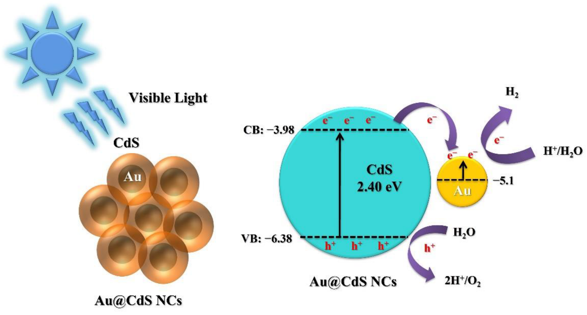
Au@CdS Nanocomposites as a Visible-Light Photocatalyst for Hydrogen Generation from Tap Water

1
Department of Materials Science and Engineering, Feng Chia University, Taichung 407, Taiwan
2
Department of Materials Science and Engineering, National Yang Ming Chiao Tung University, Hsinchu 30010, Taiwan
*
Author to whom correspondence should be addressed.
Catalysts 2023, 13(1), 33; https://doi.org/10.3390/catal13010033
Submission received: 22 November 2022 / Revised: 16 December 2022 / Accepted: 22 December 2022 / Published: 24 December 2022
(This article belongs to the Special Issue From Design to Application of Nanomaterials in Catalysis)

Abstract

:
The Au@CdS nanocomposites have been synthesized using a combination of wet chemical and hydrothermal approaches at lower reaction temperatures. The concentrations of CdS precursors and reaction temperature can be essential in influencing photocatalytic water splitting under blue-LED light excitation. The optimized Au@CdS nanocomposites (5 mM CdS precursors and 100 °C) exhibited the highest hydrogen evolution rate of 1.041 mmolh−1 g−1, which is 175.3 times higher than CdS nanoparticles for de-ionized water under blue-LED light excitation. This result is ascribed to separate photogenerated charge carriers and increased light absorption by the Au core. The Au@CdS nanocomposites (1.204 mmolh−1 g−1) revealed significant applications in photocatalytic tap water splitting under blue-LED light excitation, which is 512.3 times higher than CdS nanoparticles. In addition, reusability experiments demonstrate that Au@CdS nanocomposites exhibit excellent stability for the long-term photocatalytic tap water splitting process. Furthermore, this research shows that Au nanoparticles decorated with CdS shells effectively achieve high-efficiency conversion from light to hydrogen energy.

Graphical Abstract

1. Introduction

Metal–semiconductor core–shell heterostructures have received much attention due to their unique properties, combining two components to achieve a newly improved property by the synergistic effect [1,2,3,4,5]. When acting as a catalyst, metal–semiconductor core–shell heterostructures can exhibit two essential advantages to enhance their photocatalytic performance [6,7,8]. First, the metal–semiconductor interface with the Schottky barrier can accelerate photogenerated electron transfer and reduce the recombination rate of electrons and holes [4,9,10]. Second, metal (such as Ag and Au) on the semiconductor can increase light absorption via surface plasmon resonance [11,12]. Different kinds of metal–semiconductor core–shell heterostructures can be used as a photocatalyst, such as Ag@ZnO [12,13], Au@Cu2 O [1,14], Au@SnO2 [15], Au@MoS2 [16,17], Au@NiSx [18], Au@TiO2 [10,19,20], Ag@TiO2 [21,22], and Au@CdS [23,24,25], respectively.
Of these, Au@CdS nanocomposites can provide two potential benefits to improve their photocatalytic activity [24,25]. First, a CdS shell with a suitable bandgap (2.4 eV) can produce hydrogen under visible light excitation [26,27,28]. Second, Au nanoparticles (NPs) on the CdS nanostructures trap electrons to improve the separation of charge carriers [7]. Recently, Au@CdS nanocomposites have proven to be a promising material for photocatalytic degradation of organic compounds under visible-light excitation [24,29,30,31]. However, there are fewer reports on applying Au@CdS nanocomposites for photocatalytic hydrogen production under visible-light excitation [32,33,34]. Furthermore, the plasmonic Au nanosphere-modified visible-light-responsive CdS shells facilitate the generation and separation of electron–hole pairs and reduce the overpotential for hydrogen evolution [33].
On the other hand, Au NP-based plasmonic photocatalysts can generate “hot carriers” that may drive high-energy reactions by exciting low-energy photons for water splitting, which common semiconductor photocatalysts cannot utilize [34]. In addition, there are no reports on the use of Au@CdS nanocomposites for photocatalytic tap water splitting under blue-LED light (as the visible light) excitation. De-ionized water refers to pure water that has been obtained by removing ionic impurities from tap water [35]. Therefore, if hydrogen can be generated by directly decomposing tap water through photocatalysts, the cost and time of purifying tap water into de-ionized water can be effectively reduced, thereby contributing to the practical application of photocatalysts.
Herein, we synthesized Au@CdS nanocomposites and evaluated their visible-light-driven photocatalytic hydrogen production. Compared with CdS NPs, Au@CdS nanocomposites can increase electron–hole pairs separation to enhance their photocatalytic water splitting under blue-LED light (as the visible light) excitation. Additionally, Au@CdS nanocomposites can be used as a high-performance photocatalyst for tap water-splitting. The as-prepared photocatalyst may provide a strategy for designing stable and inexpensive technologies for highly efficient hydrogen production.

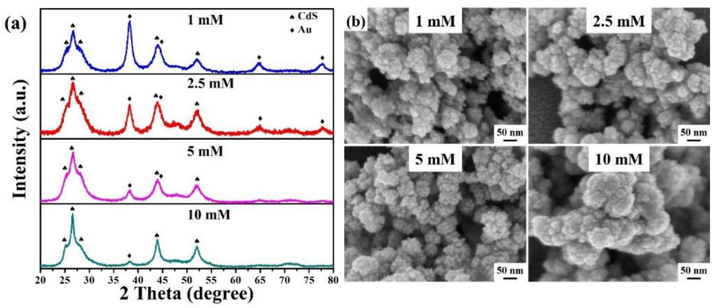
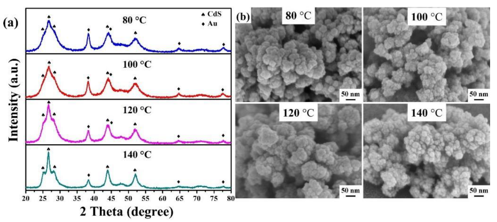
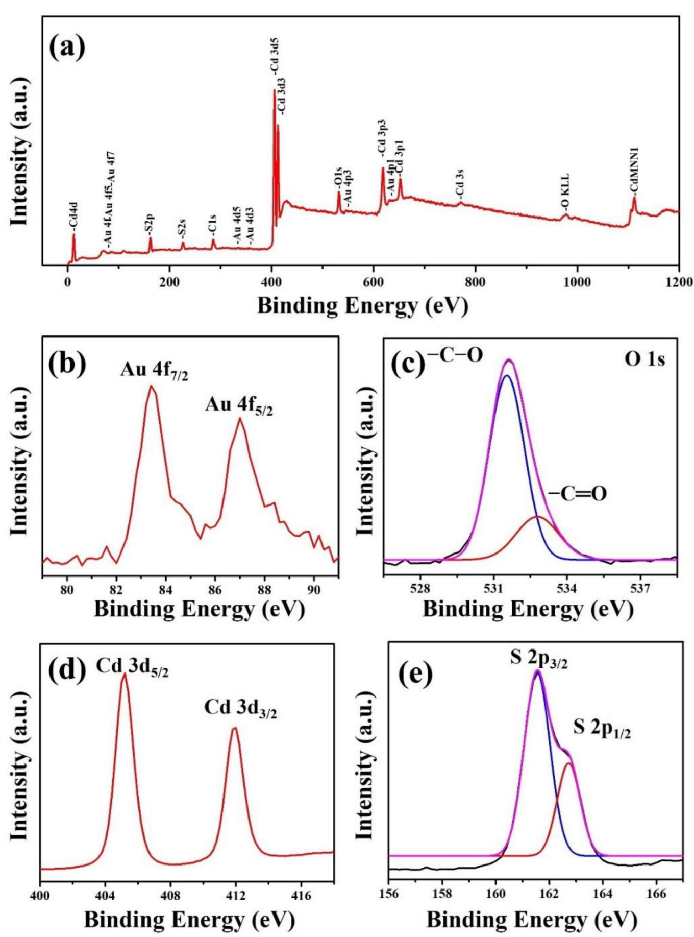
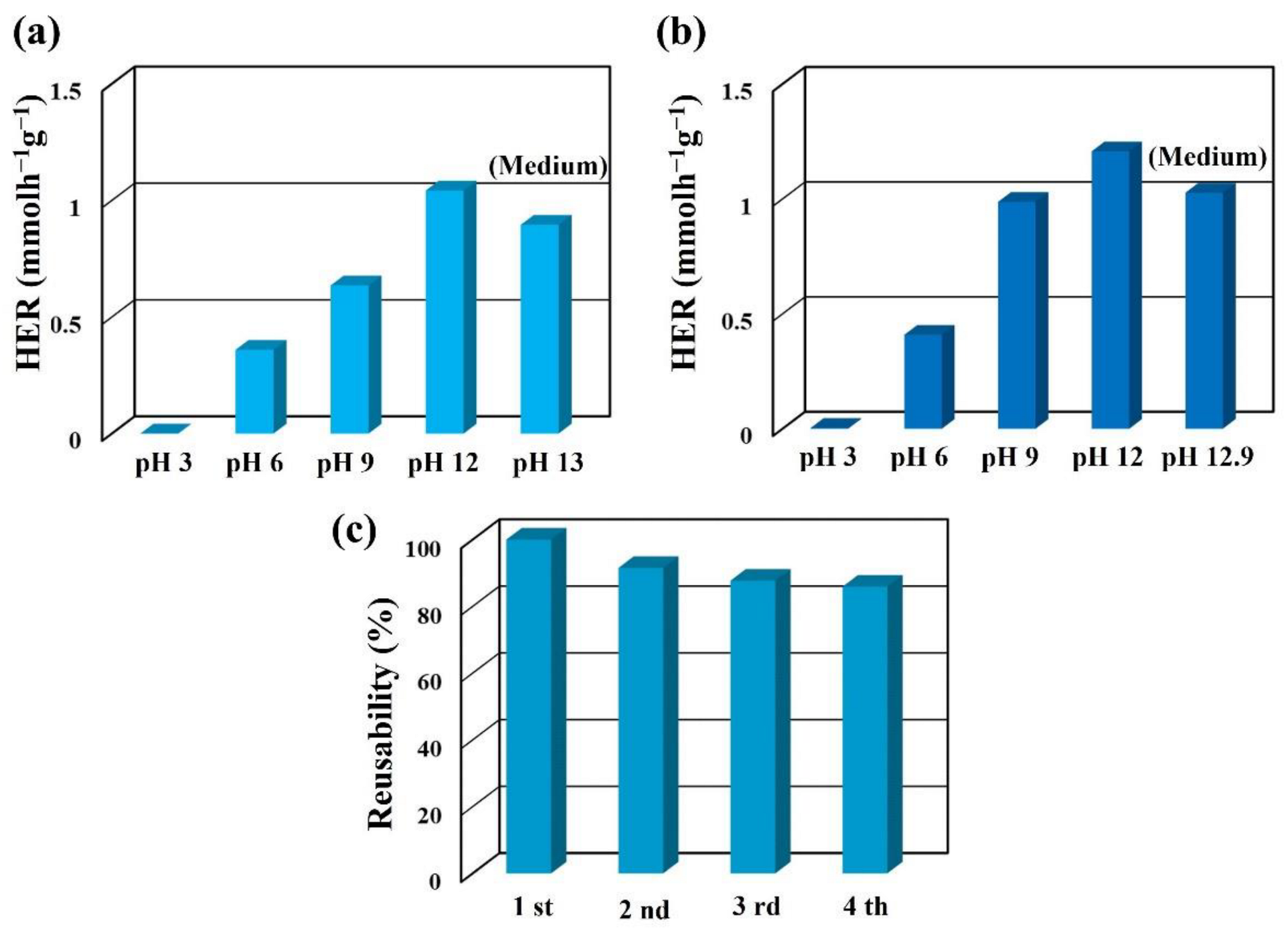
2. Results and Discussion

Figure 1 reveals the fabrication processes of Au@CdS nanocomposites by combining wet chemical and hydrothermal methods. First, Au nanoparticles were synthesized via a facile wet chemical method. Second, tween 20 can stabilize Au nanoparticles through 1h of vigorous stirring. Third, Au nanoparticles were decorated with CdS to form Au@CdS nanocomposites under different CdS precursor concentrations and reaction temperatures for 3 h.
The XRD pattern (Figure 2a) of Au@CdS nanocomposites was grown at the reaction temperature of 120 °C for 3 h in the different concentrations of CdS precursors of 1, 2.5, 5, and 10 mM, respectively. The observed diffraction peaks for CdS at 24.9°, 26.7°, 28.3°, 43.9°, and 52.1° corresponded to (100), (002), (101), (110), and (112) crystal planes of the hexagonal phase of CdS (JCPDS No. 80–0006), respectively. The observed diffraction peaks for Au at 38.2°, 44.4°, 64.6°, and 77.6° corresponded to (111), (200), (220), and (311) crystal planes of the cubic phase of Au (JCPDS No. 04-0784), respectively. The intensity of diffraction peaks for Au gradually decreases with the increased concentration of CdS precursors. In addition, the average crystalline sizes of Au@CdS nanocomposites can be calculated by using X-ray line broadening from Scherrer’s formula: D = 0.9λ/βcosθ, where D is the crystallite size, λ (=1.5405 Å) the wavelength of X-rays used, β the full width at half-maximum (FWHM), and θ is the diffraction angle [36,37,38]. The average crystalline sizes of the as-prepared samples are calculated using the (112) peak of CdS under the different concentrations of CdS precursors. The average crystalline sizes are 17.3 (1 mM), 21.6 (2.5 mM), 23.6 (5 mM), and 15.4 nm (10 mM), respectively. The average crystalline sizes of Au@CdS nanocomposites increase as the concentrations of CdS precursors increase from 1 to 5 mM. However, the further increase leads to a decrease in the average crystalline sizes of Au@CdS nanocomposites. This result is probably ascribed to Au NPs acting as nucleation sites for the growth of Au@CdS composites at the low concentration of CdS precursor. In addition, self-nucleation tends to be more and more evident at a high concentration of CdS precursor, which reduces the average crystalline sizes of Au@CdS nanocomposites by increasing the amount of CdS NPs [39]. FESEM can be used to examine the surface morphologies of the as-prepared Au@CdS nanocomposites. Figure 2b shows that the FESEM images of Au@CdS nanocomposites were grown in the different concentrations of CdS precursors. It can be seen that the degree of agglomeration of Au@CdS nanocomposites is quite severe for different concentrations of CdS precursors.
The XRD pattern (Figure 3a) of Au@CdS nanocomposites (5 mM CdS precursors) was grown at different reaction temperatures of 80, 100, 120, and 140 °C for 2 h. With the increase in reaction temperature, the crystallinity of CdS gradually increases. The XRD results demonstrate that the Au@CdS nanocomposites are only composed of Au and CdS. In addition, the average crystalline sizes are 22.7 (80 °C), 26.1 (100 °C), 20.5 (120 °C), and 15.6 nm (140 °C), respectively. Thus, the biggest average crystalline sizes occur at the reaction temperature of 100 °C. The average crystalline sizes tend to decrease gradually with the increase in reaction temperature. The average crystalline sizes of Au@CdS nanocomposites decrease with the accelerated self-nucleation growth at the higher reaction temperatures by increasing the amount of CdS NPs [40,41]. Figure 3b shows the FESEM images of Au@CdS nanocomposites were grown at different reaction temperatures. The agglomeration of Au@CdS nanocomposites is also quite severe at different reaction temperatures.
FETEM characterized the morphologies of the Au NPs and Au@CdS nanocomposites, as shown in Figure 1. The FETEM image (Figure 4a) of Au NPs was identifiable and appeared to be uniform in size, with an average diameter of 12.6 nm. The lattice fringe of 0.24 nm corresponds to the (111) plane of the cubic Au plane (JCPDS No. 04-0784), which can be observed from the HRTEM image (Figure 4b) of an Au NP. The FETEM image (Figure 4c) of Au@CdS nanocomposites clearly shows that Au NPs have been completely decorated with CdS NPs to form Au@CdS nanocomposites (5 mM CdS precursors and 100 °C). The selected area electron diffraction pattern (Figure 4d) can further confirm the polycrystalline of the Au@CdS nanocomposites. The concentric rings (from inside to outside) are indexed to the cubic Au phase (JCPDS No. 04-0784) and hexagonal CdS phase (JCPDS No. 80-0006), respectively. HRTEM images (Figure 4e,f) of Au@CdS nanocomposites show two kinds of lattice fringes with interplanar spacings of 0.31 nm and 0.24 nm pointing to the (101) plane of hexagonal CdS and (111) plane of cubic gold, respectively. This result indicates that the shell of the Au@CdS nanocomposite is CdS.
The XPS spectrum (Figure 5a) reveals the existence of Au, C, O, Cd, and S in Au@CdS nanocomposites (5 mM CdS precursors and 100 °C). The XPS peak for C 1 s may be ascribed to the pump oil in the vacuum system of the XPS equipment or the organic layer (tween 20) decorated on the Au NPs. Figure 5b shows that the two peaks at 83.4 eV and 87 eV are assigned to Au 4 f7/2 and Au 4 f5/2, respectively. The two peaks reveal that lower binding energy is ascribed to Au species in the form of a metallic state [42]. The XPS spectrum of O 1 s (Figure 5c) can be divided into two peaks at 531.5 and 532.8 eV, originating from -C-O and -C=O functional groups of tween 20 coated on the Au NPs [43,44]. The XPS spectrum (Figure 5d) of Cd 3 d doublet peaks at 405.3 and 412.1 eV are assigned to Cd 3 d5/2 and Cd 3 d3/2, respectively, indicating the presence of a + 2 oxidation state of Cd in CdS [45,46]. The XPS spectrum of the S 2 p peak (Figure 5e) can be divided into two peaks at 161.6 and 162.7 eV, originating from the characteristic peaks of S 2 p3/2 and S 2 p1/2 of S2− in CdS [47,48]. The XPS results confirm the coexistence of Au and CdS in the Au@CdS nanocomposites, which is consistent with the XRD results.
The UV-vis absorption spectra of CdS NPs and Au@CdS nanocomposites (5 mM CdS precursors and 100 °C) are shown in Figure 6a. The Au@CdS nanocomposites exhibited higher absorption from 350–800 nm. This result proves that the combination of CdS and Au can significantly improve the light-absorption capacity of surface plasmon resonance and enhances its photocatalytic hydrogen production capacity [42,49]. Furthermore, PL emission intensity on semiconductor materials can be used to evaluate photogenerated charge carrier recombination [50]. The PL spectra of CdS NPs and Au@CdS nanocomposites (5 mM CdS precursors and 100 °C) are shown in Figure 6b. The CdS NPs exhibited stronger emission properties than the Au@CdS nanocomposites, indicating severe recombination of photogenerated charge carriers in the photocatalyst. This result is ascribed to Au in the Au@CdS nanocomposites that can efficiently separate photogenerated charge carriers [51,52]. Since the separation and transfer efficiency of photogenerated charge carriers can be significantly improved in the Au@CdS nanocomposite, applying this binary composite photocatalyst in water splitting is more promising.
The photocatalytic performance of as-prepared Au@CdS nanocomposites was evaluated by monitoring the hydrogen evolution rate (HER) from 50 mL de-ionized water with 0.1 M sodium sulfide as a scavenger at pH = 12 under blue-LED light excitation (5 W, λmax = 420 nm). Figure 7 a shows the average HER of Au@CdS nanocomposites synthesized at the different concentrations of CdS precursors under a reaction temperature of 120 °C for 2 h. The average HER of Au@CdS nanocomposites are 0.262 (1 mM CdS precursors), 0.415 (2.5 mM CdS precursors), 0.676 (5 mM CdS precursors), and 0.131 mmolh−1 g−1 (10 mM CdS precursors), respectively. This variation in the HER of Au@CdS nanocomposites is consistent with the observation of average crystalline sizes. The maximum average crystalline sizes and HER have been obtained at CdS precursors of 5 mM. This result is attributed to the self-nucleation to form CdS NPs becomes more and more evident under the high concentration of CdS precursor, resulting in more CdS NPs in the photocatalyst per unit mass, which makes the photocorrosion phenomenon more obvious, thereby reducing the photocatalytic hydrogen production.
Figure 7b shows the average HER of Au@CdS nanocomposites synthesized at the different reaction temperatures for 2 h under the CdS precursor concentration of 5 mM. The average HER of Au@CdS nanocomposites are 0.673 (80 °C), 1.043 (100 °C), 0.676 (120 °C), and 0.083 mmolh−1 g−1 (140 °C), respectively. This variation in the HER of Au@CdS nanocomposites is also consistent with the observation of average crystalline sizes. However, the maximum average crystalline sizes and HER are obtained at a reaction temperature of 100 °C. This result shall be attributed to the accelerated self-nucleation to form CdS NPs becoming more and more evident at higher reaction temperatures, resulting in more CdS NPs in the photocatalyst per unit mass, which makes the photocorrosion phenomenon more obvious, thereby reducing the photocatalytic hydrogen production. Therefore, based on the above results, the appropriate reaction conditions of 5 mM CdS precursors and 100 °C can grow Au@CdS nanocomposites with the highest HER under blue-LED light excitation.
Figure 7c shows the average HER of Au@CdS nanocomposites (5 mM CdS precursors and 100 °C) under blue-LED light excitation with methanol, methanoic acid, sodium sulfide, and sodium sulfate as the four different kinds of sacrificial reagents. The average HER of Au@CdS nanocomposites are 0.0428 (methanol), 0.00282 (methanoic acid), 1.041 (sodium sulfide), and 0.198 mmolh−1 g−1 (sodium sulfate), respectively. For photocatalytic hydrogen production, the sacrificial agent not only acts as an electron donor to provide electrons for proton reduction but also acts as a hole scavenger to prevent the recombination of electron–hole pairs and improve process efficiency [53,54]. Therefore, using sodium sulfide or sodium sulfate as a sacrificial reagent can enhance the reduction/oxidation reaction and reduce the photocorrosion of CdS materials, thereby improving their photocatalytic hydrogen production capacity [55,56]. The possible reactions of sodium sulfide during photocatalytic hydrogen production are as follows [54,57]:
Na2S + H2O → 2Na+ + S2−
S2− + H2O → HS + OH
HS + hv → HS*
HS* + HS− → [(HS)2]* → H2 + S2−
Tap water with a relatively high concentration of ions can quickly react with photogenerated electrons to reduce the efficiency of photocatalytic tap water splitting [58,59]. To demonstrate that Au@CdS nanocomposites can also be used for photocatalytic tap water splitting, herein, we use CdS NPs (without Au NPs) and Au@CdS nanocomposites (5 mM CdS precursors and 100 °C) to disperse in 50 mL tap water with 0.1 M sodium sulfide as a scavenger at pH = 12 under blue-LED light excitation (5 W, λmax = 420 nm), as shown in Figure 7d. The average HER of Au@CdS nanocomposites are 1.041 (de-ionized water) and 1.204 mmolh−1 g−1 (tap water). The average HER of CdS NPs is 0.00525 (de-ionized water) and 0.00235 mmolh−1 g−1 (tap water). The amount of Au@CdS nanocomposites was almost 175.3 times higher than CdS NPs in de-ionized water. In addition, Au@CdS nanocomposites were almost 512.3 times higher than CdS NPs in tap water. Au@CdS nanocomposites exhibited better photocatalytic performance than CdS NPs for de-ionized water or tap water. Au@CdS nanocomposites exhibit higher HER than other literature, such as Au(core)–CdS(shell) half-cut nanoegg [51], MoS2/ZnO heterostructures [60], flower-like ZnO/Au/CdS nanorods [61], ZnIn2 S4/ZnO heterostructures [50], and multilayer core–shell MoS2/CdS nanorods [45].
The mechanism of photocatalytic water splitting of Au@CdS nanocomposites is shown in Figure 8. Under blue-LED light excitation (as the visible light), photogenerated electrons of CdS are excited from the valence band (VB, −6.38 V) to the conduction band (CB, −3.98 V) [62]. Since the CB edge potential of CdS (−3.98 V) is higher than the work function of Au (−5.1 V), the photogenerated electrons in the CB of CdS can transfer to Au [63,64]. Meanwhile, Au can also generate electrons via surface plasmon resonance under blue-LED light excitation [42,52]. Thus, Au shall become electron sinks to capture the electrons and reduce H + to H2. In addition, the photogenerated holes in the VB of CdS can oxidize water to oxygen or hydrogen ions.This result indicates that Au in the Au@CdS nanocomposites can benefit from separating photogenerated charge carriers and absorbing visible light to increase their efficiency of photocatalytic water splitting under blue-LED light excitation.
The influence of pH value on the HER of Au@CdS nanocomposites (5 mM CdS precursors and 100 °C) dispersed in 50 mL de-ionized water with 0.1 M sodium sulfide as a scavenger under blue-LED light excitation is shown in Figure 9a. The different pH values can be adjusted from their initial value by dropwise addition of HCl. The initial pH value of 0.1 M sodium sulfide dissolved in de-ionized water is 13.0. The average HER of Au@CdS nanocomposites are 0 (pH = 3), 0.359 (pH = 6), 0.635 (pH = 9), 1.042 (pH = 12), and 0.895 mmolh−1 g−1 (pH = 13.0, without adjusted), respectively. From pH 3 to 12, the efficiency of photocatalytic hydrogen production gradually increases with the increase in pH values. This phenomenon is ascribed to the gradual increase in HS and S2− dissociation with increasing pH values [65]. When the concentration of hydroxide ions becomes too high, it is evident that many photogenerated hydrogen ions can also react with the hydroxide ions to produce water [66]. In addition, the hydrolysis of S2− also reduces the efficiency of photocatalytic hydrogen production under higher pH values [67]. For tap water, the average HER of Au@CdS nanocomposites are 000346 (pH = 3), 0.409 (pH = 6), 0.987 (pH = 9), 1.207 (pH = 12), and 1.027 mmolh−1 g−1 (pH = 12.9, without adjusted), respectively, as shown in Figure 9b. The initial pH value of 0.1 M sodium sulfide dissolved in tap water is 12.9. This result shows that the Au@CdS nanocomposites exhibit the best hydrogen production performance at pH = 12 for de-ionized water or tap water.
The reusability of the Au@CdS nanocomposites is also evaluated by performing the tap water-splitting reaction for 3 h with four cycles under blue-LED light excitation, as shown in Figure 9c. After the four cycles, the HER of Au@CdS nanocomposites exhibits a small decrease (~14.1%). It is speculated that the loss of photocatalysts may be caused by multiple centrifugations, resulting in a slight reduction in the efficiency of photocatalytic hydrogen production. On the other hand, this phenomenon suggests that the as-synthesized Au@CdS nanocomposites revealed highly efficient photocatalytic reusability and stability for long-term photocatalytic reactions. This result proves that high-activity and stable Au@CdS nanocomposites shall also be applied to large-scale hydrogen production processes for conventional water supply.

3. Material and Methods

3.1. Chemicals

Hydrogen tetrachloroaurate(III) trihydrate (99.99%), trisodium citrate dihydride (99%), polyoxyethylene sorbitan monolaurate (tween 20), cadmium acetate dihydrate (98%), thioacetamide (TAA, 98%), sodium sulfide nonahydrate (98%), sodium sulfite (98%), methanoic acid (97%), and methanol (99%), were purchased from Alfa Aesar without further purification. De-ionized water (>18 MΩ·cm) was used throughout the experimental processes. The compositions of tap water can be qualitatively analyzed using an Inductively Coupled Plasma Optima Optical Emission Spectrometer (ICP-OES). The compositions of tap water are Mg, Ni, Ba, Ca, Cs, Cu, Fe, K, Li, Mn, Na, Sr, Ir, B, Zn, Si, and W.

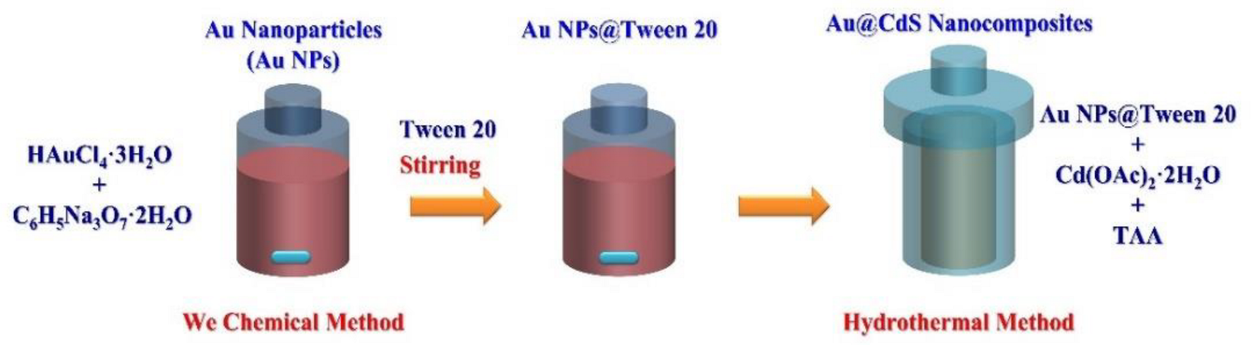
3.2. Synthesis of Au@CdS Nanocomposites

According to our previous work, a wet chemical process synthesized Au NPs [68]. Typically, an aqueous solution (20 mL) containing 1 mM hydrogen tetrachloroaurate (III) trihydride was heated to boiling, and 2 mL of 38.8 mM trisodium citrate was added under vigorous stirring for 10 min. The Au NPs solution was cooled to room temperature and stored to facilitate subsequent experiments. Tween 20 is used as a stabilizer to protect Au NPs against the CdS growth process. Tween 20-stabilized Au NPs were synthesized using 24 μL Tween 20 to 22 mL Au NPs under vigorous stirring for 1 h [69]. Au@CdS nanocomposites were synthesized on the Au NPs via a hydrothermal approach in a 50 mL aqueous solution containing different concentrations of CdS precursors (equimolar cadmium acetate dihydrate and TAA) at different reaction temperatures for 3 h.

3.3. Characterization

The microstructures, crystal structure, and photocatalytic performance of Au@CdS nanocomposites have been characterized by field-emission scanning electron microscopy (FESEM, Hitachi S-4800, Kyoto, Japan), field-emission transmission electron microscopy (FETEM, JEOL 2100 F, Kyoto, Japan), X-ray diffraction (XRD, Bruker D2 phaser, Billerica, MA, USA), X-ray photoelectron spectroscopy (XPS, ULVAC-PHI PHI 5000 VersaProbe, Kanagawa, Japan), UV-Vis spectroscopy (Hitachi U-2900, Kyoto, Japan), and photoluminescence (PL) spectroscopy (RAMaker, Protrustech, Tainan, Taiwan, He-Cd laser (λ = 532 nm)).

3.4. Photocatalytic Hydrogen Production Measurement

A multi-channel photocatalytic reaction system (5 W blue LED light, λmax = 420 nm, PCX50 B Discover, Perfect Light technology) was used to perform the photocatalytic hydrogen production on the as-prepared photocatalysts under the magnetic stirring. In a typical experiment, 50 mg of as-prepared photocatalysts was put into a mixed solution with the different sacrificial reagents (such as methanol, methanoic acid, sodium sulfide, and sodium sulfate) and 50 mL of water bases (such as de-ionized water (≥18.3 MΩ·cm) and tap water). After degassing pretreatment, the experimental device was carried out for 30 min to remove air. Then, the hydrogen amounts were evaluated using gas chromatography (GC, Shimadzu GC-2014) with a thermal conductivity detector (TCD).

4. Conclusions

Au@CdS nanocomposites have been successfully synthesized by the combination of feasible wet chemical and hydrothermal approaches at lower reaction temperatures. The CdS precursor concentrations and reaction temperatures were essential for controlling the efficiency of photocatalytic water splitting. The optimal Au@CdS nanocomposites at 5 mM CdS precursors and 100 °C achieve 1.041 (de-ionized water) and 1.204 mmolh−1 g−1 (tap water) under blue-LED light excitation, up to 175.3 and 512.3 times over CdS NPs, respectively. These reasons are mainly ascorbic to the introduction of Au NPs and the construction of a rationally coupled reaction system, which enables the synergistic utilization of photogenerated carriers and the increased absorption of visible light by the surface plasmon resonance effect. Therefore, the Au@CdS nanocomposites can be applied as efficient visible-light-induced photocatalysts for tap water splitting.

Author Contributions

Formal analysis, investigation, and data curation, Y.-R.L. and Y.-C.C. (Yung-Chang Chiao); funding acquisition, methodology, project administration, resources, software, supervision, validation, writing—original draft, and writing—review and editing, Y.-C.C. (Yu-Cheng Chang); funding acquisition, resources, F.-H.K. All authors have read and agreed to the published version of the manuscript.

Funding

This research was funded by the Ministry of Science and Technology of Taiwan (MOST 109–2221-E-035–041-MY3).

Institutional Review Board Statement

Not applicable.

Informed Consent Statement

Not applicable.

Data Availability Statement

Where no new data were created, or where data is unavailable due to privacy or ethical restrictions.

Acknowledgments

The Ministry of Science and Technology of Taiwan supported this study financially. The authors appreciate the Precision Instrument Support Center of Feng Chia University for providing the fabrication and measurement facilities.

Conflicts of Interest

The authors declare no conflict of interest.

References

  1. Zhang, G.; Ma, Y.; Liu, F.; Tong, Z.; Sha, J.; Zhao, W.; Liu, M.; Zheng, Y. Seeded Growth of Au@Cu(x)O Core-Shell Mesoporous Nanospheres and Their Photocatalytic Properties. Front. Chem. 2021, 9, 671220. [Google Scholar] [CrossRef] [PubMed]
  2. Serrà, A.; Gómez, E.; Michler, J.; Philippe, L. Facile cost-effective fabrication of Cu@Cu2O@CuO–microalgae photocatalyst with enhanced visible light degradation of tetracycline. Chem. Eng. J. 2021, 413, 127477. [Google Scholar] [CrossRef]
  3. Chang, Y.-C.; Guo, J.-Y. Double-sided plasmonic silver nanoparticles decorated copper oxide/zinc oxide heterostructured nanomaces with improving photocatalytic performance. J. Photochem. Photobiol. A Chem. 2019, 378, 184–191. [Google Scholar] [CrossRef]
  4. Chang, Y.-C.; Guo, J.-Y. ZnO/Pt core-shell nanorods on the cotton threads with high enhanced photocatalytic properties. Mater. Chem. Phys. 2016, 180, 9–13. [Google Scholar] [CrossRef]
  5. Vemuri, S.K.; Khanna, S.; Utsav; Paneliya, S.; Takhar, V.; Banerjee, R.; Mukhopadhyay, I. Fabrication of silver nanodome embedded zinc oxide nanorods for enhanced Raman spectroscopy. Colloids Surf. A Physicochem. Eng. Asp. 2022, 639, 128336. [Google Scholar] [CrossRef]
  6. Gao, W.; Liu, Q.; Zhang, S.; Yang, Y.; Zhang, X.; Zhao, H.; Qin, W.; Zhou, W.; Wang, X.; Liu, H.; et al. Electromagnetic induction derived micro-electric potential in metal-semiconductor core-shell hybrid nanostructure enhancing charge separation for high performance photocatalysis. Nano Energy 2020, 71, 104624. [Google Scholar] [CrossRef]
  7. Jadhav, J.; Biswas, S. Hybrid ZnO:Ag core-shell nanoparticles for wastewater treatment: Growth mechanism and plasmonically enhanced photocatalytic activity. Appl. Surf. Sci. 2018, 456, 49–58. [Google Scholar] [CrossRef]
  8. Li, Y.; Zhao, J.; You, W.; Cheng, D.; Ni, W. Gold nanorod@iron oxide core–shell heterostructures: Synthesis, characterization, and photocatalytic performance. Nanoscale 2017, 9, 3925–3933. [Google Scholar] [CrossRef]
  9. Zhang, K.; Xu, N.; Jia, M.; Li, R.; Huang, M. In situ detection of hot-electron-induced photocatalytic reduction using Au@Ag, Au@Ag2S, and Au@SiO2 core-shell nanoparticles. J. Appl. Phys. 2019, 125, 183101. [Google Scholar] [CrossRef]
  10. Al-Hajji, L.A.; Ismail, A.A.; Bumajdad, A.; Alsaidi, M.; Ahmed, S.A.; Almutawa, F.; Al-Hazza, A. Construction of Au/TiO2 Heterojunction with high photocatalytic performances under UVA illumination. Ceram. Int. 2020, 46, 20155–20162. [Google Scholar] [CrossRef]
  11. Liu, J.; Feng, J.; Gui, J.; Chen, T.; Xu, M.; Wang, H.; Dong, H.; Chen, H.; Li, X.; Wang, L.; et al. Metal@semiconductor core-shell nanocrystals with atomically organized interfaces for efficient hot electron-mediated photocatalysis. Nano Energy 2018, 48, 44–52. [Google Scholar] [CrossRef]
  12. Seong, S.; Park, I.-S.; Jung, Y.C.; Lee, T.; Kim, S.Y.; Park, J.S.; Ko, J.-H.; Ahn, J. Synthesis of Ag-ZnO core-shell nanoparticles with enhanced photocatalytic activity through atomic layer deposition. Mater. Des. 2019, 177, 107831. [Google Scholar] [CrossRef]
  13. Kadam, A.N.; Bhopate, D.P.; Kondalkar, V.V.; Majhi, S.M.; Bathula, C.D.; Tran, A.-V.; Lee, S.-W. Facile synthesis of Ag-ZnO core–shell nanostructures with enhanced photocatalytic activity. J. Indust. Eng. Chem. 2018, 61, 78–86. [Google Scholar] [CrossRef]
  14. Naz, G.; Shamsuddin, M.; Butt, F.K.; Bajwa, S.Z.; Khan, W.S.; Irfan, M.; Irfan, M. Au/Cu2O core/shell nanostructures with efficient photoresponses. Chin. J. Phys. 2019, 59, 307–316. [Google Scholar] [CrossRef]
  15. Fu, X.; Li, G.G.; Villarreal, E.; Wang, H. Hot carriers in action: Multimodal photocatalysis on Au@SnO2 core–shell nanoparticles. Nanoscale 2019, 11, 7324–7334. [Google Scholar] [CrossRef] [PubMed]
  16. Yang, L.; Guo, S.; Li, X. Au nanoparticles@MoS2 core-shell structures with moderate MoS2 coverage for efficient photocatalytic water splitting. J. Alloys Compd. 2017, 706, 82–88. [Google Scholar] [CrossRef]
  17. Sun, S.; An, Q.; Watanabe, M.; Cheng, J.; Ho Kim, H.; Akbay, T.; Takagaki, A.; Ishihara, T. Highly correlation of CO2 reduction selectivity and surface electron Accumulation: A case study of Au-MoS2 and Ag-MoS2 catalyst. Appl. Catal. B 2020, 271, 118931. [Google Scholar] [CrossRef]
  18. Lv, Y.; Duan, S.; Zhu, Y.; Guo, H.; Wang, R. Interface control and catalytic performances of Au-NiSx heterostructures. Chem. Eng. J. 2020, 382, 122794. [Google Scholar] [CrossRef]
  19. Truppi, A.; Petronella, F.; Placido, T.; Margiotta, V.; Lasorella, G.; Giotta, L.; Giannini, C.; Sibillano, T.; Murgolo, S.; Mascolo, G.; et al. Gram-scale synthesis of UV–vis light active plasmonic photocatalytic nanocomposite based on TiO2/Au nanorods for degradation of pollutants in water. Appl. Catal. B 2019, 243, 604–613. [Google Scholar] [CrossRef]
  20. Ghasemi, S.; Hashemian, S.J.; Alamolhoda, A.A.; Gocheva, I.; Rahman Setayesh, S. Plasmon enhanced photocatalytic activity of Au@TiO2-graphene nanocomposite under visible light for degradation of pollutants. Mater. Res. Bull. 2017, 87, 40–47. [Google Scholar] [CrossRef]
  21. Wang, J.; Zhao, H.; Liu, X.; Li, X.; Xu, P.; Han, X. Formation of Ag nanoparticles on water-soluble anatase TiO2 clusters and the activation of photocatalysis. Catal. Commun. 2009, 10, 1052–1056. [Google Scholar] [CrossRef]
  22. Choi, J.-Y.; Hoon Sung, Y.; Choi, H.-J.; Doo Kim, Y.; Huh, D.; Lee, H. Fabrication of Au nanoparticle-decorated TiO2 nanotube arrays for stable photoelectrochemical water splitting by two-step anodization. Ceram. Int. 2017, 43, 14063–14067. [Google Scholar] [CrossRef]
  23. Wang, L.; Chong, J.; Fu, Y.; Li, R.; Liu, J.; Huang, M. A novel strategy for the design of Au@CdS yolk-shell nanostructures and their photocatalytic properties. J. Alloys Compd. 2020, 834, 155051. [Google Scholar] [CrossRef]
  24. Yang, J.-L.; Xu, J.; Ren, H.; Sun, L.; Xu, Q.-C.; Zhang, H.; Li, J.-F.; Tian, Z.-Q. In situ SERS study of surface plasmon resonance enhanced photocatalytic reactions using bifunctional Au@CdS core–shell nanocomposites. Nanoscale 2017, 9, 6254–6258. [Google Scholar] [CrossRef]
  25. Yu, G.; Wang, X.; Cao, J.; Wu, S.; Yan, W.; Liu, G. Plasmonic Au nanoparticles embedding enhances the activity and stability of CdS for photocatalytic hydrogen evolution. Chem. Commun. 2016, 52, 2394–2397. [Google Scholar] [CrossRef] [PubMed]
  26. Wang, Y.; Zhang, Y.; Jiang, Z.; Jiang, G.; Zhao, Z.; Wu, Q.; Liu, Y.; Xu, Q.; Duan, A.; Xu, C. Controlled fabrication and enhanced visible-light photocatalytic hydrogen production of Au@CdS/MIL-101 heterostructure. Appl. Catal. B 2016, 185, 307–314. [Google Scholar] [CrossRef]
  27. Liu, S.; Ma, Y.; Chi, D.; Sun, Y.; Chen, Q.; Zhang, J.; He, Z.; He, L.; Zhang, K.; Liu, B. Hollow heterostructure CoS/CdS photocatalysts with enhanced charge transfer for photocatalytic hydrogen production from seawater. Int. J. Hydrogen Energy 2022, 47, 9220–9229. [Google Scholar] [CrossRef]
  28. Zhao, H.; Fu, H.; Yang, X.; Xiong, S.; Han, D.; An, X. MoS2/CdS rod-like nanocomposites as high-performance visible light photocatalyst for water splitting photocatalytic hydrogen production. Int. J. Hydrogen Energy 2022, 47, 8247–8260. [Google Scholar] [CrossRef]
  29. Wang, L.; Liu, Z.; Han, J.; Li, R.; Huang, M. Stepwise Synthesis of Au@CdS-CdS Nanoflowers and Their Enhanced Photocatalytic Properties. Nanoscale Res. Lett. 2019, 14, 148. [Google Scholar] [CrossRef]
  30. Zhang, S.; Chen, G.; Zhu, Z.; Wang, Y.; Wang, L.; Meng, S.; Zheng, X.; Fu, X.; Zhang, F.; Huang, W.; et al. Coordinating ultra-low content Au modified CdS with coupling selective oxidation and reduction system for improved photoexcited charge utilization. J. Catal. 2021, 402, 72–82. [Google Scholar] [CrossRef]
  31. Wang, L.; Li, R.; Huang, M. Synthesis and adsorption properties of CdS-Au hybrid nanorings. Mater. Lett. 2021, 304, 130722. [Google Scholar] [CrossRef]
  32. Majeed, I.; Nadeem, M.A.; Al-Oufi, M.; Nadeem, M.A.; Waterhouse, G.I.N.; Badshah, A.; Metson, J.B.; Idriss, H. On the role of metal particle size and surface coverage for photo-catalytic hydrogen production: A case study of the Au/CdS system. Appl. Catal. B 2016, 182, 266–276. [Google Scholar] [CrossRef]
  33. Zheng, Z.; Wang, T.; Han, F.; Yang, Q.; Li, B. Synthesis of Ni modified Au@CdS core–shell nanostructures for enhancing photocatalytic coproduction of hydrogen and benzaldehyde under visible light. J. Colloid Interface Sci. 2022, 606, 47–56. [Google Scholar] [CrossRef] [PubMed]
  34. Tada, H. Overall water splitting and hydrogen peroxide synthesis by gold nanoparticle-based plasmonic photocatalysts. Nanoscale Adv. 2019, 1, 4238–4245. [Google Scholar] [CrossRef] [Green Version]
  35. Zhang, M.; Gu, P.; Yan, S.; Pan, S.; Dong, L.; Zhang, G. A novel nanomaterial and its new application for efficient radioactive strontium removal from tap water: KZTS-NS metal sulfide adsorbent versus CTA-F-MF process. Chem. Eng. J. 2020, 391, 123486. [Google Scholar] [CrossRef]
  36. Caglar, Y.; Gorgun, K.; Aksoy, S. Effect of deposition parameters on the structural properties of ZnO nanopowders prepared by microwave-assisted hydrothermal synthesis. Spectrochim. Acta A Mol. Biomol. Spectrosc. 2015, 138, 617–622. [Google Scholar] [CrossRef]
  37. Romcevic, N.; Hadzic, B.; Romcevic, M.; Paunovic, N.; Sibera, D.; Narkiewicz, U.; Kuryliszyn-Kudelska, I.; Ristic-Djurovic, J.L.; Dobrowolski, W.D. Structural and optical properties of ZnO–Al2O3 nanopowders prepared by chemical methods. J. Lumin. 2020, 224, 117273. [Google Scholar] [CrossRef]
  38. Yang, F.; Huang, J.; Zou, T.; Tang, K.; Zhang, Z.; Ma, Y.; Gou, S.; Shen, Y.; Wang, L.; Lu, Y. The influence of surface processing on properties of CdZnTe films prepared by close-spaced sublimation. Surf. Coat. Technol. 2019, 357, 575–579. [Google Scholar] [CrossRef]
  39. Zheng, G.; Mourdikoudis, S.; Zhang, Z. Plasmonic Metallic Heteromeric Nanostructures. Small 2020, 16, 2002588. [Google Scholar] [CrossRef]
  40. Ghosh Chaudhuri, R.; Paria, S. Core/Shell Nanoparticles: Classes, Properties, Synthesis Mechanisms, Characterization, and Applications. Chem. Rev. 2012, 112, 2373–2433. [Google Scholar] [CrossRef]
  41. Piella, J.; Gónzalez-Febles, A.; Patarroyo, J.; Arbiol, J.; Bastús, N.G.; Puntes, V. Seeded-Growth Aqueous Synthesis of Colloidal-Stable Citrate-Stabilized Au/CeO2 Hybrid Nanocrystals: Heterodimers, Core@Shell, and Clover- and Star-Like Structures. Chem. Mater. 2019, 31, 7922–7932. [Google Scholar] [CrossRef] [Green Version]
  42. Tahir, M.; Siraj, M.; Tahir, B.; Umer, M.; Alias, H.; Othman, N. Au-NPs embedded Z–scheme WO3/TiO2 nanocomposite for plasmon-assisted photocatalytic glycerol-water reforming towards enhanced H2 evolution. Appl. Surf. Sci. 2020, 503, 144344. [Google Scholar] [CrossRef]
  43. Yu, J.; Song, N.; Zhang, Y.-K.; Zhong, S.-X.; Wang, A.-J.; Chen, J. Green preparation of carbon dots by Jinhua bergamot for sensitive and selective fluorescent detection of Hg2+ and Fe3+. Sens. Actuators B Chem. 2015, 214, 29–35. [Google Scholar] [CrossRef]
  44. Edison, T.N.J.I.; Atchudan, R.; Sethuraman, M.G.; Shim, J.-J.; Lee, Y.R. Microwave assisted green synthesis of fluorescent N-doped carbon dots: Cytotoxicity and bio-imaging applications. J. Photochem. Photobiol. B Biol. 2016, 161, 154–161. [Google Scholar] [CrossRef] [PubMed]
  45. Yan, Z.; Du, L.; Lee Phillips, D. Multilayer core–shell MoS2/CdS nanorods with very high photocatalytic activity for hydrogen production under visible-light excitation and investigation of the photocatalytic mechanism by femtosecond transient absorption spectroscopy. RSC Adv. 2017, 7, 55993–55999. [Google Scholar] [CrossRef] [Green Version]
  46. Yan, Y.; Zhou, Z.; Li, W.; Zhu, Y.; Cheng, Y.; Zhao, F.; Zhou, J. Facile ion-exchange synthesis of urchin-shaped CdS/Bi2S3 heterostructures with enhanced photostability and visible light photocatalytic activity. RSC Adv. 2014, 4, 38558–38567. [Google Scholar] [CrossRef]
  47. Liu, Y.; Dai, F.; Zhao, R.; Huai, X.; Han, J.; Wang, L. Aqueous synthesis of core/shell/shell CdSe/CdS/ZnS quantum dots for photocatalytic hydrogen generation. J. Mater. Sci. 2019, 54, 8571–8580. [Google Scholar] [CrossRef]
  48. Hoseini, A.-A.; Farhadi, S.; Zabardasti, A.; Siadatnasab, F. A novel n-type CdS nanorods/p-type LaFeO3 heterojunction nanocomposite with enhanced visible-light photocatalytic performance. RSC Adv. 2019, 9, 24489–24504. [Google Scholar] [CrossRef] [Green Version]
  49. Chou, C.-M.; Chang, T.-T.; Chen, C.-Y.; Chang, Y.-C. Constructing Er-Doped ZnO/CuS/Au Core-Shell Nanowires with Enhanced Photocatalytic and SERS Properties. Catalysts 2021, 11, 1347. [Google Scholar] [CrossRef]
  50. Chang, Y.-C.; Tasi, C.-L.; Ko, F.-H. Construction of ZnIn2S4/ZnO heterostructures with enhanced photocatalytic decomposition and hydrogen evolution under blue LED irradiation. Int. J. Hydrogen Energy 2021, 46, 10281–10292. [Google Scholar] [CrossRef]
  51. Naya, S.-I.; Kume, T.; Akashi, R.; Fujishima, M.; Tada, H. Red-Light-Driven Water Splitting by Au(Core)–CdS(Shell) Half-Cut Nanoegg with Heteroepitaxial Junction. J. Am. Chem. Soc. 2018, 140, 1251–1254. [Google Scholar] [CrossRef] [PubMed]
  52. Wu, M.; Chen, W.-J.; Shen, Y.-H.; Huang, F.-Z.; Li, C.-H.; Li, S.-K. In Situ Growth of Matchlike ZnO/Au Plasmonic Heterostructure for Enhanced Photoelectrochemical Water Splitting. ACS Appl. Mater. Interfaces 2014, 6, 15052–15060. [Google Scholar] [CrossRef] [PubMed]
  53. Corredor, J.; Rivero, M.J.; Rangel, C.M.; Gloaguen, F.; Ortiz, I. Comprehensive review and future perspectives on the photocatalytic hydrogen production. J. Chem. Technol. Biotechnol. 2019, 94, 3049–3063. [Google Scholar] [CrossRef] [Green Version]
  54. Kumaravel, V.; Imam, M.D.; Badreldin, A.; Chava, R.K.; Do, J.Y.; Kang, M.; Abdel-Wahab, A. Photocatalytic Hydrogen Production: Role of Sacrificial Reagents on the Activity of Oxide, Carbon, and Sulfide Catalysts. Catalysts 2019, 9, 276. [Google Scholar] [CrossRef] [Green Version]
  55. Lakshmana Reddy, N.; Rao, V.N.; Kumari, M.M.; Ravi, P.; Sathish, M.; Shankar, M.V. Effective shuttling of photoexcitons on CdS/NiO core/shell photocatalysts for enhanced photocatalytic hydrogen production. Mater. Res. Bull. 2018, 101, 223–231. [Google Scholar] [CrossRef]
  56. Rao, V.N.; Reddy, N.L.; Kumari, M.M.; Ravi, P.; Sathish, M.; Neppolian, B.; Shankar, M.V. Synthesis of titania wrapped cadmium sulfide nanorods for photocatalytic hydrogen generation. Mater. Res. Bull. 2018, 103, 122–132. [Google Scholar] [CrossRef]
  57. Linkous, C.A.; Huang, C.; Fowler, J.R. UV photochemical oxidation of aqueous sodium sulfide to produce hydrogen and sulfur. J. Photochem. Photobiol. A Chem. 2004, 168, 153–160. [Google Scholar] [CrossRef]
  58. Schwarze, M.; Stellmach, D.; Schröder, M.; Kailasam, K.; Reske, R.; Thomas, A.; Schomäcker, R. Quantification of photocatalytic hydrogen evolution. Phys. Chem. Chem. Phys. 2013, 15, 3466–3472. [Google Scholar] [CrossRef] [Green Version]
  59. Zhao, J.; Yang, Y.; Dong, X.; Ma, Q.; Yu, W.; Wang, J.; Liu, G. Electrospinning construction of Bi2WO6/RGO composite nanofibers with significantly enhanced photocatalytic water splitting activity. RSC Adv. 2016, 6, 64741–64748. [Google Scholar] [CrossRef]
  60. Chang, Y.-C.; Lin, Y.-W.; Lu, M.-Y. Construction of MoS2/ZnO heterostructures as highly efficient photocatalysts for enhanced visible-light decomposition of methylene blue and hydrogen evolution. Mater. Chem. Phys. 2021, 266, 124560. [Google Scholar] [CrossRef]
  61. Li, Y.; Liu, T.; Feng, S.; Yang, W.; Zhu, Y.; Zhao, Y.; Liu, Z.; Yang, H.; Fu, W. Au/CdS Core-Shell Sensitized Actinomorphic Flower-Like ZnO Nanorods for Enhanced Photocatalytic Water Splitting Performance. Nanomaterials 2021, 11, 233. [Google Scholar] [CrossRef] [PubMed]
  62. Kahane, S.V.; Sasikala, R.; Vishwanadh, B.; Sudarsan, V.; Mahamuni, S. CdO–CdS nanocomposites with enhanced photocatalytic activity for hydrogen generation from water. Int. J. Hydrogen Energy 2013, 38, 15012–15018. [Google Scholar] [CrossRef]
  63. Li, G.; Gao, L.; Li, L.; Guo, L. An electrochromic and self-healing multi-functional supercapacitor based on PANI/nw-WO2.7/Au NPs electrode and hydrogel electrolyte. J. Alloys Compd. 2019, 786, 40–49. [Google Scholar] [CrossRef]
  64. Ma, D.; Shi, J.-W.; Sun, D.; Zou, Y.; Cheng, L.; He, C.; Wang, Z.; Niu, C. Au Nanoparticle and CdS Quantum Dot Codecoration of In2O3 Nanosheets for Improved H2 Evolution Resulting from Efficient Light Harvesting and Charge Transfer. ACS Sustain. Chem. Eng. 2019, 7, 547–557. [Google Scholar] [CrossRef]
  65. Preethi, V.; Kanmani, S. Photocatalytic hydrogen production using Fe2O3-based core shell nano particles with ZnS and CdS. Int. J. Hydrogen Energy 2014, 39, 1613–1622. [Google Scholar] [CrossRef]
  66. Strataki, N.; Antoniadou, M.; Dracopoulos, V.; Lianos, P. Visible-light photocatalytic hydrogen production from ethanol–water mixtures using a Pt–CdS–TiO2 photocatalyst. Catal. Today 2010, 151, 53–57. [Google Scholar] [CrossRef]
  67. Bao, N.; Shen, L.; Takata, T.; Domen, K. Self-Templated Synthesis of Nanoporous CdS Nanostructures for Highly Efficient Photocatalytic Hydrogen Production under Visible Light. Chem. Mater. 2008, 20, 110–117. [Google Scholar] [CrossRef]
  68. Ko, F.-H.; Tai, M.R.; Liu, F.-K.; Chang, Y.-C. Au–Ag core–shell nanoparticles with controllable shell thicknesses for the detection of adenosine by surface enhanced Raman scattering. Sens. Actuators B Chem. 2015, 211, 283–289. [Google Scholar] [CrossRef]
  69. Shih, Y.-C.; Ke, C.-Y.; Yu, C.-J.; Lu, C.-Y.; Tseng, W.-L. Combined Tween 20-Stabilized Gold Nanoparticles and Reduced Graphite Oxide–Fe3O4 Nanoparticle Composites for Rapid and Efficient Removal of Mercury Species from a Complex Matrix. ACS Appl. Mater. Interfaces 2014, 6, 17437–17445. [Google Scholar] [CrossRef]
Figure 1. Schematic diagram of the fabrication processes for the growth of Au@nanocomposites.
Catalysts 13 00033 g001
Figure 2. (a) XRD spectra and (b) FESEM images of Au@CdS nanocomposites were grown at different CdS precursor concentrations. The CdS precursor concentrations were 1, 2.5, 5, and 10 mM, respectively.
Catalysts 13 00033 g002
Figure 3. (a) XRD spectra and (b) FESEM images of Au@CdS nanocomposites were grown at different reaction temperatures. The reaction temperatures were 80, 100, 120, and 140 °C.
Catalysts 13 00033 g003
Figure 4. (a) TEM and (b) HRTEM images of Au NPs. (c) FETEM, (d) SAED, and (e,f) HRTEM images of Au@CdS nanocomposites.
Catalysts 13 00033 g004
Figure 5. The XPS spectra of Au@CdS nanocomposites: (a) survey spectrum, (b) Au 4 f, (c) O 1 s, (d) Cd 3 d, and (e) S 2 p.
Catalysts 13 00033 g005
Figure 6. The (a) UV-vis absorption and (b) PL spectra of CdS NPs and Au@CdS nanocomposites.
Catalysts 13 00033 g006
Figure 7. The average HER of Au@CdS nanocomposites grown at different (a) CdS precursor concentrations and (b) reaction temperatures. (c) The average HER of Au@CdS nanocomposites at different sacrificial reagents. (d) The average HER of CdS NPs and Au@CdS nanocomposites for de-ionized or tap water.
Catalysts 13 00033 g007
Figure 8. The schematic diagram illustrates the photocatalytic mechanism of Au@CdS nanocomposites.
Catalysts 13 00033 g008
Figure 9. The average HER of Au@CdS nanocomposites at different pH values of (a) de-ionized water and (b) tap water. (c) Four cycles of photocatalytic tap water splitting with Au@CdS nanocomposites as hydrogen-evolving photocatalysts.
Catalysts 13 00033 g009
Disclaimer/Publisher’s Note: The statements, opinions and data contained in all publications are solely those of the individual author(s) and contributor(s) and not of MDPI and/or the editor(s). MDPI and/or the editor(s) disclaim responsibility for any injury to people or property resulting from any ideas, methods, instructions or products referred to in the content.

