Article

Impacts of Dietary Selenium Nanoparticles from Spirulina platensis on Growth Performance, Physio-Biochemical Components and Alleviating Effect against Cadmium Toxicity in Pacific White Shrimp Litopenaeus vannamei

by
Radwa M. Said
1,
Safaa E. Nassar
1,*,
Bothaina A. Alaidaroos
2,
Samyah D. Jastaniah
2,
Hagar Sedeek Dighiesh
3,
El-Sayed Hemdan Eissa
4,*,
Ammar AL-Farga
5,
Zulhisyam Abdul Kari
6,7,
Guillermo Téllez-Isaías
8 and
Mai S. Attia
1
1
Zoology Department, Faculty of Science, Zagazig University, Zagazig 44519, Egypt
2
Department of Biological Science, Faculty of Sciences, King Abdulaziz University Kingdom of Saudi Arabia, Jeddah 22233, Saudi Arabia
3
Aquaculture Department, Faculty of Fish Resources, Suez University, Suez 43512, Egypt
4
Fish Research Centre, Arish University, El-Arish 45516, Egypt
5
Department of Biochemistry, Faculty of Science, University of Jeddah, Jeddah 21959, Saudi Arabia
6
Department of Agricultural Sciences, Faculty of Agro-Based Industry, Universiti Malaysia Kelantan, Jeli Campus, Jeli 17600, Kelantan, Malaysia
7
Advanced Livestock and Aquaculture Research Group, Faculty of Agro-Based Industry, Universiti Malaysia Kelantan, Jeli Campus, Jeli 17600, Kelantan, Malaysia
8
Department of Poultry Science, University of Arkansas, Fayetteville, AR 72701, USA
*
Authors to whom correspondence should be addressed.
Catalysts 2023, 13(11), 1389; https://doi.org/10.3390/catal13111389
Submission received: 4 September 2023 / Revised: 6 October 2023 / Accepted: 17 October 2023 / Published: 24 October 2023
(This article belongs to the Section Biocatalysis)

Abstract

:

Simple Summary

This study investigated the role of synthesized selenium nanoparticles from Spirulina platensis extract (SP-SeNPs) on Pacific whiteleg shrimps’ (Litopenaeus vannamei) growth performance, as well as the potential role in alleviating the negative impacts of cadmium (Cd) toxicity. Based on the detected growth performance parameters, digestive enzyme activities, and biochemical components of shrimps after 56 days of the experiment, SP-SeNPs at 0.5 mg/kg were recommended. Moreover, the antioxidative status and histological investigation of shrimp tissues after exposure to Cd for 10 days indicated that SP-SeNPs could mitigate the pathological alternations induced with Cd toxicity. The findings of this study highlight the utility of nanotechnology for the enhanced production of Pacific white shrimps which could be utilized as one of the main sources for the animal proteins needed for human consumption.

Abstract

Shrimp culture is quite important and popular across the world. This study aimed to evaluate the growth-promoting potential of synthesized selenium nanoparticles from Spirulina platensis extract (SP-SeNPs) as a food source for Pacific whiteleg shrimp (Litopenaeus vannamei). However, pollution is considered a significant element affecting shrimp health and development. The effectiveness of SP-SeNPs in alleviating the negative effects of cadmium toxicity was also evaluated. Firstly, the shrimps (about 120 individuals with 6.0 ± 0.12 g of initial weight) were divided randomly into four groups in triplicates (30 shrimps/ treatment). The control group (SP-SeNPs—0 mg/kg diet) and three treatments were fed dietary SP-SeNPs (0.250, 0.50, and 1.0 mg/kg diet) for 56 days. Growth performance, digestive enzymes activities (protease, amylase, and lipase), and other biochemical components (total protein, lipid, amino acids, and carbohydrate) were evaluated. After 56 days of growth, another 150 adult shrimps were used under laboratory conditions to determine median lethal concentration of cadmium (96 h LC50), and 30 individuals were treated with cadmium (1/2 of LC50, 0.2 mg L−1) for 10 days only. Tissue samples were collected for measuring catalase (CAT), total antioxidant capacity (TAC), superoxide dismutase (SOD), malondialdehyde (MDA) levels, cadmium bioaccumulation, and histopathological investigation. The results illustrated that the application of SP-SeNPs as feed additives at varying levels significantly improved growth performance (high weight gain, specific growth rate, and low feed conversion rates) relative to the control group. Furthermore, dietary SP-SeNPs enhanced digestive enzyme activities and the concentrations of biochemical components more than the control group. Upon concurrent exposure to cadmium, the antioxidative status was significantly enhanced, and histopathological alterations were mitigated. In conclusion, this study recommended supplementation of SP-SeNPs at 0.50 mg/kg diet to enhance optimal growth rate, digestive enzyme activities, levels of antioxidants in Litopenaeus vannamei, and mitigate the pathological alternations induced with Cd toxicity.

1. Introduction

The aquaculture sector has been identified as one of the main sources for the animal proteins needed for human consumption [1]. In the 1980s, shrimp farming experienced significant growth and is a billion dollar sector today [2]. In this regard, controlling diseases, adjusting culture temperatures and salinities [3], and improving shrimps’ immune performance by supplementing functional feed additives [4,5] are important factors for sustainable aquaculture.
Selenium (Se) is a highly important trace element needed to support the growth, antioxidant action, and metabolic function of fishes, and it considered as a principal supporter component of the glutathione peroxidase (GPx) enzymes that guard all cells membrane from oxidative damage [6]. Selenium is considered an essential micronutrient that should be supplied in aquafeed for optimal growth and physiological function [7]. Selenium deficiency can induce multiple tissue damage with dysregulation of immune and redox homeostasis under heat stress [8]. Using nanoparticles in fish feed formulations has recently gained considerable attention because it offers unique methods for increasing bioavailability, safety, and absorption in aquaculture [9]. The addition of Se nanoparticles to the diet improves the feed efficiency and growth performance of many aquatic species, including Clarias gariepinus [10] and Oreochromis niloticus [11]. Moreover, dietary selenium enhances immunity and resistance against oxidative stress in rainbow trout [12], Dicentrarchus labrax, and Penaeus vannamei [13].
Nanoparticles can be generated chemically, physically, or biologically. Chemical approaches are ecologically harmful as they cannot avoid using hazardous materials in the synthesis protocol [14]. Likewise, physical methods are expensive and more difficult [15]. Therefore, the manufacture of NPs by microorganisms, plant extracts, and metabolites of animals represent an alternative safe and eco-friendly method [16]. Algae have many active substances such as proteins, amines, phenolics, alkaloids, and pigments which help in the reduction and stability of metals; therefore, their biomasses or extracts have been used in generating metallic nanoparticles [17]. Titanium dioxide (TiO2) nanoparticles and selenium nanoparticles have all been synthesized using Spirulina spp. and its extract [18].
Cadmium (Cd) is an inorganic non-degradable metal, and it is concentrated through the food chain even if its concentration is sometimes low, and then transferred into the human body [19]. Cd accumulation can induce an increase in reactive oxygen species (ROS) production which causes a cascade of oxidative damage. Excessive ROS will also lead to changes in biological macromolecules, increase lipid peroxidation, and reduce antioxidant enzyme activity in fish. Moreover, it causes harm to the cardiovascular, immune, nervous, and reproductive systems [20].
However, aquaculture is vulnerable to various xenobiotics, such as heavy metals and pesticides. Thus, the production of fish that can resist infectious diseases and chemicals to attain higher growth performance is of considerable interest. Nutritional supplementation could be critical in minimizing the impacts of pollution and improving fish development and immunity [21].
In Egypt, in 2015, the Pacific whiteleg shrimp Litopenaeus vannamei (Boone, 1931) was introduced for aquaculture to lessen economic losses brought on by outbreaks of illnesses in a native shrimp species, penaeus japonicus and Penaeus semisulcatus [22]. Due to its rapid growth development and ecological tolerance and high survival in high densities as well as the high disease resistance [23,24], L. vannamei has been widely cultured. However, research on the effectiveness of Se-NP supplementation on L. vannamei growth performance and productivity was insufficient. Furthermore, the protective effects of selenium against xenobiotics are still unknown. Thus, this study was performed based on two directions: (1) To determine the higher inclusion level of Se-NPs recommended for optimal growth by evaluating the effects of different dietary levels of Se-NPs synthesized from Spirulina platensis extract on whiteleg shrimps. Growth performance and body composition of shrimps were measured. Furthermore, digestive enzyme activities and biochemical components were analyzed. (2) To investigate the potential role of Se-NPs in alleviating oxidative status and the negative impacts of Cd toxicity in adult L. vannamei, after determining the LC50 of Cd for the studied species.

2. Results

2.1. Growth Indices

The application of SP-SeNPs as feed additives in whiteleg shrimps significantly (p < 0.05) improved growth indices and the shrimps’ biomass in all nano-Se-treated groups compared with the control group (Table 1). T3 attained the best results.
The survival percentage was 85.57, 90.00, 92.23, and 95.33%, respectively, in all treatments. T2 and T3 achieved higher values for SGR 3.25 and 3.36% and higher values for the shrimps’ biomass in T2 and T3 (528.8 and 569 g), respectively, relative to the control group (422 g). The nano-Se application significantly (p < 0.05) improved feed utilization indices of shrimps in all nano-Se-treated groups relative to the control group (Table 1). T3 attained the best feed conversion ratio relative to the other treatments.

2.2. The Chemical Composition of the Entire Body

Various SeNPs concentrations significantly (p < 0.05) improved the entire body composition of shrimps, including protein and ash content and a small increase in moisture. Different nano-Se levels (p < 0.05) reduced the content of lipids relative to the control (Table 2).

2.3. Biochemical Indices and Digestive Enzymes Activities in Muscles and Digestive Tract of L. vannamei Fed with SP-SeNP Supplemented Diets for 56 Days

Levels of total protein, lipid, amino acids, and carbohydrate, as well as protease, lipase, and amylase activities increased in all nano-Se-treated groups with a better maximum performance in T2 groups (p < 0.05) relative to the control groups (Table 3).

2.4. Determination of Lethal and Sublethal Concentrations of Cadmium against L. vannamei after 56 Days of Growth

The 96 h LC50 level of cadmium for L. vannamei was 0.4 mg/L. There was no recorded death in the control group. The lowest mortality percentage of shrimps was observed at a level of 0.2 mg/L in 96 h and the highest mortality percentage was estimated at a level of 0.8 mg/L. However, sublethal concentrations (1/2 LC50) of cadmium for L. vannamei were 0.2 mg/L (Table 4).

2.5. Bioaccumulation of Cadmium and Selenium Residues in Hepatopancreases and Muscles of L. vannamei

Results in Table 5 illustrate that the concentrations of Cd in the muscles and hepatopancreases were significantly increased (p < 0.05) in the T4 group (control diet with Cd). However, they were decreased (p < 0.05) in the T5, T6, and T7 groups (0.250, 0.50, and 1.0 mg/kg SP-SeNP supplemented diet and 0.2 mg/L Cd) compared with the control groups, T1 to T3 groups (0.250, 0.50, and 1.0 mg/kg SP-SeNP supplemented diet and no Cd). The concentration of Se was increased (p < 0.05) after exposure to Se only in T1, T2, and T3 groups compared to the control group. The concentration of Se decreased after the exposure to Cd only in T4 group. However, the concentration of Se increased after concurrent exposure both to Cd and Se (p < 0.05), as in T5, T6, and T7 groups.

2.6. Oxidative and Antioxidant Activities in the Hepatopancreases and Muscles of L. vannamei Fed with SP-SeNP Supplemented Diets after Cadmium Exposure for 10 Days

The data in Table 6 show that the level of MDA significantly increased in the hepatopancreases and muscles of the T4 group (control diet with Cd). On the other hand, SP-SeNP supplementation in the diets decreased hepatopancreas and muscle MDA concentrations in T5, T6, and T7 groups. Moreover, hepatopancreas and muscle MDA levels were decreased in the T1, T2, and T3 groups with SP-SeNP supplemented diets and no Cd compared to the control group. The highest concentration of CAT, TAC, and SOD was measured in the T1, T2, and T3 groups both in hepatopancreas and muscle tissues. Hepatopancreas and muscle CAT, TAC, and SOD levels were higher in the T5, T6, and T7 groups (SP-SeNP supplemented diet and Cd) compared to the CAT, TAC, and SOD concentrations in the T4 group (control diet and Cd), which were less than the CAT, TAC, and SOD levels in the control group.

2.7. Histopathological Examination

The hepatopancreas consists of various tubules detached by connective tissues. Three different cell types and a central lumen may be found in each tubule: absorptive cells, fibrillar cells, and secretory cells in its wall (Figure 1a). Different histopathological lesions in shrimp hepatopancreases from the T4 group (control diet and Cd exposure), such as destruction and necrosis in the cell of tubules, were noticed (Figure 1b). The histological structures of hepatopancreases in the T5 group (0.25 mg/kg SP-SeNP supplemented diets and 0.2 mg/L Cd exposure) were improved. However, large numbers of vacuoles seemed to be in the tubular epithelial cells of the hepatopancreases (Figure 1c). In the T6 group (0.50 mg/kg SP-SeNP supplemented diets and 0.2 mg/L Cd exposure), the tubules of hepatopancreases were improved and showed normal histological structures without adverse pathological lesions (Figure 1d). The tubules of the hepatopancreases of the T7 group (1 mg/kg SP-SeNP supplemented diets and 0.2 mg/L Cd exposure) showed normal histological structure. However, some tubules were rounded off and began to slough off their basement membrane, and rupture of epithelial cells in some tubules was also seen (Figure 1e).

3. Discussion

The most recent development in the aquaculture sector is the promotion of fisheries through the management of the aquatic environment and feeding techniques. Selenium is an important micronutrient for aquatic species. Any form of selenium is essential for physiological, metabolic, and immunological activities. As the difference between safe and dangerous doses of metallic Se supplementation in the diet is narrow, the Se-nanoparticle has received a lot of attention as one of the important dietary supplements. Plant extracts and microorganisms may be used to apply the green synthesis of SeNPs as a green technique. In this work, we created SeNPs from an extract of Spirulina platensis and evaluated its effect on the growth performance of L. vannamei. For the first time, the potential role of Se-NPs in alleviating the negative impacts of Cd toxicity in adult L. vannamei was investigated.
The outcomes of this research reported that the addition of SP-SeNPs into the diet with concentrations of 0.250 (T1), 0.50 (T2), and 1.0 (T3) mg kg−1 daily for 56 days increased both feed utilization (weight gain, feed intake, ADG, SGR) and growth performance in L. vannamei relative to the control group (‘0’ mg kg−1 SP-SeNP supplemented diet). Furthermore, a higher level ((T3) 1 mg kg−1) positively affects the growth of shrimps, suggesting that SeNPs may extend their beneficial effects at high levels. Many studies have reported that selenium catalyzes the formation of triiodothyronine from thyroxin, and that it also modulates growth hormones, synthesis, and their release. Therefore, selenium clearly promotes growth performance in animals because it is the principal ingredient of 50-deiodinase [25]. Furthermore, it can boost antioxidant capacity and ameliorate stress resistance and immunocompetence by protecting cell membranes against reactive oxygen species [26]. Other investigations have also illustrated the positive impact of Se-NPs in several species: Nile tilapia [27] and European Seabass [28]. However, ref.[13] illustrated that the supplementation of Se-NPs at too low or too high levels causes disrupted metabolic and physiological processes that cause malnutrition-like symptoms. Therefore, it is crucial to use the right amounts of Se-NPs to get the greatest effects.
In this study, SP-SeNP supplementation in the diet of shrimps increased body composition, such as body protein, lipid, moisture, and ash contents, and they increased with increasing the dietary level of SP-SeNPs (only). Lipid content reduced with increasing selenium level; hence, it has hypolipidemic effects in this species of white shrimps. Also, ref.[29] revealed that selenium has hypolipidemic effects in rats nourished on a diet with high fat. This is perhaps associated with the ability of selenium to reduce the activity of lipogenic enzymes which have a function in the transmission of fatty acids into the matrix of the mitochondria to produce energy [30]. Furthermore, ref.[30] reported that high concentrations of selenium strikingly upregulated the transportation and oxidation of fatty acids.
Additionally, we found that SP-SeNP supplementation in the diet increases levels of protein, lipid, and amino acids in muscles and digestive enzymes in the digestive tract; this may be because of the ability of selenium to enhance the synthesis of digestive enzymes, as well as their ability to improve intestinal villi integrity—as mentioned by [31]. Likewise, ref.[25] found that SeNPs have an antibacterial effect that might promote the growth of beneficial bacteria in aquatic animals’ intestines that play an essential function in the excretion of digestive enzymes to digest nutrients. Furthermore, selenium performs as a pioneer for selenoproteins synthesized in the intestine, leading to an elevated activity of digestive enzymes, similarly to high feed utilization and growth performance.
The activity of aquaculture is menaced with several stressors that could alter both biological and physiological functions of aquatic animals. The contamination of heavy metals is a considerable problem. Cadmium, as one of the hazardous metals, can induce a series of oxidative stresses by raising the output of ROS [32]. These free radicals promote lipid peroxidation and lead to cell destruction [33]. Malondialdehyde is an indicator for oxidative stress [34], catalase has an efficient role in organizing hydrogen peroxide level to keep body through oxidative stress [35], and superoxide dismutase consists of metalloenzymes that are in charge of superoxide oxygen dismutation and modulating the immune function [36].
Several studies found that supplementing the diet with SeNPs enhanced antioxidant enzyme activities in different crustacean species [28]. Zoidis et al. [37] declared that selenium, when metabolized and converted into selenocysteine in order to be integrated into selenoproteins, helps in GPx synthesis. GPx has a critical role in promoting innate immunity and resistance to pathogenic infection [26]. Therefore, SeNPs are recommended as a strong antioxidative factor in aquaculture. Furthermore, ref.[38] found that supplementing ideal levels of selenium into the diet improved the activity of antioxidant enzymes. In accordance with these studies, dietary supplementation (T1, T2, and T3) of SP-SeNPs in this study indicated an improvement of antioxidant capacity in both muscles and hepatopancreases of L. vannamei when compared to the control (‘0’ mg/kg SP-SeNP supplemented diet), as well as reduction in MDA with elevating CAT, SOD, TAC levels.
Selenium mitigates the heavy metals’ toxicity by forming selenium–metal protein and selenide–metal complexes with later rearrangement [39]. In this context, we estimated the impact of SP-SeNP supplementation in the diets on oxidative and antioxidant activities in hepatopancreases and muscles of adult L. vannamei after cadmium exposure for 10 days. It was found that MDA remarkably improved in the muscles and hepatopancreases of the T4 group (control diet with Cd exposure). On the other hand, supplementation of SeNPs decreased MDA levels in the studied tissues in the T5, T6, and T7 groups (SP-SeNP supplemented diet and Cd exposure).
However, hepatopancreas and muscle CAT, TAC, and SOD levels were higher in the T5, T6, and T7 groups in contrast with the T4 group (Cd exposure with control diet). These results revealed that the dietary addition of SP-SeNPs with concurrent exposure to Cd enhanced the antioxidative status of the adult shrimps. This was consistent with other reports by [40], who estimated that the Nile tilapia’s resistance to pathogenic infection increased when they were fed with organic Se in their diets and were exposed to cadmium. They revealed that selenium minimizes the harmful effects of cadmium in water, consequently ameliorating survival, growth performance, and nutrient use competence. Also, ref.[41] estimated the effects of supplementation of selenium in fish fed with copper for 8 weeks, which showed that selenium reduced copper stress and enhanced the immune system of the fish.
The detection of accumulated Cd and Se in shrimp tissues (muscles and hepatopancreases), as well as the histopathological examination, are methods for testing trace element toxicity throughout an animal’s body [42] and to ensure the safety of the food for human consumption. As reflected in this study, it was found that the exposure to Cd influences the antioxidative status of shrimps. This may be attributed to the highest levels of Cd detected in different tissues after the exposure treatment. The highest concentration of Cd was found in the T4 group (controlled diet with Cd exposure), whereas the Se-NP supplemented groups (T5, T6, and T7) showed a significant decrease in the bioaccumulation of Cd. This could be due to the capability of Se-NPs to enhance the removal of contaminants within the body [43]; various researches have demonstrated that Se can interact with metals to form Se–metal protein and selenide–metal complexes that are secreted in bile [44]. In this study, the low concentrations of Se found in the T5, T6, and T7 groups (SP-SeNP supplemented diet and Cd exposure) compared to the T1, T2, and T3 groups (SP-SeNP supplemented diet) could prove the role of selenium in reducing the bioaccumulation of Cd.
Histological abnormalities caused by Cd toxicity in this work could be attributed to reactive oxygen species generation caused by damaged hepatopancreases. Nevertheless, dietary SP-SeNPs reduced the degenerative effects of Cd poisoning. At 0.5 mg/kg, dietary SP-SeNPs maintained the normal structure of the hepatopancreases in Pacific white shrimps for 56 days. According to [32,45], selenium reduces Cd toxicity mostly by sequestering Cd into physiologically inert compounds or by activating Se-dependent antioxidant enzymes. This was compatible with the current findings on antioxidant defense systems.

4. Materials and Methods

4.1. Algae Extraction

Dry powder of blue green algae, Spirulina platensis, was achieved from the Algal Biotechnology Unit of the National Research Centre, Cairo, Egypt. The S. platensis (50 g dry powder) was extracted with (500 mL) ethanol as a solvent using a Soxhlet extractor at 50–60 °C. The mixture was refluxed for 24 h to reach saturation and the respective extracts were dried using a vacuum rotary evaporator at 45 °C. Until further experiments, the dried extracts were kept at 4 °C.

4.2. Phycosynthesis of SeNPs from Spirulina platensis (SP-SeNPs)

A total of 2.0 mL of S. platensis extract solution was placed into 10.0 mL of 10.0 mM sodium selenite, drop by drop, while being stirred magnetically for 72 h in dark conditions at 27 ± 2° until the sodium selenite solution altered to an orange-red color, demonstrating the formation of SeNPs from S. platensis [46]. Firstly, the solution of sodium selenite was colorless. After 72 h, the sodium selenite converted into an orange-red color with adding extract of S. platensis. This finding was in agreement with [18]. The appearance of an orange-red solution could indicate that sodium selenite is being reduced into elemental selenium, and this may be caused by the activation of the surface plasmon resonance.
The action of phenols, alcohols, phycocyanins, amines, and esters in spirulina sp. can act as stabilizing and lowering substances in the reduction of sodium selenite into SP-SeNPs [18].

4.3. Description of Phycosynthesis of SeNPs

4.3.1. UV–Visible Spectroscopy

By noticing the color change, the reduction of sodium selenite using S. platensis extract was confirmed. After the production of an orange-red color, the absorbance of SeNPs was measured between the wavelengths of 200 and 400 nm using a T80 UV/VIS Spectrophotometer. One absorption peak at 265 nm was seen in the extract (Figure 2), which may indicate the existence of SP-SeNPs. A single absorption peak of SP-SeNPs at 265 nm was confirmed by [18].

4.3.2. Fourier Transform Infrared Spectroscopic Analysis (FTIR)

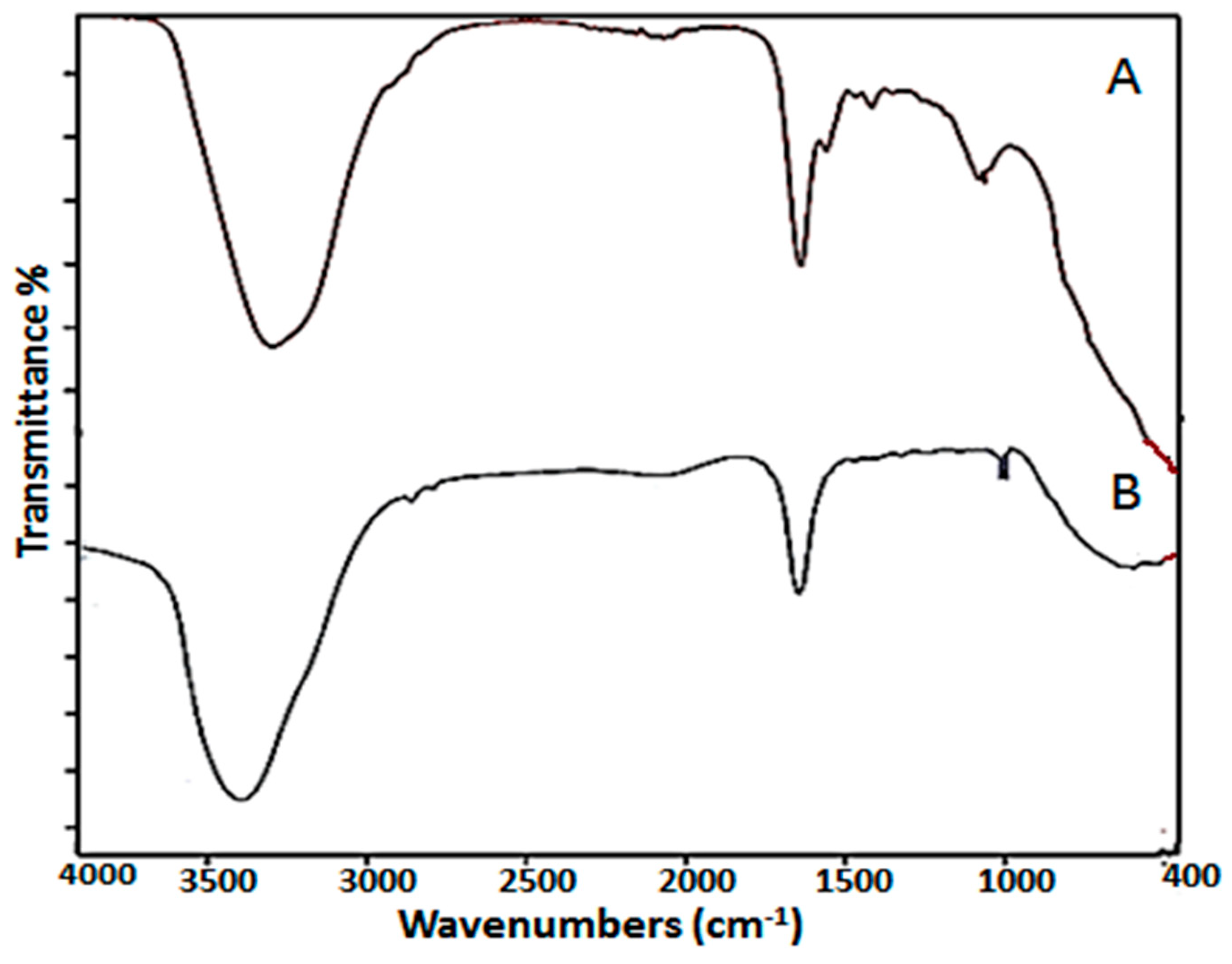
The existence of numerous stabilizing and reducing functional groups of metabolites in S. platensis was confirmed using FTIR. Spectra were obtained using a spectrophotometer (4100 Jasco, Japan) at 400–4000 cm−1 wavenumbers versus a potassium bromide background after the SP-SeNPs solution was left to dry and after being crushed to a fine powder form. The transmission peaks in the FTIR spectrum of S. platensis extract were 3362, 2982, 1633, 1551, 1455, 1401, 1233, 1050, and 436 cm−1 (Figure 3A).
The presence of phenol and alcohol groups led to the identification of the peak at 3362 as O-H stretching vibration. The peak at 2982 cm−1 is caused by the vibration stretching of aliphatic C-H. Carbonyl C=O stretch and N-O vibration stretching of nitro compounds were caused by the peaks at 1633 and 1551 cm−1, respectively. -C stretching and methylene vibrations in the proteins are responsible for the peaks at 1455 and 1401cm−1, respectively. Furthermore, the peaks at 1050 and 1233 cm−1 were responsible for the C–N stretching of aliphatic amines and the N–H stretching of the primary and secondary amines, respectively. Similar results were previously stated concerning these functional biochemical bonds [18].
On the other hand, Biosynthesized SP-SeNPs revealed that the wide intensity peak at 3362 cm−1 of S. platensis extract was displaced to 3341 cm−1 of SP-SeNPs, implying that selenium had mixed with the hydroxyl group from S. platensis extract via hydrogen bonding and aided in the biosynthesis of SP-SeNPs [46]. Similarly, the peak 1455 cm−1 in the extract proteins that corresponds to methylene vibrations has disappeared, and SP-SeNPs were able to be biosynthesized as a result of methylene vibrations in the proteins. Furthermore, the peaks at 1633 cm−1 in the S. platensis extract, which were responsible for C=O stretching, were shifted to higher frequencies 1639 cm−1 in synthesized SP-SeNPs, showing how carbonyl C=O stretch interacts with selenium [47].
The peak at 1050 cm−1 in S. platensis extract was shifted to 1033 cm−1 in SP-SeNPs, representing the typical Se-O vibration stretching and achieving SP-SeNPs biosynthesis. Peaks at 1050 cm−1 (C-O), 1551 cm−1 (C-N-H), and 1633 cm−1 (HN-H) indicate carbohydrate and protein characterization, respectively [48], as shown in Figure 3B. FTIR testing showed that carbohydrates and proteins were predominated on the outer layer of SP-SeNPs. The abovementioned alterations at the peaks indicate that products of S. platensis extract have effectively enabled the synthesis of SP-SeNPs by reducing procedure time and could assist in the protection of SP-SeNPs from aggregation and thus maintain their stability [49].

4.3.3. X-ray Diffraction Analysis (XRD)

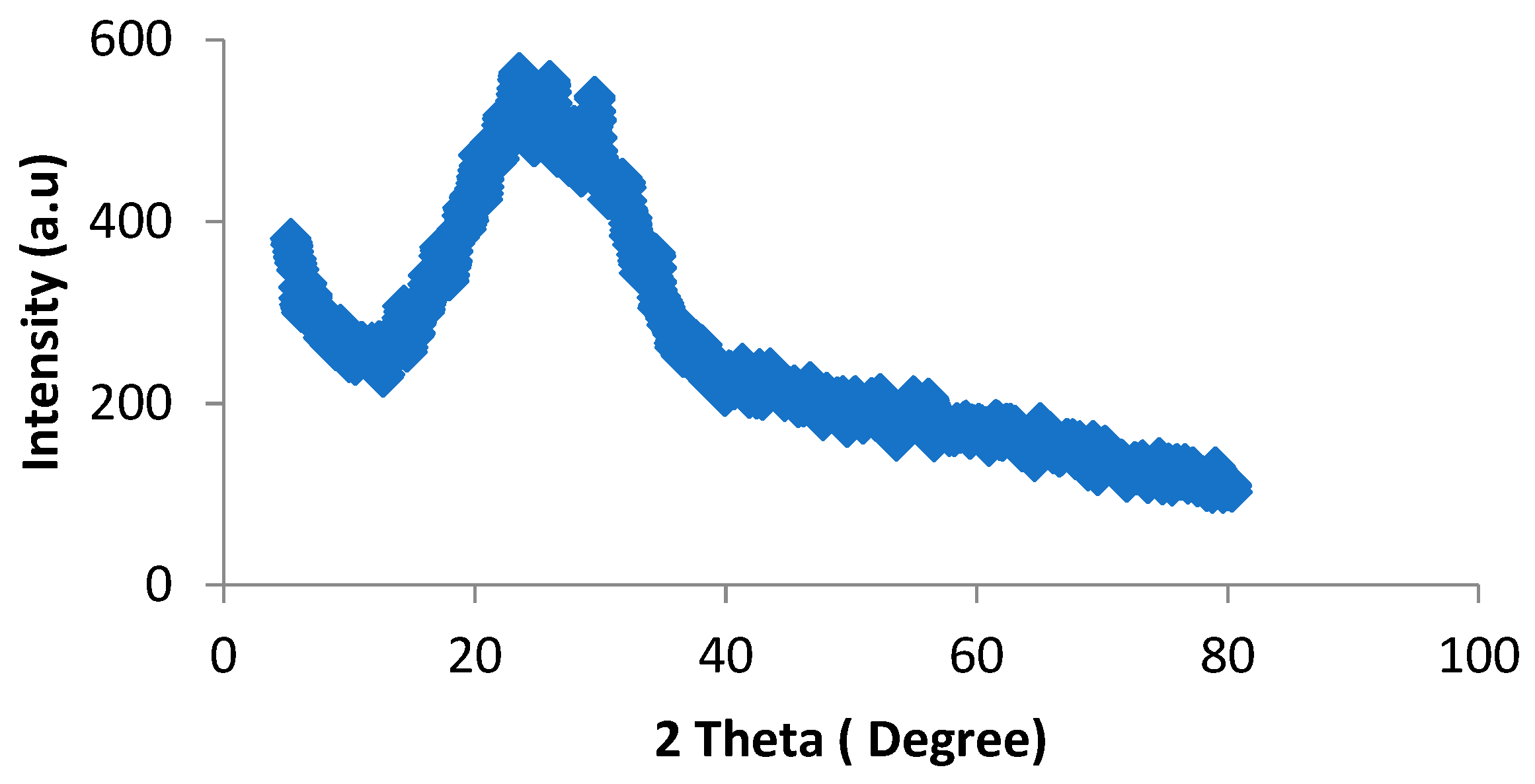
The structure of SP-SeNPs was identified using an X-ray diffractometer (XRD Brucker D8 DISCOVER) with Cu-kα radiation (λ = 1.54060 Å) at 40.0 KV and 40 mA in the 2θrange of 10–100°. The XRD testing was used to estimate the nature of the synthesized SP-SeNPs. According to Figure 3, the XRD of SP-SeNPs displayed a larger peak without any sharp Bragg’s peaks, indicating that the synthetic red elemental SP-SeNPs are undoubtedly amorphous in nature. In this context, [50] reported that synthesized SeNPs from Spirulina polysaccharides have an amorphous nature (Figure 4).

4.3.4. Energy Dispersive X-ray Testing (EDX)

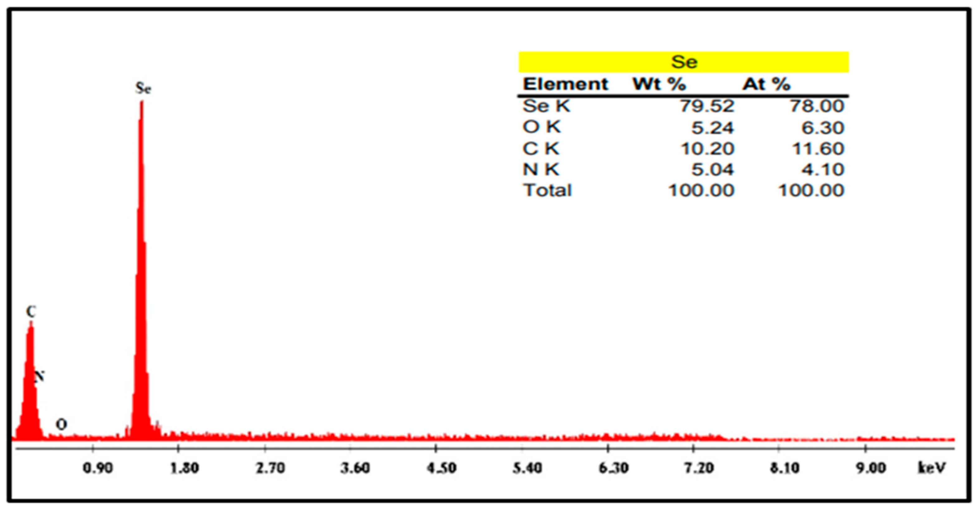
EDX spectroscopy (Hitachi S-4600) was applied to determine the elemental composition of SP-SeNPs. The EDX of SP-SeNPs revealed the presence of 79.52%, 5.24%, 10.20%, and 5.04% for Se, O, C, and N, respectively, as shown in Figure 5. This indicated that the biosynthesis of SP-SeNPs and other elements was stimulated through the existence of active substances in the S. platensis extract.

4.3.5. Transmission Electron Microscopy Analysis (TEM)

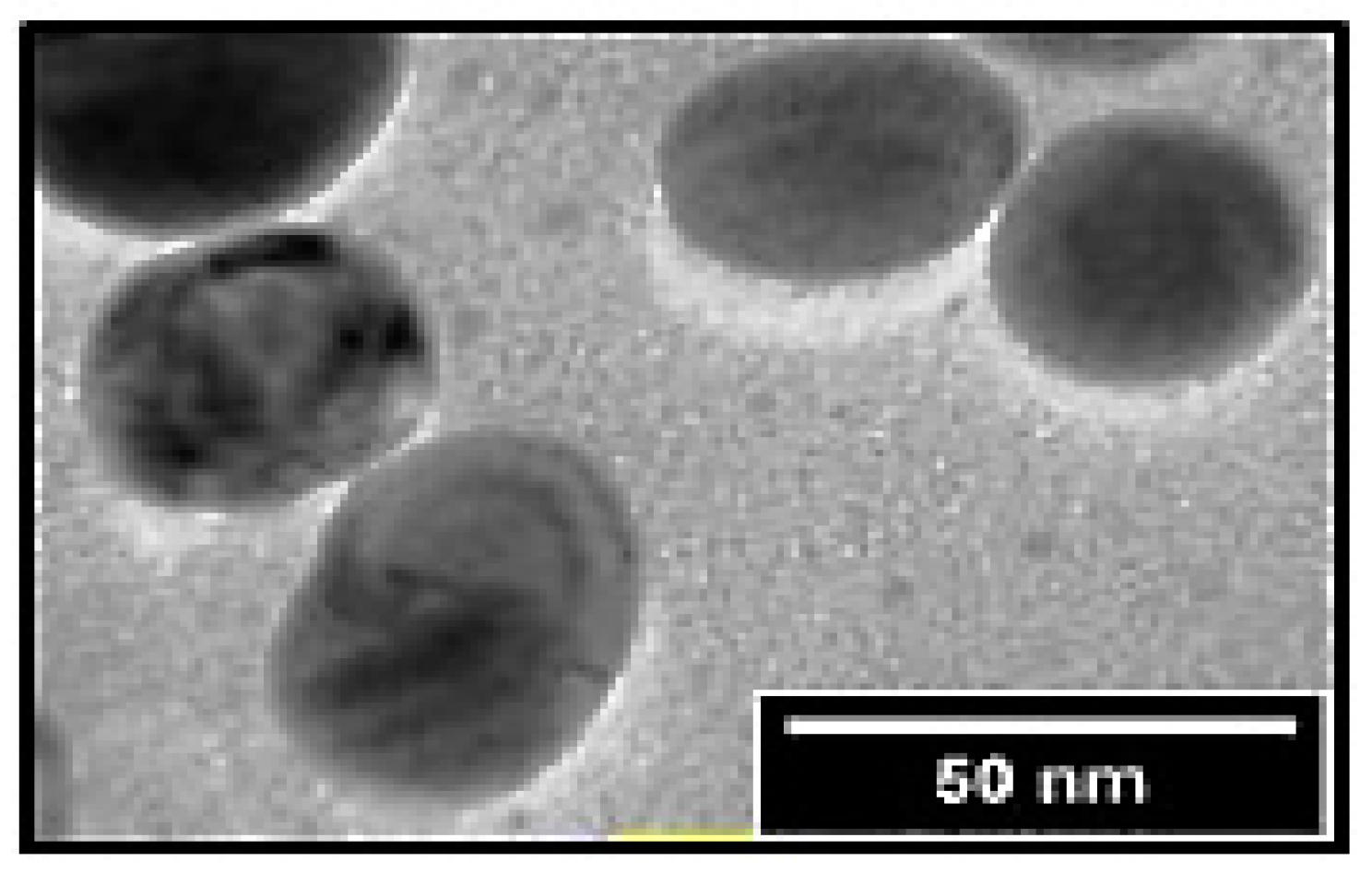
Biosynthesized SP-SeNPs’ shapes and sizes were assessed using TEM (JEOL, JEM-100S, Akishima, Tokyo, Japan). The reaction solution was sonicated for 10 min after being diluted with deionized water. After the sonicated sample was vacuum-dried for 30 min and drop-coated on carbon-coated copper grids, electron micrographs were taken. SP-SeNPs were measured using TEM, and it was found that they had a spherical form with a diameter of 50 nm (Figure 6). In this context, [18] also found that the size of SeNPs from the S. platensis extract ranged from 8.20 to 50.8 nm.

4.4. Experimental Shrimp

Pacific whiteleg shrimps, Litopenaeus vannamei (about 300 individuals with 6.0 ± 0.12 g of initial weight), were bought from a private shrimp farm and settled in 30 hapas (each 1.5 m length × 1.5 m width × 1.5 m water depth) for 14 days. In a private earthen pond shrimp farm in the Damietta Governorate of Egypt, shrimps were stocked at a stocking rate of 10 shrimps per hapa. In the earthen pond, hapas were fastened and organized. During the trial first, second, and third 20 days periods, respectively, hapa water was routinely changed every day by 5%, 10%, and 15%, and the water quality was monitored daily. The average water quality parameters were temperature: 24 ± 1.4 °C; pH: 8.5 ± 0.35; dissolved oxygen: 5.5 ± 0.36 mg/L; salinity: 37.58 ± 2.13 ppt; and ammonia: 0.018 ± 0.002 mg/L.

4.5. Feeding Procedure

Shrimp were fed on diets containing 38% protein (Table 7). Shrimp were fed three times daily at 6.0, 5.0, 4.0, and 3.0% of the biomass of shrimps in each hapa. The rates of feeding were changed in accordance with the live body weights of the shrimps every 14 days.

4.6. Experimental Design

Four groups of Pacific whiteleg shrimps (about 120 individuals with 6.0 ± 0.12 g of initial weight) were allocated in triplicate experimental design. One group represented the control and was fed with a ‘0’ mg/kg SP-SeNP supplemented diet. The residual three groups received diets supplemented with 0.26 (T1), 0.50 (T2), and 1 (T3) mg/kg of SP-SeNPs, respectively. These concentrations of dietary selenium were selected according to [13]. Shrimps were stocked at a stocking rate of 30 shrimps/treatment (10 shrimps per hapa in triplicate). Shrimps were fed 3 times daily (8, 12, 16 h) at 6, 5, 4, and 3% of the shrimps’ biomass in each hapa for 56 days, 14 days/each to adjust feeding rates. The experimental period was carried out from October to December 2022.

4.7. Growth Performance Aspects

After the end of the testing period (56 days), all shrimps/hapa were used to measure final body weight (FW), weight gain (WG), specific growth rate (SGR), average daily gain (ADG), survival %, biomass, feed intake, and feed conversion ratio (FCR), and were calculated according to [13].

4.8. Shrimp Analytical Methods

Three shrimps/treatments were collected to assess the proximate analyses of the shrimps’ bodies in accordance with [52], including protein, lipid, moisture, and ash contents at the end of the trial period.

4.9. Analysis of Biochemical Components in Muscles and Digestive Enzymes Activities in Digestive Tract of L. vannamei

Muscles and the digestive tract (total number = 6, 2 muscles and digestive tract of shrimps from 3 replicates per treatment) were dissected out from the control, T1, T2, and T3, and were homogenized with a Teflon homogenizer. The homogenates were put in a centrifuge for 10 min at 4000 rpm at 5.0 °C, and the supernatants were stored at −80 °C. Total protein was assessed with the method used in [53]; total lipid was measured according to [54]; total amino acid was assessed in accordance to [55]; total carbohydrates were assessed in accordance to [56]. Protease activity was measured in accordance to [57]. Amylase activity was estimated according to [58]. Lipase activity was assessed according to [59].

4.10. Determination of Lethal and Sublethal Toxicity of Cadmium after 56 Days of Growth under Laboratory Condition

A lethal concentration of cadmium (LC50) for 96 h was carried out under laboratory conditions. The stock solution (50 mg/L) was prepared from cadmium chloride with distilled water to give the subsequent concentrations. These concentrations were made by serially diluting the cadmium stock solution (0.20, 0.40, 0.60, and 0.80 mg/L). Selected experimental levels were set up by the addition of suitable volumes of stock solution to the seawater. A total of 150 individuals of adult shrimps (10 individuals in triplicates for each concentration) were used to determine the LC50. Ten animals (9–12 cm) were placed in each aquarium. As a control group, an equal number were left without treatment. Dead shrimps were recorded. When shrimps did not respond, they were thought to be dead after repeated touches with a probe. According to [60], probit analysis was used to determine the LC50.
Sublethal concentration (1/2 of 96 h LC50) of cadmium was used for 10 days. Shrimp groups, after the exposure to sublethal concentrations of cadmium, were divided into four groups: T4 comprised a control diet group with Cd exposure (30 individuals), T5 comprised shrimps with 0.25 mg/kg SP-SeNP supplemented diets and Cd exposure, T6 comprised shrimps with 0.50 mg/kg SP-SeNP supplemented diets and Cd exposure; T7 comprised shrimps with 1 mg/kg SP-SeNP supplemented diets and Cd exposure.

4.11. Bioaccumulation Analysis

Muscles and hepatopancreas (total number = 6, 2 muscles and hepatopancreas of shrimps from 3 replicates per treatment) were separated and dried out for 72.0 h. at 80.0 °C in an oven. The tissues were mashed to a powder form with a pestle and mortar and weighted to obtain 0.5 g. Tissues were digested in the microwave by using 5.0 mL of acid mixture for digestion (3.0 mL HNO3: 2.0 mL HCIO4) [61]. Cadmium levels were determined by an atomic absorption spectrophotometer (Buck scientific 210VGP) at the Faculty of Veterinary Medicine, Zagazig University.

4.12. Analysis of Oxidative and Antioxidant Indices before and after Cadmium Exposure in Muscles and Hepatopancreases

Muscles and hepatopancreases (total number = 6, 2 muscles and hepatopancreases from 3 replicates per treatment) were dissected and homogenized using a Teflon homogenizer from the control and the treated groups (T1 to T7). After centrifuging the homogenates, the supernatants were kept at −80 °C. Malondialdehyde (MDA) was determined using the procedure of [62]; total antioxidant capacity (TAC) was carried out by the procedure of [63]; catalase (CAT) was determined by the method of [64]; and superoxide dismutase (SOD) was measured according to [65].

4.13. Histopathological Investigations

Dissected hepatopancreases of the control groups (T4 to T7) were fixed in 10% formalin. The samples were cleaned in Xylene for 20 min following dehydration in a graded ethanol series. The specimens were set in paraffin wax. Using a microtome, sections (4 to 5 m thick) were cut, mounted, and stained with hematoxylin and eosin.

4.14. Statistical Analysis

SPSS software (version 20.0) was used to conduct the statistical analysis. Using the Kolmogorov–Smirnov and Bartlett tests, the normality and homogeneity of all of the data were tested. The significant differences between treatments were compared using one-way ANOVA followed by Tukey’s test at p < 0.05.

5. Conclusions

This study established that dietary supplementation of SP-SeNPs promote growth performance, biochemical components, digestive enzymes activities, and the antioxidative status in Litopenaeus vannamei at a specific dose. Additionally, it mitigated the pathological alternations induced with Cd toxicity. Based on Cd accumulation and histological investigation of tissues, SP-SeNPs at 0.5 mg/kg are recommended to be applied in L. vannamei aquaculture.

Author Contributions

Conceptualization, R.M.S., S.E.N. and M.S.A.; methodology, E.-S.H.E., H.S.D. and R.M.S.; software, B.A.A. and S.D.J.; validation, A.A.-F., E.-S.H.E., R.M.S. and H.S.D.; formal analysis, R.M.S., S.E.N. and M.S.A.; investigation, E.-S.H.E., H.S.D. and R.M.S.; resources, B.A.A. and S.D.J.; data curation, R.M.S.; writing—original draft preparation, E.-S.H.E., H.S.D., A.A.-F., R.M.S., S.E.N. and M.S.A.; writing—review and editing, E.-S.H.E., H.S.D., A.A.-F., R.M.S. and S.E.N.; visualization, E.-S.H.E., H.S.D., A.A.-F., R.M.S. and S.E.N.; project administration, Z.A.K. and G.T.-I. All authors have read and agreed to the published version of the manuscript.

Funding

Publication fees were paid by funds provided by the USDA-NIFA Sustainable Agriculture Systems, grant no. 2019-69012-29905. Title of project: Empowering US Broiler Production for Transformation and Sustainability USDA-NIFA (Sustainable Agriculture Systems): no. 2019-69012-29905.

Data Availability Statement

The data are included in this article.

Conflicts of Interest

The authors declare no conflict of interest.

Ethics Approval

ZU-IACUC/1/F/93/2023.

References

  1. Eissa, E.H.; Ahmed, N.H.; El-Badawi, A.A.; Munir, M.B.; Abd Al-Kareem, O.M.; Eissa, M.E.H.; Hussien, E.H.M.; Sakr, S.S. Assessing the influence of the inclusion of Bacillus subtilis AQUA-GROW® as feed additive on the growth performance, feed utilization, immunological responses and body composition of the Pacific white shrimp, Litopenaeus vannamei. Aquac. Res. 2022, 53, 6606–6615. [Google Scholar] [CrossRef]
  2. Moss, S.M.; Arce, S.M.; Moss, D.R.; Otoshi, C.A. Disease Prevention Strategies for Penaeid Shrimp Culture; The Oceanic Institute: Waimanalo Beach, HI, USA, 2006. [Google Scholar]
  3. Yang, S.P.; Liu, H.L.; Guo, W.J.; Wang, C.G.; Sun, C.B.; Chan, S.F.; Li, S.C.; Tan, Z.H. Effects of salinity and temperature on the metabolic and immune parameters of the banana shrimp Fenneropenaeus merguiensis (De Man, 1988). Iran. J. Fish. Sci. 2020, 19, 2010–2023. [Google Scholar] [CrossRef]
  4. Abdirad, Z.; Ghaednia, B.; Mirbakhsh, M.; Kakoolaki, S.; Ghorbani Vaghei, R. The effects of rearing Pacific white-leg shrimp (Litopenaeus vannamei Boone, 1931) in biofloc system on the immune responses and survival rate in challenge with Vibrio harveyi. Iran. J. Fish. Sci. 2022, 21, 705–725. [Google Scholar] [CrossRef]
  5. Eissa, E.H.; Che-Zulkifli, C.I.; El-Badawi, A.A.; Ali, M.A.M.; Baghdady, E.S.; Abd Al-Kareem, O.M.; Ahmed, R.A. Growth-Promoting and Immunomodulatory Impacts of Commercial Stimulants on Kuruma Shrimp, Penaeus japonicus (Bate, 1888) Juveniles. Egypt. J. Aquat. Biol. Fish. 2021, 25, 607–617. [Google Scholar]
  6. Khan, K.U.; Zuberi, A.; Nazir, S.; Fernandes, J.B.K.; Jamil, Z.; Sarwar, H. Effects of dietary selenium nanoparticles on physiological and biochemical aspects of juvenile Tor putitora. Turk. J. Zool. 2016, 40, 704–712. [Google Scholar] [CrossRef]
  7. Gobi, N.; Vaseeharan, B.; Rekha, R.; Vijayakumar, S.; Faggio, C. Bioaccumulation, cytotoxicity and oxidative stress of the acute exposure selenium in Oreochromis mossambicus. Ecotoxicol. Environ. Saf. 2018, 162, 147–159. [Google Scholar] [CrossRef] [PubMed]
  8. Zhao, L.; Chu, X.H.; Liu, S.; Li, R.; Zhu, Y.F.; Li, F.N.; Jiang, J.; Zhou, J.C.; Lei, X.G.; Sun, L.H. Selenium-Enriched Cardamine violifolia Increases Selenium and Decreases Cholesterol Concentrations in Liver and Pectoral Muscle of Broilers. J. Nutr. 2022, 152, 2072–2079. [Google Scholar] [CrossRef]
  9. Lang, C.; Mission, E.G.; Fuaad, A.A.H.A.; Shaalan, M. Nanoparticle tools to improve and advance precision practices in the Agrifoods Sector towards sustainability—A review. J. Clean. Prod. 2021, 293, 126063. [Google Scholar] [CrossRef]
  10. Eissa, E.H.; Alaidaroos, B.A.; Jastaniah, S.D.; Munir, M.B.; Shafi, M.E.; Abd El-Aziz, Y.M.; Bazina, W.K.; Ibrahim, S.b.; Eissa, M.E.H.; Paolucci, M. Dietary Effects of Nano Curcumin on Growth Performances, Body Composition, Blood Parameters and Histopathological Alternation in Red Tilapia (Oreochromis sp.) Challenged with Aspergillus flavus. Fishes 2023, 8, 208. [Google Scholar] [CrossRef]
  11. Dawit Moges, F.; Hamdi, H.; Al-Barty, A.; Zaid, A.A.; Sundaray, M.; Parashar, S.K.S. Effects of selenium nanoparticle on the growth performance and nutritional quality in Nile Tilapia, Oreochromis niloticus. PLoS ONE 2022, 17, e0268348. [Google Scholar] [CrossRef]
  12. Naderi, M.; Keyvanshokooh, S.; Salati, A.P.; Ghaedi, A. Combined or individual effects of dietary vitamin E and selenium nanoparticles on humoral immune status and serum parameters of rainbow trout (Oncorhynchus mykiss) under high stocking density. Aquaculture 2017, 474, 40–47. [Google Scholar] [CrossRef]
  13. Ghaffarizadeh, A.; Sotoudeh, E.; Mozanzadeh, M.T.; Sanati, A.M.; Ghasemi, A. Supplementing dietary selenium nano-particles increased growth, antioxidant capacity and immune-related genes transcription in Pacific whiteleg shrimp (Penaeus vannamei) juveniles. Aquac. Rep. 2022, 25, 101215. [Google Scholar] [CrossRef]
  14. Lee, K.X.; Shameli, K.; Yew, Y.P.; Teow, S.Y.; Jahangirian, H.; Rafiee-Moghaddam, R. Recent developments in the facile bio-synthesis of gold nanoparticles (AuNPs) and their biomedical applications. Int. J. Nanomed. 2020, 15, 275. [Google Scholar] [CrossRef] [PubMed]
  15. Gahlawat, G.; Choudhury, A.R. A review on the biosynthesis of metal and metal salt nanoparticles by microbes. RSC. Adv. 2019, 9, 12944–12967. [Google Scholar] [CrossRef] [PubMed]
  16. Abdel-Latif, H.M.; Abdel-Tawwab, M.; Khafaga, A.F.; Dawood, M.A. Dietary origanum essential oil improved antioxidative status, immune-related genes, and resistance of common carp (Cyprinus carpio L.) to Aeromonas hydrophila infection. Fish. Shellfish. Immunol. 2020, 104, 1–7. [Google Scholar] [CrossRef] [PubMed]
  17. Khanna, P.; Kaur, A.; Goyal, D. Algae-based metallic nanoparticles: Synthesis, characterization and applications. J. Microbiol. Methods 2019, 163, 105656. [Google Scholar] [CrossRef]
  18. ElSaied, B.E.F.; Diab, A.M.; Tayel, A.A.; Alghuthaymi, M.A.; Moussa, S.H. Potent antibacterial action of phycosynthesized selenium nanoparticles using Spirulina platensis extract. Green Process. Synth. 2021, 10, 49–60. [Google Scholar] [CrossRef]
  19. Wang, M.; Chen, Z.; Song, W.; Hong, D.; Huang, L.; Li, Y.A. Review on Cadmium Exposure in the Population and Intervention Strategies against Cadmium Toxicity. Bull. Environ. Contam. Toxicol. 2021, 106, 65–74. [Google Scholar] [CrossRef]
  20. Mielcarek, K.; Nowakowski, P.; Puścion-Jakubik, A.; Gromkowska-Kępka, K.J.; Soroczyńska, J.; Markiewicz-Zukowska, R.; Naliwajko, S.K.; Grabia, M.; Bielecka, J.; Żmudzińska, A.; et al. Arsenic, cadmium, lead and mercury content and health risk assessment of consuming freshwater fish with elements of chemometric analysis. Food. Chem. 2022, 379, 132167. [Google Scholar] [CrossRef]
  21. Abu-Elala, N.M.; Shaalan, M.; Ali, S.E.; Younis, N.A. Immune responses and protective efficacy of diet supplementation with selenium nanoparticles against cadmium toxicity in Oreochromis niloticus. Aquac. Res. 2021, 52, 3677–3686. [Google Scholar] [CrossRef]
  22. Megahed, M.E.; Fathi, M.; Abderhman, A.M. Molecular investigation on the production of the pacific white shrimp (Litopenaeus vannamei) using diets based on biofloc meal. J. Aquac. Mar. Biol. 2019, 8, 216–224. [Google Scholar] [CrossRef]
  23. Panutrakul, S.; Senanan, W. Abundance of introduced Pacific whiteleg shrimp Penaeus vannamei (Boone, 1931) along the east coast of Thailand. Aquat. Invasions 2021, 16, 750–770. [Google Scholar] [CrossRef]
  24. Eissa, E.-S.H.; Ahmed, R.A.; Abd Elghany, N.A.; Elfeky, A.; Saadony, S.; Ahmed, N.H.; Sakr, S.E.-S.; Dayrit, G.B.; Tolenada, C.P.S.; Atienza, A.A.C.; et al. Potential Symbiotic Effects of β-1,3 Glucan, and Fructooligosaccharides on the Growth performance, Immune Response, Redox Status, and Resistance of Pacific White Shrimp, Litopenaeus vannamei to Fusarium solani Infection. Fishes 2023, 8, 105. [Google Scholar] [CrossRef]
  25. Dawood, M.A.O.; El Basuini, M.F.; Yilmaz, S.; Abdel-Latif, H.M.R.; Abdul Kari, Z.; Abdul Razab, M.K.A.; Ahmed, H.A.; Alagawany, M.; Gewaily, M.S. Selenium Nanoparticles as a Natural Antioxidant and Metabolic Regulator in Aquaculture: A Review. Antioxidants 2021, 10, 1364. [Google Scholar] [CrossRef]
  26. Fan, J.; Li, B.; Hong, Q.; Yan, Z.; Yang, X.; Lu, K.; Chen, G.; Wang, L.; Chen, Y.A. Glutathione peroxidase gene from Litopenaeus vannamei is involved in oxidative stress responses and pathogen infection resistance. Int. J. Mol. Sci. 2022, 23, 567. [Google Scholar] [CrossRef]
  27. Ghazi, S.; Diab, A.M.; Khalafalla, M.M.; Mohamed, R.A. Synergistic effects of selenium and zinc oxide nanoparticles on growth performance, hemato-biochemical profile, immune and oxidative stress responses, and intestinal morphometry of Nile tilapia (Oreochromis niloticus). Biol. Trace. Elem. Res. 2022, 200, 364–374. [Google Scholar] [CrossRef]
  28. Abd El-Kader, M.F.; Fath El-Bab, A.F.; Abd-Elghany, M.F.; Abdel-Warith, A.W.A.; Younis, E.M.; Dawood, M.A.O. Selenium nanoparticles act potentially on the growth performance, hemato-biochemical indices, antioxidative, and immune-related genes of European seabass (Dicentrarchus labrax). Biol. Trace Elem. Res. 2021, 199, 3126–3134. [Google Scholar] [CrossRef]
  29. Zhang, Q.; Qian, Z.-Y.; Zhou, P.-H.; Zhou, X.-L.; Zhang, D.; He, N.; Zhang, J.; Liu, Y.H.; Gu, Q. Effects of oral selenium and magnesium co-supplementation on lipid metabolism, antioxidative status, histopathological lesions, and related gene expression in rats fed a high-fat diet. Lipids Health Dis. 2018, 17, 165. [Google Scholar] [CrossRef]
  30. Knight, R.; Marlatt, V.L.; Baker, J.A.; Lo, B.P.; deBruyn, A.M.H.; Elphick, J.R.; Martyniuk, C.J. Dietary selenium disrupts hepatic triglyceride stores and transcriptional networks associated with growth and Notch signaling in juvenile rainbow trout. Aquat. Toxicol. 2016, 180, 103–114. [Google Scholar] [CrossRef]
  31. Dawood, M.A.O. Nutritional immunity of fish intestines: Important insights for sustainable aquaculture. Rev. Aquac. 2021, 13, 642–663. [Google Scholar] [CrossRef]
  32. Kumar, N.; Krishnani, K.K.; Gupta, S.K.; Sharma, R.; Baitha, R.; Singh, D.K.; Singh, N.P. Immuno-protective role of biologically synthesized dietary selenium nanoparticles against multiple stressors in Pangasinodon hypophthalmus. Fish Immunol. 2018, 78, 289–298. [Google Scholar] [CrossRef]
  33. Tinggi, U. Selenium: Its role as antioxidant in human health. Environ. Health Prev. Med. 2008, 13, 102–108. [Google Scholar] [CrossRef]
  34. Dotan, Y.; Lichtenberg, D.; Pinchuk, I. Lipid peroxidation cannot be used as a universal criterion of oxidative stress. Prog. Lipid Res. 2004, 43, 200–227. [Google Scholar] [CrossRef] [PubMed]
  35. Trasvina-Arenas, C.H.; Garcia-Triana, A.; Peregrino-Uriarte, A.B.; Yepiz-Plascencia, B. White shrimp Litopenaeus vannamei catalase: Gene structure, expression and activity under hypoxia and reoxygenation. Comp. Biochem. Physiol. Part B Biochem. Mol. Biol. 2013, 164, 44–52. [Google Scholar] [CrossRef] [PubMed]
  36. Campa-Cordova, A.I.; Hern´and ez-Saavedra, N.Y.; Ascencio, F. Superoxide dismutase as modulator of immune function in American white shrimp (Litopenaeus vannamei). Comp. Biochem. Physiol. C Toxicol. Pharm. 2002, 133, 557–565. [Google Scholar] [CrossRef] [PubMed]
  37. Zoidis, E.; Seremelis, I.; Kontopoulos, N.; Danezis, G.P. Selenium-Dependent Antioxidant Enzymes: Actions and Properties of Selenoproteins. Antioxidants 2018, 7, 66. [Google Scholar] [CrossRef]
  38. Kong, Y.; Ding, Z.; Zhang, Y.; Ye, J.; Du, Z. Dietary selenium requirement of juvenile oriental river prawn Macrobrachium nipponense. Aquaculture 2017, 476, 72–78. [Google Scholar] [CrossRef]
  39. Eissa, E.H.; Bazina, W.K.; Abd El-Aziz, Y.M.; Abd Elghany, N.A.; Tawfik, W.A.; Mossa, M.I.; Abd El Megeed, O.H.; Abd El-Hamed, N.B.; El-Saeed, A.F.; El-Haroun, E.; et al. Nano-selenium impacts on growth performance, digestive enzymes, antioxidant, immune resistance and histopathological scores of Nile tilapia, Oreochromis niloticus against Aspergillus flavus infection. Aquac. Int. 2023, 1–25.–25. [Google Scholar] [CrossRef]
  40. Abdel-Tawwab, M.; Wafeek, M. Response of Niletilapia, Oreochromis niloticus (L.) to environmental cadmium toxicity during organic selenium supplementation. J. World. Aquacu. Soc. 2010, 41, 106–114. [Google Scholar] [CrossRef]
  41. Lin, Y.; Shiau, S. The effects of dietary selenium on the oxidative stress of grouper, Epinephelus malabaricus, fed high copper. Aquaculture 2007, 267, 38–43. [Google Scholar] [CrossRef]
  42. Wang, Z.; Qu, Y.; Yan, M.; Li, J.; Zou, J.; Fan, L. Physiological responses of pacific white shrimp Litopenaeus vannamei to temperature fluctuation in low salinity water. Front. Physiol. 2019, 10, 1025. [Google Scholar] [CrossRef]
  43. Kumar, N.; Gupta, S.K.; Bhushan, S.; Singh, N.P. Impacts of acute toxicity of arsenic (III) alone and with high temperature on stress biomarkers, immunological status and cellular metabolism in fish. Aquat. Toxicol. 2019, 214, 105233. [Google Scholar] [CrossRef]
  44. Gailer, J. Chronic toxicity of AsIII in mammals: The role of (GS)2AsSe. Biochimie 2009, 91, 1268–1272. [Google Scholar] [CrossRef]
  45. Zwolak, I. The role of selenium in arsenic and cadmium toxicity: An updated review of scientific literature. Biol. Trace Elem. Res. 2020, 193, 44–63. [Google Scholar] [CrossRef] [PubMed]
  46. Gunti, L.; Dass, R.S.; Kalagatur, N.K. Phytofabrication of selenium nanoparticles from Emblica officinalis fruit extract and exploring its biopotential applications: Antioxidant, anti-microbial, and biocompatibility. Front. Microbiol. 2019, 10, 931. [Google Scholar] [CrossRef] [PubMed]
  47. Ankamwar, B.; Damle, C.; Ahmad, A.; Sastry, M. Biosynthesis of gold and silver nanoparticles using Emblica officinalis fruit extract, their phase transfer and transmetallation in an organic solution. J. Nanosci. Nanotechnol. 2005, 5, 1665–1671. [Google Scholar] [CrossRef] [PubMed]
  48. Kimura, K.; Hane, Y.; Watanabe, Y. Effect of pre-coagulation on mitigating irreversible fouling during ultrafiltration of surface water. Water Sci. Technol. 2005, 51, 93–100. [Google Scholar] [CrossRef] [PubMed]
  49. Park, Y.; Hong, Y.N.; Weyers, A.; Kim, Y.S.; Linhardt, R.J. Polysaccharides and phytochemicals: A natural reservoir for the green synthesis of gold and silver nanoparticles. IET Nanobiotechnol. 2011, 5, 69–78. [Google Scholar] [CrossRef]
  50. Yang, F.; Tang, Q.; Zhong, X.; Bai, Y.; Chen, T.; Zhang, Y.; Li, Y.; Zheng, W. Surface decoration by Spirulina polysaccharide enhances the cellular uptake and anticancer efficacy of selenium nanoparticles. Int. J. Nanomed. 2012, 7, 835. [Google Scholar] [CrossRef]
  51. NRC. Nutrient Requirements of Fish and Shrimp; National Academies Press: Washington, DC, USA, 2011. [Google Scholar] [CrossRef]
  52. AOAC. Official Methods of Analysis, 17th ed.; The Association of Official Analytical Chemists: Gaithersburg, MD, USA, 2000. [Google Scholar]
  53. Bradford, M.M. A rapid and sensitive method for the quantitation of microgram quantities of protein utilizing the principle of protein-dye binding. Anal. Biochem. 1976, 72, 248–254. [Google Scholar] [CrossRef]
  54. Zöllner, N.; Kirsch, K. Colorimetric Method for Determination of Total Lipids. J. Exp. Med. 1962, 135, 545–550. [Google Scholar] [CrossRef]
  55. Moore, S.; Stein, W.H. Photometric ninhydrin method for use in the chromatography of amino acid. J. Biol. Chem. 1948, 176, 367–388. [Google Scholar] [CrossRef]
  56. Roe, J.H. The determination of sugar and blood and spinal fluid with anthrone reagent. J. Biol. Chem. 1955, 212, 335–343. [Google Scholar] [CrossRef] [PubMed]
  57. Furne, M.; Hidalgo, M.C.; Lopez, A.; Garcia-Gallego, M.; Morales, A.E.; Domezain, A. Digestive enzyme activities in Adriatic sturgeon Acipenser naccarii and rainbow trout Oncorhynchus mykiss. A comparative study. Aquaculture 2005, 250, 391–398. [Google Scholar] [CrossRef]
  58. Henry, R.J.; Chiamori, N. Profile of Alpha Amylase in vulgaris. Clin. Chem. 1961, 6, 434. [Google Scholar] [CrossRef]
  59. Lorentz, K.; Weiss, T. Pankreaslipase—Eine Übersicht. Med. Lab. 1981, 34, 272–275. [Google Scholar]
  60. Finney, D.J. Estimation of the median effective dose. In Probit Analysis, 3rd ed.; Cambridge University: Cambridge, UK, 1971; pp. 20–49. [Google Scholar] [CrossRef]
  61. Tsoumbaris, P. Heavy Metals Determination in Food Stuff. Ph.D. Thesis, Aristotle University of Thessaloniki, Thessalonica, Greece, 1990. [Google Scholar]
  62. Uchiyama, M.; Mihara, M. Determination of malonaldehyde precursor in tissues by thiobarbituric acid test. Anal. Biochem. 1978, 86, 271–278. [Google Scholar] [CrossRef]
  63. Nemec, A.; Drobnič-Košorok, M.; Skitek, M.; Pavlica, Z.; Galac, S.; Butinar, J. Total antioxidant capacity (TAC) values and their correlation with individual antioxidants in healthy beagles. Acta Vet. Brno 2000, 69, 297–303. [Google Scholar] [CrossRef]
  64. Aebi, H. Catalase. In Methods Enzymatic Analysis; Bergmeyer, H.U., Ed.; Academic Press: New York, NY, USA, 1974; pp. 673–678. [Google Scholar] [CrossRef]
  65. Nishikimi, M.; Appaji, N.; Yagi, K. The occurrence of superoxide anion in the reaction of reduced phenazine methosulfate and molecular oxygen. Biochem. Biophys. Res. Commun. 1972, 46, 849–854. [Google Scholar] [CrossRef] [PubMed]
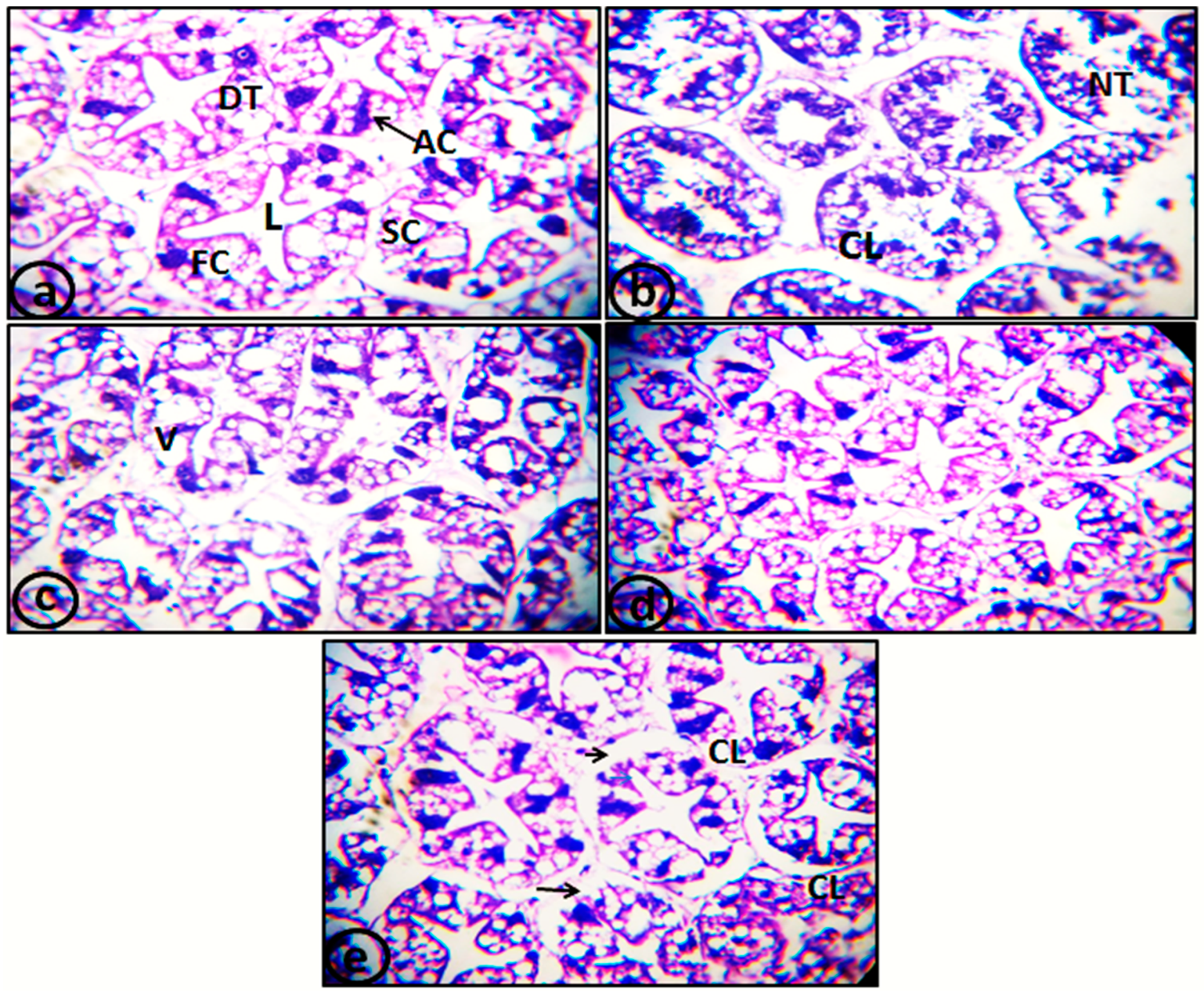
Figure 1. Photomicrograph of hepatopancreatic tubules of Pacific white shrimp L. vannamei. (a) T.S. of control hepatopancreas (control diet) displaying normal cellular structure and different types of hepatopancreatic cells (X400). (b) T.S. of hepatopancreatic tubules from the T4 group (control diet and Cd exposure) showing degeneration and necrosis in the cell of tubules (X400). (c) T.S. of hepatopancreatic tubules from the T5 group (0.25 mg/kg SP-SeNP supplemented diets and 0.2 mg/L Cd exposure) showing vacuolation (X400). (d) T.S. of hepatopancreatic tubules from the T6 group (0.50 mg/kg SP-SeNP supplemented diets and 0.2 mg/L Cd exposure) showing normal histological structures without pathological lesions (X400). (e) T.S. of hepatopancreatic tubules from the T6 group (1 mg/kg SP-SeNP supplemented diets and 0.2 mg/L Cd exposure) showing normal histological structure. However, some tubules were rounded off and began to slough off their basement membrane (arrows) and lysis of their epithelial cells occurred in some tubules (X400). AC: absorptive cell; CL: cell lysis; FC: fibrillar cell; DT: digestive tubules; L: lumen; NT: necrotic tubules; SC: secretory cell; V: vacuolation.
Catalysts 13 01389 g001
Figure 2. UV–Visible spectrum of SP-SeNPs.
Catalysts 13 01389 g002
Figure 3. FTIR spectra of (A) S. platensis extract and (B) SP-SeNPs.
Catalysts 13 01389 g003
Figure 4. XRD spectrum of SP-SeNPs.
Catalysts 13 01389 g004
Figure 5. EDX spectrum of SP-SeNPs.
Catalysts 13 01389 g005
Figure 6. TEM of SP-SeNPs.
Catalysts 13 01389 g006
Table 1. Growth performance of Pacific white shrimps (L. vannamei) supplemented with SP-SeNPs for 56 days.
ParametersConcentrations of SP-SeNPs (mg/kg)
ControlT1T2T3F-Valuep-Value
Initial weight (g/shrimp)6.00 ± 0.12 a6.03 ± 0.12 a6.13 ± 0.07 a6.03 ± 0.09 a0.330.80
Final weight (g/shrimp)16.77 ± 0.23 c18.13 ± 0.22 b19.13 ± 0.27 a19.90 ± 0.26 a29.720.00001
Weight gain (g/shrimp)10.77 ± 0.12 d12.10 ± 0.10 c13.00 ± 0.21 b13.87 ± 0.18 a70.980.00001
Weight gain %179.50 ± 1.65 d200.65 ± 2.45 c211.94 ± 1.34 b229.85 ± 0.43 a--
Feed intake (g feed/fish)18.74 ± 0.40 b19.42 ± 0.45 ab20.20 ± 0.22 a20.31 ± 0.27 a4.460.04
ADG (g/shrimp/day)0.19 ± 0.00 d0.22 ± 0.00 c0.23 ± 0.00 b0.25 ± 0.00 a47.580.00001
FCR (g)1.74 ± 0.02 a1.60 ± 0.03 b1.55 ± 0.01 b1.46 ± 0.00 c50.710.00001
SGR (%/day)1.84 ± 0.01 d1.96 ± 0.01 c2.03 ± 0.01 b2.13 ± 0.00 a140.610.00001
Initial shrimp number10.00 ± 0.0010.00 ± 0.0010.00 ± 0.0010.00 ± 0.00--
Final shrimp number8.67 ± 0.33 a9.00 ± 0.58 a9.67 ± 0.33 a9.67 ± 0.33 a1.500.29
Survival rate %86.67 ± 3.33 a90.00 ± 5.77 a96.67 ± 3.33 a96.67 ± 3.33 a1.500.29
Shrimp biomass (g/m3)145.33 ± 6.22 c163.00 ± 8.93 bc184.83 ± 5.09 ab192.50 ± 8.67 a8.350.008
Each value represents means ± SE (n = 10 shrimps per hapa in triplicates). Within the same row, mean values with various alphabetical are statistically significant at p < 0.05. ADG: average daily growth; SGR: specific growth rate; FCR: Feed conversion ratio. T1: L. vannamei received diet supplemented with 0.25 mg/kg SP-SeNPs; T2: L. vannamei received diet supplemented with 0.50 mg/kg SP-SeNPs; T3: L. vannamei received diet supplemented with 1 mg/kg SP-SeNPs.
Table 2. Impacts of SP-SeNP supplementation on the body composition of L. vannamei for 56 days.
ParametersConcentrations of SP-SeNPs (mg/kg)
ControlT1T2T3p-ValueF-Value
Moisture %79.17 ± 0.01 a79.18 ± 0.01 a79.20 ± 0.00 a79.20 ± 0.01 a2.190.17
Crude protein%15.93 ± 0.03 c16.14 ± 0.02 b16.17 ± 0.02 b16.26 ± 0.02 a41.450.00001
Crude lipid%1.88 ± 0.03 a1.87 ± 0.04 ab1.84 ± 0.02 ab1.77 ± 0.03 b2.650.12
Ash%3.09 ± 0.09 b3.12 ± 0.01 b3.17 ± 0.01 a3.18 ± 0.02 a15.230.001
Each value represents mean of 3 samples per treatment ± SE. Mean values on the same row with various alphabetical letters are statistically significant at p < 0.05. T1: L. vannamei received diet supplemented with 0.25 mg/kg SP-SeNPs; T2: L. vannamei received diet supplemented with 0.50 mg/kg SP-SeNPs; T3: L. vannamei received diet supplemented with 1 mg/kg SP-SeNPs.
Table 3. Impact of SP-SeNP supplemented diets on concentrations of biochemical components in muscles and digestive enzymes activities in the digestive tract of L. vannamei for 56 days.
ParametersConcentrations of SP-SeNPs (mg/kg)F-
Value
p-
Value
ControlT1T2T3
Biochemical
indices
(mg/g)
Protein 4.56 ± 0.20 a5.55 ± 0.45 ab6.80 ± 0.30 b6.46 ± 0.45 b7.600.04
Lipid 1.65 ± 0.1 a3.34 ± 0.35 b5.1 ± 0.12 c3.98 ± 0.13 bc51.130.001
Amino acids 95.3 ± 6.07 a112.68 ± 7.33 ab134.13 ± 3.63 b117.65 ± 4.4 ab8.30.03
Carbohydrate 35.4 ± 2.9 a41.76 ± 2.36 ab59.15 ± 1.09 c50.42 ± 1.7 bc23.660.005
Digestive
enzymes
(U/mg)
Protease2.43 ± 0.03 a2.91 ± 0.12 b3.38 ± 0.05 c3.13 ± 0.03 bc36.170.002
Amylase1.96 ± 0.04 a2.23 ± 0.12 a3.96 ± 0.04 b3.53 ± 0.42 b20.160.007
Lipase1.15 ± 0.01 a1.18 ± 0.02 a1.4 ± 0.05 b1.22 ± 0.02 a15.660.01
Each value represents mean ± SE (n = 6, 2 muscle and digestive tract of shrimps from 3 replicate per treatment). Mean values on the same row with various alphabetical letters are statistically significant at p < 0.05. T1: L. vannamei received diet supplemented with 0.25 mg/kg SP-SeNPs; T2: L. vannamei received diet supplemented with 0.50 mg/kg SP-SeNPs; T3: L. vannamei received diet supplemented with 1 mg/kg SP-SeNPs.
Table 4. Mortality percentages, lethal toxicity (LC50) value, and sublethal concentration of L. vannamei exposed to cadmium under laboratory conditions.
TimePercentage Mortality (%)
Conc 24 h48 h72 h96 h
Control0 ± 0.000 ± 0.000 ± 0.000 ± 0.00
0.20 ± 0.000 ± 0.0023.33 ± 3.3326.67 ± 3.33
0.436.7 ± 3.3336.7 ± 3.3340 ± 0.0050 ± 0.00
0.656.67 ± 6.760 ± 0.0066.67 ± 6.770 ± 0.00
0.896.6 ± 3.33100 ± 0.00100 ± 0.00100 ± 0.00
96 h LC500.4 mg/L
1/2 LC50 (for 10 days)0.2 mg/L
Each value represents mean ± SE (n = 10 individuals in triplicates for each control and treated groups).
Table 5. Levels of cadmium and selenium (µg/g dry w.) accumulated in hepatopancreas and muscle tissues of adult L. vannamei after exposure to sublethal concentrations of cadmium for 10 days.
Cd AccumulationDifferent Treatments
ControlT1T2T3T4T5T6T7F-
Value
p-
Value
Muscles0 ± 0.0 a0 ± 0.00 a0 ± 0.00 a0 ± 0.00 a0.24 ± 0.04 b 0.18 ± 0.08 ab 0.11 ± 0.02 ab 0.16 ± 0.04 ab7.910.005
Hepatopancreases0 ± 0.0 a0.01 ± 0.005 a 0.05 ± 0.02 a 0.08 ± 0.01 ab 0.6 ± 0.06 d 0.30 ± 0.01 bc 0.22 ± 0.03 b 0.43 ± 0.08 cd 17.030.002
Se accumulation
Muscles3.9 ± 0.2 abc4.5 ± 0.53 bc 5.39 ± 0.6 c 6.18 ± 0.83 c 0.9 ± 0.03 a 4.19 ± 0.35 bc 5.09 ± 0.97 c 1.79 ± 0.33 ab 10.240.002
Hepatopancreases 2.8 ± 0.2 bc 3.39 ± 0.2 bcd 5.4 ± 0.13 d5 ± 1.2 cd0.38 ± 0.1 a 2.98 ± 0.03 bc 4.1 ± 0.16 bcd 2.71 ± 0.45 b13.930.001
Each value represent mean ± SE (n = 6, 2 muscles and hepatopancreases of shrimps from 3 replicates per treatment). Mean values with various alphabetical letters within the same row are statistically significant at p < 0.05. T1: L. vannamei received diet supplemented with 0.25 mg/kg SP-SeNPs; T2: L. vannamei received diet supplemented with 0.50 mg/kg SP-SeNPs; T3: L. vannamei received diet supplemented with 1 mg/kg SP-SeNPs; T4: control diet with Cd (0.2 mg/L); T5: 0.25 mg/kg SP-SeNP supplemented diets and 0.2 mg/L Cd; T6: 0.50 mg/kg SP-SeNP supplemented diets and 0.2 mg/L Cd; T7: 1 mg/kg SP-SeNP supplemented diets and 0.2 mg/L Cd.
Table 6. Oxidative and antioxidant activities in the hepatopancreases and muscles of adult L. vannamei after cadmium exposure for 10 days.
Tested Tissues Different Treatments
ControlT1T2T3T4T5T6T7F-Valuep-Value
HepatopancreasesMDA
(µmol/mg)
123.6 ± 3.4 a61.2 ± 3.2 b77.4 ± 7.2 b85.5 ± 1 b211.4 ± 9.2 e168.7 ± 3.5 d115.6 ± 1 c52.7 ± 2.2 b86.10.0001
CAT
(U/g)
35.6 ± 0.7 ab41.22 ± 1 bc46.8 ± 1.2 cd54.8 ± 0.9 e28.4 ± 1.43 a44.1 ± 0.15 c50.2 ± 1 de51.6 ± 1.6 de68.90.001
TAC
(µmol/mg)
39.5 ± 1.5 ab49.4 ± 6.5 bc51.6 ± 1.4 bc54.7 ± 1.5 bc27.7 ± 2.4 a36.9 ± 1.7 ab67.51 ± 1.6 c49 ± 2.7 ab8.50.004
SOD
(U/g)
64.1 ± 3.2 ab74.2 ± 4.1 ab86.9 ± 3.3 b80.4 ± 4.9 ab55.6 ± 9.7 a64.3 ± 4 ab77.9 ± 7.3 ab68.4 ± 1.6 ab3.70.04
MusclesMDA
(µmol/mg)
1.5 ± 0.36 bc0.7 ± 0.03 a0.8 ± 0.04 ab0.9 ± 0.03 b2.99 ± 0.4 d0.95 ± 0.04 b1.91 ± 0.08 c1.3 ± 0.1 bc30.40.001
CAT
(U/g)
22.8 ± 0.1 ab23.2 ± 0.1 ab25.9 ± 0.7 bc23.6 ± 0.4 ab17.4 ± 1.13 a24 ± 1.6 bc30.4 ± 2.8 c29.3 ± 0.8 bc11.050.002
TAC
(µmol/mg)
20.8 ± 2.1 ab23.1 ± 0.1 ab31.6 ± 1.6 de29.4 ± 0.6 cd17.3 ± 0.5 a24.8 ± 0.3 bc36.7 ± 0.9 e30 ± 1.1 cd34.080.0001
SOD
(U/g)
8.7 ± 0.33 ab9.2 ± 0.25 ab17.1 ± 0.4 cd16.8 ± 1.5 cd6.41 ± 1.11 a12.21 ± 1 bc19.9 ± 0.4 d19.6 ± 1.4 d33.10.0001
Each value represent mean ± SE (n = 6, 2 muscle and hepatopancreases of shrimps from 3 replicates per treatment). Mean values with various alphabetical letters within the same row are statistically significant at p < 0.05. T1: L. vannamei received diet supplemented with 0.25 mg/kg SP-SeNPs; T2: L. vannamei received diet supplemented with 0.50 mg/kg SP-SeNPs; T3: L. vannamei received diet supplemented with 1 mg/kg SP-SeNPs; T4: control diet with Cd (0.2 mg/L); T5: 0.25 mg/kg SP-SeNP supplemented diets and 0.2 mg/L Cd; T6: 0.50 mg/kg SP-SeNP supplemented diets and 0.2 mg/L Cd; T7: 1 mg/kg SP-SeNP supplemented diets and 0.2 mg/L Cd.
Table 7. Chemical analysis and constitution of experimental diets.
Contentsg/kg DietProximate Chemical Analysis%
Wheat flour120.0Dry mater90.64
Shrimp meal250.0Moisture9.35
Rice bran70.0Crude protein (N × 6.25)38.79
Soybean meal150.0Crude fat10.90
Fish meal300.0Crude fiber1.74
Fish oil60.0Ash6.15
CMC10.0Carbohydrate (NFE)32.98
Vit. and min mix 120.0Gross energy kcal/100 g 3459.47 kcal/100 g 3
Min mix 220.0
1000
Symbol 1—vitamin premix (per kg of premix): 2.5 g thiamine; 2.5 g riboflavin; 2.0 g pyridoxine; 100.0 g inositol; 0.3 g biotin; 100.0 g pantothenic acid; 0.75 g folic acid; 2.5 g para-aminobenzoic acid; 200.0 g choline; 10.0 g nicotinic acid; 0.005 g cyanocobalamine; 20.1 g a-tocopherol acetate; 2.0 g menadione; 100,000 IU retinol palmitate; 500,000 IU cholecalciferol. Symbol 2—mineral premix (g/kg of premix): CaHPO4·2H2O: 727.2; MgCO4·7H2O: 127.5; KCl: 50.0; NaCl: 60.0; FeC6H5O7·3H2O: 25.0; ZnCO3: 5.5; MnCl2·4H2O: 2.5; Cu (OAc) 2·H2O: 0.785; CoCl3·6H2O: 0.477; CaIO3·6H2O: 0.295; CrCl3·6H2O: 0.128; AlCl3·6H2O: 0.54; Na2SeO3: 0.03. Symbol 3—gross energy (GE) was measured from [51] as 16.7, 37.4, and 16.7 kcal/100 g for carbohydrates, lipids, and proteins, respectively.
Disclaimer/Publisher’s Note: The statements, opinions and data contained in all publications are solely those of the individual author(s) and contributor(s) and not of MDPI and/or the editor(s). MDPI and/or the editor(s) disclaim responsibility for any injury to people or property resulting from any ideas, methods, instructions or products referred to in the content.