Article

Biogenic Synthesis Based on Cuprous Oxide Nanoparticles Using Eucalyptus globulus Extracts and Its Effectiveness for Removal of Recalcitrant Compounds

1
Departamento de Ingeniería Civil, Facultad de Ingeniería, Universidad Católica de la Santísima Concepción, Concepción 4090541, Chile
2
Centro de Estudios en Alimentos Procesados (CEAP), Campus Lircay, Talca 3460000, Chile
3
Grupo de Ingeniería y Biotecnología Ambiental (GIBA-UDEC), Facultad de Ciencias Ambientales, Universidad de Concepción, Concepción 4070386, Chile
4
Water Research Center for Agriculture and Mining (CRHIAM), ANID Fondap Center, Victoria 1295, Concepción 4070411, Chile
*
Author to whom correspondence should be addressed.
Catalysts 2024, 14(8), 525; https://doi.org/10.3390/catal14080525
Submission received: 14 July 2024 / Revised: 3 August 2024 / Accepted: 9 August 2024 / Published: 14 August 2024
(This article belongs to the Section Photocatalysis)

Abstract

:
Recalcitrant compounds resulting from anthropogenic activity are a significant environmental challenge, necessitating the development of advanced oxidation processes (AOPs) for effective remediation. This study explores the synthesis of cuprous oxide nanoparticles on cellulose-based paper (Cu2O@CBP) using Eucalyptus globulus leaf extracts, leveraging green synthesis techniques. The scanning electron microscopy (SEM) analysis found the average particle size 64.90 ± 16.76 nm, X-ray diffraction (XRD) and Raman spectroscopy confirm the Cu2O structure in nanoparticles; Fourier-transform infrared spectroscopy (FTIR) suggests the reducing role of phenolic compounds; and ultraviolet–visible diffuse reflectance spectroscopy (UV-Vis DRS) allowed us to determine the band gap (2.73 eV), the energies of the valence band (2.19 eV), and the conduction band (−0.54 eV) of Cu2O@CBP. The synthesized Cu2O catalysts demonstrated efficient degradation of methylene blue (MB) used as a model as recalcitrant compounds under LED-driven visible light photocatalysis and heterogeneous Fenton-like reactions with hydrogen peroxide (H2O2) using the degradation percentage and the first-order apparent degradation rate constant (kapp). The degradation efficiency of MB was pH-dependent, with neutral pH favoring photocatalysis (kapp = 0.00718 min−1) due to enhanced hydroxyl (·OH) and superoxide radical (O2·) production, while acidic pH conditions improved Fenton-like reaction efficiency (kapp = 0.00812 min−1) via ·OH. The reusability of the photocatalysts was also evaluated, showing a decline in performance for Fenton-like reactions at acidic pH about 22.76% after five cycles, while for photocatalysis at neutral pH decline about 11.44% after five cycles. This research provides valuable insights into the catalytic mechanisms and supports the potential of eco-friendly Cu2O nanoparticles for sustainable wastewater treatment applications.

1. Introduction

In modern industry, rapid industrialization has led to a concerning proliferation of recalcitrant contaminants. These contaminants include a diverse mixture of chemicals such as pesticides, dyes, antibiotics, and various organic compounds. This rapid increment of pollutants poses substantial risks not only to human health but also to the delicate balance of aquatic ecosystems [1]. Among these pollutants, dyes stand out due to their presence across a spectrum of industries including textiles, food, plastics, printing, leather, cosmetics, and pharmaceuticals. The extensive utilization of dyes inevitably translates into the generation of voluminous wastewater streams contaminated with these colorants, presenting a formidable challenge due to their inherent toxicity and mutagenicity [2]. Given the threat posed by water contamination, there exists an urgency for the development and deployment of efficacious and environmentally sustainable technologies for treating water contaminated with dyes and other pollutants. Traditional methodologies for wastewater treatment typically require a multifaceted approach, incorporating mechanical, biological, physical, and chemical processes adapted to the unique composition of the wastewater under treatment. These processes may include filtration, flocculation, sterilization, or chemical oxidation of organic contaminants, all aimed at achieving the desired treatment outcomes while adhering to stringent environmental standards [3]. Recent scientific attempts have increasingly turned their focus towards advanced oxidation processes (AOPs) as a promising route for the treatment of contaminated water [4]. AOPs, characterized by the in situ generation of highly reactive oxygen species (ROS), such as ·OH and O2·, offer a pathway towards significantly enhancing degradation efficiency. Moreover, these processes hold the prospect of effecting complete conversion of targeted pollutants into benign byproducts, including carbon dioxide (CO2), water (H2O), and mineral acids [4].
The homogeneous Fenton reaction, which involves the reaction of Fe2+ and H2O2 to produce ·OH, is a well-studied AOP. However, it has some drawbacks, such as its reliance on acidic pH for optimal effectiveness [5,6]. To address this issue, researchers have explored the use of peroxymonosulfate instead of H2O2 [7] or molecules from the catechol family to enhance ·OH production and generate other reactive species at a more neutral pH [8]. Despite these efforts, the problem has not been fully resolved. Within the realm of AOPs, considerable attention has been directed towards the development of heterogeneous visible-light photocatalysis and specific Fenton processes that demonstrate promise under mild operating conditions [2,9]. The search for effective catalysts is pivotal for the practical application and scalability of these processes. Notably, copper has emerged as a particularly promising candidate for Fenton-like reactions, owing to its versatile valences and favorable redox characteristics [2]. In contrast to conventional iron-based Fenton processes, copper catalysis offers the distinct advantage of reduced metal solid waste generation, thereby aligning with the principles of sustainable chemistry [10,11]. Semiconductor photocatalysis has garnered significant attention owing to its potential to catalyze the decomposition of organic pollutants into innocuous compounds through the generation of ROS [12]. This process involves the illumination of semiconductor particles in water, leading to the creation of electron-hole pairs (e/h+). While metal oxide semiconductors such as titanium dioxide (TiO2) and zinc oxide (ZnO) have historically dominated the field [13], its conventional activation has been by ultraviolet (UV) lamps for photocatalytic degradation. However, the use of UV lamps poses inherent challenges such as high energy consumption and the presence of hazardous mercury content [14]. LEDs have been developed as a cost-effective and eco-friendly light source. LEDs offer flexibility, eliminating the shape constraints of UV lamps and can emit light in visible or near-ultraviolet wavelengths for reactor design [3]. Cuprous oxide (Cu2O) emerges as a candidate for visible-light-driven photocatalysis due to its direct band gap falling within the visible light spectrum, low toxicity, adjustable band gap, and effective molecular oxygen adsorption, which render it well-suited for applications in visible-light-driven processes [13,15]. Plant extracts have shown great potential as a sustainable method for synthesizing nanoparticles [16]. The abundance of phytochemicals, such as sugars, phenolic compounds, and proteins, in these extracts serves as a natural reducing agent for metal ions [16]. This makes it a more environmentally friendly alternative to traditional physical and chemical synthesis methods. Among the various plant extracts, Eucalyptus stands out as a particularly attractive candidate due to its widespread availability and high phenolic content [16,17]. However, there is a gap in knowledge regarding the use of Eucalyptus globulus extracts specifically for Cu2O nanoparticles. Even more, while there is a significant amount of research on the use of plant extracts for Cu2O synthesis [18,19,20], the specific role of phytochemicals in the formation and stabilization of Cu2O remains unclear. Designing reactors for AOPs, particularly those integrating nanoparticles, necessitates meticulous consideration of numerous factors. Two primary approaches, namely suspended particle reactors and fixed-catalyst reactors, offer distinct advantages and challenges. While suspended particle reactors excel in contaminant-to-surface mass transfer, concerns regarding nanoparticle loss and separation complexities persist. Immobilizing photocatalysts on substrates as cellulose supports promise for easing recovery and enhancing reusability [21,22]. However, this approach introduces challenges associated with mass transfer limitations and geometric complexities for light exposition, necessitating continued exploration and innovation in reactor design. There is substantial evidence that Cu2O nanoparticles are effective in removal dye pollutants via photocatalysis [13,15,18,20,22,23,24] and heterogeneous Fenton-like reactions [2,23,25] and that immobilized nanomaterials offer advantages in these processes [2,13,21,22,23,26]. There have been studies that have utilized Cu2O nanoparticles in suspension, synthesized with ascorbic acid [27,28]. These studies have shown varying levels of MB removal, ranging from 10% to 83%, when using Cu2O concentrations of 400 and 600 mg/L, respectively. Similar evidence for varying efficiencies can be found when using Cu2O in heterogeneous Fenton-like systems using high concentrations of H2O2, high concentrations of Cu2O, and low concentrations of MB [10,29]. However, Cu2O nanoparticles synthesized from E. globulus extracts and immobilized on cellulose-based paper have not been explored. This gap leaves uncertainties regarding pH impact, LED-visible radiation efficiency in photocatalysis, and potential reactor designs. Thus, immobilizing Cu2O on cellulose supports, with a lower concentration of Cu2O than other suspension systems studied, could enhance both photocatalytic and Fenton-like reactions improving the degradation of organic pollutants.
In this study, we successfully synthesized Cu2O nanoparticles on cellulose-based paper supports using E. globulus extracts. The Cu2O nanoparticles showed photocatalytic activity under LED-visible light and catalytic activity in Fenton-like reactions at acidic and neutral pH levels. These properties were tested in a custom reactor for methylene blue removal, revealing significant performance in acidic and neutral environments. The processes were effective over five cycles, highlighting the potential of these sustainable materials for treating dye-contaminated wastewater. This research offers a sustainable approach for developing novel materials for wastewater treatment.

2. Results and Discussion

2.1. Characterization of Nanoparticles

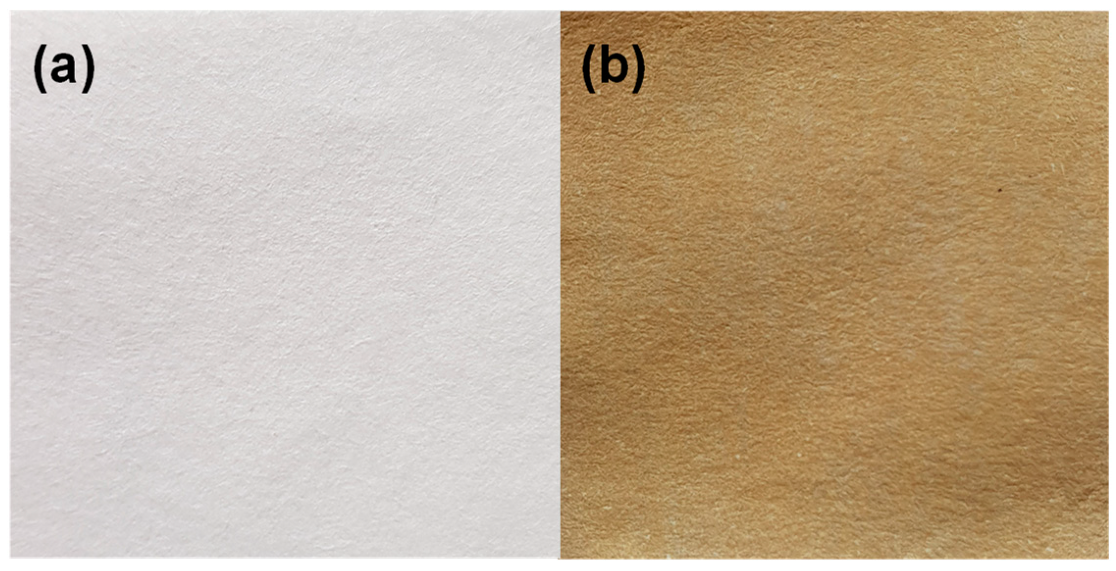
2.1.1. Synthesis of Nanoparticles Supported
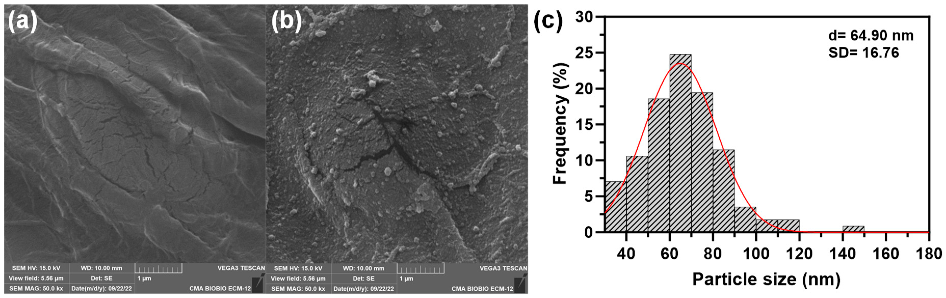
Figure 1a shows photographic images of pristine cellulose paper as support and Figure 1b shows the cellulose-based paper with CuNPs. The color change in the cellulose-based paper, typical color of Cu2O [30,31], suggests an effective formation of Cu2O nanoparticles.

2.1.2. SEM Analysis

The SEM image of cellulose-based paper is shown in Figure 2a. Figure 2b shows synthesized nanoparticles with a uniform distribution on the paper support. Figure 2c shows that size of Cu2O@CBP varied from 32.3 to 142 nm, with a mean diameter of 64.90 ± 16.76 nm, indicating the formation of primarily nanoparticles.

2.1.3. Raman Analysis

Figure 3a shows the Raman spectra of Cu2O@CBP, ranging from 50 to 1160 cm−1. Signals at 90, 107, 127, 158, 228, 311, 419, 450, 495, 528, 548, 612, 634, 658, 790, 827, and 1114 cm−1 are exhibited for Cu2O@CBP, confirming the synthesis of Cu2O nanoparticles [32,33,34,35,36,37,38] according to the results obtained from the XRD analyzes. Additional peaks at 179, 198, 332, 336, 385, 458, 571, 589, 687, 697, 721, 753, 774, 807, 842, 875, 938, 961, 970, 992, 1020, 1042, 1135, and 1150 cm−1 are related to phenolic compounds [39,40]. Peaks at 252, 349, 916, 1075, and 1095 cm−1 are related to cellulose in paper [41,42]. TGA analysis shows a mass loss stage around 340 °C in Cu2O@CBP samples corresponding to the phase transition from Cu2O to CuO [43], which was corroborated by Balık et al. [38] using XRD, FTIR, and Raman spectroscopy (Figure S1).

2.1.4. XRD Analysis

Figure 3b shows the XRD analysis for cellulose-based paper and Cu2O@CBP, which reveals diffraction peaks at 2θ values of 29.53°, 36.37°, 42.32°, and 61.39°. These peaks correspond to the lattice planes (110), (111), (200), and (220), respectively, and are consistent with the monoclinic Cu2O phase (JCPDS N° 05-0667) [44]. It is worth noting that no other signals related to other copper-derived nanoparticle structures were observed despite the fact that this is a green synthesis method. According to the Debye–Scherrer equation, the average crystalline size of Cu2O@CBP is estimated to be 78.74 nm, which closely matches the particle size obtained from SEM analysis. A diffraction peak close to 34° is typical of cellulose [45].

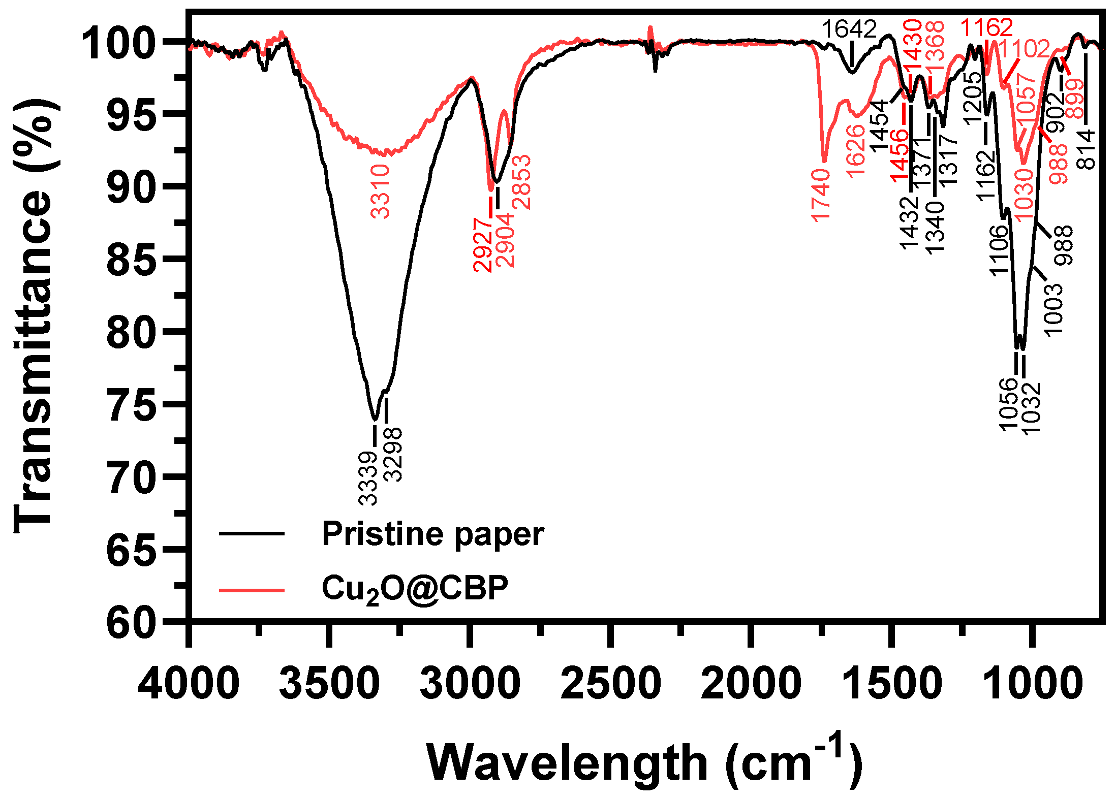
2.1.5. FTIR Analysis

Figure 4 shows the FTIR spectrum for pristine paper and Cu2O@CBP.
Table 1 summarizes the main signals of the pristine paper and Cu2O@CBP by FTIR.
The vibrational bands at 3339, 3298, 2904, 2853, 1454, 1371, 1340, 1317, 1205, 1162, 1056, 1003, 988, 902, and 814 cm−1 in pristine paper are attributed to cellulose and show a decreased intensity in Cu2O@CBP. This reduction is due to the interaction of cellulose with nanoparticles [54] and the interaction of cellulose chemical groups with Cu2O nanoparticles [45]. Additionally, the decrease in signals may be attributed to the interaction of phytochemicals in E. globulus extract with cellulose functional groups [55].
FTIR analysis of Cu2O@CBP reveals additional bands that are not present in pristine paper samples. For example, the 1626 cm−1 band, which is characteristic of phenolic compounds, is likely due to their interaction with Cu2O nanoparticles [56]. Another band appears near 1740 cm−1, which is attributed to C=O bonds. One possible explanation for this phenomenon is the oxidation of -OH groups in catechol-type phenolic compounds during the reduction of metallic ions [57], such as Cu2+ to Cu+, in the synthesis of Cu2O [58]. Additionally, there are slight shifts in the cellulose bands at 1106 and 1032 cm−1, indicating possible interactions between cellulose functional groups and Cu2O nanoparticles or phytochemicals from the E. globulus extract [45,59].

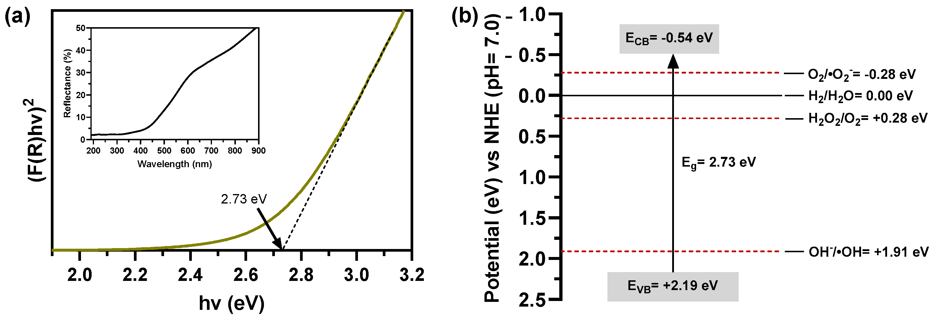
2.1.6. Optical Analysis

Figure 5a shows the reflectance diffuse and Tauc plot used to determine the optical band-gap values (2.73 eV) of Cu2O@CBP [60].
To determine the conduction and valence band positions of Cu2O on Cu2O@CBP, the following empirical relations can be used (Equations (1) and (2)) [61]:
E C B = χ E e 0.5 E g
E V B = E C B + E g
where χ is the absolute electronegativity of semiconductor which defined as the arithmetic mean of the atomic electron affinity and the first ionization energy (5.32 eV for Cu2O), Eg is the bandgap of the semiconductor, and Ee is the energy of free electrons on the hydrogen scale (~4.5 eV), ECB is the conduction band potential, and EVB is the valence band potential [62]. For Cu2O@CBP, the calculated values are ECB = −0.54 eV and EVB = 2.19 eV (Figure 5b). To generate reactive oxygen species (ROS) via redox reactions, the conduction band of Cu2O must be above the reduction potential of O2/O2· and H2O2/O2, and the valence band must be below the oxidation potential of OH/·OH [63]. Figure 5b shows that electrons in the conduction band of the synthesized Cu2O nanoparticles can be captured by O2 to produce O2· and H2O2, while holes in the valence band can react with OH to produce ·OH. The band gap energy of Cu2O is typically around 2.0–2.2 eV [22,26,64]. However, Cu2O samples synthesized on paper exhibit an unusual increase in band gap energy. This shift is primarily due to changes in particle size [64,65,66], exposed crystalline facets [65,67,68], ligand interactions [69,70], and material defects [71,72,73]. Evidence shows the effect of Cu2O nanoparticle sizes, with band gap energy impacting optical properties [65]. The authors have reported that through their analysis of different sizes and structures of Cu2O nanoparticles, they have found that a decrease in nanoparticle size leads to an increase in the band gap for each exposed facet of the crystalline structure. Specifically, they have observed that Cu2O nanoparticles with the (111) facet and diameters of 85 and 117 nm have band gaps of 2.70 and 2.61 eV, respectively, which align with the results of their study. These findings indicate that both particle size and exposed facets have a significant impact on the band gap of the synthesized Cu2O nanoparticles.

2.2. Identification of Biomolecules Involved in the Synthesis of Cu2O Nanoparticles

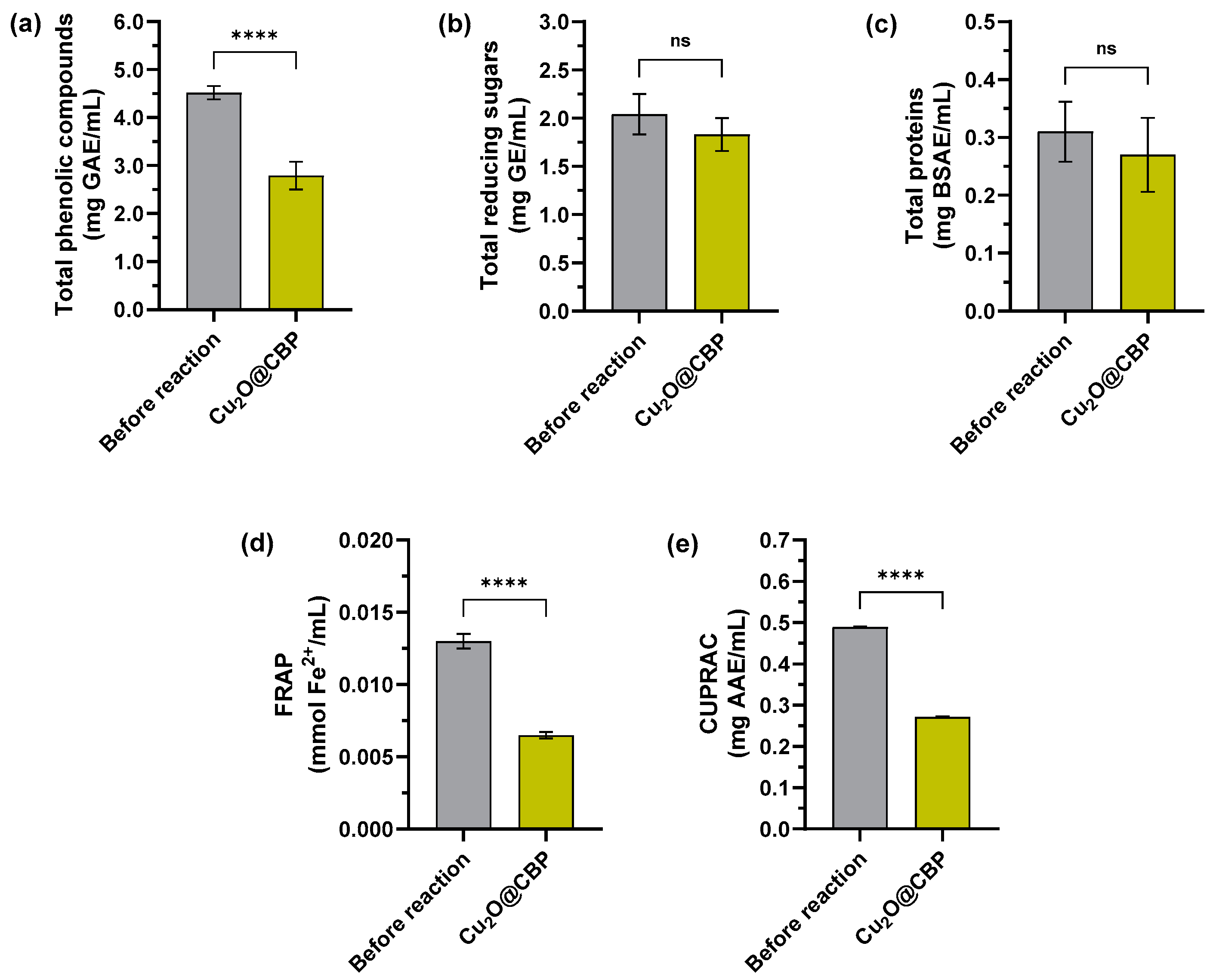
2.2.1. Spectrophotometric Analysis of Extracts before and after Formation Cu2O Nanoparticles

Changes in the phenolic compounds, reducing sugars, proteins, FRAP, and CUPRAC of E. globulus extract before and after the synthesis of Cu2O on paper are shown in Figure 6. No significant changes in the concentrations of reducing sugars or proteins were observed between the initial and post-synthesis stages of Cu2O nanoparticle production. Studies indicate that these proteins do not actively participate in the reduction of Cu2+ to Cu2O or other nanoparticles [74,75,76]. While some authors attribute a reducing function to sugars in leaf extracts during Cu2O synthesis [19,20], others suggest sugars primarily serve a stabilizing role [74,77]. In contrast, significant changes were observed in the concentrations of phenolic compounds, which play crucial roles as reducers and stabilizers in Cu2O nanoparticle synthesis [49,74,77,78,79]. This is supported by FRAP and CUPRAC analyses, highlighting the antioxidant capacity of plant extracts and the presence of phenolic compounds [80] and underscoring their crucial role in the formation of Cu2O nanoparticles.

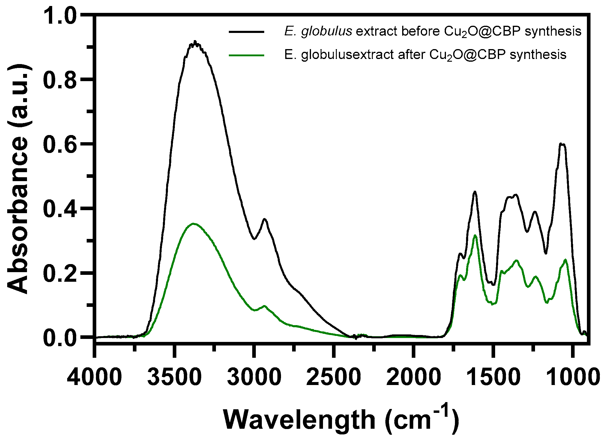
2.2.2. FTIR Analysis of Extracts before and after Formation of Cu2O Nanoparticles

A comparative experiment was conducted to examine the formation of Cu2O nanoparticles on paper substrates using E. globulus extract. The aim of the experiment was to understand the role of plant extract components in the synthesis process. FTIR measurements were used to identify the primary functional groups involved in Cu2O formation. Figure 7 shows the FTIR spectra of E. globulus extract before and after Cu2O synthesis on paper. Significant decreases were observed in signals around 3375 cm−1 (alcoholic and phenolic -OH groups) [81], 2936 cm−1 (C–H asymmetric stretching) [81], 1700 cm−1 (C=O stretching), 1610 cm−1 (C=C stretching) [81], 1350 cm−1 (C–H bending) [58], 1450 cm−1 (C=C aromatic ring stretching) [49], 1400 cm−1 (C-OH stretching) [49], 1250 cm−1 (C–O stretching in aromatic esters) [81], and 1060 cm−1 (C–O–C asymmetric stretching) [77]. These signals, attributed to various phenolic compounds in E. globulus extracts, decrease after nanoparticle synthesis, indicating their involvement in the process.

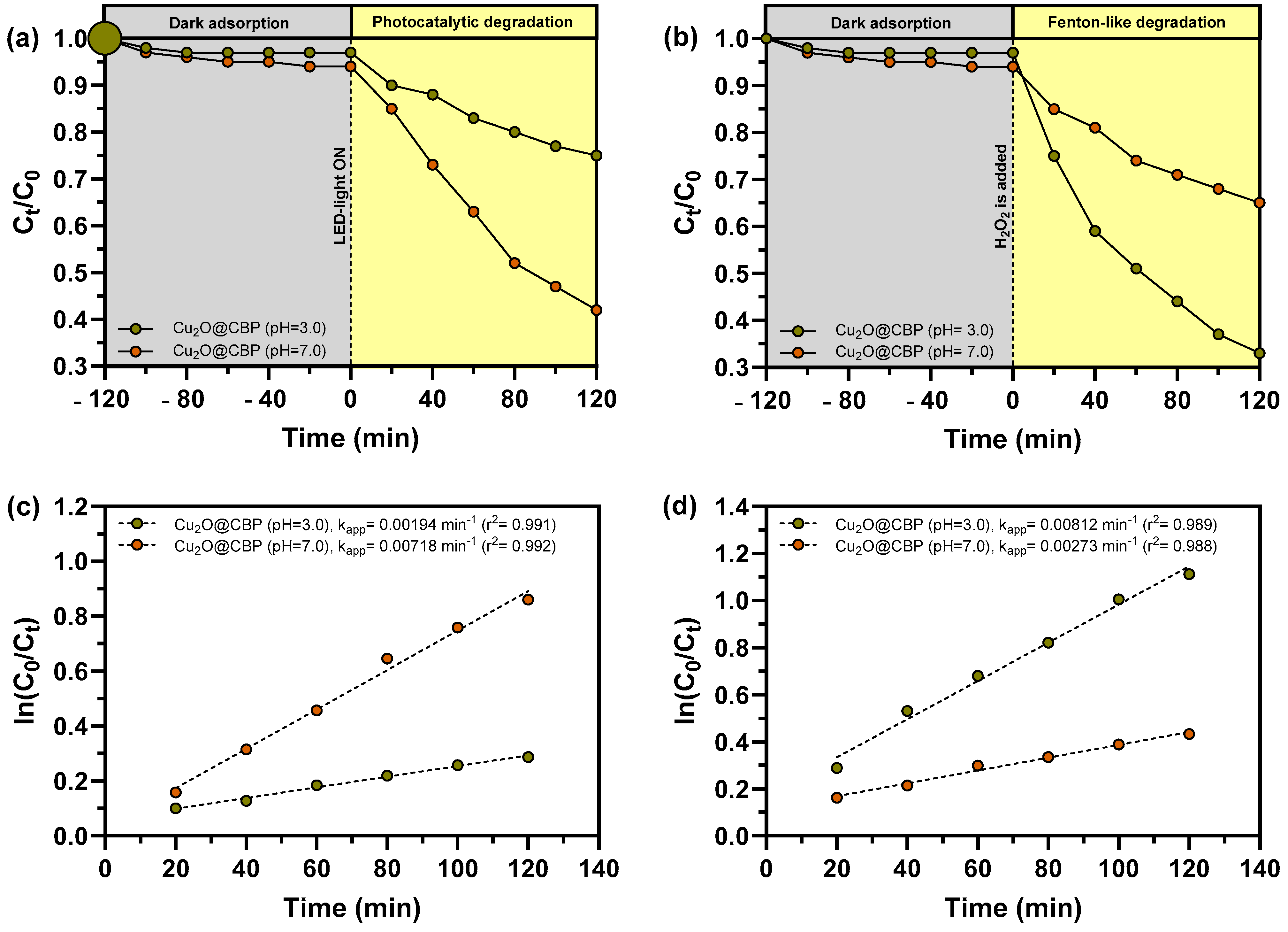
2.3. Photocatalytic and Fenton-Like Degradation of MB

The degradation efficiency of MB by Cu2O@CBP was evaluated under photocatalysis and heterogeneous Fenton-like reaction conditions at pH 3.0 and 7.0 to compare their performance. Figure 8a illustrates the photocatalytic degradation kinetics of MB catalyzed by Cu2O@CBP at pH 3.0 and 7.0. Notably, Cu2O@CBP at pH 7.0 demonstrates the highest photocatalytic efficiency, with an apparent rate constant (kapp) of 0.00718 min−1, compared to 0.00194 min−1 at pH 3.0 (Figure 8c). This highlights the significant impact of pH on the photocatalytic process. The enhanced degradation of MB at basic pH is attributed to stronger electrostatic interactions between the negatively charged Cu2O and the cationic MB. Similar findings suggest that these interactions facilitate greater MB adsorption on the Cu2O surface, thereby increasing the degradation rate [82]. Furthermore, dark adsorption experiments confirm that MB adsorption is more substantial at pH 7.0 than at pH 3.0, supporting the proposed mechanism. This pH-dependent behavior is consistent with observations in systems using other adsorbents for MB removal [83,84] as well as Cu2O nanoparticles for the removal of various contaminants [85].
Figure 8b illustrates the degradation of MB through a heterogeneous Fenton-like process using Cu2O@CBP at pH 3.0 and 7.0, highlighting the significant impact of pH on the reaction. The highest apparent rate constant (kapp) is observed for Cu2O@CBP at pH 3.0 (0.00812 min−1), followed by Cu2O@CBP at pH 7.0 (0.00273 min−1) (Figure 8d). This trend is consistent with other studies that have utilized Cu2O for Fenton-like reactions [2,23,86,87,88,89]. The enhanced degradation at acidic pH can be attributed to several factors: (i) acidic conditions promote the dissolution of Cu2O, resulting in the formation of Cu(I) ions that readily react with H2O2 to produce ·OH radicals [23,86,87,88], (ii) at higher pH levels, H2O2 decomposes, reducing its effectiveness in generating ·OH radicals [23,89], and (iii) ·OH radicals may react with OH ions at high pH, decreasing the availability of ·OH for MB degradation [90].
In this study, it is important to note that the Cu2O@CBP systems do not achieve the same efficiency in dye removal as other similar studies [91]. Several studies have utilized Cu2O or Cu2O-based nanoparticles for photocatalytic removal of MB under visible radiation. Table 2 provides a summary of these investigations, including the type and concentration of the catalyst (if reported), synthesis method, duration of MB removal, and percentage of MB removal achieved. When comparing the efficiencies and experimental conditions of MB removal achieved in this work with those reported in other studies using Cu2O-based nanomaterials, notable differences emerge. Specifically, the photocatalytic processes for MB removal (Table 2) demonstrate comparable or superior efficiencies in the system investigated here, especially considering the low mass of Cu2O used and the high concentration of MB treated. Additionally, a similar compilation to the previous one was conducted using research on MB removal through heterogeneous Fenton-like systems catalyzed by Cu2O or Cu2O-based nanoparticles. Table 3 presents the type of nanoparticle, the synthesis method, the duration of MB removal, the initial MB concentration, the initial H2O2 concentration, and the percentage of MB removed. When comparing similar systems for MB removal via heterogeneous Fenton-like reactions, the efficiencies observed in this study are also comparable or superior. This is particularly significant given the lower concentration of H2O2 used in this research compared to other studies (Table 3), highlighting the effectiveness of our approach despite lower Cu2O concentrations.

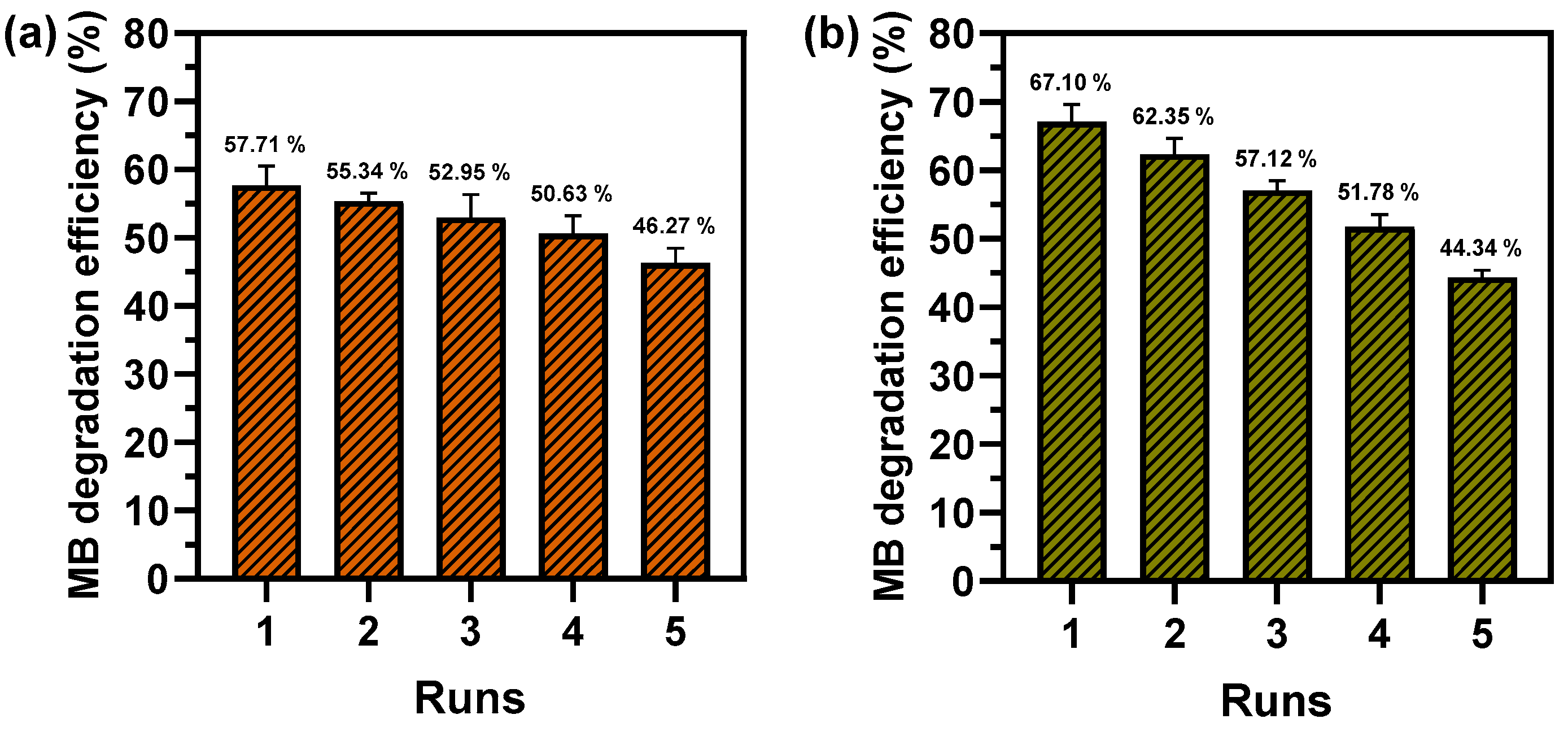
2.4. Reusability of Photocatalytic and Fenton-Like Degradation of MB

Reusability studies were conducted to assess the reusability of Cu2O@CBP for MB dye removal through photocatalysis at pH 7.0 and Fenton-like reactions at pH 3.0 (Figure 9). The results showed a decrease in photocatalytic degradation efficiency from 57.71% to 46.27% after five cycles (Figure 9a), while Fenton-like degradation efficiency dropped from 67.10% to 44.34% (Figure 9b). It was observed that the photocatalytic system maintained a more sustained efficiency, with a less significant decline in MB degradation compared to the Fenton-like system. The performance of Cu2O@CBP in Fenton-like systems was lower, which could be attributed to either greater Cu2O dissolution at acidic pH [25], or the reaction of Cu+ on the Cu2O surface with H2O2, resulting in reduced Cu+ availability, as it converts to Cu2+ [101]. A possible explanation for the decline in performance of both processes could be attributed to the inadequate washing of Cu2O@CBP between cycles. This may result in dye particles remaining attached to the surface of the nanoparticles, affecting their effectiveness in the subsequent cycle.

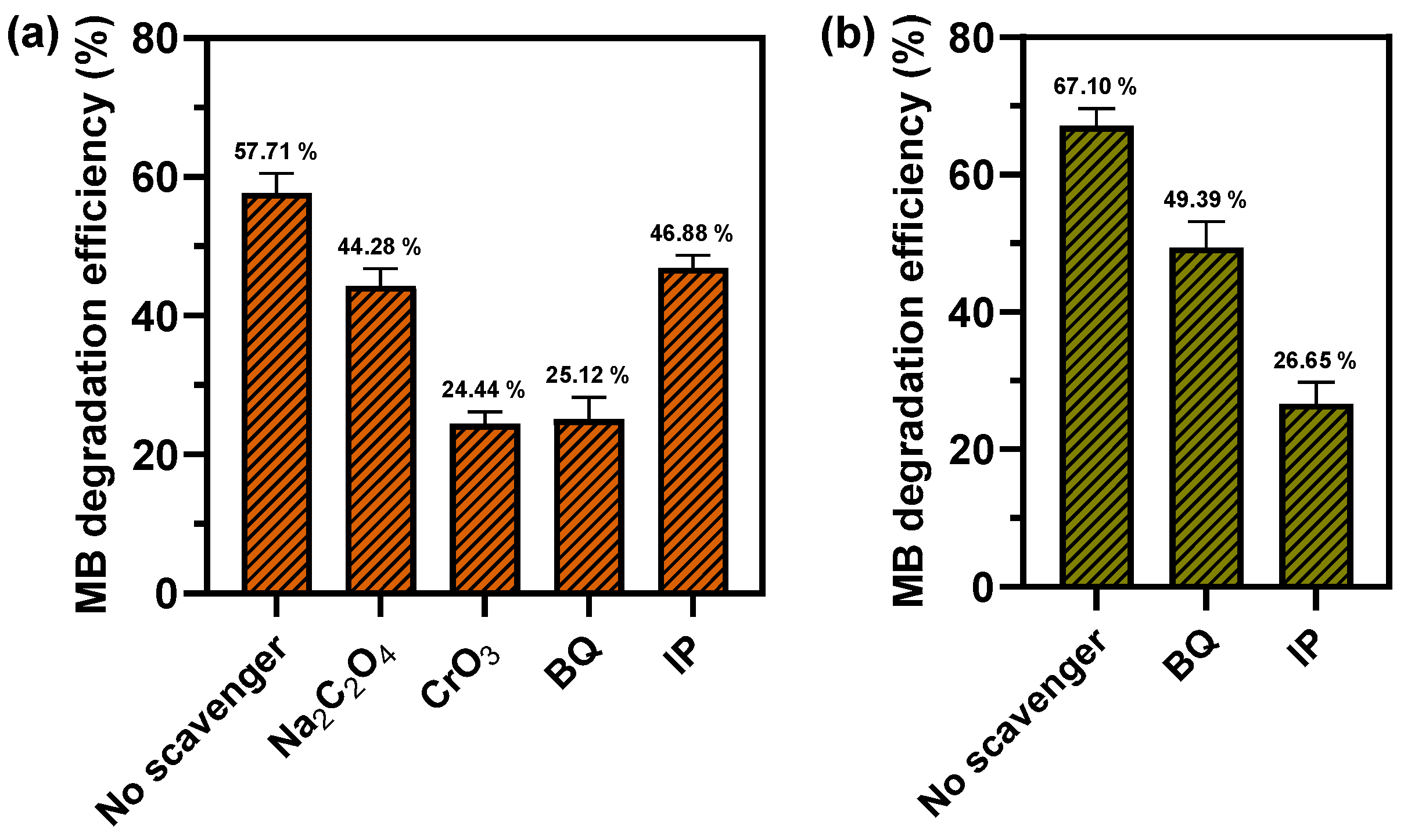
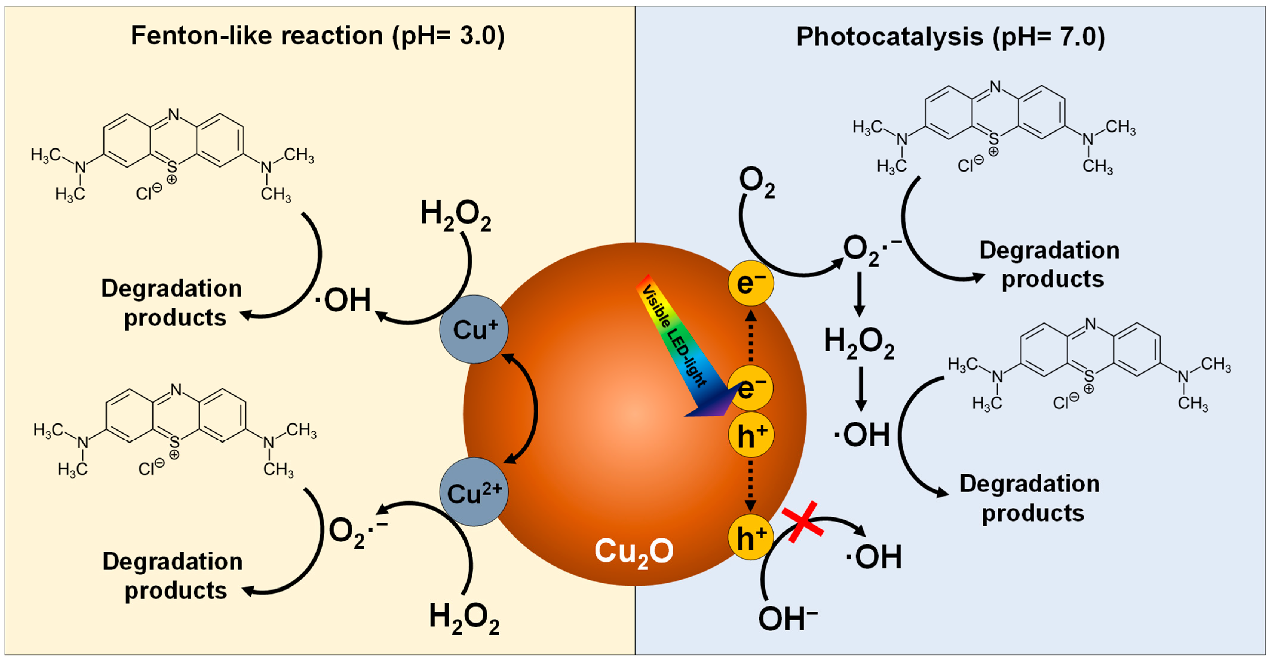
2.5. Reactive Species in Photocatalytic and Fenton-Like Degradation of MB

To clarify the mechanism behind the photocatalytic and Fenton-like degradation of MB, we investigated the reactive species involved (Figure 10). Instead of measuring the degradation efficiency in a solution containing both MB and Cu2O@CBP, we used scavengers to detect the initial steps of photocatalysis, such as the generation of electrons (e) and holes (h+), and to identify radicals such as ·OH and O2·. Specifically, we used CrO3 as an e scavenger, Na2C2O4 as an h+ scavenger, p-benzoquinone (BQ) as an O2· scavenger, and isopropanol (IP) as an ·OH scavenger.
In pH 7.0 photocatalytic systems, the addition of Na2C2O4 had a minimal effect on the degradation efficiency of MB, reducing it by only 13.43% for Cu2O@CBP compared to systems without scavengers (Figure 10a). However, the use of CrO3 significantly reduced the degradation efficiency by 39.27%, indicating effective migration of photoexcited electrons to the Cu2O surface. This suggests that reduction reactions are more critical than oxidation reactions, as the minimal impact of Na2C2O4 suggests low availability of positive holes (h+) on the Cu2O surface. Additionally, the presence of BQ resulted in a 29.83% decrease in MB degradation efficiency, while IP caused a substantial reduction of 52.45%. This highlights the primary role of ·OH in MB degradation compared to O2·. These findings are consistent with research that observed that ·OH and electrons are the main species responsible for MB degradation using a CuO/Cu2O heterostructure under visible light [102].
The low presence of h+ on the Cu2O surface suggests that ·OH, the key species in MB degradation, are produced through parallel reactions (Equations (3)–(9)) rather than the direct oxidation of hydroxide ions (OH) by h+ [24,82,102].
Cu 2 O + h ν Cu 2 O   ( h + + e )
O 2 + e O 2 ·
O 2 + 2 H 2 O + 2 e H 2 O 2 + 2 OH
2 H + + O 2 · H 2 O 2  
H 2 O 2 + e · OH + OH
H 2 O 2   2 · OH  
H 2 O 2 + O 2 · · OH + OH +   O 2
A study was conducted by Chu and Huang [103] on the facet-dependent photocatalytic properties of Cu2O using scavengers. Their results and trends were similar to those found in the present study for Cu2O nanoparticles with a predominantly octahedral structure. Their findings suggest that the primary source of photocatalytic activity in octahedral Cu2O is the migration of electrons (e) to the catalyst surface, which facilitates the production of O2· and ·OH radicals. This is in contrast with the weak migration of holes (h+) to the surface.
In the Fenton-like system at pH 3.0, the addition of BQ, an O2· scavenger, resulted in a 17.71% decrease in MB degradation efficiency for Cu2O@CBP compared to systems without scavengers (Figure 10b). Conversely, the addition of IP, an ·OH scavenger, resulted in a more significant reduction of 50.45%. This suggests that ·OH plays a more crucial role in MB degradation than O2·, which is consistent with previous findings in Cu2O/H2O2 systems at acidic pH [23,100]. The reasons for this behavior include: (i) ·OH has a higher redox potential (1.5–2.8 V) compared to O2· (O2·/H2O2 = +0.94 V, O2/O2· = −0.35 V) [104,105], (ii) The reaction between Cu+ and H2O2 to produce ·OH and Cu2+ (Equation (10)) occurs at a faster rate (k = 4 × 10⁵ M−1 s−1 at pH 6–8) compared to the reaction between Cu2+ and H2O2 (Equation (11)) to produce O2· and Cu+ (k < 1 M−1 s−1 at pH 7) [106], and (iii) H2O2 interaction on the Cu2O surface leads to ·OH generation through O-O bond cleavage [23]. These reactions could involve both surface-bound copper ions (=Cu+ and =Cu2+) and dissolved ions.
Cu + + H 2 O 2 Cu 2 + + · OH   +   OH  
Cu 2 + + H 2 O 2 Cu + + O 2 · +   2 H +
Overall, these insights into the roles of reactive species and the effect of pH provide a comprehensive understanding of the mechanisms driving the photocatalytic and Fenton-like degradation of MB in the presence of Cu2O@CBP (Figure 11).

3. Materials and Methods

3.1. Preparation of E. Globulus Extracts

To prepare the E. globulus extracts, fresh leaves were collected from Concepcion in southern Chile, thoroughly washed with deionized water to remove any surface impurities, and then air-dried at room temperature. The dried leaves were ground into a fine powder using a mechanical grinder. An aqueous extract was prepared by boiling the leaf powder (60 g/L) in deionized water at 60 °C for 20 min. The mixture was allowed to cool to room temperature and then filtered through a 0.45 µm membrane filter to remove any solid residues. The resulting filtrate was stored at 2 °C for subsequent use in the synthesis of Cu2O nanoparticles [107].

3.2. Synthesis of Cu2O Nanoparticles on Cellulose Paper

The synthesis of Cu2O nanoparticles on cellulose paper was achieved through a multi-step process. First, 4 × 4 cm pieces of cellulose paper were immersed in 50 mL of a 0.5 M CuCl2 solution and stirred (200 rpm) at 60 °C for 30 min. The paper was then removed and the CuCl2 solution was mixed with 10 mL of a 1 g/50 mL NaOH solution to precipitate Cu(OH)2. The paper pieces were re-immersed in this Cu(OH)2 suspension and stirred (200 rpm) for an additional 30 min at 60 °C. After removing the paper, it was placed in 25 mL of the prepared E. globulus extract and stirred (200 rpm) at 60 °C for 30 min to facilitate the reduction of Cu(OH)2 to Cu2O nanoparticles (approximate pH of 8.5). The paper was then washed thoroughly with deionized water and dried at 60 °C. Cellulose-based paper with Cu2O is named as Cu2O@CBP.

3.3. Characterization Techniques

To investigate the morphology and elemental composition of the synthesized Cu2O nanoparticles, scanning electron microscopy (SEM) was employed. The SEM images were captured using a Hitachi SU3500 SEM (Bruker, Germany).
X-ray diffraction (XRD) was utilized to determine the crystalline structure of the Cu2O nanoparticles. XRD patterns were obtained using a Bruker D4 ENDEAVOR X-ray diffractometer with Cu Kα radiation. The operating voltage and current were set at 40 kV and 20 mA, respectively. Diffraction peak intensities were recorded within a 10–80° (2θ) range, with increments of 0.02° and a count duration of 0.3 s per increment. The average crystallite size was calculated using the Debye–Scherrer equation (Equation (12)).
D = K λ β cos θ
Raman spectroscopy was performed using a LabRam HR Evolution Horiba Jobin Yvon spectrometer equipped with a 633 nm laser, focusing the laser spot with an Olympus 10× VIS optical lens and a NUV camera (B/S UV 50/50 + Lens F125 D25).
Fourier-transform infrared spectroscopy (FTIR) was conducted to identify functional groups before and after the synthesis, and the interactions between the Eucalyptus extract and the Cu2O nanoparticles. FTIR spectra were obtained using a Perkin Elmer Spectrum Frontier/Spotlight 400 Microscopy System with a linear array of mercury–cadmium–tellurium (MCT) detectors.
The optical properties of the nanoparticles were evaluated using UV-Vis diffuse reflectance spectroscopy (DRS). DRS spectra were recorded with a Jasco V-750 UV–Visible spectrophotometer equipped with a Jasco ISV-922 integrating sphere. The bandgap energy of the Cu2O nanoparticles was determined from Tauc plots derived from the DRS data.
Thermogravimetric analysis (TGA) was used to assess the thermal stability of the Cu2O nanoparticles on cellulose paper. TGA measurements were performed using a Perkin-Elmer STA 6000 instrument, heating the samples at a rate of 15 °C/min under a nitrogen flow of 40 mL/min from 25 °C to 600 °C.
Phenolic compounds, reducing sugars, and proteins were analyzed in the E. globulus extracts before and after synthesis of Cu2O nanoparticles [107]. Phenolic compounds were measured by Folin–Ciocalteu method and expressed as gallic acid equivalents. The reducing sugars were analyzed using the DNS method and calculated as glucose equivalents. Protein in extracts was measured using the Bradford method quantified using bovine albumin serum equivalents. Phytochemicals in the E. globulus extract with reducing capacity in the formation of Cu2O nanoparticles were measured by the ferric-reducing antioxidant power method (FRAP) and cupric reducing antioxidant capacity (CUPRAC) [80].

3.4. Photocatalytic and Fenton-Like Degradation of MB

The photocatalytic and Fenton-like activities of the Cu2O nanoparticles were evaluated using MB as a model pollutant. For photocatalytic degradation, 20 mL of a 50 mg/L MB solution in a glass reactor containing a 4 × 4 cm piece of the Cu2O supported on cellulose paper was placed in 3D printed supports (Figure 12). The reactor was stirred for 120 min in the dark to achieve equilibrium. Subsequently, the reactor was irradiated with LED light (wavelength range: 400–700 nm) for 120 min while being continuously stirred. The effect of pH on photocatalytic activity was studied by adjusting the solution to pH 3.0 and 7.0 using HCl and NaOH. Samples were taken at regular intervals, and the concentration of MB was measured using a UV–Vis spectrophotometer (Jasco, V-750, Tokyo, Japan) at 664 nm.
For Fenton-like degradation, 20 mL of a 50 mg/L MB solution was added to a glass reactor containing the Cu2O supported on cellulose paper placed in printed supports (Figure 12). The reactor was stirred for 120 min in the dark to achieve equilibrium. The reaction was started by adding 50 mM of H2O2. The pH of the solution was adjusted to 3.0 and 7.0 to study its effect on the degradation process. Samples were taken at regular intervals, and the concentration of MB was measured using a UV–Vis spectrophotometer (Jasco, V-750) at 664 nm.
The degradation percentage of MB and the first-order apparent degradation rate constant (kapp) were calculated using Equations (13) and (14) respectively:
MB   degradation   % = C 0 C t C 0 ×   100
ln C t C 0 = K a p p t
where C0 is the initial MB concentration, Ct is the remaining MB concentration after “t” min, and kapp (min−1) is the first-order apparent rate constant for MB degradation by photocatalysis and Fenton-like reaction.

3.5. Reusability Studies

To assess the reusability of the Cu2O nanoparticles, the photocatalytic and Fenton-like MB degradation experiments were repeated for five cycles. After each cycle, the Cu2O supported on cellulose paper was washed with deionized water and dried at 50 °C before being reused in the next experiment. The degradation efficiency of MB was calculated for each cycle to evaluate the stability and reusability of the catalysts.

3.6. Reactive-Species-Trapping Experiments

Reactive-species-trapping experiments were conducted to identify the primary reactive species involved in the degradation processes. Specific scavengers were used: isopropanol (IP) for ·OH, p-benzoquinone (BQ) for O2·, Na2C2O4 for holes (h+) and CrO3 for electrons (e). The experiments were performed under the same conditions as the photocatalytic and Fenton-like degradation tests, with the addition of appropriate scavengers [103].

4. Conclusions

This research demonstrates the potential of immobilizing Cu2O nanoparticles on cost-effective supports such as paper cellulose-based materials through an eco-friendly synthesis method. The synthesis of Cu2O@CBP using E. globulus extracts resulting in nanoparticles with a mean size of 64.9 nm. The degradation of MB was more effective using Cu2O@CBP in both photocatalysis (57.71% at pH 7.0, kapp = 0.00718 min−1) under visible light and Fenton-like reactions (67.1% at pH 3.0, kapp = 0.00812 min−1). Reusability studies have shown that Cu2O@CBP can be recycled multiple times with high efficiency. However, in Fenton-like systems, the efficiency declines at a faster rate (22.76% after 5 cycles) compared to photocatalysis (11.44% after five cycles). Although other systems utilizing Cu2O-based nanoparticles have demonstrated successful removal of MB through photocatalysis and Fenton-like reactions, this study highlights the effectiveness of Cu2O@CBP. Therefore, for the removal of similar recalcitrant compounds in acidic contaminated water, the use of Cu2O@CBP in Fenton-like systems would be more advantageous, while for near-neutral-pH contaminated water, the utilization of Cu2O@CBP in photocatalysis is recommended. These findings enhance the future applicability of these technologies, providing a sustainable and versatile approach to water purification and environmental remediation. In the future, we plan to investigate the impact of additional variables on the removal of contaminants. We will also examine the effectiveness of Cu2O@CBP in photocatalysis and heterogeneous Fenton-like reactions for other dyes or contaminants. Additionally, we will explore the role of copper ion leaching, particularly in heterogeneous Fenton-like processes. Furthermore, we aim to identify the specific reasons for the decrease in removal effectiveness after multiple uses.

Supplementary Materials

The following supporting information can be downloaded at: https://www.mdpi.com/article/10.3390/catal14080525/s1, Figure S1: TGA curves of (a) paper pristine and (b) Cu2O@CBP.

Author Contributions

Conceptualization, P.S.; methodology, P.S.; software, K.M.; validation, K.M.; formal analysis, P.S. and K.M.; investigation, P.S.; resources, P.S.; writing—original draft preparation, P.S.; writing—review and editing, G.V.; visualization, P.S.; project administration, G.V. and P.S.; funding acquisition, G.V. All authors have read and agreed to the published version of the manuscript.

Funding

This study was funded by ANID/FONDAP/1523A0001 and ANID Convocatoria Nacional Subvención a Instalación en la Academia convocatoria año 2019 Folio PAI77190082.

Data Availability Statement

Data are contained within the article and Supplementary Materials.

Acknowledgments

The authors thank the Centro de Microscopía Avanzada, CMA BIO-BIO, Proyecto PIA-ANID ECM-12.

Conflicts of Interest

The authors declare that they have no known competing financial interests or personal relationships that could have appeared to influence the work reported in this paper.

References

  1. Yao, J.; Huang, L.; Li, Y.; Zhu, M.; Liu, S.; Shu, S.; Huang, L.; Wu, X. Enhanced photo-degradation of pollutants in three dimensions by rGO/Bi12O15Cl6/InVO4: LED-light photocatalytic performance and mechanism investigation. Ceram. Int. 2023, 49, 19422–19433. [Google Scholar] [CrossRef]
  2. Sun, B.; Li, H.; Li, X.; Liu, X.; Zhang, C.; Xu, H.; Zhao, X.S. Degradation of Organic Dyes over Fenton-Like Cu2O–Cu/C Catalysts. Ind. Eng. Chem. Res. 2018, 57, 14011–14021. [Google Scholar] [CrossRef]
  3. Jo, W.-K.; Tayade, R.J. New Generation Energy-Efficient Light Source for Photocatalysis: LEDs for Environmental Applications. Ind. Eng. Chem. Res. 2014, 53, 2073–2084. [Google Scholar] [CrossRef]
  4. Ma, D.; Yi, H.; Lai, C.; Liu, X.; Huo, X.; An, Z.; Li, L.; Fu, Y.; Li, B.; Zhang, M.; et al. Critical review of advanced oxidation processes in organic wastewater treatment. Chemosphere 2021, 275, 130104. [Google Scholar] [CrossRef] [PubMed]
  5. Salgado, P.; Frontela, J.L.; Vidal, G. Optimization of Fenton Technology for Recalcitrant Compounds and Bacteria Inactivation. Catalysts 2020, 10, 1483. [Google Scholar] [CrossRef]
  6. Salgado, P.; Melin, V.; Durán, Y.; Mansilla, H.; Contreras, D. The Reactivity and Reaction Pathway of Fenton Reactions Driven by Substituted 1,2-Dihydroxybenzenes. Environ. Sci. Technol. 2017, 51, 3687–3693. [Google Scholar] [CrossRef] [PubMed]
  7. Guo, S.; Chen, M.; You, L.; Wei, Y.; Cai, C.; Wei, Q.; Zhang, H.; Zhou, K. 3D printed hierarchically porous zero-valent copper for efficient pollutant degradation through peroxymonosulfate activation. Sep. Purif. Technol. 2023, 305, 122437. [Google Scholar] [CrossRef]
  8. Salgado, P.; Melin, V.; Albornoz, M.; Mansilla, H.; Vidal, G.; Contreras, D. Effects of pH and substituted 1,2-dihydroxybenzenes on the reaction pathway of Fenton-like systems. Appl. Catal. B Environ. 2018, 226, 93–102. [Google Scholar] [CrossRef]
  9. Salgado, P.; Melin, V.; Contreras, D.; Moreno, Y.; Mansilla, H.D. Fenton reaction driven by iron ligands. J. Chil. Chem. Soc. 2013, 58, 2096–2101. [Google Scholar] [CrossRef]
  10. Zhou, M.; Ji, C.; Ji, F.; Chen, M.; Zhong, Z.; Xing, W. Micro-Octahedron Cu2O-Based Photocatalysis-Fenton for Organic Pollutant Degradation: Proposed Coupling Mechanism in a Membrane Reactor. Ind. Eng. Chem. Res. 2022, 61, 7255–7265. [Google Scholar] [CrossRef]
  11. Tony, M.A.; Mansour, S.A. Microwave-assisted catalytic oxidation of methomyl pesticide by Cu/Cu2O/CuO hybrid nanoparticles as a Fenton-like source. Int. J. Environ. Sci. Technol. 2020, 17, 161–174. [Google Scholar] [CrossRef]
  12. Lopis, A.D.; Choudhari, K.S.; Sai, R.; Kanakikodi, K.S.; Maradur, S.P.; Shivashankar, S.A.; Kulkarni, S.D. Laddered type-1 heterojunction: Harvesting full-solar-spectrum in scavenger free photocatalysis. Sol. Energy 2022, 240, 57–68. [Google Scholar] [CrossRef]
  13. Dong, J.; Xu, H.; Zhang, F.; Chen, C.; Liu, L.; Wu, G. Synergistic effect over photocatalytic active Cu2O thin films and their morphological and orientational transformation under visible light irradiation. Appl. Catal. A Gen. 2014, 470, 294–302. [Google Scholar] [CrossRef]
  14. Akbal, F. Photocatalytic degradation of organic dyes in the presence of titanium dioxide under UV and solar light: Effect of operational parameters. Environ. Prog. 2005, 24, 317–322. [Google Scholar] [CrossRef]
  15. Su, Y.; Nathan, A.; Ma, H.; Wang, H. Precise control of Cu2O nanostructures and LED-assisted photocatalysis. RSC Adv. 2016, 6, 78181–78186. [Google Scholar] [CrossRef]
  16. Salgado, P.; Mártire, D.O.; Vidal, G. Eucalyptus extracts-mediated synthesis of metallic and metal oxide nanoparticles: Current status and perspectives. Mater. Res. Express 2019, 6, 082006. [Google Scholar] [CrossRef]
  17. Salgado, P.; Márquez, K.; Rubilar, O.; Contreras, D.; Vidal, G. The effect of phenolic compounds on the green synthesis of iron nanoparticles (FexOy-NPs) with photocatalytic activity. Appl. Nanosci. 2019, 9, 371–385. [Google Scholar] [CrossRef]
  18. Kerour, A.; Boudjadar, S.; Bourzami, R.; Allouche, B. Eco-friendly synthesis of cuprous oxide (Cu2O) nanoparticles and improvement of their solar photocatalytic activities. J. Solid State Chem. 2018, 263, 79–83. [Google Scholar] [CrossRef]
  19. Amrani, M.A.; Srikanth, V.V.S.S.; Labhsetwar, N.K.; Al-Fatesh, A.S.; Shaikh, H. Phoenix dactylifera mediated green synthesis of Cu2O particles for arsenite uptake from water. Sci. Technol. Adv. Mater. 2016, 17, 760–768. [Google Scholar] [CrossRef]
  20. Muthukumaran, M.; Niranjani, S.; Barnabas, K.S.; Narayanan, V.; Raju, T.; Venkatachalam, K. Green Route Synthesis and Characterization of Cuprous Oxide (Cu2O): Visible light Irradiation photocatalytic activity of MB Dye. Mater. Today Proc. 2019, 14, 563–568. [Google Scholar] [CrossRef]
  21. Hodges, B.C.; Cates, E.L.; Kim, J.-H. Challenges and prospects of advanced oxidation water treatment processes using catalytic nanomaterials. Nat. Nanotechnol. 2018, 13, 642–650. [Google Scholar] [CrossRef] [PubMed]
  22. Tu, K.; Wang, Q.; Lu, A.; Zhang, L. Portable Visible-Light Photocatalysts Constructed from Cu2O Nanoparticles and Graphene Oxide in Cellulose Matrix. J. Phys. Chem. C 2014, 118, 7202–7210. [Google Scholar] [CrossRef]
  23. Zhou, H.; Kang, L.; Zhou, M.; Zhong, Z.; Xing, W. Membrane enhanced COD degradation of pulp wastewater using Cu2O/H2O2 heterogeneous Fenton process. Chin. J. Chem. Eng. 2018, 26, 1896–1903. [Google Scholar] [CrossRef]
  24. Su, X.; Chen, W.; Han, Y.; Wang, D.; Yao, J. In-situ synthesis of Cu2O on cotton fibers with antibacterial properties and reusable photocatalytic degradation of dyes. Appl. Surf. Sci. 2021, 536, 147945. [Google Scholar] [CrossRef] [PubMed]
  25. Zhang, L.; Li, J.; Chen, Z.; Tang, Y.; Yu, Y. Preparation of Fenton reagent with H2O2 generated by solar light-illuminated nano-Cu2O/MWNTs composites. Appl. Catal. A Gen. 2006, 299, 292–297. [Google Scholar] [CrossRef]
  26. Sabbaghan, M.; Argyropoulos, D.S. Synthesis and characterization of nano fibrillated cellulose/Cu2O films; micro and nano particle nucleation effects. Carbohydr. Polym. 2018, 197, 614–622. [Google Scholar] [CrossRef] [PubMed]
  27. Kalubowila, K.D.R.N.; Gunewardene, M.S.; Jayasingha, J.L.K.; Dissanayake, D.; Jayathilaka, C.; Jayasundara, J.M.D.; Gao, Y.; Jayanetti, J.K.D.S. Reduction-Induced Synthesis of Reduced Graphene Oxide-Wrapped Cu2O/Cu Nanoparticles for Photodegradation of Methylene Blue. ACS Appl. Nano Mater. 2021, 4, 2673–2681. [Google Scholar] [CrossRef]
  28. Kumar, M.; Das, R.R.; Samal, M.; Yun, K. Highly stable functionalized cuprous oxide nanoparticles for photocatalytic degradation of methylene blue. Mater. Chem. Phys. 2018, 218, 272–278. [Google Scholar] [CrossRef]
  29. Pariona, N.; Basurto Cereceda, S.; Mondaca, F.; Carrión, G.; Mtz-Enriquez, A.I. Antifungal activity and degradation of methylene blue by ZnO, Cu, and Cu2O/Cu nanoparticles, a comparative study. Mater. Lett. 2021, 301, 130182. [Google Scholar] [CrossRef]
  30. Zhang, Z.; Che, H.; Wang, Y.; Gao, J.; Zhao, L.; She, X.; Sun, J.; Gunawan, P.; Zhong, Z.; Su, F. Facile Synthesis of Mesoporous Cu2O Microspheres with Improved Catalytic Property for Dimethyldichlorosilane Synthesis. Ind. Eng. Chem. Res. 2012, 51, 1264–1274. [Google Scholar] [CrossRef]
  31. Park, J.Y.; Lee, S.; Kim, Y.; Ryu, Y.B. Antimicrobial Activity of Morphology-Controlled Cu2O Nanoparticles: Oxidation Stability under Humid and Thermal Conditions. Materials 2024, 17, 261. [Google Scholar] [CrossRef]
  32. Prabhakaran, G.; Murugan, R. Synthesis of Cu2O Nanospheres and Cubes: Their Structural, Optical and Magnetic Properties. Adv. Mater. Res. 2014, 938, 114–117. [Google Scholar] [CrossRef]
  33. Sahai, A.; Goswami, N.; Kaushik, S.D.; Tripathi, S. Cu/Cu2O/CuO nanoparticles: Novel synthesis by exploding wire technique and extensive characterization. Appl. Surf. Sci. 2016, 390, 974–983. [Google Scholar] [CrossRef]
  34. Yu, P.Y.; Shen, Y.R.; Petroff, Y. Resonance Raman scattering in Cu2O at the blue and indigo excitons. Solid State Commun. 1973, 12, 973–975. [Google Scholar] [CrossRef]
  35. Solache-Carranco, H.; Juarez-Diaz, G.; Galvan-Arellano, M.; Martinez-Juarez, J.; Romero-Paredes, R.G.; Pena-Sierra, R. Raman scattering and photoluminescence studies on Cu2O. In Proceedings of the 2008 5th International Conference on Electrical Engineering, Computing Science and Automatic Control, Mexico City, Mexico, 12–14 November 2008; pp. 421–424. [Google Scholar]
  36. Ansari, A.; Jha, A.K.; Akhtar, M.J. Preparation and characterization of oxidized electrolytic copper powder and its dielectric properties at microwave frequency. Powder Technol. 2015, 286, 459–467. [Google Scholar] [CrossRef]
  37. Sander, T.; Reindl, C.T.; Klar, P.J. Breaking of Raman selection rules in Cu2O by intrinsic point defects. MRS Online Proc. Libr. 2013, 1633, 81–86. [Google Scholar] [CrossRef]
  38. Balık, M.; Bulut, V.; Erdogan, I.Y. Optical, structural and phase transition properties of Cu2O, CuO and Cu2O/CuO: Their photoelectrochemical sensor applications. Int. J. Hydrogen Energy 2019, 44, 18744–18755. [Google Scholar] [CrossRef]
  39. Krysa, M.; Szymańska-Chargot, M.; Zdunek, A. FT-IR and FT-Raman fingerprints of flavonoids—A review. Food Chem. 2022, 393, 133430. [Google Scholar] [CrossRef] [PubMed]
  40. Espina, A.; Sanchez-Cortes, S.; Jurašeková, Z. Vibrational Study (Raman, SERS, and IR) of Plant Gallnut Polyphenols Related to the Fabrication of Iron Gall Inks. Molecules 2022, 27, 279. [Google Scholar] [CrossRef]
  41. Edwards, H.G.M.; Farwell, D.W.; Williams, A.C. FT-Raman spectrum of cotton: A polymeric biomolecular analysis. Spectrochim. Acta Part A Mol. Spectrosc. 1994, 50, 807–811. [Google Scholar] [CrossRef]
  42. Agarwal, U.P.; Ralph, S.A.; Baez, C.; Reiner, R.S. Detection and quantitation of cellulose II by Raman spectroscopy. Cellulose 2021, 28, 9069–9079. [Google Scholar] [CrossRef]
  43. Gupta, D.; Meher, S.R.; Illyaskutty, N.; Alex, Z.C. Facile synthesis of Cu2O and CuO nanoparticles and study of their structural, optical and electronic properties. J. Alloys Compd. 2018, 743, 737–745. [Google Scholar] [CrossRef]
  44. Gupta, A.; Maruthapandi, M.; Das, P.; Saravanan, A.; Jacobi, G.; Natan, M.; Banin, E.; Luong, J.H.T.; Gedanken, A. Cuprous Oxide Nanoparticles Decorated Fabric Materials with Anti-biofilm Properties. ACS Appl. Bio Mater. 2022, 5, 4310–4320. [Google Scholar] [CrossRef] [PubMed]
  45. Sedighi, A.; Montazer, M.; Samadi, N. Synthesis of nano Cu2O on cotton: Morphological, physical, biological and optical sensing characterizations. Carbohydr. Polym. 2014, 110, 489–498. [Google Scholar] [CrossRef]
  46. Abidi, N.; Cabrales, L.; Haigler, C.H. Changes in the cell wall and cellulose content of developing cotton fibers investigated by FTIR spectroscopy. Carbohydr. Polym. 2014, 100, 9–16. [Google Scholar] [CrossRef] [PubMed]
  47. Yang, A.-L.; Li, S.-P.; Wang, Y.-J.; Wang, L.-L.; Bao, X.-C.; Yang, R.-Q. Fabrication of Cu2O@Cu2O core–shell nanoparticles and conversion to Cu2O@Cu core–shell nanoparticles in solution. Trans. Nonferrous Met. Soc. China 2015, 25, 3643–3650. [Google Scholar] [CrossRef]
  48. Grasel, F.d.S.; Ferrão, M.F.; Wolf, C.R. Development of methodology for identification the nature of the polyphenolic extracts by FTIR associated with multivariate analysis. Spectrochim. Acta Part A Mol. Biomol. Spectrosc. 2016, 153, 94–101. [Google Scholar] [CrossRef] [PubMed]
  49. Mohamed, E.A. Green synthesis of copper & copper oxide nanoparticles using the extract of seedless dates. Heliyon 2020, 6, e03123. [Google Scholar] [CrossRef]
  50. Ventura-Cruz, S.; Tecante, A. Extraction and characterization of cellulose nanofibers from Rose stems (Rosa spp.). Carbohydr. Polym. 2019, 220, 53–59. [Google Scholar] [CrossRef]
  51. Chinnaiah, K.; Maik, V.; Kannan, K.; Potemkin, V.; Grishina, M.; Gohulkumar, M.; Tiwari, R.; Gurushankar, K. Experimental and Theoretical Studies of Green Synthesized Cu2O Nanoparticles Using Datura metel L. J. Fluoresc. 2022, 32, 559–568. [Google Scholar] [CrossRef]
  52. Muthulakshmi, L.; Varada Rajalu, A.; Kaliaraj, G.S.; Siengchin, S.; Parameswaranpillai, J.; Saraswathi, R. Preparation of cellulose/copper nanoparticles bionanocomposite films using a bioflocculant polymer as reducing agent for antibacterial and anticorrosion applications. Compos. Part B Eng. 2019, 175, 107177. [Google Scholar] [CrossRef]
  53. Patle, T.K.; Shrivas, K.; Kurrey, R.; Upadhyay, S.; Jangde, R.; Chauhan, R. Phytochemical screening and determination of phenolics and flavonoids in Dillenia pentagyna using UV–vis and FTIR spectroscopy. Spectrochim. Acta Part A Mol. Biomol. Spectrosc. 2020, 242, 118717. [Google Scholar] [CrossRef] [PubMed]
  54. Musino, D.; Rivard, C.; Landrot, G.; Novales, B.; Rabilloud, T.; Capron, I. Hydroxyl groups on cellulose nanocrystal surfaces form nucleation points for silver nanoparticles of varying shapes and sizes. J. Colloid Interface Sci. 2021, 584, 360–371. [Google Scholar] [CrossRef] [PubMed]
  55. Phan, A.D.T.; D’Arcy, B.R.; Gidley, M.J. Polyphenol–cellulose interactions: Effects of pH, temperature and salt. Int. J. Food Sci. Technol. 2016, 51, 203–211. [Google Scholar] [CrossRef]
  56. Mannarmannan, M.; Biswas, K. Phytochemical-Assisted Synthesis of Cuprous Oxide Nanoparticles and Their Antimicrobial Studies. ChemistrySelect 2021, 6, 3534–3539. [Google Scholar] [CrossRef]
  57. Salgado, P.; Contreras, D.; Mansilla, H.D.; Márquez, K.; Vidal, G.; Cobos, C.J.; Mártire, D.O. Experimental and computational investigation of the substituent effects on the reduction of Fe3+ by 1,2-dihydroxybenzenes. New J. Chem. 2017, 41, 12685–12693. [Google Scholar] [CrossRef]
  58. Taherzadeh Soureshjani, P.; Shadi, A.; Mohammadsaleh, F. Algae-mediated route to biogenic cuprous oxide nanoparticles and spindle-like CaCO3: A comparative study, facile synthesis, and biological properties. RSC Adv. 2021, 11, 10599–10609. [Google Scholar] [CrossRef] [PubMed]
  59. Mohammadi, F.; Montazer, M.; Mianehro, A.; Eslahi, N.; Mahmoudi Rad, M. Preparation of Antibacterial Cellulose Fabric via Copper (II) Oxide and Corn Silk (Stigma maydis) Nanocomposite. Starch Stärke 2023, 75, 2200156. [Google Scholar] [CrossRef]
  60. Tauc, J.; Grigorovici, R.; Vancu, A. Optical Properties and Electronic Structure of Amorphous Germanium. Phys. Status Solidi (B) 1966, 15, 627–637. [Google Scholar] [CrossRef]
  61. Alp, E. The Facile Synthesis of Cu2O-Cu hybrid cubes as efficient visible-light-driven photocatalysts for water remediation processes. Powder Technol. 2021, 394, 1111–1120. [Google Scholar] [CrossRef]
  62. Xu, Y.; Schoonen, M.A.A. The absolute energy positions of conduction and valence bands of selected semiconducting minerals. Am. Mineral. 2000, 85, 543–556. [Google Scholar] [CrossRef]
  63. Das, S.; Deka, T.; Ningthoukhangjam, P.; Chowdhury, A.; Nair, R.G. A critical review on prospects and challenges of metal-oxide embedded g-C3N4-based direct Z-scheme photocatalysts for water splitting and environmental remediation. Appl. Surf. Sci. Adv. 2022, 11, 100273. [Google Scholar] [CrossRef]
  64. da Costa, W.V.; Pereira, B.d.S.; Montanha, M.C.; Kimura, E.; Hechenleitner, A.A.W.; de Oliveira, D.M.F.; Pineda, E.A.G. Hybrid materials based on cotton fabric-Cu2O nanoparticles with antibacterial properties against S. aureus. Mater. Chem. Phys. 2017, 201, 339–343. [Google Scholar] [CrossRef]
  65. Huang, J.-Y.; Madasu, M.; Huang, M.H. Modified Semiconductor Band Diagrams Constructed from Optical Characterization of Size-Tunable Cu2O Cubes, Octahedra, and Rhombic Dodecahedra. J. Phys. Chem. C 2018, 122, 13027–13033. [Google Scholar] [CrossRef]
  66. Sreeju, N.; Rufus, A.; Philip, D. Nanostructured copper (II) oxide and its novel reduction to stable copper nanoparticles. J. Phys. Chem. Solids 2019, 124, 250–260. [Google Scholar] [CrossRef]
  67. Chen, T.-N.; Kao, J.-C.; Zhong, X.-Y.; Chan, S.-J.; Patra, A.S.; Lo, Y.-C.; Huang, M.H. Facet-Specific Photocatalytic Activity Enhancement of Cu2O Polyhedra Functionalized with 4-Ethynylanaline Resulting from Band Structure Tuning. ACS Cent. Sci. 2020, 6, 984–994. [Google Scholar] [CrossRef]
  68. Ke, W.-H.; Hsia, C.-F.; Chen, Y.-J.; Huang, M.H. Synthesis of Ultrasmall Cu2O Nanocubes and Octahedra with Tunable Sizes for Facet-Dependent Optical Property Examination. Small 2016, 12, 3530–3534. [Google Scholar] [CrossRef]
  69. Higashimoto, S.; Nishi, T.; Yasukawa, M.; Azuma, M.; Sakata, Y.; Kobayashi, H. Photocatalysis of titanium dioxide modified by catechol-type interfacial surface complexes (ISC) with different substituted groups. J. Catal. 2015, 329, 286–290. [Google Scholar] [CrossRef]
  70. Savić, T.D.; Čomor, M.I.; Nedeljković, J.M.; Veljković, D.Ž.; Zarić, S.D.; Rakić, V.M.; Janković, I.A. The effect of substituents on the surface modification of anatase nanoparticles with catecholate-type ligands: A combined DFT and experimental study. Phys. Chem. Chem. Phys. 2014, 16, 20796–20805. [Google Scholar] [CrossRef]
  71. Wang, Y.; Miska, P.; Pilloud, D.; Horwat, D.; Mücklich, F.; Pierson, J.F. Transmittance enhancement and optical band gap widening of Cu2O thin films after air annealing. J. Appl. Phys. 2014, 115, 073505. [Google Scholar] [CrossRef]
  72. Kumar, A.; Kumar, R.; Verma, N.; Anupama, A.V.; Choudhary, H.K.; Philip, R.; Sahoo, B. Effect of the band gap and the defect states present within band gap on the non-linear optical absorption behaviour of yttrium aluminium iron garnets. Opt. Mater. 2020, 108, 110163. [Google Scholar] [CrossRef]
  73. Norouzzadeh, P.; Mabhouti, K.; Golzan, M.M.; Naderali, R. Investigation of structural, morphological and optical characteristics of Mn substituted Al-doped ZnO NPs: A Urbach energy and Kramers-Kronig study. Optik 2020, 204, 164227. [Google Scholar] [CrossRef]
  74. Yadav, S.; Jain, A.; Malhotra, P. A review on the sustainable routes for the synthesis and applications of cuprous oxide nanoparticles and their nanocomposites. Green Chem. 2019, 21, 937–955. [Google Scholar] [CrossRef]
  75. Santos, S.A.O.; Pinto, R.J.B.; Rocha, S.M.; Marques, P.A.A.P.; Neto, C.P.; Silvestre, A.J.D.; Freire, C.S.R. Unveiling the Chemistry behind the Green Synthesis of Metal Nanoparticles. ChemSusChem 2014, 7, 2704–2711. [Google Scholar] [CrossRef] [PubMed]
  76. Waris, A.; Din, M.; Ali, A.; Ali, M.; Afridi, S.; Baset, A.; Ullah Khan, A. A comprehensive review of green synthesis of copper oxide nanoparticles and their diverse biomedical applications. Inorg. Chem. Commun. 2021, 123, 108369. [Google Scholar] [CrossRef]
  77. Djamila, B.; Eddine, L.S.; Abderrhmane, B.; Nassiba, A.; Barhoum, A. In vitro antioxidant activities of copper mixed oxide (CuO/Cu2O) nanoparticles produced from the leaves of Phoenix dactylifera L. Biomass Convers. Biorefinery 2024, 14, 6567–6580. [Google Scholar] [CrossRef]
  78. Dou, L.; Zhang, X.; Zangeneh, M.M.; Zhang, Y. Efficient biogenesis of Cu2O nanoparticles using extract of Camellia sinensis leaf: Evaluation of catalytic, cytotoxicity, antioxidant, and anti-human ovarian cancer properties. Bioorg. Chem. 2021, 106, 104468. [Google Scholar] [CrossRef]
  79. Atri, A.; Echabaane, M.; Bouzidi, A.; Harabi, I.; Soucase, B.M.; Ben Chaâbane, R. Green synthesis of copper oxide nanoparticles using Ephedra Alata plant extract and a study of their antifungal, antibacterial activity and photocatalytic performance under sunlight. Heliyon 2023, 9, e13484. [Google Scholar] [CrossRef] [PubMed]
  80. Özyürek, M.; Güçlü, K.; Apak, R. The main and modified CUPRAC methods of antioxidant measurement. TrAC Trends Anal. Chem. 2011, 30, 652–664. [Google Scholar] [CrossRef]
  81. Dhara, M.; Kisku, K.; Naik, U.C. Biofunctionalized cuprous oxide nanoparticles synthesized using root extract of Withania somnifera for antibacterial activity. Appl. Nanosci. 2022, 12, 3555–3571. [Google Scholar] [CrossRef]
  82. Akter, J.; Sapkota, K.P.; Hanif, M.A.; Islam, M.A.; Abbas, H.G.; Hahn, J.R. Kinetically controlled selective synthesis of Cu2O and CuO nanoparticles toward enhanced degradation of methylene blue using ultraviolet and sun light. Mater. Sci. Semicond. Process. 2021, 123, 105570. [Google Scholar] [CrossRef]
  83. Bayomie, O.S.; Kandeel, H.; Shoeib, T.; Yang, H.; Youssef, N.; El-Sayed, M.M.H. Novel approach for effective removal of methylene blue dye from water using fava bean peel waste. Sci. Rep. 2020, 10, 7824. [Google Scholar] [CrossRef] [PubMed]
  84. Salazar-Rabago, J.J.; Leyva-Ramos, R.; Rivera-Utrilla, J.; Ocampo-Perez, R.; Cerino-Cordova, F.J. Biosorption mechanism of Methylene Blue from aqueous solution onto White Pine (Pinus durangensis) sawdust: Effect of operating conditions. Sustain. Environ. Res. 2017, 27, 32–40. [Google Scholar] [CrossRef]
  85. Messaadia, L.; Kiamouche, S.; Lahmar, H.; Masmoudi, R.; Boulahbel, H.; Trari, M.; Benamira, M. Solar photodegradation of Rhodamine B dye by Cu2O/TiO2 heterostructure: Experimental and computational studies of degradation and toxicity. J. Mol. Model. 2023, 29, 38. [Google Scholar] [CrossRef] [PubMed]
  86. Qin, H.; Wang, Z.; Yang, S.; Jiang, W.; Gu, Y.; Chen, J.; Yang, G. High efficiency degradation of 2,4-dichlorophenol using Cu2S/Cu2O decorated nanoscale zerovalent iron via adsorption and photocatalytic performances. J. Clean. Prod. 2022, 378, 134587. [Google Scholar] [CrossRef]
  87. Ai, Z.; Xiao, H.; Mei, T.; Liu, J.; Zhang, L.; Deng, K.; Qiu, J. Electro-Fenton Degradation of Rhodamine B Based on a Composite Cathode of Cu2O Nanocubes and Carbon Nanotubes. J. Phys. Chem. C 2008, 112, 11929–11935. [Google Scholar] [CrossRef]
  88. Xia, H.; Zhang, Y.; Zhou, P.; Li, C.; Yang, D. Oxidative degradation of organic pollutants using cuprous oxide in acidic solution: Hydroxyl radical generation. Desalin. Water Treat. 2019, 171, 262–269. [Google Scholar] [CrossRef]
  89. Zayed, M.F.; Eisa, W.H.; Anis, B. Gallic acid-assisted growth of cuprous oxide within polyvinyl alcohol; a separable catalyst for oxidative and reductive degradation of water pollutants. J. Clean. Prod. 2021, 279, 123826. [Google Scholar] [CrossRef]
  90. Wu, M.; He, S.; Ha, E.; Hu, J.; Ruan, S. A facile synthesis of PEGylated Cu2O@SiO2/MnO2 nanocomposite as efficient photo−Fenton−like catalysts for methylene blue treatment. Front. Bioeng. Biotechnol. 2022, 10, 1023090. [Google Scholar] [CrossRef]
  91. Salgado, P.; Rubilar, O.; Salazar, C.; Márquez, K.; Vidal, G. In Situ Synthesis of Cu2O Nanoparticles Using Eucalyptus globulus Extract to Remove a Dye via Advanced Oxidation. Nanomaterials 2024, 14, 1087. [Google Scholar] [CrossRef]
  92. Aguilar, M.S.; Rosas, G. A new synthesis of Cu2O spherical particles for the degradation of methylene blue dye. Environ. Nanotechnol. Monit. Manag. 2019, 11, 100195. [Google Scholar] [CrossRef]
  93. Xu, L.; Xu, H.; Wu, S.; Zhang, X. Synergy effect over electrodeposited submicron Cu2O films in photocatalytic degradation of methylene blue. Appl. Surf. Sci. 2012, 258, 4934–4938. [Google Scholar] [CrossRef]
  94. Sorekine, G.; Anduwan, G.; Waimbo, M.N.; Osora, H.; Velusamy, S.; Kim, S.; Kim, Y.S.; Charles, J. Photocatalytic studies of copper oxide nanostructures for the degradation of methylene blue under visible light. J. Mol. Struct. 2022, 1248, 131487. [Google Scholar] [CrossRef]
  95. Dustgeer, M.R.; Asma, S.T.; Jilani, A.; Raza, K.; Hussain, S.Z.; Shakoor, M.B.; Iqbal, J.; Abdel-wahab, M.S.; Darwesh, R. Synthesis and characterization of a novel single-phase sputtered Cu2O thin films: Structural, antibacterial activity and photocatalytic degradation of methylene blue. Inorg. Chem. Commun. 2021, 128, 108606. [Google Scholar] [CrossRef]
  96. Athariq, M.; Rauf, M.R.; Khairany, I.W.; Adani, I.F.; Sutrisno, M.G. Synthesis and characterization of nanocube Cu2O thin film at room temperature for methylene blue photodegradation application. Chem. Mater. 2023, 2, 67–71. [Google Scholar] [CrossRef]
  97. Kumar, B.; Smita, K.; Debut, A.; Cumbal, L. Andean Sacha Inchi (Plukenetia volubilis L.) Leaf-Mediated Synthesis of Cu2O Nanoparticles: A Low-Cost Approach. Bioengineering 2020, 7, 54. [Google Scholar] [CrossRef] [PubMed]
  98. Ramesh, P.; Rajendran, A. Photocatalytic dye degradation activities of green synthesis of cuprous oxide nanoparticles from Sargassum wightii extract. Chem. Phys. Impact 2023, 6, 100208. [Google Scholar] [CrossRef]
  99. Sun, H.W.; Chu, D.Q.; Liu, L.; Wang, L.M. Preparation of Nanocrystalline Cu2O and Its Catalytic Degradation of Methylene Blue. In Proceedings of the 2010 4th International Conference on Bioinformatics and Biomedical Engineering, Chengdu, China, 18–20 June 2010; pp. 1–3. [Google Scholar]
  100. Cheng, Y.; Lin, Y.; Xu, J.; He, J.; Wang, T.; Yu, G.; Shao, D.; Wang, W.-H.; Lu, F.; Li, L.; et al. Surface plasmon resonance enhanced visible-light-driven photocatalytic activity in Cu nanoparticles covered Cu2O microspheres for degrading organic pollutants. Appl. Surf. Sci. 2016, 366, 120–128. [Google Scholar] [CrossRef]
  101. Zhao, W.; Liang, C.; Wang, B.; Xing, S. Enhanced Photocatalytic and Fenton-like Performance of CuOx-Decorated ZnFe2O4. ACS Appl. Mater. Interfaces 2017, 9, 41927–41936. [Google Scholar] [CrossRef]
  102. Bayat, F.; Sheibani, S. Enhancement of photocatalytic activity of CuO-Cu2O heterostructures through the controlled content of Cu2O. Mater. Res. Bull. 2022, 145, 111561. [Google Scholar] [CrossRef]
  103. Chu, C.-Y.; Huang, M.H. Facet-dependent photocatalytic properties of Cu2O crystals probed by using electron, hole and radical scavengers. J. Mater. Chem. A 2017, 5, 15116–15123. [Google Scholar] [CrossRef]
  104. Li, Y.; Dong, H.; Xiao, J.; Li, L.; Chu, D.; Hou, X.; Xiang, S.; Dong, Q.; Zhang, H. Advanced oxidation processes for water purification using percarbonate: Insights into oxidation mechanisms, challenges, and enhancing strategies. J. Hazard. Mater. 2023, 442, 130014. [Google Scholar] [CrossRef] [PubMed]
  105. Winterbourn, C.C. Biological chemistry of superoxide radicals. ChemTexts 2020, 6, 7. [Google Scholar] [CrossRef]
  106. Liu, Y.; Tan, N.; Guo, J.; Wang, J. Catalytic activation of O2 by Al0-CNTs-Cu2O composite for Fenton-like degradation of sulfamerazine antibiotic at wide pH range. J. Hazard. Mater. 2020, 396, 122751. [Google Scholar] [CrossRef]
  107. Salgado, P.; Bustamante, L.; Carmona, D.J.; Meléndrez, M.F.; Rubilar, O.; Salazar, C.; Pérez, A.J.; Vidal, G. Green synthesis of Ag/Ag2O nanoparticles on cellulose paper and cotton fabric using Eucalyptus globulus leaf extracts: Toward the clarification of formation mechanism. Surf. Interfaces 2023, 40, 102928. [Google Scholar] [CrossRef]
Figure 1. Photographic images of (a) pristine cellulose-based paper and (b) Cu2O@CBP for 4 × 4 cm pieces.
Catalysts 14 00525 g001
Figure 2. SEM images of (a) pristine paper, (b) Cu2O@CBP, and (c) particle size distribution of Cu2O@CBP (The red line represents the best curve fitting using the Gaussian distribution function).
Catalysts 14 00525 g002
Figure 3. (a) Raman spectra for Cu2O@CBP (red numbers for Cu2O nanoparticles, green numbers for phenolic compounds, and black numbers for cellulose), (b) XRD patterns of pristine cellulose-based paper and Cu2O@CBP.
Catalysts 14 00525 g003
Figure 4. FTIR analyses for pristine cellulose-based paper and Cu2O@CBP.
Catalysts 14 00525 g004
Figure 5. (a) Band gap energy (Eg) determination from the Tauc plot (green line) for Cu2O@CBP. The linear part of the plot is extrapolated to the x-axis using a dotted line for Eg determination. (inset graph: DRS spectra for Cu2O@CBP). (b) Potential band diagram for Cu2O@CBP.
Catalysts 14 00525 g005
Figure 6. Analysis of (a) total phenolic compounds, (b) total reducing sugars, (c) total proteins, (d) FRAP, and (e) CUPRAC in E. globulus extract before and after the synthesis of Cu2O@CBP. Differences between groups were compared using ANOVA with Tukey post hoc analysis. **** p < 0.0001, ns: no significant differences.
Catalysts 14 00525 g006
Figure 7. FTIR spectra of E. globulus extract before (black spectra) and after synthesis (green spectra) of Cu2O@CBP.
Catalysts 14 00525 g007
Figure 8. MB removal efficiency by (a) photocatalysis under LED visible light and (b) Fenton-like reaction catalyzed by Cu2O@CBP at pH 3.0 and 7.0 (inset: legend represents samples at different pH). (c) Photocatalytic and (d) Fenton-like calculated degradation rate constant (kapp) for Cu2O@CBP at pH 3.0 and 7.0 (inset: legend represents samples at different pH, kapp, and r2 of a pseudo-first order model).
Catalysts 14 00525 g008
Figure 9. Cycling performance of Cu2O@CBP for MB degradation by (a) photocatalysis under LED visible light at pH = 7.0 and (b) Fenton-like at pH = 3.0 under dark. The data obtained are presented as mean ± standard deviation.
Catalysts 14 00525 g009
Figure 10. MB removal by Cu2O@CBP under influence of scavenging agents (a) Na2C2O4, CrO3, BQ, and IP on photocatalytic degradation under LED visible light, and (b) BQ and IP on Fenton-like under darkness. The data obtained are presented as mean ± standard deviation.
Catalysts 14 00525 g010
Figure 11. Scheme of Fenton-like reaction at acidic pH under dark and photocatalysis at neutral pH under LED visible light to generate reactive species and MB degradation process.
Catalysts 14 00525 g011
Figure 12. (a) Internal view and (b) view from above of the reactor used for photocatalysis and Fenton-like reaction degradation of MB by Cu2O@CBP.
Catalysts 14 00525 g012
Table 1. FTIR bands in pristine paper and Cu2O@CBP.
Wavenumber (cm−1)VibrationReference
Pristine PaperCu2O@CBP
3339-Intra-molecular hydrogen bonding C(3)OH⋯O(5) or C(6)O⋯(O)H[46]
32983310Inter-molecular hydrogen bonding C(3)OH⋯C(6)O[46]
29042927Asymmetric stretching vibration of –CH2[47]
28532853Symmetric stretching vibration of –CH2[47]
-1740C=O stretching[48]
1642-O–H bending of adsorbed water[46]
-1626C=C of aromatic ring[49]
14541456O–H in-plane deformation[46]
14321430CH2 scissoring[50]
13711368C–OH bending[48]
1340-C–O stretching[48]
1317-C–N stretch of aromatic amines[51]
1205-C–O stretching[46]
11621162Anti-symmetrical bridge C–O–C stretching[50]
11061102C–OH bending[49]
10561057Stretching vibration of C–O–C in the pyranose skeletal ring[18]
10321030C–OH groups of cellulose[46]
1003-C–O–H stretching vibration[52]
988988C–O and ring stretching modes[46]
902899Glycosidic deformation –C1–O–C4 characteristic of the β-glycosidic bond of cellulose[50]
814Plane bending of =C–H[53]
Table 2. Comparison of photocatalysis under visible radiation for methylene blue degradation using Cu2O-based nanoparticles.
CatalystNanoparticles
Synthesis
Time (min)MB (mg/L)Removal (%)Ref.
Cu2O@CBP (~50 mg/L of Cu2O) Green synthesis1205057.71This study
Cu2O (5 mg/L)NaBH4 reduction120100070[82]
Cu2O (3150 mg/L)Green synthesis80560[92]
Cu/Cu2O (250 mg/L)Green synthesis15010<10[29]
Cu2O (400 mg/L)Ascorbic acid12010~10[27]
Cu2O on film Electrodeposition1506.25<10[93]
Cu/Cu2O/CuO (1000 mg/L)Chemical synthesis1201046[94]
Cu2O thin filmsMagnetron sputtering1055069.6[95]
Cu2O thin filmElectrodeposition120562[96]
Cu2O (600 mg/L)Ascorbic acid1051083[28]
Cu2OGreen synthesis1501078.9[97]
Cu2O-octaedral (60 mg/L)Membrane-assisted precipitation1205068[10]
Cu2O-spherical (60 mg/L)Membrane-assisted precipitation1205049[10]
Cu2O-micro-octahedrons (60 mg/L)Membrane-assisted precipitation1205033[10]
Cu2O-microspheres (60 mg/L)Membrane-assisted precipitation120506.5[10]
Cu2O (50 mg/L)Green synthesis1801000~60[98]
Table 3. Comparison of Fenton-like heterogeneous reaction for methylene blue degradation using Cu2O-based nanoparticles.
CatalystNanoparticles SynthesisTime (min)MB (mg/L)H2O2 ConcentrationRemoval (%)Ref.
Cu2O@CBP (~50 mg/L of Cu2O)Green
synthesis
120500.05 M67.10This study
Cu/Cu2O (250 mg/L)Green
synthesis
150100.55 M~70[29]
Cu2O (60 mg/L)Membrane-
assisted
precipitation
120500.1 M74[10]
Cu2O (200 mg/L)Sol-gel1301012.63%~75[99]
Cu2O/Cu (300 mg/L)D-glucose60109.8 × 10−3 M28[100]
Disclaimer/Publisher’s Note: The statements, opinions and data contained in all publications are solely those of the individual author(s) and contributor(s) and not of MDPI and/or the editor(s). MDPI and/or the editor(s) disclaim responsibility for any injury to people or property resulting from any ideas, methods, instructions or products referred to in the content.

