Article

α-L-Arabinofuranosidases of Glycoside Hydrolase Families 43, 51 and 62: Differences in Enzyme Substrate and Positional Specificity between and within the GH Families

by
Walid Fathallah
1,2 and
Vladimír Puchart
1,*
1
Institute of Chemistry, Slovak Academy of Sciences, Dúbravská cesta 9, SK-845 38 Bratislava, Slovakia
2
Botany and Microbiology Department, Faculty of Science, Beni-Suef University, Beni-Suef 625 11, Egypt
*
Author to whom correspondence should be addressed.
Catalysts 2024, 14(8), 536; https://doi.org/10.3390/catal14080536 (registering DOI)
Submission received: 14 July 2024 / Revised: 11 August 2024 / Accepted: 15 August 2024 / Published: 17 August 2024
(This article belongs to the Section Biocatalysis)

Abstract

:
The increasing number of uncharacterized proteins in the CAZy database highlights the importance of their functional characterization. Therefore, the substrate and positional specificity of 34 α-L-arabinofuranosidases classified into GH43, GH51, and GH62 families was determined on arabinoxylan, arabinan, and derived oligosaccharides (many enzyme–substrate combinations were examined for the first time) covering all possible kinds of arabinofuranosyl branches using TLC. Arabinoxylan was efficiently debranched by the majority of the tested proteins. Most of them showed AXH-m specificity, acting on 2- or 3-monoarabinosylated substrates, while AXH-d3 specificity (liberation of 3-linked arabinose solely from 2,3 doubly decorated substrates) was found mainly in the subfamily GH43_10, harbouring enzymes of both types. Several GH51 enzymes, however, released arabinose also from a xylooligosaccharide doubly arabinosylated at the non-reducing end. The AXH-m and AXH-d3 specificities correlated well with the dearabinosylation of arabinan and arabinooligosaccharides, which were debranched by all GH51 representatives and some GH43 and GH62 members. The GH51 and GH62 arabinan-debranching enzymes also hydrolyzed debranched arabinan, while within the GH43 family the linear arabinan-degrading ability was found only in the GH43_26 subfamily, comprising specific exo 1,5-α-L-arabinofuranosidases. This study demonstrates a first attempt in the systematic examination of a relationship between CAZy classification and substrate and positional specificities of various α-L-arabinofuranosidases.

Graphical Abstract

1. Introduction

Microorganisms produce a plethora of enzymes in order to decompose and utilize carbohydrates present in plant biomass. Plant biomass represents a complex material encompassing various polysaccharides that are decomposed to oligosaccharides, both of which serve as substrates for many catalysts collectively named Carbohydrate-Active enZymes (CAZymes) [1]. The CAZymes are regularly updated in the CAZy database (http://www.cazy.org/, accessed on 14 July 2024), which covers different classes of enzymes, all of which are organized into families based on similarity in their 3-D structure and the mode of action.
α-L-arabinofuranosidases (EC 3.2.1.55) are a diverse group of glycoside hydrolases that remove the non-reducing-end terminal arabinofuranosyl (Araf) residues from plant polysaccharides such as arabinoxylan, arabinan, and arabinogalactan or the corresponding oligosaccharides by an exo-wise cleavage of α-1,5-, α-1,2-, or α-1,3-arabinofuranosidic linkages [2]. The decoration of arabinoxylan and arabinan is tissue-, species-, and season-specific and the main chain residues of both polysaccharides are substituted by arabinose at position 2 and/or 3 [3]. The frequency and pattern of branching may hinder the enzymatic hydrolysis of the oligo- and polysaccharides [4] or at least result in variations in the primary dearabinosylation step [5], which is regarded as a critical stage for microbial degradation [3].
In CAZy, the α-L-arabinofuranosidases are classified into three glycoside hydrolase families, GH51, GH54, GH62 [6], and sixteen subfamilies of the GH43 family (GH43_1, GH43_10, GH43_11, GH43_12, GH43_14, GH43_16, GH43_18, GH43_19, GH43_21, GH43_26, GH43_27, GH43_29, GH43_33, GH43_34, GH43_35 and GH43_36) [7]. The specificity of these glycosidases towards structurally well-defined substrates varies considerably. There are enzymes attacking arabinose side chains present in both arabinan and arabinoxylan, whereas other types of enzymes may exhibit much narrower substrate specificity towards specifically either arabinan or arabinoxylan [3]. The enzymes active on arabinoxylan (AX) are named arabinoxylan arabinofuranohydrolases (AXHs) [5]. Based on their regiospecificity, the AXHs were classified into two types. Widespread AXH-m enzymes act only on 2- and 3-monosubstituted xylopyranosyl (Xylp) residues. This regiospecificity has been reported to be exhibited by GH51 [8], GH54, GH62 [9], and the majority of GH43 enzymes, namely from the following subfamilies: GH43_10, GH43_12, GH43_16, GH43_19, GH43_29, and GH43_36 [10,11,12,13,14]. The other type, designated AXH-d3, represents the enzymes which specifically release 3-O-linked Araf side chains only from 2,3-diarabinosylated Xylp residues. The AXH-d3 enzymes are hitherto found in subfamilies GH43_10 and GH43_36 [15,16], although lower activity of this type was reported for a GH51 member [12].
Elucidating the catalytic properties of uncharacterized α-L-arabinofuranosidases plays a key role in the determination of a relationship between their CAZy classification into (sub)families and substrate/positional specificity. Such a relationship may serve as a guide to select the best representatives suited for a given application. In this paper, we determined the hydrolytic activity and specificity of 34 α-L-arabinofuranosidases belonging to families GH43, GH51 and GH62 towards four polymers (arabinoxylan, branched and linear arabinan, and arabinogalactan) and twelve corresponding oligosaccharides.

2. Results

2.1. Arabinofuranosidase Activity on the Chromogenic Substrate

The 34 examined α-L-arabinofuranosidases belonging to different GH (sub)families are listed in Table 1. Most enzymes exhibited reasonable activity on 4-nitrophenyl α-L-arabinofuranoside (NPA) as typical α-L-arabinofuranosidases. Their activity on this artificial chromogenic substrate, however, varied considerably and the difference between the most (BsAbf51B; 118 U/mg) and the least active enzyme (BtAbf43A; 3.6 mU/mg) was in a range of five orders of magnitude (Table 1). Large differences were observed among the GH62 and GH43 enzymes (often even within the subfamilies), while NPA is generally a very good substrate for GH51 members (Table 1).

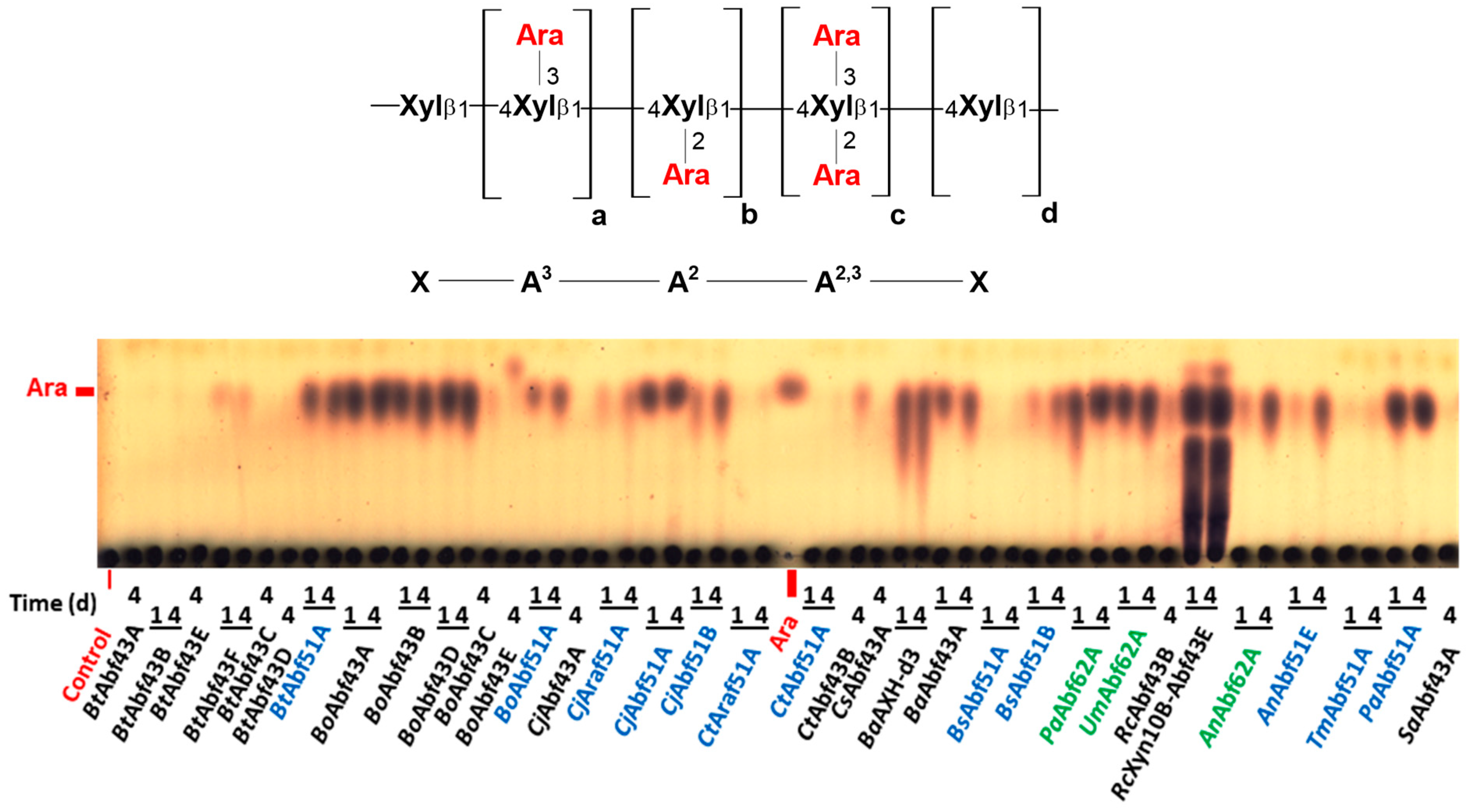
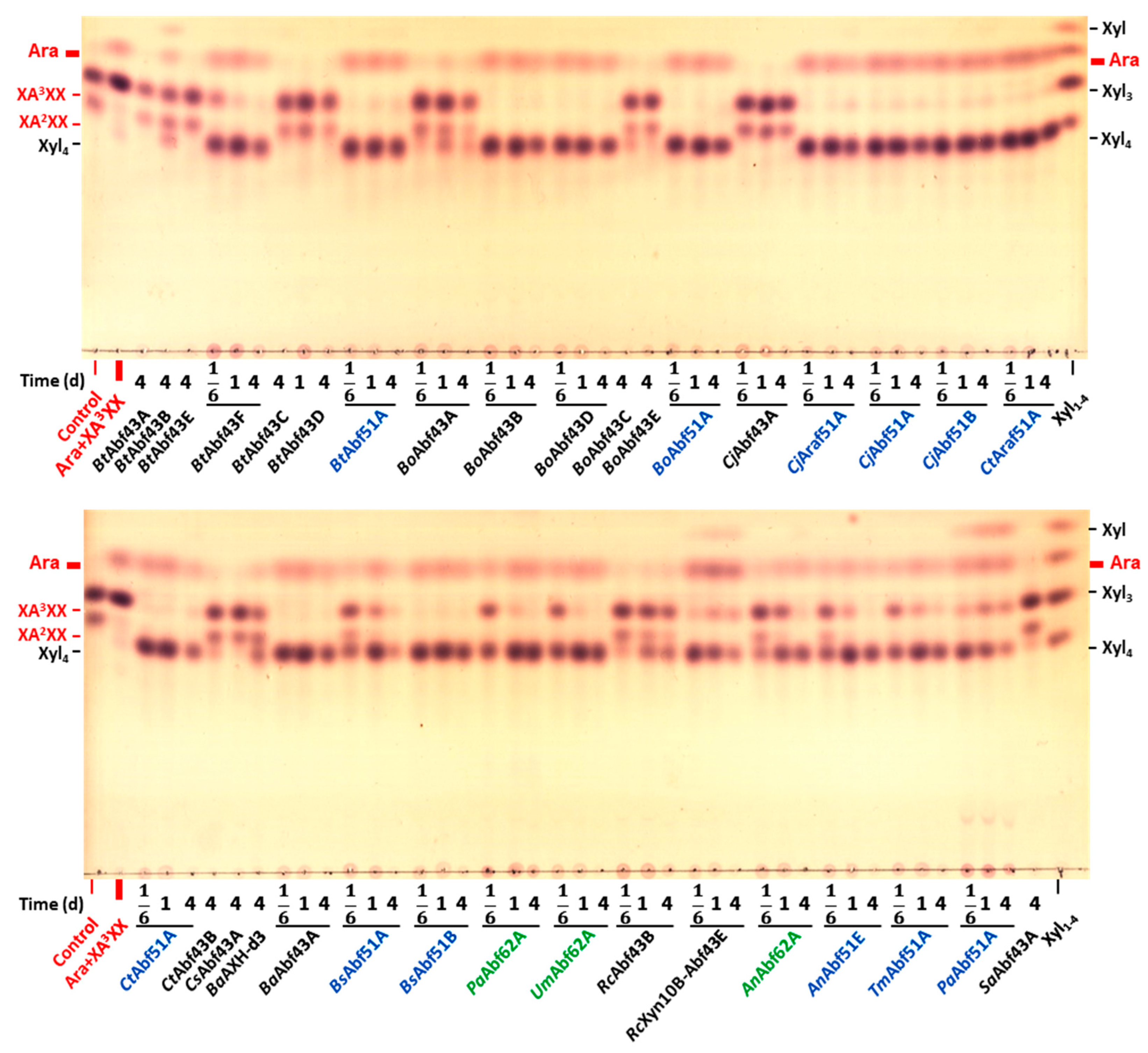
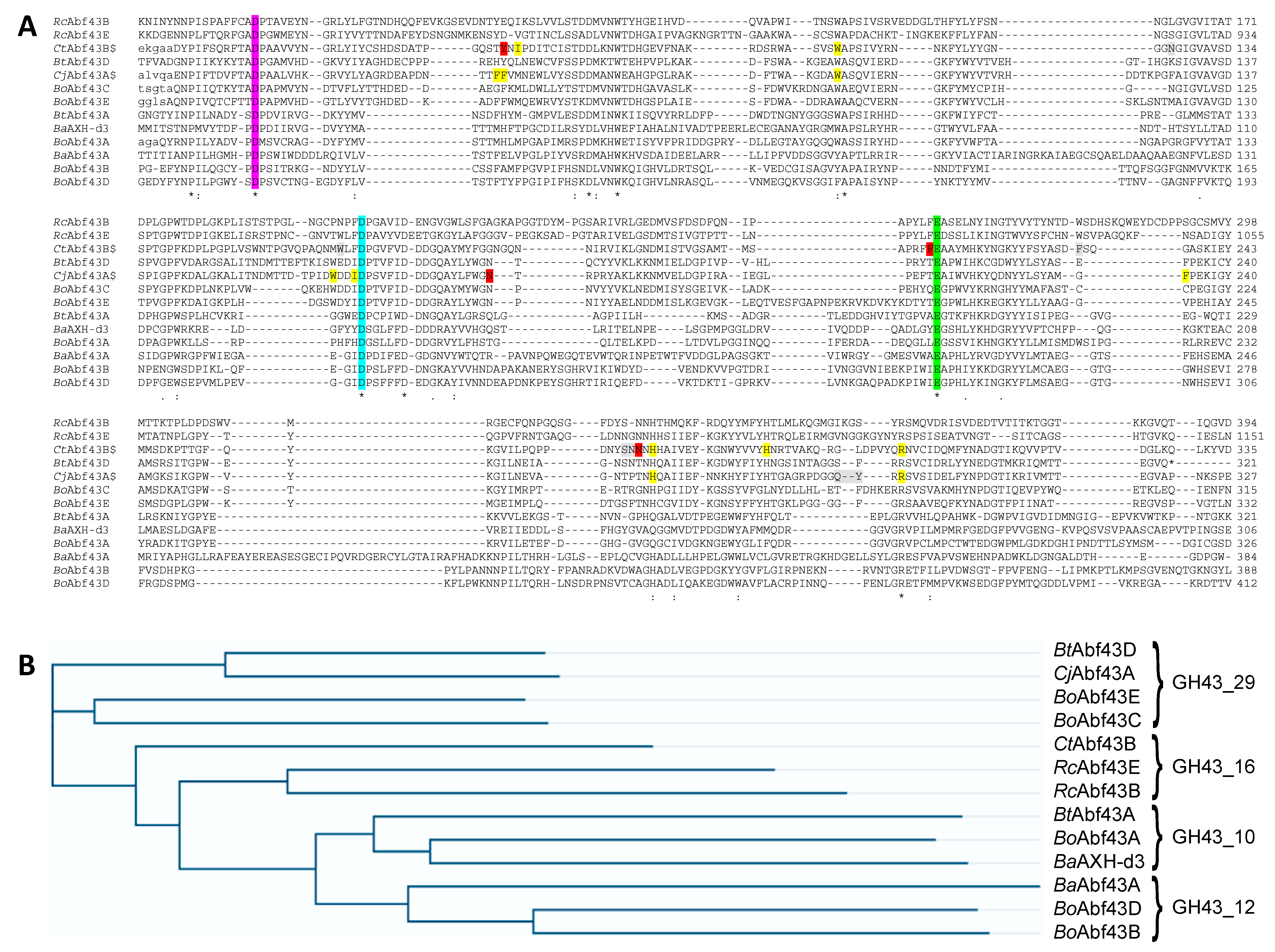
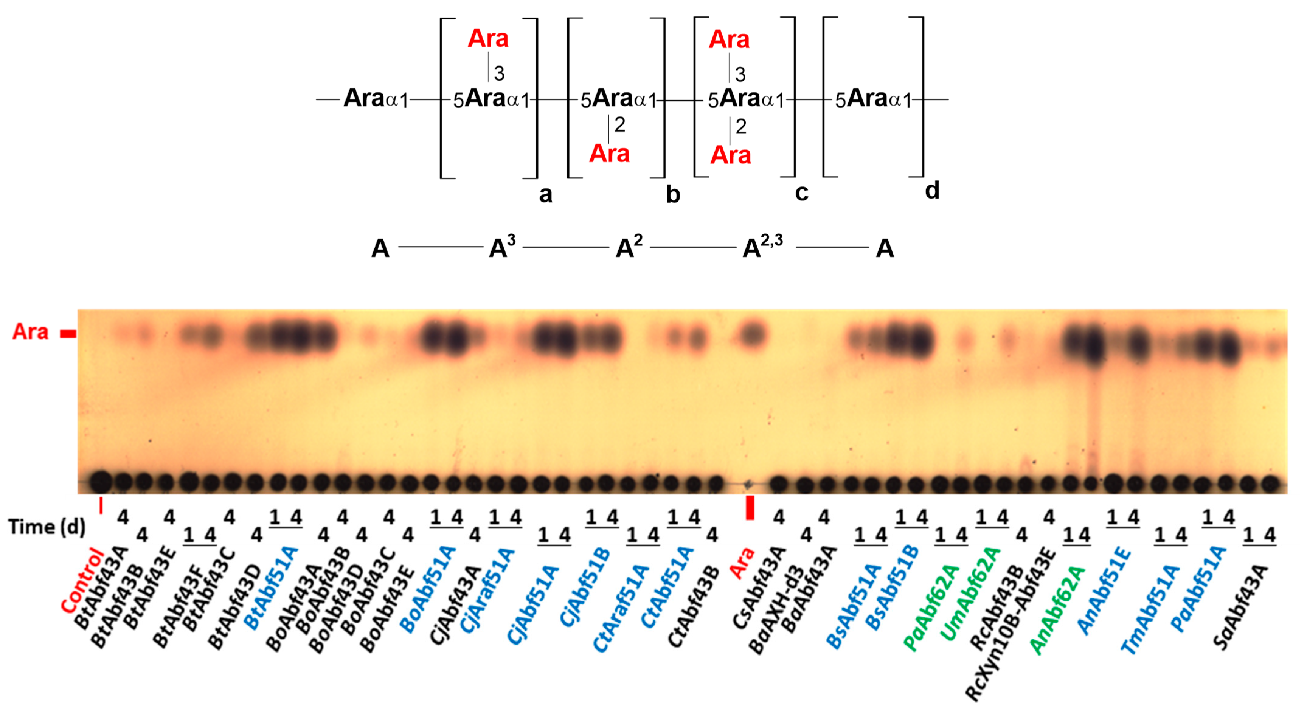
2.2. Specificity of GH43 α-L-Arabinofuranosidases

The hydrolysis of the polysaccharides and oligosaccharides is summarized in Table 2. Many of the GH43 enzymes displayed high activity on polymeric wheat and rye arabinoxylans (WAX and RAX), releasing arabinose as a single product (Figure 1, Supplementary Figure S1; GH43 enzymes are marked in black). Their polymer debranching activity coincided with the dearabinosylation of arabinoxylooligosaccharides (AXOSs) (Figure 2, Supplementary Figures S3–S7).
The proteins belonging to the GH43_12 subfamily (BaAbf43A, BoAbf43B and BoAbf43D) liberated arabinose exclusively from the substrates having xylo-based main chains (Table 2), although the amino acid sequences of the B. ovatus enzymes are more similar to each other than to BaAbf43A (Figure 3, Supplementary Table S1). The members of GH43_16 subfamily (CtAbf43B, RcAbf43B and RcXyn10B-Abf43E) behaved similarly, although some of them additionally showed weak activity on the branched arabinooligosaccharides (AOSs), which is in line with comparison of their amino acid sequences (Figure 3, Supplementary Table S1). On the other hand, the members of subfamilies GH43_18, GH43_19, GH43_29, and GH43_35 either showed no release of arabinose from polymeric AXs or released it extremely slowly (RAX + GH43_19 BtAbf43B) (Figure 1, Supplementary Figure S1); however, some GH43_29 members weakly hydrolyzed some AXOSs (Supplementary Figures S3–S7). It is likely that these enzymes either target another substrate(s) than those having xylo-based main chains or they are specific for an epitope that is present in the AXs and derived AXOSs at a very low frequency.
A vast majority of the GH43 enzymes releasing arabinose from AXs and/or AXOSs showed AXH-m regiospecificity, i.e., they solely attacked singly arabinosylated Xylp residues (Figure 2, Supplementary Figures S3–S5). On the other hand, the enzymes BoAbf43A and BaAXH-d3, both belonging to the GH43_10 subfamily exhibited AXH-d3 specificity, i.e., cleaved 3-O-linked α-L-Araf side chain solely from 2,3 doubly arabinosylated AXOS (Supplementary Figures S6 and S7). Mixed specificity, i.e., activity on both mono- and 2,3-di-O-arabinosylated Xylp residues, was found for BoAbf43C (GH43_29), and particularly BtAbf43F having very broad specificity (GH43_34; examined for the first time on AXOS and AOS) (Table 2). Of special interest is a pair of Bacteroides ovatus enzymes BoAbf43C and BoAbf43E, both from the GH43_29 subfamily, which slightly hydrolyzed both arabinoxylans (WAX and RAX), releasing product(s) other than arabinose (Figure 1, Supplementary Figure S1) whose precise structure needs further investigation.
Some GH43 hydrolases showed moderate activity on sugar beet arabinan (SBA) (Figure 4). Unambiguous arabinan-debranching capacity was found for six GH43 enzymes only, namely GH43_10 BoAbf43A, GH43_19 BtAbf43B, GH43_29 members (BoAbf43E, CjAbf43A and BtAbf43D), and GH43_34 BtAbf43F (Figure 4, Table 2). This was also confirmed by AOS debranching, although the branched AOSs were attacked by a slightly higher number of the enzymes (Figure 5, Supplementary Figure S10, Table 2). In all cases, if the branched arabinan or AOS served as a substrate, regiospecific removal of arabinose side chains was maintained. In other words, the AXH-d3 specificity exhibited on AX and AXOS coincided with a similar specificity on the 2,3-diarabinosylated arabinose residues in SBA and AOS. The same holds true for the AXH-m enzymes that attacked singly arabinosylated Xylp residues (AX, AXOS) as well as singly arabinosylated Araf residues (arabinan, AOS) (Table 2).
None of the GH43 enzymes released a significant amount of arabinose from debranched arabinan (DA) and linear AOS except a GH43_26 member (SaAbf43A) (Figure 6, Supplementary Figures S8 and S9). It is an exo-1,5-α-L-arabinofuranosidase liberating the non-reducing-end terminal undecorated 1,5-linked α-L-arabinose from the main chain of arabinans and AOS. It is specific for the cleavage of α-1,5-linkages and liberates neither α-1,2- nor α-1,3-attached arabinose (side chains of branched arabinan and derived AOS) (Figure 4 and Figure 5, Supplementary Figure S10). However, decoration of the penultimate main chain residue (counted from the reducing end) at position 3 renders the non-reducing end terminal 1,5-linked arabinofuranosyl residue inaccessible to the enzyme, which is reflected in the resistance of AA3A (Supplementary Figure S10, last lane) and AA2,3A (Figure 5, last lane).
Arabinose liberation from larchwood arabinogalactan (AG) was rare among the GH43 enzymes (Supplementary Figure S2). Such ability was exhibited by BtAbf43B, BtAbf43C, BtAbf43F, BoAbf43C, BoAbf43E, and RcAbf43B, although the amount of arabinose released was low in all cases (Table 2, Supplementary Figure S2).

2.3. Specificity of GH51 α-L-Arabinofuranosidases

The GH51 enzymes significantly hydrolyzed DA, SBA, AOS, and AXOS (Figure 2 and Figure 4, Figure 5 and Figure 6, Supplementary Figures S3–S6 and S8–S10; GH51 enzymes are marked in blue) but differed in their ability to debranch arabinoxylans, which are resistant to two enzymes BsAbf51A and CtAbf51A (Table 3). With a single exception (BsAbf51A), larchwood AG is the poorest polysaccharidic substrate, being recalcitrant to a half of the investigated GH51 representatives (Supplementary Figure S2). All tested GH51 enzymes exhibited AXH-m regiospecificity on AXOS and AOS, although most of them were able to release arabinose also from the non-reducing-end terminal doubly arabinosylated Xylp residue present in A2,3XX, yielding completely dearabinosylated linear xylooligosaccharide (Supplementary Figures S6 and S11A). We did not observe any significant differences in the catalytic properties between GH51_1 and GH51_2 members (Table 3).

2.4. Specificity of GH62 α-L-Arabinofuranosidases

We examined only three proteins (AnAbf62A, PaAbf62A, and UmAbf62A) classified into the GH62 family. None of them efficiently released arabinose from AG. In contrast, AXs as well as AXOSs served as good substrates for all three enzymes, which exhibited AXH-m regiospecificity (Figure 1 and Figure 2, Supplementary Figures S1 and S3–S5; GH62 enzymes are marked in green). However, AnAbf62A hydrolyzed both arabinose moieties attached to the non-reducing-end terminal Xylp residue of A2,3XX (Supplementary Figure S11A). Moreover, this enzyme was the only GH62 representative, which was able to hydrolyze arabinans (Figure 4 and Figure 6) and AOS. The hydrolysis products suggest that the enzyme catalyzed debranching of SBA, AA3A, and AAA3A (Figure 5, Supplementary Figure S10), as well as depolymerization of DA and Ara2–4 (Supplementary Figures S8 and S9), indicating that AnAbf62A is able to hydrolyze 1,5-linkages too. In this way, monomeric arabinose was the only final product generated from all the AOSs including the doubly arabinosylated substrate AA2,3A. Its hydrolysis is explained by an initial cleavage of 1,5-linked arabinose from the non-reducing end, resulting in an intermediate tetrasaccharide A2,3A, which is then debranched (similarly to the debranching of A2,3XX) to 1,5-arabinobiose Ara2, the latter being finally hydrolyzed to two molecules of arabinose.

3. Discussion

The enzyme activities of some members belonging to the Gh43_16 subfamily on natural arabinose-containing substrates have never been examined before. Moreover, many enzymes grouped into subfamilies GH43_10, GH43_12, GH43_18, GH43_19, GH43_29, GH43_34, and GH51_1, in particular those originating from Bifidobacterium adolescentis, Bacteroides ovatus, and Bacteroides thetaiotaomicron, have been tested for the first time using AXOS and AOS (cf. Table 2 and Table 3). Their specificity is analyzed in further sections and compared with other enzymes showing similar properties.

3.1. Specificity of GH43 α-L-Arabinofuranosidases

GH43 is one of the largest and most diverse CAZy families [17]. Currently, it is divided into 38 subfamilies (GH43_1 to GH43_39, but GH43_38 has been deleted). Our results show that there is no relation between the capacity of the GH43 arabinofuranosidases to hydrolyze artificial chromogenic glycoside NPA, commonly used for screening purposes, and natural substrates. In agreement with a previously published subdivision of the GH43 proteins into subfamilies based on detailed analysis of their amino acid sequences [17], we observed that the enzymes differ significantly in substrate and positional specificities. For instance, three proteins, BaAbf43A, BoAbf43B, and BoAbf43D, belonging to GH43_12 as well as three proteins, CtAbf43B, RcAbf43B, and RcXyn10B-Abf43E, classified into GH43_16 were found to be xylan-specific debranching catalysts without noticeable action on arabinan and AOS. Moreover, these six proteins exhibited AXH-m specificity. In previous studies, the regiospecificity of BoAbf43B and BoAbf43D has been examined [4,12], while the others have not been studied in this regard, although specific activities determined here on the synthetic substrate NPA are similar to the values reported in the literature [12,18]. In contrast, five other α-L-arabinofuranosidases (CsAbf43A, CjAbf43A, BtAbf43D, SaAbf43A, and BtAbf43E) from four different GH43 subfamilies did not release Araf side chains from the polymeric WAX and RAX, thus confirming the literature data. Moreover, we demonstrated the enzymes’ inability to release arabinose attached to main chain xylose from the majority of the tested AXOSs. Nevertheless, our results are fully consistent with earlier studies indicating that (a) the functional role of CsAbf43A (GH43_35) [19] still remains unknown; (b) BtAbf43E from GH43_18 displayed activity on NPA and rhamnogalacturonan-II (RGII) [20]; (c) BtAbf43B, a founding member of the GH43_19 subfamily, seems to specifically release 3-linked arabinose attached to singly substituted xylose and galactose residues, which agrees with the reported weak activity on NPA and WAX (this enzyme has never been examined on AXOS and AOS) [10]; and (d) CjAbf43A, BtAbf43D (both belonging to GH43_29), and SaAbf43A (GH43_26) are arabinan-specific α-L-arabinofuranosidases [10,21]. The substrate specificity of the latter enzyme underscores the functional diversity within the GH43 family. The enzyme SaAbf43A released pentose from the substrates comprising non-substituted non-reducing-end terminal 1,5-linked Araf residue regardless of their degree of polymerization, only when the neighbouring main chain residue was not decorated at position 3. Any other GH43 member was unable to cleave α-1,5 linkages found in the arabinan backbone. We thus confirmed that the GH43_26 members are arabinan-specific and act as exo-1,5-α-L-arabinofuranosidases [21,22].
The GH43_29 representatives CjAbf43A and BtAbf43D have already been demonstrated to be specific enzymes releasing 2-linked arabinose from arabinan regardless of the occupation of vicinal position 3 with another arabinose moiety [10,21]. Our data on the two enzymes are fully consistent with this conclusion. Moreover, analysis of their amino acid sequences revealed that both enzymes form a separate clade (Figure 3, Supplementary Table S1). The other clade within the GH43_29 subfamily is formed by BoAbf43C and BoAbf43E (Figure 3). This subgrouping is also reflected in their specificities since the latter two enzymes were inactive on SBA and branched AOS, however, they yielded so far unidentified products from arabinoxylans (WAX and RAX). Their observation is in line with the enzymes’ performance on a different type of xylan originating from corn bran [4]; however, to the best of our knowledge, we report for the first time the action of these GH43_29 members on AXOS and branched AOS.
The 2,3-diarabinosylated residues of arabinan are specifically recognized by another type of GH43 arabinofuranosidases. Both enzymes, namely BaAXH-d3 and BoAbf43A (the latter being tested on AXOS and AOS for the first time), exhibiting this regioselectivity are classified into the GH43_10 subfamily and selectively cleave 3-linked arabinose. The recognition of 2,3 doubly arabinosylated main chain residues and selective dearabinosylation at position 3 was also observed for AX and AXOS, by which we confirmed previous reports [4,16]. It seems that the 3-dearabinosylation of 2,3 doubly decorated main chain residues of AX as well as arabinan is a common property of arabinofuranosidases exhibiting the AXH-d3 specificity since such a selective partial debranching of both polysaccharides has been demonstrated also for a fungal enzyme HiAXHd3 classified into the GH43_36 subfamily [23]. We should, however, note that membership to a subfamily comprising the AXH-d3 enzymes subfamily does not automatically confer this regiospecificity since BtAbf43A (also tested on AXOS and AOS for the first time) from the GH43_10 subfamily is an enzyme of the AXH-m type and forms a separate clade within the GH43_10 subfamily (Figure 3), although the degrees of identity of these three enzymes do not differ too much (Supplementary Table S1). Similarly, the GH43_36 subfamily also consists of AXH-m enzymes [14], implying differences in the enzyme regiospecificity even within at least these two GH43 subfamilies.
For a vast majority of the enzymes examined, larchwood arabinogalactan was used as a substrate for the first time. However, arabinose liberation was very rarely observed. Only six GH43 members produced by Bacteroides ovatus, B. thetaiotaomicron, and Ruminococcus champenellensis (BtAbf43B, BtAbf43C, BtAbf43F, BoAbf43C, BoAbf43E, and RcAbf43B) unambiguously liberated small amounts of arabinose from this polysaccharide. It is, therefore, possible that the arabinofuranosidases targeting this polysaccharide and falling into the GH43 family have not been hitherto discovered and/or will be classified into subfamilies established in the future.

3.2. Specificity of GH51 α-L-Arabinofuranosidases

All 12 examined proteins from the GH51 family attacked 1,5- as well as 1,2- and 1,3-linkages. Accordingly, the enzymes were active on all arabinoxylan- and arabinan-derived polysaccharides and oligosaccharides except those comprising internally 2,3-diarabinosylated residues. Interestingly, the majority of the enzymes were able to liberate arabinose from the xylooligosaccharide doubly arabinosylated at the non-reducing end (A2,3XX). With a few exceptions, all our findings are in accordance with the literature data about these enzymes [2,3,24,25,26,27,28,29] which were generally reported to exhibit broad specificity, being active on both arabinoxylan- and arabinan-based substrates and/or exhibiting both main chains as well as debranching activity towards branched arabinan. However, none of the enzymes have previously been tested on such a large set of oligosaccharide and polysaccharide substrates as was used in this study. Despite the wide spectrum of the investigated substrates, we found no significant difference in the substrate and positional specificity between the studied members of the GH51_1 and GH51_2 subfamilies. The differences were insignificant also in case of AG, from which BsAbf51A, BtAbf51A, BoAbf51A, PaAbf51A, AnAbf51E, and CjAbf51A were found to unambiguously liberate arabinose. Such a capacity is shown here for the first time for the latter five enzymes. Generally, the dearabinosylation of larchwood AG has hitherto been studied very rarely because there are only few reports in this regard. This polysaccharide has previously been included in the tested substrates only in the case of BsAbf51A (positive result), BsAbf51B, and TmAbf51A (negative result) [25,26,27].

3.3. Specificity of GH62 α-L-Arabinofuranosidases

The catalytic properties of PaAbf62A and UmAbf62A were very similar. Both seem to be arabinoxylan-specific AXH-m enzymes, not attacking linear arabinan and slowly processing NPA. Although the enzymes have been reported to also hydrolyze branched arabinan [30], we observed only negligible activity on this branched polysaccharide, similarly to another previous study [31]. Significantly broader specificity was exhibited by AnAbf62A. In addition to at least two orders of magnitude higher specific activity towards the chromogenic substrate NPA (1880 mU/mg), the enzyme efficiently hydrolyzed all tested natural substrates except AG and XA2,3XX. Thus, we confirm the xylan debranching capacity of the enzyme, although we extend its hydrolytic potential also towards arabinan including its main chain 1,5-linkages, which have been reported to be recalcitrant [32,33]. Moreover, in contrast to [32], we unambiguously detected the liberation of arabinose from A2,3XX, i.e., an oligosaccharide comprising doubly decorated non-reducing-end xylose residue, thus leaving only internal doubly arabinosylated epitopes (XA2,3XX) resistant to AnAbf62A.

4. Materials and Methods

4.1. Enzymes and Substrates

The 34 examined α-L-arabinofuranosidases belonging to different GH (sub)families are listed in Table 1. These proteins were purchased from NZYTech (Lisbon, Portugal) and Megazyme (Bray, Ireland). The suppliers report the electrophoretic purity of the enzymes in the product data sheets, as well as their pH and temperature optima (Supplementary Table S2). For hydrolysis experiments, the enzymes (in most cases supplied at 1 mg/mL concentration) were diluted with 50 mM sodium phosphate buffer, pH 6.0, to obtain their working solutions with a 0.1 mg/mL concentration. The dilution was typically 10-fold; however, if the enzymes were supplied at a lower protein concentration, their working solutions had proportionally lower concentrations after the 10-fold dilution. All natural substrates were procured from Megazyme. These included polysaccharides, namely medium-viscosity wheat flour arabinoxylan (WAX) [Arabinose/Xylose = 38:62]; high-viscosity rye flour arabinoxylan (RAX) [Ara:Xyl = 40:60]; sugar beet arabinan (SBA) [Ara:Gal:Rha:GalA/other sugars = 69:18.7:1.4:10.2:0.7]; debranched arabinan (DA) [Ara:Gal:Rha = 71:26:3], larchwood arabinogalactan (AG) [Gal:Ara/other sugars = 81:14:5], arabinoxylooligosaccharides (AXOSs), specifically 32-α-L-arabinofuranosyl-xylobiose (A3X); 23-α-L-arabinofuranosyl-xylotriose (A2XX); 33-α-L-arabinofuranosyl-xylotetraose (XA3XX); a mixture of 23-α-L-arabinofuranosyl-xylotetraose (XA2XX) and XA3XX; 23,33-di-α-L-arabinofuranosyl-xylotriose (A2,3XX); 23,33-di-α-L-arabinofuranosyl-xylotetraose (XA2,3XX), and arabinooligosaccharides (AOSs), namely α-1,5-L-arabinobiose (Ara2); α-1,5-L-arabinotriose (Ara3); α-1,5-L-arabinotetraose (Ara4); 32-α-L-arabinofuranosyl-arabinotriose (AA3A); a mixture of 32-α-L-arabinofuranosyl-arabinotetraose (AAA3A) and 22,32-di-α-L-arabinofuranosyl-arabinotriose (AA2,3A). A synthetic chromogenic substrate, 4-nitrophenyl α-L-arabinofuranoside (NPA), was obtained from Carbosynth (Compton, UK).

4.2. Enzyme Assays

For routine determination of enzyme activity, NPA was used as a substrate. After preincubation of 120 μL of 1 mM substrate solution in 50 mM sodium phosphate buffer, pH 6.0, at 35 °C, the reaction was started by the addition of 5 μL of an appropriately diluted enzyme. The reactions were performed at 35 °C and terminated by 0.75 mL of saturated aqueous solution of sodium tetraborate. The amount of released chromophore was determined spectrophotometrically at 405 nm using 4-nitrophenol as a standard. One unit of arabinofuranosidase activity is defined as the amount of enzyme producing 1 μmol of 4-nitrophenol in 1 min.

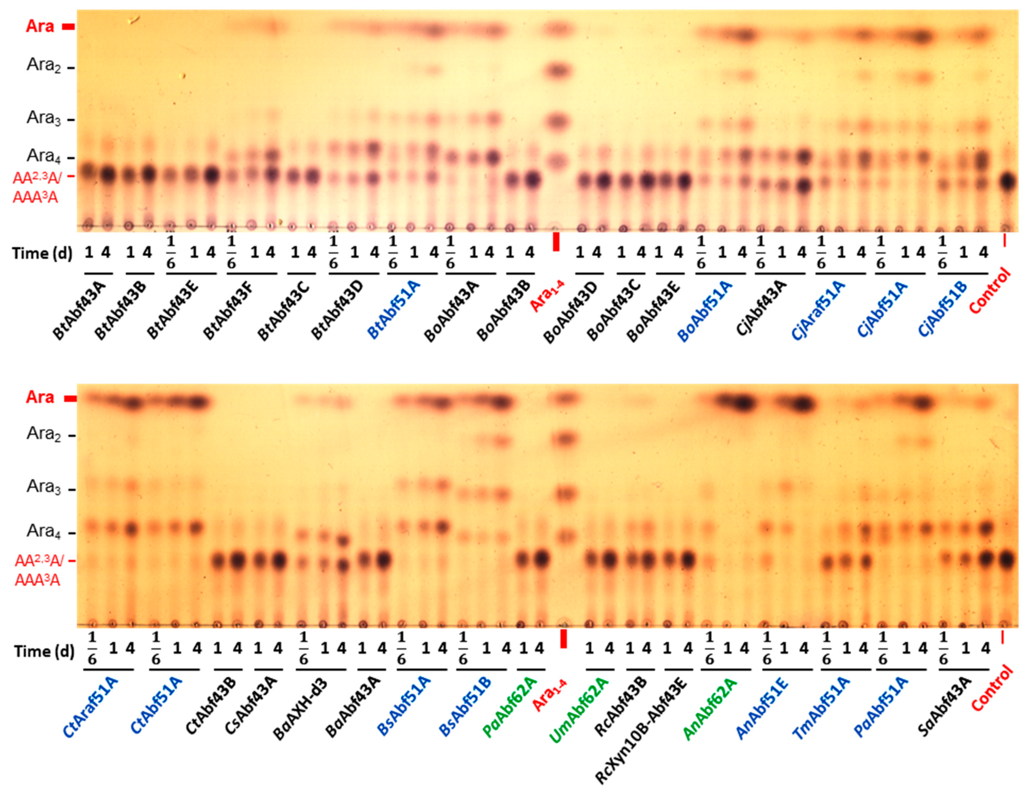
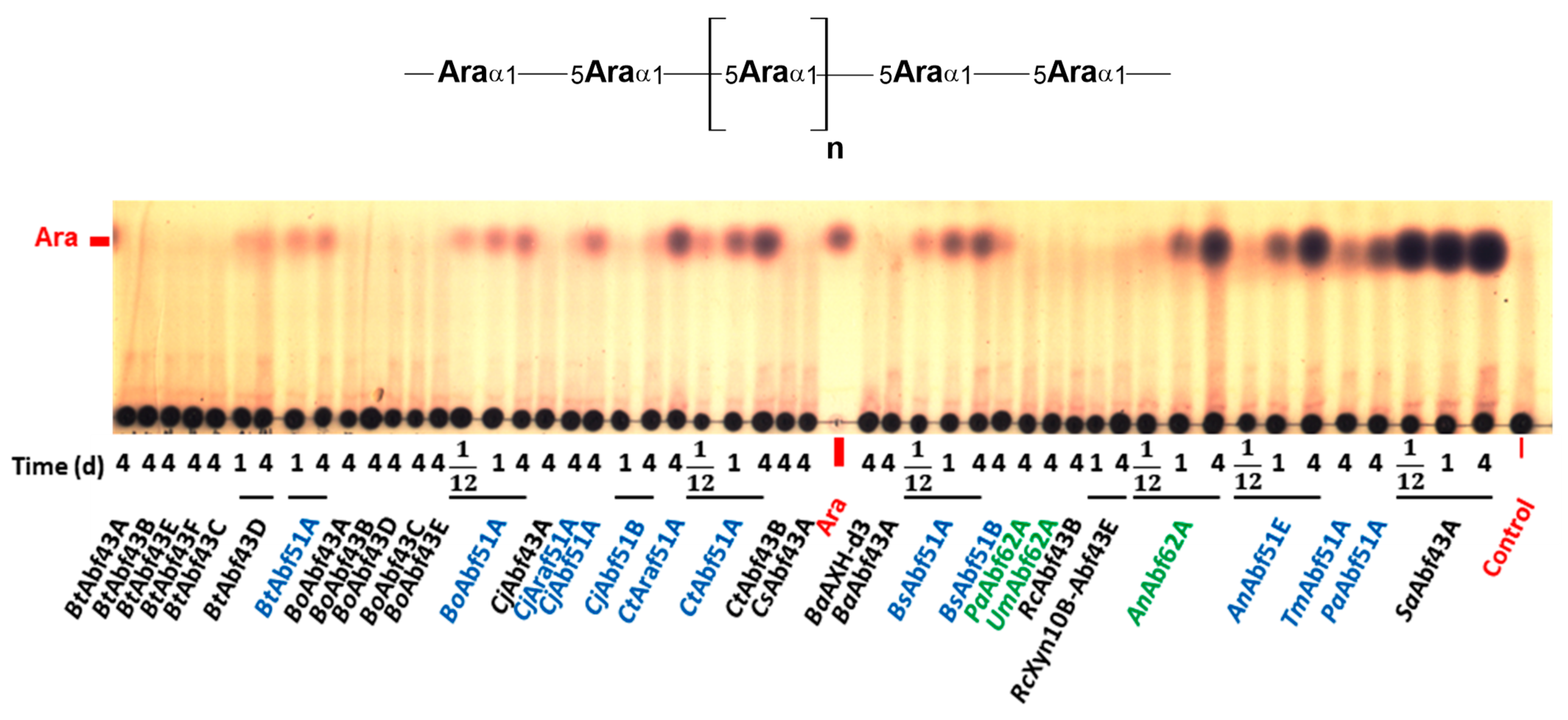
4.3. Hydrolysis of Natural Substrates

The catalytic properties of the enzymes were tested on a series of polysaccharides and oligosaccharides listed above in the Section 4.1. The substrates (20 μL), i.e., 1% (w/v) polysaccharides or 0.1% (w/v) oligosaccharides dissolved in 50 mM sodium phosphate buffer, pH 6.0, were thermally preequilibrated at 35 °C and then the reaction was started by the addition of 2 μL of the enzyme working solution. At time intervals, 2 μL aliquots were spotted onto the TLC plate.

4.4. Analysis of the Hydrolysates

The hydrolysates were analyzed by TLC, which was performed on aluminium-coated silica plates (Merck, Darmstadt, Germany). The plates were developed in the following chromatographic solvent systems: 1-butanol/ethanol/water (10:8:5, by vol.; system 1, used for polysaccharides, Ara2 and AXOS except the XA2XX/XA3XX mixture); chloroform/acetic acid/water (6:7:1, by vol.; system 2, used for AOS except Ara2; developed twice) or 1-propanol/ethanol/water (7:1:2, by vol.; system 3, used for the XA2XX/XA3XX mixture). The plates were air-dried and after dipping into the detection reagent [0.5% orcinol (w/v) in 5% sulfuric acid in ethanol (v/v)], they were heated to 120 °C to visualize the carbohydrates. The sugars were quantified by densitometry (UN-SCAN-IT; Silk Scientific, Orem, UT, USA), i.e., by comparison of the product spot intensities with that of different amounts of standard arabinose. Again, 1 unit of arabinofuranosidase activity is defined as the amount of enzyme producing 1 μmol of L-arabinose in 1 min.

4.5. Comparison of Amino Acid Sequences

The amino acid sequences of the enzymes belonging to those GH43 subfamilies, which were represented by multiple members in this study (GH43_10, GH43_12, GH43_16 and GH43_29), were compared. All sequences were analyzed for the presence of a signal sequence using the server of SignalP, version 6.0 (https://services.healthtech.dtu.dk/services/SignalP-6.0/, accessed on 14 July 2024). If the signal sequence was predicted, it is indicated by small letters in Figure 3A. Only GH43 catalytic domains were included in the multiple sequence alignment, which was generated by Uniprot server (https://www.uniprot.org/align, accessed on 14 July 2024). Simultaneously with the multiple sequence alignment, the phylogenetic tree (Figure 3B) and percent identity matrix table (Supplementary Table S1) were obtained.

5. Conclusions

α-L-arabinofuranosidases (EC 3.2.1.55) participate in the enzymatic saccharification of arabinose-rich plant biomass to produce value-added bioproducts and prebiotics. The present study gives experimental evidence for the substrate and positional specificity of 34 α-L-arabinofuranosidases from GH43, GH51, and GH62 families on a wide spectrum of natural substrates derived from arabinoxylan and arabinan. For many of the enzymes, at least some of the substrates were tested for the first time. Generally, GH51 members from GH51_1 as well as GH51_2 subfamilies may be classified as non-specific α-L-arabinofuranosidases, liberating arabinose from arabinoxylan, branched as well as linear arabinan, and their oligomeric derivatives. In contrast, most studied GH43 members were arabinoxylan-debranching enzymes inactive on arabinans. Only a few GH43 hydrolases were arabinan-specific, e.g., the enzymes from subfamily GH43_29 (arabinan-specific α-1,2-arabinofuranosidases), in addition to subfamily GH43_26, which encompasses solely exo-1,5-α-L-arabinofuranosidases, i.e., the enzymes involved in the arabinan main chain degradation. Moreover, among the GH43 arabinoxylan-debranching enzymes, there were significant differences in the regiospecificity between the subfamilies and sometimes also within a given subfamily (e.g., subfamily GH43_10). Despite the functional diversity in this family, we did not find an enzyme able to efficiently dearabinosylate larchwood arabinogalactan, which was a poor substrate, if any, also for α-L-arabinofuranosidases from the other two families studied.
All the arabinoxylan-debranching enzymes behave either as AXH-m- or AXH-d3-type catalysts, although some AXH-m enzymes from both examined GH51 subfamilies were able to hydrolyze xylooligosaccharides doubly arabinosylated at the non-reducing-end terminal, but not internal Xylp residues. Moreover, if a given arabinoxylan-debranching enzyme (having either AXH-m or AXH-d3 specificity) is active on arabinooligosaccharides and/or arabinan, the regiospecificity is maintained. Taken together, the diversity in the specificity of arabinofuranosidases coincides with their multiplicity in microbial genomes and reflects the complexity of lignocellulosic materials. Furthermore, the CAZy classification system is a valuable database that can serve as a guide to identify the substrate and positional specificity of uncharacterized and novel α-L-arabinofuranosidases; however, experimental verification of their catalytic properties is still needed before their industrial application in targeted exploitation of isolated or native complex polysaccharides. This study thus not only highlights subfamilies requiring additional functional characterization but also enhances the functional predictive power of GH43, GH51, and GH62 sequences, facilitating more accurate annotations and our understanding of these GH families that can potentially provide reliable and stable functional predictions of their enzymes based solely on sequence information. This approach seems to be of general use since it has been recently demonstrated not only for glycoside hydrolases [7,17,34,35,36,37,38] but also for other types of carbohydrate active enzymes such as glycoside phosphorylases [39], glycosyl transferases [40], and carbohydrate esterases [41].

Supplementary Materials

The following supporting information can be downloaded at: https://www.mdpi.com/article/10.3390/catal14080536/s1, Table S1: Identity percentage between the GH43 enzymes, the amino acid sequences of which are aligned in Figure 3. Table S2: pH and temperature optima of the enzymes used in this study. Figure S1: TLC analysis of rye arabinoxylan (RAX) hydrolysates. Figure S2: TLC analysis of larchwood arabinogalactan (AG) hydrolysates. Figure S3: TLC analysis of A3X hydrolysates. Figure S4: TLC analysis of A2XX hydrolysates. Figure S5: TLC analysis of XA3XX hydrolysates. Figure S6: TLC analysis of A2,3XX hydrolysates. Figure S7: TLC analysis of XA2,3XX hydrolysates. Figure S8: TLC analysis of α-1,5-L-arabinobiose (Ara2) hydrolysates. Figure S9: TLC analysis of linear α-1,5-L arabinotriose (Ara3) and α-1,5-L-arabinotetraose (Ara4) hydrolysates. Figure S10: TLC analysis of 32-α-L arabinofuranosyl-(1,5)-α-L-arabinotriose (AA3A) hydrolysates. Figure S11: TLC analysis of A2,3XX and XA2,3XX hydrolysates by selected α-L-arabinofuranosidases.

Author Contributions

W.F.: Methodology, Validation, Formal Analysis, Investigation, Writing—original draft, Writing—Review and Editing, Visualization. V.P.: Conceptualization, Validation, Formal Analysis, Writing—Review and Editing, Supervision, Funding acquisition. All authors have read and agreed to the published version of the manuscript.

Funding

This work was supported by the Slovak Research and Development Agency under the contract no. APVV-20-0591, and by the Scientific Grant Agency under the contract no. 2/0171/22.

Data Availability Statement

The original contributions presented in the study are included in the article/Supplementary Material; further inquiries can be directed to the corresponding author.

Conflicts of Interest

The authors declare no conflicts of interest. The funders had no role in the design of the study; in the collection, analyses, or interpretation of data; in the writing of the manuscript; or in the decision to publish the results.

References

  1. Wardman, J.F.; Bains, R.K.; Rahfeld, P.; Withers, S.G. Carbohydrate-active enzymes (CAZymes) in the gut microbiome. Nat. Rev. Microbiol. 2022, 20, 542–556. [Google Scholar] [CrossRef]
  2. Taylor, E.J.; Smith, N.L.; Turkenburg, J.P.; D’Souza, S.; Gilbert, H.J.; Davies, G.J. Structural insight into the ligand specificity of a thermostable family 51 arabinofuranosidase, Araf51, from Clostridium thermocellum. Biochem. J. 2006, 395, 31–37. [Google Scholar] [CrossRef]
  3. Beylot, M.-H.; Mckie, V.A.; Voragen, A.G.J.; Doeswijk-Voragen, C.H.L.; Gilbert, H.J. The Pseudomonas cellulosa glycoside hydrolase family 51 arabinofuranosidase exhibits wide substrate specificity. Biochem. J. 2001, 358, 607–614. [Google Scholar] [CrossRef] [PubMed]
  4. Rogowski, A.; Briggs, J.A.; Mortimer, J.C.; Tryfona, T.; Terrapon, N.; Lowe, E.C.; Baslé, A.; Morland, C.; Day, A.M.; Zheng, H.; et al. Glycan complexity dictates microbial resource allocation in the large intestine. Nat. Commun. 2015, 6, 7481. [Google Scholar] [CrossRef]
  5. Kormelink, F.J.M.; Searl-Van Leeuwen, M.J.F.; Wood, T.M.; Voragen, A.G.J. Purification and characterization of a (1,4)-β-D-arabinoxylan arabinofuranohydrolase from Aspergillus awamori. Appl. Microbiol. Biotechnol. 1991, 35, 753–758. [Google Scholar] [CrossRef]
  6. Lagaert, S.; Pollet, A.; Courtin, C.M.; Volckaert, G. β-Xylosidases and α-L-arabinofuranosidases: Accessory enzymes for arabinoxylan degradation. Biotechnol. Adv. 2014, 32, 316–332. [Google Scholar] [CrossRef]
  7. Jones, D.R.; Thomas, D.; Alger, N.; Ghavidel, A.; Inglis, G.D.; Abbott, D.W. SACCHARIS: An automated pipeline to streamline discovery of carbohydrate-active enzyme activities within polyspecific families and de novo sequence datasets. Biotechnol. Biofuels 2018, 11, 27. [Google Scholar] [CrossRef] [PubMed]
  8. Borsenberger, V.; Dornez, E.; Desrousseaux, M.-L.; Massou, S.; Tenkanen, M.; Courtin, C.M.; Dumon, C.; O’Donohue, M.J.; Fauré, R. A 1H NMR study of the specificity of α-L-arabinofuranosidases on natural and unnatural substrates. Biochim. Biophys. Acta 2014, 1840, 3106–3114. [Google Scholar] [CrossRef]
  9. Sakamoto, T.; Ogura, A.; Inui, M.; Tokuda, S.; Hosokawa, S.; Ihara, H.; Kasai, N. Identification of a GH62 α-L-arabinofuranosidase specific for arabinoxylan produced by Penicillium chrysogenum. Appl. Microbiol. Biotechnol. 2011, 90, 137–146. [Google Scholar] [CrossRef] [PubMed]
  10. Cartmell, A.; McKee, L.S.; Peña, M.J.; Larsbrink, J.; Brumer, H.; Kaneko, S.; Ichinose, H.; Lewis, R.J.; Viksø-Nielsen, A.; Gilbert, H.J.; et al. The structure and function of an arabinan-specific α-1,2-arabinofuranosidase identified from screening the activities of bacterial GH43 glycoside hydrolases. J. Biol. Chem. 2011, 286, 15483–15495. [Google Scholar] [CrossRef]
  11. Goyal, A.; Ahmed, S.; Sharma, K.; Gupta, V.; Bule, P.; Alves, V.D.; Fontes, C.M.G.A.; Najmudin, S. Molecular determinants of substrate specificity revealed by the structure of Clostridium thermocellum arabinofuranosidase 43A from glycosyl hydrolase family 43 subfamily 16. Acta Cryst. 2016, D72, 1281–1289. [Google Scholar] [CrossRef]
  12. Lagaert, S.; Pollet, A.; Delcour, J.A.; Lavigne, R.; Courtin, C.M.; Volckaert, G. Substrate specificity of three recombinant α-L-arabinofuranosidases from Bifidobacterium adolescentis and their divergent action on arabinoxylan and arabinoxylan oligosaccharides. Biochem. Biophys. Res. Commun. 2010, 402, 644–650. [Google Scholar] [CrossRef] [PubMed]
  13. Till, M.; Goldstone, D.; Card, G.; Attwood, G.T.; Moon, C.D.; Arcus, V.L. Structural analysis of the GH43 enzyme Xsa43E from Butyrivibrio proteoclasticus. Acta Cryst. 2014, F70, 1193–1198. [Google Scholar] [CrossRef]
  14. Leschonski, K.P.; Kaasgaard, S.G.; Spodsberg, N.; Krogh, K.B.R.M.; Kabel, M.A. Two subgroups within the GH43_36 α-L-arabinofuranosidase subfamily hydrolyze arabinosyl from either mono- or disubstituted xylosyl units in wheat arabinoxylan. Int. J. Mol. Sci. 2022, 23, 13790. [Google Scholar] [CrossRef]
  15. Sørensen, H.; Jørgensen, C.T.; Hansen, C.H.; Jørgensen, C.I.; Pedersen, S.; Meyer, A.S. A novel GH43 α-L-arabinofuranosidase from Humicola insolens: Mode of action and synergy with GH51 α-L-arabinofuranosidases on wheat arabinoxylan. Appl. Microbiol. Biotechnol. 2022, 73, 850–861. [Google Scholar] [CrossRef]
  16. Van Laere, K.; Beldman, G.; Voragen, A.G.J. A new arabinofuranohydrolase from Bifidobacterium adolescentis able to remove arabinosyl residues from double-substituted xylose units in arabinoxylan. Appl. Microbiol. Biotechnol. 1997, 47, 231–235. [Google Scholar] [CrossRef]
  17. Mewis, K.; Lenfant, N.; Lombard, V.; Henrissat, B. Dividing the large glycoside hydrolase family 43 into subfamilies: A motivation for detailed enzyme characterization. Appl. Environ. Microbiol. 2016, 82, 1686–1692. [Google Scholar] [CrossRef]
  18. Moraïs, S.; David, Y.B.; Bensoussan, L.; Duncan, S.H.; Koropatkin, N.M.; Martens, E.C.; Flint, H.J.; Bayer, E.A. Enzymatic profiling of cellulosomal enzymes from the human gut bacterium, Ruminococcus champanellensis, reveals a fine-tuned system for cohesin-dockerin recognition. Environ. Microbiol. 2016, 18, 542–556. [Google Scholar] [CrossRef]
  19. Sakka, K.; Yoshikawa, K.; Kojima, Y.; Karita, S.; Ohmiya, K.; Shimada, K. Nucleotide sequence of the Clostridium stercorarium xylA gene encoding a bifunctional protein with β-D-xylosidase and α-L-arabinofuranosidase activities, and properties of the translated product. Biosci. Biotechnol. Biochem. 1993, 57, 268–272. [Google Scholar] [CrossRef]
  20. Ndeh, D.; Rogowski, A.; Cartmell, A.; Luis, A.S.; Baslé, A.; Gray, J.; Venditto, I.; Briggs, J.; Zhang, X.; Labourel, A.; et al. Complex pectin metabolism by gut bacteria reveals novel catalytic functions. Nature 2017, 544, 65–70. [Google Scholar] [CrossRef]
  21. Ichinose, H.; Yoshida, M.; Fujimoto, Z.; Kaneko, S. Characterization of a modular enzyme of exo-1,5-α-L-arabinofuranosidase and arabinan binding module from Streptomyces avermitilis NBRC14893. Appl. Microbiol. Biotechnol. 2008, 80, 399–408. [Google Scholar] [CrossRef]
  22. Fujimoto, Z.; Ichinose, H.; Maehara, T.; Honda, M.; Kitaoka, M.; Kaneko, S. Crystal structure of an exo-1,5-α-L-arabinofuranosidase from Streptomyces avermitilis provides insights into the mechanism of substrate discrimination between exo- and endo-type enzymes in glycoside hydrolase family 43. J. Biol. Chem. 2010, 285, 34134–34143. [Google Scholar] [CrossRef] [PubMed]
  23. McKee, L.S.; Peña, M.J.; Rogowski, A.; Jackson, A.; Lewis, R.J.; York, W.S.; Krogh, K.B.R.M.; Viksø-Nielsen, A.; Skjøt, M.; Gilbert, H.J.; et al. Introducing endo-xylanase activity into an exo-acting arabinofuranosidase that targets side chains. Proc. Natl. Acad. Sci. USA 2012, 109, 6537–6542. [Google Scholar] [CrossRef] [PubMed]
  24. Hoffmam, Z.B.; Oliveira, L.C.; Cota, J.; Alvarez, T.M.; Diogo, J.A.; Neto, M.O.; Citadini, A.P.S.; Leite, V.B.P.; Squina, F.M.; Murakami, M.T.; et al. Characterization of a hexameric exo-acting GH51 α-L-arabinofuranosidase from the mesophilic Bacillus subtilis. Mol. Biotechnol. 2013, 55, 260–267. [Google Scholar] [CrossRef] [PubMed]
  25. Kaneko, S.; Sano, M.; Kusakabe, I. Purification and some properties of α-L-arabinofuranosidase from Bacillus subtilis 3-6. Appl. Environ. Microbiol. 1994, 60, 3425–3428. [Google Scholar] [CrossRef] [PubMed]
  26. Inácio, J.M.; Correia, I.L.; de Sá-Nogueira, I. Two distinct arabinofuranosidases contribute to arabino-oligosaccharide degradation in Bacillus subtilis. Microbiology 2008, 154, 2719–2729. [Google Scholar] [CrossRef] [PubMed]
  27. Miyazaki, K. Hyperthermophilic α-L-arabinofuranosidase from Thermotoga maritima MSB8: Molecular cloning, gene expression, and characterization of the recombinant protein. Extremophiles 2005, 9, 399–406. [Google Scholar] [CrossRef] [PubMed]
  28. Luis, A.S.; Briggs, J.; Zhang, X.; Farnell, B.; Ndeh, D.; Labourel, A.; Baslé, A.; Cartmell, A.; Terrapon, N.; Stott, K.; et al. Dietary pectic glycans are degraded by coordinated enzyme pathways in human colonic Bacteroides. Nat. Microbiol. 2018, 3, 210–219. [Google Scholar] [CrossRef]
  29. Couturier, M.; Haon, M.; Coutinho, P.M.; Henrissat, B.; Lesage-Meessen, L.; Berrin, J.-G. Podospora anserina hemicellulases potentiate the Trichoderma reesei secretome for saccharification of lignocellulosic biomass. Appl. Environ. Microbiol. 2011, 77, 237–246. [Google Scholar] [CrossRef]
  30. Siguier, B.; Haon, M.; Nahoum, V.; Marcellin, M.; Burlet-Schiltz, O.; Coutinho, P.M.; Henrissat, B.; Mourey, L.; O’Donohue, M.J.; Berrin, J.-G.; et al. First structural insights into α-L-arabinofuranosidases from the two GH62 glycoside hydrolase subfamilies. J. Biol. Chem. 2014, 289, 5261–5273. [Google Scholar] [CrossRef]
  31. McCleary, B.V.; McKie, V.A.; Draga, A.; Rooney, E.; Mangan, D.; Larkin, J. Hydrolysis of wheat flour arabinoxylan, acid-debranched wheat flour arabinoxylan and arabino-xylo-oligosaccharides by β-xylanase, α-L-arabinofuranosidase and β-xylosidase. Carbohydr. Res. 2015, 407, 79–96. [Google Scholar] [CrossRef]
  32. Wilkens, C.; Andersen, S.; Petersen, B.O.; Li, A.; Busse-Wicher, M.; Birch, J.; Cockburn, D.; Nakai, H.; Christensen, H.E.M.; Kragelund, B.B.; et al. An efficient arabinoxylan-debranching α-L-arabinofuranosidase of family GH62 from Aspergillus nidulans contains a secondary carbohydrate binding site. Appl. Microbiol. Biotechnol. 2016, 100, 6265–6277. [Google Scholar] [CrossRef]
  33. Contesini, F.J.; Liberato, M.V.; Rubio, M.V.; Calzado, F.; Zubieta, M.P.; Riaño-Pachón, D.M.; Squina, F.M.; Bracht, F.; Skaf, M.S.; Damasio, A.R. Structural and functional characterization of a highly secreted α-L-arabinofuranosidase (GH62) from Aspergillus nidulans grown on sugarcane bagasse. Biochim. Biophys. Acta 2017, 1865, 1758–1769. [Google Scholar] [CrossRef]
  34. Aspeborg, H.; Coutinho, P.M.; Wang, Y.; Brumer, H., III; Henrissat, B. Evolution, substrate specificity and subfamily classification of glycoside hydrolase family 5 (GH5). BMC Ecol. Evol. 2012, 12, 186. [Google Scholar] [CrossRef]
  35. Orlando, M.; Buchholz, P.C.F.; Lotti, M.; Pleiss, J. The GH19 Engineering Database: Sequence diversity, substrate scope, and evolution in glycoside hydrolase family 19. PLoS ONE 2021, 16, e0256817. [Google Scholar] [CrossRef] [PubMed]
  36. Marchetti, A.; Orlando, M.; Bombardi, L.; Fusco, S.; Mangiagalli, M.; Lotti, M. Evolutionary history and activity towards oligosaccharides and polysaccharides of GH3 glycosidases from an Antarctic marine bacterium. Int. J. Biol. Macromol. 2024, 275, 133449. [Google Scholar] [CrossRef]
  37. Lebreton, A.; Vuillemin, M.; Pilgaard, B.; Hornung, B.V.H.; Drula, E.; Lombard, V.; Garron, M.-L.; Helbert, W.; Henrissat, B.; Terrapon, N. Subdivision of Family GH2 for improved functional prediction. In Proceedings of the 15th Carbohydrate Bioengineering Meeting, Ghent, Belgium, 5–8 May 2024; Ghent University: Ghent, Belgium, 2024; p. 230. [Google Scholar]
  38. Pilgaard, B.; Vuillemin, M.; Gippert, G.P.; Fjermedal, S.; Vincentelli, R.; Henrissat, B. Large scale functional exploration of underexplored subfamilies in the GH5 family. In Proceedings of the 15th Carbohydrate Bioengineering Meeting, Ghent, Belgium, 5–8 May 2024; Ghent University: Ghent, Belgium, 2024; p. 23. [Google Scholar]
  39. De Doncker, M.; De Graeve, C.; Franceus, J.; Beerens, K.; Křen, V.; Pelantová, H.; Vercauteren, R.; Desmet, T. Exploration of GH94 sequence space for enzyme discovery reveals a novel glucosylgalactose phosphorylase specificity. ChemBioChem 2021, 22, 3319–3325. [Google Scholar] [CrossRef]
  40. Meitil, I.K.S.; Gippert, G.P.; Barrett, K.; Hunt, C.J.; Henrissat, B. Diversity of sugar-diphospholipid-utilizing glycosyltransferase families. Commun. Biol. 2024, 7, 285. [Google Scholar] [CrossRef] [PubMed]
  41. Dilokpimol, A.; Mäkelä, M.R.; Aguilar-Pontes, M.V.; Benoit-Gelber, I.; Hildén, K.S.; de Vries, R.P. Diversity of fungal feruloyl esterases: Updated phylogenetic classification, properties, and industrial applications. Biotechnol. Biofuels 2016, 9, 231. [Google Scholar] [CrossRef] [PubMed]
Figure 1. TLC analysis of wheat arabinoxylan (WAX) hydrolysates. A schematic structure of the polysaccharide in non-abbreviated and abbreviated forms is shown above the TLC plate. The plate was developed in the solvent system of 1-butanol/ethanol/water (10:8:5, by vol.; system 1), and the sugars were visualized using orcinol detection reagent. The GH43, GH51, and GH62 enzymes are marked in black, blue, and green colours, respectively. Aliquots of the reaction mixtures were spotted after 1 and/or 4 days of incubation.
Catalysts 14 00536 g001
Figure 2. TLC analysis of hydrolysates of branched arabinoxylooligosaccharide mixture comprising 33-α-L-arabinofuranosyl-xylotetraose (XA3XX) and 23-α-L-arabinofuranosyl-xylotetraose (XA2XX). The plate was developed twice in the solvent system of 1-propanol/ethanol/water (7:1:2, by vol.; system 3), and the sugars were visualized using orcinol detection reagent. The GH43, GH51, and GH62 enzymes are marked in black, blue, and green colours, respectively. Aliquots of the reaction mixtures were spotted after 4 h, 1, and/or 4 days of incubation.
Catalysts 14 00536 g002
Figure 3. Comparison of amino acid sequences of the proteins used in this study classified into GH43 family. Enzymes belonging to subfamilies that are represented by multiple members used in this work were only chosen for this comparison. Panel (A): multiple sequence alignment of the sequences. The enzymes with solved 3-D structures are marked with a $ symbol (accessible under PDB codes 5A8C and 3QEF for BtAbf43B and CjAbf43A). The asterisk at the end of the BtAbf43D sequence denotes C-terminus of the protein. Catalytically competent amino acids, i.e., catalytic base, catalytic acid, and modulator of catalytic acid, are marked in violet, green, and cyan, respectively. The residues forming subsites −1 and +1 are highlighted in yellow and red, respectively. The residues interacting with the substrate in other subsites are indicated in grey. Panel (B): phylogenetic tree corresponding to the multiple sequence alignment. Classification of the enzymes into the GH43 subfamilies is indicated.
Catalysts 14 00536 g003
Figure 4. TLC analysis of sugar beet arabinan (SBA) hydrolysates. Schematic structure of the polysaccharide in non-abbreviated and abbreviated forms is shown above the TLC plate. The plate was developed in the solvent system of 1-butanol/ethanol/water (10:8:5, by vol.; system 1), and the sugars were visualized using orcinol detection reagent. The GH43, GH51, and GH62 enzymes are marked in black, blue, and green colours, respectively. Aliquots of the reaction mixtures were spotted after 1 and/or 4 days of incubation.
Catalysts 14 00536 g004
Figure 5. TLC analysis of hydrolysates of branched arabinooligosaccharide mixture comprising 22,32-di-α-L-arabinofuranosyl-(1,5)-α-L-arabinotriose (AA2,3A) and 32-α-L-arabinofuranosyl-(1,5)-α-L-arabinotetraose (AAA3A). The plate was developed twice in the solvent system of chloroform/acetic acid/water (6:7:1, by vol.; system 2), and the sugars were visualized using orcinol detection reagent. The GH43, GH51. and GH62 enzymes are marked in black, blue. and green colours, respectively. Aliquots of the reaction mixtures were spotted after 4 h, 1, and/or 4 days of incubation.
Catalysts 14 00536 g005
Figure 6. TLC analysis of debranched arabinan (DA) hydrolysates. The schematic structure of the polysaccharide is shown above the TLC plate. The plate was developed in the solvent system of 1-butanol/ethanol/water (10:8:5, by vol.; system 1), and the sugars were visualized using orcinol detection reagent. The GH43, GH51, and GH62 enzymes are marked in black, blue, and green colours, respectively. Aliquots of the reaction mixtures were spotted after 2 h, 1, and/or 4 days of incubation.
Catalysts 14 00536 g006
Table 1. List of α-L-arabinofuranosidases examined in this study. Their specific activity was determined on 4-nitrophenyl α-L-arabinofuranoside.
Catalogue Number (a)Abbreviated
Enzyme Name
OrganismGH (Sub)Family ClassificationGeneGenBank
Accession
Uniprot
Accession
Specific
Activity (mU/mg)
E-AFASEAnAbf51EAspergillus niger CBS 513.8851_2abfEACE00420.1B3GQR23620
E-ABFANAnAbf62AAspergillus nidulans FGSC A462AN7908.2EAA59562.1Q5AUX21880
CZ0951BaAbf43ABifidobacterium adolescentis ATCC 1570343_12BAD_0423BAF39204.1A1A0H1319
E-AFAM2BaAXH-d3Bifidobacterium adolescentis ATCC 1570343_10BAD_0301AAO67499.1Q5JB5615.9
CZ0485BoAbf43ABacteroides ovatus ATCC 848343_10BACOVA_03417ALJ48320.1A7LZZ14630
CZ0487BoAbf43BBacteroides ovatus ATCC 848343_12BACOVA_03421ALJ48322.1A7LZZ4421
CZ0489BoAbf43CBacteroides ovatus ATCC 848343_29BACOVA_03424ALJ48325.1A7LZZ747.0
CZ0490BoAbf43DBacteroides ovatus ATCC 848343_12BACOVA_03425ALJ48326.1A7LZZ886.5
CZ0492BoAbf43EBacteroides ovatus ATCC 848343_29BACOVA_03436ALJ48337.1A7M009347
CZ0778BoAbf51ABacteroides ovatus V975 (ATCC 8483)51_1asdII
(BACOVA_01708)
AAA50393.1
(EDO12566.1)
Q59219
(A7LV58)
74,200
CZ0807BsAbf51ABacillus subtilis subsp. subtilis str. 16851_1abfA
BSU_28720
CAA99595.1P9453187,700
CZ0771BsAbf51BBacillus subtilis subsp. subtilis str. 16851_1abf2
BSU_28510
CAA99576.1P94552118,000
CZ0136BtAbf43ABacteroides thetaiotaomicron VPI-548243_10Bt2852AAO77958.1Q8A3V23.60
CZ0137BtAbf43BBacteroides thetaiotaomicron VPI-548243_19Bt3655AAO78760.1Q8A1K6110
CZ0138BtAbf43CBacteroides thetaiotaomicron VPI-548243_NCBT3094AAO78200.1Q8A36172.0
CZ0201BtAbf43DBacteroides thetaiotaomicron VPI-548243_29BT0369AAO75476.1Q8AAU510,400
CZ0826BtAbf43EBacteroides thetaiotaomicron VPI-548243_18BT1021AAO76128.1Q8A8Z66.20
CZ0882BtAbf43FBacteroides thetaiotaomicron VPI-548243_34BT3662AAO78767.1Q8A1J95320
CZ0841BtAbf51ABacteroides thetaiotaomicron VPI-548251_1BT0348AAO75455.1Q8AAW495,300
CZ0198CjAbf43ACellvibrio japonicus Ueda10743_29CJA_3018ACE83886.1B3PD6039,100
CZ0554CjAbf51ACellvibrio japonicus51_1abfAAAK84947.1Q93LE052,900
CZ0707CjAbf51BCellvibrio japonicus Ueda 10751_1abf51A
CJA_2769
ACE86344.1B3PBK267,900
E-ABFCJCjAraf51ACellvibrio japonicus Ueda10751_1CJA_2769ACE86344.1B3PBK27070
CZ0329CsAbf43AThermoclostridium stercorarium F-943_35xylABAA02527.1P487908.99
CZ0209CtAbf43BAcetivibrio thermocellus ATCC 2740543_16Cthe_1271ABN52503.1A3DEX48.04
CZ0024CtAbf51AAcetivibrio thermocellus ATCC 2740551_1Cthe_2548ABN53749.1A3DIH045,900
E-ABFCTCtAraf51AAcetivibrio thermocellus ATCC 2740551_1Cthe_2548ABN53749.1A3DIH017,800
CZ0291PaAbf51APodospora anserina S mat+51_2abf51ACAP62201.1B2AFI227,000
CZ0292PaAbf62APodospora anserina S mat+62Pa_0_1370CAP62336.1B2AFW510.5
CZ0903RcAbf43BRuminococcus champanellensis 18P1343_16RUM_14020CBL17514.1D4LD19367
CZ0995RcXyn10B-Abf43E (b)Ruminococcus champanellensis 18P1343_16RUM_15940CBL17682.1D4LDI7907
CZ0313SaAbf43AStreptomyces avermitilis MA-468043_26SAV1043BAC68753.1Q82P9010,500
CZ0805TmAbf51AThermotoga maritima MSB851_1TM0281AAD35369.1Q4R1J95950
E-ABFUMUmAbf62AUstilago maydis 52162UM04309KIS67202.1 (c)A0A0D1BZQ427.9
(a) The enzymes were purchased from NZYTech, except the products whose catalogue number starts with “E-”, which were obtained from Megazyme. (b) The enzyme RcXyn10B-Abf43E is a natural multi-domain protein consisting of a GH10 endo-1,4-β-xylanase and a GH43 exo-α-L-arabinofuranosidase domain. (c) The gene accession number EAK85571.1 mentioned on the CAZy webpage has been removed from GenBank and replaced with KIS67202.1.
Table 2. Specific activities (mU/mg protein) of GH43 α-L-arabinofuranosidases on arabinogalactan, arabinoxylan, arabinan, and the corresponding oligosaccharides. The highlighted combinations were tested for the first time.
Abbreviated Enzyme NameSubfamilyWAXRAXAXOSDALinear AOSSBABranched AOSAG
A3XA2XXXA3XXXA2XXA2,3XXXA2,3XXAra2Ara3Ara4AA3AAAA3AAA2,3A
BaAXH-d3GH43_10229172n.d.n.d.n.d.n.d.245179n.d.n.d.n.d.n.d.10.3n.d.n.d.202n.d.
BoAbf43A344115n.d.n.d.n.d.n.d.306256n.d.n.d.n.d.n.d.71.6n.d.n.d.331n.d.
BtAbf43An.d.n.d.n.d.n.d.<6n.d.n.d.n.d.n.d.n.d.n.d.n.d.<6n.d.n.d.n.d.n.d.
BaAbf43AGH43_1240.1103958328199178n.d.n.d.n.d.n.d.n.d.n.d.n.d.n.d.n.d.n.d.n.d.
BoAbf43B5044581010686429368n.d.n.d.n.d.n.d.n.d.n.d.n.d.n.d.n.d.n.d.n.d.
BoAbf43D286258494343214184n.d.n.d.n.d.n.d.n.d.n.d.n.d.n.d.n.d.n.d.n.d.
CtAbf43BGH43_16138206n.d.n.d.10.2n.d.n.d.n.d.n.d.n.d.n.d.n.d.n.d.n.d.n.d.n.d.n.d.
RcAbf43Bn.d.n.d.1519.5320.419.97.66n.d.n.d.n.d.n.d.n.d.n.d.11.16.13n.d.<6
RcXyn10B-Abf43E (a)11501030504229355331<6n.d.n.d.n.d.n.d.n.d.n.d.<620.4n.d.n.d.
BtAbf43EGH43_18n.d.n.d.n.d.n.d.n.d.n.d.n.d.n.d.n.d.n.d.n.d.n.d.n.d.n.d.n.d.n.d.n.d.
BtAbf43BGH43_19n.d.<6353n.d.10.2n.d.n.d.n.d.n.d.n.d.n.d.n.d.11.57.94<6n.d.57.3
SaAbf43AGH43_26n.d.n.d.<6n.d.n.d.n.d.n.d.n.d.96301480101076357.3n.d.98.0n.d.n.d.
BoAbf43CGH43_2940.1 (b)28.6 (b)25.2n.d.<6<6<617.1n.d.n.d.n.d.n.d.n.d.<66.139.1917.2
BoAbf43E114 (b)45.8 (b)6.30n.d.n.d.n.d.n.d.n.d.n.d.n.d.n.d.n.d.8.59<6<6n.d.<6
BtAbf43Dn.d.n.d.n.d.n.d.n.d.n.d.n.d.n.d.34.4n.d.n.d.n.d.28.612.724.5221n.d.
CjAbf43An.d.n.d.n.d.12.7n.d.n.d.n.d.n.d.n.d.n.d.n.d.n.d.68.8n.d.n.d.588n.d.
BtAbf43FGH43_3468.86.55131045824534930.6154n.d.n.d.n.d.n.d.160610147n.d.34.4
CsAbf43AGH43_35n.d.n.d.n.d.n.d.n.d.n.d.n.d.n.d.n.d.n.d.n.d.n.d.n.d.n.d.n.d.n.d.n.d.
BtAbf43CGH43_NCn.d.n.d.<6n.d.<6n.d.n.d.n.d.n.d.n.d.n.d.n.d.n.d.19.116.3n.d.22.9
GH43_NC refers to an enzyme not classified yet in any subfamily in the CAZy classification. n.d., not detected. (a) The enzyme RcXyn10B-Abf43E is a natural multi-domain protein consisting of a GH10 endo-1,4-β-xylanase and a GH43_16 exo-α-L-arabinofuranosidase domain. (b) Generation of product(s) not migrating as arabinose on TLC.
Table 3. Specific activities (mU/mg protein) of GH51 and GH62 α-L-arabinofuranosidases on arabinogalactan, arabinoxylan, arabinan, and the corresponding oligosaccharides. The highlighted combinations were tested for the first time.
Abbreviated Enzyme NameGH FamilyWAXRAXAXOSDALinear AOSSBABranched AOSAG
A3XA2XXXA3XXXA2XXA2,3XXXA2,3XXAra2Ara3Ara4AA3AAAA3AAA2,3A
BoAbf51AGH51_192.867.696731320818140.8n.d.550210336381539763172n.d.11.5
BsAbf51An.d.n.d.403076372344128.3n.d.33005330403030503213040980147019.5
BsAbf51B11522.9202038146634651n.d.28.62471511535161530490735n.d.
BtAbf51A17297.492734322517512317.157.318550441934476332.7n.d.28.6
CjAraf51A51.624.1995240199177245n.d.12.037.042.031.834.4381123148n.d.
CjAbf51A2861091010267208184276n.d.20.1173353343441763221331<6
CjAbf51B32165.540301220735712980n.d.34.41232022544582750196294n.d.
CtAbf51An.d.n.d.97434316517930.6n.d.1001410101076368.8763245368n.d.
CtAraf51A<6n.d.93332915916727.5n.d.85.9133096174610.6757239348n.d.
TmAbf51A45.813.13630610613740598n.d.114493121091591.7610196n.d.n.d.
AnAbf51EGH51_245.859.095811412311030.6n.d.22922155537871.6458208312<6
PaAbf51A10304132020458404368245n.d.1722716335344011520490705<6
AnAbf62AGH6257.332.787676.320.492.920.4n.d.11018550445522964818029417.2
PaAbf62A20123521295.3135165n.d.n.d.n.d.n.d.n.d.n.d.14.3n.d.n.d.n.d.<6
UmAbf62A258228252153195171n.d.n.d.n.d.n.d.n.d.n.d.15.8n.d.n.d.n.d.n.d.
n.d., not detected.
Disclaimer/Publisher’s Note: The statements, opinions and data contained in all publications are solely those of the individual author(s) and contributor(s) and not of MDPI and/or the editor(s). MDPI and/or the editor(s) disclaim responsibility for any injury to people or property resulting from any ideas, methods, instructions or products referred to in the content.

