Article

Green Synthesis of LaMnO3-Ag Nanocomposites Using Citrus limon (L.) Burm Peel Aqueous Extract: Photocatalytic Degradation of Rose Bengal Dye and Antibacterial Applications

1
Department of Physical Sciences, Chemistry Division, College of Science, Jazan University, P.O. Box 114, Jazan 45142, Saudi Arabia
2
Nanotechnology Research Unit, College of Science, Jazan University, P.O. Box 114, Jazan 45142, Saudi Arabia
Catalysts 2024, 14(9), 609; https://doi.org/10.3390/catal14090609
Submission received: 29 June 2024 / Revised: 22 August 2024 / Accepted: 23 August 2024 / Published: 11 September 2024

Abstract

:
Perovskites can absorb solar energy and are extensively used in various catalytic and photocatalytic reactions. However, noble metal particles may enhance the catalytic, photocatalytic, and antibacterial activities. This study demonstrates the cost-effective green synthesis of the photocatalyst perovskite LaMnO3 and its modification with noble metal Ag nanoparticles. The green synthesis of nanocomposite was achieved through a hydrothermal method employing aqueous extract derived from Citrus limon (L.) Burm peels. The properties of fabricated perovskites LaMnO3 and LaMnO3-Ag nanocomposites were evaluated and characterized by Ultraviolet-Visible spectroscopy (UV-Vis), Diffuse Reflectance Spectroscopy (DRS), X-ray diffraction (XRD), Fourier-Transform Infrared Spectroscopy (FT-IR), High-Resolution Transmission Electron Microscopy (HRTEM), Scanning Electron Microscopy (SEM), Energy-Dispersive X-ray spectroscopy (EDX) and Brunauer–Emmett–Teller (BET) surface area techniques. The particle size distribution % of LaMnO3 and LaMnO3-Ag was observed to be 20 to 60 nm after using TEM images. The maximum percentage size distribution was 37 nm for LaMnO3 and 43 nm for LaMnO3-Ag. In addition, LaMnO3-Ag nanocomposite was utilized as a photocatalyst for the degradation of Rose Bengal (RB) dye and its antibacterial activities against Staphylococcus aureus (S. aureus) and Escherichia coli (E. coli). The surface area and band gap for perovskite LaMnO3 nanoparticles were calculated as 12.642 m2/g and 3.44 eV, respectively. The presence of noble metal and hydrothermal-bio reduction significantly impacted the crystallinity. The BET surface area was found to be 16.209 m2/g, and band gap energy was calculated at 2.94 eV. The LaMnO3 nanocomposite with noble metal shows enhanced photocatalytic effectiveness against RB dye (20 PPM) degradation (92%, R2 = 0.995) with pseudo-first-order chemical kinetics (rate constant, k = 0.05057 min−1) within 50 min due to the ultimate combination of the hydrothermal and bio-reduction technique. The photocatalytic activity of the LaMnO3-Ag nanocomposite was optimized at different reaction times, photocatalyst doses (0.2, 0.4, 0.6, and 0.8 g/L), and various RB dye concentrations (20, 30, 40, and 50 ppm). The antibacterial activities of green synthesized LaMnO3 and LaMnO3-Ag nanoparticles were explored based on colony-forming unit (cfu) reduction and TEM images of bacterial and nanoparticle interactions for S. aureus and E. coli. An amount of 50 µg/mL LaMnO3-Ag nanocomposite was sufficient to work as the highest antibacterial activity for both bacteria. The perovskite LaMnO3-Ag nanocomposite synthesis process is economically and environmentally friendly. Additionally, it has a wide range of effective and exclusive applications for remediating pollutants.

1. Introduction

Recently, significant research has focused on solar photocatalysis and water treatment, utilizing diverse heterogeneous semiconductors that are widely applicable due to their photocatalytic characteristics [1,2,3]. In the photocatalytic process, e -h+ couples can occur when photocatalysts absorb photons with energies greater than or equal to their band gap [4]. Highly Reactive Oxygen Species (ROSs), such as the hydroxyl radical (OH) and superoxide radical (O2•−), produced by the photo-generated conduction band electron ( e C B ) and valence band hole ( h V B + ), can take part in several reactions and eventually mineralize organic contaminants into H2O and CO2 [5]. However, due to the high band gap, which can only be activated by UV light, 4% of solar radiation intensity, metal oxide’s practical photocatalytic uses are constrained [6,7]. Low photo-conversion efficiency is another consequence of unwanted electron-hole recombination [8]. Furthermore, photocatalytic ROS production in water treatment has a low quantum yield (less than 10% for most semiconductors) in comparison to other Advanced Oxidation Processes (AOPs) that can induce in situ ROS generation, such as Fenton-based methods, UV/oxidant methods, and electrochemical oxidation methods) [9]. Numerous other semiconductors, such as ZnO, SnO2, Fe2O3, NiFe2O4, SiO2, CdS, g-C3N4, and Mn3O4/PVP, have been extensively researched and exploited in various photocatalytic destructions of organic contaminants [9,10,11,12,13,14,15,16]. Therefore, it is crucial to research new photocatalysts with high photo-conversion efficiency and exceptional activity for photocatalytic water treatment and antibacterial effects.
Perovskite oxides, a subclass of complex metal oxides, have emerged as a focal point of extensive scientific research owing to their unique and versatile properties. The fundamental structural motif of perovskite oxides is represented by the formula ABX3, where ‘A’ is typically a rare-earth or alkaline-earth metal, ‘B’ is a transition metal, and ‘X’ is oxygen. This arrangement results in a cubic crystal lattice with the oxygen atoms forming octahedral coordination around the metal ions [17]. Perovskite oxides have also drawn significant interest in photocatalytic water remediation in comparison to others because of their exceptional optical features, distinctive structural properties, and high chemical stability [18,19,20,21,22]. Because of their unique physicochemical characteristics, which include high stability, excellent optical performance, catalytic properties, optoelectronic performance, and compositional flexibility, perovskite oxides have been investigated for a variety of applications, including thermoelectric devices [23], optical switches [24], electro-catalysts [25], electro-optical materials [26], and dielectric resonators [27]. Perovskite has chemical stability and optical and electrical properties, making it a suitable photocatalyst for wastewater treatment. Perovskites’ optical and structural characteristics greatly impact how well they work as photocatalysts. Since they control how light is used and transferred in perovskites, optical characteristics such as light absorption coefficient, scattering coefficient, and refractive index are crucial for perovskite photocatalytic reactivity [28,29]. Additionally, the crystal structure and electronic structure of perovskite, which both affect the lifetime and fate of photo-generated electrons and holes, have a significant impact on the photocatalytic performance [30]. These properties determine the thermodynamic viability of photo-oxidation and photo-reduction reactions for water decontamination under light irradiation.
Due to its excellent light photocatalytic activity, perovskite (ABO3) structure has received significant interest lately [21,31,32,33]. Lanthanum and manganese are selected for their chemical properties, which enable the formation of the desired perovskite structure. Lanthanum, a rare-earth metal, contributes to the A-site of the perovskite structure, while manganese occupies the B-site. This arrangement stabilizes the LaMnO3 perovskite lattice and ensures the desired electronic configuration. LaMnO3 has caught researchers’ attention due to its characteristics, including its electrical, magnetic, and catalytic capabilities [34,35]. LaMnO3 is ideal for various applications, including batteries, oxygen reduction reactions, and sensing [36,37,38]. The advantages and ease of the chemical route approaches, such as sol-gel [39], solution and solid-state combustion [40,41], co-precipitation [42], and hydrothermal [43] synthesis, have led to their application for producing LaMnO3. The hydrothermal method is one of the simple tools for producing regulated size and homogenous nanoparticles among numerous chemical processes. This approach has several key benefits, including high uniformity, simplicity, and affordability. Biogenic synthesis of metal oxide-based nanoparticles is a novel, environmentally friendly, and economical approach that has grown in popularity in recent years [44]. Different parts of the plant contain distinctive phytochemicals responsible for the metal reduction and formation of oxide nanoparticles. Citrus limon (L.) Burm is part of the Rutaceae family and contains vitamin C, alkaloids, and flavonoids in lemon juice. Citrus limon (L.) yellow peel is usually a waste product after squeezing to obtain citrus juice. However, lemon peels are high in phytochemicals like flavanones and many polymethylated flavones such as nomilin, limonin, cineole, naringin, and octanol, which generate a bitter taste [45,46]. Hence, Citrus limon (L.) Burm peel extract is highly efficient for the green synthesis of stable LaMnO3 nanoparticles and noble metal Ag nanoparticle-decorated LaMnO3 nanocomposite.
The wastewater of many businesses, including those that produce textiles, leather goods, plastics, cosmetics, consumer electronics, and food, frequently contains organic colors. Due to its potential to decompose azo dye molecules into mineralization end products, such as CO2 and H2O, photocatalytic technologies have received much attention. Numerous investigations have shown that adorning lanthanum-based perovskites with additional metals or semiconductors or adding to them considerably enhances photocatalysis [47,48]. Noble silver metal nanoparticles placed on the surface of polar semiconductors or insulator particles may enhance photocatalytic activity by absorbing irradiating light. This happens due to the hindrance of the recombination of electrons and holes by the silver noble metal surface activities of Surface Plasma Resonance (SPR) [49,50,51]. The goal of the current study is to fabricate noble Ag metal on the surface of perovskite LaMnO3 nanocomposites via the hydrothermal cum green synthesis method using aqueous extract from the peel of Citrus limon (L.) Burm. LaMnO3 and LaMnO3-Ag nanocomposites were characterized via sophisticated techniques, and further photocatalytic RB dye degradation and antibacterial activities were checked for S. aureus (Gram-positive) and E. coli (Gram-negative) bacteria.

2. Results and Discussion

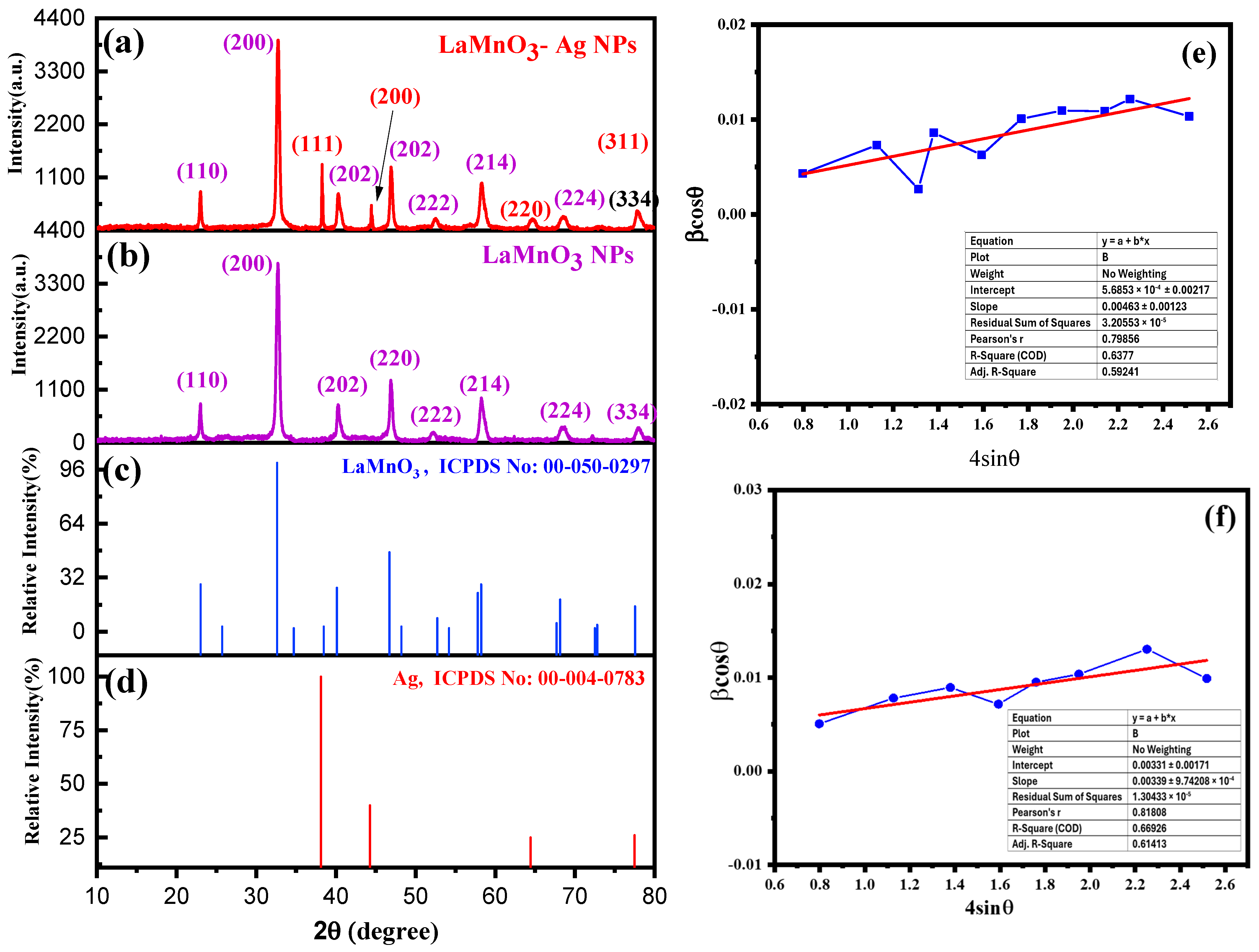
2.1. X-ray Diffraction

Figure 1a,b display the results for LaMnO3 and the noble metal-deposited LaMnO3-Ag nanocomposite. LaMnO3 showed an excellent crystallization impact since the diffraction peaks were visible in the XRD pattern. Sharp reflection peaks are visible in the XRD pattern of LaMnO3 at 2θ = 23°, 32.8°, 40.2°, 46.9°, 52.8°, 58.2°, 68.1°, 73.4°, and 77.9°. These diffraction peaks were matched with the reference card number JCPDS PDF card no. 00-050-0297, as shown in Figure 1c. The (200) crystal plane is the most pronounced distinctive peak of the catalyst. The other peaks, such as (110), (202), (220), (222), (024), (224), (134), and (332), are crystal faces and significantly matched with the orthorhombic single-phase perovskite structure [41]. The cubic phase of Ag nanoparticles and the orthorhombic structure of LaMnO3 were both confirmed by the XRD pattern of Ag-modified LaMnO3 nanoparticles. The presence of the Ag diffraction peaks at 2θ = 38.24° (111), 44.46° (200), 64.58° (220), and 77.47° (311) were detected and confirmed with JCPDS PDF card no. 00-004-0783 [52], as shown in Figure 1d. The XRD confirmed the loading of noble metals onto single-phase LaMnO3 nanoparticles.
Further, the Debye–Scherer equation, Equation (1), was used to calculate the size of perovskite LaMnO3 and LaMnO3-Ag nanoparticles.
D = 0.9 λ β c o s θ
where D is the crystallite size of the nanomaterial and λ is the X-ray radiation wavelength (0.15405 nm). At the same time, β is Full Width at Half Maximum (FWHM) of the peaks corresponding to the all-plane line obtained from LaMnO3 and LaMnO3-Ag nanoparticles of broadened XRD peaks. The crystallite size of the perovskites LaMnO3 and LaMnO3-Ag are shown in Tables S1 and S2, corresponding to all obtained lattice peaks. The average crystallite size of LaMnO3 nanoparticles was calculated as 13.77 nm (as shown in Table S1). In comparison, perovskite LaMnO3-Ag nanocomposite crystallite size was calculated for all diffracted peaks (11 peaks), and the average crystallite size was deemed to be 19.34 nm. The increment in crystallite size demonstrates that perovskite LaMnO3 nanoparticles contain Ag nanoparticle coverage on their structure as Ag nanoparticle plane peaks were observed in XRD and shown in the EDX plot. The micro-strain (ε) of LaMnO3-Ag and LaMnO3 nanocomposites were calculated using the Williamson–Hall (W–H) plot, as shown in Figure 1e,f. Micro-strain (ε) appertains the perovskite structure strain in LaMnO3 nanocomposite due to green-synthesized Ag nanoparticles. Micro-strain for LaMnO3 was calculated as 3.39 × 10⁻³, shown in Table S1, and is attributed to the good crystallinity. In the case of LaMnO3- Ag nanocomposite, the micro-strain was increased and measured as 4.63 × 10⁻³, shown in Table S2. The increment in micro-strain, appertaining to the presence of Ag nanoparticles on the LaMnO3 nanocomposite surface, significantly enhances the photocatalytic effects of these green synthesized nanoparticles.

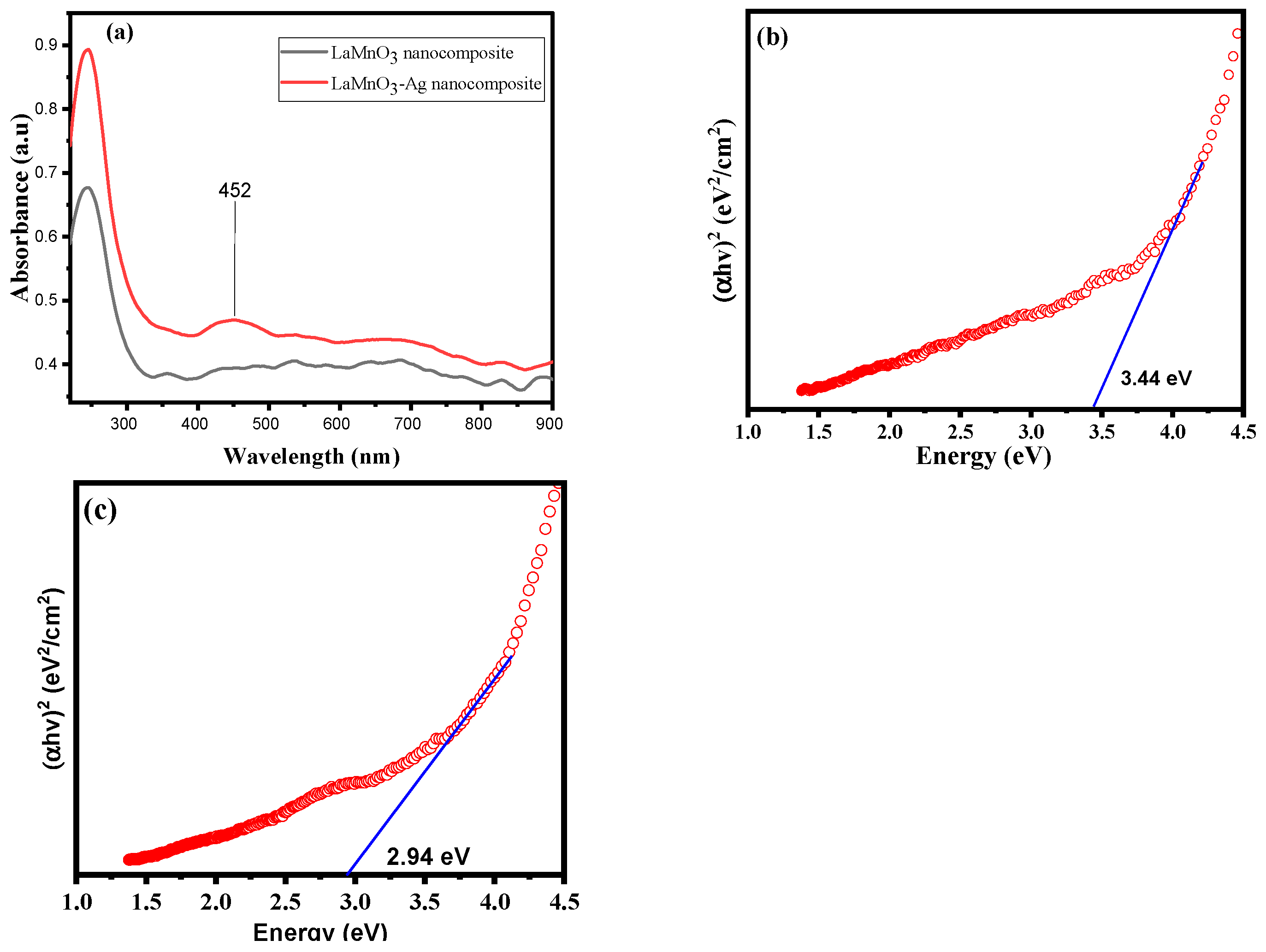
2.2. UV-Vis Diffuse Reflectance Spectroscopy (UV-Vis DRS)

UV-Vis Diffuse Reflectance Spectroscopy (DRS) was utilized to measure the optical properties of pristine materials and nanocomposites incorporating noble metals. Figure 2 displays the resultant spectra of nanomaterials. A low response intensity spectrum of LaMnO3 was observed between 240 and 800 nm, as shown in Figure 2a. The electronic transition from the Valance Band (VB) to the Conduction Band (CB), O2p, Mn3d, are primarily responsible for the strong absorption edge in perovskite (ABO3)-type materials [34]. Because of the charge transfer from the VB of O atoms to the CB of La atoms, the LaMnO3 displayed absorption bands below 400 nm. The optical band gap energy (Eg), which was computed using the Tauc plot and found to be 3.44 eV, as shown in Figure 2b, matches the previously measured absorption [41]. Recording the UV-Vis in the DRS spectra of LaMnO3-Ag nanocomposite further validated the deposition of noble metals. The absorption bands at 452 nm for Ag, which are seen in the visible region, were measured, and they were consistent with the successful deposition of Ag on LaMnO3 perovskite [53]. The charge transfer transition, which excites Ag 4d electrons into the conduction band, is what causes the apparent absorption bands to exist [53]. As depicted in Figure 2c, LaMnO3-Ag has a much higher absorption intensity than LaMnO3 nanoparticles. The absorption edge of LaMnO3-Ag nanocomposites showed a clear redshift. Additionally, the band gap calculation results showed that the value for LaMnO3-Ag was 2.94 eV, as shown in the Tauc plot in Figure 2c [41]. This finding suggests that the band of LaMnO3 can become narrower through the deposition of noble metals. As a result, it is possible to use solar energy more effectively. LaMnO3 with noble metals also exhibits a noticeable improvement in the photodegradation process. The leading cause of the bandgap alteration is the interaction between LaMnO3 and Ag nanoparticles. The Ag nanoparticles create trap levels between LaMnO3′s conduction and valence bands. This drop indicates that the electrical characteristics have improved the strong electron transport ability in LaMnO3-Ag structures due to Ag. LaMnO3-Ag can absorb a wide range of light irradiation with decreased bandgap energy. Additionally, Ag nanoparticles also have proficient light irradiation absorbers and plasmonic abilities. LaMnO3 contains Ag, which enhances perovskite’s ability to absorb light irradiation. The composite contacts considerably shortened the photoelectron transfer path from Ag to LaMnO3 nanoparticles. Effective charge transfer reduced the rate of electron-hole recombination and enhanced their capacity for photocatalysis [50].

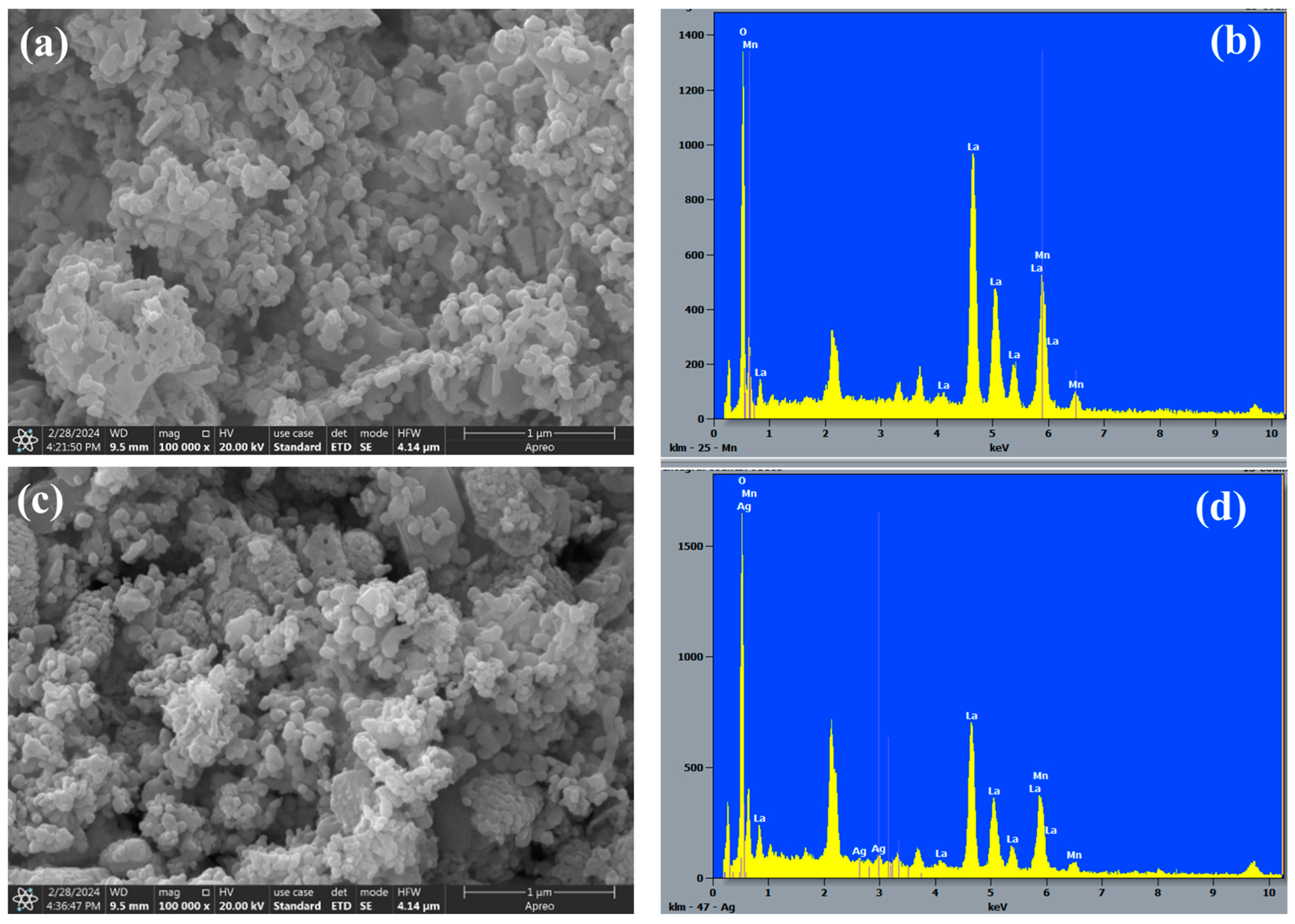
2.3. Morphological Analysis
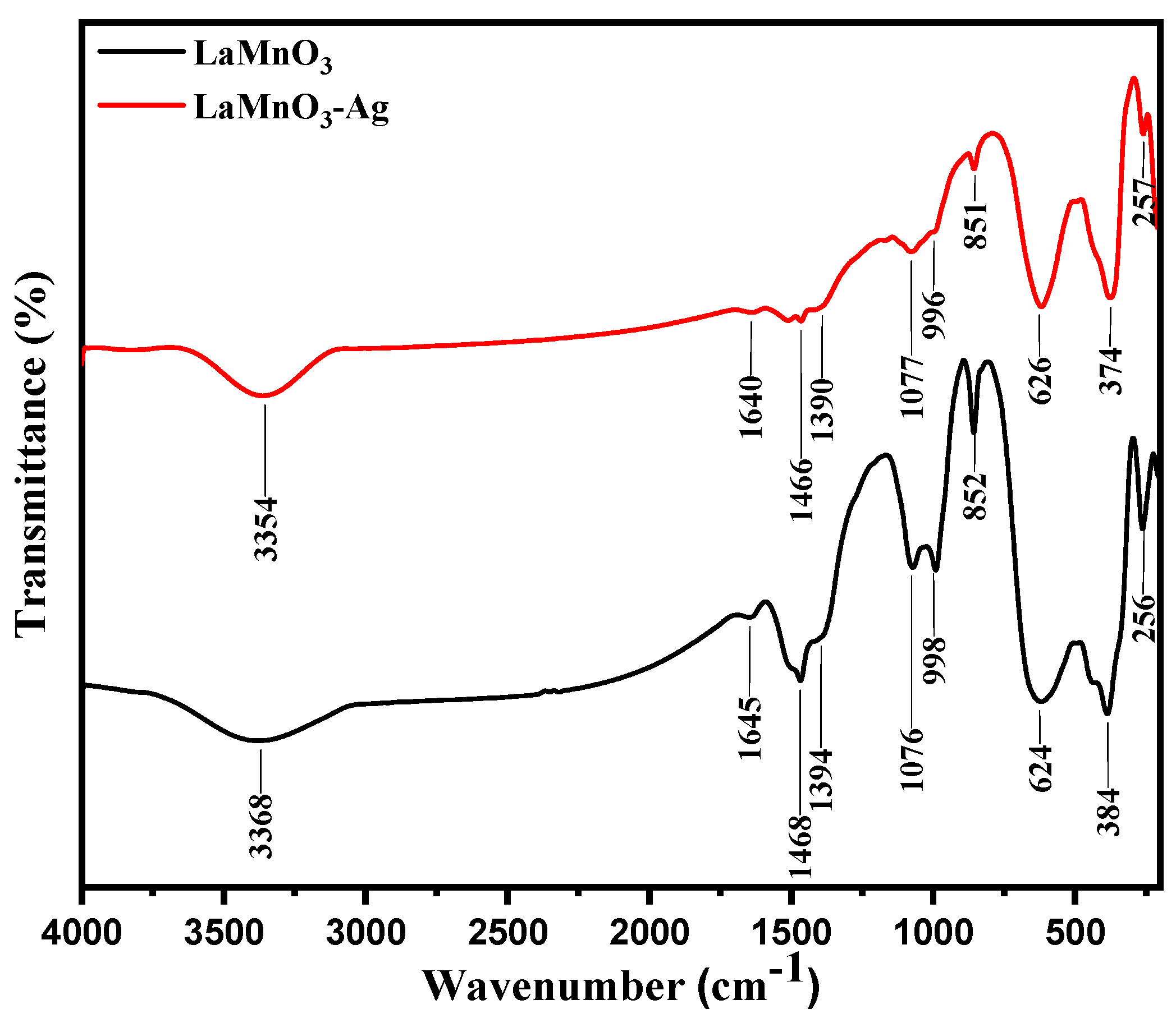
Figure 3 depicts the TEM images of the synthesized perovskites LaMnO3 and LaMnO3-Ag nanocomposites. The bare perovskite shows the agglomeration or clustering of nanoparticles, as seen in Figure 3a. The particle sizes ranged from 20 to 60 nm, with the most significant percentage of particles distributed at 37 nm. Compared to bare perovskite, denser nanoparticles were observed in noble metal-deposited perovskites; as a result, particles became oblong and erratic in size (Figure 3c,d). It is interesting to note that, in the TEM images, fabricated materials have spherical particles with a wide range of sizes. The TEM pictures of the LaMnO3-Ag nanocomposite show that the LaMnO3 nanomaterials are covered with black spots that are visible on the LaMnO3 spherical materials, indicating that Ag nanoparticles were successfully deposited on the LaMnO3 surface. Particle sizes were observed to be between 20 and 60 nm. The lattice fringes of the composite sample can be seen in Figure 3b,d. The crystal plane (110) belongs to LaMnO3 nanoparticles, with 0.32 nm spacing, while Ag nanoparticles indicate the crystal plane (111) with a 0.30 nm fringe space. The (200), (220), and (024) crystal planes of LaMnO3 were consistent with the spectral results of the preceding XRD, and the spacing of 0.274 nm, 0.194 nm, and 0.158 nm, respectively, conform to these results.
Figure 4a shows the HRSEM image of perovskite LaMnO3 nanoparticles and the LaMnO3-Ag nanocomposite morphology. The LaMnO3 contained a homogenous silver nanoparticle distribution with a spherical shape. Elemental analyses of the perovskite LaMnO3 and LaMnO3-Ag nanocomposites were confirmed by Energy Dispersive analysis of X-rays (EDX) characterization, as shown in Figure 4b,d. EDX confirmed the successful formation of crystalline LaMnO3 nanoparticles by the Ag nanoparticles’ homogenous distribution. Figures S1 and S2 represent the elemental mapping of HRSEM-selected images during EDX analysis. Figure S1a depicts the perovskite LaMnO3 nanoparticle-selected portion of HRSEM, while Figure S1b–d show the elemental mapping for oxygen (yellow dots), manganese (green dots), and lanthanum (purple dots). Figure S2a shows the Ag nanoparticle distribution on the perovskite LaMnO3 nanocomposite selected part to analyze the elemental mapping. Figure S2b–e represents the elemental mapping of O (yellow dots), Mn (green dots), Ag (red dots), and La (purple dots) using different spot colors, respectively, which confirmed the successful formation of perovskite LaMnO3-Ag nanocomposite using the Citrus limon (L.) Burm peels extract.

2.4. FT-IR Analysis

Figure 5 displays the FT-IR spectra of bare LaMnO3 and LaMnO3-Ag samples in the 200–4000 cm−1 spectral region. LaMnO3, in its purest form, has bands in its FT-IR spectra at 257, 384, 624, 852, 998, 1076, 1394, 1468, 1645, and 3368 cm−1. The stretching vibration of the La-O and Mn-O bonds in the perovskite phase of LaMnO3 is attributed to the bands at 256 and 384 cm−1 in the infrared spectra, respectively [54]. The band visible at 624 cm−1 is the stretching mode resulting from the Mn-O-Mn bond inside the octahedron MnO6, moving internally. This motion was attributed to a vibration of the perovskite-type ABO3 [54]. Two more strong infrared bands are seen at 1468 and 1394 cm−1, which are related to the OH vibrational modes of water molecules, respectively [55]. Similar to the perovskite LaMnO3 materials from a previous study that utilized hydrothermal synthesis, the important absorption bands for the LaMnO3 materials from this work are roughly placed in the same spectral region. Each sample showed a diffused band between 3150 and 3650 cm−1, which was attributed to the v O-H stretching vibration caused by H2O molecules that were adsorbed to the surface [55].
While bending vibrations in the N-O bonds (nitrates) are responsible for the bands around 1390 and 1466 cm−1, bending vibrations in the N-H bonds (secondary amines) are assumed to be responsible for the bands around 1645 and 1640 cm−1 [42]. The presence of nitrate and secondary amine bands on the stabilized surface of LaMnO3 and LaMnO3-Ag nanocomposites is attributed to the lemon extract, which contains complex phytochemicals with amino acids and proteins [56].

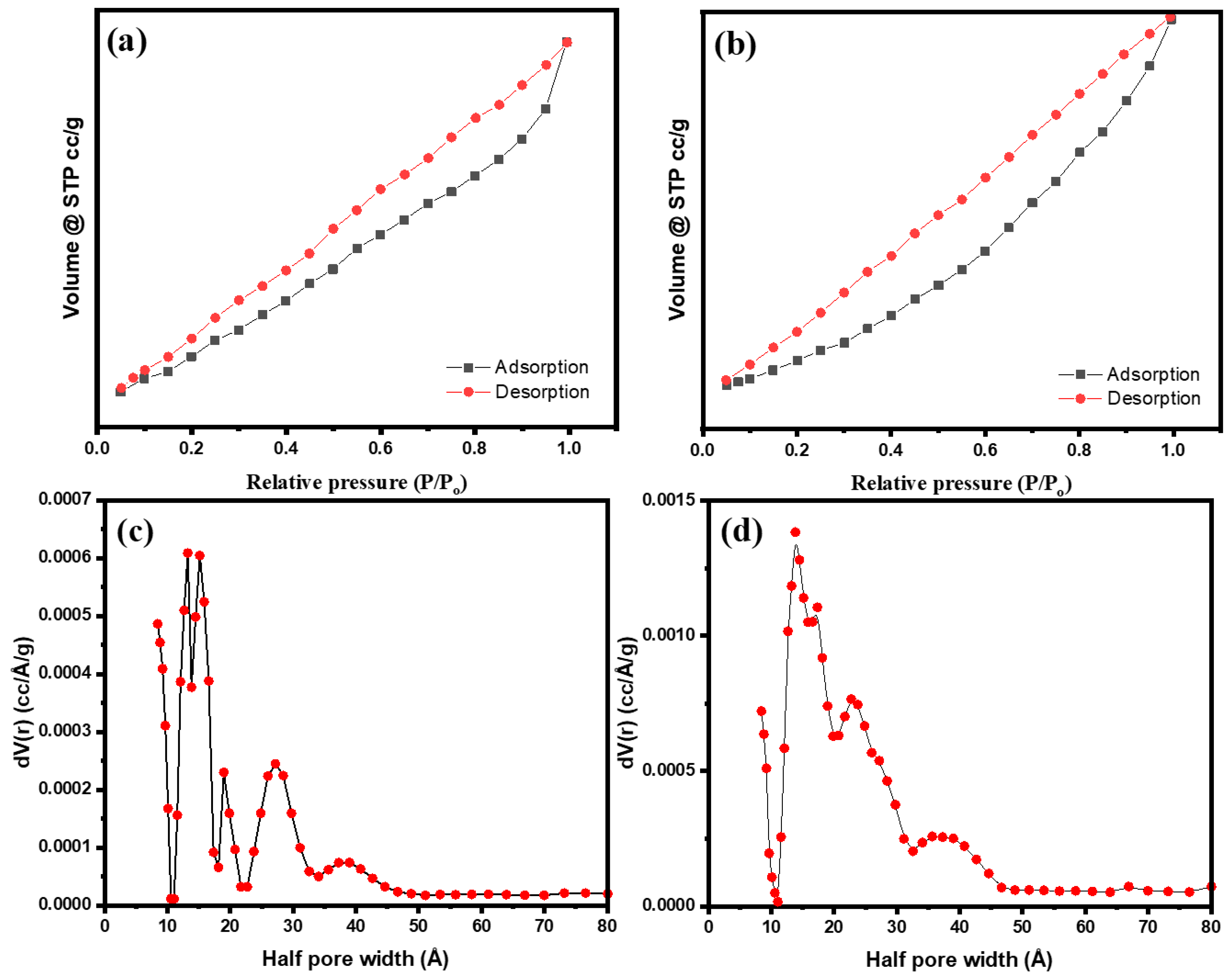
2.5. BET Surface Area

Figure 6a,b display the N2 adsorption-desorption isotherms and pore size distributions (Figure 6c,d) of the fabricated and green synthesized perovskite LaMnO3 and LaMnO3-Ag nanocomposites. According to Figure 6, some changes in the hysteresis loop are seen after the deposition of silver noble metals. Table 1 lists the samples’ textural characteristics, including BET surface area, pore size, and pore volume. It was observed that bare LaMnO3 and LaMnO3-Ag have BET values of 12.642 m2/g and 16.209 m2/g, respectively, and pore volumes of 0.012 cc/g and 0.023 cc/g, respectively [53]. Intriguingly, the findings show that compared to LaMnO3 and LaMnO3-Ag nanocomposite, green synthesized perovskite LaMnO3-Ag has the largest surface area and total pore volume. Additionally, the DFT model was used to calculate the pore size distributions of LaMnO3 and LaMnO3-Ag on the adsorption branch of the isotherms.

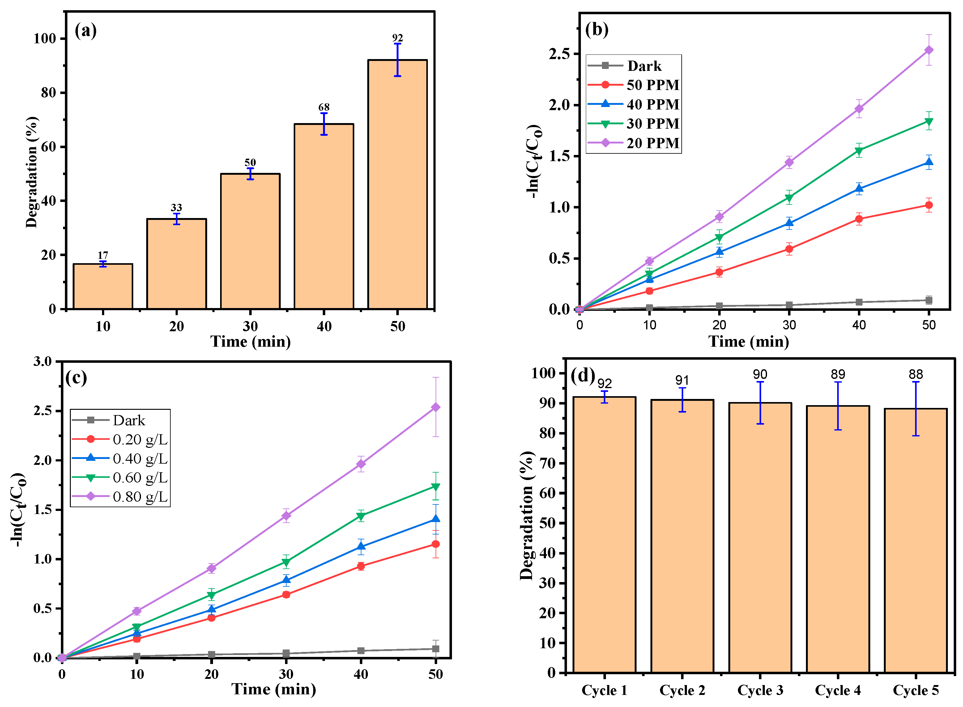
2.6. Photocatalytic Activity

In contrast to bare perovskite LaMnO3 nanoparticles, the LaMnO3-Ag nanocomposite was found to have a better band gap energy and surface area and is considered a more suitable candidate for RB degradation. The band gap energy of the perovskite oxide material plays a promising and favorable role, besides crystallinity, surface area, and shape, in defining its photocatalytic activity [35]. In the preliminary test, the initial absorption intensity of RB dye is unchanged in the control experiment (dye + light irradiation; dye + catalyst). The RB dye molecules’ adsorption on the LaMnO3-Ag photocatalyst surface caused a modest reduction in the initial absorption. The absorption intensity reduced over time when the photocatalyst was applied in light irradiation, indicating that the dye gradually degraded in the presence of a photocatalyst under direct light irradiation. Several photocatalytic degradation tests using the LaMnO3-Ag catalyst on RB dye were conducted to verify the prepared catalysts’ adaptability in light irradiation-driven catalytic behavior. It was best to optimize the factors that affect photocatalytic processes, such as catalyst dose, dye concentration, pH, and temperature, a few of which are discussed below. As the exposure duration prolongs, the vibrant pink color of the initial solution gradually diminishes. It almost loses color, demonstrating the degradation of RB dye under light irradiation.

2.7. Dye Concentration

The catalytic activity of the synthesized LaMnO3-Ag photocatalyst (40 mg/50 mL dye solution) was investigated using RB dye solution. According to studies on photolysis and adsorption, there was no degradation in the absence of the LaMnO3-Ag nanocomposite. However, there was a slight decrease in dye concentration when light irradiation was absent, and a photocatalyst was present. It is indicated that some adsorption may have occurred due to the increased surface area of the LaMnO3-Ag nanocomposite. Therefore, it can be determined by the photocatalytic studies that LaMnO3-Ag is quite effective at photodegrading the RB dye with light irradiation. The effectiveness of RB’s breakdown declines as dye concentration rises. This is caused by an accumulation of dye molecules on the surface of LaMnO3-Ag, which reduces photocatalytic activity due to the increased absorption of irradiation photons [54].
As the RB dye concentration rose from 20 to 50 ppm, the degradation rate constant (k) steadily reduced from 0.05057 to 0.02129 min−1. Furthermore, the optimal concentration at which the greatest degradation efficiency was observed was 20 ppm (Figure 7a). The results show a considerable concentration dependence for the photocatalytic activity. Figure 7b shows the plot of −ln(Ct/Co) versus light irradiation time, with the slopes representing the rate constant (k) for RB dye degradation [55]. From the plot, it is clear that the decay process has a characteristic pattern of typical pseudo-first-order kinetics, and the calculated rate constant (k) is shown in Table 2.

2.8. Photocatalyst Different Doses

The degradation of RB dye by LaMnO3-Ag was studied as a function of catalyst dosage and initial dye concentration under light irradiation. The time-dependent UV-Vis absorption spectra were recorded for the degradation of RB dye at LaMnO3-Ag nanocomposite dosages ranging from 10 to 40 mg (0.2 to 0.8 g/L) during 50 min of light irradiation. With an increase in photocatalyst dosage and light irradiation period, the characteristic RB absorption at 548 nm gradually declines. The degradation efficiency reaches 92% after 50 min of exposure to light irradiation, indicating that the RB has completely broken down. It is noted that no new absorption peaks emerged during the procedure, which ensures the complete RB dye degradation. Additionally, the degrading behavior initially enhances as the dosage of the LaMnO3-Ag nanocomposite rises, peaking at 40 mg. The optimal concentration of the LaMnO3-Ag catalyst for the degradation of RB dye in aqueous solution is 40 mg per 50 mL of 20 ppm RB dye. The vacancy of metal cations and O2− anions in the perovskite structure causes a striking increase in oxygen adsorbed onto the surface, which may be the cause of the increased photocatalytic activity of the perovskite LaMnO3-Ag nanocomposite. As a result, the dose of LaMnO3-Ag nanocomposite significantly impacts photocatalytic activity. The fluctuation of normalized ln C t C 0 dye concentration as a function of light irradiation time and the photocatalytic degradation of RB dye (20 ppm) in the presence of different photocatalysts (20–40 mg doses) yields a straight line (Figure 7c) [57]. The plot of ln C t C 0 over time illustrates how to compute the rate constant (k) for the degrading reaction using the slopes of linearized straight lines (Table 2). The findings unequivocally demonstrate that the photocatalytic degradation reaction follows pseudo-first-order chemical kinetics.

2.9. Stability and Reusability

To effectively combat organic pollutants, photocatalyst stability and reusability are crucial. Reusing materials is both a practical requirement and a cost-effective measure of many water treatment methods. LaMnO3-Ag nanocomposite was separated using a centrifuge and used for five consecutive cycles of photocatalytic degradation of RB to test the reusability of the material. RB was photodegraded over five reuse cycles, as shown in Figure 7d, with degradation rates of 92%, 91%, 90%, 89%, and 88%, respectively. Figure 7e attributes the UV-Vis spectra for the degradation of RB dye (20 ppm) using 40 mg of the LaMnO3-Ag nanocomposite photocatalyst. The loss of the catalyst explains the lower photocatalytic stability of LaMnO3-Ag composite photocatalysts [53,54,55,57]. Consequently, enhancing sample recovery is an effective strategy to improve photocatalytic stability. The degradation efficiency of the LaMnO3-Ag nanocomposite for RB dye is slightly reduced due to their multiple recycling processes, such as washing, filtering, and mixing. The deterioration of the material structure, likely caused by factors such as oxidation or mechanical stress, may be responsible for reducing the removal rate of RB dye from 92% to 88% over five cycles. In Figure S3a, the XRD spectra of the LaMnO3-Ag nanocomposite were analyzed to check for any phase changes or Ag leaching from the nanocomposite. The XRD spectra reveal that the LaMnO3-Ag phase remains unchanged and perfectly matches the reference card number JCPDS PDF card no. 00-050-0297 (Figure S3b). The Ag diffraction peaks were also observed and perfectly matched the reference JCPDS PDF card no. 00-004-0783 (Figure S3c).
RB dye photocatalytic degradation scavenger tests were also performed in the presence of various scavenging agents, such as Ethylenediaminetetraacetic acid (EDTA. h + scavenger), Potassium persulfate (KPS, e scavenger), Isopropanol (IPA, O H scavenger), and 1, 4 benzoquinone (BQ, O 2 scavenger), as shown in Figure 7f. These reactive species are responsible for photocatalytic RB dye degradation over the surface of LaMnO3-Ag nanocomposites using light irradiation. In the presence of IPA, RB dye photodegradation was reduced to 40 %, while using EDTA, BQ, and KPS reduced it to 55, 68, and 87 %. These scavenger studies indicate that the addition of IPA in RB dye solution in the presence of LaMnO3-Ag nanocomposite suspension leads to photocatalytic activities being significantly decreased due to the scavenging of O H species, which are more effectively involved in degrading the dye. Nevertheless, the LaMnO3-Ag nanocomposite significantly generates other scavenger species observed during RB dye photocatalytic degradation, as shown in the scavenger test in Figure 7f.

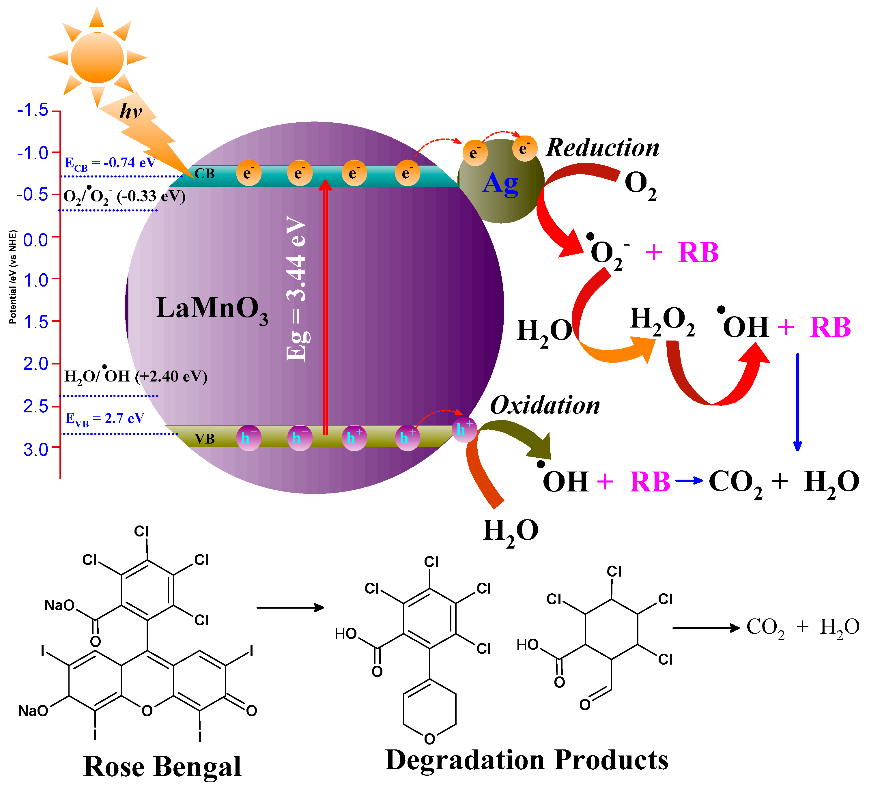
2.10. Mechanism of Dye

The photocatalytic activity’s reaction mechanism is depicted in Figure 8. The photo-generated electrons are transferred from the Valence Band (VB) to the Conduction Band (CB), leaving positive holes in the VB when the catalyst is exposed to light irradiation that has energy above the threshold during the degradation process of RB dye. Pollutants are trapped by the hydroxyl groups on the catalyst surface or are directly oxidized by the photoinduced holes in the VB, producing hydroxyl radicals (OH) [57]. These radicals transform the color into non-toxic metabolites. Superoxide anion radicals (O2−•) are created when conduction band electrons contact dissolved oxygen molecules. These radicals then protonate to form hydroperoxy radicals (HO2). Because of their large surface area and band gap energy, LaMnO3-Ag nanostructures help generate more electron (e) and hole (h+) pairs, eventually stopping the e and h+ from recombining. This results in radiation-less e and h + pair recombination within the perovskite material; however, this increases the photocatalytic activity during light irradiation [34,53]. Additionally, compared to bare LaMnO3, ultra-small plasmonic metal Ag- NP-decorated LaMnO3 has more significant photocatalytic activity [58]. Due to their electron-accepting nature, the Fermi levels of the ultra-small Ag NPs and the LaMnO3 begin to equilibrate this electron flow from the conduction band of the latter to the surface of the former during light irradiation. Ultra-small Ag NPs have a significantly altered Fermi level towards a more negative potential (Figure 8), dramatically enhancing the interfacial charge-transfer process [54].
The Fermi level of the LaMnO3-Ag is lower than the pure LaMnO3 conduction band, and photogenerated electrons migrate from the CB of LaMnO3 to the plasmonic Ag. Therefore, Ag on the surface of the LaMnO3 serves as an electron trap to impede the e h + pair recombination. The electrons migrate to the Ag and react with the surface-adsorbed O2, producing superoxide radicals (O2−•).
Mulliken electronegativity theory was used to calculate the valance band (EVB) and conduction band (ECB) edge potentials by following the empirical formula shown in Equations (2) and (3).
E V B =   χ E e     0.5 E g
E C B = E V B     E g
where χ is the absolute electronegativity (geometric mean of electronegativity of the constituent atoms), Ee is the energy of the free electron on the hydrogen scale with a fixed value of 4.5 eV, and Eg is the band gap energy. The χ and Eg of LaMnO3 were found to be 5.48 eV and 3.44 eV, respectively. Using the above empirical formula of Equations (1) and (2), the EVB and ECB of LaMnO3 were +2.7 eV and −0.74 eV, respectively. The O2 in the water was reduced to O 2 radicals by photogenerated electrons due to the ECB of the LaMnO3 equal to −0.74 eV being more negative than the standard potential of O2/ O 2   ( 0.33   e V ) . Similarly, the photogenerated h + created in the VB of LaMnO3 by the transfer of electrons from VB to CB could generate O H radicals by reacting with H2O molecules due to an EVB equal to +2.7 eV, which is higher than the standard potential of H2O/ O H (+2.40 eV) [59]. As a result, there are fewer electrons in LaMnO3’s conduction band, which raises the number of active holes in the photocatalytic system. The injected electrons then move from the molecular oxygen that is always present to a multitude of potent oxidative radicals, which effectively degrades the RB dyes [53]. The Ag NPs work efficiently, scavenging excitons and avoiding charge carrier recombination, which increases activity. According to this work, the evenly dispersed ultra-small plasmonic Ag NPs on the LaMnO3 boost the light irradiation absorption, which improves incident photo-conversion efficiency. It is possible to alter the Fermi level to a more negative potential by creating Schottky barriers between the ultra-small plasmonic Ag NPs and LaMnO3 after exposure to light irradiation, which lengthens the lifetime of charge carriers. This energy barrier will, therefore, prevent the photo-generated electrons and holes from recombination. Thus, efficient electron transfers combine with surface-adsorbed oxygen to produce more active oxidative species. For the efficient breakdown of dyes, these active oxidative species are reasonable. Table 3 presents various procedures for synthesizing perovskite LaMnO3 nanoparticles and their applications as photocatalysts. Additionally, it compares the LaMnO3-Ag nanocomposite with previous research work.

2.11. Antibacterial Activities against S. aureus (Gram-Positive) and E. coli (Gram-Negative) Bacteria

LaMnO3 and LaMnO3-Ag nanocomposite were confirmed to have effective antibacterial activities against Gram-positive (S. aureus) and Gram-negative (E. coli) bacteria. Figure S4 (Supporting Information) shows the effective antibacterial activities in the presence of LaMnO3 and LaMnO3-Ag nanocomposites. The green synthesized perovskite LaMnO3 and LaMnO3-Ag nanocomposite antibacterial efficiency was calculated using Equation (4).
A n t i b a c t e r i a l   e f f i c i e n c y = L o g N N o
N is the bacterial concentration after applying LaMnO3 and LaMnO3-Ag nanocomposites, while No is the initial bacterial concentration in cfu/mL, respectively. In the presence of the LaMnO3 nanocomposite, antibacterial efficiency was enhanced from 3.14 for S. aureus to 0.049. However, E. coli shows high antibacterial efficiency, calculated as 4.32 from 0.011 by Equation (4), as represented in Figure S4. Phosphate ( PO 4 3 ) is a vital component for bacterial growth and is essential for various cellular processes. Phospholipids, which make up the lipid bilayer of cell membranes, contain phosphate groups. These phospholipids contribute to the structural integrity and fluidity of cell membranes. The phosphate ( PO 4 3 ) group is ubiquitously present in proteins, nucleic acid, peptides, and enzymes, and it works as an energy carrier in cells that effectively possess bacterial cell growth. Phosphate translocation and exchange in living microorganisms is fast and carries the phosphorylation and dephosphorylation process to control cell growth and division [63]. The phosphate depletion in media during bacterial cell growth may limit it or cause death. Recently, lanthanum-based oxide nanocomposites were reported as strong binders toward phosphate removal [64,65,66,67,68]. Lanthanum-based nanomaterial reacts with available phosphate from the growth medium or microorganisms to form stable LaPO4 [65]. In our case, 50 µg/mL of LaMnO3 or LaMnO3-Ag nanocomposites in a known cfu/mL concentration of S. aureus and E. coli, for both bacteria LaMnO3 nanocomposite, was found effective in controlling the microorganism cfu due the effect of lanthanum phosphate formation. However, the LaMnO3-Ag nanocomposite shows more highly effective reduction efficiency for S. aureus and E. coli and works as an antibacterial material [69,70,71,72]. Silver nanoparticles are well-known and influential antibacterial material; however, specific mechanisms are still not known to show toxicity [73]. The LaMnO3-Ag nanocomposite contains the synergistic effect due to LaMnO3 and Ag nanoparticles. The LaMnO3 nanocomposite reacts with PO 4 3 , while Ag NPs exert their antimicrobial effects at the membrane level [73,74]. Ag NPs can penetrate the outer membrane of bacterial cells, accumulating in the inner membrane. Figure 9a,d represent the control S. aureus and E. coli TEM images representing the sharp bacterial cell biofilm. After the application of LaMnO3-Ag nanocomposite on both S. aureus and E. coli., the adhesion of nanoparticles occurs, which triggers destabilization and damages the cells, resulting in an elevation of membrane permeability. This heightened permeability leads to the leakage of cellular content, ultimately culminating in cell death, as shown in the TEM of Figure 9b,e for S. aureus and E. coli. The antibacterial efficiency was calculated as 5.37 and 6.22 for S. aureus and E. coli, respectively, which are much higher than the LaMnO3 nanocomposite, as shown in Figure S4. The sequence of events highlights the disruptive impact of the LaMnO3-Ag nanocomposite on bacterial cell membranes, as shown in Figure 9c,f for S. aureus and E.coli, respectively, showcasing their potential as effective agents against microbial growth.
The observed morphology and synergistic effects of the Ag-decorated LaMnO3 nanocomposite are critical for understanding its enhanced photodegradation of RB dye and antibacterial activity. The LaMnO3 nanoparticles exhibit a well-defined morphology that provides a high surface area, which is crucial for catalytic activity. When Ag nanoparticles are decorated on the LaMnO3 surface, they introduce additional active sites, further enhancing the material’s catalytic properties. This synergistic effect between LaMnO3 and Ag is vital for improving the nanocomposite’s efficiency in degrading RB dye and inhibiting S. aureus and E. coli growth. This activity enhancement is due to the efficient charge separation and transfer facilitated by Ag nanoparticles, as shown in Figure 8. Ag acts as an electron sink, reducing the recombination rate of electron-hole pairs generated in LaMnO3 under light irradiation, demonstrating synergic effects. This process leads to a higher generation of Reactive Oxygen Species (ROS), essential for breaking down RB dye, and increased antibacterial properties. Nevertheless, the perovskite LaMnO3-Ag nanocomposite, fabricated through green-synthesis routes, exhibits significant effectiveness in photocatalytic and antibacterial activities. However, a substantial quantity of the LaMnO3-Ag nanocomposite is required to enhance photocatalytic efficiency for the degradation of higher concentrations (PPM) of RB dye, which limits its applicability. The LaMnO3-Ag nanocomposite holds promising potential for environmental and biomedical research applications.

3. Methods

3.1. Materials

The reagents used for the preparation of perovskite oxide were lanthanum (II) nitrate hexahydrate (La(NO3)3·6H2O, 99.99%), manganese (II) nitrate tetrahydrate (Mn(NO3)2.4H2O, 99.98%), ammonia (NH3) solution, Ethylenediaminetetraacetic acid (EDTA), 1,4-bezoquinone, Potassium persulfate, Isopropanol, Ethanol (99.8–99.98%), Silver nitrate (AgNO3, 99.0%) and Rose Bengal dye (C20H2Cl4I4Na2O5, 95%) which were purchased from Sigma Aldrich (Taufkirchen, Germany). All the chemicals were analytical grade and used without further purification. Dried lemons were acquired from the local market in Jazan, Saudi Arabia.

3.2. Synthesis of LaMnO3 and LaMnO3-Ag Nanocomposite

Firstly, the precursors La(NO3)3 and Mn(NO3)2 were magnetically dissolved in deionized Milli Q water at room temperature. Then, 20 mL of aqueous extract of lemon peel, which was prepared at 80 °C, was added to a stoichiometric amount of La(NO3)3 (0.1M) and Mn(NO3)2 (0.1M) solutions in stirring conditions. The pH of the reaction mixture was kept constant, and ammonia solution was added dropwise while continuously heated and stirred. The resulting reaction mixture was then gradually heated until 30% of the water had been removed to produce a slightly viscous substance. Furthermore, a 100 mL Teflon-lined stainless-steel autoclave was filled with the resulting precursor solution and heated over a hot air oven for 24 h at 180 °C. After cooling, it was centrifuged at 5000 rpm for 10 min to separate the sediment. The collected sediment was repeatedly washed with ethanol and DI water to make a pure LaMnO3 precursor. The obtained product was dried overnight and calcined (10 °C/min) at 650 °C for four hours. In the same way, LaMnO3 was fabricated and decorated with Ag nanoparticles utilizing the lemon peel aqueous extract. Approximately 2 g of LaMnO3 nanocomposites were mixed continuously with 10 mL of double-distilled water in two distinct beakers to form the mixture. After adding 20 mL of lemon extract, each beaker received 10 mL of a 0.15 M aqueous solution of AgNO3. A color change allowed the detection of the early reduction of noble metals. The LaMnO3 nanocomposites with noble metal loading (12.5 % silver with 2 g LaMnO3 perovskite) were separated by centrifugation, and the obtained material was repeatedly washed with DI water and calcined at the same temperature. Further, the silver leaching test from the LaMnO3 nanoparticles was negative after using the HCl diluted solution. The diagram in Figure 10 shows the process flow for creating nanocomposite materials.

3.3. Characterization Techniques

The phase purity and crystallinity of the fabricated perovskites were determined by quantifying the X-ray diffraction (XRD) patterns using an X-ray diffractometer (D8, Advance, Bruker, Germany) with C u K α radiation (1.542 Å). The surface chemistry and chemical bonding were determined using Fourier Transform Infrared (FTIR) spectra recorded using KBr pellets and the Bruker Tensor-27 in transmission mode between 400 and 4000 cm 1 . The reflectance spectra of the solid samples were acquired using a Thermo-Scientific evolution UV-Vis spectrophotometer outfitted with an integrating sphere in the 200–800 nm wavelength range. The specific surface area and pore structure were measured using an automatic volumetric apparatus (Quantachrome ASiQ adsorption) following standard Brunauer–Ettor–Teller (BET) theory and DFT procedures. Using a Transmission Electron Microscope (TEM) (JEM-2100, JEOL, Japan) operated at 200 kV, the surface morphology of the synthesized photocatalysts and nanoparticle–bacteria interactions were observed. High-resolution scanning Electron Microscopy (HRSEM) images, EDX, and atomic mapping were measured using SEM (Quanta FEG 250 with field emission gun, FEI, Eindhoven, The Netherlands).

3.4. Photocatalytic Application of LaMnO3-Ag Nanocomposites

The degradation of RB dye in an aqueous solution, illuminated by visible light, was used to assess the photocatalytic activity of the perovskite catalysts. A 150 W mercury lamp was placed 15 cm from the reactor to trigger a photochemical reaction. To 50 mL of RB dye (20 ppm), 40 mg of the photocatalyst was added at constant stirring and kept in the dark for 30 min to achieve the adsorption–desorption equilibrium before irradiation. The degradation procedure at room temperature then involved exposing the solution to visible light irradiation. During the different time intervals, aliquots of the RB dye solution were withdrawn at 10 min intervals. UV-Vis spectra were measured using a UV-2550 spectrophotometer (Shimadzu UV-2550) in a wavelength range of 200 to 800 nm. To determine how quickly the RB was breaking down, its maximum absorbance was measured at 548 nm.
The degradation efficiency was calculated using Equation (5).
Degradation   efficiency   ( % ) = C o C t C o × 100 %
where the initial absorbance of the RB dye is denoted by Co and the absorbance at time t by Ct. Equation (6) was utilized to determine the degradation rate constant, which is based on the simplified pseudo-first-order kinetic model of the Lang–Muir–Hinshewood equation.
l n C t C o = k t
where t is degradation time (min) and k is the rate constant (min−1).

3.5. Antibacterial Activities of LaMnO3 and LaMnO3-Ag Nanoparticles

The antimicrobial activities of LaMnO3 NPs and LaMnO3-Ag nanocomposite were checked against Staphylococcus aureus (Gram-positive) and E. coli (Gram-negative) bacteria isolates.
The bacteria were cultured at 37 °C on Mueller and Hinton agar. cDNA gene sequencing was used to identify molecularly of the S. aureus and E. coli strains. Standard biochemical assays identified the used samples.

3.6. Bacterial Sample Preparation and Antibacterial Study Using Colony Forming Unit (cfu)

S. aureus and E. coli bacteria colonies were picked from freshly grown culture agar plates and placed into buffer saline; the concentration was calculated using cfu/mL. Different weights of LaMnO3-Ag nanomaterial were applied for E. coli and S. aureus using a 108 cfu/ mL concentration in Mueller and Hinton agar solution at 37 °C, gently shaking in an incubator overnight.

3.7. LaMnO3-Ag Nanomaterial and Bacterial Surface Analysis Using Transmission Electron Microscopy (TEM), a Sample Preparation

Centrifugation at 3000 rpm separated the culture treated with coupled bacteria and the LaMnO3-Ag nanomaterial. After discarding the supernatant, the conjugated bacterial cell and LaMnO3-Ag nanomaterial that had been separated were gently vortexed for five minutes and then rinsed with deionized water. Lastly, 0.5 μL of the conjugated bacterial cell suspension was applied to the copper grid for TEM examination (JEM-2100, JEOL, Japan; Tokyo, Japan) at a 75 kV accelerating voltage. The cells were resuspended in deionized water.

4. Conclusions

The hydrothermal process with Citrus limon (L.) Burm peel extract was used to create LaMnO3 and its nanocomposite materials with Ag noble metal, and their photocatalytic and antibacterial activities against S. aureus and E. coli were assessed. The XRD analysis reveals an orthorhombic perovskite structure. FT-IR examination validated the structure and type of the chemical bonds, while HRTEM analysis revealed spherical and slightly clumped nanoparticles. LaMnO3 nanoparticles exhibit a high absorption band at 247 nm, and a band gap energy of 3.44 eV was calculated by UV-Vis analysis. To lower the band gap energy of LaMnO3, noble metal nanoparticles of Ag nanoparticles were successfully added to the LaMnO3 perovskite surface. LaMnO3-Ag has a lowered band gap, calculated as 2.94 eV, indicating that it absorbs light irradiation. The surface areas of the LaMnO3 and LaMnO3-Ag nanocomposites were calculated using BET and were found to be 12.642 m2/g and 16.209 m2/g, respectively. The photocatalytic activity of LaMnO3-Ag demonstrates favorable results when subjected to light irradiation for degrading RB dye. The H O and O 2 radicals formation study unequivocally supported the aforementioned photocatalytic activity. Because of their greater photocatalytic effectiveness, LaMnO3-Ag showed above 92% RB dye degradation in 50 min when exposed to light irradiation. The LaMnO3 and the LaMnO3-Ag nanocomposites exhibited antibacterial activity for S. aureus and E. coli, with the interaction analyzed through TEM images of bacterial cells and nanocomposites. Green synthesized LaMnO3-Ag nanocomposite is a viable option for quickly cleansing a significant amount of industrial effluent with light irradiation and has efficient antibacterial properties.

Supplementary Materials

The following supporting information can be downloaded at: https://www.mdpi.com/article/10.3390/catal14090609/s1, Figure S1: Elemental mapping on selected HRSEM (a), Oxygen (yellow dots) (b), Manganese (green dots) (c) and Lanthanum (purple dots) (d). Figure S2: Elemental mapping on selected HRSEM (a), Oxygen (yellow dots) (b), Manganese (green dots) (c), Silver (red dots) (d), and Lanthanum (purple dots) (e). Figure S3: (a), XRD pattern of LaMnO3-Ag nanocomposite after using fifth cycle RB dye degradation, (b), ICPDS reference peaks of LaMnO3 with card number 00-050-0297, (c), ICPDS reference peaks of Ag with card number 00-004-0783, Figure S4: Antibacterial activity of LaMnO3 and LaMnO3-Ag nanomaterials for S.aureus and E.coli bacteria. Table S1: Calculation of crystallite size (D) and micro-strain (ε) using XRD diffraction peaks LaMnO3 nanoparticles alone, Table S2: Calculation of crystallite size (D) and micro-strain (ε) using XRD diffraction peaks of LaMnO3- Ag nanoparticles.

Funding

The Deanship of Graduate Studies and Scientific Research of Jazan University, Saudi Arabia have funded this research article by project number GSSRD-24.

Data Availability Statement

The data presented in this study are available on request from the corresponding author.

Acknowledgments

The author gratefully acknowledges the funding of the Deanship of Graduate Studies and Scientific Research, Jazan University, Saudi Arabia, through Project Number: GSSRD-24.

Conflicts of Interest

The author declares no conflict of Interest.

References

  1. Salomatina, E.; Fukina, D.; Koryagin, A.; Titaev, D.; Suleimanov, E.; Smirnova, L. Preparation and photocatalytic properties of titanium dioxide modified with gold or silver nanoparticles. J. Environ. Chem. Eng. 2021, 9, 106078. [Google Scholar] [CrossRef]
  2. Narkbuakaew, T.; Sujaridworakun, P. Role of Ag(0) deposited on TiO2 nanoparticles for superior photocatalytic performance induced by calcination. Opt. Mater. 2019, 98, 109407. [Google Scholar] [CrossRef]
  3. Bak, T.; Li, W.; Nowotny, J.; Atanacio, A.J.; Davis, J. Photocatalytic properties of TiO2: Evidence of the key role of surface active sites in water oxidation. J. Phys. Chem. A 2015, 119, 9465–9473. [Google Scholar] [CrossRef]
  4. Ajmal, A.; Majeed, I.; Malik, R.N.; Idriss, H.; Nadeem, M.A. Principles and mechanisms of photocatalytic dye degradation on TiO2 based photocatalysts: A comparative overview. RSC Adv. 2014, 4, 37003–37026. [Google Scholar] [CrossRef]
  5. Banerjee, S.; Pillai, S.C.; Falaras, P.; O’Shea, K.E.; Byrne, J.A.; Dionysiou, D.D. New insights into the mechanism of visible light photocatalysis. J. Phys. Chem. Lett. 2014, 5, 2543–2554. [Google Scholar] [CrossRef]
  6. Salas, S.E.; Rosales, B.S.; de Lasa, H. Quantum yield with platinum modified TiO2 photocatalyst for hydrogen production. Appl. Catal. B Environ. 2013, 140, 523–536. [Google Scholar] [CrossRef]
  7. Grabowska, E. Selected perovskite oxides: Characterization, preparation and photocatalytic properties—A review. Appl. Catal. B Environ. 2016, 186, 97–126. [Google Scholar] [CrossRef]
  8. Devi, L.G.; Kavitha, R. A review on nonmetal ion doped titania for the photocatalytic degradation of organic pollutants under UV/solar light: Role of photogenerated charge carrier dynamics in enhancing the activity. Appl. Catal. B Environ. 2013, 140, 559–587. [Google Scholar] [CrossRef]
  9. Jabbar, Z.H.; Ebrahim, S.E. Recent advances in nano-semiconductors photocatalysis for degrading organic contaminants and microbial disinfection in wastewater: A comprehensive review. Environmental Nanotechnology. Monit. Manag. 2022, 17, 100666. [Google Scholar] [CrossRef]
  10. Kar, P.; Shukla, K.; Jain, P.; Sathiyan, G.; Gupta, R.K. Semiconductor-based photocatalysts for detoxification of emerging pharmaceutical pollutants from aquatic systems: A critical review. Nano Mater. Sci. 2021, 3, 25–46. [Google Scholar] [CrossRef]
  11. Lee, K.M.; Lai, C.W.; Ngai, K.S.; Juan, J.C. Recent developments of zinc oxide based photocatalyst in water treatment technology: A review. Water Res. 2016, 88, 428–448. [Google Scholar] [CrossRef]
  12. Chen, P.; Cai, Y.; Wang, J.; Wang, K.; Tao, Y.; Xue, J.; Wang, H. Preparation of protonized titanate nanotubes/Fe3O4/TiO2 ternary composites and dye self-sensitization for visible-light-driven photodegradation of Rhodamine B. Powder Technol. 2018, 326, 272–280. [Google Scholar] [CrossRef]
  13. Cheng, Z.; Ling, L.; Wu, Z.; Fang, J.; Westerhoff, P.; Shang, C. Novel visible-light-driven photocatalytic chlorine activation process for carbamazepine degradation in drinking water. Environ. Sci. Technol. 2020, 54, 11584–11593. [Google Scholar] [CrossRef]
  14. Meneceur, S.; Hemmami, H.; Bouafia, A.; Laouini, S.E.; Tedjani, M.L.; Berra, D.; Mahboub, M.S. Photocatalytic activity of iron oxide nanoparticles synthesized by different plant extracts for the degradation of diazo dyes Evans blue and Congo red. Biomass Convers. Biorefinery 2024, 14, 5357–5372. [Google Scholar] [CrossRef]
  15. Mohammed, H.A.; Eddine, L.S.; Souhaila, M.; Hasan, G.G.; Kir, I.; Abdullah, J.A.A. Green Synthesis of SnO2 Nanoparticles from Laurus nobilis L. Extract for Enhanced Gelatin-Based Films and CEF@SnO2 for Efficient Antibacterial Activity. Food Bioprocess Technol. 2024, 17, 1364–1382. [Google Scholar] [CrossRef]
  16. Salmi, C.; Souhaila, M.; Eddine, L.S.; Mohammed, H.A.M.; Hasan, G.G.; Mahboub, M.S. Biosynthesis of Mn3O4/PVP Nanocomposite for Enhanced Photocatalytic Degradation of Organic Dyes Under Sunlight Irradiation. J. Clust. Sci. 2024, 35, 201–215. [Google Scholar] [CrossRef]
  17. Rao, C.N.R. Perovskites. In Encyclopedia of Physical Science and Technology, 3rd ed.; Meyers, R.A., Ed.; Academic Press: Cambridge, MA, USA, 2003; pp. 707–714. [Google Scholar]
  18. Irshad, M.; Ain, Q.T.; Zaman, M.; Aslam, M.Z.; Kousar, N.; Asim, M.; Rafique, M.; Siraj, K.; Tabish, A.N.; Usman, M. Photocatalysis and perovskite oxide-based materials: A remedy for a clean and sustainable future. RSC Adv. 2022, 12, 7009–7039. [Google Scholar] [CrossRef]
  19. Khalil, K.M.; Mahmoud, A.H.; Khairy, M. Formation and textural characterization of size-controlled LaFeO3 perovskite nanoparticles for efficient photocatalytic degradation of organic pollutants. Adv. Powder Technol. 2022, 33, 103429. [Google Scholar] [CrossRef]
  20. Das, N.; Kandimalla, S. Application of perovskites towards remediation of environmental pollutants: An overview: A review on remediation of environmental pollutants using perovskites. Int. J. Environ. Sci. Technol. 2017, 14, 1559–1572. [Google Scholar] [CrossRef]
  21. Yang, L.; Jiao, Y.; Xu, X.; Pan, Y.; Su, C.; Duan, X.; Sun, H.; Liu, S.; Wang, S.; Shao, Z. Superstructures with Atomic-Level Arranged Perovskite and Oxide Layers for Advanced Oxidation with an Enhanced Non-Free Radical Pathway. ACS Sustain. Chem. Eng. 2022, 10, 1899–1909. [Google Scholar] [CrossRef]
  22. Xu, X.; Wang, W.; Zhou, W.; Shao, Z. Recent Advances in Novel Nanostructuring Methods of Perovskite Electrocatalysts for Energy-Related Applications. Small Methods 2018, 2, 1800071. [Google Scholar] [CrossRef]
  23. Vulchev, V.; Vassilev, L.; Harizanova, S.; Khristov, M.; Zhecheva, E.; Stoyanova, R. Improving the thermoelectric efficiency of LaCoO3 by double substitution with nickel and iron. J. Phys. Chem. C 2012, 116, 13507–13515. [Google Scholar] [CrossRef]
  24. Han, X.; Ni, H.; Zhao, K.; Xiang, W. Highly sensitive reversible light-driven switches based on LaSrAlO4 single crystals. Opt. Commun. 2011, 284, 5702–5704. [Google Scholar] [CrossRef]
  25. Wang, H.; Zhou, M.; Choudhury, P.; Luo, H. Perovskite oxides as bifunctional oxygen electrocatalysts for oxygen evolution/reduction reactions–A mini-review. Appl. Mater. Today 2019, 16, 56–71. [Google Scholar] [CrossRef]
  26. Choi, J.-J.; Park, G.-T.; Kim, H.-E.; Kim, D.-Y. Transverse and longitudinal electrooptic properties of highly (100) oriented Pb(Zr, Ti)O3 films grown on glass substrates. Thin Solid Film. 2006, 515, 2437–2441. [Google Scholar] [CrossRef]
  27. Chen, Y.-C.; Chang, K.-C.; Tsai, D.-Y. A hybrid dielectric resonator antenna based upon novel complex perovskite microwave ceramic. Ceram. Int. 2013, 39, 8043–8048. [Google Scholar] [CrossRef]
  28. Hou, J.; Liu, H.; Zhao, L. Optimization of radiation absorption model in solar photocatalytic reactors: Coupling photocatalyst concentration distribution. Appl. Therm. Eng. 2019, 160, 114090. [Google Scholar] [CrossRef]
  29. Acosta-Herazo, R.; Mueses, M.A.; Puma, G.L.; Machuca-Martinez, F. Impact of photocatalyst optical properties on the efficiency of solar photocatalytic reactors rationalized by the concepts of initial rate of photon absorption (IRPA) dimensionless boundary layer of photon absorption and apparent optical thickness. Chem. Eng. J. 2019, 356, 839–849. [Google Scholar] [CrossRef]
  30. Jong, U.-G.; Yu, C.-J.; Kye, Y.-H. Computational prediction of structural, electronic, and optical properties and phase stability of double perovskites K2SnX6 (X = I, Br, Cl). RSC Adv. 2020, 10, 201–209. [Google Scholar] [CrossRef]
  31. Thinley, T.; Prabagar, J.S.; Yadav, S.; Anusha, H.S.; Anilkumar, K.M.; Kitirote, W.; Shahmoradi, B.; Shivaraju, H.P. LaNiO3-rGO perovskite interface for sustainable decontaminants of emerging concerns under visible light photocatalysis. J. Mol. Struct. 2023, 1285, 135413. [Google Scholar] [CrossRef]
  32. Shi, J.; Guo, L. ABO3-based photocatalysts for water splitting. Prog. Nat. Sci. Mater. Int. 2012, 22, 592–615. [Google Scholar] [CrossRef]
  33. Hu, J.; Liu, Y.; Men, J.; Zhang, L.; Huang, H. Ag modified LaMnO3 nanorods-reduced graphene oxide composite applied in the photocatalytic discoloration of direct green. Solid State Sci. 2016, 61, 239–245. [Google Scholar] [CrossRef]
  34. Kumar, R.D.; Thangappan, R.; Jayavel, R. Enhanced visible light photocatalytic activity of LaMnO3 nanostructures for water purification. Res. Chem. Intermed. 2018, 44, 4323–4337. [Google Scholar] [CrossRef]
  35. Kumar, S.R.; Abinaya, C.; Amirthapandian, S.; Ponpandian, N. Enhanced visible light photocatalytic activity of porous LaMnO3 sub-micron particles in the degradation of rose bengal. Mater. Res. Bull. 2017, 93, 270–281. [Google Scholar] [CrossRef]
  36. Flores-Lasluisa, J.X.; Huerta, F.; Cazorla-Amorós, D.; Morallón, E. Manganese oxides/LaMnO3 perovskite materials and their application in the oxygen reduction reaction. Energy 2022, 247, 123456. [Google Scholar] [CrossRef]
  37. Luo, K.; Zheng, Q.; Yu, Y.; Wang, C.; Jiang, S.; Zhang, H.; Liu, Y.; Guo, Y. Urea-Assisted Sol-Gel Synthesis of LaMnO3 Perovskite with Accelerated Catalytic Activity for Application in Zn-Air Battery. Batteries 2023, 9, 90. [Google Scholar] [CrossRef]
  38. Song, L.; Zhu, Y.; Yang, Z.; Wang, C.; Lu, X. Oxidase-mimicking activity of perovskite LaMnO3+ δ nanofibers and their application for colorimetric sensing. J. Mater. Chem. B 2018, 6, 5931–5939. [Google Scholar] [CrossRef] [PubMed]
  39. Zhang, C.; Zhang, W.; Li, X.; Zhu, Z.; Wang, Q.; Luo, S.; Xie, A. Honeycomb LaMnO3 perovskite synthesized by a carbon sphere as a self-sacrificing template for supercapacitors. Energy Fuels 2021, 35, 13457–13465. [Google Scholar] [CrossRef]
  40. Ashok, A.; Kumar, A.; Bhosale, R.R.; Almomani, F.; Malik, S.S.; Suslov, S.; Tarlochan, F. Combustion synthesis of bifunctional LaMO3 (M= Cr, Mn, Fe, Co, Ni) perovskites for oxygen reduction and oxygen evolution reaction in alkaline media. J. Electroanal. Chem. 2018, 809, 22–30. [Google Scholar] [CrossRef]
  41. Jawhari, A.H.; Hasan, N.; Radini, I.A.; Rao, K.N.; Malik, M.A. Noble Metals Deposited LaMnO3 Nanocomposites for Photocatalytic H2 Production. Nanomaterials 2022, 12, 2985. [Google Scholar] [CrossRef]
  42. Zhang, C.; Guo, Y.; Guo, Y.; Lu, G.; Boreave, A.; Retailleau, L.; Baylet, A.; Giroir-Fendler, A. LaMnO3 perovskite oxides prepared by different methods for catalytic oxidation of toluene. Appl. Catal. B Environ. 2014, 148, 490–498. [Google Scholar] [CrossRef]
  43. Priyatharshni, S.; Kumar, S.R.; Viswanathan, C.; Ponpandian, N. Morphologically tuned LaMnO3 as an efficient nanocatalyst for the removal of organic dye from aqueous solution under sunlight. J. Environ. Chem. Eng. 2020, 8, 104146. [Google Scholar] [CrossRef]
  44. Karunakaran, G.; Sudha, K.G.; Ali, S.; Cho, E.-B. Biosynthesis of Nanoparticles from Various Biological Sources and Its Biomedical Applications. Molecules 2023, 28, 4527. [Google Scholar] [CrossRef]
  45. Okwu, D.E.; Awurum, A.N.; Okoronkwo, J.I. Phytochemical composition and in vitro antifungal activity screening of extracts from citrus plants against Fusarium oxysporum of okra plant (Hibiscus esculentus). Pest Technol. 2007, 1, 145–148. [Google Scholar]
  46. John, S.; Monica, S.J.; Priyadarshini, S.; Arumugam, P. Investigation on phytochemical profile of Citrus limonum peel extract. Int. J. Food Sci. Nutr. 2017, 2, 65–67. [Google Scholar]
  47. Arabi, A.; Fazli, M.; Ehsani, M. Synthesis and characterization of calcium-doped lanthanum manganite nanowires as a photocatalyst for degradation of methylene blue solution under visible light irradiation. Bull. Mater. Sci. 2018, 41, 77. [Google Scholar] [CrossRef]
  48. Chen, C.; Zhou, J.; Geng, J.; Bao, R.; Wang, Z.; Xia, J.; Li, H. Perovskite LaNiO3/TiO2 step-scheme heterojunction with enhanced photocatalytic activity. Appl. Surf. Sci. 2020, 503, 144287. [Google Scholar] [CrossRef]
  49. Alzahrani, E.A.; Nabi, A.; Kamli, M.R.; Albukhari, S.M.; Althabaiti, S.A.; Al-Harbi, S.A.; Khan, I.; Malik, M.A. Facile Green Synthesis of ZnO NPs and Plasmonic Ag-Supported ZnO Nanocomposite for Photocatalytic Degradation of Methylene Blue. Water 2023, 15, 384. [Google Scholar] [CrossRef]
  50. Jawhari, A.H.; Hasan, N.; Radini, I.A.; Malik, M.A.; Narasimharao, K. Pt-Ag/Ag3PO4-WO3 nanocomposites for photocatalytic H2 production from bioethanol. Fuel 2023, 344, 127998. [Google Scholar] [CrossRef]
  51. Susanti, Y.D.; Afifah, N.; Saleh, R. Ag modified LaMnO3 nanoparticles for methylene blue degradation via photosonocatalytic activities. IOP Conf. Ser. Mater. Sci. Eng. 2019, 496, 012037. [Google Scholar] [CrossRef]
  52. Saha, P.; Mahiuddin, M.; Islam, A.N.; Ochiai, B. Biogenic synthesis and catalytic efficacy of silver nanoparticles based on peel extracts of citrus macroptera fruit. ACS Omega 2021, 6, 18260–18268. [Google Scholar] [CrossRef] [PubMed]
  53. Hu, J.; Men, J.; Liu, Y.; Huang, H.; Jiao, T. One-pot synthesis of Ag-modified LaMnO3–graphene hybrid photocatalysts and application in the photocatalytic discoloration of an azo-dye. RSC Adv. 2015, 5, 54028–54036. [Google Scholar] [CrossRef]
  54. Farhadi, S.; Mahmoudi, F.; Amini, M.M.; Dusek, M.; Jarosova, M. Synthesis and characterization of a series of novel perovskite-type LaMnO3/Keggin-type polyoxometalate hybrid nanomaterials for fast and selective removal of cationic dyes from aqueous solutions. Dalton Trans. 2017, 46, 3252–3264. [Google Scholar] [CrossRef]
  55. Sfirloaga, P.; Ivanovici, M.-G.; Poienar, M.; Ianasi, C.; Vlazan, P. Investigation of Catalytic and Photocatalytic Degradation of Methyl Orange Using Doped LaMnO3 Compounds. Processes 2022, 10, 2688. [Google Scholar] [CrossRef]
  56. Mathew, B.B.; Jatawa, S.K.; Tiwari, A. Phytochemical analysis of Citrus limonum pulp and peel. Int. J. Pharm. Pharm. Sci. 2012, 4, 369–371. [Google Scholar]
  57. Venkatesh, N.; Murugadoss, G.; Mohamed, A.A.A.; Kumar, M.R.; Peera, S.G.; Sakthivel, P. A Novel Nanocomposite Based on Triazine Based Covalent Organic Polymer Blended with Porous g-C3N4 for Photo Catalytic Dye Degradation of Rose Bengal and Fast Green. Molecules 2022, 27, 7168. [Google Scholar] [CrossRef] [PubMed]
  58. Bamola, P.; Sharma, M.; Dwivedi, C.; Singh, B.; Ramakrishna, S.; Dalapati, G.K.; Sharma, H. Interfacial interaction of plasmonic nanoparticles (Ag, Au) decorated floweret TiO2 nanorod hybrids for enhanced visible light driven photocatalytic activity. Mater. Sci. Eng. B 2021, 273, 115403. [Google Scholar] [CrossRef]
  59. Chen, Y.-X.; Jiang, X.; Luan, Z.-Y.; Guo, J. Rational Design and Synthesis of Fe-Doped Co-Based Coordination Polymer Composite Photocatalysts for the Degradation of Norfloxacin and Ciprofloxacin. Inorg. Chem. 2024, 63, 6514–6525. [Google Scholar]
  60. Shaterian, M.; Enhessari, M.; Rabbani, D.; Asghari, M.; Salavati-Niasari, M. Synthesis, characterization and photocatalytic activity of LaMnO3 nanoparticles. Appl. Surf. Sci. 2014, 318, 213–217. [Google Scholar] [CrossRef]
  61. Tran, T.H.; Phi, T.H.; Nguyen, H.N.; Pham, N.H.; Nguyen, C.V.; Ho, K.H.; Doan, Q.K.; Le, V.Q.; Nguyen, T.T.; Nguyen, V.T. Sr doped LaMnO3 nanoparticles prepared by microwave combustion method: A recyclable visible light photocatalyst. Results Phys. 2020, 19, 103417. [Google Scholar] [CrossRef]
  62. Nayak, H.; Padhi, B. Degradation of methylene blue using Ca-doped LaMnO3 as a photocatalyst under visible light irradiation. Results Chem. 2023, 6, 101104. [Google Scholar] [CrossRef]
  63. van Veen, H.W.; Abee, T.; Kortstee, G.J.; Konings, W.N.; Zehnder, A.J. Translocation of metal phosphate via the phosphate inorganic transport system of Escherichia coli. Biochemistry 1994, 33, 1766–1770. [Google Scholar] [CrossRef]
  64. Persy, V.P.; Behets, G.J.; Bervoets, A.R.; De Broe, M.E.; D’Haese, P.C. Lanthanum: A Safe Phosphate Binder. Semin. Dial. 2006, 19, 195–199. [Google Scholar] [CrossRef]
  65. Zhang, X.; Wang, W.; Shi, W.; He, J.; Feng, H.; Xu, Y.; Cui, F.; Wang, C. Carbon nanofiber matrix with embedded LaCO3OH synchronously capture phosphate and organic carbon to starve bacteria. J. Mater. Chem. A 2016, 4, 12799–12806. [Google Scholar] [CrossRef]
  66. Zhang, X.; Wang, W.; Dai, S.; Cui, F. Synchronous, efficient and fast removal of phosphate and organic matter by carbon-coated lanthanum nanorods. RSC Adv. 2018, 8, 11754–11763. [Google Scholar] [CrossRef]
  67. He, J.; Wang, W.; Shi, W.; Cui, F. La2O3 nanoparticle/polyacrylonitrile nanofibers for bacterial inactivation based on phosphate control. RSC Adv. 2016, 6, 99353–99360. [Google Scholar] [CrossRef]
  68. Shi, W.; Fu, Y.; Jiang, W.; Ye, Y.; Kang, J.; Liu, D.; Ren, Y.; Li, D.; Luo, C.; Xu, Z. Enhanced phosphate removal by zeolite loaded with Mg-Al–La ternary (hydro)oxides from aqueous solutions: Performance and mechanism. Chem. Eng. J. 2019, 357, 33–44. [Google Scholar] [CrossRef]
  69. Bruna, T.; Maldonado-Bravo, F.; Jara, P.; Caro, N. Silver Nanoparticles and Their Antibacterial Applications. Int. J. Mol. Sci. 2021, 22, 7202. [Google Scholar] [CrossRef]
  70. Alamier, W.M.; Hasan, N.; Syed, I.S.; Bakry, A.M.; Ismail, K.S.; Gedda, G.; Girma, W.M. Silver Nanoparticles’ Biogenic Synthesis Using Caralluma subulata Aqueous Extract and Application for Dye Degradation and Antimicrobials Activities. Catalysts 2023, 13, 1290. [Google Scholar] [CrossRef]
  71. Alamier, W.M.; Oteef, M.D.Y.; Bakry, A.M.; Hasan, N.; Ismail, K.S.; Awad, F.S. Green Synthesis of Silver Nanoparticles Using Acacia ehrenbergiana Plant Cortex Extract for Efficient Removal of Rhodamine B Cationic Dye from Wastewater and the Evaluation of Antimicrobial Activity. ACS Omega 2023, 8, 18901–18914. [Google Scholar] [CrossRef]
  72. Fouda, A.; Awad, M.A.; Al-Faifi, Z.E.; Gad, M.E.; Al-Khalaf, A.A.; Yahya, R.; Hamza, M.F. Aspergillus flavus-Mediated Green Synthesis of Silver Nanoparticles and Evaluation of Their Antibacterial, Anti-Candida, Acaricides, and Photocatalytic Activities. Catalysts 2022, 12, 462. [Google Scholar] [CrossRef]
  73. Boutalbi, A.; Meneceur, S.; Laouini, S.E.; Mohammed, H.A.M.; Hasan, G.G.; Bouafia, A. Synthesis of Ag nanoparticles loaded with potassium polyacrylate hydrogel for rose bengal dye removal and antibacterial activity. Biomass Convers. Biorefinery 2023. [Google Scholar] [CrossRef]
  74. Vazquez-Muñoz, R.; Meza-Villezcas, A.; Fournier, P.G.J.; Soria-Castro, E.; Juarez-Moreno, K.; Gallego-Hernández, A.L.; Bogdanchikova, N.; Vazquez-Duhalt, R.; Huerta-Saquero, A. Enhancement of antibiotics antimicrobial activity due to the silver nanoparticles impact on the cell membrane. PLoS ONE 2019, 14, e0224904. [Google Scholar] [CrossRef] [PubMed]
Figure 1. XRD pattern of the green-synthesized (a) LaMnO3-Ag and (b) LaMnO3 nanocomposites (XRD peaks are highlighted and matched), (c) ICPDS reference peaks of LaMnO3 with card number 00-050-0297, (d) ICPDS reference peaks of Ag with card number 00-004-0783, and W–H plot for (e) LaMnO3-Ag and (f) LaMnO3 nanocomposite.
Catalysts 14 00609 g001
Figure 2. UV-Vis DRS spectra of LaMnO3 and LaMnO3-Ag nanocomposites (a), band gap energy (eV) of LaMnO3 (b), band gap of LaMnO3-Ag nanocomposite (c).
Catalysts 14 00609 g002
Figure 3. TEM and HRTEM images of LaMnO3 (a,b), LaMnO3-Ag (c,d), and their respective particle size distribution %.
Catalysts 14 00609 g003
Figure 4. HRSEM images; (a) LaMnO3 and (b) LaMnO3-EDX, (c) LaMnO3-Ag and (d) LaMnO3-Ag EDX.
Catalysts 14 00609 g004
Figure 5. FTIR spectra of the synthesized LaMnO3 and LaMnO3-Ag nanocomposites.
Catalysts 14 00609 g005
Figure 6. N2 adsorption–desorption isotherms ((a) LaMnO3 and (b) LaMnO3-Ag nanocomposites) and pore size distribution patterns ((c) LaMnO3 and (d) LaMnO3-Ag nanocomposites).
Catalysts 14 00609 g006
Figure 7. (a) Photodegradation percentage efficiency, (b) apparent rate constant for the degradation of different ppm of dye, (c) apparent rate constant for the degradation of 20 ppm dye after using various amounts of LaMnO3 nanocomposite, (d) recyclability of LaMnO3-Ag nanocomposite as catalyst, (e) UV-Vis spectra of RB dye (20 PPM, 50 mL) degradation using 40 mg LaMnO3-Ag nanocomposite catalyst, (f) scavenging agents effect on RB dye degradation.
Catalysts 14 00609 g007aCatalysts 14 00609 g007b
Figure 8. Schematic diagram of the reaction mechanism involved in the photocatalytic activity of LaMnO3-Ag nanocomposites.
Catalysts 14 00609 g008
Figure 9. TEM images of S. aureus and E. coli interaction with LaMnO3-Ag nanoparticles. Images (a) and (d) show control bacterial cells, (b,e) represent the interaction of LaMnO3-Ag NPs towards S. aureus and E. coli, respectively. In contrast (c,f) represent S. aureus and E. coli. cell debris after the antibacterial effects of LaMnO3-Ag NPs.
Catalysts 14 00609 g009
Figure 10. Schematic preparation of LaMnO3 and LaMnO3-Ag nanocomposites.
Catalysts 14 00609 g010
Table 1. The texture parameters of synthesized nanocomposites.
SampleBET Surface Area (m2/g)Pore Volume (cc/g)Average Pore Size (nm)
LaMnO312.6420.0121.26
LaMnO3-Ag16.2090.0231.38
Table 2. Rate constant values of RB degradation at different dye concentrations and catalyst dosages.
Initial Dye Concentration (ppm)Rate Constant k (min−1)R2Catalyst Dosage (mg)Rate Constant k (min−1)R2
200.050570.997100.023460.995
300.03780.996200.028430.995
400.028990.998300.035440.995
500.021290.990400.050570.995
Table 3. Scientific literature review of dye degradation by photocatalyst perovskite LaMnO3 nanoparticles/nanocomposites based on synthesis procedures and various conditions (* weight of catalyst (g), Dye concentration (ppm), and dye degradation time (min).
S. NoPerovskite Type and Synthesis RoutesType of DyeEfficiency of Method (g/ppm/min) *Dye Degradation (%)Dark or LightReferences
1LaMnO3–graphene-Ag (Sol-gel)Direct Green BE0.02/20/12097.8Light irradiation[53]
2LaMnO3 (sol-gel)Methyl orange0.12/12/8096.0Dark[55]
3LaMnO3 (sol-gel)Methyl orange 0.06/6/9098.0Visible light irradiation [60]
4LaMnO3-Sr (microwave)Methylene blue0.20/10/12096.0Light irradiation[61]
5LaMnO3-Ca (sol-gel)Methylene blue0.05/5/10068.5Light irradiation[62]
6LaMnO3-Ag (green-synthesis)Rose Bengal0.8/20/5092.0Light irradiationThis work
Disclaimer/Publisher’s Note: The statements, opinions and data contained in all publications are solely those of the individual author(s) and contributor(s) and not of MDPI and/or the editor(s). MDPI and/or the editor(s) disclaim responsibility for any injury to people or property resulting from any ideas, methods, instructions or products referred to in the content.

