Article

Calcium Tungstate Doped with Rare Earth Ions Synthesized at Low Temperatures for Photoactive Composite and Anti-Counterfeiting Applications

Division of Materials Science and Engineering, Silla University, Busan 46958, Korea
*
Author to whom correspondence should be addressed.
Crystals 2021, 11(10), 1214; https://doi.org/10.3390/cryst11101214
Submission received: 27 August 2021 / Revised: 4 October 2021 / Accepted: 5 October 2021 / Published: 8 October 2021
(This article belongs to the Special Issue Optical and Spectroscopic Properties of Rare-Earth-Doped Crystals)

Abstract

:
A precursor was prepared using a co-precipitation method to synthesize crystalline calcium tungstate. The prepared precursor was dried in an oven at 80 °C for 18 h. The dried powders, prepared without a heat treatment process, were observed in XRD analysis to be a crystalline CaWO4 phase, confirming that the synthesis of crystalline CaWO4 is possible even at low temperature. To use this crystalline CaWO4 as a light emitting material, rare earth ions were added when preparing the precursor. The CaWO4 powders doped with terbium (Tb3+) and europium (Eu3+) ions, respectively, were also observed to be crystalline in XRD analysis. The luminescence of the undoped CaWO4 sample exhibited a wide range of 300~600 nm and blue emission with a central peak of 420 nm. The Tb3+-doped sample showed green light emission at 488, 545, 585, and 620 nm, and the Eu3+-doped sample showed red light emission at 592, 614, 651, and 699 nm. Blue, green, and red CaWO4 powders with various luminescence properties were mixed with glass powder and heat-treated at 600 °C to fabricate a blue luminescent PiG disk. In addition, a flexible green and red light-emitting composite was prepared by mixing it with a silicone-based polymer. An anti-counterfeiting application was prepared by using the phosphor in an ink, which could not be identified with the naked eye but can be identified under UV light.

1. Introduction

Rare earth ion-doped luminescent materials are attracting a lot of attention because of their various applications as materials in lighting, leisure luminescent materials, and luminescent diodes [1,2,3]. In particular, the crystalline material tungstic acid is a suitable host material for doping rare earth ions because of its excellent thermal stability, and high energy transfer efficiency from tungsten ions to activator ions [4,5]. In general, tungstic acid hosts are classified into two groups according to their crystal structure: scheelite [6] and wolframites [7]. Representative materials are BaWO4, SrWO4, CaWO4, PbWO4, MgWO4, CdWO4, and ZnWO4 [8,9,10]. Among them, calcium tungstate (CaWO4) is Ca2+ and WO4 with the coordination numbers 8 and 4 [11]. There is a scheelite structure composed of Ca2+ and WO 4 2 [12]. Because CaWO4 with these characteristics also exhibits excellent optical properties and high chemical stability, it is widely applicable to phosphors for X-Ray augmentation screens, fluorescent lamps, light emitting diodes, scintillators, field emission displays, and white LEDs. In addition, phosphors made by doping rare earth ions with CaWO4 as the host have the advantage of strong luminescence intensity with a narrow bandgap, caused by energy transfer between the 4f-4f shells of the doped rare earth ions, emitting light at various wavelengths [13,14]. Oh et al. reported a crystalline CaWO4 synthesis method in which calcium chloride (CaCl2) and sodium tungstate (Na2WO4·2H2O) in a molar ratio of 1:1 was dried at 100 °C for 12 h and exposed to microwaves (2.45 GHz, 1250 W, 15 min) after reheating at 600 °C [15]. To synthesize CaWO4, Phurangr et al. prepared 0.005 mole of calcium nitrate (Ca(NO3)2) and sodium tungstate (Na2WO4·2H2O) and dissolved them in 15 mL of ethylene glycol. This solution was put in an autoclave and heated for 20 min using a microwave (600 W), and studies on CaWO4 crystallinity, chemical bond formation, and surface shape have been reported [16]. Du et al. prepared calcium carbonate, tungsten oxide, and dysprosium oxide in a chemically quantitative ratio then pulverized and kneaded the compound in a mortar. The mixture was placed in an alumina crucible and sintered at 1100 °C for 6 h in air to synthesize crystalline CaWO4. In addition, a phosphor having light emission characteristics at 572 nm by adding the dysprosium ion was presented [17].
Previous studies have mainly synthesized CaWO4 by supplying additional energy using high temperature or microwaves. Alternatively, it would be practically valuable to use relatively little energy during synthesis and to expand the utility of the phosphor powder.
In this study, a precursor was prepared by co-precipitation with calcium nitrate and sodium tungstate and drying at 80 °C to synthesize crystalline CaWO4 white powder. It can be potentially used as a light emitting material by doping with rare earth ions such as terbium (Tb3+) and europium (Eu3+) in order to impart various luminescent properties.
The synthesized CaWO4 phosphor was mixed with glass powder and a silicone-based polymer to prepare a disk as a flexible composite light emitter under UV light. It has possible application in the field of anti-counterfeiting when used in a solution, since it cannot be observed with the naked eye and can only be confirmed using UV light.

2. Materials and Methods

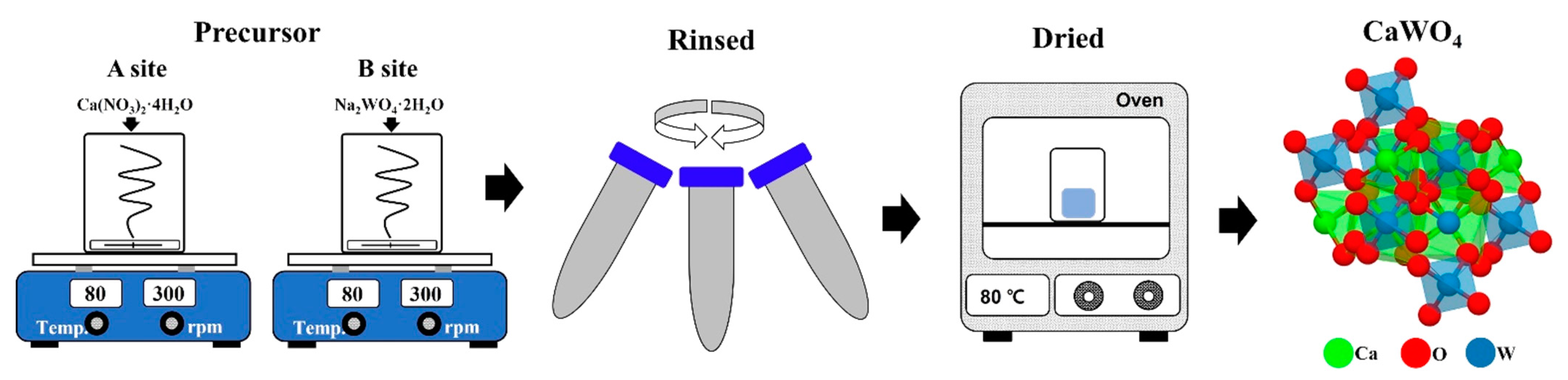
2.1. Crystalline CaWO4 Synthesized at Low Temperature

The starting materials were Calcium nitrate (Ca(NO3)2), Sodium tungstate (Na2WO4), Turbium(Ⅲ) nitrate hydrate (Tb(NO3)3·xH2O, Tb3+) and Europium(Ⅲ) nitrate hydrate (Eu(NO3)3·xH2O, Eu3+).
A total of 1 mmol of Ca(NO3)2 was dissolved in beaker ‘A’ containing 50 mL (80 °C) of distilled water. Na2WO4 was put in beaker ‘B’, under the same conditions as in beaker ‘A’ and dissolved (Figure 1). The solution in beaker ‘B’ after being completely dissolved was slowly poured into beaker ‘A’ while stirring and maintained for about 30 min. After that, a white powder was recovered using a centrifuge. The white powder was prepared by rinsing with distilled water three times to remove the remaining sodium. The white powder was dried in an oven at 80 °C for 16 h to investigate its crystallinity and luminescent properties. In addition, Tb(NO3)3·xH2O or Eu(NO3)3·xH2O (0.05 mol%) ions were added to beaker ‘A’ during the co-precipitation reaction to impart luminescent properties [18]. The experiment was carried out at 25 °C and 55% humidity.

2.2. Characterization

Structural characterizing was performed by X-ray diffraction (XRD, Rigaku Ultima IV) with Cu Kα radiation (λ = 1.5406 Å). The chemical composition of samples was studied by X-ray photoelectron spectroscopy (XPS; Thermo Fisher Scientific, Gloucester, UK) using Al-Kα lines. The C1s at 284.6 eV was used to calibrate the peak position of the insulating samples. The surface morphology was investigated using a field emission scanning electron microscope (FE-SEM, SU-8220, Hitach, Tokyo, Japan). The photoluminescence spectra were obtained using a fluorescence spectrophotometer (Scinco, FS-2, Seoul, Korea) with a 150 W Xenon lamp as the excitation source and a photomultiplier tube operating at 350 V.

2.3. Fabrication of Photoactive Composite and Anti-Counterfeiting Application

CaWO4 powder was mixed with glass frits (BaO-ZnO-B2O3-SiO2) at a weight ratio of 1:3 wt%. The mixed powder was placed in a metal mold and pressed with a press to prepare a round disk. A disk which absorbs UV light and emits light was finally produced by heat treatment in an air atmosphere at 600 °C for 5 h. In addition, a composite that was flexible and emits light by absorbing UV light was prepared by mixing a silicon-based polymer (Polydimethylsiloxane (PDMS)) and CaWO4:RE3+ (RE = Tb, Eu) powder. For anti-counterfeiting application, a solution was prepared by adding 10 wt% polyvinylpyrolidone (PVP, M.W. = 14,000) and 1 wt% of the synthesized powder to 10 mL of ethanol. The prepared solution, which cannot be confirmed with the naked eye and can only be confirmed with UV light, was stamped and painted on a banknote [19].

3. Results and Discussion

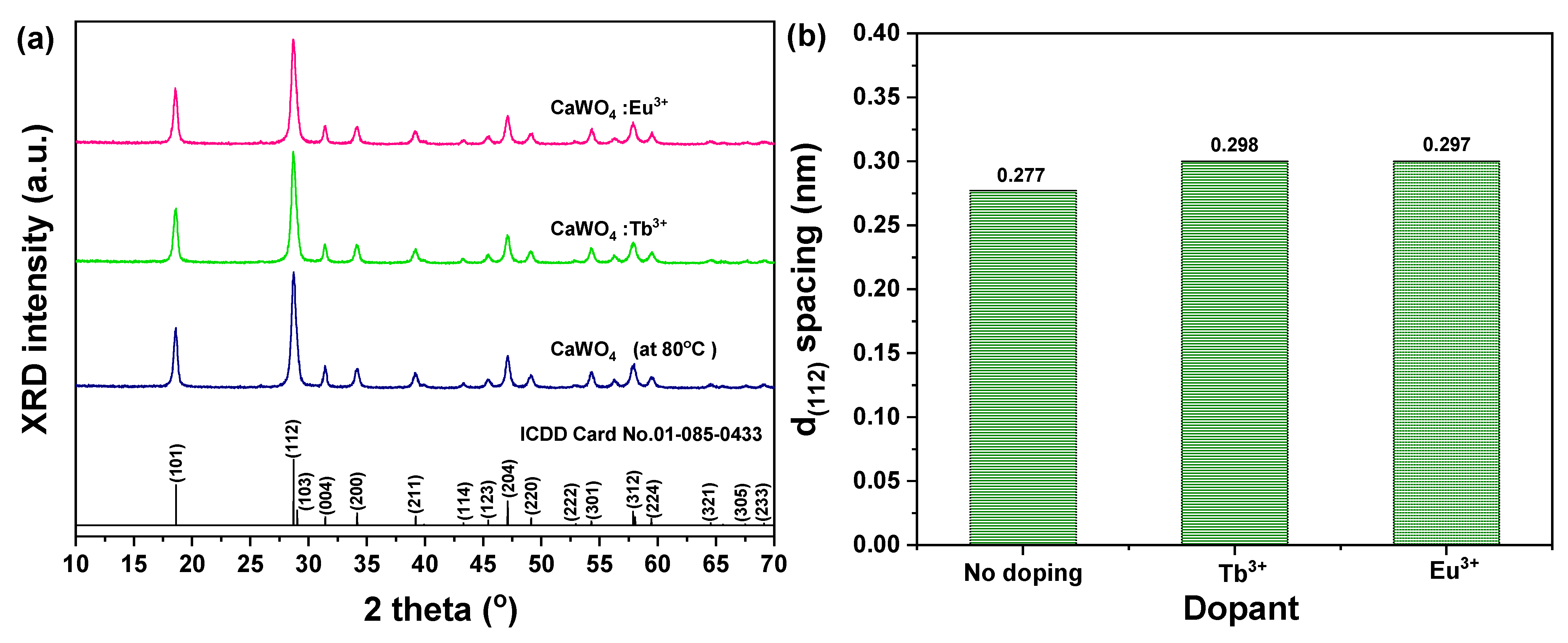
3.1. Structural and Morphology

Figure 2a shows the XRD analysis of the crystallinity and structure of the CaWO4 white powder prepared by the co-precipitation method after dried in an oven at 80 °C. The synthesized CaWO4 was consistent with the ICDD card (NO. 01-085-0433) and tetragonal structure of scheelite [20]. In addition, phases (101), (112), (204), and (312), which are the main peaks, were identified. It has been shown that the synthesis of crystalline CaWO4 is possible at a low temperature and a simple process without a heat treatment process. The crystallinity and structure of CaWO4:Tb3+ and CaWO4:Eu3+ powder doped with rare earth ions were also confirmed in the same manner as pure CaWO4. There was no secondary phase of CaWO4 due to rare earth doping. However, as shown in Figure 2b, when the lattice spacing was compared with the main peak (112) phase, it showed a change due to the addition of rare earth. For pure CaWO4, the lattice spacing was 0.277 nm. The lattice spacing of the rare earth doped CaWO4:Tb3+ (0.298 nm) and CaWO4:Eu3+ (0.279 nm) were increased. This is thought to be the result of doping with rare earth ions with relatively large ionic radii (r(Tb3+) = 0.92 Å, r(Eu3+) = 0.95 Å) in the CaWO4 lattice [21].
In addition, the size of each sample particle was investigated by substituting the half width and peak position of the main peak (112) in Scherrer’s equation [22]. CaWO4 samples were calculated at 76 nm, CaWO4:Tb3+ at 85 nm, and CaWO4:Eu3+ at 90 nm.
Titipun et al. synthesized CaMoO4, SrMoO4, CaWO4, and SrWO4 using the co-precipitation method at room temperature. The MXO4 (M = Ca and Sr, X = Mo and W) nanoparticles precipitated—M2+ cations as electron pair acceptors (Lewis acid) and reacted with XO4 2 anions as electron pair donors (Lewis base). The reaction between these two species (M2+←:XO42) proceeded to produce bonding. The lowest molecular orbital energy of the Lewis acid interacted with the highest molecular orbital energy of the Lewis base, and MXO4 nanoparticles were finally synthesized [23]. It is thought that the CaWO4 powder synthesized at low temperature in this study can also be synthesized without additional energy supply, as in the previous case.
In addition, Puneet et al. identified the oxide phase of rare earth ions doped in a synthesized CaWO4 lattice through synchrotron X-ray diffraction analysis [24]. In this study, when the doped rare earth ions were calculated using a single unit cell of CaWO4, it was calculated that the doped amount was about 1.59 × 10¹⁹ RE atoms/cm3 (RE = Tb3+, Eu3+).
The size and surface morphology of the synthesized crystalline CaWO4, CaWO4:Tb3+, and CaWO4:Eu3+ particles were observed by FE-SEM. In addition, EDS mapping was performed to confirm the components of the synthesized samples, as shown in Figure 3. The size of the synthesized particles was observed to be about 5 μm and spherical at low magnification regardless of doping with rare earth ions, but when observed at high magnification, smaller particles of about 75 nm (CaWO4), 83 nm (CaWO4:Tb3+), and 86 nm (CaWO4:Eu3+) were observed to be agglomerated.
In addition, in the rare-earth-doped CaWO4:Tb3+ (Figure 3b) and CaWO4:Eu3+ (Figure 3c) samples, each rare-earth component was confirmed, and it was confirmed that the rare-earth ions were evenly distributed without agglomeration.

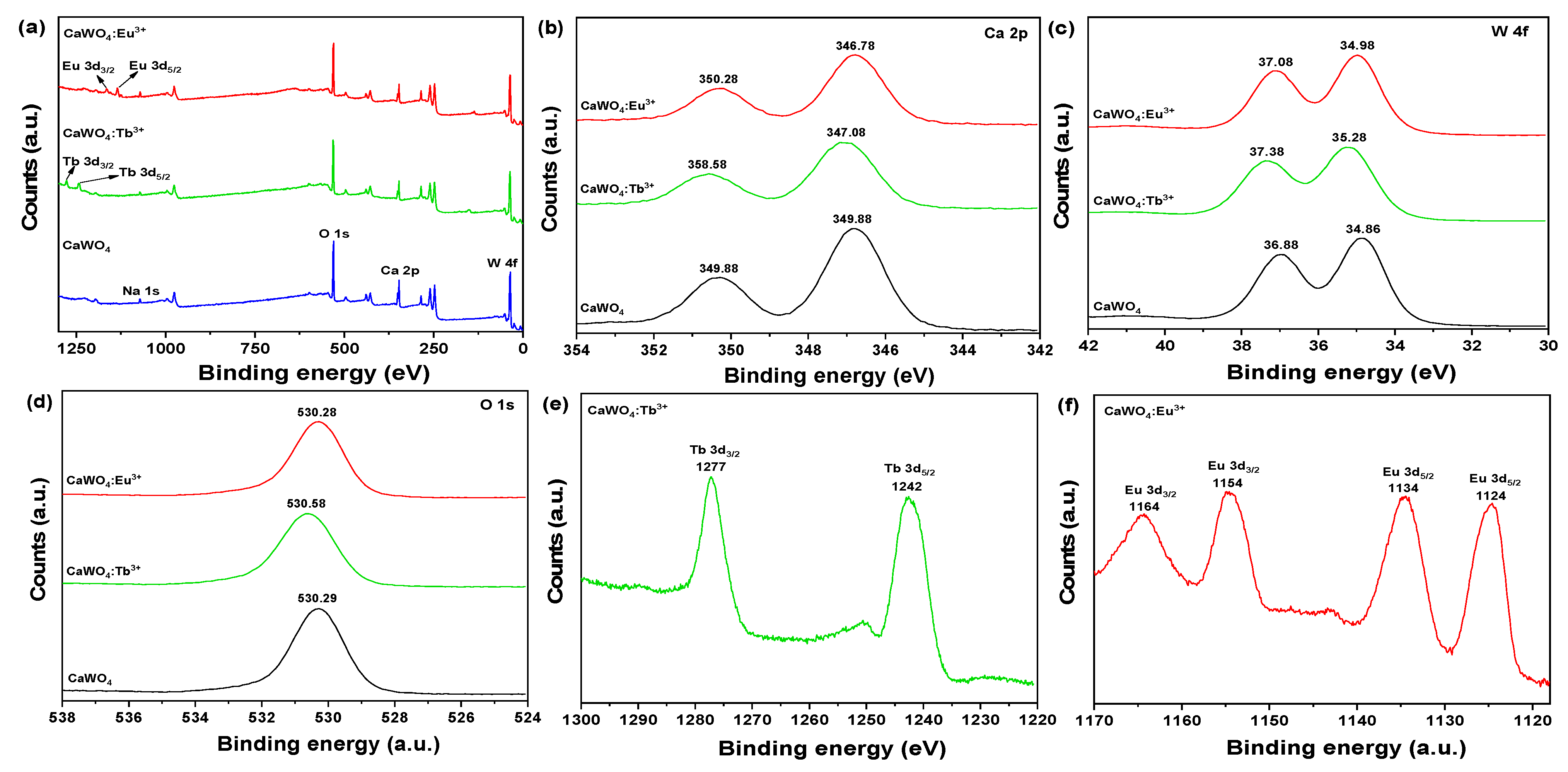
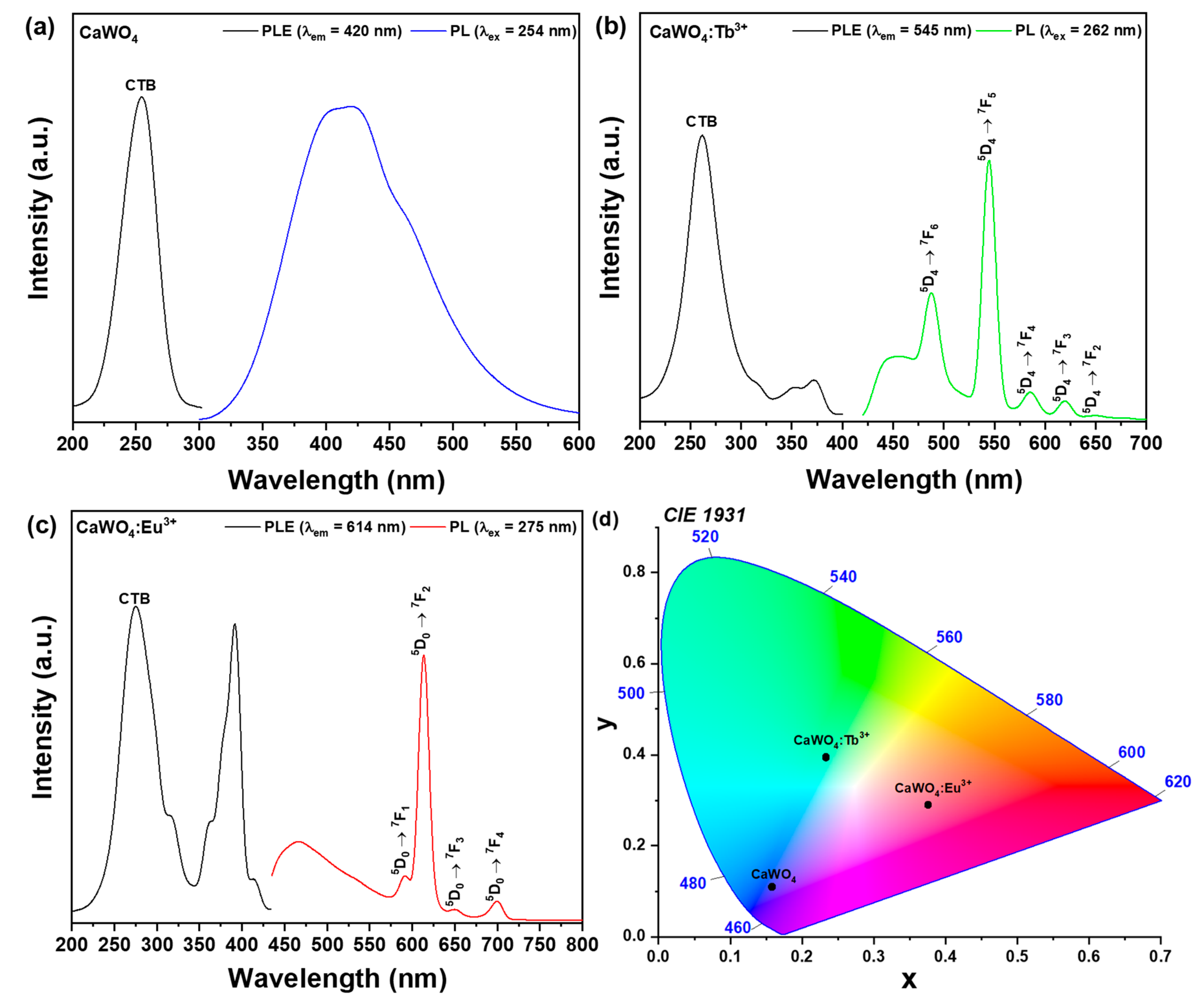
3.2. Chemical States and Phtoluminescence Proeprties

Figure 4 shows the XPS measurements used to determine the chemical state of the synthesized CaWO4, CaWO4:Tb3+, and CaWO4:Eu3+. Ca 2p, W 4f, and O 1s were confirmed as shown in Figure 4a. A trace amount of Na 1s was detected. This is thought to be due to sodium tungsten in the starting material, and it is thought to be a leftover that was not removed during the washing process when preparing the precursor.
The peaks of Ca 2p had core binding energies of 346.78 eV (2P3/2) and 349.88 eV (2P1/2), indicating that Ca is in the +2 oxidation state [25]. At the peak of W 4f, the detected core binding energies were 34.86 eV (4f7/2) and 36.88 eV (4f5/2), which is considered to be the +6 state of W [26]. The peak of the binding energy of O 1s was detected as 530.29 eV, which is considered to indicate the crystal lattice oxygen with increasing binding energy [27].
In the rare earth-doped sample (CaWO4:Tb3+, CaWO4:Eu3+), the binding energy of the Ca 2p (Figure 4b), W 4f (Figure 4c), and O 1s (Figure 4d) components was slightly changed. This change in binding energy is considered due to rare earth doping, and is related to the change in the lattice spacing observed in the XRD result and SEM-EDS component analysis, which means that the rare earth is doped in CaWO4. In addition, the respective binding energy spectra were observed in the samples doped with Tb3+ and Eu3+. In Figure 4e, binding energies of 1277 eV (Tb 3d3/2) and 1242 eV (Tb 3d5/2) were observed, and in Figure 4f, binding energy peaks of 1164 (Eu 3d3/2), 1154 (Eu 3d3/2), 1134 (Eu 3d5/2), and 1124 (Eu 3d5/2) eV were obtained. This indicates the presence of rare earth ions in the +3 oxidation state following the synthesis of the sample [27].
The excitation and emission spectra of CaWO4 are shown in Figure 5a. A signal having an excitation wavelength of 254 nm peak was detected, and a blue signal having a broad bandwidth of 420 nm was observed in the emission spectrum. This is thought to be due to the transfer of ions from the 2p orbital of oxygen to the 5d orbital of the vacant W6+ [28,29,30,31]. The absorption and emission spectra of CaWO4:Tb3+ powder are shown in Figure 5b.
The photoluminescence excitation (PLE) spectrum controlled with a photoluminescence (PL) wavelength of 545 nm has a peak at 262 nm and is widely distributed over a 200 ~ 300 nm region, which is an absorption spectrum by charge transfer band (CTB) generated between O−2-W6+ and O−2-Tb3+ ions [32]. The relatively weak absorption signals between 330 and 400 nm are the 4f-4ftransition signals of Tb3+ ions [33]. The peak signals at 350 and 372 nm were generated by the 7F65G5 and 7F65G6 transition signals, respectively. When excited with the strongest absorption wavelength of 262 nm, the PL spectra of CaWO4:Tb3+ were observed at 488 (5D47F6), 545 (5D47F5), 585 (5D47F4), 620 (5D47F3), and 648 (5D47F2) nm. Among these emission peaks, the green emission spectrum by the magnetic dipole transition at 545 nm was about 2.4 times stronger than the blue emission intensity by the electric dipole transition at 488 nm. This means that the Tb3+ ions located in the parent crystal are located at sites of inversion symmetry [34].
In CaWO4:Eu3+ the PLE spectrum controlled with a PL wavelength of 614 nm has a peak at 275 nm and is widely distributed over a 200–340 nm region (Figure 5b), which is the absorption spectrum by CTB generated between O−2-W6+ and O−2-Eu3+ ions [35]. The relatively weak absorption signals between 350 and 440 nm are the 4f-4f transition signals of Eu3+ ions [34]. The peak signals at 362, 391, and 414 nm were generated by the 7F05D4, 7F05G2, and 7F05L6 transition signals, respectively. When excited with the strongest absorption wavelength of 275 nm, the PL spectra of CaWO4:Eu3+ were observed at 592 (5D07F1), 614 (5D07F2), 650 (5D07F3), and 700 (5D07F4) nm. Here, 614 nm is a spectrum by electric dipole transition, 592 nm is a magnetic dipole transition, and 650 and 700 nm are electric dipole signals [36].
At this time, the PL intensity of 614 nm was observed to be about 6 times stronger than that of 592 nm, which means that Eu3+ ions in the parent lattice are located at non-inverting symmetric sites. The ratio between the intensity of the red emission and the intensity of the orange emission is also called an asymmetry ratio [37]. The color coordinates (CIE 1931) according to the emission spectra of CaWO4, CaWO4:Tb3+, and CaWO4:Eu3+ are shown in Figure 5c. They were found to be located at the blue, green, and red coordinates, respectively.

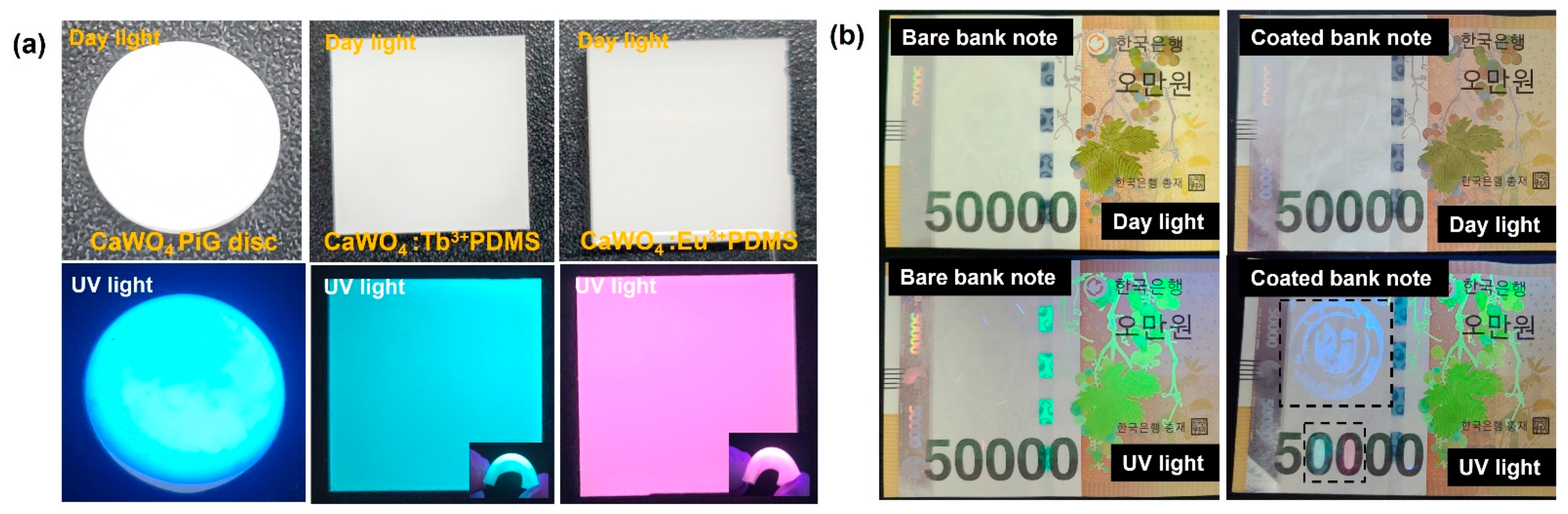
3.3. Photoactive Composite and Anti-Counterfeiting Application

Figure 6 shows the application of the synthesized phosphors as a photoreactive composite for anti-counterfeiting. The disk composite made by mixing glass powder and CaWO4 showed no reaction in daylight but showed a blue light emission in response to UV light [38]. In addition, the composite made by mixing with a silicone-based polymer (PDMS) could be flexibly bent, and each unique color was realized in UV light. These materials are thought to be applicable to the display and laser industries.
In addition, a solution made by mixing PVP polymer and CaWO4 phosphor with ethanol was stamped and applied to banknotes. The location could not be recognized with the naked eye, and the shape and unique color could be confirmed only by UV light, suggesting that the synthesized phosphor can be applied to anti-counterfeiting.

4. Conclusions

Calcium nitrate and sodium tungstate were dissolved in distilled water, and crystalline CaWO4 white powder was synthesized without a high-temperature heat treatment process by co-precipitation. CaWO4:Tb3+ and CaWO4:Eu3+ phosphors were synthesized by doping trace amounts of Tb3+ or Eu3+ ions, respectively, for use as various light-emitting materials. The synthesized powders were confirmed to have a crystalline scheelite structure from XRD results, and a secondary phase resulting from rare earth doping was not observed. However, it was found that the lattice spacing was slightly changed by doping with rare earth with a relatively large ionic radius. The particles synthesized by SEM-EDS analysis were about 75–85 nm in size and were aggregated in a spherical shape. Ca, W, and O were confirmed in the component analysis, and it was found that the doped Tb and Eu were evenly distributed by mapping. The binding energies of Ca 2p, W 4f, and O 1s were identified by XPS component analysis, and the signals of Tb 3d and Eu 3d energy binding by rare earth doping were detected. In the PLE and PL spectra, when each specimen was excited with CTB, blue emission was observed over a wide area of 420 nm for CaWO4, green emission at 545 nm for CaWO4:Tb3+, and red emission from CaWO4:Eu3+ at 614 nm. The synthesized phosphor was mixed with glass powder and PDMS polymer to prepare a disk-shaped and flexible composite that can be applied to a display. In addition, it was suggested that the solution made by mixing with PVP polymer can be applied to anti-counterfeiting because it is impossible to visually confirm when applied to banknotes and can only be confirmed by UV light.

Author Contributions

Conceptualization, J.-Y.J.; data curation, J.-Y.J. and S.-S.Y.; formal analysis, J.-Y.J.; funding acquisition, S.-S.Y.; investigation, J.-Y.J. and S.-S.Y.; methodology, J.-Y.J.; project administration, J.-Y.J.; software, J.-Y.J.; supervision, J.-Y.J.; visualization, J.-Y.J.; writing—original draft, J.-Y.J. and S.-S.Y.; writing—review and editing, J.-Y.J. All authors have read and agreed to the published version of the manuscript.

Funding

This research received no external funding.

Institutional Review Board Statement

Not applicable.

Data Availability Statement

The data presented in this study are available in the database of the authors at the Faculty of Materials Science and Engineering.

Acknowledgments

This research was supported by Basic Science Research Program through the National Research Foundation of Korea (NRF) funded by the Ministry of Education (NRF-2020R1F1A1072616).

Conflicts of Interest

The authors declare no conflict of interest.

References

  1. Liu, C.; Pokhrel, S.; Tessarek, C.; Li, H.; Schowalter, M.; Rosenauer, A.; Eickhoff, M.; Li, S.; Mädler, L. Rare-Earth-Doped Y4Al2O9 Nanoparticles for Stable Light-Converting Phosphors. ACS Appl. Nano Mater. 2019, 3, 699. [Google Scholar] [CrossRef] [Green Version]
  2. Withnall, R.; Silver, J. Physics of Light Emission from Rare Earth-Doped Phosphors. Handb. Vis. Disp. Technol. 2015, 1, 1019–1028. [Google Scholar] [CrossRef]
  3. Kim, D. Recent Developments in Lanthanide-Doped Alkaline Earth Aluminate Phosphors with Enhanced and Long-Persistent Luminescence. Nanomaterials 2021, 11, 723. [Google Scholar] [CrossRef] [PubMed]
  4. Parhi, P.; Karthik, T.N.; Manivannan, V. Synthesis and characterization of metal tungstates by novel solid-state metathetic approach. J. Alloy Compd. 2008, 465, 380–386. [Google Scholar] [CrossRef]
  5. Zhang, J.; Zhu, X.; Huang, H.; Zhang, Y. Synthesis of Crystallized BaWO4 Nanorods in a Microemulsion System. Adv. Chem. Eng. Sci. 2017, 7, 228–234. [Google Scholar] [CrossRef] [Green Version]
  6. Kuzmin, A.; Purans, J. Local atomic and electronic structure of tungsten ions in AWO4 crystals of scheelite and wolframite types. Radiat. Meas. 2001, 33, 583–586. [Google Scholar] [CrossRef]
  7. Smith, J.; Desgreniers, S.; Tse, J.; Klug, D. High-pressure phase transition observed in barium hydride. J. Appl. Phys. 2007, 102, 043520. [Google Scholar] [CrossRef]
  8. Liao, J.; Qiu, B.; Wen, H.; Chen, J.; You, W.; Liu, L. Synthesis process and luminescence properties of Tm3+ in AWO4 (A = Ca, Sr, Ba) blue phosphors. J. Alloy. Compd. 2009, 487, 758–762. [Google Scholar] [CrossRef]
  9. Sota, E.N.; Che Ros, F.; Hassan, J. Synthesis and Characterisation of AWO4 (A=Mg, Zn) Tungstate Ceramics. J. Phys. Conf. Ser. 2018, 1083, 12002. [Google Scholar] [CrossRef] [Green Version]
  10. Cooper, T.; De Leeuw, N. A combined ab initio and atomistic simulation study of the surface and interfacial structures and energies of hydrated scheelite: Introducing a CaWO4 potential model. Surf. Sci. 2003, 531, 159–176. [Google Scholar] [CrossRef]
  11. Oliveira, M.C.; Gracia, L.; Nogueira, I.C.; do Carmo Gurgel, M.F.; Mercury, J.M.R.; Longo, E.; Andrés, J. Synthesis and morphological transformation of BaWO4 crystals: Experimental and theoretical insights. Ceram. Int. 2016, 42, 10913–10921. [Google Scholar] [CrossRef] [Green Version]
  12. Zhang, H.; Liu, T.; Zhang, Q.; Wang, X.; Yin, J.; Song, M.; Guo, X. First-principles study on electronic structures of BaWO4 crystals containing F-type color centers. J. Phys. Chem. Solids 2008, 69, 1815–1819. [Google Scholar] [CrossRef]
  13. Yekta, S.; Sadeghi, M.; Babanezhad, E. Synthesis of CaWO4 nanoparticles and its application for the adsorption-degradation of organophosphorus cyanophos. J. Water Process. Eng. 2016, 14, 19–27. [Google Scholar] [CrossRef]
  14. Balestrieri, M.; Colis, S.; Gallart, M.; Schmerber, G.; Ziegler, M.; Gilliot, P.; Dinia, A. Photoluminescence properties of rare earth (Nd, Yb, Sm, Pr)-doped CeO2 pellets prepared by solid-state reaction. J. Mater. Chem. C Mater. Opt. Electron. Devices 2015, 3, 7014–7021. [Google Scholar] [CrossRef] [Green Version]
  15. Oh, W.; Chong, Y.; Park, C.S. Lim Solid-State Metathetic Synthesis and Photoluminescence of Calcium Tungstate Particles Assisted by Cyclic Microwave Irradiation. Asian J. Chem. 2012, 24, 3319. [Google Scholar]
  16. Phuruangrat, A.; Thongtem, T.; Thongtem, S. Synthesis, characterisation and photoluminescence of nanocrystalline calcium tungstate. J. Exp. Nanosci. 2010, 5, 263–270. [Google Scholar] [CrossRef]
  17. Li, J.; Zhang, J.; Hao, Z.; Zhang, X.; Zhao, J.; Luo, Y. Upconversion properties and dynamics study in Tm3+ and Yb3+ codoped CaSc2O4 oxide material. J. Appl. Phys. 2013, 113, 223507. [Google Scholar] [CrossRef]
  18. Jung, J.; Kim, J.; Shim, Y.; Hwang, D.; Son, C.S. Structure and Photoluminescence Properties of Rare-Earth (Dy3+, Tb3+, Sm3+)-Doped BaWO4 Phosphors Synthesized via Co-Precipitation for Anti-Counterfeiting. Materials 2020, 13, 4165. [Google Scholar] [CrossRef]
  19. Jung, J.; Yi, S.; Hwang, D.; Son, C. Structure, Luminescence, and Magnetic Properties of Crystalline Manganese Tungstate Doped with Rare Earth Ion. Materials 2021, 14, 3717. [Google Scholar] [CrossRef]
  20. Wang, S.; Gao, H.; Sun, G.; Li, Y.; Wang, Y.; Liu, H.; Chen, C.; Yang, L. Structure characterization, optical and photoluminescence properties of scheelite-type CaWO4 nanophosphors: Effects of calcination temperature and carbon skeleton. Opt. Mater. 2020, 99, 109562. [Google Scholar] [CrossRef]
  21. Jia, Y.Q. Crystal radii and effective ionic radii of the rare earth ions. J. Solid State Chem. 1991, 95, 184–187. [Google Scholar] [CrossRef]
  22. Bokuniaeva, A.O.; Vorokh, A.S. Estimation of particle size using the Debye equation and the Scherrer formula for polyphasic TiO2 powder. J. Phys. Conf. Ser. 2019, 1410, 012057. [Google Scholar] [CrossRef]
  23. Thongtem, T.; Kungwankunakorn, S.; Kuntalue, B.; Phuruangrat, A.; Thongtem, S. Luminescence and absorbance of highly crystalline CaMoO4, SrMoO4, CaWO4 and SrWO4 nanoparticles synthesized by co-precipitation method at room temperature. J. Alloy Compd. 2010, 506, 475–481. [Google Scholar] [CrossRef]
  24. Kaur, P.; Khanna, A.; Singh, M.N.; Sinha, A.K. Structural and optical characterization of Eu and Dy doped CaWO4 nanoparticles for white light emission. J. Alloy Compd. 2020, 834, 154804. [Google Scholar] [CrossRef]
  25. Maheshwary, M.; Singh, B.P.; Singh, R.A. Color tuning in thermally stable Sm3+-activated CaWO4 nanophosphors. New J. Chem. 2015, 39, 4494–4507. [Google Scholar] [CrossRef]
  26. Terohid, S.; Heidari, S.; Jafari, A.; Asgary, S. Effect of growth time on structural, morphological and electrical properties of tungsten oxide nanowire. Appl. Phys. A 2018, 124, 1–9. [Google Scholar] [CrossRef] [Green Version]
  27. Pawlak, D.A.; Ito, M.; Oku, M.; Shimamura, K.; Fukuda, T. Interpretation of XPS O (1s) in Mixed Oxides Proved on Mixed Perovskite Crystals. J. Phys. Chem. B 2002, 106, 504–507. [Google Scholar] [CrossRef]
  28. Fang, Z.B.; Wang, J.J.; Yang, X.F.; Chen, T.H.; Ni, H.N.; Li, Z.B.; Tan, Y.S. Reliability Study of the Band Gap of Rare Earth Oxides Measured by XPS Spectra. Key Eng. Mater. 2013, 562–565, 891–895. [Google Scholar]
  29. Park, K.; Ahn, H.; Nguyen, H.; Jang, H.; Mho, S. Optical Properties of Eu2(WO4)3 and Tb2(WO4)3 and of CaWO4 Doped with Eu3+ or Tb3+—Revisited. J. Korean Phys. Soc. 2008, 53, 2220–2223. [Google Scholar] [CrossRef]
  30. Wang, W.; Yang, P.; Gai, S.; Niu, N.; He, F.; Lin, J. Fabrication and luminescent properties of CaWO4: Ln3+ nanocrystals. J. Nanoparticle Res. 2010, 12, 2295. [Google Scholar] [CrossRef]
  31. Cavalli, E.; Boutinaud, P.; Mahiou, R.; Bettinelli, M.; Dorenbos, P. Luminescence Dynamics in Tb3+-Doped CaWO4 and CaMoO4 Crystals. Inorg. Chem. 2010, 49, 4916–4921. [Google Scholar] [CrossRef]
  32. Sun, S.; Wu, L.; Yi, H.; Wu, L.; Ji, J.; Zhang, C.; Zhang, Y.; Kong, Y.; Xu, J. Energy transfer between Ce3+ and Tb3+ and the enhanced luminescence of a green phosphor SrB2O4:Ce3+, Tb3+, Na. Opt. Mater. Express 2016, 6, 1172. [Google Scholar] [CrossRef]
  33. Alaria, J.; Borisov, P.; Dyer, M.S.; Manning, T.D.; Lepadatu, S.; Cain, M.G.; Mishina, E.D.; Sherstyuk, N.E.; Ilyin, N.A.; Hadermann, J.; et al. Engineered spatial inversion symmetry breaking in an oxide heterostructure built from isosymmetric room-temperature magnetically ordered components. Chem. Sci. 2014, 5, 1599–1610. [Google Scholar] [CrossRef] [Green Version]
  34. Nayak, P.; Nanda, S.S.; Sharma, R.K.; Dash, S. Tunable luminescence of Eu3+-activated CaWO4 nanophosphors via Bi3+ incorporation. Luminescence 2020, 35, 1068–1076. [Google Scholar] [CrossRef]
  35. Malta, O.L.; Ribeiro, S.J.L.; Faucher, M.; Porcher, P. Theoretical intensities of 4f-4f transitions between Stark levels of the Eu3+ ion in crystals. J. Phys. Chem. Solids 1991, 52, 587–593. [Google Scholar] [CrossRef]
  36. Savinov, M.; Nuzhnyy, D.; Ležaić, M.; Eckel, S.; Kamba, S.; Laufek, F.; Spaldin, N.A.; Knížek, K.; Vaněk, P.; Lamoreaux, S.K.; et al. A multiferroic material to search for the permanent electric dipole moment of the electron. Nat. Mater. 2010, 9, 649–654. [Google Scholar] [CrossRef]
  37. Kolesnikov, I.E.; Povolotskiy, A.V.; Mamonova, D.V.; Kolesnikov, E.Y.; Kurochkin, A.V.; Lähderanta, E.; Mikhailov, M.D. Asymmetry ratio as a parameter of Eu3+ local environment in phosphors. J. Rare Earths 2018, 36, 474–481. [Google Scholar] [CrossRef]
  38. Jung, J.Y.; Shim, Y.; Son, C.S.; Kim, Y.; Hwang, D. Boron Nitride Nanoparticle Phosphors for Use in Transparent Films for Deep-UV Detection and White Light-Emitting Diodes. ACS Appl. Nano Mater. 2021, 4, 3529–3536. [Google Scholar] [CrossRef]
Figure 1. Schematic of the CaWO4 synthesis procedure.
Crystals 11 01214 g001
Figure 2. (a) XRD patterns and (b) change in d(112) spacing; CaWO4, CaWO4:Tb3+, and CaWO4:Eu3+.
Crystals 11 01214 g002
Figure 3. SEM-EDS analysis; (a) CaWO4, (b) CaWO4:Tb3+, and (c) CaWO4:Eu3+.
Crystals 11 01214 g003
Figure 4. XPS spectra of (a) survey, (b) Ca 2p, (c) W 4f, (d) O 1s, (e) Tb 3d, and (f) Eu 3d.
Crystals 11 01214 g004
Figure 5. Luminescence spectra of (a) CaWO4, (b) CaWO4:Tb3+, and (c) CaWO4:Eu3+, and (d) CIE coordination.
Crystals 11 01214 g005
Figure 6. Photograph of (a) phosphors in glass disc and flexible composite, (b) phosphors painted on bank notes for anti-counterfeiting.
Crystals 11 01214 g006
Publisher’s Note: MDPI stays neutral with regard to jurisdictional claims in published maps and institutional affiliations.

