Article

Biosynthesis of CeO2 Nanoparticles Using Egg White and Their Antibacterial and Antibiofilm Properties on Clinical Isolates

1
Department of Physics, College of Science, King Faisal University, P.O. Box 400, Al-Ahsa 31982, Saudi Arabia
2
Department of Physics, School of Engineering, University of Petroleum & Energy Studies, Dehradun 248007, India
3
Physics Department, Faculty of Science, Assiut University, Assiut 71516, Egypt
*
Author to whom correspondence should be addressed.
Crystals 2021, 11(6), 584; https://doi.org/10.3390/cryst11060584
Submission received: 8 April 2021 / Revised: 18 May 2021 / Accepted: 19 May 2021 / Published: 22 May 2021
(This article belongs to the Section Inorganic Crystalline Materials)

Abstract

:
Bio-inspired synthesis is a novel and attractive environmentally friendly route to generating inorganic materials. In this work, the preparation of CeO2 NPs using egg white and investigation of their antibacterial properties both in liquid and solid growth medium against Escherichia coli and Staphylococcus aureus bacteria were reported. The CeO2 nanoparticles were characterized using X-ray diffraction (XRD), Field emission transmission electron microscope (FETEM), UV-Vis, Raman, and antibacterial measurements. The results from XRD and TEM analysis showed that the prepared nanoparticles were a single phase in the nano regime (5–7 nm) with spherical shape and uniform size distribution. Optical properties reflected the characteristics peaks of CeO2 in the UV-Vis range with a bandgap ~2.80 eV. The antibacterial activity of the synthesized NPs was achieved under ambient conditions with different bacteria and the results showed that the properties were different for both the bacteria. The highest activity with an inhibition zone of about 22 mm against S. aureus was obtained as compared with the 19 mm zone of inhibition obtained with E. coli. This finding will be of major significance that indicates a possibility to develop CeO2 NPs as antibacterial agents against extensive microorganisms to control and prevent the spread and persistence of bacterial infections.

1. Introduction

During the last few decades, there has been a significant advancement in the design of nanomaterials and their potential applications in multidisciplinary science [1]. Microorganisms have produced pollution and contamination by causing degradation and infection in various fields such as the textile industry, water treatment, medicine, and food packaging. The current antibacterial agents are composed of chemically modified natural compounds that exhibit antibacterial properties that locally kill bacteria or hamper their growth, still being non-toxic to the surrounding environment [2]. The mostly used antibacterial agents include purely natural products, e.g., aminoglycosides, as well as purely synthetic antibiotics; e.g., sulfonamides. Antibacterial agents are paramount to fight infectious diseases. However, these advantages come along with detriments. The emergence of bacterial resistance to antibacterial drugs has arisen as a major problem. Thus, as the bacteria developed resistance against many conventional antibacterial agents, infectious diseases remain one of the extreme challenges to health worldwide. Nonetheless, researchers have been working to develop new pasteurization and antibacterial techniques to treat these bacterial diseases to solve such health issues [3,4]. The search for new antibacterial agents is based on withholding the use of conventional methods which use organic compounds and to investigate the inorganic nanomaterials for the required antibacterial properties. The main advantages that inorganic nanomaterials like oxides possess over organic antimicrobial agents are their stability, robustness, and long shelf life.
Among inorganic nanomaterials, cerium oxide (CeO2) nanoparticles are excellent candidates for multiple industrial, commercial, and biomedical uses. They have a wide range of industrial applications such as catalysts [4], fuel cells [5], ultraviolet absorbers [6], hydrogen storage materials [7], oxygen sensors [8], optical devices [9], and polishing materials [10]. Recent reports have revealed that CeO2 NPs exhibit antioxidant activity at physiological pH values [11]. Soren et al. demonstrated the antioxidant potential and toxicity study of CeO2 nanoparticles [11]. This property enables it to protect cells against radiation damage, oxidative stress, or inflammation. Thereby, CeO2 is utilized in biomedical applications [12,13]. Various methods developed to prepare CeO2 nanopowders include sol-gel [14,15], hydrothermal [16,17,18], chemical precipitation with urea or hexamethylenetetramine [19], flame spray pyrolysis [20], reverse micelles route [21,22], combustion method [23], sonochemical, and microwave-assisted heating routes [24,25] as well as a complex thermo-decomposition method [26]. Additionally, Nyoka et al. reported a review article on the synthesis of CeO2 nanoparticles. In this review article, they described the various approaches used for the synthesis of CeO2 nanoparticles and discussed their biomedical applications [27]. However, most of these techniques involve multistep syntheses and aging processes. This leads to an undesirable consumption of time and energy. These methods are also not environmentally friendly. In addition, the synthesis of particles with extremely small sizes causes a huge upsurge in the complexity and the cost of manufacture. Therefore, finding simple and cost-effective procedures to prepare CeO2 NPs, which are non-toxic and environmentally friendly is still a key issue.
While searching for new methods, bio-inspired synthesis has emerged as a novel and attractive route to generate inorganic substances, which address the above-mentioned issues effectively. The bio-inspired procedures are directed through the utilization of proteins and peptides. This makes the reaction conditions much milder as compared to those provided by the conventional methods of material processing. Peptides and proteins facilitate chemical reactions of inorganic materials in a aqueous solutions at or near room temperature and at neutral pH. In addition to this, the use of proteins and peptides drives the bio-enabled synthesis towards inherent “green” processing of materials. This happens because they lower the energy input and facilitate the reaction without the use of any kind of organic solvents and thereby synthesize the materials naturally. There are other benefits of exploiting biomolecules, for instance, the exquisite control that is intruded by proteins and peptides on the size, shape, chemistry, and geometrical structure of the inorganic product. This is significant as, in general, various properties of the synthesized materials are influenced by these characteristics. For example, egg albumen, being a complex mixture of proteins, has been broadly utilized as a functional protein ingredient for the processing of food items due to its excellent foaming, emulsification, and gelation properties. Each protein component in egg white albumen possesses a different isoelectric point (pI) [28,29,30]. Maensiri et al. reported the optical properties of CeO2 nanoparticles synthesized using the egg white (EW) method [29]. They have synthesized CeO2 nanoparticles using cerium (III) acetate hydrate as a starting precursor. In their typical synthesis, the mixed solution was evaporated by heating on a hot plate at 80 °C under vigorous stirring for several hours until a dried precursor was obtained. They obtained the platelike structure of CeO2 nanoparticles having a particle size of 6–30 nm.
Kargar et al. [30] also prepared the CeO2 nanoparticles by keeping the reaction time for 8 h. The resulted CeO2 nanoparticles were 25 nm in size. By comparing the biological applications, Kargar’s work reported in vitro cytotoxicity effects; however, in our work, we have shown the potential of CeO2 nanoparticles as an effective antibacterial agent for both the Gram-negative and Gram-positive bacteria. These groups demonstrated that the synthesis of CeO2 nanoparticles by utilizing food and bio-derived materials (i.e., EW) is cost-effective, simple, and environmentally friendly. This technique is an alternative route for the synthesis of nanoparticles in comparison to those obtained from conventional preparation method that uses hazardous material.
In this work, CeO2 NPs were prepared using egg white and investigated their antibacterial properties using two types of clinical isolates of E. coli and S. aureus. Since egg white is an abundant, cheap, and environmentally benign biomaterial, it synthesizes the inorganic functional materials via bio-inspired manufactures. The prepared CeO2 product was characterized by using XRD, FETEM, UV-Vis, Raman, and its antibacterial, and antibiofilm potential were also identified. This method will provide a simple and green route for biomaterials assisted synthesis of advanced functional materials for their use in biomedical applications.

2. Experimental Details

2.1. Synthesis Method for the Preparation of CeO2 Nanoparticles

Analytical grade and as received reagents were utilized in the experiments without further purification. For the synthesis, cerium (III) nitrate hexahydrate, Ce(NO3)3·6H2O (99.9% purity, Aldrich, St. Louis, MO, USA), and freshly extracted egg white (ovalbumin) were taken as the raw materials. The typical bio-inspired procedure was followed. First, a homogeneous solution was prepared by mixing 50 mL of egg white in 40 mL of deionized water under vigorous stirring at room temperature (27 °C). Subsequently, the desired amount of Ce(NO3)3·6H2O precursor was added gradually to the above obtained solution while maintaining vigorous stirring at room temperature. The stirring was continued for another 2 h to get a well-dissolved solution. During the stirring, the reaction was facilitated by the extracted egg white which acted as a matrix for the entrapment of cerium ions resulting in a gelled precursor. During the process, no pH adjustments were carried. The heating was started on the hot plate and maintained at 70 °C while keeping the solution under vigorous stirring for several hours. The solution was allowed to evaporate until a dried precursor was obtained. The remained product was ground to make a fine powder with the help of a mortar and pestle. Finally, the nanoparticles were obtained when the crushed powder was put in a furnace for annealing in the air.

2.2. Characterizations

The purity of the phase and structural studies of the synthesized nanoparticles were studied through XRD, SAED pattern, UV-vis, and Raman spectroscopy. The XRD pattern was recorded using the Philips X-pert X-ray diffractometer X’pert; (MPD 3040, EA Almelo, The Netherlands) with Cu Kα (λ ~ 1.5418 Å). Morphological studies were carried out by FETEM (JEM 2100 F, Corporation Place, Singapore). The Raman spectrum was measured using a micro-Raman spectrometer (NRS-3100, JASCO, Easton, MD, USA). The optical absorption spectrum was measured in the range of 250–800 nm at room temperature using a UV–VIS spectrophotometer (S-4100, SINCO Instrument Co., Seoul, Korea)

2.3. Antibacterial Assessment

The antibacterial activity of CeO2 NPs was tested by the standard agar well diffusion method. The standard strains of E. coli and S. aureus bacteria were used for testing. These bacterial strains were grown on LB (Luria Bertani) Broth at a temperature of 37 °C overnight up to a turbidity of 0.5 Mac Farland standard (108 CFU/mL). The required amount of CeO2 NPs suspension was used to inoculate petridish filled with LB (Luria Bertani) agar. After punching the wells (diameter ~ 6 mm) in the agar plates, they were filled with nanoparticles solutions. The incubation of LB agar plates was carried out overnight at 37 °C. After the treatment with nanoparticles, the samples were spread on nutrient agar plates. The inhibition was determined using the inhibition zone surrounding the disk. The average diameter of the zone was noted. Further, to determine the turbidity of the samples, optical density (OD) was measured in a spectrophotometer. At the last, the bacterial inoculum was transferred into a liquid suspension of bacteria in a nutrient broth.

2.4. Antibiofilm Assessment

Antibiofilm activity of the synthesized NPs was performed using a method as described by Dwivedi et al. [26]. The microtiter plate was seeded with 100 μL of freshly grown bacterial cells, after 96 h incubation, the wells were then stained with 0.25% crystal violet followed by incubation at 37 °C for 30 min. The wells were, then, washed and air-dried. Lastly, the bound stain was solubilized in 200 μL of ethanol (99.9% Aldrich, St. Louis, MO, USA).

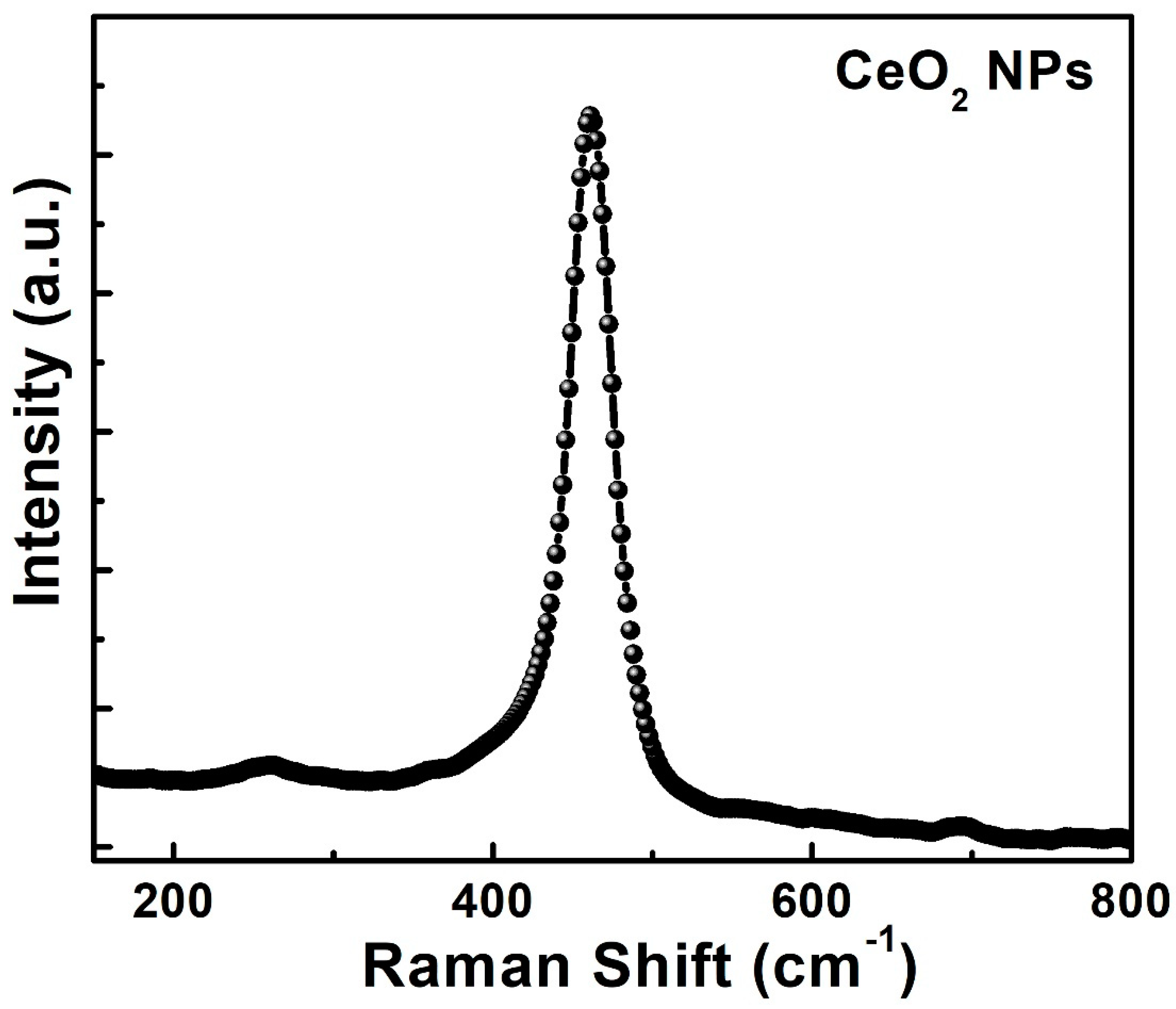
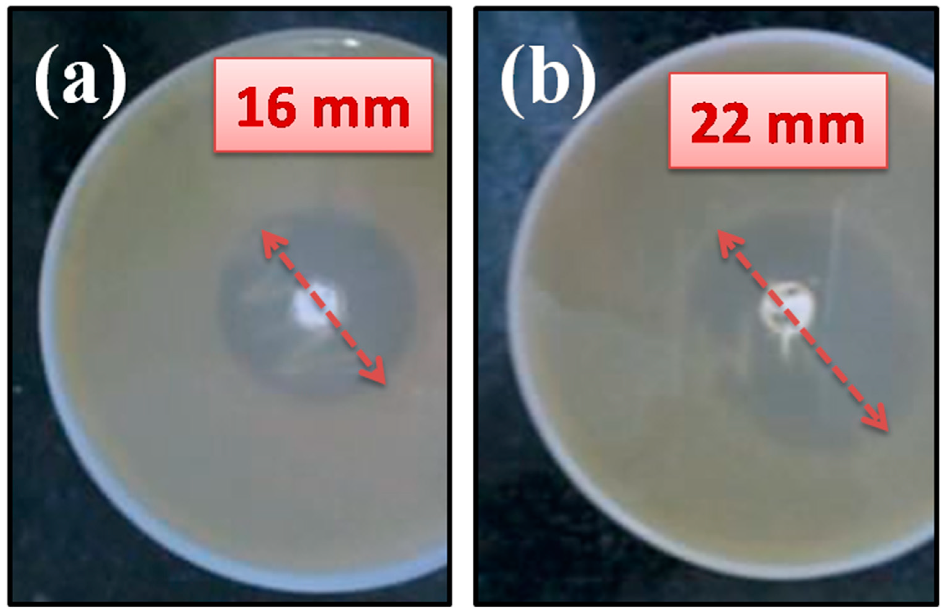
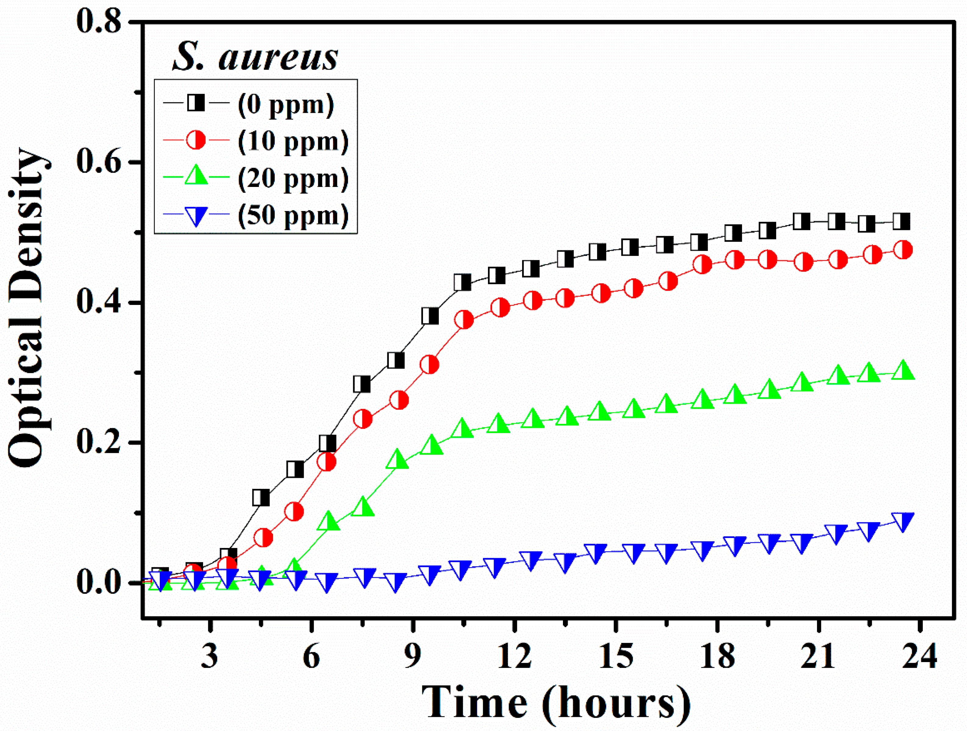
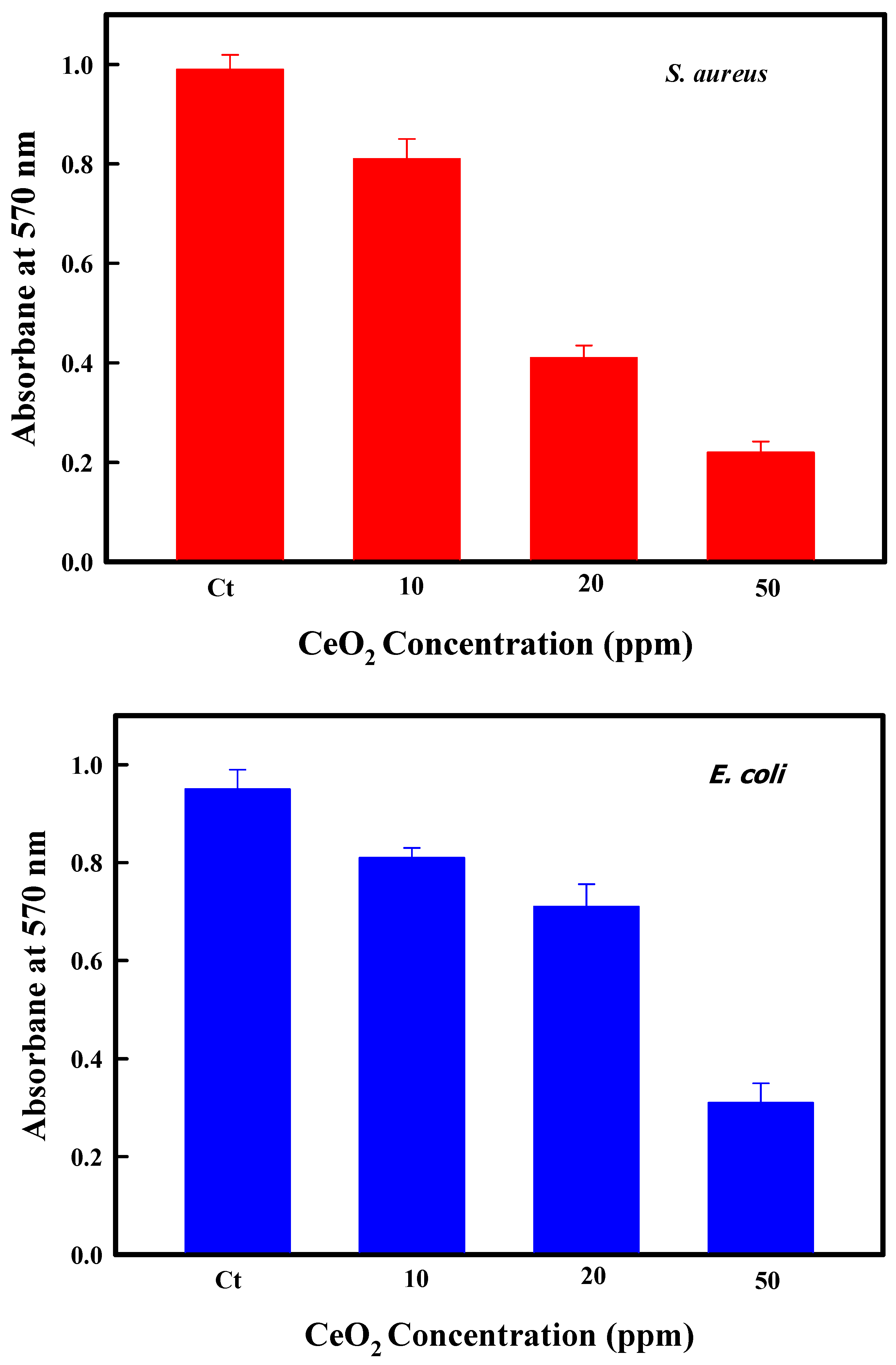
3. Results and Discussion

The purity of the phase of CeO2 NPs and their crystalline structure was investigated using XRD analysis. Figure 1 displays the XRD profile of CeO2 NPs. The analysis was carried precisely by the Rietveld refinement process using the FullProof program [31].
The spectral lines marked by the black sphere represent the experimental curve while the fine purple line indicates the calculated spectra. The green line represents the difference between experimental and fitting spectra. The location of Bragg’s positions is demonstrated by the red lines seen in Figure 1. The goodness factor of the fitting (χ2) was found between ~0.4. It can be seen from Figure 1 that the Bragg’s positions observed at 28.46°, 33.04°, 47.43°, 56.32°, 59.23°, 69.32°, 76.67°, and 79.12° correspond to Miller planes (111), (200), (220), (311), (222), (400), (331), and (420), respectively. All the planes observed in the XRD pattern are successfully indexed to possess face-centered cubic (FCC) crystal structure with a space group Fm-3m (JCPDS card no.75-0151) [32,33] and observed peaks are consistent with the standard peaks of CeO2 with Fm3m (O5h) space group symmetry. The absence of any extra peak within the range of detection limit of the XRD diffractometer infers the single-phase nature of CeO2 NPs. The crystallite dimensions of CeO2 estimated by Scherrer’s equation [34] were found ~5 nm. Additionally, the lattice strain calculated using equation 4ϵ = β cos θ [35], was found 1.2 × 10⁻².
A transmission electron microscope was used for morphological analysis by measuring the size and shape of the CeO2 NPs. The TEM micrographs of CeO2 NPs are illustrated in Figure 2. The TEM micrograph shows a narrow distribution of particles. It has been observed from the TEM image that the NPs have quasi-spherical shapes with moderate agglomeration. The histogram measuring the particle size distribution, shown in the upper inset of Figure 2, was determined using ~200 randomly selected individual nanoparticles. The average size of the particles is observed ~6.8 nm. Generally, the crystallite indicates the crystal quality as associated with the increases in surface area to volume ratio. This, further, is accompanied with an increase in deformation of surface texture and oxygen vacancy related defects. Further, the crystal structure of CeO2 NPs was studied using the selective area electron diffraction (SAED) pattern. The rings observed in these patterns were successfully indexed to the FCC structure of CeO2 NPs. The SAED pattern was determined by focusing the electron beam on the individual nanoparticles, which indicate the presence of any impurity phase. The location of the planes was identified with (111), (200), (220), and (311) orientations with respect to the colorful ring patterns of increasing diameters respectively, and the crystalline planes are identified in agreement with the cubic fluorite structure of CeO2 NPs. The SAED results are analogous with the XRD indexing.
Raman spectroscopy is a powerful method for getting evidence of the crystal phase, lattice vibrations, sublattice stoichiometry of metal-oxides, defects, and the modifications in the crystallite dimensions. Indeed, the detailed information of the geometrical structure of the CeO2 system is important for understanding the nature of defects that arise at the surfaces during the synthesis and their influence on the optical properties of the synthesized product. Figure 3 shows the Raman spectra of CeO2 NPs.
It can be easily observed that this spectral peak completely resembles to that of CeO2, in which the location of the main Raman active mode (F2g) at 460 cm−1 has been ascribed to the symmetrical vibrations of Ce-O [36] bonds. This symmetrical stretching is affected by any kind of molecular disorders in the local environment around Ce. Hence, the F2g mode sensitive to the non-stoichiometry induced in O-sub-lattice. Further, the F2g mode observed for synthesized CeO2 (462.43 cm−1) lies very close to its characteristic Raman frequency (460 cm−1) [37]. We also learn by scrutinizing the peak that the peak corresponding to CeO2 NPs is absolutely symmetric and the broadening in F2g mode compare to the bulk CeO2 may be originated as a result of an increase in the lattice point defect such as the oxygen vacancies.
Inset in Figure 4 represents the UV-Vis spectrum of CeO2 nanoparticles measured in the 250–800 nm range. The analysis of the UV absorption spectra of CeO2 NPs shows sharp absorption edges. The absorption spectra are observed when the electronic transitions occur from valance band maximum to conductivity band minimum in the lattice. The average absorption below 374 nm has been observed which means that the radiations with energy corresponding to wavelength below 374 nm are capable enough to set up the electronic transitions between the valence band and the conduction band. Further, the bandgap has been determined using Schuster–Kubelka–Munk absorption function for direct gap semiconductors [38]. The graph between (αhv)2 and hv [39] has been plotted as shown in Figure 4, where, the absorption coefficient, α = A b s o r b a n c e T h i c k n e s s   o f   s a m p l e   ( n m ) and hv = 1240 λ [40]. The straight region of the curve corresponds to direct gap energy. This straight regime of the plot (αhv)2 Vs hv is extrapolated in order to obtain an intercept on the x-axis which has been found to be 2.8 eV for CeO2 NPs.
It is observed that as-prepared CeO2 NPs demonstrated the broad-spectrum antimicrobial activities to cease the Gram-negative and Gram-positive bacteria. The CeO2 NPs exposure to two different test microorganisms including E. coli as well as S. aureus resulted in the formation of inhibition zones. Figure 5 shows the inhibition zones obtained in the presence of CeO2 NP solutions, the diameter of which has been found to be 16 and 22 mm for E. coli and S. aureus, respectively. The largest zone of inhibition was observed for CeO2 NPs toward S. aureus while keeping the concentration of nanoparticles fixed. The large surface area may have played a vital role towards the efficient antimicrobial property of CeO2 NPs as this delivers enhanced contact with the microorganisms.
The optical density (OD), which is used to measure the turbidity, is measured at 570 nm using an ELISA reader from Thermo Fisher Scientific (Waltham, MA, USA). The Bacterial inoculum is, for the measurement, introduced in a nutrient broth within a liquid suspension of bacteria. It is observed that, in the suspension, along with the development of bacterial population, the increase in the turbidity (OD) of the suspension also takes place. The higher value of turbidity infers that the bacterial inhabitants are also greater. It is found that the turbidity (OD) of the solution reduces after introducing an antimicrobial agent. This may happen due to the killing of the bacteria by the antimicrobial agent. This method delivers a fast as well as a convenient approach for testing the anti-microbial activities of liquid formulations. ODs, as a function of time, have been measured periodically up to 24 h of the control (0 ppm) and the solutions possessing different concentrations (10, 20, and 50 ppm) of CeO2 NPs towards E. coli (Figure 6) as well as S. aureus (Figure 7).
It could be clearly observed from Figure 6 and Figure 7 that the increasing concentration of CeO2 NPs affects the growth of bacteria. The growth of both the bacteria viz. E. coli, as well as S. aureus, was found to be reduced with increasing concentration of CeO2 and the highest concentration of the CeO2 NPs (50 ppm) was determined to strongly impede the growth of all the bacterial test strains. Thereby, in particular, CeO2 NPs produced higher toxicity to S. aureus with a fixed concentration of 50 ppm, thus the growth was inhibited much more than E. coli. Further, the antibiofilm potential of CeO2 NPs was analyzed on these clinical isolates and data revealed the 80% and 72% inhibition of formation of biofilm in S. aureus as well as E. coli at the concentration of 50 ppm respectively (Figure 8).
The nanomaterials are illustrious to exhibit long-lasting inhibitory effects on a wide spectrum of bacterial strains [41]. Various possible mechanisms have been suggested by many investigations which explain the interaction of nanomaterials with the biological macromolecules. Cerium is the most abundant lanthanide out of all the rare-earth elements found in the earth’s crust (average concentration of 50 ppm) [42]. Cerium is a very reactive rare earth metal and is a strong oxidizing agent with dual oxidation states that is stabilized when associated with an oxygen ligand [43]. Generally, nanomaterials coordinate to electron-donating groups such as thiols, carboxylates, amides, imidazoles, indoles, and hydroxyls and deactivate cellular enzymes and DNA. They create pits in the walls of the bacterial cell leading to the enhanced permeability through the wall causing cell death [44]. The small size of CeO2 NPs infers that it exposes a greater surface area to come in contact with the wall of the bacterial cells as a result it will exhibit a higher degree of interaction [45]. The CeO2 NPs with a size smaller than 10 nm interact effectively with the bacteria and generate electronic effects, which boost the reactivity of NPs.

4. Conclusions

In brief, spherical shape and uniformly distributed CeO2 NPs were successfully synthesized by using an egg white assisted biological approach. XRD and SAED results show a high crystalline nature and face-centered cubic geometry of the CeO2 NPs. TEM micrographs confirmed the formation of spherical, and highly dispersed CeO2 NPs (~6.8 nm) with fcc structure. UV-Vis absorption spectrum shows the characteristic of CeO2 NPs with an energy bandgap of 2.80 eV. Raman spectrum measured at room temperature further confirms the highly crystalline quality of the prepared CeO2 NPs. The measured antibacterial activity of the prepared CeO2 NPs clearly demonstrates the excellent activity of these nanoparticles to curb Gram-positive and Gram-negative pathogenic bacteria, showing the highest activity for S. aureus. The presented approach to synthesize CeO2 NPs by using the biological approach of egg white could provide a novel and cost-effective way for the inhibition of bacterial infections by the use of CeO2 NPs.

Author Contributions

Conceptualization, S.K., F.A.; Data curation, F.A., N.M.S.; Formal analysis, S.K., Funding acquisition, S.K.; Resources, O.S., N.M.S.; Visualization, S.K., F.A.; Writing—original draft, S.K., F.A.; Writing—review and editing, O.S., N.M.S. All authors have read and agreed to the published version of the manuscript.

Funding

This research was funded by Deanship of Scientific Research at King Faisal University for financial support under Nasher Track (Grant No. 206075).

Acknowledgments

The authors acknowledge the Deanship of Scientific Research at King Faisal University for financial support under Nasher Track (Grant No. 206075). The authors are also grateful to Sourabh Dwivedi for the antibacterial and antibiofilm measurements.

Conflicts of Interest

The authors declare no conflict of interest.

References

  1. Alpaslan, E.; Geilich, B.M.; Yazici, H.; Webster, T.J. PH-Controlled Cerium Oxide Nanoparticle Inhibition of Both Gram-Positive and Gram-Negative Bacteria Growth. Sci. Rep. 2017, 7, 1–12. [Google Scholar] [CrossRef]
  2. Von Nussbaum, F.; Brands, M.; Hinzen, B.; Weigand, S.; Häbich, D. Antibacterial natural products in medicinal chemistry—Exodus or revival? Angew. Chem.-Int. Ed. 2006, 45, 5072–5129. [Google Scholar] [CrossRef] [PubMed]
  3. Yamamoto, O. Influence of particle size on the antibacterial activity of zinc oxide. Int. J. Inorg. Mater. 2001, 3, 643–646. [Google Scholar] [CrossRef]
  4. Bera, P.; Gayen, A.; Hegde, M.S.; Lalla, N.P.; Spadaro, L.; Frusteri, F.; Arena, F. Promoting effect of CeO2 in combustion synthesized Pt/CeO2 catalyst for CO oxidation. J. Phys. Chem. B 2003, 107, 6122–6130. [Google Scholar] [CrossRef]
  5. Jacobs, G.; Williams, L.; Graham, U.; Sparks, D.; Davis, B.H. Low-Temperature Water-Gas Shift: In-Situ DRIFTS-Reaction Study of a Pt/CeO2 Catalyst for Fuel Cell Reformer Applications. J. Phys. Chem. B 2003, 107, 10398–10404. [Google Scholar] [CrossRef]
  6. Li, R.; Yabe, S.; Yamashita, M.; Momose, S.; Yoshida, S.; Yin, S.; Sato, T. Synthesis and UV-shielding properties of ZnO- and CaO-doped CeO2 via soft solution chemical process. Solid State Ion. 2002, 151, 235–241. [Google Scholar] [CrossRef]
  7. Sohlberg, K.; Pantelides, S.T.; Pennycook, S.J. Interactions of hydrogen with CeO2. J. Am. Chem. Soc. 2001, 123, 6609–6611. [Google Scholar] [CrossRef] [PubMed]
  8. Jasinski, P.; Suzuki, T.; Anderson, H.U. Nanocrystalline undoped ceria oxygen sensor. Sens. Actuators B Chem. 2003, 95, 73–77. [Google Scholar] [CrossRef]
  9. Goubin, F.; Rocquefelte, X.; Whangbo, M.; Montardi, Y.; Brec, R.; Jobic, S. Experimental and Theoretical Characterization of the. Chem. Mater. 2004, 16, 662–669. [Google Scholar] [CrossRef]
  10. Shchukin, D.G.; Caruso, R.A. Template synthesis and photocatalytic properties of porous metal oxide spheres formed by nanoparticle infiltration. Chem. Mater. 2004, 16, 2287–2292. [Google Scholar] [CrossRef]
  11. Soren, S.; Jena, S.R.; Samanta, L.; Parhi, P. Antioxidant Potential and Toxicity Study of the Cerium Oxide Nanoparticles Synthesized by Microwave-Mediated Synthesis. Appl. Biochem. Biotechnol. 2015, 177, 148–161. [Google Scholar] [CrossRef] [PubMed]
  12. Perez, J.M.; Asati, A.; Nath, S.; Kaittanis, C. Synthesis of biocompatible dextran-coated nanoceria with pH-dependent antioxidant properties. Small 2008, 4, 552–556. [Google Scholar] [CrossRef] [PubMed]
  13. Tarnuzzer, R.W.; Colon, J.; Patil, S.; Seal, S. Vacancy engineered ceria nanostructures for protection from radiation-induced cellular damage. Nano Lett. 2005, 5, 2573–2577. [Google Scholar] [CrossRef] [PubMed]
  14. Alifanti, M.; Baps, B.; Blangenois, N.; Naud, J.; Grange, P.; Delmon, B. Characterization of CeO2-ZrO2 mixed oxides. Comparison of the citrate and sol-gel preparation methods. Chem. Mater. 2003, 15, 395–403. [Google Scholar] [CrossRef]
  15. Laberty-Robert, C.; Long, J.W.; Lucas, E.M.; Pettigrew, K.A.; Stroud, R.M.; Doescher, M.S.; Rolison, D.R. Sol-gel-derived ceria nanoarchitectures: Synthesis, characterization, and electrical properties. Chem. Mater. 2006, 18, 50–58. [Google Scholar] [CrossRef]
  16. Hirano, M.; Inagaki, M. Preparation of monodispersed cerium. J. Mater. Chem. 2000, 10, 473–477. [Google Scholar] [CrossRef]
  17. Hirano, M.; Fukuda, Y.; Iwata, H.; Hotta, Y.; Inagaki, M. Preparation and Spherical Agglomeration of Crystalline Cerium(IV) Oxide Nanoparticles by Thermal Hydrolysis. J. Am. Ceram. Soc. 2004, 83, 1287–1289. [Google Scholar] [CrossRef]
  18. Zhou, Y.; Rahaman, M.N. Effect of redox reaction on the sintering behavior of cerium oxide. Acta Mater. 1997, 45, 3635–3639. [Google Scholar] [CrossRef]
  19. Gopal, C.B.; Garcıa, M.; Lee, S.C.; Shi, Y.; Shavorskiy, A.; Monti, M.; Guan, Z.; Sinclair, R.; Bluhm, H.; Vojvodic, A.; et al. Equilibrium oxygen storage capacity of ultrathin CeO2-δ depends non-monotonically on large biaxial strain. Nat. Commun. 2017, 8, 15360. [Google Scholar] [CrossRef]
  20. Mädler, L.; Stark, W.J.; Pratsinis, S.E. Flame-made ceria nanoparticles. J. Mater. Res. 2002, 17, 1356–1362. [Google Scholar] [CrossRef] [Green Version]
  21. Masui, T.; Fujiwara, K.; Machida, K.I.; Adachi, G.Y.; Sakata, T.; Mori, H. Characterization of Cerium(IV) Oxide Ultrafine Particles Prepared Using Reversed Micelles. Chem. Mater. 1997, 9, 2197–2204. [Google Scholar] [CrossRef]
  22. Sathyamurthy, S.; Leonard, K.J.; Dabestani, R.T.; Paranthaman, M.P. Reverse micellar synthesis of cerium oxide nanoparticles. Nanotechnology 2005, 16, 1960–1964. [Google Scholar] [CrossRef]
  23. Mokkelbost, T.; Kaus, I.; Grande, T.; Einarsrud, M.A. Combustion synthesis and characterization of nanocrystalline CeO2-based powders. Chem. Mater. 2004, 16, 5489–5494. [Google Scholar] [CrossRef]
  24. Liao, X.H.; Zhu, J.M.; Zhu, J.J.; Xu, J.Z.; Chen, H.Y. Preparation of monodispersed nanocrystalline CeO2 powders by microwave irradiation. Chem. Commun. 2001, 10, 937–938. [Google Scholar] [CrossRef]
  25. Wang, L.; Yu, Y.; He, H.; Zhang, Y.; Qin, X.; Wang, B. Oxygen vacancy clusters essential for the catalytic activity of CeO2 nanocubes for o-xylene oxidation. Sci. Rep. 2017, 7, 1–11. [Google Scholar] [CrossRef] [PubMed]
  26. Miri, A.; Sarani, M. Biosynthesis, characterization and cytotoxic activity of CeO2 nanoparticles. Ceram. Int. 2018, 44, 12642–12647. [Google Scholar] [CrossRef]
  27. Nyoka, M.; Choonara, Y.E.; Kumar, P.; Kondiah, P.P.D.; Pillay, V. Synthesis of Cerium Oxide Nanoparticles Using Various Methods: Implications for Biomedical Applications. Nanomaterials 2020, 10, 242. [Google Scholar] [CrossRef] [Green Version]
  28. Ali, K.; Ahmed, B.; Dwivedi, S.; Saquib, Q.; Al-Khedhairy, A.A.; Musarrat, J. Microwave accelerated green synthesis of stable silver nanoparticles with Eucalyptus globulus leaf extract and their antibacterial and antibiofilm activity on clinical isolates. PLoS ONE 2015, 10, e0131178. [Google Scholar] [CrossRef]
  29. Maensiri, S.; Masingboon, C.; Laokul, P.; Jareonboon, W.; Promarak, V.; Anderson, P.L.; Seraphin, S. Egg White Synthesis and Photoluminescence of Platelike Clusters of CeO2 Nanoparticles. Cryst. Growth Design 2007, 7, 950–955. [Google Scholar] [CrossRef]
  30. Kargar, H.; Ghazavi, H.; Darroudi, M. Size-controlled and bio-directed synthesis of ceria nanopowders and their in vitro cytotoxicity effects. Ceram. Int. 2015, 41, 4123–4128. [Google Scholar] [CrossRef]
  31. Casas-Cabanas, M.; Reynaud, M.; Rikarte, J.; Horbach, P.; Rodríguez-Carvajal, J. FAULTS: A program for refinement of structures with extended defects. J. Appl. Crystallogr. 2016, 49, 2259–2269. [Google Scholar] [CrossRef]
  32. Kumar, S.; Kim, Y.J.; Koo, B.H.; Lee, C.G. Structural and magnetic properties of Ni doped CeO2 nanoparticles. J. Nanosci. Nanotechnol. 2010, 10, 7204–7207. [Google Scholar] [CrossRef] [PubMed] [Green Version]
  33. Kumari, K.; Vij, A.; Hashim, M.; Chawla, A.K.; Kumar, S. Structural and optical properties of Cu doped CeO2 nanoparticles. AIP Conf. Proc. 2019, 2093, 2–6. [Google Scholar]
  34. Kumar, S.; Koo, B.H.; Sharma, S.K.; Knobel, M.; Lee, C.G. Influence of Co doping on structural, optical and magnetic studies of Co doped CeO2 nanoparticles. Nano 2010, 5, 349–355. [Google Scholar] [CrossRef]
  35. Soni, S.; Kumar, S.; Dalela, B.; Kumar, S.; Alvi, P.A.; Dalela, S. Defects and oxygen vacancies tailored structural and optical properties in CeO2 nanoparticles doped with Sm3+ cation. J. Alloy. Compd. 2018, 752, 520–531. [Google Scholar] [CrossRef]
  36. Kumari, K.; Aljawfi, R.N.; Katharria, Y.S.; Dwivedi, S.; Chae, K.H.; Kumar, R.; Alshoaibi, A.; Alvi, P.A.; Dalela, S.; Kumar, S. Study the contribution of surface defects on the structural, electronic structural, magnetic, and photocatalyst properties of Fe: CeO2 nanoparticles. J. Electron Spectrosc. Relat. Phenom. 2019, 235, 29–39. [Google Scholar] [CrossRef]
  37. Kumari, K.; Aljawfi, R.N.; Chawla, A.K.; Kumar, R.; Alvi, P.A.; Alshoaibi, A.; Vij, A.; Ahmed, F.; Abu-samak, M.; Kumar, S. Engineering the optical properties of Cu doped CeO2 NCs for application in white LED. Ceram. Int. 2020, 46, 7482–7488. [Google Scholar] [CrossRef]
  38. Channei, D.; Inceesungvorn, B.; Wetchakun, N.; Ukritnukun, S.; Nattestad, A.; Chen, J.; Phanichphant, S. Photocatalytic degradation of methyl orange by CeO2 and Fe-doped CeO2 films under visible light irradiation. Sci. Rep. 2014, 4, 5757. [Google Scholar] [CrossRef]
  39. Khan, M.A.M.; Kumar, S.; Ahamed, M. Structural, electrical and optical properties of nanocrystalline silicon thin films deposited by pulsed laser ablation. Mater. Sci. Semicond. Process. 2015, 30, 169–173. [Google Scholar] [CrossRef]
  40. Ranjith, K.S.; Saravanan, P.; Chen, S.-H.; Dong, C.-L.; Chen, C.L.; Chen, S.-Y.; Asokan, K.; Rajendra Kumar, R.T. Enhanced Room-Temperature Ferromagnetism on Co-Doped CeO 2 Nanoparticles: Mechanism and Electronic and Optical Properties. J. Phys. Chem. C 2014, 118, 27039–27047. [Google Scholar] [CrossRef]
  41. Hiramatsu, K. Vancomycin-resistant Staphylococcus aureus: A new model of antibiotic resistance. Lancet Infect. Dis. 2001, 1, 147–155. [Google Scholar] [CrossRef]
  42. Barnard, A.S. Nanohazards: Knowledge is our first defence. Nat. Mater. 2006, 5, 245–248. [Google Scholar] [CrossRef] [PubMed]
  43. De Faria, L.A.; Trasatti, S. The Point of Zero Charge of CeO2. J. Colloid Interface Sci. 1994, 167, 352–357. [Google Scholar] [CrossRef]
  44. Donaldson, K.; Stone, V.; Seaton, A.; MacNee, W. Ambient particle inhalation and the cardiovascular system: Potential mechanisms. Environ. Health Perspect. 2001, 109, 523–527. [Google Scholar]
  45. Morones, J.R.; Elechiguerra, J.L.; Camacho, A.; Holt, K.; Kouri, J.B.; Ramírez, J.T.; Yacaman, M.J. The bactericidal effect of silver nanoparticles. Nanotechnology 2005, 16, 2346–2353. [Google Scholar] [CrossRef] [Green Version]
Figure 1. XRD patterns of CeO2 NPs at room temperature.
Crystals 11 00584 g001
Figure 2. TEM micrograph of CeO2 NPs. The upper inset shows the particle distribution histogram and the lower inset corresponding SAED patterns of CeO2 NPs.
Crystals 11 00584 g002
Figure 3. Room temperature Raman spectrum of CeO2 NPs.
Crystals 11 00584 g003
Figure 4. Tauc’s plot for the absorption of CeO2 NPs. Inset shows the UV-Vis spectrum of CeO2 nanoparticles.
Crystals 11 00584 g004
Figure 5. Zone of inhibition obtained with CeO2 NPs for (a) E. coli and (b) S. aureus.
Crystals 11 00584 g005
Figure 6. Optical densities as a function of time of E. coli in the presence of CeO2 NPs.
Crystals 11 00584 g006
Figure 7. Optical densities as a function of time of S. aureus in the presence of CeO2 NPs.
Crystals 11 00584 g007
Figure 8. Inhibition on E. coli and S. aureus biofilm formation at different concentrations of CeO2 NPa.
Crystals 11 00584 g008
Publisher’s Note: MDPI stays neutral with regard to jurisdictional claims in published maps and institutional affiliations.

