Article

Synthesis and X-ray Crystal Structure of New Substituted 3-4′-Bipyrazole Derivatives. Hirshfeld Analysis, DFT and NBO Studies

by
Ahmed T. A. Boraei
1,*,
Matti Haukka
2,
Ahmed A. M. Sarhan
3,
Saied M. Soliman
4,
Abdullah Mohammed Al-Majid
5 and
Assem Barakat
5,*
1
Chemistry Department, Faculty of Science, Suez Canal University, Ismailia 41522, Egypt
2
Department of Chemistry, University of Jyväskylä, P.O. Box 35, FI-40014 Jyväskylä, Finland
3
Chemistry Department, Faculty of Science, Arish University, Al-Arish 45511, Egypt
4
Department of Chemistry, Faculty of Science, Alexandria University, P.O. Box 426, Ibrahimia, Alexandria 21321, Egypt
5
Department of Chemistry, College of Science, King Saud University, P.O. Box 2455, Riyadh 11451, Saudi Arabia
*
Authors to whom correspondence should be addressed.
Crystals 2021, 11(8), 953; https://doi.org/10.3390/cryst11080953
Submission received: 10 July 2021 / Revised: 5 August 2021 / Accepted: 13 August 2021 / Published: 15 August 2021
(This article belongs to the Special Issue New Trends in Crystals at Saudi Arabia)

Abstract

:
A new compounds named 3-4′-bipyrazoles 2 and 3 were synthesized in high chemical yield from a reaction of pyran-2,4-diketone 1 with aryl hydrazines under thermal conditions in MeOH. Compound 2 was unambiguously confirmed by single-crystal X-ray analysis. It crystalizes in a triclinic crystal system and space group P-1. Its crystal structure was found to be in good agreement with the spectral characterizations. With the aid of Hirshfeld calculations, the H…H (54.8–55.3%) and H…C (28.3–29.2%) intermolecular contacts are the most dominant, while the O…H (5.8–6.5%), N…H (3.8–4.6%) and C…C (3.0–4.9%) are less dominant. The compound has a polar nature with a net dipole moment of 6.388 Debye. The BD(2)C31-C32→BD*(2)N4-C34 (27.10 kcal/mol), LP(1)N5→BD*(2)C31-C32 (36.90 kcal/mol), BD(1)C32-C34→BD*(1)C18-C31 (6.78 kcal/mol) and LP(1)N4→BD*(1)N5-C31 (7.25 kcal/mol) are the strongest π→π*, n→π*, σ-σ* and n→σ* intramolecular charge transfer processes, respectively.

Graphical Abstract

1. Introduction

Pyranones are examples of ester-functional heterocyclic molecules which have been employed as a synthon for several targeted organic compounds [1]. These synthetic pyranones have been reported to have several applications related to medicine, including HIV protease inhibitors [2], anticonvulsants [3], antitumor [4,5], antifungal [3], and anti-microbial [6] agents, and as plant-growth regulators [2,7]. On the other hand, some other naturally occurring pyranones were reported to have interesting actions against bacteria, and also have cytotoxic effects (e.g., Marin natural product, Pectinatone) [8]. Another representative example works against inflammation and rheumatism (e.g., Bufalin) [9], and many others such as Pentylpyran-2-one [10] and Griseulin [11,12] exhibited antibiotic and mosquitocidal effects, respectively. These substituted pyranones scaffold have been studied extensively in the literature [13,14,15].
Heterocycles comprising five-membered rings with two adjacent nitrogen atoms are called pyrazoles, and these pharmacophore-containing heterocycles have been shown to have several therapeutic applications. They were reported to have interesting actions against different diseases, including cancer, inflammation, and bacterial infection, and can also be used as analgesic and antidepressant agents [16,17,18]. Pyrazolines are also widely distributed in nature in the cells of animals and plants, and also exist in alkaloids, pigments, and vitamins [19]. Many approaches have been reported for the synthesis of pyrazoline scaffolds [20,21,22,23,24,25,26]. One representative common approach is a reaction of carbonyl compounds with hydrazines under thermal conditions [27], in addition to other reported methods for the synthesis of pyrazolines scaffold, such as the metal catalyzed approach [28].
In this context and continuation of our research program [29,30] here, we are reporting a facile and direct straightforward approach for the synthesis of substituted 3-4′-bipyrazoles in a catalyst free one-pot reaction, from pyran-2,4-dione and arylhydrazines. The structure of the newly synthesized 3-4′-bipyrazoles was confirmed by using different spectral characterizations. The X-ray structure of the bipyrazole 2 was further confirmed by single-crystal X-ray structure. Additionally, DFT calculations were used to predict its electronic parameters and calculate the different intramolecular charge transfer processes which stabilize the structure of 2.

2. Materials and Methods

2.1. General

Melting points were measured using a melting-point apparatus (SMP10) in open capillaries and are uncorrected. The progress of the reaction was observed by thin-layer chromatography (TLC), and detection was achieved by UV light. Nuclear magnetic resonance (1H- and 13C-NMR) spectra were determined in DMSO-d6 and were recorded on a Bruker AC 400 MHz spectrometer using tetramethylsilane as an internal standard. Chemical shifts are described in δ (ppm) and coupling constants are given in Hz. Elemental analysis was performed on a Flash EA-1112 instrument. X-ray crystallographic analysis was collected on a Rigaku Oxford Diffraction Supernova diffractometer using Cu Kα radiation. The UV-Vis electronic spectra were recorded in EtOH using a T90+UV/VIS spectrometer.

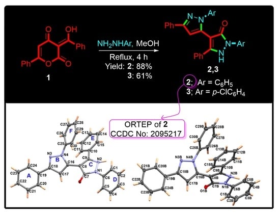
2.2. Synthesis of 2 and 3

A methanolic solution of pyran-2,4-dione 1 (1.0 mmol) was mixed with phenylhydrazine or 4-chlorophenylhydrazine HCl (2.2 mmol) in methanol (10 mL), then the mixture was refluxed for 4 h. The resulting solutions were cooled to room temperature. The formed precipitates were collected by filtration and dried. Purifications were performed by recrystallization from EtOH in case of 2 and DCM/MeOH in case of 3. Only good quality single crystals were obtained for 2, which were found to be suitable for single-crystal X-ray diffraction analysis.

2.2.1. 2,2′,5,5′-Tetraphenyl-1′,2′-Dihydro-2H,3′H-[3,4′-Bipyrazol]-3′-One (2)

Yield: 88%, m.p. 270–271 °C. 1H-NMR (400 MHz, DMSO-d6) δ 12.01 (s, 1H), 7.99 (d, J = 7.6 Hz, 2H), 7.84 (d, J = 8.0 Hz, 2H), 7.53 (t, J = 7.6 Hz, 2H), 7.48 (t, J = 7.6 Hz, 2H),7.40–7.23 (m, 12H), 7.16 (s, 1H); 13C-NMR (101 MHz, DMSO-d6) δ 151.31, 140.28, 135.72, 133.33, 129.55, 129.30, 129.12, 128.79, 128.47, 127.57, 126.79, 126.59, 125.75, 124.21, 122.07, 108.40 (Figures S1 and S2 (Supplementary data)); CHN analysis for [C30H22N4O] C, 79.27; H, 4.88; N, 12.33; O, 3.52 found C, 79.38; H, 4.99; N, 12.21. UV-Vis (EtOH): 264 nm (Figure S7 (Supplementary data)).

2.2.2. 2,2′-Bis(4-Chlorophenyl)-5,5′-Diphenyl-1′,2′-Dihydro-2H,3′H-[3,4′-Bipyrazol]-3′-One (3)

Yield: 61%, m.p. 227–228 °C. 1H-NMR (400 MHz, DMSO-d6) δ 12.25 (s, 1H), 7.97 (d, J = 7.6 Hz, 2H), 7.89 (d, J = 8.8 Hz, 2H), 7.60 (d, J = 8.8 Hz, 2H), 7.48(t, J = 7.6 Hz, 2H), 7.41–7.27 (m, 10H), 7.16 (s, 1H); 13C-NMR (101 MHz, DMSO-d6) δ 151.68, 138.97, 135.66, 133.04, 131.88, 130.34, 129.54, 129.33, 129.16, 129.04, 128.91, 128.83, 128.65, 126.67, 126.36, 125.80, 125.65, 123.35, 108.53 (Figures S3 and S4 (Supplementary data)); CHN analysis for [C30H20Cl2N4O] C, 68.84; H, 3.85; Cl, 13.55; N, 10.70 found C, 68.99; H, 3.86; Cl, 13.67; N, 10.62. UV-Vis (EtOH): 269 nm and 320 nm (Sh); (Figure S7 (Supplementary data)).

2.3. Crystal Structure Determination

The collecting experimental data for compound 2 are provided in the Supplementary Information.

2.4. Computational Study Protocols

All software and protocols employed for Hirshfeld surface analysis and DFT calculations are provided in the Supplementary Information.

3. Results and Discussion

3.1. Synthesis of 2 and 3

In our recent publication [29], we reported that the pyran-2,4-diketone 1 could react with different amines and also amino acid esters to afford β-enaminones in a region-selective conversion. Interestingly, when pyran-2,4-dione 1 was reacted with aromatic hydrazines (phenylhydrazine and 4-chlorophenylhydrazine HCl) in MeOH under reflux conditions, the 3,4′-bipyrazol-3′-ones 2 and 3 were obtained. We assumed that the reaction occurred under a set of chemical transformations, including keto-enol tautomerism, aryl hydrazone formation, ring opening–closure, then keto-enol tautomerism, followed by aryl hydrazone formation with the second molecule of aryl hydrazine, and finally, ring closure upon the dehydration process (Scheme 1). The chemical structure of the final compounds was assigned based on various spectroscopic techniques (1H- and 13C-NMR, and CHN elemental analysis). Furthermore, compound 2 was unambiguously confirmed by single-crystal X-ray diffraction analysis.

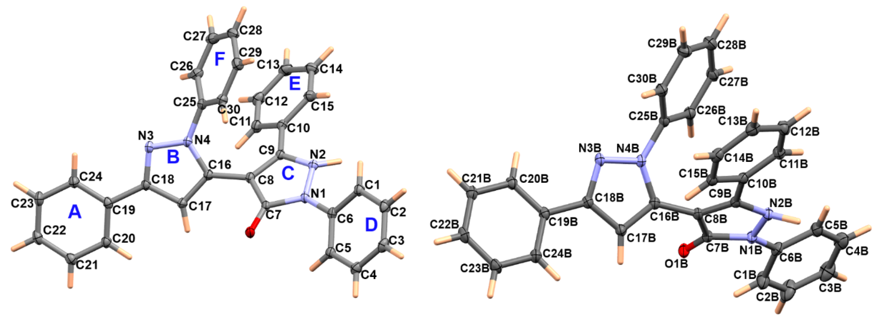
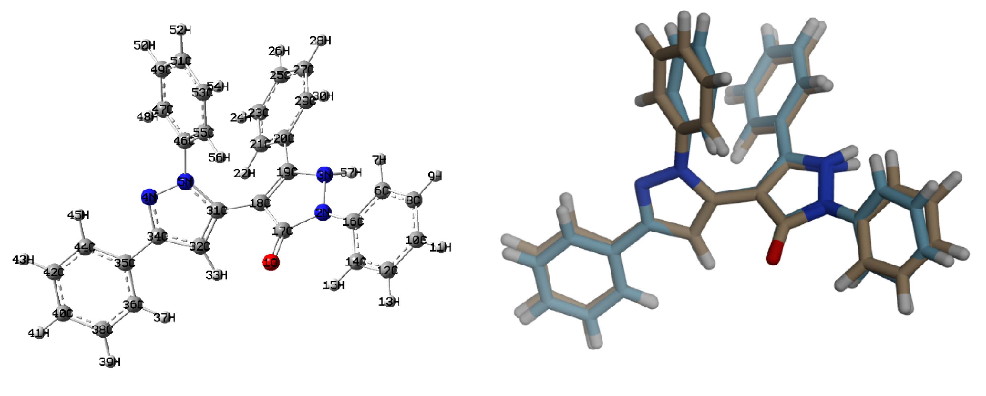
3.2. Crystal Structure Description of 2

The X-ray structure of 2 is shown in Figure 1. The structure agreed very well with the spectral analyses. The crystal data and structure refinement details are depicted in Table 1. The compound crystallized in the triclinic system and P-1 space group with unit cell parameters of a = 11.0023(4) Å, b = 11.9332(5) Å, c = 18.0128(7) Å, α = 85.276(3)°, β = 84.517(3)° and γ = 85.240(3)°. The unit cell volume is 2339.40(16) Å3 and Z = 4, and there are two molecules per asymmetric unit. Selected bond distances and angles are listed in Table 2.
The studied compound comprised six ring systems. The three rings B, D and E are twisted from the mean plane of ring C by 46.59, 36.61 and 30.90°, respectively. Similarly, rings A and F are twisted from ring B by 3.93 and 58.05°, respectively. The corresponding values of the second molecular unit are 58.02, 23.86, 24.74, 20.68 and 45.95°, respectively.
The molecules of 2 are connected with one another via strong N-H…O hydrogen bonding interactions. The hydrogen bond parameters are listed in Table 3 and presentation of the hydrogen bonding interactions among molecular units is shown in Figure 2 (upper part). In addition, the crystal structure of 2 comprised a large number of intra- and intermolecular π–π interactions. Summary of the short intra- and intermolecular C…C contacts are depicted in Table 4 and shown in the lower part of Figure 2.

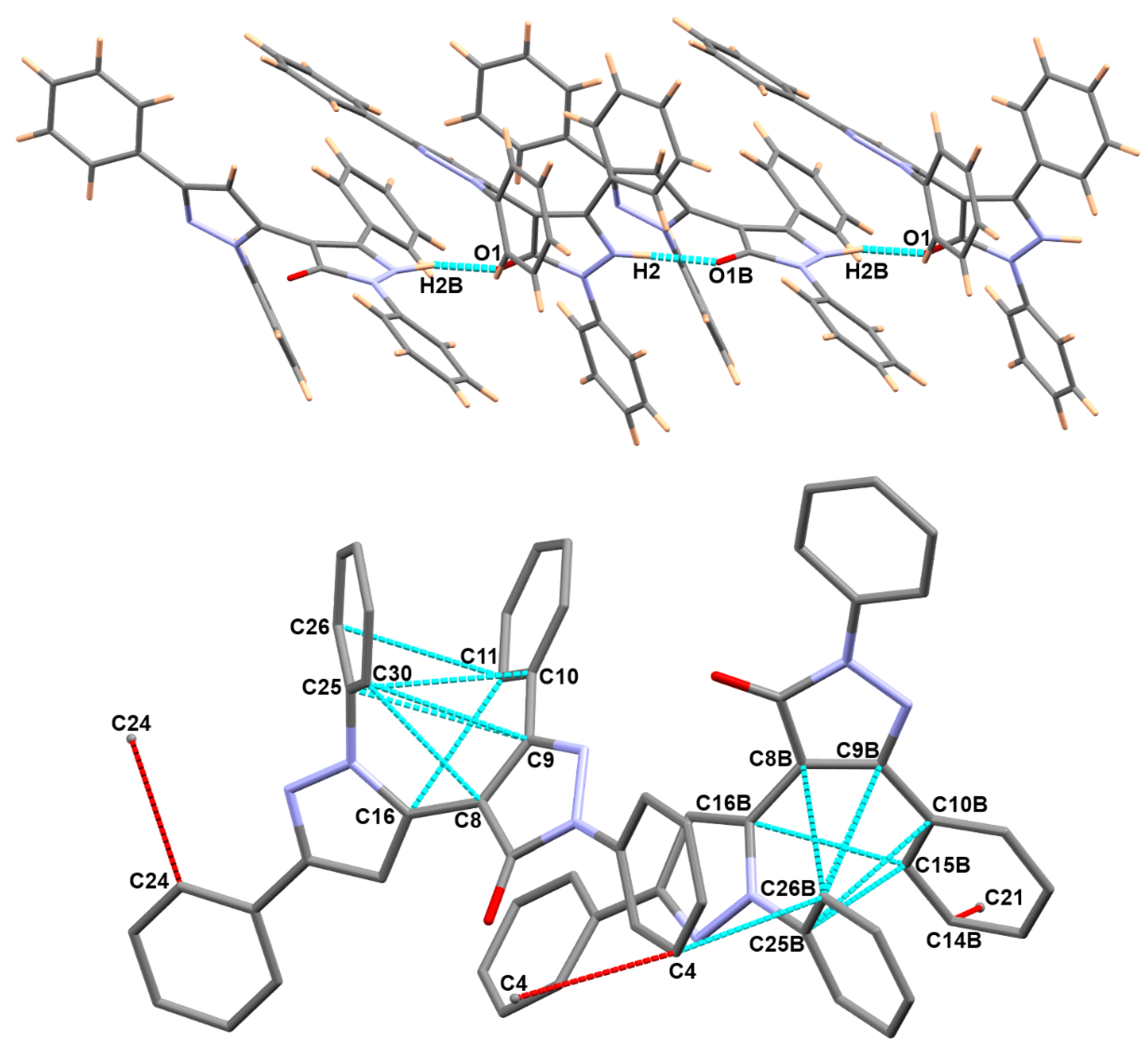
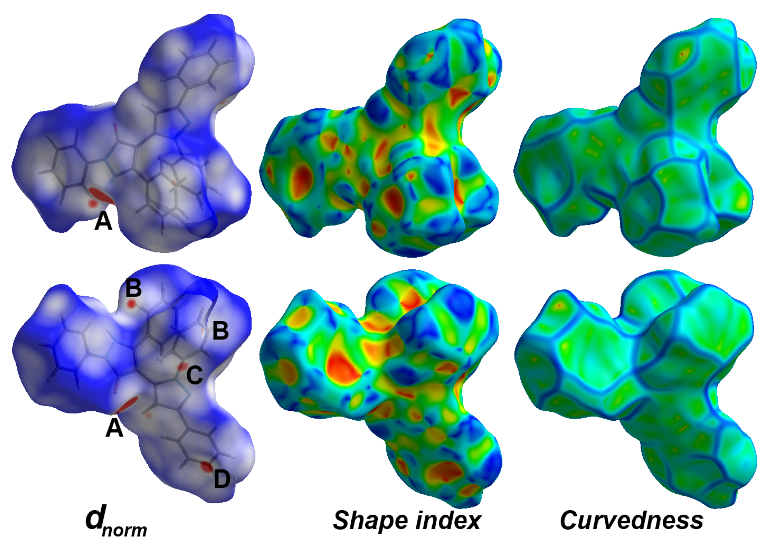
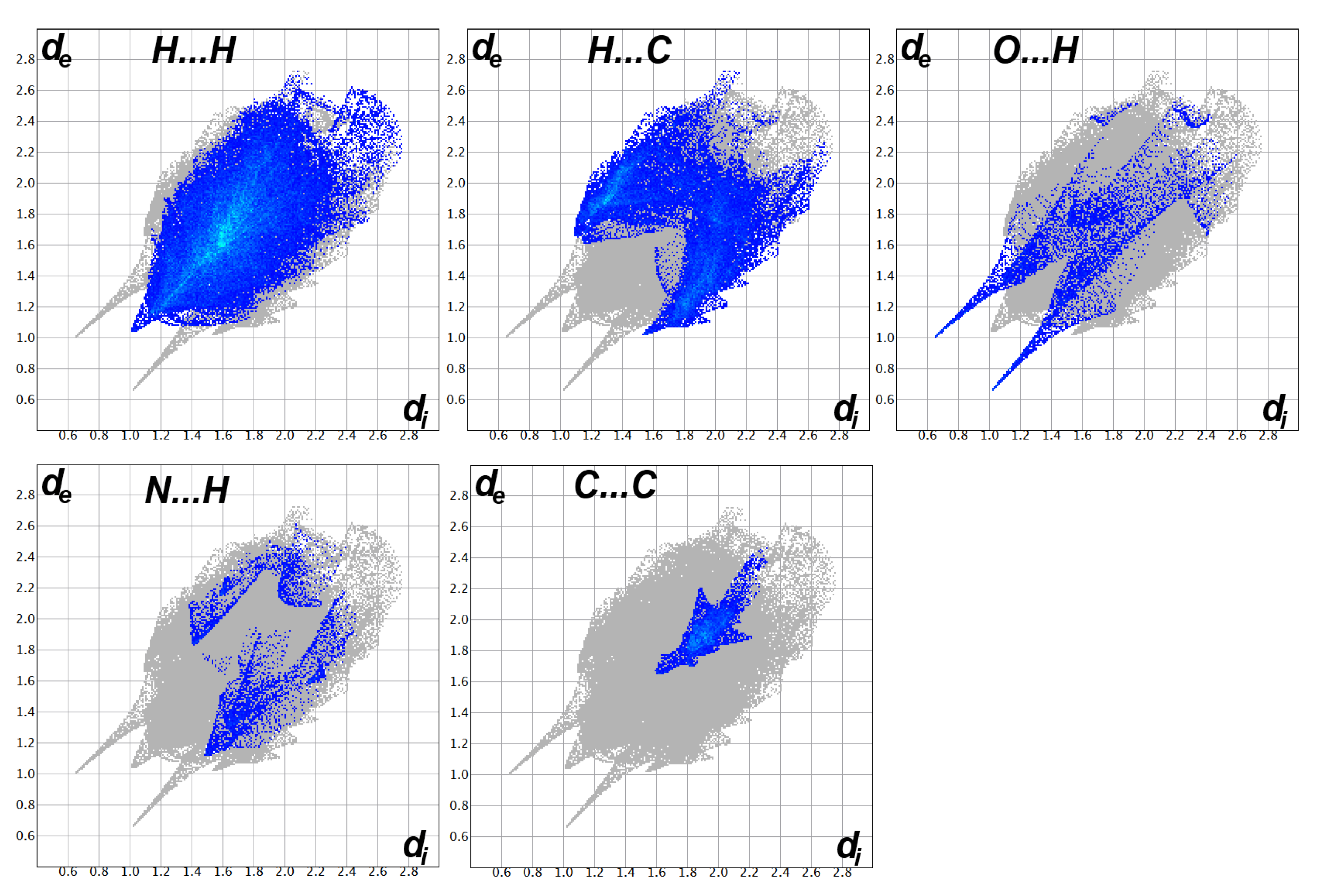
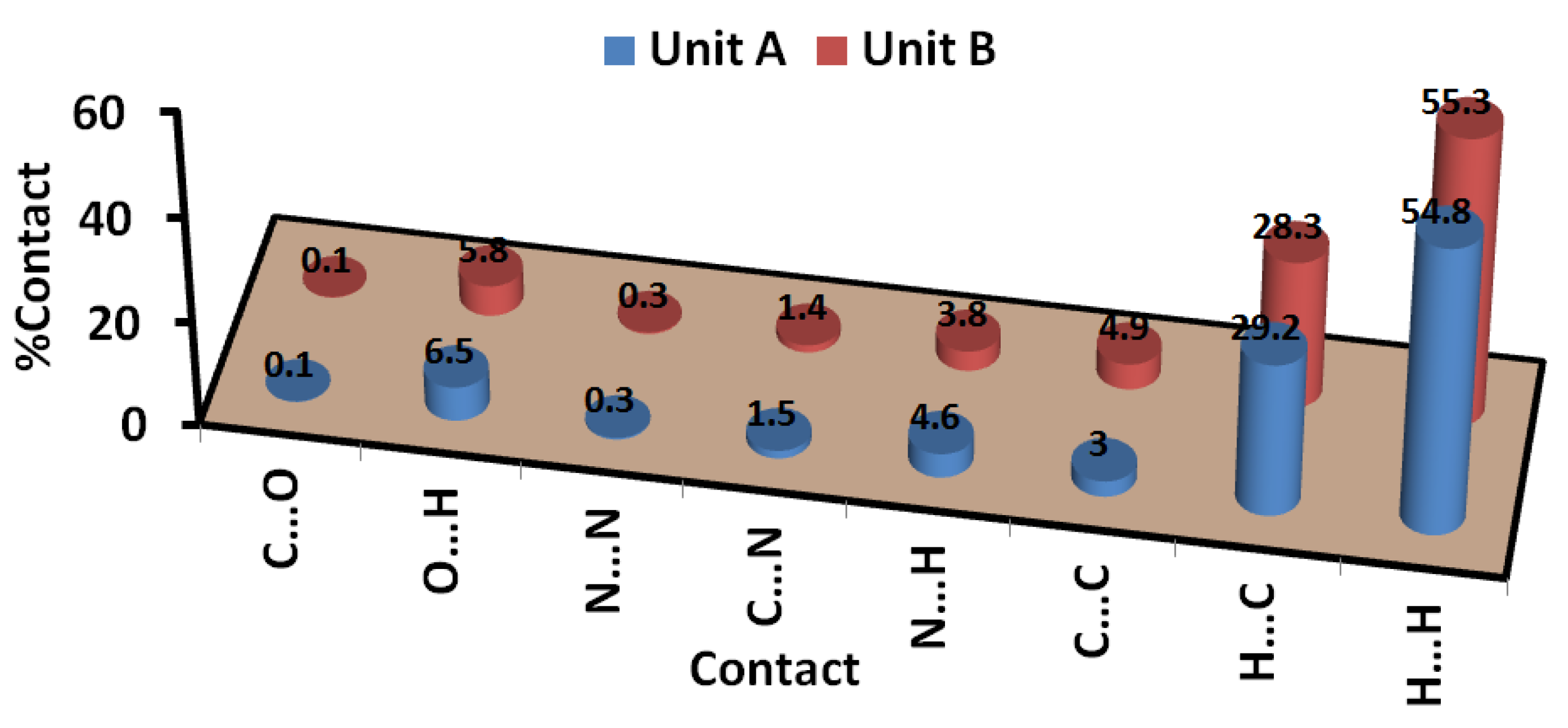
3.3. Analysis of Molecular Packing
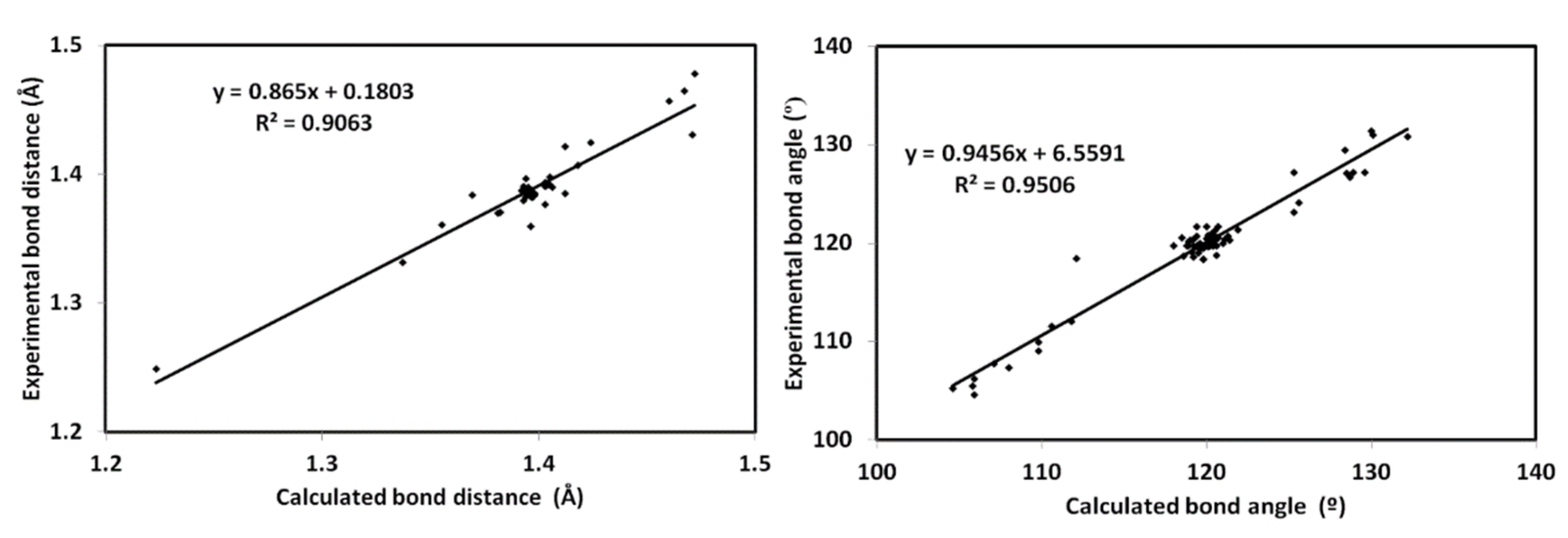
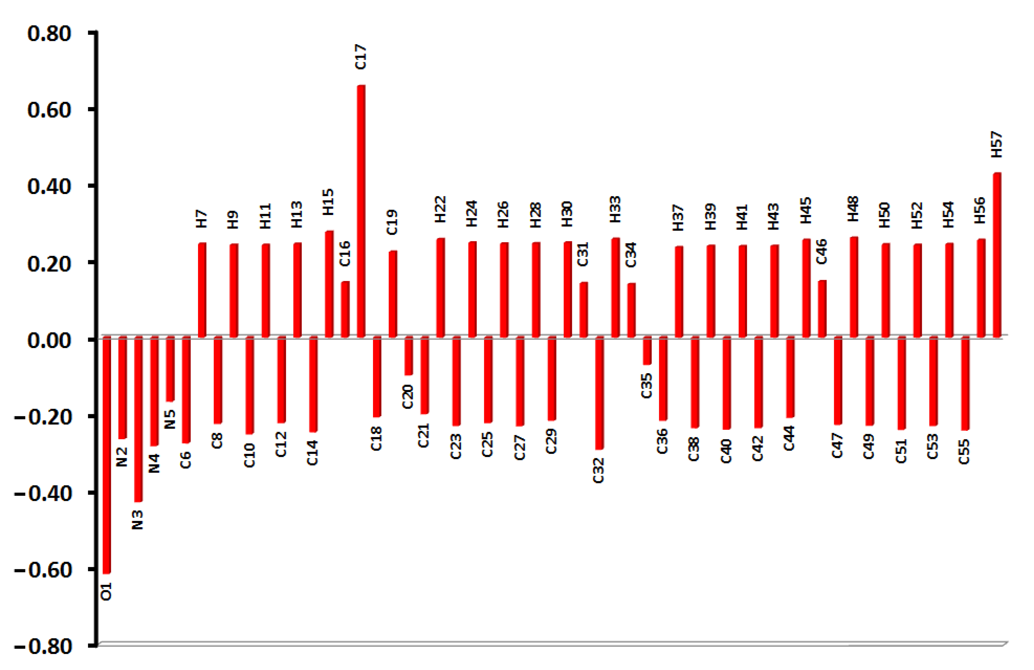
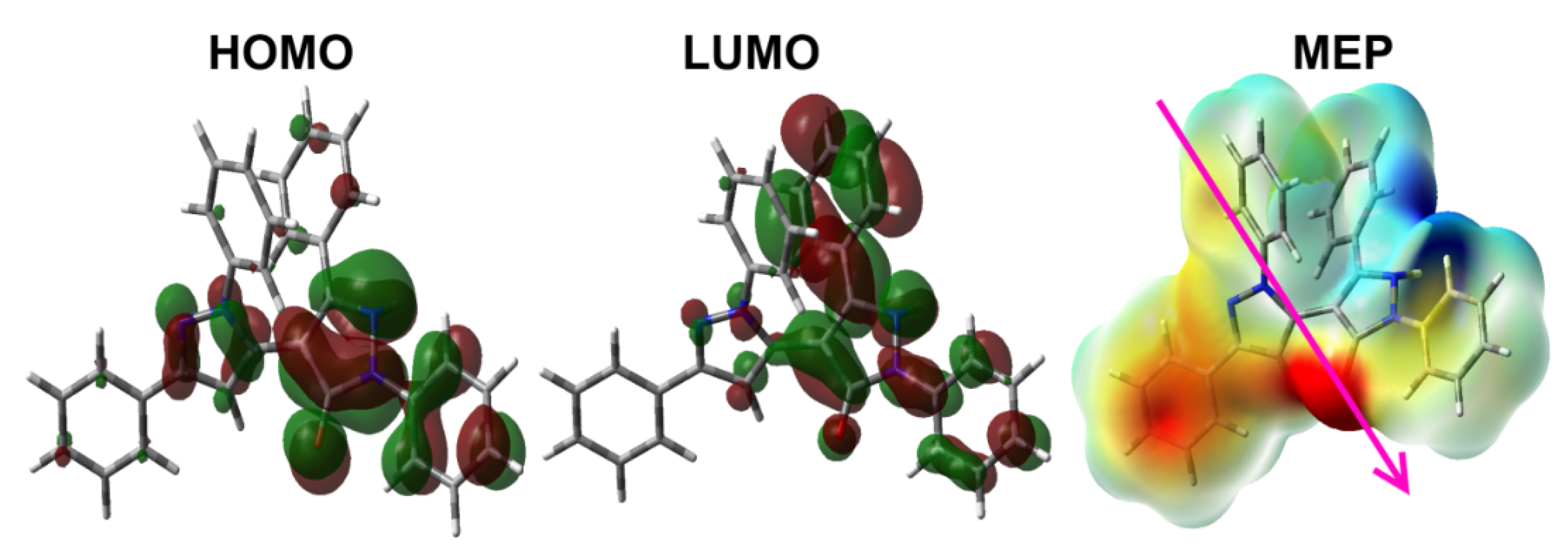
The Hirshfeld surfaces, including the dnorm, shape index and curvedness maps of 2 are shown in Figure 3. In the dnorm map, there are many short distance contacts appearing as red spots. In the studied crystal, there are two molecular units per asymmetric formula; hence, the Hirshfeld results are discussed for the two molecular units.
The decomposition of all possible contacts with the aid of fingerprint plots gave the percentages of all intermolecular interactions controlling the molecular packing of 2 (Figure 4). It is clear that the H…H and H…C interactions are the most dominant, which comprised 54.8–55.3% and 28.3–29.2% of the whole fingerprint area. The other contacts such as O…H (5.8–6.5%), N…H (3.8–4.6%) and C…C (3.0–4.9%) are less dominant in the crystal structure of 2. A summary of all possible contacts in both units are presented in Figure 5.
For simplicity, the most important short contacts are labelled A to D in the dnorm map shown in Figure 3. These red spots represent the intermolecular interactions with shorter distances than the van der Waals radii sum of the interacting atoms. The interaction distances of these short contacts are summarized in Table 5. The O…H hydrogen bonds are significantly shorter. The O1B…H2 and O1…H2B contacts have contact distances of 1.677 and 1.658 Å, respectively. In contrast, the H…C interactions are significantly longer with interaction distances ranging from 2.559 Å (H17…C21B) to 2.776 Å (H30…C4B). On the other hand, the shortest H…H and C…C contacts are H3…H11B and C4…C26B with interaction distances of 2.056 and 3.264Å, respectively. These intermolecular interactions have an important role in the stability of the crystalline structure of the studied compound.

3.4. DFT Studies

The structure of 2 was optimized and the resulting minimum energy structure was shown in Figure 6. An overlay of the calculated structure with the experimental one is presented in the same figure. There are good agreements between the calculated bond distances and angles with the experimental ones (Table S1, Supplementary data). In Figure 7, there are excellent straight-line correlations (R2 = 0.906–0.951) between the calculated and experimental geometric parameters. Little variation between the calculated and experimental structures could be attributed to the crystal packing effects.
The distribution of charges at the different atomic sites was predicted with the aid of natural population analysis (Figure 8). The results indicated the high electronegative nature of the nitrogen and oxygen atoms. Their natural charges were calculated to be −0.6160 and −0.1673 to −0.4294 e, respectively. Additionally, all carbons are negatively charged except those bonded to electronegative atoms. In contrast, all hydrogen atoms are electropositive. Of these atomic sites, the NH proton (0.427 e) and the carbonyl carbon (0.655 e) are the most electropositive. The studied molecule is highly polar with a net dipole moment of 6.388 Debye, and the direction of the dipole moment vector is presented in Figure 9.
In the MEP map shown in the right part of Figure 9, there is an intense red region found close to the carbonyl oxygen atom, while a blue region is close to the NH proton, which represent molecular sites with the highest and lowest electron density, respectively. Hence, the carbonyl oxygen atom and the NH proton are the most probable sites as hydrogen bond acceptors and donors, respectively. This analysis is in accord with the observed X-ray structure of the studied system. In Figure 9, the HOMO and LUMO levels are also presented. It is clear that the HOMO and LUMO are mainly located over the delocalized π system of the compound. Hence, the HOMO–LUMO intramolecular charge transfer could be described as π–π* transition.
Based on the HOMO and LUMO energies, the ionization potential (I = −EHOMO), electron affinity (A= −ELUMO), chemical potential (μ = −(I + A)/2), hardness (η= (I − A)/2), as well as electrophilicity index (ω= μ2/2η) were calculated [31,32,33,34,35,36,37]. These reactivity indices are calculated to be 5.571, 1.645, −3.608, 3.926, and 1.658 eV, respectively. It was believed that these electronic parameters have important rule in the biomolecular reactivity.

3.5. NBO Analysis

The stabilization of molecular systems through electron delocalization from occupied orbitals to antibonding empty orbitals was investigated using NBO calculations [38,39]. The results of the stabilization energies (E(2)) due to the different electron delocalization processes are summarized in Table 6. The compound is stabilized by a large number of π→π* intramolecular charge transfer (IMCT) processes. The strongest π→π* IMCT processes is BD(2)C31-C32→BD*(2)N4-C34, which stabilized the structure by 27.10 kcal/mol. The structure is also stabilized by a number of strong n→π* IMCT processes which stabilized the system up to 36.90 kcal/mol for the LP(1)N5→BD*(2)C31-C32 IMCT process. In contrast, the σ–σ* and n→σ* IMCT processes stabilized the system to weaker extents. The BD(1)C32-C34→BD*(1)C18-C31 (6.78 kcal/mol) and LP(1)N4→BD*(1)N5-C31 (7.25 kcal/mol) are the strongest σ–σ* and n→σ* IMCT in the studied molecule.

4. Conclusions

In conclusion, newly substituted 3-4′-bipyrazoles were synthesized for the first time using a simple one-pot method from pyran-2,4-diketone and arylhydrazines. The structure of the newly synthesized compounds were elucidated using elemental analysis and spectroscopic techniques. The supramolecular structure of the crystallized compound 2 was analyzed using Hirshfeld calculations. Moreover, the optimized geometry calculations showed good agreement with the experimental data. Different electron delocalization processes which stabilized the system through conjugation effects were also calculated using natural bond orbital calculations. HOMO, LUMO and MEP as well as the electronic reactivity descriptors were also presented and discussed.

Supplementary Materials

The following are available online at https://www.mdpi.com/article/10.3390/cryst11080953/s1, X-Ray single-crystal determination of 2; Table S1: The calculated geometric parameters of 2, Figure S1–S4. 1H- and 13C-NMR for compounds 2 and 3 in DMSO-d6, Figure S5. Hirshfeld surfaces for the other molecular unit in the crystal structure of 2; Figure S6. Decomposed fingerprint plots for the other molecular unit the crystal structure of 2; Figure S7. Uv-Vis spectra of compounds 2 and 3.

Author Contributions

Conceptualization, A.T.A.B., S.M.S. and A.B.; synthesis and characterization, A.T.A.B., A.A.M.S. and A.M.A.-M.; X-ray crystal structure was carried out by: M.H.; writing—original manuscript, A.T.A.B., S.M.S. and A.B.; revision and editing, A.T.A.B., S.M.S. and A.B. All authors have read and agreed to the published version of the manuscript.

Funding

Researchers Supporting Project (RSP-2021/64), King Saud University, Riyadh, Saudi Arabia.

Institutional Review Board Statement

Not applicable.

Informed Consent Statement

Not applicable.

Data Availability Statement

Not applicable.

Acknowledgments

The authors would like to extend their sincere appreciation to the Researchers Supporting Project (RSP-2021/64), King Saud University, Riyadh, Saudi Arabia.

Conflicts of Interest

The authors declare no conflict of interest.

References

  1. Lee, J.S. Recent advances in the synthesis of 2-pyrones. Mar. Drugs 2015, 13, 1581–1620. [Google Scholar] [CrossRef] [Green Version]
  2. Fisch, M.H.; Flick, B.H.; Arditti, J. Structure and antifungal activity of hircinol, loroglossol and orchinol. Phytochemistry 1973, 12, 437–441. [Google Scholar] [CrossRef] [Green Version]
  3. Kelly, T.R.; Li, Q.; Bhushan, V. Intramolecular biaryl coupling: Asymmetric synthesis of the chiral B-ring diol unit of pradimicinone. Tetrahedron Lett. 1990, 31, 161–164. [Google Scholar] [CrossRef]
  4. Boger, D.L.; Mullican, M.D. Regiospecific total synthesis of juncusol. J. Org. Chem. 1984, 49, 4045–4050. [Google Scholar] [CrossRef]
  5. Rabideau, P.W.; Harvey, R.G. Metal-ammonia reduction. VII. Stereospecific re- duction in the phenanthrene series. J. Org. Chem. 1970, 35, 25–30. [Google Scholar] [CrossRef]
  6. Goel, A.; Ram, V.J. Natural and synthetic 2 H-pyran-2-ones and their versatility in organic synthesis. Tetrahedron 2009, 65, 7865–7913. [Google Scholar] [CrossRef]
  7. Tsuchiya, K.; Kobayashi, S.; Nishikiori, T.; Nakagawa, T.; Tatsuta, K. NK10958P, a novel plant growth regulator produced by Streptomyces sp. J. Antibiot. 1997, 50, 259–260. [Google Scholar] [CrossRef] [Green Version]
  8. Birkbeck, A.A.; Enders, D. The total synthesis of (+)-pectinatone: An iterative alkylation approach based on the SAMP-hydrazone method. Tetrahedron Lett. 1998, 39, 7823–7826. [Google Scholar] [CrossRef]
  9. Costa, S.S.; Jossang, A.; Bodo, B. 4′′′′-Acetylsagittatin A, a kaempferol triglycoside from Kalanchoe streptantha. J. Nat. Prod. 1996, 59, 327–329. [Google Scholar] [CrossRef]
  10. Parker, S.R.; Cutler, H.G.; Jacyno, J.M.; Hill, R.A. Biological activity of 6-pentyl-2-H-pyran-2-one and its analogs. J. Agric. Food Chem. 1997, 45, 2774–2776. [Google Scholar] [CrossRef]
  11. Nair, M.G.; Chandra, A.; Thorogood, D.L. Griseulin, a new nitro-containing bioactive metabolite produced by Streptomyces spp. J. Antibiot. 1993, 46, 1762–1763. [Google Scholar] [CrossRef] [Green Version]
  12. Ishibashi, Y.; Nishiyama, S.; Yamamura, S. Structural revision of griseulin, a bioactive pyrone possessing a nitrophenyl unit. Chem. Lett. 1994, 1747–1748. [Google Scholar] [CrossRef]
  13. Xue, F.; Li, X.; Wan, B. A class of benzene backbone-based olefin-sulfoxide ligands for Rh-catalyzed enantioselective addition of arylboronic acids to enones. J. Org. Chem. 2011, 76, 7256–7262. [Google Scholar] [CrossRef]
  14. Gianni, J.; Pirovano, V.; Abbiati, G. Silver triflate/p-TSA co-catalysed synthesis of 3-substituted isocoumarins from 2-alkynylbenzoates. Org. Biomol. Chem. 2018, 16, 3213–3219. [Google Scholar] [CrossRef] [PubMed] [Green Version]
  15. Hosny, M.; Fawzy, M.; Abdelfatah, A.M.; Fawzy, E.E.; Eltaweil, A.S. Comparative study on the potentialities of two halophytic species in the green synthesis of gold nanoparticles and their anticancer, antioxidant and catalytic efficiencies. Adv. Powder Technol. 2021, in press. [Google Scholar] [CrossRef]
  16. Shaaban, M.R.; Mayhoub, A.S.; Farag, A.M. Recent advances in the therapeutic applications of pyrazolines. Expert. Opin. Ther. Pat. 2012, 22, 253–291. [Google Scholar] [CrossRef] [PubMed]
  17. Varghese, B.; Al-Busafi, S.N.; Suliman, F.O.; Al-Kindy, S.M.Z. Unveiling a versatile heterocycle: Pyrazoline—A review. RSC Adv. 2017, 7, 46999–47016. [Google Scholar] [CrossRef] [Green Version]
  18. Tok, F.; Abas, B.I.; Çevik, O.; Koçyiğit-Kaymakçıoğlu, B. Design, synthesis and biological evaluation of some new 2-pyrazoline derivatives as potential anticancer agents. Bioorg. Chem. 2020, 102, 104063. [Google Scholar] [CrossRef]
  19. Yusuf, M.; Jain, P. Synthetic and biological studies of pyrazolines and related heterocyclic compounds. Arab. J. Chem. 2014, 7, 553–596. [Google Scholar] [CrossRef] [Green Version]
  20. Kariuki, B.M.; Abdel-Wahab, B.F.; El-Hiti, G.A. Synthesis and structural characterization of isostructural 4-(4-Aryl)-2-(5-(4-fluorophenyl)-3-(1-(4-fluorophenyl)-5-methyl-1H-1,2,3-triazol-4-yl)-4,5-dihydro-1H-pyrazol-1-yl)thiazoles. Crystals 2021, 11, 795. [Google Scholar] [CrossRef]
  21. Mabkhot, Y.N.; Al-Majid, A.M.; Barakat, A.; Alshahrani, S.; Siddiqui, Y. 1,1′-(3-Methyl-4-phenylthieno[2,3-b]thiophene-2,5-diyl)diethanone as building block in heterocyclic synthesis. Novel synthesis of some pyrazoles, and pyrimidines derivatives. Molecules 2011, 16, 6502–6511. [Google Scholar] [CrossRef] [Green Version]
  22. Mabkhot, Y.N.; Barakat, A.; Al-Majid, A.M.; Al-Othman, Z.A.; Alamary, A.S. A Facile and convenient synthesis of some novel hydrazones, schiff’s base and pyrazoles incorporating thieno [2,3-b]thiophenes. Int. J. Mol. Sci. 2011, 12, 7824–7834. [Google Scholar] [CrossRef] [Green Version]
  23. Mabkhot, Y.N.; Kaal, N.A.; Alterary, S.; Al-Showiman, S.S.; Barakat, A.; Ghabbour, H.A.; Frey, W. Synthesis, in-vitro antibacterial, antifungal, and molecular modeling of potent anti-microbial agents from a combined pyrazole and thiophene pharmacophore. Molecules 2015, 20, 8712–8729. [Google Scholar] [CrossRef] [Green Version]
  24. Elshaier, Y.A.M.M.; Barakat, A.; Al-Qahtany, B.M.; Al-Majid, A.M.; Al-Agamy, M.H. Synthesis of pyrazole-thiobarbituric acid derivatives: Antimicrobial activity and docking studies. Molecules 2016, 21, 1337. [Google Scholar] [CrossRef] [PubMed] [Green Version]
  25. Barakat, A.; Al-Qahtani, B.M.; Al-Majid, A.M.; Ali, M.; Mabkhot, Y.N.; Al-Agamy, M.H.M.; Wadood, A. Synthesis, characterization, anti-microbial activity and molecular docking studies of combined pyrazol-barbituric acid pharmacophore. Trop. J. Pharm. Res. 2016, 15, 1319–1326. [Google Scholar] [CrossRef]
  26. Barakat, A.; Al-Majid, A.; Al-Qahtani, B.M.; Ali, M.; Teleb, M.; Al-Agamy, M.H.M.; Naz, S.; Ul-Haq, Z. Synthesis, antimicrobial activity, pharmacophore modeling and molecular docking studies of new pyrazole-dimedone hybrid architectures. Chem. Cent. J. 2018, 12, 29. [Google Scholar] [CrossRef] [Green Version]
  27. Lellek, V.; Chen, C.-Y.; Yang, W.; Liu, J.; Ji, X.; Faessler, R. An efficient synthesis of substituted pyrazoles from one-pot reaction of ketones, aldehydes, and hydrazine monohydrochloride. Synlett 2018, 29, 1071–1075. [Google Scholar] [CrossRef] [Green Version]
  28. Alex, K.; Tillack, A.; Schwarz, N.; Beller, M. Zinc-catalyzed synthesis of pyrazolines and pyrazoles via hydrohydrazination. Org. Lett. 2008, 10, 2377–2379. [Google Scholar] [CrossRef] [PubMed]
  29. Sarhan, A.A.M.; Haukka, M.; Barakat, A.; Boraei, A. A novel synthetic approach to pyran-2,4-dione scaffold production:Microwave-assisted dimerization, cyclization, and expeditiousregioselective conversion intob-enamino-pyran-2,4-diones. Tetrahedron Lett. 2020, 61, 152660. [Google Scholar] [CrossRef]
  30. Boraei, A.T.A.; Haukka, M.; Sarhan, A.A.M.; Soliman, S.M.; Barakat, A. Intramolecular hydrogen bond, hirshfeld analysis, AIM; DFT studies of pyran-2,4-dione derivatives. Crystals 2021, 11, 896. [Google Scholar] [CrossRef]
  31. Foresman, J.B.; Frisch, E. Exploring Chemistry with Electronic Structure Methods, 2nd ed.; Gaussian: Pittsburgh, PA, USA, 1996. [Google Scholar]
  32. Chang, R. Chemistry, 7th ed.; McGraw-Hill: New York, NY, USA, 2001. [Google Scholar]
  33. Kosar, B.; Albayrak, C. Spectroscopic investigations and quantum chemical computational study of (E)-4-methoxy-2-[(p-tolylimino) methyl] phenol. Spectrochim. Acta 2011, 78, 160–167. [Google Scholar] [CrossRef] [PubMed]
  34. Koopmans, T.A. Ordering of wave functions and eigenenergies to the individual electrons of an atom. Physica 1933, 1, 104–113. [Google Scholar] [CrossRef]
  35. Parr, R.G.; Yang, W. Density-Functional Theory of Atoms and Molecules; Oxford University Press: New York, NY, USA, 1989. [Google Scholar]
  36. Parr, R.G.; Szentpaly, L.V.; Liu, S. Electrophilicity index. J. Am. Chem. Soc. 1999, 121, 1922–1924. [Google Scholar] [CrossRef]
  37. Singh, R.N.; Kumar, A.; Tiwari, R.K.; Rawat, P.; Gupta, V.P. A combined experimental and quantum chemical (DFT and AIM) study on molecular structure, spectroscopic properties, NBO and multiple interaction analysis in a novel ethyl 4-[2-(carbamoyl) hydrazinylidene]-3, 5-dimethyl-1H-pyrrole-2-carboxylate and its dimer. J. Mol. Strut. 2013, 1035, 427–440. [Google Scholar] [CrossRef]
  38. Joe, I.H.; Kostova, I.; Ravikumar, C.; Amalanathan, M.; Pinzaru, S.C. Theoretical and vibrational spectral investigation of sodium salt of acenocoumarol. J. Raman Spectrosc. 2009, 40, 1033–1038. [Google Scholar]
  39. Sebastian, S.; Sundaraganesan, N. The spectroscopic (FT-IR, FT-IR gas phase, FT-Raman and UV) and NBO analysis of 4-Hydroxypiperidine by density functional method. Spectrochim. Acta Part A Mol. Biomol. Spectrosc. 2010, 75, 941–952. [Google Scholar] [CrossRef]
Scheme 1. Synthesis and proposed mechanism for compounds 2 and 3 starting from pyran-2,4-diketone 1.
Crystals 11 00953 sch001
Figure 1. X-ray structure of 2. Atom thermal ellipsoids are drawn at 30% probability level.
Crystals 11 00953 g001
Figure 2. Packing of molecular units via N-H…O H-bonding interactions (upper) and the important intra- and intermolecular π–π stacking interactions (lower).
Crystals 11 00953 g002
Figure 3. Hirshfeld surfaces for the molecular unit with atom numbering comprising B of 2. The results for the other unit are presented in Figure S5 (Supplementary data). A: O…H, B: H…H, C: C…C and D: H…C contacts.
Crystals 11 00953 g003
Figure 4. Decomposed fingerprint plots for the molecular unit with atom numbering comprising B. The results for the other unit are presented in Figure S6 (Supplementary data).
Crystals 11 00953 g004
Figure 5. The percentages of all possible intermolecular interactions in the crystal structure of 2.
Crystals 11 00953 g005
Figure 6. The calculated geometry (left) and its overlay with the experimental one, (right) for 2. For the experimental model used for the comparison with the optimized one, see Figure 1 (left part).
Crystals 11 00953 g006
Figure 7. The correlations between the calculated and experimental geometric parameters.
Crystals 11 00953 g007
Figure 8. Natural charge distributions in 2.
Crystals 11 00953 g008
Figure 9. The MEP, HOMO and LUMO of 2. In molecular electrostatic potential (MEP): red (most negative) and blue (most positive).
Crystals 11 00953 g009
Table 1. Crystal Data for 2.
2
CCDC no.2095217
empirical formulaC30H22N4O
Fw454.51
temp (K)120(2)
λ(Å)1.54184
cryst systTriclinic
space groupP?1
a (Å)11.0023(4)
b (Å)11.9332(5)
c (Å)18.0128(7)
α(deg)85.276(3)
β (deg)84.517(3)
γ(deg)85.240(3)
V3)2339.40(16)
Z4
ρcalc (Mg/m3)1.290
μ(Mo Kα) (mm−1)0.634
No. reflns.17900
Unique reflns.9577
Completeness to θ = 67.684° (%)99.8
GOOF (F2)1.034
Rint0.0419
R1 a (I ≥ 2σ)0.0485
wR2 b (I ≥ 2σ)0.1153
a R1 = Σ||Fo| − |Fc||/Σ|Fo|. b wR2 = [Σ[w(Fo2Fc2)2]/Σ[w(Fo2)2]]1/2.
Table 2. Selected bond lengths [Å] and angles [°] for 2.
AtomsDistanceAtomsDistance
O1–C71.249(2)O1B–C7B1.250(2)
N1–N21.377(2)N1B–N2B1.387(2)
N1–C71.385(2)N1B–C7B1.395(2)
N1–C61.422(2)N1B–C6B1.410(2)
N2–C91.360(2)N2B–C9B1.354(2)
N3–C181.332(3)N3B–C18B1.345(2)
N3–N41.361(2)N3B–N4B1.366(2)
N4–C161.370(2)N4B–C16B1.365(2)
N4–C251.425(2)N4B–C25B1.431(2)
AtomsAngleAtomsAngle
N2–N1–C7110.01(15)C5–C6–N1118.42(17)
N2–N1–C6121.68(15)O1–C7–N1123.25(16)
C7–N1–C6127.14(15)O1–C7–C8131.41(17)
C9–N2–N1107.80(15)N1–C7–C8105.28(15)
C18–N3–N4104.58(15)C9–C8–C7107.45(16)
N3–N4–C16112.08(15)C9–C8–C16130.99(17)
N3–N4–C25120.64(16)C7–C8–C16121.41(16)
C16–N4–C25127.25(16)N2–C9–C8109.10(16)
C6–C1–C2118.41(19)N2–C9–C10119.81(16)
C3–C2–C1120.4(2)C8–C9–C10130.96(17)
C2–C3–C4120.37(19)C15–C10–C11119.13(19)
C3–C4–C5120.4(2)C15–C10–C9120.99(17)
C4–C5–C6118.60(19)C11–C10–C9119.84(17)
C1–C6–C5121.75(18)C12–C11–C10120.09(19)
C1–C6–N1119.82(18)
Table 3. Hydrogen bonds for 2 [Å and °].
D-H…Ad(D-H)d(H…A)d(D…A)<(DHA)
N2-H2…O1B0.95(3)1.73(3)2.681(2)173(3)
N2B-H2B…O1 #11.01(3)1.65(3)2.648(2)166(3)
#1 x + 1,y,z.
Table 4. The intra- and intermolecular π-π interactions for 2 [Å].
ContactDistanceContactDistance
C8…C303.195C8B…C26B3.127
C9…C253.287C9B…C25B3.342
C9…C303.316C9B…C26B3.296
C10…C253.307C10B…C25B3.331
C11…C163.303C15B…C16B3.142
C11…C263.335C15B…C25B3.336
C4…C26B3.264C4…C43.273 ii
C24…C243.39 iC21…C14B3.369 iii
Symm. Codes: i −1 − x,−y,2 − z; ii −x,1 − y,1 − z; iii −x,1 − y,1 − z.
Table 5. Most important contacts and the corresponding shortest interaction distances.
ContactDistanceContactDistance
O1B…H21.677H30…C4B2.776
O1…H2B1.658H2…C7B2.613
O1…H5B2.465H12B…C282.761
H23…C122.726H2B…C72.608
H14B…C212.769H3…H11B2.056
H17…C21B2.559C4…C26B3.264
Table 6. The E(2) (kcal/mol) values for the IMCT interactions in 2a.
NBO iNBO jE(2)Donor NBO iAcceptor NBO jE(2)
BD(1) C17–C18BD*(1) C19–C206.61BD(2) N4–C34BD*(2) C31–C3212.59
BD(1) C18–C31BD*(1) C18–C195.00BD(2) N4–C34BD*(2) C35–C36 9.42
BD(1) C31–C32BD*(1) N5–C465.06BD(2) C6–C16BD*(2) C8–C1020.71
BD(1) C31–C32BD*(1) C34–C355.39BD(2) C6–C16BD*(2) C12–C1417.34
BD(1) C32–C34BD*(1) C18–C316.78BD(2) C8–C10BD*(2) C6–C1619.45
BD(2) C8–C10BD*(2) C12–C1420.87
LP(1) N4BD*(1) N5–C317.25BD(2) C12–C14BD*(2) C6–C1621.86
LP(1) N4BD*(1) C32–C345.38BD(2) C12–C14BD*(2) C8–C1019.25
BD(2) C18–C19BD*(2) C20–C297.95
LP(1) N2 BD*(2) C6–C1629.57BD(2) C18–C19BD*(2) C31–C325.57
LP(1) N3 BD*(2) C18–C1920.16BD(2) C20–C29 BD*(2) C18–C1913.95
LP(1) N5BD*(2) N4–C3428.45BD(2) C20–C29 BD*(2) C21–C2319.16
LP(1) N5BD*(2) C31–C3236.90BD(2) C20–C29 BD*(2) C25–C2719.32
LP(1) N5 BD*(2) C46–C5517.17BD(2) C21–C23BD*(2) C20–C2919.89
BD(2) C21–C23BD*(2) C25–C2720.53
BD(2) C25–C27BD*(2) C20–C2921.42
BD(2) C25–C27BD*(2) C21–C2319.25
BD(2) C31–C32BD*(2) N4–C3427.10
BD(2) C31–C32BD*(2) C18–C19 6.37
BD(2) C35–C36BD*(2) N4–C3420.01
BD(2) C35–C36BD*(2) C38–C4020.92
BD(2) C35–C36BD*(2) C42–C4419.13
BD(2) C38–C40BD*(2) C35–C3619.85
BD(2) C38–C40BD*(2) C42–C4419.52
BD(2) C42–C44BD*(2) C35–C3619.90
BD(2) C42–C44BD*(2) C38–C4020.31
BD(2) C46–C55BD*(2) C47–C4919.29
BD(2) C46–C55BD*(2) C51–C5320.04
BD(2) C47–C49BD*(2) C46–C5520.93
BD(2) C47–C49BD*(2) C51–C5320.13
BD(2) C51–C53BD*(2) C46–C5520.39
BD(2) C51–C53BD*(2) C47–C49 20.21
i Donor; j Acceptor.
Publisher’s Note: MDPI stays neutral with regard to jurisdictional claims in published maps and institutional affiliations.

