Article

Cerium Niobate Hollow Sphere Engineered Graphitic Carbon Nitride for Synergistic Photothermal/Chemodynamic Cancer Therapy

1
Department of Chemical Engineering and Biotechnology, National Taipei University of Technology (Taipei Tech), Taipei 106, Taiwan
2
Centre for the Cellular Microenvironment, Division of Biomedical Engineering, James Watt School of Engineering, Mazumdar-Shaw Advanced Research Centre, University of Glasgow, Glasgow G116EW, UK
3
Scrona AG, Grubenstrasse 9, 8045 Zürich, Switzerland
4
Genomics Research Center, Academia Sinica, Taipei 115, Taiwan
5
Department of Biochemistry, College of Medicine, Kaohsiung Medical University, Kaohsiung 807, Taiwan
6
High-Value Biomaterials Research and Commercialization Center, National Taipei University of Technology (Taipei Tech), Taipei 10608, Taiwan
*
Author to whom correspondence should be addressed.
Crystals 2023, 13(6), 954; https://doi.org/10.3390/cryst13060954
Submission received: 18 May 2023 / Revised: 10 June 2023 / Accepted: 12 June 2023 / Published: 15 June 2023
(This article belongs to the Special Issue Pharmaceutical Crystals (Volume III))

Abstract

:
Reactive oxygen species (ROS)-mediated chemodynamic therapy (CDT) and photothermal therapy (PTT) have potential for various cancer treatments. However, they are still bound by the demands of Fenton reaction conditions such as oxygen dependence, inherent defects in common standard photosensitizers (PSs), and the continuous availability of laser sources. Herein, we designed Ce3NbO7/g-C3N4 nanocomposites (NCs) and investigated their ability to evaluate the performance of PTT/CDT synergistically to enhance cancer treatment. The activation of Ce3NbO7/g-C3N4 NCs in the tumor microenvironment (TME) causes the generation of cytotoxic ROS via the Fenton reaction. Additionally, the g-C3N4 in NCs absorbs NIR, generating hyperthermia in the TME. The photothermal conversion efficiency (ƞ) of the Ce3NbO7/g-C3N4 NCs was found to be 49.5%. A photocatalytic reaction with PTT-enhanced Fenton reagents, without consuming additional photothermal agents (PTA) or Fenton reagents, generates the hydroxyl radical (OH•) primarily by direct electron transfer in the TME. Almost 68% of cells experienced programmed cell death due to the combinational effect (PTT/CDT), making it an efficient and biocompatible therapy. Furthermore, this work provides a basis for developing numerous innovative materials that can be used to treat cancer, overcome general limitations, and enhance ROS production under single-wavelength (808 nm) laser irradiation.

1. Introduction

Cancer has the highest mortality rate, accounting for 9.6 million deaths globally in 2018 [1,2]. According to the World Health Organization (WHO), nearly 35% of cancer deaths occur due to changes in lifestyles (including smoking and alcohol consumption), dietary factors, and exposure to ultraviolet rays and ionizing radiation [3,4,5,6]. Hepatitis B and C viruses, fatty liver disease, cirrhosis brought on by alcohol use, smoking, obesity, diabetes, an excess of iron in the diet, and other dietary exposures are the risk factors for liver cancer (LC) [7]. LC is one of the most common cancers [8,9] and ranks third in mortality rate among all malignancies, with an estimated 830,130 deaths globally in 2020 [10,11,12]. Worldwide, LC is the leading form of cancer [9,13]. The WHO estimates around 905,677 new cases annually and reports that 1,276,679 people will die from LC in 2040 [10,14]. Several traditional cancer treatments, including surgery, radiation therapy, and chemotherapy, have been widely used; however, all these therapies are “double-edged swords” due to their high rates of recurrence, harsh side effects, and multidrug resistance [15]. Therefore, there is a need to develop a novel methodology for attaining more efficacy with negligible side effects [16,17,18,19,20], such as sonodynamic therapy (SDT) [18], photodynamic therapy (PDT) [19], chemodynamic therapy (CDT) [20], and photothermal ablation therapy (PAT) [17,19]. A nanocatalytic medicine is an emerging therapeutic concept, producing toxic ROS that actively targets the tumor tissues without harming the normal tissues [21]. The tumor microenvironment (TME) is mildly acidic and hypoxic, expressing H2O2, promoting tumor growth and spread. Therefore, the creation of nanoplatforms for TME-responsive tumor therapy appears to be promising. CDT is the most widely used approach in nanocatalytic medicine and is wholly based on biological characteristics linking various metabolic pathways between tumor cells and normal cells. These biological qualities can produce distinctive biochemical conditions, such as moderate acidity and abundant H2O2, to stimulate specific chemical reactions, particularly the Fenton/Fenton-like reaction within tumors. These interactions produce the highly toxic hydroxyl radical (OH•), which causes tissue damage and tumor cell death. The produced OH• induces primary tumor death in TME, causing these tumor cells to produce tumor-associated antigens (TAAs). The TAAs are captured by dendritic cells (DCs) and migrate to immune organs such as the spleen [22,23]. Some of the metals involved in CDT are Fe [24,25], Mn [26,27], Co [28,29], Ag [30,31], and Cu [32,33]. Recently, a new class of bio-antioxidants called cerium-oxide-based nanozymes has been developed [34,35]. In nature, cerium has two different oxidation states, Ce3+ and Ce4+, and the enzymatic activity of CeO2-x scavenging ROS is assumed to be related to the self-regeneration cycle of Ce3+/Ce4+ and the oxygen vacancies on the cerium oxide surface. Most researchers believe Ce3+/Ce4+ redox cycling is directly associated with CeO2-xs antioxidant properties. The potential significance of oxygen vacancies in the rapid redox cycling of CeO2-x is still under discussion [2,35,36]. Herein, we constructed cerium niobate hollow spheres (CeO2/Ce3NbO7/g-C3N4 NCs), where Ce3NbO7 is meant for CDT purposes.
Owing to its inherent noninvasiveness and negligible side effects, photothermal therapy (PTT) is widely used in cancer therapy. It uses photothermal agents (PTAs) that convert light energy into thermal energy to generate localized hyperthermia; this has been considered an alternative treatment for various cancers [19,37]. Graphitic polymeric carbon nitride (g-C3N4) has gained attention as a novel material that resembles graphene due to its distinct elemental makeup and photoelectric properties. The exceptional biocompatibility of g-C3N4, a compound made exclusively of carbon and nitrogen, is advantageous for use in biomedicine [38]. Due to its superior physical and chemical characteristics, such as ease of preparation, stable fluorescence, appropriate energy level, a wide excitation wavelength range, and high biocompatibility, g-C3N4 is an emerging polymeric material with excellent performance in PTT and photocatalyst applications. However, evidence on its application as a biomedicine is scarce [39].
In this work, we designed NCs composed of cerium niobate (CeO2/Ce3NbO7) hollow spheres coated with g-C3N4 nanosheets (Ce3NbO7/g-C3N4 NCs) with excellent biocompatibility, enhancing both CDT and PTT for synergistically killing tumor cells. The overall scheme of the work is given in Figure 1. The designed Ce3NbO7/g-C3N4 NCs possess the Ce3+ ions that actively take part in the Fenton reaction via the cascade mechanism to yield the OH• radical; the g-C3N4 nanosheets to the hollow spheres act as excellent PTA agents and exhibit an outstanding photothermal conversion efficiency (ƞ) of 49.5% due to higher carbon content. Notably, the synthesized NCs feature an “all in one” therapeutic platform for performing both PTT/CDT without adding any external photothermal or chemodynamic (Fenton) agents. Hence, our synthesized NCs show a novel approach for accessible design and hold great potential in therapeutic applications. Integrating PTT/CDT significantly improves the progression of cell apoptosis by enhancing one mode of therapy with the other through synergistic performance by heat generation in PTT and the production of OH• in CDT. The excellent synergistic performance of the in vitro results of both PTT and CDT illustrates the potential of NCs as a cancer-curing treatment technique.

2. Materials and Methods

2.1. Required Chemicals

A total of 99.5% cerium nitrate hexahydrate (Ce(NO3)3·6H2O), 99.9% niobium pentachloride (NbCl5), 99% urea (CH4N2O), 35% hydrogen peroxide (H2O2), 99% ethanol (C6H5OH), graphitic carbon nitride (g-C3N4), double distilled water (DD H2O), dimethyl sulfoxide (DMSO), 3-(4,5-dimethylthiazol-2-yl)-2,5-diphenyl tetrazolium bromide (MTT), and methylene blue (MB) were purchased from Sigma-Aldrich, Burlington, MA, USA, or Merck, Darmstadt, Germany

2.2. Preparation for CeO2/Ce3NbO7 Hollow Spheres

The CeO2/Ce3NbO7 were prepared by the facile hydrothermal method. Initially, about 0.303 g of cerium nitrate hexahydrate (Ce (NO3)3·6H2O) and 0.18 g of niobium pentachloride (NbCl5) were dissolved in 70 mL of DD H2O. In the above solution, 1 g of urea (CH4N2O) was added dropwise with constant stirring. Then, 4–5 drops of H2O2 were added; the mixture was kept on a magnetic stirrer for 1 h. The clear solution was transferred to a Teflon-lined autoclave and sealed. The above solution was subjected to a hydrothermal process at 180 °C for 20 h. The pale green precipitate obtained was washed four times with ethanol and DD H2O and dried in an oven at 60 °C for 15 h. After drying, the sample was calcined at 800 °C for 2 h. The resultant black powder was stored and labeled as cerium niobate hollow spheres.

2.3. Preparation of g-C3N4

The g-C3N4 was prepared by using the pyrolysis method. Initially, 20 mg of CH4N2O was placed in a crucible and subjected to pyrolysis at 550 °C for 3 h.

2.4. Modification of g-C3N4 on CeO2/Ce3NbO7 Hollow Spheres

The CeO2/Ce3NbO7/g-C3N4 NCs were prepared by coating the CeO2/Ce3NbO7 hollow spheres on the surface of g-C3N4 nanosheets by the ultrasonication method. The calculated amounts of the CeO2/Ce3NbO7 hollow spheres and g-C3N4 were taken in the molar ratio of 1:4 by weight (20% of CeO2/Ce3NbO7 hollow spheres and 80% of g-C3N4). The desired amount of CeO2/Ce3NbO7 hollow spheres was dissolved in ethanol, followed by the addition of g-C3N4 nanosheets during ultrasonication for 10 min, and the residue was dried in an oven at 60 °C overnight. Later, the dried mixture of CeO2/Ce3NbO7/g-C3N4 was placed in a boat-shaped crucible and set in a muffle furnace at 550 °C with a heating ramp rate of 10 °C /min in an argon atmosphere for 4 h. Finally, the obtained CeO2/Ce3NbO7/g-C3N4 NCs were dried and ground into powder.

2.5. Characterization of Synthesized g-C3N4-Coated NCs

The morphological examination of CeO2/Ce3NbO7 hollow spheres and CeO2/Ce3NbO7/g-C3N4 NCs was determined by transmission electron microscopy (TEM). The crystallinity of the g-C3N4-coated NCs was confirmed by X-ray diffraction spectroscopy (XRD), and their surface conformation was predicted by energy-dispersive X-ray spectroscopy (EDS). The stretching of various vibrational bands and the presence of different functional groups were indicated using Fourier transform infrared spectroscopy (FT-IR). The elemental composition and orbital range of the synthesized NCs were determined by X-ray photoelectron spectroscopy (XPS), and their photothermal response was determined using an infrared (IR) camera. The respective cell viability and biocompatibility of the NCs were monitored by MTT assay.

2.6. Photothermal Response of the Synthesized CeO2/Ce3NbO7/g-C3N4 NCs

The time-dependent temperature profiles of the CeO2/Ce3NbO7/g-C3N4 NCs were assessed at an irradiation of 808 nm using a near-infrared (NIR) laser. The images were obtained using the infrared (IR) camera. The synthesized CeO2/Ce3NbO7/g-C3N4 NCs were dissolved in 1 mL of DD H2O at different concentrations (0.1, 0.2, 0.5, 1, and 2 mg/mL), placed in a 2 mL cuvette, and subjected to laser exposure at 1 W/cm2 for 5 min. The thermal images and temperature elevation of the synthesized NCs concerning different concentrations and their cooling curves were recorded using an IR camera. The photothermal conversion efficiency (ƞ) was calculated using the formula given below:
ƞ = hs   Tmax Tsurr   Qdis   I 1 10 A 808
where h = heat transfer coefficient, S = superficial area, Tmax = maximum equilibrium temperature, Tsurr = ambient surrounding temperature, Qdis = heat dissipated by the surrounding, I = laser power, and A808 = absorbance of the NCs at 808 nm.

2.7. Cell Culture

Human liver cancer HepG-2 cells (ATCC-CCL107, Manassas, VA, USA) and mouse fibroblasts L929 cells (ATCC-CCL1, Manassas, VA, USA) were cultured in Dulbecco’s modified Eagle’s medium (DMEM) with 10% fetal bovine serum (GIBCO, Ann Arbor, MI, USA), 100 µg/mL of penicillin, and 100 µg/mL of streptomycin. The incubators were maintained at 37 °C with a 5% CO2 atmosphere and 95% relative humidity.

2.8. OH• Generation of CeO2/Ce3NbO7/g-C3N4 NCs by MB

Initially, 0.2 mg of the CeO2/Ce3NbO7/g-C3N4 NCs were added to 10 µL of H2O2 and 1 mL of MB solution (10 µM). After incubation for approximately 20 min at room temperature, this solution was subjected to UV spectrometry, and its absorbance was measured at 660 nm.

2.9. In Vitro Cytotoxicity Assay

To evaluate the biocompatibility of the prepared CeO2/Ce3NbO7/g-C3N4 NCs, 5 × 10⁴/mL of both L929 and HepG-2 cells were seeded in a 96-well plate, cultured and kept in an oven at 37 °C with 5% of CO2 for 12 h. Later, the DMEM medium was removed, and fresh DMEM containing different concentrations of NCs (0, 12.5, 25, 50, and 100 µg/mL) was added to a 96-well plate and incubated for 24 h. The medium in the 96-well plate was removed the following day, and MTT solution (100 µL) was added and incubated for 3 h. Then, the MTT solution was discarded and DMSO solution was added to the 96-well plate; this was shaken well at 100 rpm for 20 min, and absorbance at 570 nm was measured using a Varioscan® flash microplate reader (Thermo Scientific, Waltham, MA, USA).
In the cytotoxicity analysis, 5 × 10⁴ HepG-2 cells were seeded in a 96-well plate with different groups such as control (DMEM medium), CeO2/Ce3NbO7/g-C3N4 NCs, and CeO2/Ce3NbO7/g-C3N4 NCs + NIR (808 nm laser source) at two different pH (7.4 and 6.5) to mimic TME. Finally, the cell viability was determined using the MTT assay.

2.10. Statistical Analysis

All the obtained data in the present work are represented as mean ± standard deviation. Significance between the groups was calculated using the using Student’s t-test method. A p-value less than 0.5 was considered to be statistically significant.

3. Results and Discussion

3.1. Synthesis and Characterization of CeO2/Ce3NbO7/g-C3N4 NCs

Using the facile hydrothermal method, the CeO2/Ce3NbO7 hollow spheres were prepared [40], and their g-C3N4 synthesis was performed using the pyrolysis method [41]. The surface modification of g-C3N4 towards the CeO2/Ce3NbO7 hollow spheres via the ultrasonication method is given in Figure 1.
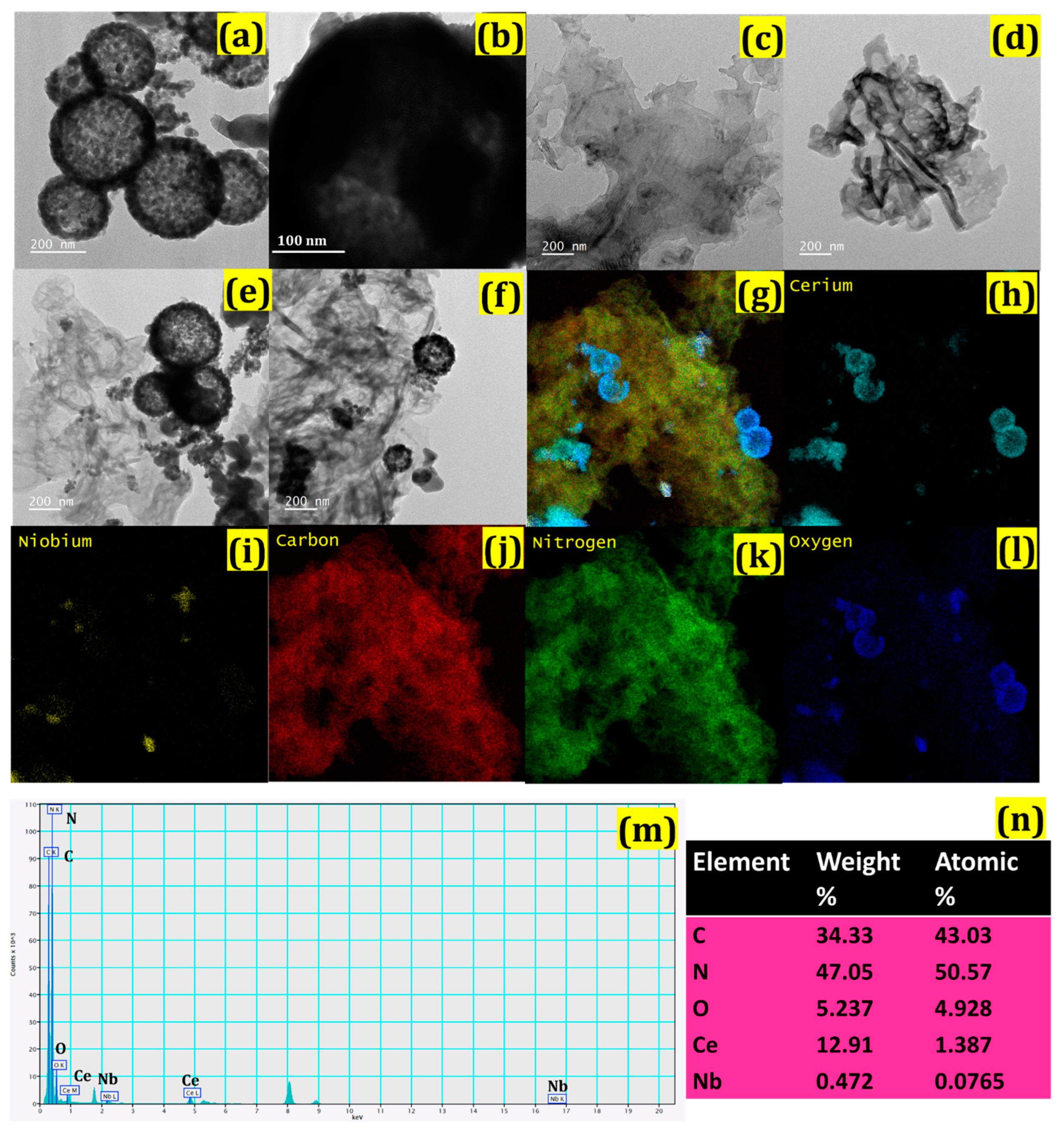
Figure 2a,b shows the structure of the prepared CeO2/Ce3NbO7 hollow spheres. TEM images of g-C3N4 show a nanosheet-like structure (Figure 2c,d), and Figure 2e,f shows the hollow-sphere-shape of the CeO2/Ce3NbO7/g-C3N4 NCs that were modified on the surface of the g-C3N4 nanosheets. Herein, the sphere-shaped structures of CeO2/Ce3NbO7 were produced during the calcination process (800 °C for 2 h). The EDX spectrum revealed the elemental constitution and weight percentage of the NCs. Figure 2g represents the EDX mapping of the CeO2/Ce3NbO7/g-C3N4 NCs and confirms the presence of all elements such as cerium (Ce), niobium (Nb), oxygen (O), carbon (C), and nitrogen (N). Various colors in the signal indicated the presence of these components. Figure 2h presents the EDX images of Ce, while those of Nb (2i), C (2j), N (2k), and O (2l), respectively, are also provided. Numerous bright electron diffraction spots confirm the crystalline structure of CeO2/Ce3NbO7/g-C3N4 NCs. The EDX analysis was performed to elucidate the existence of elements such as Ce, Nb, C, N, and O in the prepared CeO2/Ce3NbO7/g-C3N4 NCs. Figure 2m represents the EDX mapping of the respective NCs. Figure 2n shows the weight percentage and the elemental composition of the NCs, where the elements Ce, Nb, C, N, and O were present at 34.33, 47.05, 5.23, 12.91, and 0.47 wt%, respectively.
The XRD patterns of Ce3NbO7, g-C3N4, and CeO2/Ce3NbO7/g-C3N4 NCs are given in Figure 3a. The XRD pattern of CeO2/Ce3NbO7 hollow spheres shows the characteristic peaks at 28.6, 33.1, 47.5, 56.4, 59.2, and 69.5°, which correspond to the (111), (200), (220), (311), (222), and (400) planes of the cubic lattice of Ce3NbO7 [PDF No.: 00-023-0144], while the presence of tiny, intense peaks at 28, 76.7, and 79.1° correspond to the (111), (331), and (420) planes of CeO2, with a cubic structure [PDF No.: 00-001-0800] [42]. The XRD stick pattern of the composition of Ce3NbO7 [PDF No.: 00-023-0144] [43] confirms the composition of Ce and Nb in the CeO2/Ce3NbO7 hollow spheres. The modification of g-C3N4 nanosheets towards CeO2/Ce3NbO7 hollow spheres shows the presence of a characteristic peak at 27.5°, corresponding to the (001) coordinates confirming the presence of g-C3N4 in the synthesized CeO2/Ce3NbO7/g-C3N4 NCs [41]. Thus, XRD analysis confirmed that the g-C3N4 was successfully modified on CeO2/Ce3NbO7 hollow spheres. The functional groups and structural moieties of the NCs were determined by FT-IR spectroscopy. Figure 3b represents the FT-IR spectrum of the NCs, with peaks at 605, 926, 1633, 2848, and 2919 cm−1. The peaks in the 605 and 926 cm−1 region strongly convey Ce-O and Nb-O stretching vibrational modes. The C=O groups, -C-H- stretching, and hydroxy -OH groups were attributed to the presence of peaks at 1633, 2848, and 2919 cm−1 [40]. The appearance of a peak in the 1600–1900 cm−1 range confirms the presence of aromatic C-N stretching mode, and the sharp characteristic peak at 806 cm−1 was due to the presence of the s-triazine ring system [41]. The UV-visible spectrum of the CeO2/Ce3NbO7, g-C3N4, and CeO2/Ce3NbO7/g-C3N4 NCs upon 808 nm of NIR laser irradiation is given in Figure 3c. It is clearly observed that g-C3N4 exhibits a significant absorption spectrum in the range of 300–500 nm. Moreover, it is confirmed that the up-converted UV and visible emissions from the CeO2/Ce3NbO7 NPs activate the g-C3N4 nanosheets. Therefore, our NCs possess a broad band spectrum from the visible to Near IR region [44].
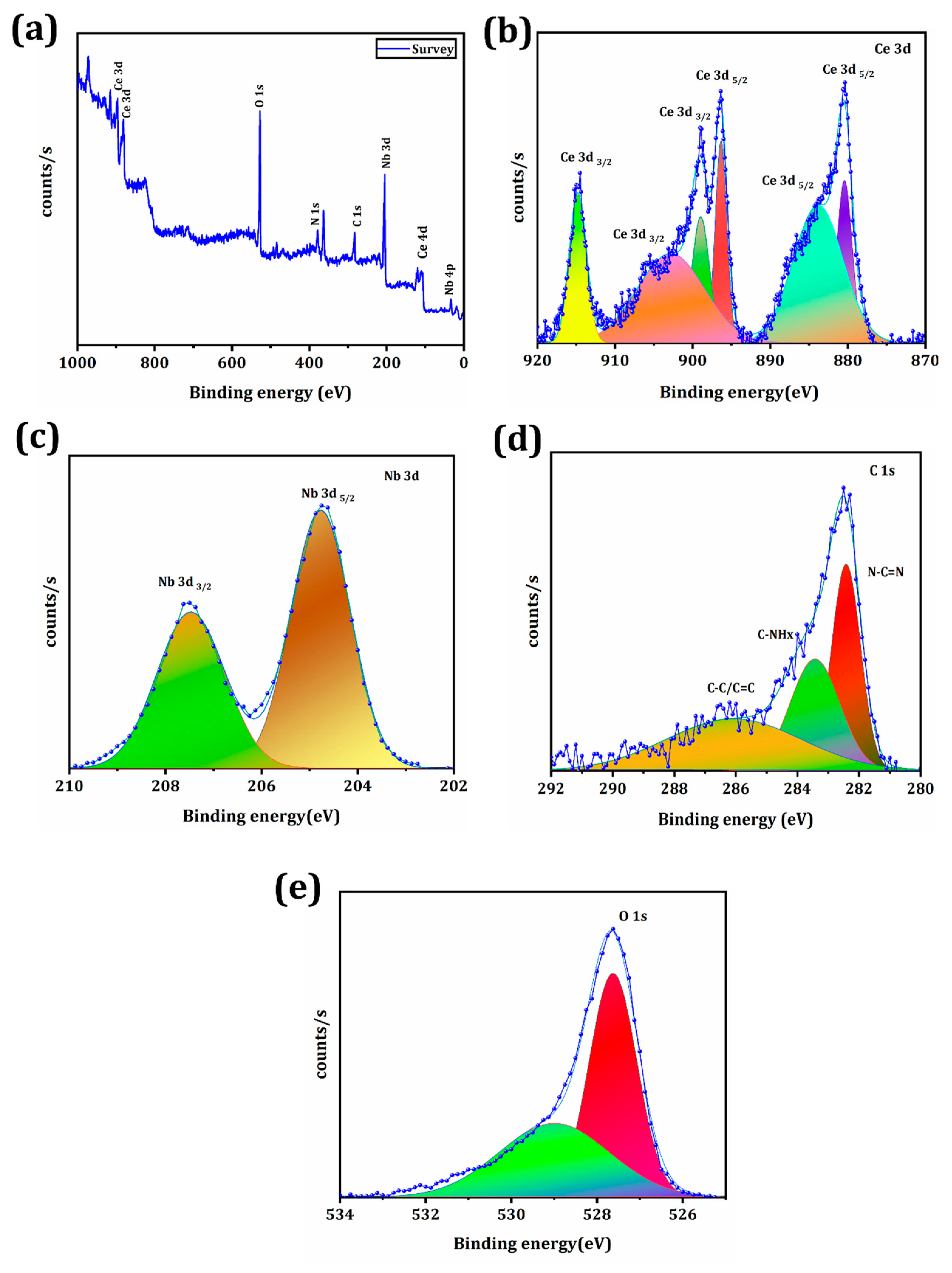
The XPS spectra of the constructed CeO2/Ce3NbO7/g-C3N4 NCs are depicted in Figure 4, validating the presence of elements along with the structural behavior of NCs. Figure 4a shows the overall survey spectrum of CeO2/Ce3NbO7/g-C3N4 NCs, confirming the existence of elements such as Ce, Nb, O, C, and N. This leads to the conformation of CeO2/Ce3NbO7 modification towards the g-C3N4 in the NCs. Here, small, intense peaks at 283.8 eV (C1s) and 378.2 eV (N 1s) (g-C3N4) were obtained. Figure 4b depicts the deconvoluted spectra of Ce 3d 5/2 and Ce 3d 3/2, which show the appearance of six peaks attributed to the presence of Ce4+ and Ce3+ at 883.6 eV (v), 890.1 eV (v′), 899.4 eV (v″), 902 eV (u), 908.7 eV (u′), and 917.8 eV (u″) [45]. The presence of the Nb+5 oxidation state was confirmed by the appearance of the noticeable peaks at 208 eV (Nb 3d 5/2) and 211.1 eV (Nb 3d 3/2) in Figure 4c [46]. The special sp2 (C-C) bond in the CN from g-C3N4 was responsible for three peaks in the region of 284 eV in Figure 4d. The existence of the C atom in the aromatic ring linked to NHx is shown by the peak at 286.5 eV [47]. The N-C=N triazine ring sp2 hybrid carbon was responsible for the appearance of a strong peak at 282 eV. In Figure 4e, the presence of metal oxide (M-O) bindings such as Ce-O, Nb-O, and O with a hydrocarbon or hydroxyl group (O-C or O-H) are depicted by the two convoluted peaks of O 1s at 531.7 eV and 533.4 eV [40]. The above results confirmed the successful modification of Ce3NbO7 over g-C3N4 in NCs.

3.2. Photothermal Performance of CeO2/Ce3NbO7/g-C3N4 NCs

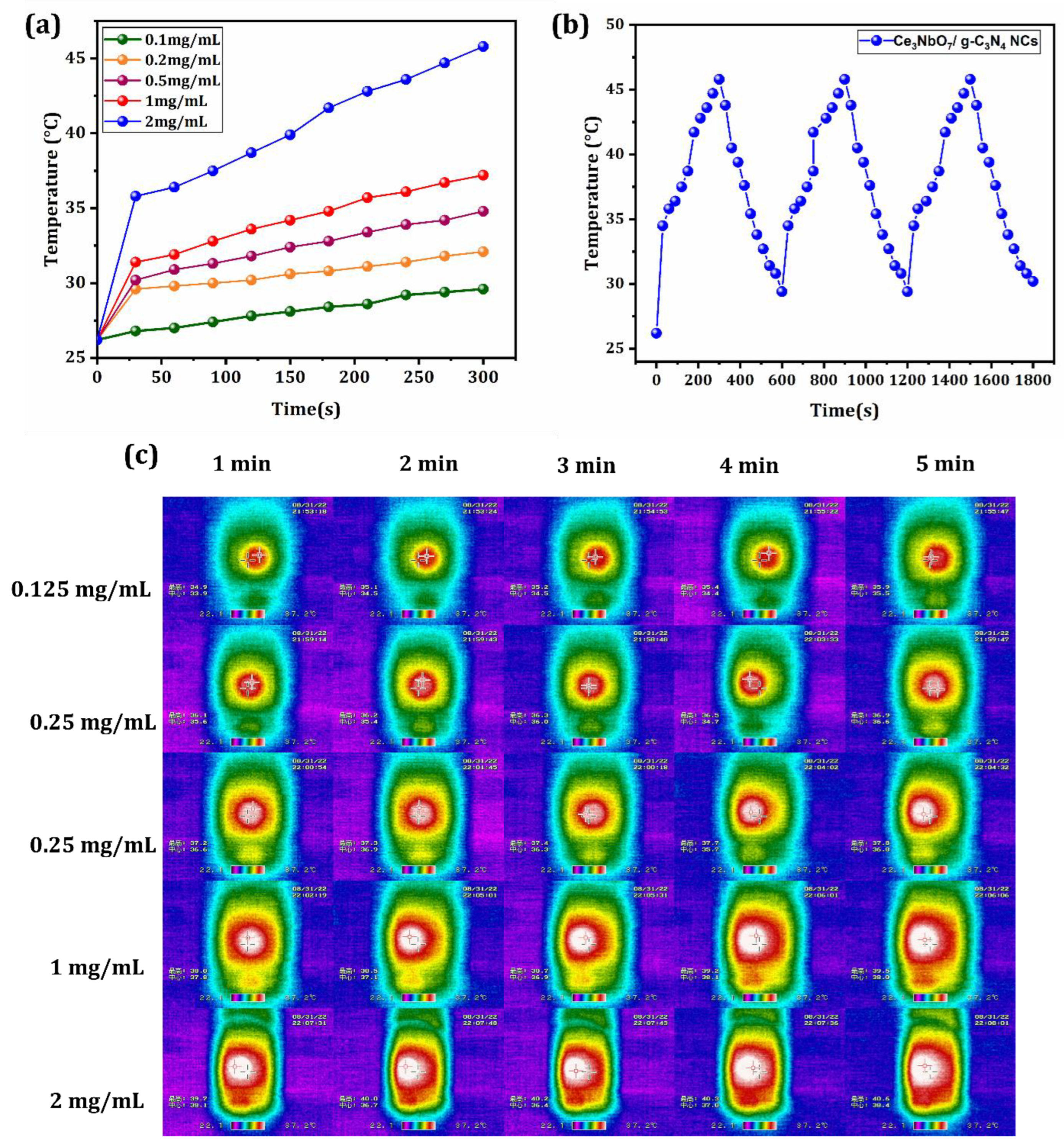
g-C3N4 is a superior photothermal agent due to its rich carbon supply, and it can be easily dispersed in H2O, is photostable, and is environmentally friendly in nature [48]. The CeO2/Ce3NbO7/g-C3N4 NCs were expected to perform effectively in PTT therapy utilizing 808 nm laser irradiation. Figure 5a shows the time-dependent temperature elevation curve of CeO2/Ce3NbO7/g-C3N4 NCs for various concentrations (0, 0.1, 0.2, 0.5, 1, and 2 mg/mL), which confirms that the temperature is a concentration-dependent one. The heating–cooling curve of the NCs proved that they possess good thermal stability, as presented in Figure 5b. Figure 5c shows the IR images of NCs, demonstrating that the temperature elevation of the NCs has increased over time (0 to 5 min). Furthermore, the NCs PCE (ƞ) was calculated as 49.5%. These results confirm that the prepared NCs have good photothermal stability under NIR irradiation.

3.3. Fenton Catalytic Property of the Synthesized CeO2/Ce3NbO7/g-C3N4 NCs

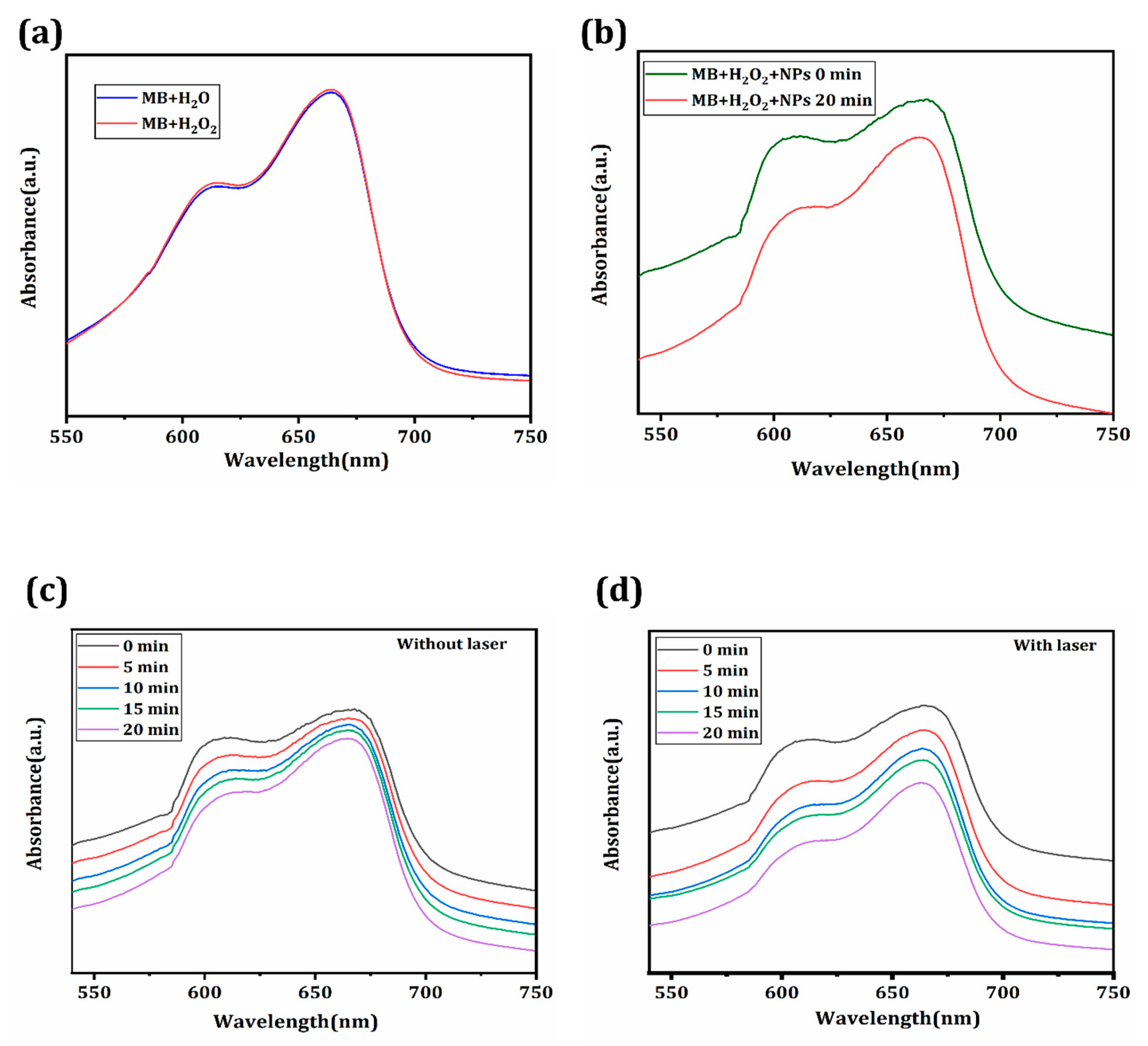
Methylene blue (MB) was employed for the detection and quantification of hydroxyl radical (OH•) to investigate the capacity of CeO2/Ce3NbO7/g-C3N4 NCs to generate OH• under the circumstances of TME for CDT. Ce mediates the reaction of H2O2 to generate OH• via Fenton-type reactions. Figure 6a depicts the UV-visible curve for MB in water (pH = 7.4) and H2O2 (pH = 6.5) solutions, where the absorbance peak was observed at the maximum wavelength of 664.6 nm in room temperature with little changes, indicating MB could not be degraded under various pH conditions. In Figure 6b, the degradation curve of MB with H2O2 is given for varying time intervals from 0 to 20 min at 37 °C. Figure 6c presents the MB degradation curve and the calculated amount of synthesized NCs in the absence of laser source for different time durations of 0, 10, 15, and 20 min. Here, the Ce in the NCs actively participates in the Fenton reaction via a cascade mechanism to yield the highly toxic OH• that rapidly kills cancer cells inside the TME. The degradation efficiency slightly decreased, mainly caused by more Ce4+ occupying the H2O2 active sites. These results demonstrate that CeO2/Ce3NbO7/g-C3N4 NCs have the best catalytic performance, and they can efficiently catalyze the production of sufficient hydroxyl radicals from H2O2. Figure 6d reveals the photothermally enhanced Fenton reaction. The degradation of MB following exposure to an 808 nm NIR laser was studied to evaluate the impact of the photothermal effect on the Fenton reaction of NCs. It is evident that when the NCs were exposed to an 808 nm NIR laser, MB degradation efficiency increased in the presence of the laser compared to the normal Fenton reaction, supporting the theory that the synthesized NCs firmly control the photothermal enhancement of the Fenton reaction. NCs could generate heat by the photothermal reaction to accelerate the Fenton reaction and the subsequent production of OH•. Additionally, combining photothermal and chemodynamic performance, the prepared NCs act as an optimized therapeutic platform in cancer applications.

3.4. In Vitro PTT/CDT Performance of CeO2/Ce3NbO7/g-C3N4 NCs towards Liver Cancer

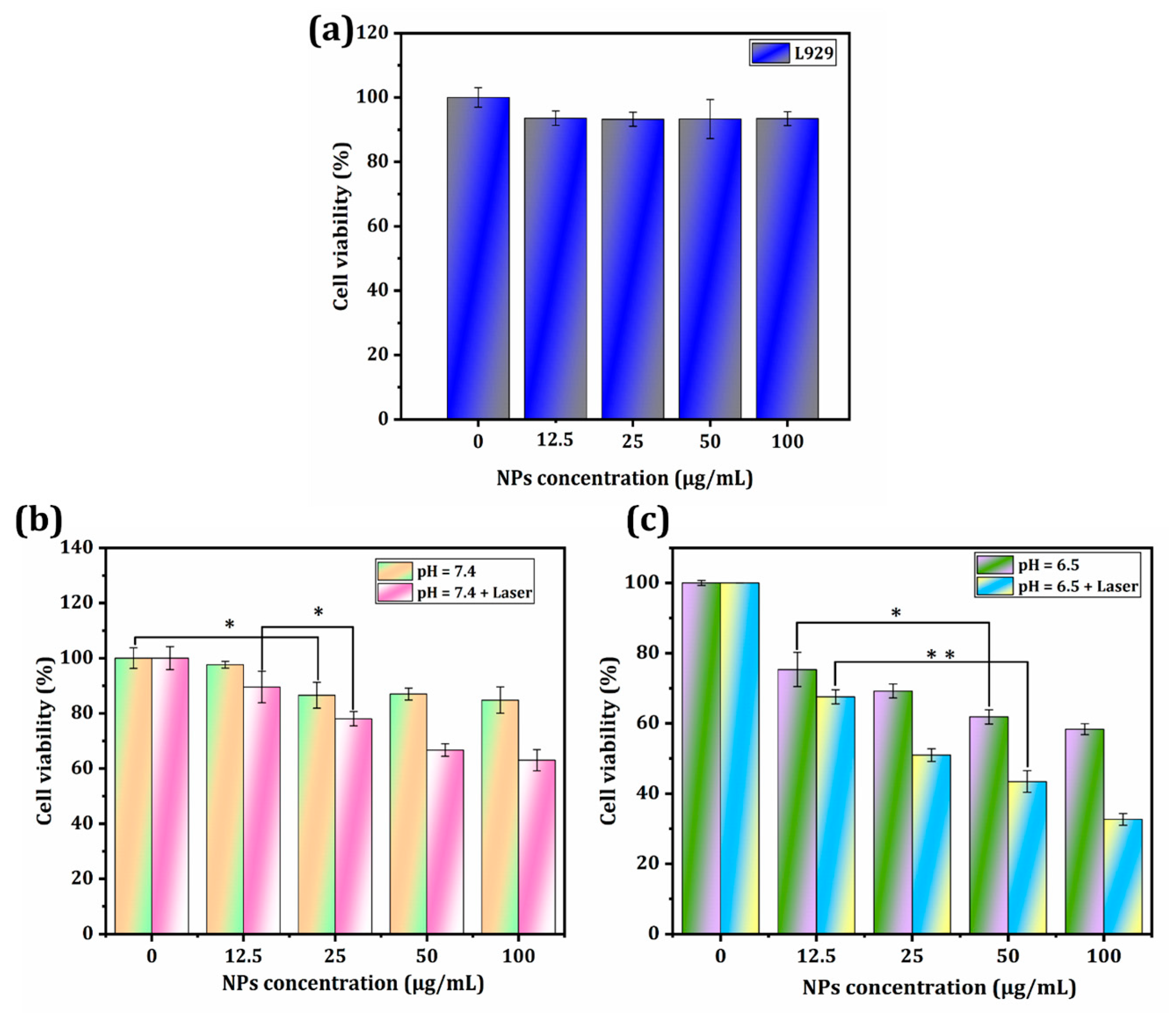
The synergistic therapeutic applications of the NCs were examined using a cell viability assay. Human cancer cells (HepG2) and L929 cells were chosen here to investigate the in vitro cytotoxicity of CeO2/Ce3NbO7/g-C3N4 NCs by utilizing an MTT assay. Designing nanomaterials for various biomedical applications requires careful consideration of biocompatibility. Here, the primary intrinsic cytotoxicity of the CeO2/Ce3NbO7/g-C3N4 NCs of L929 was assessed in Figure 7a, where the cell viability of L929 is high around 93% compared to HepG2 cells. From this, we conclude that NCs are biocompatible. The relative cell viability of HepG2 cells without laser at pH 7.4 was estimated at 84% for the highest concentration of 100 µg/mL of NCs, as shown in Figure 7a. These findings proved that CeO2/Ce3NbO7/g-C3N4 NCs show negligible cytotoxicity compared to other targeting-agent-based biomedical systems, ensuring their eligibility for further therapeutic application in research [49]. The in vitro tumor-cell-killing effectiveness of CeO2/Ce3NbO7/g-C3N4 NCs was assessed after 808 nm laser radiation (1 W/cm2) for 5 min (Figure 7b). After laser radiation, as seen in Figure 7b, CeO2/Ce3NbO7/g-C3N4 NCs significantly increased the concentration-dependent cell death of HepG2 cells. This demonstrated that NCs possess the capacity for benign photothermal cell death. Figure 7c represents the respective cell viability under pH = 6.5 (CDT). The NCs powerfully enhance the production of OH•. Endogenous CDT is made possible without external stimuli owing to the CeO2/Ce3NbO7/g-C3N4 NCs’ potent catalysis of H2O2 and dissociation to create OH•. Because of their metabolism, cancer cells inside the TME typically create H2O2 naturally. Additionally, the effectiveness of CeO2/Ce3NbO7/g-C3N4 NCs against the HepG2 cells in the tumor environment (pH = 6.5) was further evaluated. The HepG2 cell viability marginally decreased without laser irradiation when the CeO2/Ce3NbO7/g-C3N4 NCs concentration increased. In order to kill cancer cells, CeO2/Ce3NbO7/g-C3N4 NCs can catalyze H2O2 dissociation to produce toxic OH• (Figure 7c). The reduction in cell viability was more significant when CDT and PTT (an 808 nm laser) were coupled. Approximately 68% of HepG2 cells were destroyed at a concentration of 100 µg/mL of CeO2/Ce3NbO7/g-C3N4 NCs (Figure 7c). Overall, MTT cell viability results suggested that the PTT could enhance CDT efficacy, leading to a synergistic effect in the ablation of cancer cells by combination therapy.

4. Discussion

We propose a distinctive perspective on conjugated g-C3N4 with CeO2/Ce3NbO7 hollow spheres to form CeO2/Ce3NbO7/g-C3N4 NCs. g-C3N4 exhibits excellent NIR absorption rates, which suggests they have a strong potential for PTT applications. CeO2/Ce3NbO7/g-C3N4 NCs (2.0 mg/mL) increased in temperature after 300 s. Under 808nm of laser irradiation exposure, CeO2/Ce3NbO7/g-C3N4 NCs displayed an excellent photothermal conversion efficiency (ƞ) of around 49.5% and better thermal stability after three consecutive cycles.
Ce3+ in the NCs actively participates in the Fenton mechanism to yield the reactive oxygen species (OH•) that drastically kills the tumor cells inside the TME. Here, the MB degradation plot provides clear evidence for the production of OH• radicals with and without a laser source. From those results, we confirm that the NCs show the best catalytic performance along with laser (808 nm) in cancer theranostics.
Furthermore, concentration-dependent cell death occurs due to the PTT/CDT effect in CeO2/Ce3NbO7/g-C3N4 NCs. Compared to PTT and CDT, the in vitro studies convey that the synergistic PTT/CDT exhibits decreased cell survival of 30% HepG2 cells for (100 mg/mL) NCs. The excellent cell-killing ability is mainly due to the dual therapeutic performance of NCs toward cancer cells. Based on these results, the synthesized NCs hold great potency in both PTT and CDT for ablating cancer cells, and are biocompatible.

5. Conclusions

Herein, we developed a CeO2/Ce3NbO7 hollow sphere modified with graphitic carbon nitride (g-C3N4) to create a synergistic therapeutic application using CDT and PTT for cancer treatment. Moreover, the cerium niobate hollow spheres were prepared by a facile hydrothermal method for cancer theranostics, and the NCs showed outstanding photothermal conversion efficiency (ƞ) of 49.5% due to the presence of g-C3N4. Under laser irradiation, it not only ablates the tumor cells but also enhances the Fenton reaction, boosting the conversion of Ce3+ to Ce2+ to generate ROS and showing excellent photothermal behavior. In vitro, studies demonstrated the excellent ability for cell apoptosis of tumor cells using synergistic therapeutic modes. The current research can assist in developing novel materials and various photothermal agents for multifunctional therapeutic applications in cancer treatment. As a result of our study, novel nanoplatforms can be created for the precise and highly efficient treatment of liver tumors.

Author Contributions

K.S.M.: Writing—original draft, methodology, formal analysis, data curation, visualization, writing—review and editing. S.T.: Writing—review and editing, methodology, formal analysis, data curation, visualization. Y.-C.L.: Writing—review and editing, methodology, formal analysis, data curation, visualization. R.S.: Investigation, formal analysis, data curation. U.D.: Writing—review and editing, methodology, formal analysis, data curation, visualization. A.-N.W.: Resources. M.H.: Resources. R.-J.C.: Conceptualization, methodology, supervision, project administration, funding acquisition, writing—review and editing. All authors have read and agreed to the published version of the manuscript.

Funding

The authors are grateful for the financial support provided by the Ministry of Science and Technology of Taiwan (MOST 108-2628-E-027-003-MY3; MOST 111-2221-E-027-105).

Data Availability Statement

The data presented in this study are available upon request from the corresponding author.

Acknowledgments

Technical assistance from the Precision Analysis and Material Research Center of National Taipei University of Technology (Taipei Tech) is appreciated.

Conflicts of Interest

The authors declare no conflict of interest.

References

  1. Deng, Z.; Fang, C.; Ma, X.; Li, X.; Zeng, Y.-J.; Peng, X. One stone two birds: Zr-Fc metal–organic framework nanosheet for synergistic photothermal and chemodynamic cancer therapy. ACS Appl. Mater. Isnterfaces 2020, 12, 20321–20330. [Google Scholar] [CrossRef]
  2. Bray, F.; Ferlay, J.; Soerjomataram, I.; Siegel, R.L.; Torre, L.A.; Jemal, A. Global cancer statistics 2018: GLOBOCAN estimates of incidence and mortality worldwide for 36 cancers in 185 countries. CA A Cancer J. Clin. 2018, 68, 394–424. [Google Scholar] [CrossRef] [Green Version]
  3. Weiderpass, E. Lifestyle and cancer risk. J. Prev. Med. Public Health 2010, 43, 459–471. [Google Scholar] [CrossRef]
  4. Chen, Y.C.; Hunter, D.J. Molecular epidemiology of cancer. CA A Cancer J. Clin. 2005, 55, 45–54. [Google Scholar] [CrossRef]
  5. Arico, M.; Schrappe, M.; Hunger, S.P.; Carroll, W.L.; Conter, V.; Galimberti, S.; Manabe, A.; Saha, V.; Baruchel, A.; Vettenranta, K. Clinical outcome of children with newly diagnosed Philadelphia chromosome–positive acute lymphoblastic leukemia treated between 1995 and 2005. J. Clin. Oncol. 2010, 28, 4755. [Google Scholar] [CrossRef] [PubMed]
  6. Monsalve, J.; Kapur, J.; Malkin, D.; Babyn, P.S. Imaging of cancer predisposition syndromes in children. Radiographics 2011, 31, 263–280. [Google Scholar] [CrossRef] [PubMed] [Green Version]
  7. Center, M.M.; Jemal, A. International Trends in Liver Cancer Incidence RatesInternational Liver Cancer Incidence Trends. Cancer Epidemiol. Biomark. Prev. 2011, 20, 2362–2368. [Google Scholar] [CrossRef] [Green Version]
  8. Siegel, R.L.; Miller, K.D.; Jemal, A. Cancer statistics, 2019. CA A Cancer J. Clin. 2019, 69, 7–34. [Google Scholar] [CrossRef] [PubMed] [Green Version]
  9. Anwanwan, D.; Singh, S.K.; Singh, S.; Saikam, V.; Singh, R. Challenges in liver cancer and possible treatment approaches. Biochim. Et Biophys. Acta (BBA)-Rev. Cancer 2020, 1873, 188314. [Google Scholar] [CrossRef]
  10. Pérez-López, A.; Martín-Sabroso, C.; Gómez-Lázaro, L.; Torres-Suárez, A.I.; Aparicio-Blanco, J. Embolization therapy with microspheres for the treatment of liver cancer: State-of-the-art of clinical translation. Acta Biomater. 2022, 149, 1–15. [Google Scholar] [CrossRef] [PubMed]
  11. World Health Organization. International Agency for Research on Cancer Cancer Today; World Health Organization: Geneva, Switzerland, 2016. [Google Scholar]
  12. Sung, H.; Ferlay, J.; Siegel, R.L.; Laversanne, M.; Soerjomataram, I.; Jemal, A.; Bray, F. Global cancer statistics 2020: GLOBOCAN estimates of incidence and mortality worldwide for 36 cancers in 185 countries. CA A Cancer J. Clin. 2021, 71, 209–249. [Google Scholar] [CrossRef]
  13. Starley, B.Q.; Calcagno, C.J.; Harrison, S.A. Nonalcoholic fatty liver disease and hepatocellular carcinoma: A weighty connection. Hepatology 2010, 51, 1820–1832. [Google Scholar] [CrossRef]
  14. Feng, M.; Pan, Y.; Kong, R.; Shu, S. Therapy of primary liver cancer. Innovation 2020, 1, 100032. [Google Scholar] [CrossRef] [PubMed]
  15. Wang, Z.; Yu, W.; Yu, N.; Li, X.; Feng, Y.; Geng, P.; Wen, M.; Li, M.; Zhang, H.; Chen, Z. Construction of CuS@ Fe-MOF nanoplatforms for MRI-guided synergistic photothermal-chemo therapy of tumors. Chem. Eng. J. 2020, 400, 125877. [Google Scholar] [CrossRef]
  16. Fan, W.; Yung, B.; Huang, P.; Chen, X. Nanotechnology for multimodal synergistic cancer therapy. Chem. Rev. 2017, 117, 13566–13638. [Google Scholar] [CrossRef]
  17. Liu, Y.; Bhattarai, P.; Dai, Z.; Chen, X. Photothermal therapy and photoacoustic imaging via nanotheranostics in fighting cancer. Chem. Soc. Rev. 2019, 48, 2053–2108. [Google Scholar] [CrossRef]
  18. Zhu, P.; Chen, Y.; Shi, J. Nanoenzyme-augmented cancer sonodynamic therapy by catalytic tumor oxygenation. ACS Nano 2018, 12, 3780–3795. [Google Scholar] [CrossRef]
  19. Cheng, L.; Wang, C.; Feng, L.; Yang, K.; Liu, Z. Functional nanomaterials for phototherapies of cancer. Chem. Rev. 2014, 114, 10869–10939. [Google Scholar] [CrossRef] [PubMed]
  20. Tang, Z.; Liu, Y.; He, M.; Bu, W. Chemodynamic therapy: Tumour microenvironment-mediated Fenton and Fenton-like reactions. Angew. Chem. 2019, 131, 958–968. [Google Scholar] [CrossRef]
  21. Jiang, F.; Ding, B.; Liang, S.; Zhao, Y.; Cheng, Z.; Xing, B.; Lin, J. Intelligent MoS2–CuO heterostructures with multiplexed imaging and remarkably enhanced antitumor efficacy via synergetic photothermal therapy/chemodynamic therapy/immunotherapy. Biomaterials 2021, 268, 120545. [Google Scholar] [CrossRef]
  22. Ng, C.W.; Li, J.; Pu, K. Recent progresses in phototherapy-synergized cancer immunotherapy. Adv. Funct. Mater. 2018, 28, 1804688. [Google Scholar] [CrossRef]
  23. Wang, C.; Xu, L.; Liang, C.; Xiang, J.; Peng, R.; Liu, Z. Immunological responses triggered by photothermal therapy with carbon nanotubes in combination with anti-CTLA-4 therapy to inhibit cancer metastasis. Adv. Mater. 2014, 26, 8154–8162. [Google Scholar] [CrossRef]
  24. Wu, C.; Wang, S.; Zhao, J.; Liu, Y.; Zheng, Y.; Luo, Y.; Ye, C.; Huang, M.; Chen, H. Biodegradable Fe (III)@ WS2-PVP nanocapsules for redox reaction and TME-enhanced nanocatalytic, photothermal, and chemotherapy. Adv. Funct. Mater. 2019, 29, 1901722. [Google Scholar] [CrossRef]
  25. Liu, Y.; Zhen, W.; Jin, L.; Zhang, S.; Sun, G.; Zhang, T.; Xu, X.; Song, S.; Wang, Y.; Liu, J. All-in-one theranostic nanoagent with enhanced reactive oxygen species generation and modulating tumor microenvironment ability for effective tumor eradication. ACS Nano 2018, 12, 4886–4893. [Google Scholar] [CrossRef]
  26. Wan, S.-S.; Cheng, Q.; Zeng, X.; Zhang, X.-Z. A Mn (III)-sealed metal–organic framework nanosystem for redox-unlocked tumor theranostics. ACS Nano 2019, 13, 6561–6571. [Google Scholar] [CrossRef]
  27. Ding, B.; Shao, S.; Jiang, F.; Dang, P.; Sun, C.; Huang, S.; Ma, P.a.; Jin, D.; Kheraif, A.A.A.; Lin, J. MnO2-disguised upconversion hybrid nanocomposite: An ideal architecture for tumor microenvironment-triggered UCL/MR bioimaging and enhanced chemodynamic therapy. Chem. Mater. 2019, 31, 2651–2660. [Google Scholar] [CrossRef]
  28. Gao, S.; Jin, Y.; Ge, K.; Li, Z.; Liu, H.; Dai, X.; Zhang, Y.; Chen, S.; Liang, X.; Zhang, J. Self-supply of O2 and H2O2 by a Nanocatalytic medicine to enhance combined chemo/Chemodynamic therapy. Adv. Sci. 2019, 6, 1902137. [Google Scholar] [CrossRef] [Green Version]
  29. Liu, Y.; Jia, Q.; Guo, Q.; Wei, W.; Zhou, J. Simultaneously activating highly selective ratiometric MRI and synergistic therapy in response to intratumoral oxidability and acidity. Biomaterials 2018, 180, 104–116. [Google Scholar] [CrossRef]
  30. Duan, L.-Y.; Wang, Y.-J.; Liu, J.-W.; Wang, Y.-M.; Li, N.; Jiang, J.-H. Tumor-selective catalytic nanosystem for activatable theranostics. Chem. Commun. 2018, 54, 8214–8217. [Google Scholar] [CrossRef] [PubMed]
  31. Park, J.; Lim, D.-H.; Lim, H.-J.; Kwon, T.; Choi, J.-s.; Jeong, S.; Choi, I.-H.; Cheon, J. Size dependent macrophage responses and toxicological effects of Ag nanoparticles. Chem. Commun. 2011, 47, 4382–4384. [Google Scholar] [CrossRef]
  32. Hu, R.; Fang, Y.; Huo, M.; Yao, H.; Wang, C.; Chen, Y.; Wu, R. Ultrasmall Cu2-xS nanodots as photothermal-enhanced Fenton nanocatalysts for synergistic tumor therapy at NIR-II biowindow. Biomaterials 2019, 206, 101–114. [Google Scholar] [CrossRef]
  33. Wang, Z.; Liu, B.; Sun, Q.; Dong, S.; Kuang, Y.; Dong, Y.; He, F.; Gai, S.; Yang, P. Fusiform-like copper (II)-based metal–organic framework through relief hypoxia and GSH-depletion co-enhanced starvation and chemodynamic synergetic cancer therapy. ACS Appl. Mater. Interfaces 2020, 12, 17254–17267. [Google Scholar] [CrossRef] [PubMed]
  34. Feng, N.; Liu, Y.; Dai, X.; Wang, Y.; Guo, Q.; Li, Q. Advanced applications of cerium oxide based nanozymes in cancer. RSC Adv. 2022, 12, 1486–1493. [Google Scholar] [CrossRef]
  35. Asati, A.; Santra, S.; Kaittanis, C.; Nath, S.; Perez, J.M. Oxidase-like activity of polymer-coated cerium oxide nanoparticles. Angew. Chem. 2009, 121, 2344–2348. [Google Scholar] [CrossRef]
  36. Gao, Y.; Chen, K.; Ma, J.-l.; Gao, F. Cerium oxide nanoparticles in cancer. OncoTargets Ther. 2014, 7, 835–840. [Google Scholar] [CrossRef] [Green Version]
  37. Song, X.; Chen, Q.; Liu, Z. Recent advances in the development of organic photothermal nano-agents. Nano Res. 2015, 8, 340–354. [Google Scholar] [CrossRef]
  38. Liao, G.; He, F.; Li, Q.; Zhong, L.; Zhao, R.; Che, H.; Gao, H.; Fang, B. Emerging graphitic carbon nitride-based materials for biomedical applications. Prog. Mater. Sci. 2020, 112, 100666. [Google Scholar] [CrossRef]
  39. Bosio, G.N.; Mártire, D.O. Carbon nitride nanomaterials with application in photothermal and photodynamic therapies. Photodiagnosis Photodyn. Ther. 2022, 37, 102683. [Google Scholar] [CrossRef]
  40. Liu, X.; Tseng, C.-L.; Lin, L.-Y.; Lee, C.-A.; Li, J.; Feng, L.; Song, L.; Li, X.; He, J.-H.; Sakthivel, R. Template-free synthesis of mesoporous Ce3NbO7/CeO2 hollow nanospheres for label-free electrochemical immunosensing of leptin. Sens. Actuators B Chem. 2021, 341, 130005. [Google Scholar] [CrossRef]
  41. Sanjay, B.P.; Swamy, N.K.; Yashas, S.R.; Sandeep, S. Design of an electrochemical sensor using 2D sheet-like Cu@ g-C3N4 transducer matrix for electroanalysis of catechol. J. Electrochem. Soc. 2021, 168, 076511. [Google Scholar] [CrossRef]
  42. Guoguang, Z.; Qin, L.; Qianfeng, F. Structure and Dielectric Study of Ce3 NbO7+ δ. J. Rare Earths 2007, 25, 730–733. [Google Scholar] [CrossRef]
  43. Farahmandjou, M.; Zarinkamar, M.; Firoozabadi, T. Synthesis of Cerium Oxide (CeO2) nanoparticles using simple CO-precipitation method. Rev. Mex. De Física 2016, 62, 496–499. [Google Scholar]
  44. Feng, L.; He, F.; Yang, G.; Gai, S.; Dai, Y.; Li, C.; Yang, P. NIR-driven graphitic-phase carbon nitride nanosheets for efficient bioimaging and photodynamic therapy. J. Mater. Chem. B 2016, 4, 8000–8008. [Google Scholar] [CrossRef]
  45. Sakthivel, R.; Annalakshmi, M.; Chen, S.-M.; Kubendhiran, S.; Anbazhagan, R.; Tsai, H.-C. A novel sensitive and reliable electrochemical determination of palmatine based on CeO2/RGO/MWCNT ternary composite. J. Taiwan Inst. Chem. Eng. 2019, 96, 549–558. [Google Scholar] [CrossRef]
  46. Liu, F.; Wang, Y.; Kong, X.; Lei, D.; Zhang, F.; Lei, X. A hierarchical Nb2O5@ NiFe-MMO rod array, fabricated and used as a structured photocatalyst. RSC Adv. 2019, 9, 6177–6183. [Google Scholar] [CrossRef] [Green Version]
  47. Baranowska, D.; Kędzierski, T.; Aleksandrzak, M.; Mijowska, E.; Zielińska, B. Influence of hydrogenation on morphology, chemical structure and photocatalytic efficiency of graphitic carbon nitride. Int. J. Mol. Sci. 2021, 22, 13096. [Google Scholar] [CrossRef] [PubMed]
  48. Yan, S.; Li, Z.; Zou, Z. Photodegradation performance of g-C3N4 fabricated by directly heating melamine. Langmuir 2009, 25, 10397–10401. [Google Scholar] [CrossRef]
  49. Zhang, L.; Jiang, H.; Selke, M.; Wang, X. Selective cytotoxicity effect of cerium oxide nanoparticles under UV irradiation. J. Biomed. Nanotechnol. 2014, 10, 278–286. [Google Scholar] [CrossRef] [PubMed]
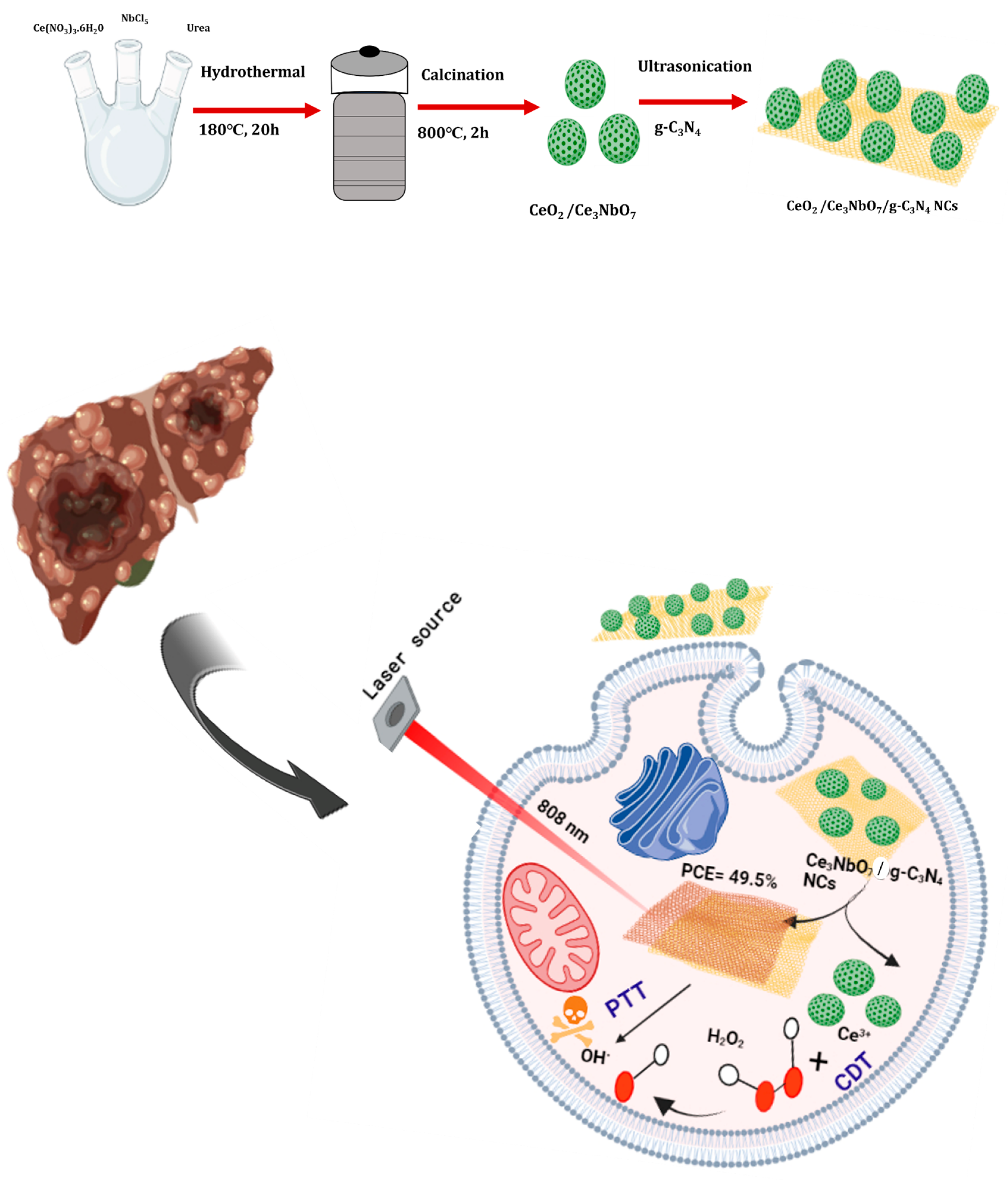
Figure 1. Schematic representation of the synthesis and therapeutic application of the CeO2/Ce3NbO7/g-C3N4 NCs. The modifications of g-C3N4 to the Ce3NbO7 hollow spheres synergistically accomplish PTT/CDT to treat LC.
Crystals 13 00954 g001
Figure 2. Characterization of CeO2/Ce3NbO7/g-C3N4 NCs, (a,b) show TEM images of CeO2/Ce3NbO7 hollow spheres; (c,d) g-C3N4; (e,f) CeO2/Ce3NbO7/g-C3N4 NCs; (g) elemental mapping images of CeO2/Ce3NbO7/g-C3N4 NCs; (h) cerium (Ce); (i) niobium (Nb); (j) carbon (C); (k) nitrogen (N); (l) oxygen (O); (m) EDX mapping of CeO2/Ce3NbO7/g-C3N4 NCs; (n) elemental composition and weight percentage.
Crystals 13 00954 g002
Figure 3. XRD patterns of CeO2/Ce3NbO7, g-C3N4, and CeO2/Ce3NbO7/g-C3N4 NCs (a); FT-IR spectra of CeO2/Ce3NbO7, g-C3N4, and CeO2/Ce3NbO7/g-C3N4 NCs (b), and (c) UV-visible spectrum of CeO2/Ce3NbO7, g-C3N4, and CeO2/Ce3NbO7/g-C3N4 NCs.
Crystals 13 00954 g003
Figure 4. XPS spectra of the CeO2/Ce3NbO7/g-C3N4 NCs. (a) The XPS survival spectrum. (be) The core level spectrum of Ce 3d, Nb 3d, C 1s, and O 1 s, respectively.
Crystals 13 00954 g004
Figure 5. In vitro photothermal responses of the CeO2/Ce3NbO7/g-C3N4 NCs (those Chinese characters in the figure represents the respective temperature range): (a) temperature changes of the CeO2/Ce3NbO7/g-C3N4 NCs aqueous solutions at different concentrations in 1 W/cm2. (b) Thermal stability of the synthesized NCs under irradiation with a 1 W/cm2 NIR laser source. (c) Infrared thermal images of the NCs at a power density of 1 W/cm2.
Crystals 13 00954 g005
Figure 6. UV-Visible spectra of MB + H2O and MB + H2O2 (a). Degradation plot of MB+ CeO2/Ce3NbO7/g-C3N4 NCs at different time intervals (0 and 20 min) (b). The MB degradation of the NCs without laser for different time durations (c). The PTT/CDT of MB degradation efficiency of NCs in the presence of laser (d).
Crystals 13 00954 g006
Figure 7. In vitro cytotoxicity analysis of L929 cells (a); synergistic therapeutic application of CeO2/Ce3NbO7/g-C3N4 NCs towards HepG2 cells with the incorporation of 808 nm of NIR laser at pH = 7.4 (b); cytotoxicity analysis of the CeO2/Ce3NbO7/g-C3N4 NCs + laser at pH = 6.5 (c). Statistical analysis (*, **) was performed by student’s t-test method.
Crystals 13 00954 g007
Disclaimer/Publisher’s Note: The statements, opinions and data contained in all publications are solely those of the individual author(s) and contributor(s) and not of MDPI and/or the editor(s). MDPI and/or the editor(s) disclaim responsibility for any injury to people or property resulting from any ideas, methods, instructions or products referred to in the content.