Article

Novel Salts of Heterocyclic Polyamines and 5-Sulfosalicylic Acid: Synthesis, Crystal Structure, and Hierarchical Supramolecular Interactions

by
Joanna Bojarska
1,
Krzysztof Łyczko
2 and
Adam Mieczkowski
3,*
1
Institute of Faculty of Chemistry, Institute of General & Inorganic Chemistry, Technical University of Lodz, Zeromskiego 116, 90-924 Lodz, Poland
2
Institute of Nuclear Chemistry and Technology, Dorodna 16, 03-195 Warsaw, Poland
3
Institute of Biochemistry and Biophysics, Polish Academy of Sciences, Pawinskiego 5a, 02-106 Warsaw, Poland
*
Author to whom correspondence should be addressed.
Crystals 2024, 14(6), 497; https://doi.org/10.3390/cryst14060497
Submission received: 26 April 2024 / Revised: 10 May 2024 / Accepted: 20 May 2024 / Published: 24 May 2024

Abstract

:
A series of novel salts of heterocyclic polyamines with 5-sulfosalicylic acid (C4H7N4+)(C7H5O6S)∙2(H2O) (1), (C4H6ClN4+)(C7H5O6S)∙H2O (2), (C5H8N3+)(C7H5O6S)∙H2O (3), (C5H7N6+)(C7H5O6S)∙H2O (4), (C6H14N22+)(C7H4O6S2−)∙H2O (5), and (C14H19N2+)(C7H5O6S) (6) have been successfully synthesized. Their crystal structures have been determined by single-crystal X-ray diffraction. Overall, compounds adopt a layered structure with aminium cations and 5-sulfosalicylic anions linked via water molecules. The solid-state architectures of these compounds are dominated by O(N,H)-H⋯O and N-H⋯N hydrogen bonds and stabilized by weak interconnects. C-Cl⋯π and S-O⋯π interactions, apart from π⋯π and C-H(O)⋯π, were reported. Diverse approaches were used to study the effect of substituents in the polyamines in solid-state arrangement. A Hirshfeld surface analysis, with associated 3D Hirshfeld surface maps and 2D fingerprint plots, molecular electrostatic potential, and energy frameworks were used to comprehensively investigate the nature and hierarchy of non-covalent interactions and inspect supramolecular differences. The contact enrichment ratio calculations provided deeper insight into the propensity of interconnects to influence crystal packing. The evaluation of the effects of H-bonding synthons resulting from different substituents in the polyamines on self-assemblies is also presented. In the context of crystal engineering, a specific intramolecular synthon via O-H⋯O observed in nearly all crystals can be employed in the pseudo-cyclic replacement strategy in the design of new molecules.

1. Introduction

Organic salts based on 2-hydroxy-5-sulfobenzoic (5-sulfosalicylic) acid are eagerly studied due to their interesting optical and pharmaceutical properties [1,2,3,4,5,6]. Many salts, including one negative 3-carboxy-4-hydroxybenzenesulfonate anion, C7H5O6S (with a negative charge at the sulfonyl group), have been presented. These include compounds mostly with organic [7,8,9,10] but also inorganic cations [11,12]. Still, there are only a few of its salts with cations originating from pyrimidine. One salt with 2-aminopyrimidin-1-ium ion [13] and two with 2,4-diaminopyrimidin-1-ium ion derivatives can be distinguished. Among these 2,4-diaminopyrimidin-1-ium ions, the first has the trimethoxybenzyl group in the 5-position of the pyrimidine ring [14], and the second o-chlorophenyl and ethyl groups in the 5- and 6-positions, respectively [15]. The 2,4-diaminopyrimidin-1-ium cation has been reported in several ion pairs with simple inorganic [16] and some organic anions [17,18,19,20,21]. The crystal structures of a series of 2,4-diaminopyrimidin-1-ium salts with chain dicarboxylate ions have been discussed by us recently [22].
Of the presented organic connections, only the crystal structure of salt 3, derived from 5-sulfosalicylic acid and 2,6-diaminopyridine, has been measured at room temperature earlier [23]. In addition, the crystal structure of the C7H5O6S and 2,3-diaminopyridinium ion pair has also been reported [24]. Moreover, a few other structures with 2-aminopyridinium ion [13] and its derivatives substituted by chloro [25,26], bromo [27] or methyl [28] group can be found in the CSD. For 2,6-diamino-9H-purin-1-ium cation, only its salts with 2-(2-carboxylatophenyl)acetate and closo-dodecafluorododecaborate (B12F122−) anions have been described [29,30]. In turn, for 3-carboxy-4-hydroxybenzenesulfonate anion, its salts with 9H-purin-6-aminium, 6-aminopurin-1-ium and 6-(benzylamino)-9H-purin-1-ium cations have been reported [31,32]. The ion pairs of 8-(dimethylamino)-N,N-dimethylnaphthalen-1-aminium (dmanH+) and the salicylic acid derivatives have also been discussed [33,34].
Recently, salts of the dipositive 1,4-diazabicyclo[2.2.2]octane-1,4-diium cation (dabcoH22+) have been widely presented as attractive materials for use in opto-electronics [35,36,37,38,39,40]. In addition, crystal structures containing 3-carboxylato-4-hydroxybenzenesulfonate dianion, C7H4O6S2− (with a negative charge at both sulfonyl and carboxyl groups), and singly or doubly charged organic cations have been structurally characterized [1,41,42,43,44]. Among them, the salts of C7H4O6S2− with 2-aminopyridinium [45], 2,6-diaminopyridinium [46] 2-amino-4,6-dimethylpyrimidinium [47], and 6-(benzylamino)-3H-purin-7-ium ions [48] can also be distinguished.
N-rich heterocyclic polyamines, together with popular multifunctional 5-sulfosalicylic acid, have received a lot of interest in both academic and industrial studies and applications as building blocks of appealing multifunctional supramolecular architectures. Supramolecular chemistry, as a key branch of crystal engineering, has wide implications in the rational design of novel compounds, especially in the context of control of the self-organization and, in further consequence, predictable and desirable structural complex assemblies (such as salts or co-crystals). H-bonding, as well as other non-covalent interactions, have emerged to be the most important tool in crystal engineering [49,50,51]. Organic crystals composed of specific functional groups with diverse applications attracted much attention in the context of the predictable assemblies of supramolecular architectures. The utilization of self-assembly of small compounds with either strong O(N)-H⋯O or weak C-H⋯O hydrogen bonds, halogen bonds, and π-based interactions to the formation and stabilization of 3D supramolecular networks is a vital aspect. The rational design of well-defined structures from complex multifunctional moieties is possible via supramolecular synthons—structural units within crystals composed of hydrogen bonds and/or other non-covalent interactions [52]. The synthons can be classified as homo- or heterosynthons, which are formed from self-complementary half units and different components/functionalities, respectively [53].
Considering all these points in the course of our supramolecular studies on the hierarchy of non-covalent interactions [22,54,55,56,57] here, we report the synthesis and thorough supramolecular investigations of the series of new salts, namely, 2,4-diaminopyrimidin-1-ium 3-carboxy-4-hydroxybenzenesulfonate dihydrate (1), 2,4-diamino-6-chloropyrimidin-1-ium 3-carboxy-4-hydroxybenzenesulfonate monohydrate (2), 2,6-diaminopyridin-1-ium 3-carboxy-4-hydroxybenzenesulfonate monohydrate (3), 2,6-diamino-9H-purin-1-ium 3-carboxy-4-hydroxybenzenesulfonate monohydrate (4), 1,4-diazabicyclo[2.2.2]octane-1,4-diium 2-hydroxy-5-sulfonatobenzoate monohydrate (5), and 8-(dimethylamino)-N,N-dimethylnaphthalen-1-aminium 3-carboxy-4-hydroxybenzenesulfonate (6) (Figure 1). It should be mentioned that these structures have not previously been structurally authenticated. They are presented for the first time to the best of our knowledge; only one salt (3) is a better re-determination, at low temperatures with a higher precision, of previously published structure (CSD refcode: KAXAE) [23]. These compounds can construct appealing supermolecules and diverse either strong or weak synthons because of the presence of a large amount of oxygen and nitrogen atoms, excellent acceptors and donors, and different functional groups such as the sulfonic, carboxylic, hydroxyl, amino, and methyl groups. In addition, water solvent molecules offer an enriched portfolio of H-bonding patterns. This study was undertaken to understand the nature and relevance of the hierarchy of non-covalent interactions either between the cations or between the anions, as well as the cation–anion interplay, focusing on the effect of the H-bonding synthons on novel self-assemblies. The analysis of interaction preferences was supported by Full Interaction Maps. The supramolecular topology of H-bonding networks was examined by an extended Hirshfeld surface analysis, including the enrichment ratios, the molecular electrostatic potentials, and the energy frameworks.

2. Materials and Methods

2.1. Synthesis of Compounds 16

The commercially available chemicals were of reagent grade and used as received. The ATR-FTIR spectra were recorded on a Thermo Scientific Nicolet iS10 FTIR spectrometer using the ATR technique with ZnSe crystal. The following procedure was applied to obtain the requested crystals of heterocyclic polyamines and 5-sulfonylsalicylic acid.
The 5-sulfosalicylic acid (0.1 mmol, 1 equiv., 22 mg) and appropriate organic diamine (0.1 mmol, 1 equiv., 11 mg of 2,4-diaminopyrimidine, 15 mg of 2,4-diamino-6-chloropyrimidine, 11 mg of 2,6-diaminopyridine, 15 mg of 2,6-diaminopurine, 12 mg of 1,4-diazabicyclo[2.2.2]octane, and 22 mg of 1,8-bis(dimethylamino)naphthalene) were dissolved in 2 mL of warm distilled water, the solution was filtered through a small cotton pad and left in the room temperature for solvent evaporation (about two weeks). The obtained crystals of the salts were used for the X-ray measurements and recording the ATR-FTIR spectra.
Compound 1: ATR-FTIR (ZnSe), νmax/cm−1: 3434 s, 3350 s (N–H), 3245 m (N–H), 3089 w (C–H), 3039 w (C–H), 2952 w, 2837 w, 1658 vs (C=O), 1636 m sh (C–O), 1606 sh (C–O), 1519 m, 1475 w, 1451 w, 1395 w, 1334 w (SO3), 1295 w, 1236 sh, 1211 m sh (SO3), 1169 s (SO3), 1156 sh (SO3), 1119 m, 1078 w, 1034 m, and 792 w.
Compound 2: ATR-FTIR (ZnSe), νmax/cm−1: 3507 m, 3455 w (N–H), 3304 m br (N–H), 3117 s, 2772 w br, 1665 vs (C=O), 1652 sh (C=O), 1605 m (C–O), 1551 m (C–O), 1530 w, 1479 w, 1371 w (SO3), 1305 w, 1217 m (SO3), 1169 s (SO3), 1151 s (SO3), 1126 s, 1079 m, 1032 s, 987 m, 796 w, 711 w, and 664 w.
Compound 3: ATR-FTIR (ZnSe), νmax/cm−1: 3471 m, 3424 m, 3372 m (N–H), 3343 (N–H), 3271 m, 3221 m, 3124 w, 3096 w (C–H), 3075 w (C–H), 2974 w br, 2863 m br, 2790 m br, 2703 w br, 1652 vs (C=O), 1631 s (C=O), 1582 m (C–O), 1474 w, 1432 w, 1406 w, 1303 w (SO3), 1225 m (SO3), 1208 m (SO3), 1169 m sh (SO3), 1158 m (SO3), 1144 m, 1116 s, 1017 s, 922 w, 830 w, 775 w, 714 w, and 662 w.
Compound 4: ATR-FTIR (ZnSe), νmax/cm−1: 3397 vs br, 3360 vs br (N–H), 3209 vs br (N–H), 3048 m (C–H), 2851 w br, 1655 s sh (C=O), 1619 s (C=O), 1588 s sh (C–O), 1478 m, 1437 m, 1406 w, 1377 w (SO3), 1347 w sh, 1156 m (SO3), 1125 m (SO3), 1086 w, 1029 w, and 668 w.
Compound 5: ATR-FTIR (ZnSe), νmax/cm−1: 3394 w br (N–H), 3045 w (C–H), 2566 w vbr, 1644 w (C=O), 1595 m (C–O), 1474 m, 1377 w (SO3), 1291 w, 1270 w, 1291 s (SO3), 1180 s (SO3), 1159 m (SO3), 1145 s (SO3), 1122 s, 1084 m, 1076 m, 1057 w, 1034 s, 1021 vs, 888 w br, 832 m, 777 w br, 728 w, and 668 s.
Compound 6: ATR-FTIR (ZnSe), νmax/cm−1: 3050 w (C–H), 3004 w (C–H), 2973 w, 1662 m (C=O), 1603 m (C–O), 1585 w, 1474 m, 1465 m, 1288 w, 1250 s (SO3), 1216 s (SO3), 1188 w, 1157 m (SO3), 1140 s (SO3), 1119 m, 1080 m, 1016 vs, 885 w, 841 m, 831 m, 784 w, 767 m, 746 w br, 711 m, and 664 m.

2.2. Single-Crystal X-ray Diffraction

Diffraction data of single crystals of the studied compounds were collected at T = 100 K using a mirror-monochromated Cu Kα radiation (λ = 1.54184 Å) from a microfocus Nova X-ray source on a Rigaku SuperNova (dual source) four-circle diffractometer operating with an Eos CCD detector. CrysAlis PRO software (version CrysAlisPro 1.171.41.112a) was used to perform data collection and reduction, and multi-scan absorption correction. Direct method and full matrix least-squares treatment on F2 data were applied to solve and refine the crystal structures. All non-hydrogen atoms were refined with anisotropic atomic displacement parameters. Hydrogen atoms bonded to carbon atoms were placed in calculated positions and refined isotropically as a riding model with standard parameters. The H atoms bonded to the N and O atoms were located from a different Fourier map, and their positions were freely refined. All calculation procedures were carried out using SHELXTL programs [58], integrated with the OLEX2 crystallographic software (version 1.3) [59]. Mercury (version 2023.3.1) [60] and PLATON (version 2023.1) [61] programs were applied for the graphical representation of the structures and geometry analysis.

2.3. Computational Details

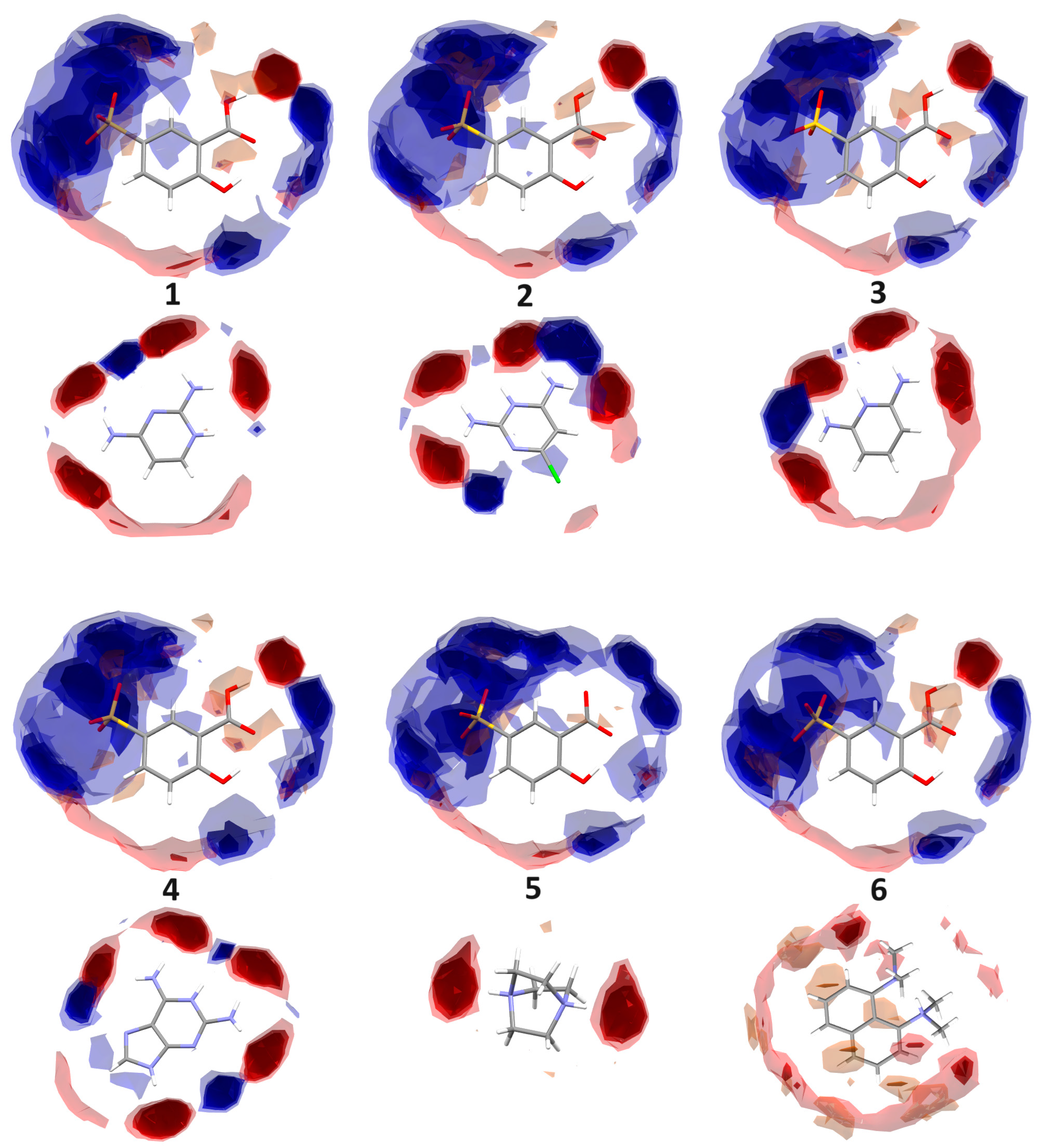
2.3.1. Full Interaction Maps

3D Full Interaction Maps (FIMs) based on CSD interaction data [62] for all analyzed crystals to check their preferred interaction behavior were calculated using Mercury software [60]. This tool generates the landscape of interactions using 3D coordinates from an X-ray experiment. The comparison of the predicted most likely positions of functional groups with a crystal packing can help evaluate whether a corresponding crystal fulfills the desired interactions.

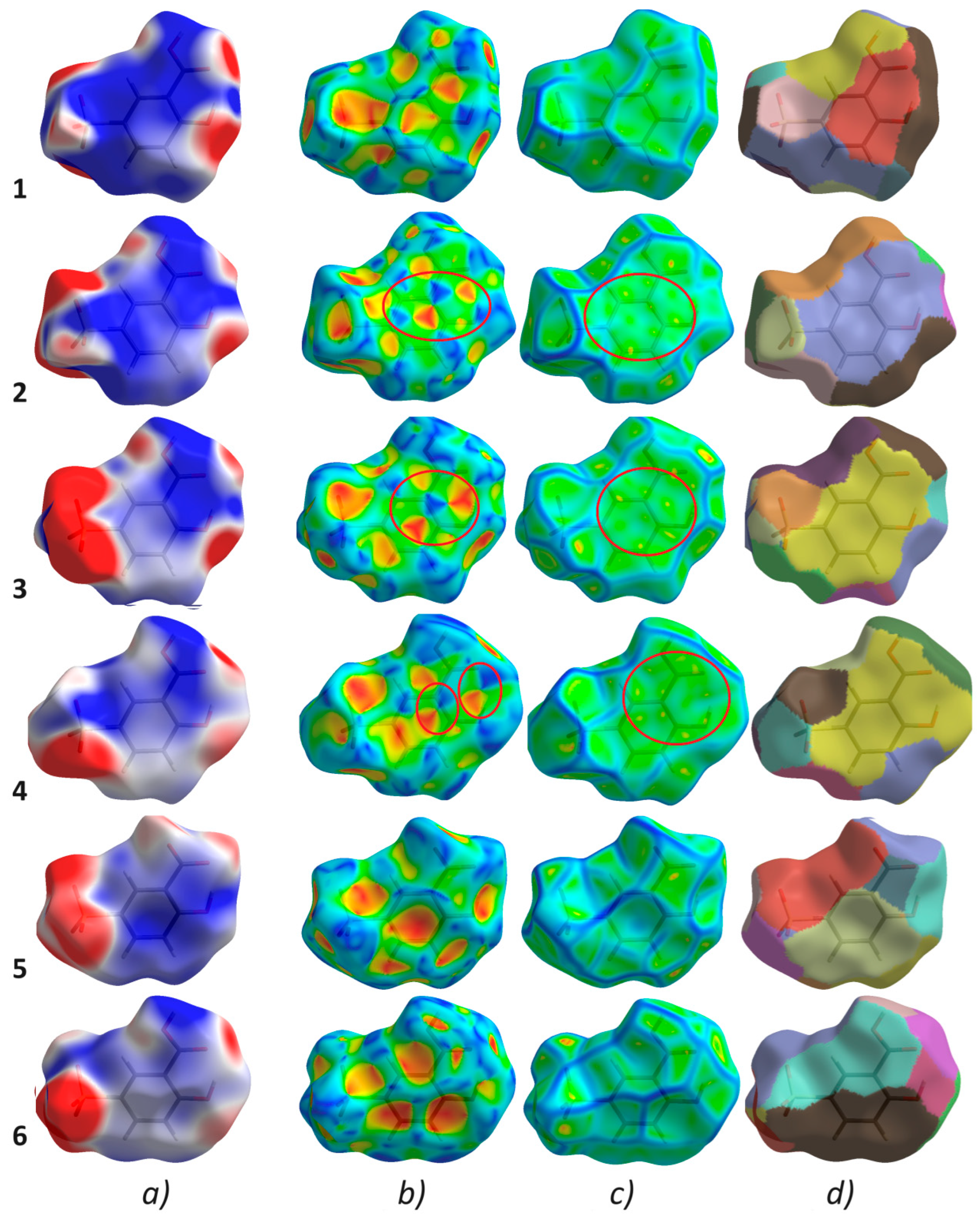
2.3.2. Hirshfeld Surface Analysis

A Hirshfeld analysis was performed using the newest version of CrystalExplorer 21.5 [63,64]. The 3D maps of the Hirshfeld’s surfaces revealing qualitative information on non-covalent interactions were mainly mapped with the dnorm property by a colored scheme, where red spots denote the shortest inter-contacts, the white regions illustrate distances close to the van der Waals (vdW) contacts (dnorm equal to zero), and the blue areas signify the interactions longer than the sum of the vdW radii (positive values of dnorm) [64]. The map was generated by calculation of the normalized distances from the contact points on the surface to the nearest nucleus inside (di) or outside (de) the Hirshfeld surface where all H-bond lengths adopted the neutron-derived values [65]. Two-dimensional fingerprint plots, drawn as a function of di and de values, reveal quantitative data on close inter-contacts [64].

2.3.3. Molecular Electrostatic Potential

The molecular electrostatic potential mapped onto Hirshfeld surfaces was obtained at the wave function of the HF/STO-3G level using Crystal Explorer 21.5 [63,66].

2.3.4. The Enrichment Ratio

The enrichment ratios (ER) of the intermolecular interactions in the analyzed crystal structures 16 were calculated based on the HS methodology. ER for an element pair (X, Y) is the ratio of the actual percentage of random inter-contacts within the crystal (CXY) to the percentage of theoretically equivalently distributed random contacts (RXY), when EXY = CXY/RXY. In this way privileged (when ER > 1) and disfavored inter-contacts (ER < 1) can be highlighted in the crystal structures [67].

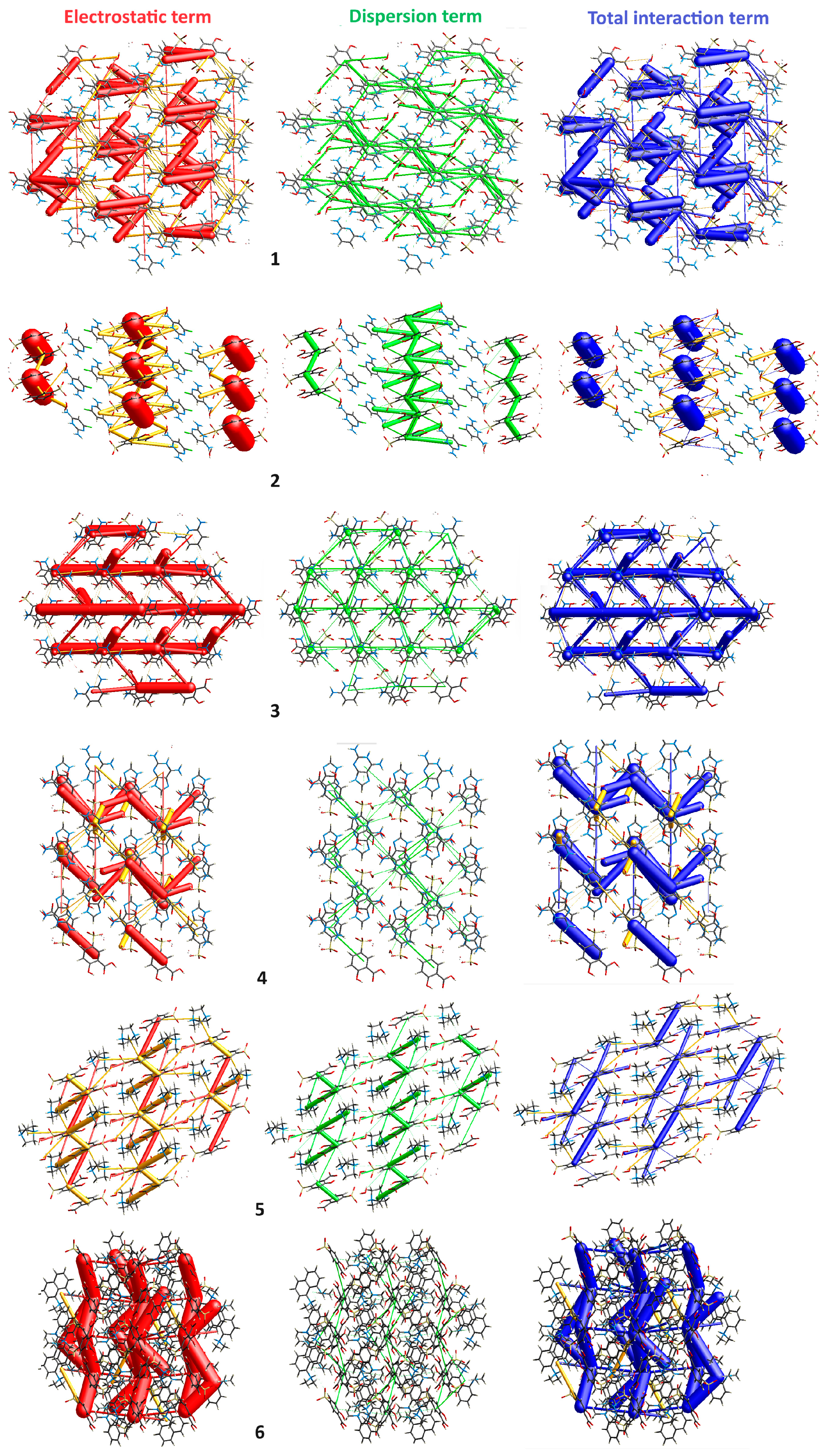
2.3.5. Energy Frameworks

The pairwise interaction energies between the moieties within the crystal were computed by Crystal Explorer 21.5 program [63,64,66,68,69] using the wavefunction calculated at the B3LYP/6-31G(d,p) functional basis set that is widely accepted in computational chemistry. The electrostatic (Eele), polarization (Epol), dispersion (Edisp), and exchange repulsion (Erep) energy components, according to Equation (1), were obtained. More specifically, the electrostatic term is characterized by the forces among charged particles, the polarization term is concerned with the interactions resulting from the distortion of a molecule’s electron cloud via other near-charge distributions, dispersion term is considered to be the weak forces caused by fluctuations in a molecule’s electron distribution, while repulsion term means the energy needed to overcome the forces that prevent a molecules pair from interaction [70,71].
To calculate the total energy (Etot) framework, a molecular cluster with a radius of 3.8 Å was generated around a selected single moiety of the reference compound. Symmetry operations were applied to build molecular wave functions and calculate the electron densities of a cluster. A tube size of 100 was used for the images.
Etot = Eele + Epol + Edisp + Erep

3. Results and Discussion

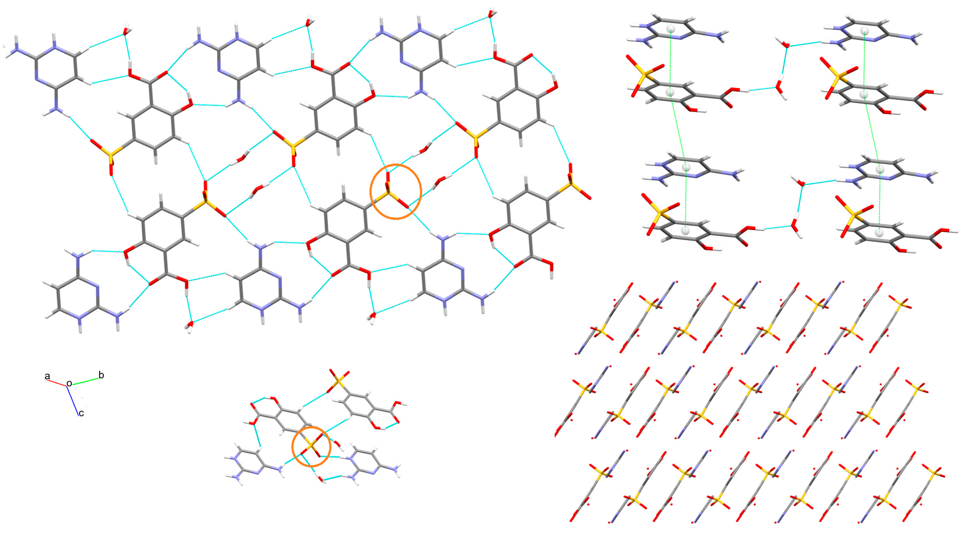
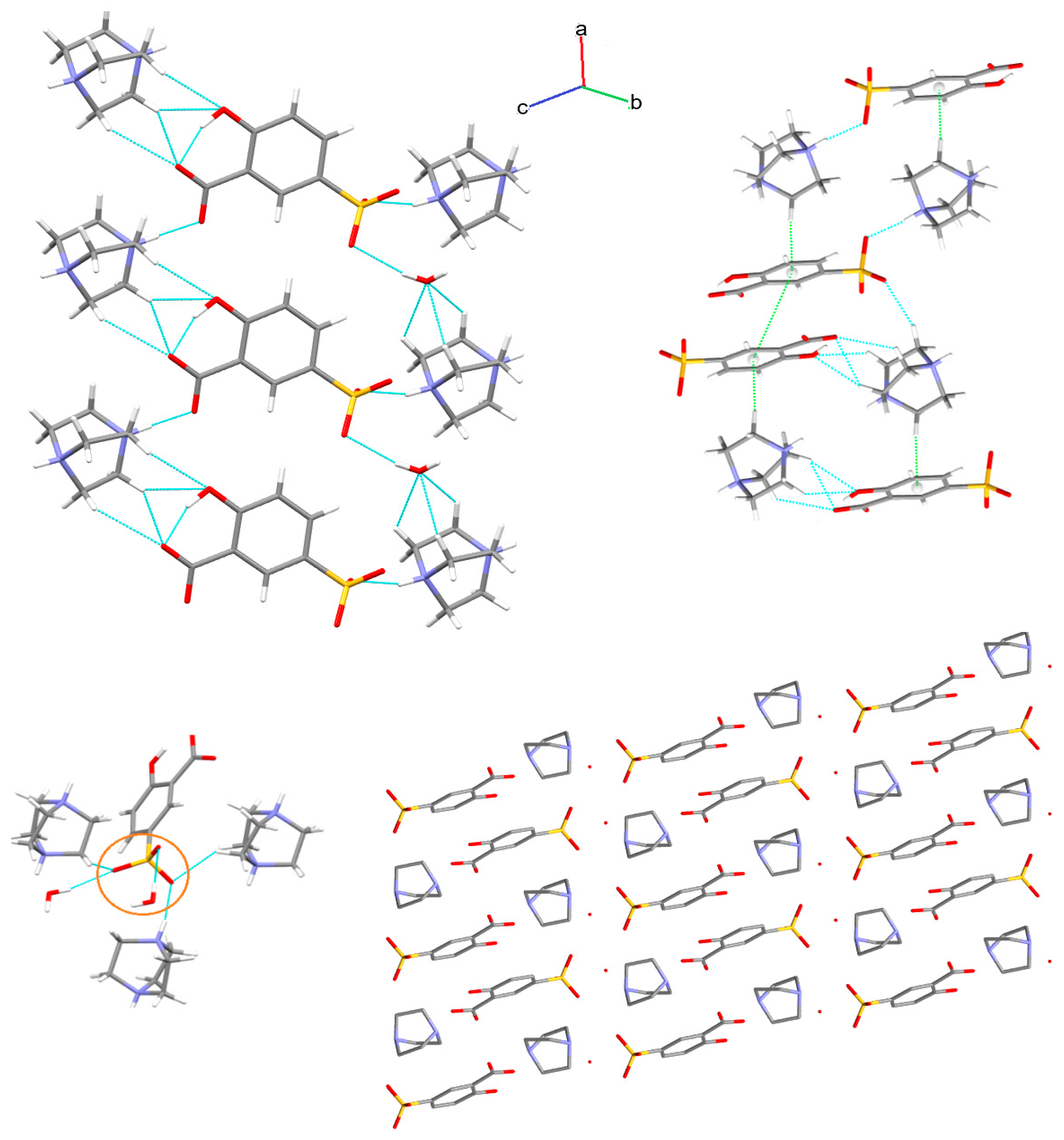
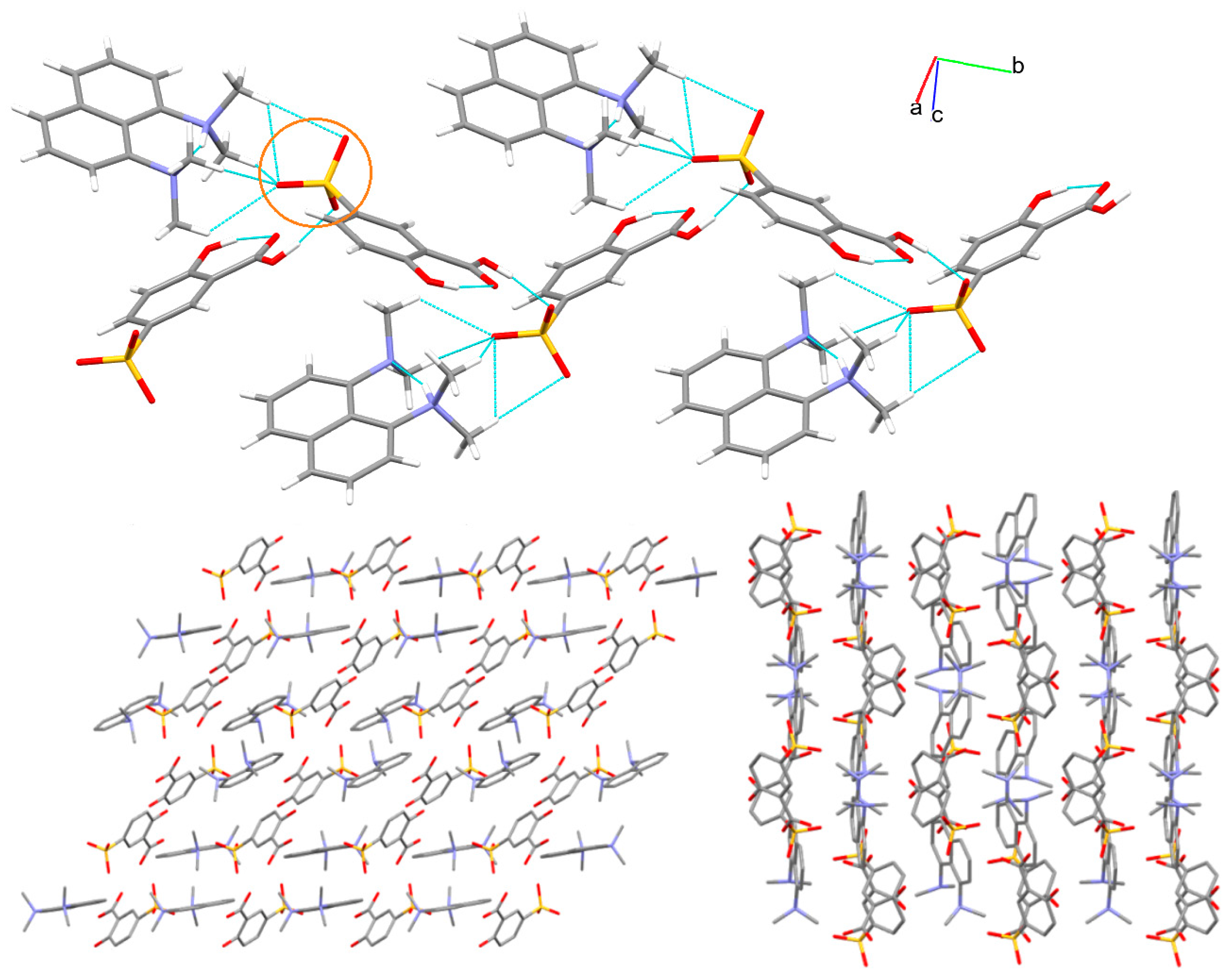
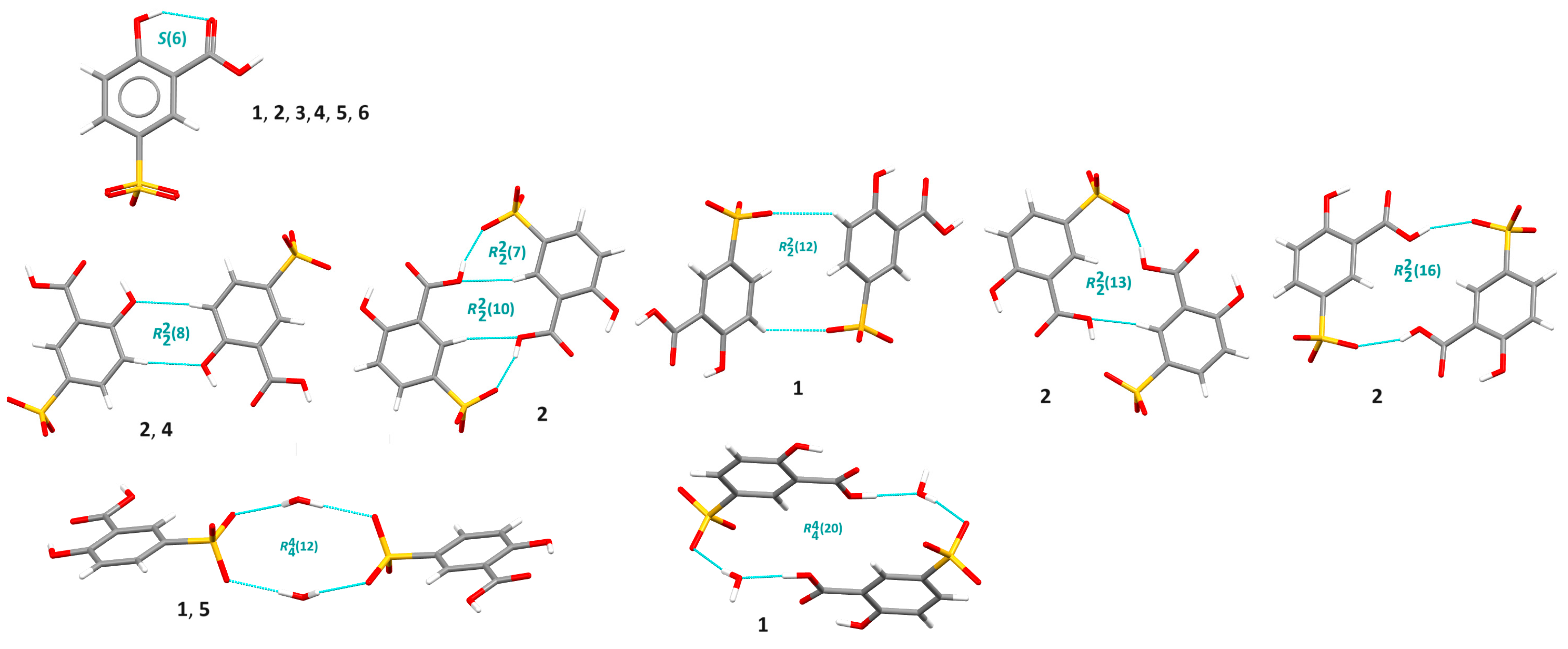
3.1. X-ray Structure and Supramolecular Features

The crystal structures of 16 were determined by single crystal X-ray diffraction with high precision at low temperature. Crystals 1, 2, 4, 5, and 6 are novel, while 3 is a better equivalent of the previously published structure KASXAE, which was earlier determined at ambient temperature [23]. The molecular structures of 16 are shown in Figure 2. Compounds 1, 2, 4, and 5 crystallize in the triclinic P 1 ¯ space group, while compounds 3 and 6 crystallize in the monoclinic space groups—P21 and P21/c, respectively. Full crystallographic data of studied salts are collected in Table 1. The unit cells of 15 contain solvent (water) molecules, apart from aminium cation and 5-sulfosalicylate anion. The unit cell of 6 consists of two crystallographically independent cations and anions. The structure of 5 represents the dicationic and dianionic species. The deprotonated carboxylic (COO) and sulfonic (SOO) groups of the anion show bond lengths of 1.275 and 1.261, as well as 1.453 and 1.456 Å, respectively. It indicates a delocalized anionic charge. Selected bond lengths and angles of 16 can be found in Table S1. The values are comparable to those found in the literature.
The crystal packing can be quantified by the Kitaigorodsky packing index (K.P.I.) [72,73]. It was calculated with the ‘calc void’ procedure implemented in the PLATON program [61]. The results showed that 6 is the most, while 1 is the least closely packed structure with 74.4% and 70.2% of filled space, respectively. No space accessible for voids was found (Table 1). The appealing feature of analyzed salts is the presence of diverse functional groups (sulfonate, carboxyl, hydroxyl, amine, methyl) with available H-atom donor and acceptor atoms of both cation and anion species, as well as water molecules, which results in extensive H-bonding interactions. The crystal packing of all salts is formed mainly by O-H⋯O, N-H⋯O, and C-H⋯O interactions, with the distances shorter than the sum of the mean van der Waals radii of the two corresponding atoms, from 1.63 Å for O-H⋯O in 4 and 6 to 2.59 Å for C-H⋯O in 5. Furthermore, O-H⋯N in 1 and N-H⋯N interactions in 2, 4, and 6 are observed. The intramolecular network is affected by O-H⋯O and C-H⋯O, and also N-H⋯N in 6. The geometrical parameters of H-bonds are listed in Table 2. It can be mentioned that nearly all available acceptors in all crystals, apart from 6, interact with all available donors. The two oxygen atoms in the sulfonic group play the role of bifurcated acceptors participating in the formation of O(N,C)-H⋯O interactions with anion, cation, and water molecules. A rare trifurcated (and tetrafurcated) acceptor is observed in 6 (Figure 3, Figure 4, Figure 5, Figure 6, Figure 7 and Figure 8).
Overall, at first glance, novel salts exhibit similar self-assembly behavior. Nevertheless, a thorough examination revealed that the structures differ markedly as demonstrated in Figure 3, Figure 4, Figure 5, Figure 6, Figure 7 and Figure 8. More specifically, compounds feature extensive H-bonding 3D-layered structures with significant π⋯π interactions in 24 with a Cg-Cg distance below 4 Å; and with the shortest distance—3.4111(12) Å in 4, and C-H⋯π in 5 and 6 (H⋯Cg distance below 3 Å) between the cations and anions, as demonstrated in Figure 3, Figure 4, Figure 5, Figure 6, Figure 7 and Figure 8. With respect to other weak π-based interactions, C-O⋯π (O⋯Cg distance below 4 Å) exist in 2, 4, and 5. It can be noted that the S-O⋯π [3.841(2) Å] inter-contact and C-Cl⋯π [Cl-Cg distance 3.6827(13) Å] halogen interaction were found in 2 (see Tables S2 and S3). In 2, a specific zig-zag layered structure is formed via alternating perpendicular separate anionic and cationic layers. Notably, water solvent molecules are significant contributors to building supramolecular architectures in all suitable cases (15). They play an important role as either a donor or acceptor of a bridge-linking moieties. The interplay of strong and weak H-bonding interactions and π-based inter-contacts gives rise to interesting supramolecular motifs at diverse levels of self-assembly. First of all, a specific intramolecular synthon, namely S(6), via O-H⋯O between the phenol OH group and a carboxyl O atom is observed in all crystals (Figure 9). It is key information from the crystal engineering point of view. This synthon can be employed in the pseudo-ring replacement strategy in the design of molecules. Another intramolecular H-bonding supramolecular motif via (NH)N-H⋯N(N) interactions in cationic moiety, but denoted by a similar descriptor S(6), is observed in crystal 6 (Table S4). In 2 and 4, a robust and very popular homosynthon R22(8) via O-H⋯O interaction is observed. Additionally, other centrosymmetric dimers, such as R22(16) in 2 by O-H⋯O, R22(12) in 1, R22(10) and R22(13) in 2 through C-H⋯O, were found. Moreover, heterosynthon R22(7) via O-H⋯O and C-H⋯O interactions is generated. Here, the OH group plays as role of either donor or acceptor simultaneously (Table S4). Moving forward, water molecules, linking ions, are involved in the formation of cyclic homosynthons as a donor and acceptor. Figure 9 illustrates the above supramolecular motifs taking into account anions. On the other hand, the protonated pyridine N atom of a cation forms diverse, interesting H-bonding supramolecular motifs with either COOH/COO or SO groups of anions or the water molecules as shown in Figure S1 (in Supplementary Materials). A library of supramolecular H-bonding synthons, including the first and second levels of the graph-set theory is provided (see Table S4). It is noteworthy that novel salts demonstrate remarkably varied supramolecular features arising mainly from different cationic polyamine species and their substituents.
The Full Interaction Maps (FIMs), related to statistical interaction data extracted from the CSD [62], helped us understand the effect of diverse substituents in the polyamines on supramolecular interactions and synthons formed by them. FIMs generated for all analyzed salts in the Mercury program [55] are presented in Figure 10. They illustrate zones around cations and anions where interactions are expected. It allowed us to estimate whether synthon preferences within the corresponding crystal lattices are satisfied. The FIMs of analyzed compounds 16 visualize the expected directions of the formation of either H-bonding or π-based interactions. Notably, nearly identical maps were obtained for the anions in all analyzed crystals, apart from 5. On the other hand, FIMs for cations are completely different. Areas of H-bonding donor probabilities are shown in blue while H-bonding acceptors are demonstrated in red. Aromatic/hydrophobic interactions are denoted in beige/light brown. It can be mentioned that the intensity of the color areas is compatible with the likelihood of the relevant inter-contacts occurring. It can be seen that cation 6 has the largest probability of being involved in hydrophobic interactions, while anion 5 can participate in forming synthons as acceptors only. The substituent effect on multifaceted classical and non-classical non-covalent interactions was thoroughly examined using complex Hirshfeld surfaces analysis, described in the next section.

3.2. Hirshfeld Surface Analysis

To gain a deep understanding of the crystal-packing behavior and the hierarchy of non-covalent interactions Hirshfeld surface analysis was performed. The 3D Hirshfeld surface maps for 16 were generated using either a standard surface solution of three-dimensional dnorm surfaces or shape index and curvedness properties (Figure 11). The normalized contact distance, called dnorm, is the sum of the distance of the point from the nearest nucleus within the Hirshfeld surface (di) and the distance of the point from the nearest nucleus outside the Hirshfeld surface (de) normalized via the van der Waals radius of the corresponding atom [66]. The surfaces were calculated for the 5-sulfosalicylate moiety, which is the same in all compounds to emphasize nuanced differences. The bright, larger red spots on the views of the dnorm surfaces indicate closer distances than the van der Waals radii. In particular, they display strong H-bonding interactions such as O-H⋯O and N-H⋯O between carboxyl and amino groups of anions and cations, respectively. The smaller spots signify weaker C-H⋯O interactions with neighboring moieties. The blue regions present longer inter-contacts than the van der Waals radii, while the white spots represent the close interactions equal to the sum of van der Waals radii. On the other hand, the convex blue spots on the shape index maps indicate hydrogen donor groups, while the concave red regions characterize acceptor groups. The π⋯π inter-contacts are represented via adjacent red and blue triangles on the shape index surfaces and as flat green areas around the rings, delineated by the blue outlines, on the curvature maps. Moving forward, orange-colored deformations of the Hirshfeld surfaces in crystals 5 and 6 indicate C-H⋯π interactions. For a better overview, the colored fragment patch surfaces were also generated to specify the nearest surrounding moiety [66], as shown in Figure 11. It can be used to calculate the number of moieties interacting with the main one.

3.2.1. Molecular Electrostatic Potential
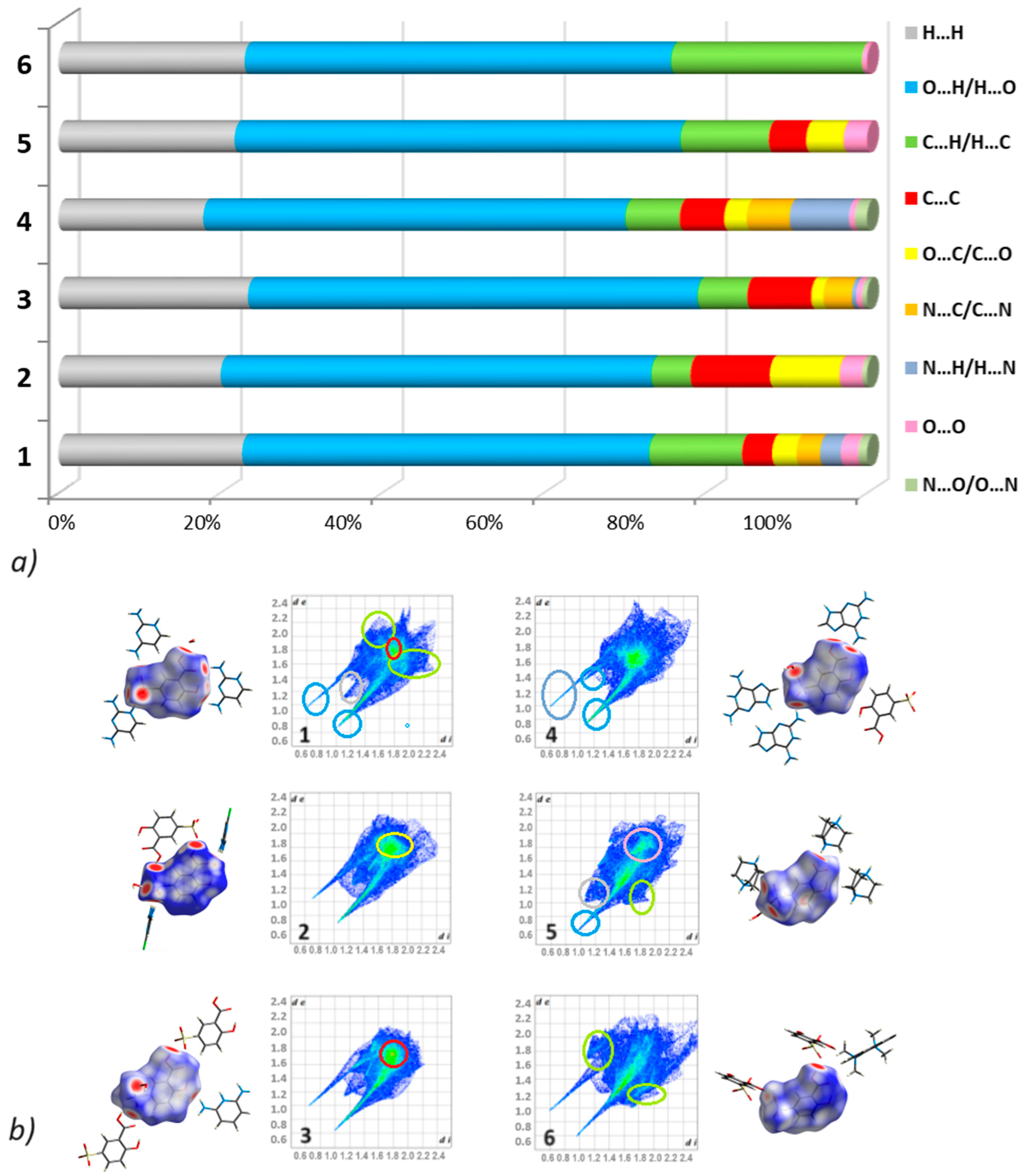
In Figure 11, the electrostatic potential mapped onto the Hirshfeld surfaces is also presented. It visualizes molecular electrostatic complementarities verifying the acceptor’s and donors’ functions in the 3D supramolecular network. Blue areas indicate a positive electrostatic potential (H-bond donors), while red regions denote a negative electrostatic potential (acceptors) [64].

3.2.2. Fingerprints

The 2D fingerprint plots are constructed based on the distances de and di from the Hirshfeld surfaces. The shapes of the graphs are unique for each compound. They manifest differences and summarize the intricate quantitative information contained in molecular crystals 16 as illustrated in Figure 12. The percentage contribution of close interactions in these crystal structures is included in the same Figure. It is clear at first sight that the O⋯O/H⋯O interactions have the highest contribution in all crystals, at the level of 50%. They are characterized by long symmetrical/parallel ‘spikes’ in the fingerprint histograms. The absence of the second spike in 5 means that the anionic moiety only serves as an H-bonding acceptor, while the cationic is a donor. The H⋯H and C⋯H/H⋯C interactions are second and third contributors, representing an average of 21.5% and 11%, respectively. The latter interactions change from 4.9% in 2 to 23.5% in 6. The H⋯H contacts are visualized in the central region between the spikes, while the C⋯H/H⋯C interactions are visualized as ‘wings’ in the upper side of fingerprint plots. The C⋯C inter-contacts are significant to share in the 24 crystal structures, showing from 3.7% in 1 to 9.8% in 2. The C⋯O/O⋯C contacts, observed in 15, represent from 1.5% in 3 to 8.6% in 2. In addition, N⋯C/C⋯N contacts are observed in 1 (2.9%), 3 (3.5%), and 4 (5.3%), while N⋯H/H⋯N are reported in 1 (2.6%) and 4 (7.4%). A much smaller share falls on O⋯O (in 1, 2, and 5) and N⋯O/O⋯N (in 1 and 4), at a level of 3% and 1.5%, respectively (Table S5).
Enrichment ratios were calculated based on the Hirshfeld surface concept for all analyzed compounds to analyze the interactions with a high propensity to form contacts in the crystal packing. The obtained values are tabulated in Table 3 and Table S6. Privileged interactions are as follows: O⋯H/H⋯O in all crystals, C⋯H/H⋯C—in 1 and 6, N⋯H/H⋯N in 4, C⋯C in nearly all structures (apart from 6), O⋯C/C⋯O in 2, C⋯N/N⋯C in 1 and 4. The highest value is obtained for the contacts O⋯H in 1, C⋯C in 15, and C⋯N in 1 and 4. However, C⋯C and C⋯N represent only ~5.5% and 4% of the contact surfaces, respectively. Surprisingly, H⋯H is one of the most abundant interactions in all structures (~20%), but they are disfavored in the crystal packing. The enrichment ratio values are below 1. O⋯N are disfavored, while O⋯O are impoverished.
The energy framework concept enables the visualization of strong interaction chains and an understanding of the distinct energy types. The three-dimensional energy frameworks for all six analyzed compounds are depicted in Figure 13. It emphasizes the neighboring moieties located in a radius of 3.8 Å from the analyzed molecule. Energy types are symbolized by different colors. In particular, red is for classical electrostatic (the coulombic), while green is for dispersion terms. In addition, the total energy is presented in blue. Notably, the cylinder/tube size (diameter) signifies the energy value; thicker cylinders mean higher energies. The tube thicknesses denote the relative interaction strengths. The calculated values of interaction energies, including the crystallographic symmetry operations, are summarized in Table 4. It depicts the energy contributions of all components, such as electrostatic (Eele), polarization (Epol), dispersion (Edisp), and repulsion (Erep), as well as total (Etot) for various intermolecular interactions in the analyzed crystals. Details presented in Table 4 are pivotal for the calculations of lattice energy [71]. The results revealed several types of interactions between the center moiety and the neighboring moieties. The thorough analysis revealed that in 1, total interaction energy (Etot = −85.9 kJ/mol) is associated with a symmetric pair of molecules distanced at R = 5.66, 5.80, and 7.34 Å, respectively. Similarly, in 2, a closer look at the obtained values of energies shows significant insights. As an example, in 1, the electrostatic, polarization, dispersion, and repulsion energies were calculated to be −218.9, −92.5, −162.7, and 57.2 kJ/mol, respectively (see Table 4). Further analysis indicated total lattice energies: −383.5 kJ/mol for 1, −153 kJ/mol for 2, −469.5 kJ/mol for 3, −237 kJ/mol for 4, −86.8 kJ/mol for 5, and −333.4 kJ/mol for 6. Thus, Etot values show significant differences. The most thermodynamically stable salt is considered to be 3. Interestingly, 3 presents remarkable red cylinders that joined the molecular pairs forming a hexagon energy topology. It is noteworthy that overall, the electrostatic energy is clearly a dominant component in 1, 3, 4, and 6, while dispersive energy is in 2 and 5. This means that van der Waals forces, contributing to dispersion interactions, are significant in the supramolecular assembly in 2 and 5. In this regard, the interactions between the reference moiety and the symmetry-related molecule at −x, −y, and −z in 2 and 5 are the most important interactions, with Etot = 175.6 and −49.7 kJ/mol, respectively.

4. Conclusions

In summary, a series of novel salts of heterocyclic polyamines with 5-sulfosalicylic acid, namely 2,4-diaminopyrimidin-1-ium 3-carboxy-4-hydroxybenzenesulfonate dihydrate (1), 2,4-diamino-6-chloropyrimidin-1-ium 3-carboxy-4-hydroxybenzenesulfonate hydrate (2), 2,6-diaminopyridin-1-ium 3-carboxy-4-hydroxybenzenesulfonate monohydrate (3), 2,6-diamino-9H-purin-1-ium 3-carboxy-4-hydroxybenzenesulfonate monohydrate (4), and 8-(dimethylamino)-N,N-dimethylnaphthalen-1-aminium 3-carboxy-4-hydroxybenzenesulfonate (6) have been reported for the first time, to the best of our knowledge. We managed to successfully synthesize and crystallize new salts and determine their crystal structures by single-crystal X-ray diffraction. The hierarchical supramolecular features were characterized by diverse modern approaches, including mainly extended Hirshfeld surface concepts. In addition, in the analysis, we included 1,4-diazabicyclo[2.2.2]octane-1,4-dium 2-hydroxy-5-sulfonatobenzoate monohydrate (5) as a better re-determination of its previously published equivalent (CSD refcode: KAXAE). The new compounds 1, 2, 4, and 5 crystallize in the triclinic P 1 ¯ space group, while compounds 3 and 6 crystallize in the monoclinic P21 and P21/c space groups, respectively. Differences in the intermolecular interactions in 16 were seen on the Hirshfeld surface maps and fingerprint plots, as well as the energy frameworks. More specifically, O⋯H/H⋯O interactions are significant contributors (~ 50%). However, C⋯H/H⋯C (5–23%) N⋯H/H⋯N (below 7%) and C⋯C (below 10%) inter-contacts also govern the crystal packing of the studied structures. The latter represents π⋯π contacts with the shortest interplanar distance of 3.4111(12) Å in 4. The electrostatic interactions in most crystals (in 1, 3, 4, and 6), while the dispersion forces only in 2 and 5, were regarded as dominant in stabilizing the crystal packing. Notably, the presence of water molecules has a pivotal role in determining the crystal packing, as either a donor or acceptor, as well as, to some extent, the electrostatic properties. A library of hierarchical supramolecular H-bonding motifs is provided. A specific intramolecular synthon via O-H⋯O is observed in nearly all crystals. It can be employed in the pseudo-cyclic replacement strategy in the design of new molecules.

Supplementary Materials

The following supporting information can be downloaded at: https://www.mdpi.com/article/10.3390/cryst14060497/s1. Figure S1. Other types of supramolecular H-bonding motifs observed in crystals 16. Figure S2. The ATR-FTIR spectrum of 1. Figure S3. The ATR-FTIR spectrum of 2. Figure S4. The ATR-FTIR spectrum of 3. Figure S5. The ATR-FTIR spectrum of 4. Figure S6. The ATR-FTIR spectrum of 5. Figure S7. The ATR-FTIR spectrum of 6. Table S1. Selected bond length and angles of 16. Table S2. The π⋯π interactions (<4.5 Å) in 16. Table S3. Other π-based interactions in 16. Table S4. Portfolio of selected H-bonding motifs in 16. Table S5. Percentage contribution of non-covalent interactions in crystals 16 based on Hirshfeld surface analysis. Table S6. Enrichment ratios in crystals 16 based on Hirshfeld surface analysis.

Author Contributions

Conceptualization, A.M. and J.B.; methodology A.M., K.Ł. and J.B.; validation, K.Ł. and J.B.; formal analysis, K.Ł. and J.B.; investigation, K.Ł., J.B. and A.M.; data curation, K.Ł. and J.B.; writing—original draft preparation, K.Ł., J.B. and A.M. All authors have read and agreed to the published version of the manuscript.

Funding

This research received no external funding.

Data Availability Statement

The CIF files have been deposited at the Cambridge Crystallographic Data Centre (CCDC) with the following numbers: 2348280–2348285, accessed on 15 May 2024. These data can be obtained free of charge via https://www.ccdc.cam.ac.uk/structures.

Conflicts of Interest

The authors declare no conflicts of interest.

References

  1. Ganie, A.A.; Vishnoi, P.; Dar, A.A. Utility of Bis-4-pyridines as Supramolecular Linkers for 5-Sulfosalicylic Acid Centers: Structural and Optical Investigations. Cryst. Growth Des. 2019, 19, 2289–2297. [Google Scholar] [CrossRef]
  2. Wu, Y.; Hao, X.; Li, J.; Guan, A.; Zhou, Z.; Guo, F. New insight into improving the solubility of poorly soluble drugs by preventing the formation of their hydrogen-bonds: A case of dapsone salts with camphorsulfonic and 5-sulfosalicylic acid. CrystEngComm 2021, 23, 6191–6198. [Google Scholar] [CrossRef]
  3. Du, H.; Xu, L.; Yue, M.; Xu, F.; Wang, Y. Studies on crystal structures, optical, solubility and dyeing properties of two new crystalline dye salts based on berberine with aromatic carboxylic acid. J. Mol. Struct. 2022, 1260, 132856. [Google Scholar] [CrossRef]
  4. Zhang, Y.-N.; Duan, Y.; Liu, L.-X.; Chang, L.; Feng, Y.-R.; Wu, L.-L.; Zhang, L.; Zhang, Y.-J.; Zou, D.-Y.; Liu, Y.-L.; et al. On improving the hygroscopic stability of palmatine chloride with crystalline palmatine sulfosalicylate pharmaceutical salt. J. Struct. Chem. 2022, 63, 52–61. [Google Scholar] [CrossRef]
  5. Hua, X.-N.; Pan, X.; Zhu, Y.; Cai, Z.; Song, Q.; Li, Y.; Feng, W.; Chen, X.; Zhang, H.; Sun, B. Novel pharmaceutical salts of cephalexin with organic counterions: Structural analysis and properties. RSC Adv. 2022, 12, 34843–34850. [Google Scholar] [CrossRef] [PubMed]
  6. Dar, A.A.; Lone, S.H.; Ahmad, I.; Ahangar, A.A.; Ganie, A.A.; Femina, C. Engineering the solid-state luminescence of organic crystals and cocrystals. Mater. Adv. 2024, 5, 1056–1064. [Google Scholar] [CrossRef]
  7. Duan, W.; Liu, B.; Gong, N.; Famulari, A.; Guo, F. Polymorphs and Transformations of the Solid Forms of Organic Salts of 5-Sulfosalicylic Acid and Isonicotinamide. Cryst. Growth Des. 2020, 20, 7606–7614. [Google Scholar] [CrossRef]
  8. Ganie, A.A.; Ismail, T.M.; Sajith, P.K.; Dar, A.A. Validation of the supramolecular synthon preference through DFT and physicochemical property investigations of pyridyl salts of organo-sulfonates. New J. Chem. 2021, 45, 4780–4790. [Google Scholar] [CrossRef]
  9. Ganie, A.A.; Marimuthu, R.; Islam, S.T.; Narang, S.; Dar, A.A. Molecular salts of the isoniazid derivatives. Expanding the scope of sulfonate-pyridinium synthon to design materials. J. Solid State Chem. 2022, 307, 122762. [Google Scholar] [CrossRef]
  10. Ramesh, K.S.; Saravanabhavan, M.; Muhammad, S.; Edison, D.; Ho, M.-S.; Sekar, M.; Al-Sehemi, A.G. Synthesis, physico-chemical characterization and quantum chemical studies of 2, 3-dimethyl quinoxalinium-5-sulphosalicylate crystal. J. Mol. Struct. 2023, 1285, 135425. [Google Scholar] [CrossRef]
  11. Smith, G.; Wermuth, U.D. Hydrogen bonding in two ammonium salts of 5-sulfosalicylic acid: Ammonium 3-carboxy-4-hydroxybenzenesulfonate monohydrate and triammonium 3-carboxy-4-hydroxybenzenesulfonate 3-carboxylato-4-hydroxybenzenesulfonate. Acta Crystallogr. Sect. C Cryst. Struct. Commun. 2013, 69, 534–537. [Google Scholar] [CrossRef]
  12. Selvaraju, D.; Venkatesh, R.; Sundararajan, V. Hydrazine-1,2-diium bis(3-carboxy-4-hydroxybenzenesulfonate) tetrahydrate. Acta Crystallogr. Sect. E Struct. Rep. Online 2011, 67, o1236–o1237. [Google Scholar] [CrossRef]
  13. Smith, G.; Wermuth, U.D.; Healy, P.C. Hydrogen bonding in proton-transfer compounds of 5-sulfosalicylic acid with ortho-substituted monocyclic heteroaromatic Lewis bases. J. Chem. Crystallogr. 2006, 36, 841–849. [Google Scholar] [CrossRef]
  14. Raj, S.B.; Sethuraman, V.; Francis, S.; Hemamalini, M.; Muthiah, P.T.; Bocelli, G.; Cantoni, A.; Rychlewska, U.; Warzajtis, B. Supramolecular organization via hydrogen bonding in trimethoprim sulfonate salts. CrystEngComm 2003, 5, 70–76. [Google Scholar] [CrossRef]
  15. Hemamalini, M.; Muthiah, P.T.; Sridhar, B.; Rajaram, R.K. Pyrimethaminium sulfosalicylate monohydrate. Acta Crystallogr. Sect. E Struct. Rep. Online 2005, 61, o1480–o1482. [Google Scholar] [CrossRef]
  16. Matulkova, I.; Mathauserova, J.; Cisarova, I.; Nemec, I.; Fabry, J. The study of crystal structures and vibrational spectra of inorganic salts of 2,4-diaminopyrimidine. J. Mol. Struct. 2016, 1103, 82–93. [Google Scholar] [CrossRef]
  17. Bertolasi, V.; Pretto, L.; Gilli, P.; Ferretti, V.; Gilli, G. Hydrogen-bonded supramolecular structures in co-crystals of β- or ζ-diketone enols with 2,6-diaminopyridine or 2,4-diaminopyrimidine. New J. Chem. 2002, 26, 1559–1566. [Google Scholar] [CrossRef]
  18. Draguta, S.; Fonari, M.S.; Masunov, A.E.; Zazueta, J.; Sullivan, S.; Antipin, M.Y.; Timofeeva, T.V. New acentric materials constructed from aminopyridines and 4-nitrophenol. CrystEngComm 2013, 15, 4700–4710. [Google Scholar] [CrossRef]
  19. Hutzler, W.M.; Egert, E.; Bolte, M. 6-Propyl-2-thiouracil versus 6-methoxymethyl-2-thiouracil: Enhancing the hydrogen-bonded synthon motif by replacement of a methylene group with an O atom. Acta Crystallogr. Sect. C Cryst. Struct. Commun. 2016, 72, 634–646. [Google Scholar] [CrossRef]
  20. Hutzler, W.M.; Egert, E.; Bolte, M. One barbiturate and two solvated thiobarbiturates containing the triply hydrogen-bonded ADA/DAD synthon, plus one ansolvate and three solvates of their coformer 2,4-diaminopyrimidine. Acta Cryst. C 2016, 72, 705–715. [Google Scholar] [CrossRef]
  21. Hall, V.M.; Thornton, A.; Miehls, E.K.; Bertke, J.A.; Swift, J.A. Uric Acid Crystallization Interrupted with Competing Binding Agents. Cryst. Growth Des. 2019, 19, 7363–7371. [Google Scholar] [CrossRef]
  22. Bojarska, J.; Łyczko, K.; Mieczkowski, A. Synthesis, Crystal Structure and Supramolecular Features of Novel 2,4-Diaminopyrimidine Salts. Crystals 2024, 14, 133. [Google Scholar] [CrossRef]
  23. Smith, G.; Wermuth, U.D.; Healy, P.C. Layered structures in proton-transfer compounds of 5-sulfosalicylic acid with the aromatic polyamines 2,6-diaminopyridine and 1,4-phenylenediamine. Acta Crystallogr. Sect. C Cryst. Struct. Commun. 2005, 61, o555–o558. [Google Scholar] [CrossRef] [PubMed]
  24. Hemamalini, M.; Goh, J.H.; Fun, H.-K. 2,3-Diaminopyridinium 3-carboxy-4-hydroxybenzenesulfonate monohydrate. Acta Crystallogr. Sect. E Struct. Rep. Online 2011, 67, o3122. [Google Scholar] [CrossRef] [PubMed]
  25. Hemamalini, M.; Fun, H.-K. 2-Amino-5-chloropyridinium 3-carboxy-4-hydroxybenzenesulfonate. Acta Crystallogr. Sect. E Struct. Rep. Online 2010, 66, o2323–o2324. [Google Scholar] [CrossRef] [PubMed]
  26. Manickam, R.; Rajakannan, V.; Prabhaharan, M.; Srinivasan, G. 2-Amino-6-chloropyridinium 3-carboxy-4-hydroxybenzenesulfonate. IUCrData 2019, 4, x190566. [Google Scholar] [CrossRef]
  27. Hemamalini, M.; Fun, H.-K. 2-Amino-5-bromopyridinium 3-carboxy-4-hydroxybenzenesulfonate. Acta Crystallogr. Sect. E Struct. Rep. Online 2010, 66, o2408–o2409. [Google Scholar] [CrossRef] [PubMed]
  28. Hemamalini, M.; Fun, H.-K. 2-Amino-5-methylpyridinium 3-carboxy-4-hydroxybenzenesulfonate. Acta Crystallogr. Sect. E Struct. Rep. Online 2010, 66, o2153–o2154. [Google Scholar] [CrossRef]
  29. Atria, A.M.; Garland, M.T.; Baggio, R. 2,6-Diamino-9H-purine monohydrate and bis(2,6-diamino-9H-purin-1-ium) 2-(2-carboxylatophenyl)acetate heptahydrate: Two simple structures with very complex hydrogen-bonding schemes. Acta Crystallogr. Sect. C Cryst. Struct. Commun. 2010, 66, o547–o552. [Google Scholar] [CrossRef]
  30. Belletire, J.L.; Schneider, S.; Shackelford, S.A.; Peryshkov, D.V.; Strauss, S.H. Pairing heterocyclic cations with closo-dodecafluorododecaborate (2−): Synthesis of binary heterocyclium(1+) salts and a Ag4(heterocycle)84+ salt of B12F122−. J. Fluor. Chem. 2011, 132, 925–936. [Google Scholar] [CrossRef]
  31. de Vries, E.J.C.; Kantengwa, S.; Ayamine, A.; Báthori, N.B. Testing the limits of synthon engineering: Salts of salicylic and sulfosalicylic acid with nucleobases and derivatives. CrystEngComm 2016, 18, 7573–7579. [Google Scholar] [CrossRef]
  32. Xia, M.; Ma, K.-R.; Zhu, Y. Synthesis and Crystal Structure of Hydrate Adduct of 6-Benzylaminopurine and 5-Sulfosalicylic Acid [(C12H12N5)(C7H5O6S)·H2O]. J. Chem. Crystallogr. 2010, 40, 634–638. [Google Scholar] [CrossRef]
  33. Jones, A.O.F.; Kallay, A.A.; Lloyd, H.; McIntyre, G.J.; Wilson, C.C.; Thomas, L.H. The Effect of Local Crystalline Environment on Hydrogen Atom Behavior in Molecular Complexes of a Proton Sponge. Cryst. Growth Des. 2016, 16, 2123–2129. [Google Scholar] [CrossRef]
  34. Sigala, P.A.; Ruben, E.A.; Liu, C.W.; Piccoli, P.M.B.; Hohenstein, E.G.; Martinez, T.J.; Schultz, A.J.; Herschlag, D. Determination of Hydrogen Bond Structure in Water versus Aprotic Environments To Test the Relationship Between Length and Stability. J. Am. Chem. Soc. 2015, 137, 5730–5740. [Google Scholar] [CrossRef] [PubMed]
  35. Ye, H.-Y.; Tang, Y.-Y.; Li, P.-F.; Liao, W.-Q.; Gao, J.-X.; Hua, X.-N.; Cai, H.; Shi, P.-P.; You, Y.-M.; Xiong, R.-G. Metal-free three-dimensional perovskite ferroelectrics. Science 2018, 361, 151–155. [Google Scholar] [CrossRef] [PubMed]
  36. Jiang, X.; Duan, H.-B.; Jellen, M.J.; Chen, Y.; Chung, T.S.; Liang, Y.; Garcia-Garibay, M.A. Thermally Activated Transient Dipoles and Rotational Dynamics of Hydrogen-Bonded and Charge-Transferred Diazabicyclo [2.2.2]Octane Molecular Rotors. J. Am. Chem. Soc. 2019, 141, 16802–16809. [Google Scholar] [CrossRef]
  37. Song, X.; Cui, Q.; Liu, Y.; Xu, Z.; Cohen, H.; Ma, C.; Fan, Y.; Zhang, Y.; Ye, H.; Peng, Z.; et al. Metal-Free Halide Perovskite Single Crystals with Very Long Charge Lifetimes for Efficient X-ray Imaging. Adv. Mater. 2020, 32, 2003353. [Google Scholar] [CrossRef] [PubMed]
  38. Chu, L.-L.; Zhang, T.; Zhang, W.-Y.; Shi, P.-P.; Gao, J.-X.; Ye, Q.; Fu, D.-W. Three-Dimensional Metal-Free Molecular Perovskite with a Thermally Induced Switchable Dielectric Response. J. Phys. Chem. Lett. 2020, 11, 1668–1674. [Google Scholar] [CrossRef]
  39. Budzianowski, A.; Petřiček, V.; Katrusiak, A. Metal-free enantiomorphic perovskite [dabcoH2]2+[H3O]+Br3 and its one-dimensional polar polymorph. IUCrJ 2022, 9, 544–550. [Google Scholar] [CrossRef]
  40. Song, X.; Cohen, H.; Yin, J.; Li, H.; Wang, J.; Yuan, Y.; Huang, R.; Cui, Q.; Ma, C.; Liu (Frank), S.; et al. Low Dimensional, Metal-Free, Hydrazinium Halide Perovskite-Related Single Crystals and Their Use as X-Ray Detectors. Small 2023, 19, 2300892. [Google Scholar] [CrossRef]
  41. Jin, S.; Guo, J.; Liu, L.; Wang, D. Five organic salts assembled from carboxylic acids and bis-imidazole derivatives through collective noncovalent interactions. J. Mol. Struct. 2011, 1004, 227–236. [Google Scholar] [CrossRef]
  42. Guo, F.; Zhang, M.-Q.; Famulari, A.; Marti-Rujas, J. Solid state transformations in stoichiometric hydrogen bonded molecular salts: Ionic interconversion and dehydration processes. CrystEngComm 2013, 15, 6237–6243. [Google Scholar] [CrossRef]
  43. Liu, H.-l.; Xie, Y.-F.; Pan, Z.-G.; Famulari, A.; Guo, F.; Zhou, Z.; Martí-Rujas, J. Cyclic Interconversion among Molecular Salts via Neat Grinding and Related Photoluminescence Properties. Cryst. Growth Des. 2014, 14, 6528–6536. [Google Scholar] [CrossRef]
  44. Fu, Q.; Xu, X.-K.; Liu, B.-K.; Guo, F. Solid state transformations of different stoichiometric forms of an organic salt formed from 5-sulfosalicylic acid and hexamethylenetetramine upon dehydration and rehydration. CrystEngComm 2018, 20, 1844–1852. [Google Scholar] [CrossRef]
  45. Yang, D.-J.; Qu, S.-H. Bis(2-aminopyridinium) 5-sulfonatosalicylate. Acta Crystallogr. Sect. E Struct. Rep. Online 2006, 62, o5127–o5129. [Google Scholar] [CrossRef]
  46. Jin, S.; Zhao, Y.; Liu, B.; Jin, X.; Zhang, H.; Wen, X.; Liu, H.; Jin, L.; Wang, D. Hydrogen bonded supramolecular structures of eight organic salts based on 2,6-diaminopyridine, and organic acids. J. Mol. Struct. 2015, 1099, 601–615. [Google Scholar] [CrossRef]
  47. Balasubramani, K.; Muthiah, P.T.; Lynch, D.E. R22(8) motifs in aminopyrimidine sulfonate/carboxylate interactions: Crystal structures of pyrimethaminium benzenesulfonate monohydrate (2:2:1) and 2-amino-4,6-dimethylpyrimidinium sulfosalicylate dihydrate (4:2:2). Chem. Cent. J. 2007, 1, 28. [Google Scholar] [CrossRef] [PubMed]
  48. Jin, S.; Jin, L.; Ye, X.; Li, J.; Jin, B.; Zheng, L.; Wang, D. Crystal and Molecular Structure of Three Organic Salts from N6-Benzyladenine, 5-Sulfosalicylic Acid, Naphthalene-1,5-Disulfonic Acid, and α-Ketoglutaric Acid. J. Chem. Crystallogr. 2015, 45, 9–19. [Google Scholar] [CrossRef]
  49. Krishnamohan, S.C.V. Crystal engineering––Where do we go from here? Cryst. Growth Des. 2002, 2, 465–474. [Google Scholar] [CrossRef]
  50. Braga, D.; Brammer, L.; Champness, N.R. New trends in crystal engineering. CrystEngComm 2005, 7, 1–19. [Google Scholar] [CrossRef]
  51. Bond, A.D.; Jones, W. Supramolecular Organization And Materials Design; Cambridge University Press: Cambridge, UK, 2002. [Google Scholar]
  52. Desiraju, G.R. Supramolecular Synthons in Crystal Engineering—A New Organic Synthesis. Angew. Chem. Int. Ed. Engl. 1995, 34, 2311–2327. [Google Scholar] [CrossRef]
  53. Walsh, R.D.B.; Bradner, M.W.; Fleischman, S.; Morales, L.A.; Moulton, B.; Rodríguez-Hornedo, N.; Zaworotko, M.J. Crystal Engineering of the Composition of Pharmaceutical Phases. Chem. Commun. 2003, 2, 186–187. [Google Scholar] [CrossRef]
  54. Bojarska, J.; Breza, M.; Remko, M.; Yuan, Y.; Ziora, Z.; Chai, T.-T.; Madura, I.D.; Kaczmarek, K.; Blaskovich, M.A.T.; Wolf, M.W. A supramolecular self-assembly of peptide-derived compounds via 1,5-disubstituted tetrazole-based supramolecular synthons: An experimental and computational study. J. Mol. Struct. 2023, 1288, 135732. [Google Scholar] [CrossRef]
  55. Bojarska, J.; Breza, M.; Remko, M.; Borowiecki, P.; Fruziński, A.; Madura, I.D.; Kaczmarek, K.; Leśnikowski, Z.; Kraj, A.; Zielenkiewicz, P.; et al. Supramolecular synthon hierarchy in cyclopropyl containing peptide-derived compounds. CrystEngComm 2022, 24, 8372–8389. [Google Scholar] [CrossRef]
  56. Bojarska, J.; Breza, M.; Remko, M.; Czyz, M.; Gajos-Michniewicz, A.; Zimecki, M.; Kaczmarek, K.; Madura, I.D.; Wojciechowski, J.M.; Wolf, W.M. Structural and Biofunctional Insights into the Cyclo(Pro-Pro-Phe-Phe-)Scaffold from Experimental and In Silico Studies: Melanoma and Beyond. Int. J. Mol. Sci. 2022, 23, 7173. [Google Scholar] [CrossRef]
  57. Bojarska, J.; Remko, M.; Madura, I.D.; Kaczmarek, K.; Zabrocki, J.; Wolf, W.M. Synthesis, experimental and in silico studies of N-fluorenylmethoxycarbonyl-O-tert-butyl-N-methyltyrosine, coupled with CSD data: A survey of interactions in the crystal structures of Fmoc-amino acids. Acta Crystallogr. C 2020, 76, 328–345. [Google Scholar] [CrossRef]
  58. Sheldrick, G.M. Crystal Structure Refinement with SHELXL. Acta Cryst. C Struc. Chem. 2015, 71, 3–8. [Google Scholar] [CrossRef] [PubMed]
  59. Dolomanov, O.V.; Bourhis, L.J.; Gildea, R.J.; Howard, J.A.K.; Puschmann, H. OLEX2: A complete structure solution, refinement and analysis program. J. Appl. Crystallogr. 2009, 42, 339–341. [Google Scholar] [CrossRef]
  60. Macrae, C.F.; Sovago, I.; Cottrell, S.J.; Galek, P.T.A.; McCabe, P.; Pidcock, E.; Platings, M.; Shields, G.P.; Stevens, J.S.; Towler, M.; et al. Mercury 4.0: From visualization to analysis, design and prediction. J. Appl. Cryst. 2020, 53, 226–235. [Google Scholar] [CrossRef]
  61. Spek, A.L. Structure Validation in chemical crystallography. Acta Cryst. D Biol. Crystallogr. 2009, 65, 148–155. [Google Scholar] [CrossRef]
  62. Wood, P.A.; Olsson, T.S.G.; Cole, J.C.; Cottrell, S.J.; Feeder, N.; Galek, P.T.A.; Groom, C.R.; Pidcock, E. Evaluation of Molecular Crystal Structures Using Full Interaction Maps. CrystEngComm 2013, 15, 65–72. [Google Scholar] [CrossRef]
  63. Turner, M.J.; McKinnon, J.J.; Wol, S.K.; Grimwood, D.J.; Spackman, P.R.; Jayatilaka, D.; Spackman, M.A. CrystalExplorer, Version 3.1; The University of Western Australia: Perth, Australia, 2017. [Google Scholar]
  64. Spackman, M.A.; Jayatilaka, D. Hirshfeld surface analysis. CrystEngComm 2009, 11, 19–32. [Google Scholar] [CrossRef]
  65. Jayatilaka, D.; Grimwood, D.J. Tonto: A Fortran Based Object-Oriented System for Quantum Chemistry and Crystallography. In Computational Science—ICCS 2003; Sloot, P.M.A., Abramson, D., Bogdanov, A.V., Gorbachev, Y.E., Dongarra, J.J., Zomaya, A.Y., Eds.; Lecture Notes in Computer Science; Springer: Berlin/Heidelberg, Germany, 2003; Volume 2660, pp. 142–151. [Google Scholar] [CrossRef]
  66. Spackman, P.R.; Turner, M.J.; McKinnon, J.J.; Wolff, S.K.; Grimwood, D.J.; Jayatilaka, D.; Spackman, M.A. CrystalExplorer: A program for Hirshfeld surface analysis, visualization and quantitative analysis of molecular crystals. J. Appl. Crystallogr. 2021, 54, 1006. [Google Scholar] [CrossRef] [PubMed]
  67. Jelsch, C.; Ejsmont, K.; Huder, L. The enrichment ratio of atomic contacts in crystals, an indicator derived from the Hirshfeld surface analysis. IUCrJ 2014, 1, 119–128. [Google Scholar] [CrossRef] [PubMed]
  68. Spackman, M.A.; McKinnon, J.J. Fingerprinting intermolecular interactions in molecular crystals. CrystEngComm 2002, 4, 378. [Google Scholar] [CrossRef]
  69. McKinnon, J.J.; Mitchell, A.S.; Spackman, M.A. Hirshfeld Surfaces: A New Tool for Visualizing and Exploring Molecular Crystals. Chem. Eur. J. 1998, 4, 2136. [Google Scholar] [CrossRef]
  70. Mackenzie, C.F.; Spackman, P.R.; Jayatilaka, D.; Spackman, M.A. CrystalExplorer model energies and energy frameworks: Extension to metal coordination compounds, organic salts, solvates and open-shell systems. IUCrJ 2017, 4, 575. [Google Scholar] [CrossRef]
  71. Tan, S.L.; Jotani, M.M.; Tiekink, E.R.T. Utilizing Hirshfeld surface calculations, non-covalent interaction (NCI) plots and the calculation of interaction energies in the analysis of molecular packing. Acta Crystallogr. E Crystallogr. Commun. 2019, 75, 308. [Google Scholar] [CrossRef]
  72. Kitaigorodskii, A.I. Organic Chemical Crystallography; Consultants Bureau: New York, NY, USA, 1961; pp. 1–30, 65–112. [Google Scholar]
  73. Kitaigorodskii, A.I. Molecular Crystals and Molecules; Academic Press: London, UK, 1973; pp. 1–133. [Google Scholar]
Figure 1. Structures of analysed salts: 2,4-diaminopyrimidin-1-ium 3-carboxy-4-hydroxybenzenesulfonate dihydrate (1), 2,4-diamino-6-chloropyrimidin-1-ium 3-carboxy-4-hydroxybenzenesulfonate monohydrate (2), 2,6-diaminopyridin-1-ium 3-carboxy-4-hydroxybenzenesulfonate monohydrate (3), 2,6-diamino-9H-purin-1-ium 3-carboxy-4-hydroxybenzenesulfonate monohydrate (4), 1,4-diazabicyclo[2.2.2]octane-1,4-diium 2-hydroxy-5-sulfonatobenzoate monohydrate (5), and 8-(dimethylamino)-N,N-dimethylnaphthalen-1-aminium 3-carboxy-4-hydroxybenzenesulfonate (6).
Crystals 14 00497 g001
Figure 2. The molecular structures of 16 showing the atom numbering schemes. Displacement ellipsoids are drawn at a 30% probability level.
Crystals 14 00497 g002
Figure 3. Crystal packing of 1.
Crystals 14 00497 g003
Figure 4. Crystal packing of 2.
Crystals 14 00497 g004
Figure 5. Crystal packing of 3.
Crystals 14 00497 g005
Figure 6. Crystal packing of 4.
Crystals 14 00497 g006
Figure 7. Crystal packing of 5.
Crystals 14 00497 g007
Figure 8. Crystal packing of 6.
Crystals 14 00497 g008
Figure 9. Supramolecular H-bonding synthonic patterns formed by anionic species in 16.
Crystals 14 00497 g009
Figure 10. Full interactions maps for 16.
Crystals 14 00497 g010
Figure 11. Views of the transparent Hirshfeld surfaces of analyzed salts 16 mapped with shape index (b), curvedness (c), and fragment patch (d). In addition, molecular electrostatic potential mapped on the Hirshfeld surfaces is presented (a). Red circles on the shape index and curvedness maps signify C⋯C interactions.
Crystals 14 00497 g011
Figure 12. (a) Percentage contributions of individual close inter-contacts to the Hirshfeld surfaces of 16 (contacts with >0.5% are taken into account); (b) full fingerprint plots and Hirshfeld surfaces of the 5-sulfosalicylic moiety for 16 mapped with dnorm property over a range between −0.50 and 1.50 Å and drawn with the surrounding moieties. Circles on the fingerprint plots characterize corresponding interactions: C...C in red, C...H/H...C in green, H...H in blue, O...O in pink (according to the legend).
Crystals 14 00497 g012
Figure 13. A comparison of energy frameworks in crystals 16 presented in a cluster (a radius of 3.8 Å) viewed down the a-axis.
Crystals 14 00497 g013
Table 1. Crystal data and structure refinement details obtained from measurements at 100 K for the studied compounds.
Compound123456
Chemical formulaC11H16N4O8SC11H13ClN4O7SC12H15N3O7SC12H14N6O7SC13H20N2O7SC21H24N2O6S
Formula weight364.34380.76345.33386.35348.37432.48
λ (Cu Kα) (Å)1.541841.541841541841.541841.541841.54184
Crystal systemtriclinictriclinicmonoclinictriclinictriclinicmonoclinic
Space groupP 1 ¯ P 1 ¯ P 21P 1 ¯ P 1 ¯ P 21/c
a (Å)7.9539(3)6.8906(6)6.6358(2)6.6755(4)6.7903(4)19.7312(3)
b (Å)9.5529(4)6.9077(6)12.9481(4)10.6495(5)8.7284(8)12.33559(11)
c (Å)11.2394(4)16.9140(11)8.4812(3)10.9153(5)13.3336(7)18.4437(3)
α (°)67.904(4)96.621(6)9084.427(4)101.707(6)90
β (°)85.375(3)96.880(6)104.703(3)87.076(5)103.408(5)114.7954(16)
γ (°)78.700(3)108.636(8)9083.698(5)94.585(6)90
Volume (Å)775.92(6)747.20(11)704.84(4)767.01(7)746.02(9)4075.28(10)
Z222228
Dcalc (g·cm−3)1.5591.6921.6271.6731.5511.775
μ (mm−1)2.3444.0222.4702.4052.3121.410
F (000)3803923604003681824
Crystal size (mm)0.18 × 0.16 × 0.080.15 × 0.08 × 0.030.18 × 0.10 × 0.050.22 × 0.04 × 0.030.18 × 0.14 × 0.100.20 × 0.18 × 0.10
θ range (°)4.245–70.4222.667–70.2825.392–70.3284.073–70.3523.500–70.2252.467–70.187
Reflections collected11,295541445019276472828,523
Unique reflections292627922451285827767674
Reflections I > 2σ(I)280524192365248425136949
Rint0.02200.02900.02110.03610.01870.0222
Restraints/parameters1/2611/2531/2440/2741/2232/567
Goodness-of-fit1.0861.1161.0731.0731.0471.043
R1, wR2 (I > 2σ(I))0.0261, 0.07350.0379, 0.09860.0276, 0.06950.0432, 0.10960.0335, 0.08530.0303, 0.0827
R1, wR2 (all data)0.0273, 0.07450.0443, 0.10470.0293, 0.07160.0533, 0.11610.0373, 0.08810.0338, 0.0855
Max. peak/hole (e·Å−3)0.359/−0.4680.307/−0.6020.202/−0.2980.674/−0.4730.398/−0.4270.302/−0.482
K.P.I. [%]70.272.672.774.270.674.4
K.P.I.—Kitaigorodsky’s packing index.
Table 2. Geometric parameters of H-bonds for 16.
9D-H [Å]H⋯A [Å]D⋯A [Å]D-H⋯A [o]
1
N1-H1⋯O2 i0.883(19)1.869(19)2.7472(14)172.7(17)
N3-H3A⋯O8 ii0.886(19)2.154(19)2.9655(16)152.1(17)
N3-H3B⋯O5 iii0.853(19)2.136(19)2.9698(16)165.5(19)
O4-H4⋯O70.90(2)1.69(2)2.5685(13)166(3)
N4-H4A⋯O6 ii0.857(19)2.141(19)2.9205(17)151.1(17)
N4-H4B⋯O3 iv0.869(19)2.082(19)2.9313(16)165.5(18)
*O6-H6⋯O50.86(2)1.87(2)2.6397(14)149(2)
O6-H6⋯N2 iii0.86(2)2.44(2)2.9052(15)114.9(17)
O7-H7A⋯O1 v0.83(2)1.96(2)2.7803(14)172(2)
O7-H7B⋯O80.84(2)1.90(2)2.7306(15)168(2)
O8-H8A⋯O3 iv0.81(2)2.10(2)2.8640(14)157(2)
O8-H8B⋯O1 vi0.85(3)1.93(3)2.7594(15)166(2)
*C2-H2⋯O30.952.532.9197(17)105
C5-H5⋯O1 vii0.952.573.2961(16)134
C10-H10⋯O4 iv0.952.543.2942(18)137
C11-H11⋯O7 iv0.952.423.3590(18)168
(i) 1 − x, 1 − y, 2 − z; (ii) x, y, 1 + z; (iii) −x, 2 − y, 1 − z; (iv) 1 − x, 1 − y, 1 − z; (v) −x, 1 − y, 1 − z; (vi) x, 1 + y, −1 + z; (vii) −x, 1 − y, 2 − z
2
O1-H1⋯O4 i0.80(3)1.93(3)2.666(3)154(3)
N2-H2⋯O60.94(4)1.84(4)2.758(3)166(3)
*O3-H3⋯O20.89(4)1.85(4)2.615(3)144(3)
N3-H3B⋯N1 ii0.87(3)2.10(3)2.953(3)170(3)
N3-H3C⋯O50.89(4)1.98(3)2.862(3)169(3)
N4-H4A⋯O2 iii0.86(3)2.08(3)2.925(3)168(3)
N4-H4B⋯O70.92(4)1.92(4)2.813(3)163(3)
O7-H7A⋯O5 iv0.82(3)2.02(3)2.838(3)174(3)
O7-H7B⋯O6 v0.78(5)2.11(5)2.867(3)164(5)
C3-H3A⋯O3 vi0.952.543.355(4)144
*C6-H6⋯O40.952.482.885(3)106
C6-H6⋯O1 i0.952.543.339(3)142
(i) 3 − x, 1 − y, 1 − z; (ii) 1 − x, −y, −z; (iii) 2 − x, 1 − y, 1 − z; (iv) −1 + x, 1 + y, z; (v) −1 + x, y, z; (vi) 1 − x, −y, 1 − z
3
N1-H1⋯O70.86(4)1.91(4)2.754(4)170(4)
N2-H2A⋯O3 i0.85(5)2.38(5)3.206(3)163(4)
N3-H3A⋯O6 ii0.85(5)2.24(5)3.012(3)152(4)
N3-H3B⋯O2 iii0.87(4)2.09(4)2.924(3)162(4)
O4-H4⋯O1 iii0.88(4)1.76(4)2.613(2)165(4)
*O6-H6⋯O50.80(5)1.89(5)2.606(3)148(5)
O7-H7A⋯O30.81(5)2.07(5)2.842(4)161(5)
O7-H7B⋯O1 iv0.81(5)2.00(5)2.768(3)160(5)
*C2-H2⋯O30.952.532.917(4)104
(i) 1 − x, −1/2 + y, 2 − z; (ii) 1 − x, ½ + y, 1 − z; (iii) x, y, −1+z; (iv) 1 + x, y, z
4
N2-H2A⋯O70.91(3)1.84(3)2.730(3)166(3)
O4-H4⋯N3 i0.97(4)1.63(4)2.573(2)165(4)
N4-H4A⋯N1 ii0.87(3)2.04(3)2.891(3)170(3)
N5-H5A⋯O70.88(3)2.36(3)3.091(3)141(3)
N5-H5B⋯O2 iii0.84(3)1.99(3)2.817(3)170(3)
*O6-H6⋯O50.86(3)1.82(3)2.594(2)150(3)
N6-H6B⋯O30.89(3)1.94(3)2.781(3)159(2)
N6-H6C⋯O5 iv0.84(3)2.02(3)2.851(3)168(3)
O7-H7A⋯O1 v0.83(4)2.07(4)2.827(3)153(4)
O7-H7B⋯O3 vi0.80(4)2.09(4)2.877(3)165(3)
*C2-H2⋯O30.952.472.868(3)105
C5-H5⋯O6 vii0.952.583.455(3)153
C11-H11⋯O2 viii0.952.373.098(3)133
(i) x, 1 + y, z; (ii) 1 − x, −y, 2 − z; (iii) x, y, 1 + z; (iv) x, −1 + y, z; (v) −x, 1 − y, 1 − z; (vi) 1 − x, 1 − y, 1 − z; (vii) −x, 2 − y, −z; (viii) 1 − x, −y, 1 − z
5
N1-H1⋯O6 i0.91(2)1.84(2)2.7102(18)161(2)
N2-H2⋯O1 ii0.916(17)1.632(17)2.5451(18)174.0(17)
N2-H2⋯O2 ii0.916(17)2.59(2)3.1463(19)119.9(14)
*O3-H3⋯O20.91(3)1.67(3)2.5278(19)155(2)
O7-H7A⋯O4 iii0.85(3)2.13(2)2.9623(19)168(2)
O7-H7B⋯O5 iv0.81(3)2.05(3)2.841(2)167(2)
*C6-H6⋯O40.952.592.951(2)103
C8-H8A⋯O5 v0.992.473.423(2)161
C8-H8B⋯O7 vi0.992.443.314(3)147
C9-H9B⋯O3 vi0.992.473.329(2)145
C11-H11A⋯O2 vi0.992.593.363(2)135
C13-H13B⋯O7 vi0.992.593.356(2)134
(i) 1 − x, 1 − y, 2 − z; (ii) −x, 1 − y, 1 − z; (iii) 1 + x, y, −1 + z; (iv) 1 − x, −y, 1 − z; (v) 1 + x, 1 + y, z; (vi) 1 − x, 1 − y, 1 − z
6
*N1-H1⋯N20.930(18)1.713(18)2.5973(15)157.6(17)
O1-H1A⋯O5 i0.93(2)1.63(2)2.5468(13)169(2)
*N3-H3⋯N40.93(2)1.707(19)2.5907(16)158.8(17)
*O3-H3A⋯O20.87(2)1.83(2)2.6134(17)148(2)
O7-H7⋯O11 ii0.92(2)1.66(2)2.5714(13)169(2)
*O9-H9⋯O80.89(2)1.79(2)2.6005(16)149(2)
C25-H25B⋯O4 iii0.982.523.4370(16)157
C25-H25C⋯O2 iv0.982.513.3061(18)138
C26-H26A⋯O5 v0.982.463.3977(18)161
C27-H27C⋯O100.982.573.381(2)140
C30-H30⋯O11 vi0.952.493.4349(16)173
C39-H39C⋯O8 iii0.982.513.0482(17)114
C40-H40A⋯O10 vi0.982.393.3235(18)159
C41-H41B⋯O120.982.503.1961(17)128
C42-H42C⋯O120.982.583.2782(16)128
(i) −x, ½ + y, ½ − z; (ii) 1 − x, −1/2 + y, ½ − z; (iii) x, −1 + y, z; (iv) x, −1 + y, z; (v) −x, −1/2 + y, ½ − z; (vi) 1 − x, 1 − y, −z
Table 3. Enrichment ratios for 16.
123456
H⋯H0.7560.830.790.6860.730.62
O⋯H3.1311.6071.7911.8041.531.64
C⋯H0.9620.4070.420.5120.811.64
N⋯H0.719 1.138
O⋯O0.2460.247 0.27
O⋯C0.4711.0430.2020.3820.57
O⋯N0.57 0.418
C⋯C1.9676.534.33.2792.99
C⋯N4.03 3.223
Table 4. The interaction energies via energy framework calculations. Energy values are given in kJ mol−1. (R means the distance between molecular centroids; N is the number of interactions involving the central moiety).
NREeleEpolEdispErepEtotSymmetry Operation
1
15.987.10.0−21.25.8−7.1x, −y, −z
15.907.1−12.5−21.25.8−15.3
16.55−66.60.0−16.59.3−75.2x, −y, −z
15.66−66.6−16.4−16.59.3−85.9
17.8515.5−4.0−12.91.32.6x, −y, −z
17.64−7.9−0.7−0.50.0−9.0
15.68−7.9−0.7−0.50.0−9.0
15.80−66.6−16.4−16.59.3−85.9
13.469.2−1.4−1.40.07.2
16.387.1−12.5−21.25.8−15.3
16.9315.5−4.0−12.91.32.6
16.169.2−1.4−1.40.07.2
18.58−7.4−6.1−3.50.3−14.5x, −y, −z
17.34−66.6−16.4−16.59.3−85.9
−218.9−92.5−162.757.2−383.5
2
15.1328.7−18.6−43.821.2−5.2x, −y, −z
18.396.8−4.2−10.48.11.4x, −y, −z
04.0719.2−37.6−57.830.3−32.4x, −y, −z
16.8926.6−8.0−10.92.313.8x, y, z
17.000.0−4.20.00.0−2.7
06.9635.9−2.5−2.20.032.9
14.7926.6−8.0−10.92.313.8
06.09−176.3−61.8−22.779.7−175.6x, −y, −z
05.658.7−2.3−1.80.05.7
06.538.7−2.3−1.80.05.7
18.6528.7−18.6−43.821.2−5.2
17.9028.7−18.6−43.821.2−5.2
42.3−186.7−249.9186.3−153
3
19.138.5−1.1−5.62.95.2
14.05−93.4−30.6−11.155.8−79.8
13.65−46.1−18.2−50.132.6−77.5
29.01−23.9−3.7−2.30.1−28.7x, y + 1/2, z
28.48−93.4−30.6−11.155.8−79.8x, y, z
14.14−12.0−2.4−1.90.0−15.5
16.210.0−0.20.00.0−0.1
13.49−55.6−21.0−48.528.3−91.1
18.131.4−2.5−8.013.53.6
16.200.9−8.3−14.28.4−10.4
28.49−25.3−5.5−3.80.2−32.6x, y + 1/2, z
15.82−0.9−5.1−9.21.2−11.5
17.81−11.2−8.7−12.915.0−16.5
17.58−33.3−14.8−9.020.8−34.8
−384.3−152.7−187.7234.6−469.5
4
17.40−76.4−14.8−11.52.8−95.6x, −y, −z
18.816.5−3.1−8.95.71.3x, −y, −z
19.380.0−2.40.00.0−1.6
19.4515.2−2.7−5.50.49.1x, −y, −z
17.046.5−3.1−8.95.71.3
18.27−76.4−14.8−11.52.8−95.6
14.1143.4−2.9−2.60.040.1
17.8815.2−2.7−5.50.49.1
18.55−10.7−0.6−0.30.0−11.6
17.63−57.3−6.7−4.60.0−66.8
14.30−57.3−6.7−4.60.0−66.8
13.9743.4−2.9−2.60.040.1
−148−63.4−66.517.8−237
5
14.8144.8−37.4−40.716.9−1.7x, −y, −z
17.06−3.7−0.6−1.30.0−5.2
15.3810.1−0.5−0.70.09.3
18.750.0−6.40.00.0−4.1x, −y, −z
15.8710.7−1.4−1.50.08.6
16.64−13.6−9.5−10.72.6−27.6
17.38−39.5−4.5−7.60.5−49.7x, −y, −z
17.2944.8−37.4−40.716.9−1.7
14.834.5−0.2−0.20.04.3
16.01−13.6−9.5−10.72.6−27.6
16.9610.7−1.4−1.50.08.6
55.2−108.8−115.639.5−86.8
6
27.91−123.7−40.9−8.774−100.5x, y + 1/2, −z + 1/2
16.17−16.6−2.4−1.70.0−19.9
29.34−16.6−2.4−1.70.0−19.9x, −y + 1/2, z + 1/2
18.0628.9−1.4−0.50.028.1
16.45−123.7−40.9−8.774−100.5
16.940.0−0.10.00.0−0.1
19.53−16.6−2.4−1.70.0−19.9
19.600.0−0.30.00.0−0.2
19.73−123.7−40.9−8.774−100.5
−392−131.7−31.7222−333.4
Energy modelk_elek_polk_dispk_rep
CE-HF⋯HF/3-21G electron densities1.0190.6510.9010.811
CE-B3LYP⋯B3LYP/6-31G(d,p) electron densities1.0570.7400.8710.618
k—scale factor. Each form of energy should be multiplied by its relevant factor.
Disclaimer/Publisher’s Note: The statements, opinions and data contained in all publications are solely those of the individual author(s) and contributor(s) and not of MDPI and/or the editor(s). MDPI and/or the editor(s) disclaim responsibility for any injury to people or property resulting from any ideas, methods, instructions or products referred to in the content.

