Review

Advancements in ZnO-Based Photocatalysts for Water Treatment: A Comprehensive Review

by
Souad Abou Zeid
and
Yamin Leprince-Wang
*
ESYCOM, CNRS-UMR9007, Université Gustave Eiffel, F-77420 Champs sur Marne, France
*
Author to whom correspondence should be addressed.
Crystals 2024, 14(7), 611; https://doi.org/10.3390/cryst14070611
Submission received: 4 June 2024 / Revised: 21 June 2024 / Accepted: 25 June 2024 / Published: 30 June 2024
(This article belongs to the Section Inorganic Crystalline Materials)

Abstract

:
Water contamination remains a pressing global concern, necessitating the development of effective and sustainable water treatment solutions. Zinc oxide (ZnO) has garnered significant attention for its potential applications in photocatalysis due to its unique properties and versatile nature. This review synthesizes recent research findings on the advancement in ZnO-based photocatalysts for water treatment, encompassing synthesis methods, structure modifications for photocatalytic efficiency enhancement, toxicity assessments, and applications in diverse water treatment processes. By critically analyzing the strategies to enhance the photocatalytic performance of ZnO and its role in addressing water pollution challenges, this review provides valuable insights into the evolving landscape of ZnO-based photocatalysts for achieving efficient and environmentally friendly water treatment systems. This review emphasizes the transformative potential of ZnO-based photocatalysts in revolutionizing water treatment methodologies and underscores the importance of continued research and innovation in harnessing ZnO’s capabilities for sustainable water purification.

1. Introduction

Water, the cradle of life, faces escalating challenges of global scarcity and pollution, primarily due to rapid population growth, unchecked industrial expansion, and extensive environmental degradation [1,2]. With over seven billion people—comprising more than 15% of the world’s population—experiencing acute freshwater shortages, the ability to maintain basic living standards and engage in productive activities is hindered [3,4]. Consequently, the purification of polluted water before its discharge into the environment is of paramount importance. Many traditional techniques like coagulation, filtration, adsorption, reverse osmosis, and treatment with ozone have been proposed to remove such organic pollutants in water. Although these methods are somewhat effective, they possess limitations such as high energy consumption and the potential for secondary pollution [5,6,7]. Hence, the development of more effective and energy-efficient techniques is imperative, with a focus on environmentally friendly, cost-effective approaches. Semiconductor-assisted photocatalysis has emerged as a green, low-cost technology that utilizes sunlight to dissociate organic pollutants into harmless byproducts, thereby contributing to a reduction in environmental pollution [8,9,10]. This approach has gained considerable attention due to its non-selective oxidative properties and its capability to mineralize organic pollutants into carbon dioxide and water. A crucial part of the ongoing research in the field of photocatalysis revolves around the exploration of various semiconductor materials to harness their potential in environmental purification and the conversion of light energy into chemical energy. Notably, semiconductors such as TiO2 [11,12], ZnO [13,14], Fe2O3 [15,16], BiVO4 [17,18], SnO2 [19], MgO [20], SiO2 [21], and others [22,23] have been utilized in photocatalytic applications, showing promise in addressing environmental challenges and energy conversion. Among these semiconductors, titanium dioxide (TiO2) has been extensively studied for its exceptional photocatalytic characteristics, including high stability, potent oxidizing power, and long-term durability [24,25]. However, concerns about the potential nanoscale toxicity of TiO2 have prompted a shift in focus toward zinc oxide (ZnO), which exhibits lower toxicity profiles. The exceptional properties of ZnO, including stability, ease of synthesis, low material cost, optical transparency, piezoelectric properties, and high biocompatibility, have positioned it as a promising alternative to TiO2 [26,27,28]. Although ZnO possesses a wide band gap of 3.37 eV and a high exciton binding energy of 60 meV [29], it demonstrates superior photocatalytic performance compared to TiO2 in the degradation of organic pollutants [30,31,32,33]. This is attributed to its higher electron mobility (200–300 cm2/V·s) than TiO2 (0.1–4.0 cm2/V·s) [34,35], which accelerates electron transfer, contributing to high quantum efficiency. Additionally, the lower valence band (VB) position of ZnO compared to TiO2 results in a higher oxidation potential of hydroxyl radicals, leading to the more effective degradation of pollutants [36].
However, the challenge lies in the limited light absorption of ZnO, which is largely confined to the UV spectrum (~5% of the solar spectrum) and lacks efficiency in harnessing the visible spectrum (~43% of the solar spectrum) [37]. To address this limitation, various methods have been employed to enhance ZnO’s performance and widen its light response range. Strategies such as doping with metals and non-metals [38,39,40,41], depositing noble metals [42,43,44], creating heterostructured photocatalysts based on ZnO [45,46,47,48,49,50,51,52,53,54,55,56], and incorporating carbon materials [57,58,59] have been instrumental in improving the efficacy of ZnO. Doping ZnO with metals and non-metals like Fe, Cu, Ag, Sn, C, N, F, P, and S introduces localized donor levels within the forbidden energy band, enabling enhanced light absorption and electron transfer. Additionally, depositing noble metals onto ZnO surfaces and developing heterostructured photocatalysts by combining ZnO with other semiconducting metal oxides with varying band gaps have shown promising results, augmenting ZnO’s photocatalytic capabilities.
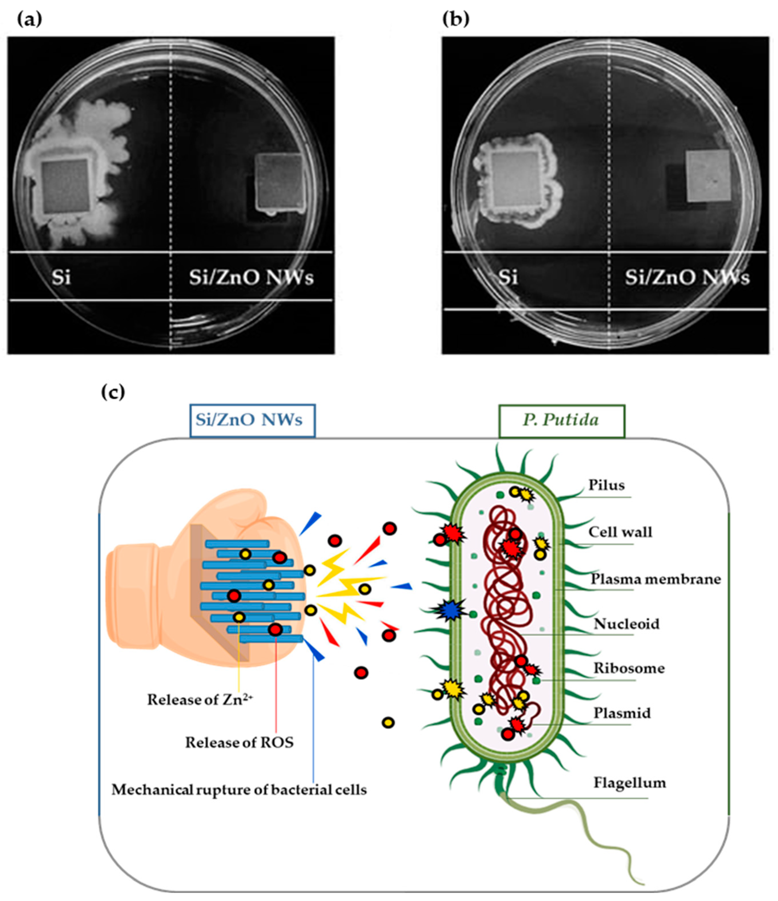
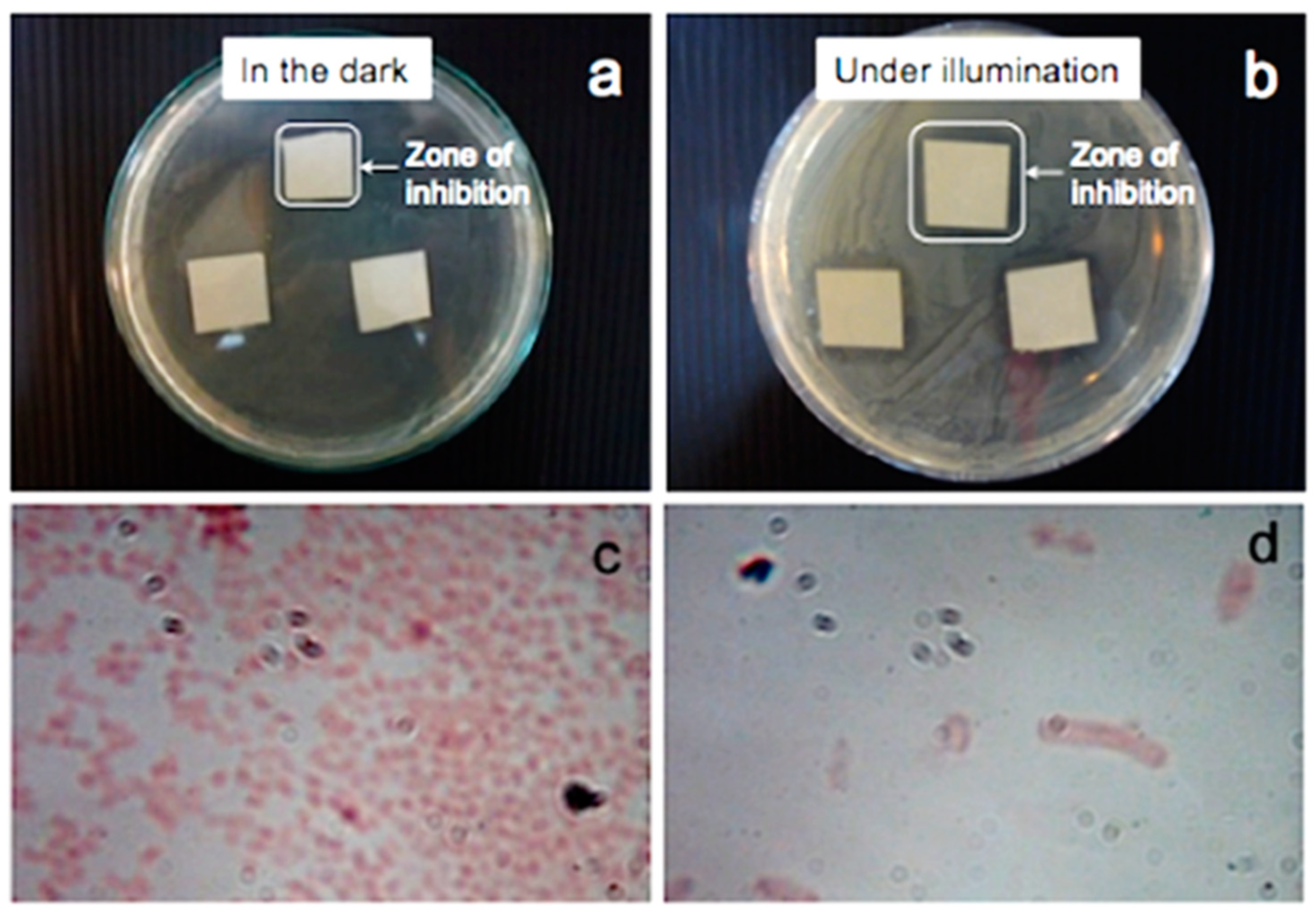
Furthermore, the remarkable antibacterial activity of ZnO has positioned it as a valuable solution for addressing bacterial contamination in water sources. Various strategies, including the integration of antimicrobial agents and precise control of particle size and concentration, have been implemented to amplify the antibacterial performance of ZnO. The mechanisms underlying ZnO’s antibacterial action involve the generation of reactive oxygen species (ROS) through photocatalysis and the release of Zn2+, presenting a multifaceted approach to combat waterborne bacterial challenges efficiently [60,61,62,63]. Moreover, beyond its roles as a photocatalyst and antibacterial agent, ZnO plays a vital function as a transparent conductive oxide (TCO) in a diverse array of optoelectronic devices like solar cells, LEDs, and displays [64,65,66]. The exceptional properties of ZnO thin films, such as high transparency, excellent conductivity, and a wide bandgap, make them indispensable components in the realm of optoelectronic technologies. Furthermore, the compatibility of ZnO with flexible substrates further enhances its appeal for the burgeoning domains of flexible electronics and wearable technology [67], underscoring its versatility and significance beyond its traditional usage in environmental cleanup and water treatment applications.
In this comprehensive review, we strive to offer a detailed overview of zinc ZnO, focusing prominently on its photocatalytic and antibacterial properties, while integrating recent studies to enrich our analysis. Our primary objective was to present a thorough understanding of ZnO by exploring its fundamental properties and highlighting its diverse applications beyond traditional water treatment and disinfection methods. Unlike previous reviews that often segregate synthesis methods or application aspects, our approach integrates these facets to provide a cohesive narrative. We extensively cover various synthesis methods and their impacts on ZnO’s properties, emphasizing strategies to enhance its photocatalytic efficiency. Additionally, we underscore ZnO’s versatility in addressing environmental challenges and beyond, showcasing its potential in broader scientific and technological domains. This review endeavors to deliver a clear and comprehensive study of ZnO, consolidating existing knowledge while offering insights that contribute to its broader relevance in contemporary research and applications.

2. Generalities of ZnO

Zinc oxide emerges as a versatile and robust photocatalyst, offering a plethora of exceptional properties that underpin its efficacy in photocatalytic applications. This versatility is showcased by its extensive range of structures, spanning from one-dimensional (1D) nanorods, nanoneedles, nanohelixes, nanosprings, nanorings, nanoribbons, nanotubes, nanobelts, nanowires, and nanocombs to two-dimensional (2D) nanoplates/nanosheets and nanopellets and, finally, to intricate three-dimensional (3D) forms like flowers, dandelions, snowflakes, and coniferous urchin-like structures [68,69,70,71,72]. Examples of ZnO nanostructures are shown in Figure 1. The ability to exist in such diverse structural forms highlights the potential of ZnO for multifaceted applications across nanotechnology, making it a material of significant interest and research focus in various fields. Additionally, the outstanding characteristics of ZnO, including high photocatalytic efficiency, excellent physical and chemical stability, a high electrochemical coupling coefficient, a comprehensive radiation absorption range, notable photostability, cost-effectiveness, and robust environmental compatibility, render it highly suitable for environmental remediation and water treatment technologies [73,74]. At the structural level, ZnO encompasses various crystalline forms, with the hexagonal wurtzite structure emerging as the most energetically stable under ambient conditions. This crystalline arrangement endows ZnO with piezoelectric and spontaneous polarization properties, significantly influencing its defect formation and exceptional chemical reactivity. Moreover, the material’s exceptional luminescence properties stem from defect-related transitions and excitonic processes, characterizing robust UV emission bands, photoluminescence behavior, and distinct electrical attributes. ZnO’s lattice dynamics, including its interactions with optical phonons, markedly influence its optical properties and light–matter interactions, underpinning its pivotal role in optoelectronic and technological applications. Notably, ZnO also boasts remarkable electrical attributes, including enhanced electron mobility, high breakdown voltages, a substantial exciton binding energy of 60 meV at room temperature, and an exceptional breakdown field strength. These electrical properties further highlight ZnO’s versatility and utility, positioning it as a preferred material for diverse applications in high-powered electronic devices, electro-optical applications, and UV/blue light emitter applications. Additionally, the material’s mechanical properties, such as hardness and piezoelectric properties, position ZnO as a promising candidate for diverse device applications [75]. Furthermore, ZnO’s magnetic properties, particularly its classification as a magnetic semiconductor, diluted magnetic semiconductor (DMS), or nonmagnetic semiconductor, have garnered significant research interest, with clear implications for spintronic applications and innovative material design [76]. Finally, understanding the thermal properties of ZnO—including its thermal expansion coefficient, thermal conductivity, and specific heat—is crucial for leveraging its performance in high-power and demanding conditions, signifying its relevance in engineering and materials science applications [77]. Beyond these fundamental attributes, ZnO demonstrates remarkable antibacterial capabilities attributed to its diverse mechanisms of action. One key mechanism involves the induction of ROS generation, although the exact role of ROS remains debated due to factors like surface area, hydroxylation, interfacial traps, and particle size [78]. Furthermore, ZnO nanoparticles effectively disrupt biofilms—microorganisms’ defensive structures—through electrostatic interactions with extracellular polymeric matrices (EPMs) [62,63]. The positively charged ZnO nanoparticles can penetrate the negatively charged EPMs, impeding bacterial growth. Moreover, the release of Zn2+ ions by ZnO nanomaterials heightens their toxicity to bacteria, pathogens, and viruses, surpassing the efficacy of other elements like Cu+, Fe2+, and Al3+ [79]. Notably, ZnO demonstrates notably potent direct interactions with the surfaces of bacterial cells and pathogens, underlining its efficacy as a highly effective agent in combatting microbial threats [80]. Collectively, these antibacterial mechanisms aim to destabilize bacterial cell structures and disintegrate them into inert substances. Incorporating ZnO nanoparticles into water treatment systems can effectively control the growth of harmful bacteria, reducing the risk of waterborne diseases.

3. Synthesis Methods for ZnO Nanostructures for Photocatalysis

The unique properties of ZnO nanostructures, stemming from their nanoscale dimensions, surface effects, and quantum phenomena, differentiate them from their bulk counterparts. At the nanoscale, these materials demonstrate enhanced performance, particularly in photocatalytic applications, due to their substantial surface area and abundant active sites, resulting in significantly improved degradation rates [87,88,89]. A diverse array of ZnO nanostructures have been synthesized using various fabrication techniques, including solution processes, hydrothermal and solvothermal methods, sol–gel processes, precipitation, emulsions and microemulsions, vapor transport methods, thermal evaporation processes, chemical vapor deposition, physical vapor deposition, electrochemical methods, and environmentally friendly green synthesis approaches, documented in the literature [90,91]. In the forthcoming subsections, the focus will be on commonly employed methods such as hydrothermal and solvothermal techniques and chemical vapor deposition, among others, which aim to showcase the inherent advantages of each technique, such as cost-effectiveness, environmental considerations, scalability, and other pertinent factors. By elucidating the diverse synthesis techniques and their respective outcomes, this section aims to provide valuable insights into the selection and utilization of these methods for tailoring ZnO nanostructures to excel in advanced photocatalytic applications.

3.1. Hydrothermal Method

Hydrothermal synthesis emerges as a promising low-cost and low-temperature method yielding anhydrous materials with diverse sizes and morphologies directly from aqueous solutions, offering distinct advantages over traditional synthesis techniques. Notably, this approach enables one-step synthesis without necessitating high-temperature calcination or milling, leading to low aggregation levels and a narrow crystallite size distribution, making it a straightforward and environmentally friendly method. The technique excels in controlling particle morphology, resulting in enhanced purity exceeding that of starting materials as crystallites can expel impurities during growth. Operating at lower temperatures promotes unique defect structures, distinct from those obtained via conventional methods, potentially imparting exceptional properties for specific applications [92,93]. Hydrothermal synthesis conditions for ZnO powders and films span a wide range, encompassing temperatures from 20 to 400 °C, pressures from 0 to 4000 psi, diverse process times ranging from several hours to several days, various precursor types, reactant concentrations from 0.001 to 1.5 M, and pH values spanning 5–14, as well as employing additives, diverse precursor preparation methods, and autoclave types including both batch and continuous flow systems. Resulting ZnO powders exhibit a diverse array of sizes (ranging from 20 nm to tens of microns), morphologies (e.g., equiaxed, needles/whiskers, rods, fibers, sheets, flakes, plates, polyhedral, ellipsoid), and aggregation levels [94,95]. The synthesis of well-controlled ZnO nanostructured films via the hydrothermal process typically involves two key steps. First is the deposition of a ZnO buffer or seed layer on the substrate using methods like spin-coating, dip-coating, or sol–gel techniques; this seed layer plays a crucial role in nucleating and guiding the subsequent growth of ZnO nanostructures. Second, the growth of ZnO nanostructures on the seeded layer is governed by various synthesis parameters such as pH, temperature, and processing time, influencing the morphological and optical properties of the resulting nanostructures [96,97,98]. Various substrates, including glass, silicon, quartz, F-doped tin oxide (FTO), In-doped tin oxide (ITO), Al-doped zinc oxide (AZO), polyethylene terephthalate (PET), and other metals, are employed for ZnO hydrothermal deposition, each impacting nanostructure formation [98,99,100,101,102,103,104]. The surface type and condition of substrates play a crucial role in facilitating the formation of suitable ZnO layers, offering potential applications in a wide range of industrial and technological fields.
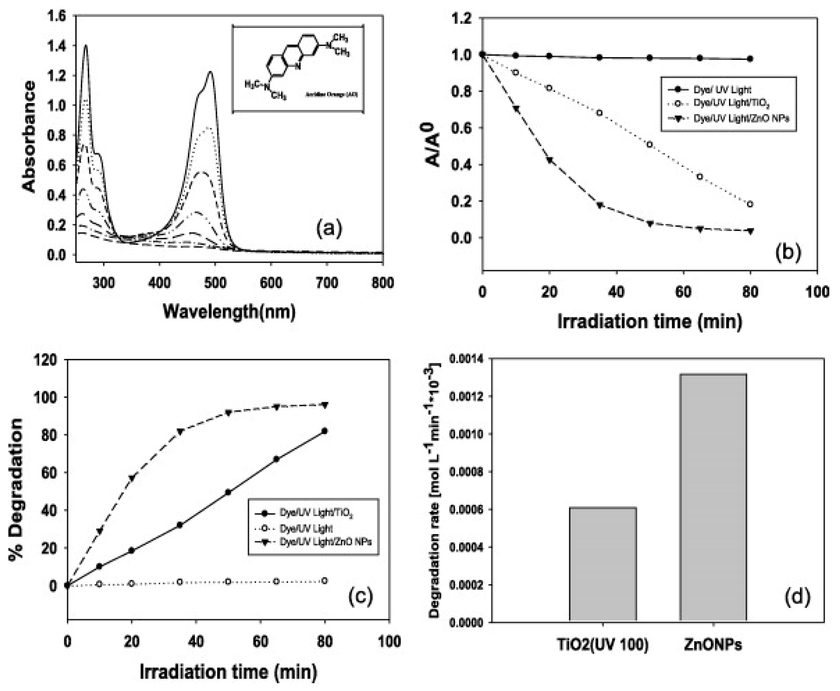
Khan et al. [105] conducted a study on the large-scale synthesis of well-crystalline ZnO NPs through a simple hydrothermal process using aqueous mixtures of (ZnCl) and ammonium hydroxide (NH4OH). The resulting NPs exhibited a wurtzite hexagonal phase with an almost spherical shape and an average diameter of ~50 ± 10 nm. Their structural properties were extensively characterized, and the NPs demonstrated high efficacy as a photocatalyst for degrading acridine orange (AO) and as a chemical sensor for electrochemically sensing acetone in the liquid phase. Comparative analysis revealed that the ZnO NPs exhibited superior photocatalytic activity compared to commercially available TiO2-UV100, achieving almost complete degradation of AO after 80 min of irradiation, as Figure 2 shows. Additionally, the ZnO NPs showed excellent performance as a chemical sensor for detecting acetone, with high sensitivity (~0.14065 μA/cm2 mM) and a lower detection limit (0.068 ± 0.01 mM) within a short response time of 10 s.
Our research group has been dedicated to refining the hydrothermal method for synthesizing ZnO nanowire arrays (ZnO NWs). Through our previous endeavors, we successfully demonstrated the synthesis of well-aligned ZnO NWs using a silicon substrate coated with a pre-deposited ZnO seed layer. This involved depositing the seed layer on the Si substrate through spin-coating and then undergoing calcination to produce ZnO nanocrystallites as seeds. Subsequently, the growth of nanowires was achieved through hydrothermal treatment, enabling us to adjust growth conditions to fine-tune the morphology and defect concentration within the ZnO band gap. We highlighted the significant influence of key parameters of the hydrothermal method on the microstructure and morphology of the ZnO NWs. Factors such as the pH value of the aqueous solution, growth time, and solution temperature during ZnO NWs growth were found to affect the characteristics of the nanowires [98]. In our 2013 publication, we detailed the preparation of the growth solution and the optimal conditions for hydrothermal growth, emphasizing the importance of these parameters in achieving homogeneous ZnO NWs with higher aspect ratios. This research exemplified the optimization of hydrothermal synthesis conditions for ZnO NWs arrays, paving the way for their potential applications across various fields, particularly in water treatment [98].
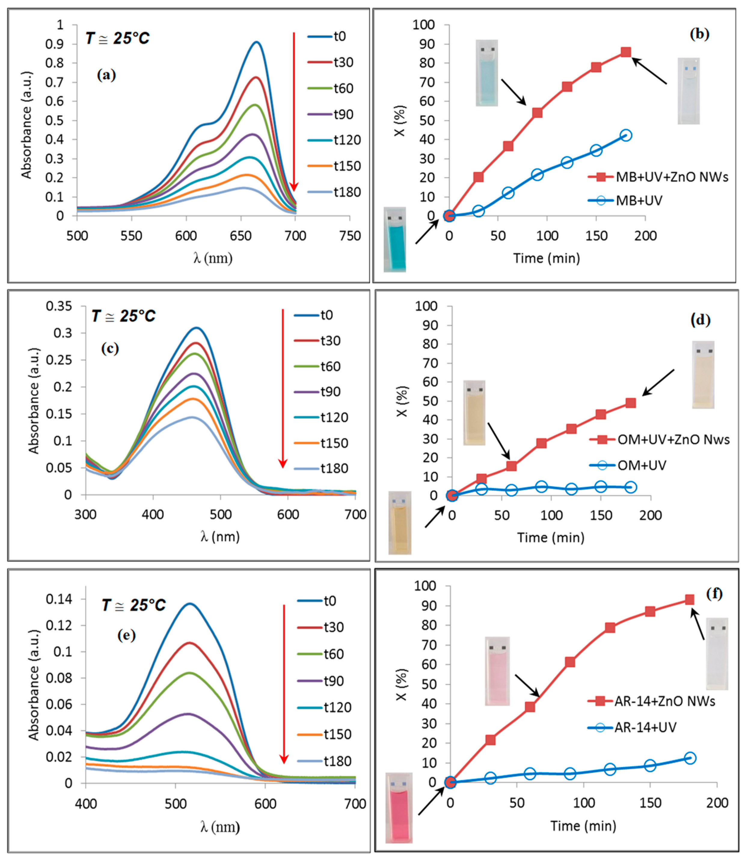
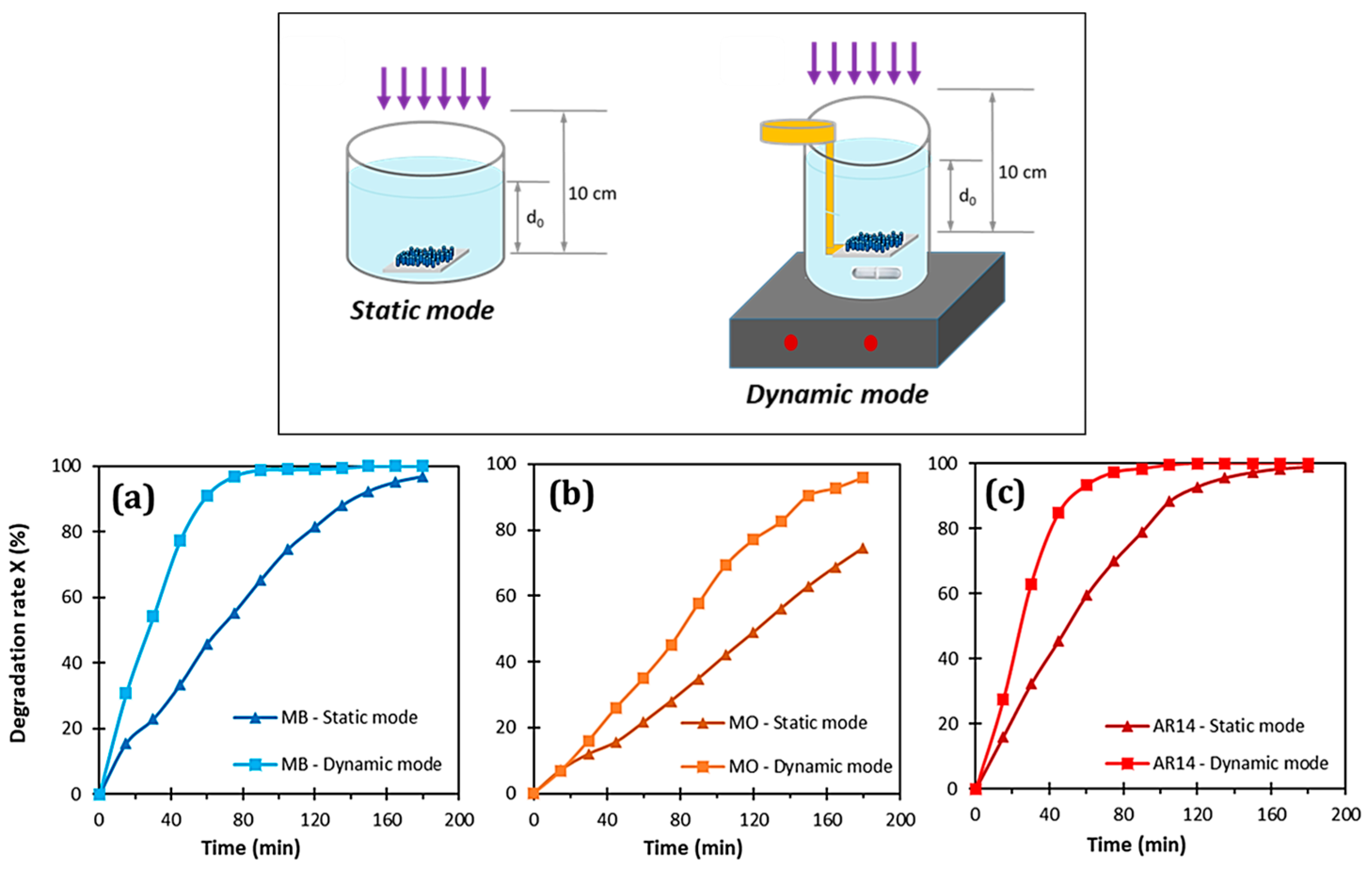
In subsequent studies, we evaluated the photocatalytic performance of ZnO NWs in degrading three dyes (methylene blue (MB), methyl orange (MO), and acid red 14 (AR14)) under UV irradiation [106]. The results revealed substantial enhancements in degradation rates, with approximately 49% degradation for methyl orange, 86% degradation for methylene blue, and 93% degradation for AR14 after 180 min of UV exposure in the presence of ZnO nanowires (see Figure 3). Furthermore, our investigations into the effects of photocatalysis under static and dynamic modes highlighted the pivotal role of ZnO NWs in expediting degradation processes, particularly under dynamic conditions, where both MB and AR14 achieved total mineralization in less than 2 h (see Figure 4) [70].
Our contributions to the field include publications focusing on the synthesis of ZnO NWs through the hydrothermal method and exploring the impact of doping on their photocatalytic properties. If you require more detailed insights, we invite you to refer to the publication by Le Pivert et al. [107].

3.2. Solvothermal Method

The solvothermal method, a variation of the hydrothermal technique, uses organic solvents instead of aqueous ones (such as ethanol or ethylene glycol). This method allows for the precise control of temperature and pressure in an autoclave to synthesize materials under specific conditions. The higher temperatures that are possible with the solvothermal method, along with the use of organic solvents with high boiling points, provide better control over the size, shape, and crystallinity of ZnO NPs compared to hydrothermal methods. This method offers a versatile approach to tailor materials with specific characteristics based on the chosen solvent, expanding the range of achievable material properties and potential applications.
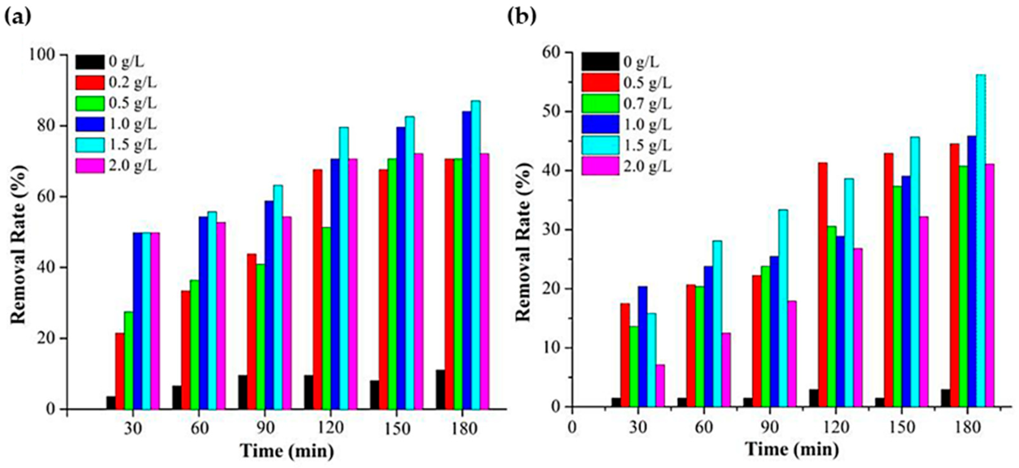
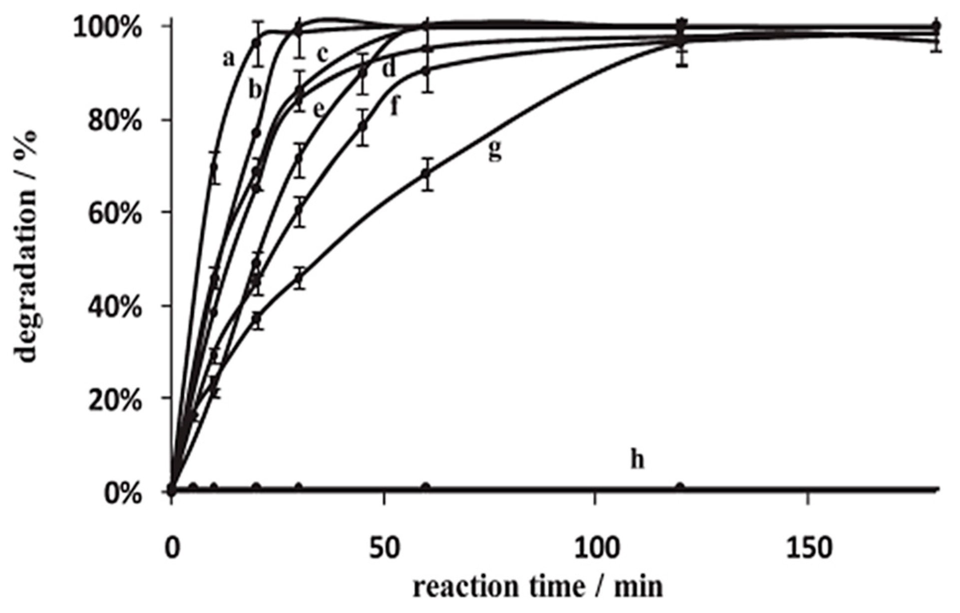
In a study by Wang et al. [108], the solvothermal method was used to produce uniform, sphere-like ZnO NPs ranging from 25 to 40 nm without the use of surfactants or agents. At a concentration of 1.5 g/L, these ZnO nanostructures exhibited exceptional effectiveness in degrading MO and p-Nitrophenol (PNP) at a concentration of 20 mg/L (Figure 5). The photocatalytic degradation rates, analyzed using pseudo-first-order kinetics, resulted in 92% degradation for MO and 56.2% for PNP after 180 min under UV light.
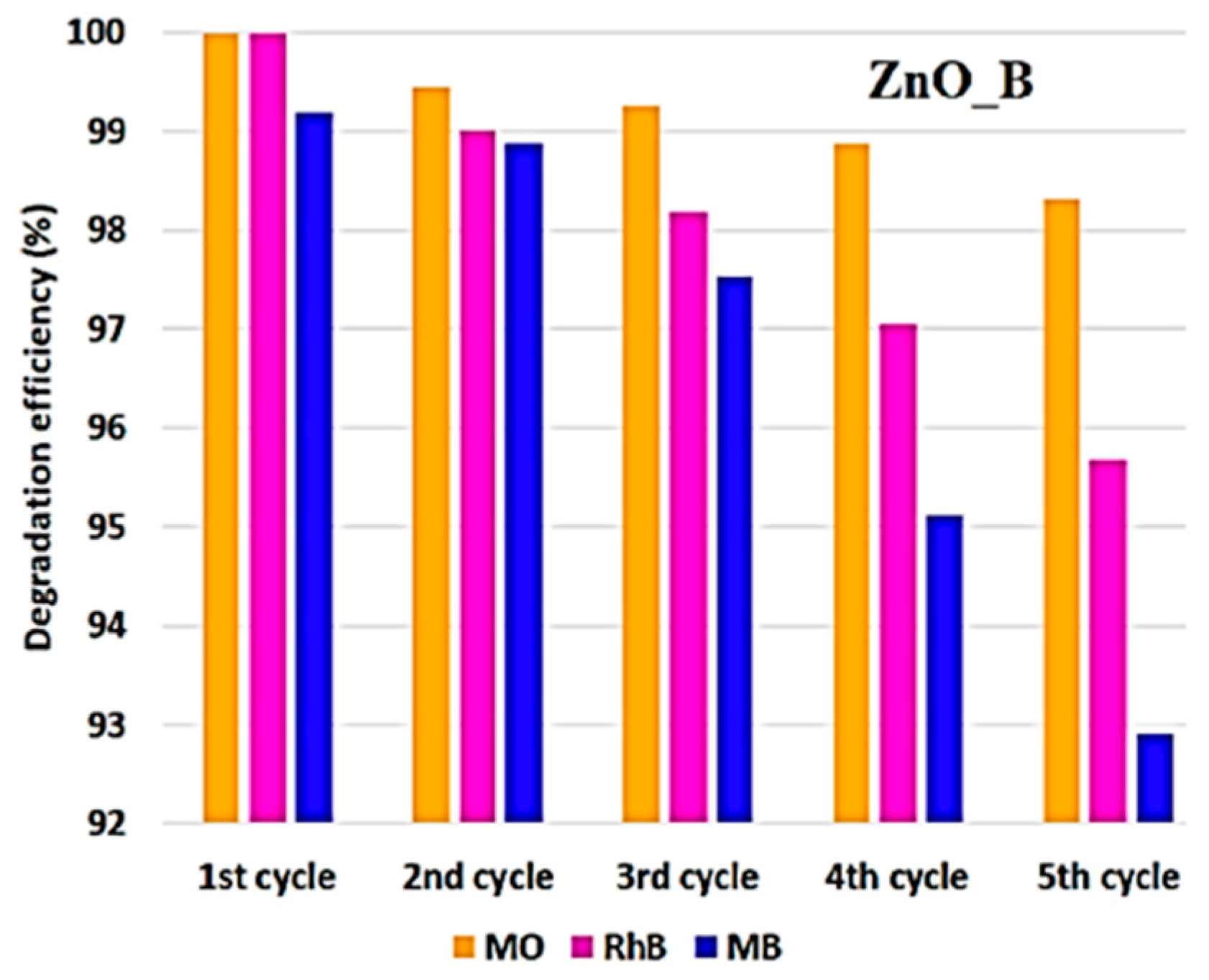
Additionally, Motelica et al. [109] conducted a study on ZnO nanoparticle synthesis through forced solvolysis using Zn(CH3COO)2·2H2O in n-butanol, ethylene glycol, and glycerin. They found that the choice of alcohol influenced nanoparticle size and morphology, with alcohols containing a single –OH group resulting in smaller particles. ZnO nanoparticles produced in n-butanol were monocrystalline and polyhedral-shaped (ZnO_B), averaging 27 nm, while those from ethylene glycol were monocrystalline and round-shaped at approximately 44 nm. Glycerin synthesis led to larger, hexagonal, and polycrystalline particles around 120 nm. Their investigation into the photocatalytic performance with MB, rhodamine B (RhB), and MO revealed superior degradation efficiency in the order of MO > RhB > MB, increasing with nanoparticle size. The smallest nanoparticles exhibited exceptional degradation rates exceeding 99% and maintained high efficiency over multiple cycles (Figure 6). Furthermore, these NPs showed potent disinfection capabilities against various Gram-positive and Gram-negative bacterial strains.
In a different study, Xu et al. [110] made ZnO materials in various shapes using a solvothermal method with different solvents, and, for the first time, they used zinc acetylacetonate (Zn(C5H7O2)2) as the zinc source. They found that the ZnO took on shapes like cauliflower-like, truncated hexagonal conical, tubular and rod-like, hourglass-like, nanorod, and spherical forms, depending on the solvent used (THF, decane, water, toluene, ethanol, and acetone, respectively). They looked at how well these ZnO samples broke down phenol, and they found that ZnO made in THF broke it down the fastest at a rate of 0.1496 min−1, nine times faster than commercial ZnO. Figure 7 illustrates the photodegradation results for phenol on the ZnO catalysts.

3.3. Chemical Vapor Deposition (CVD)

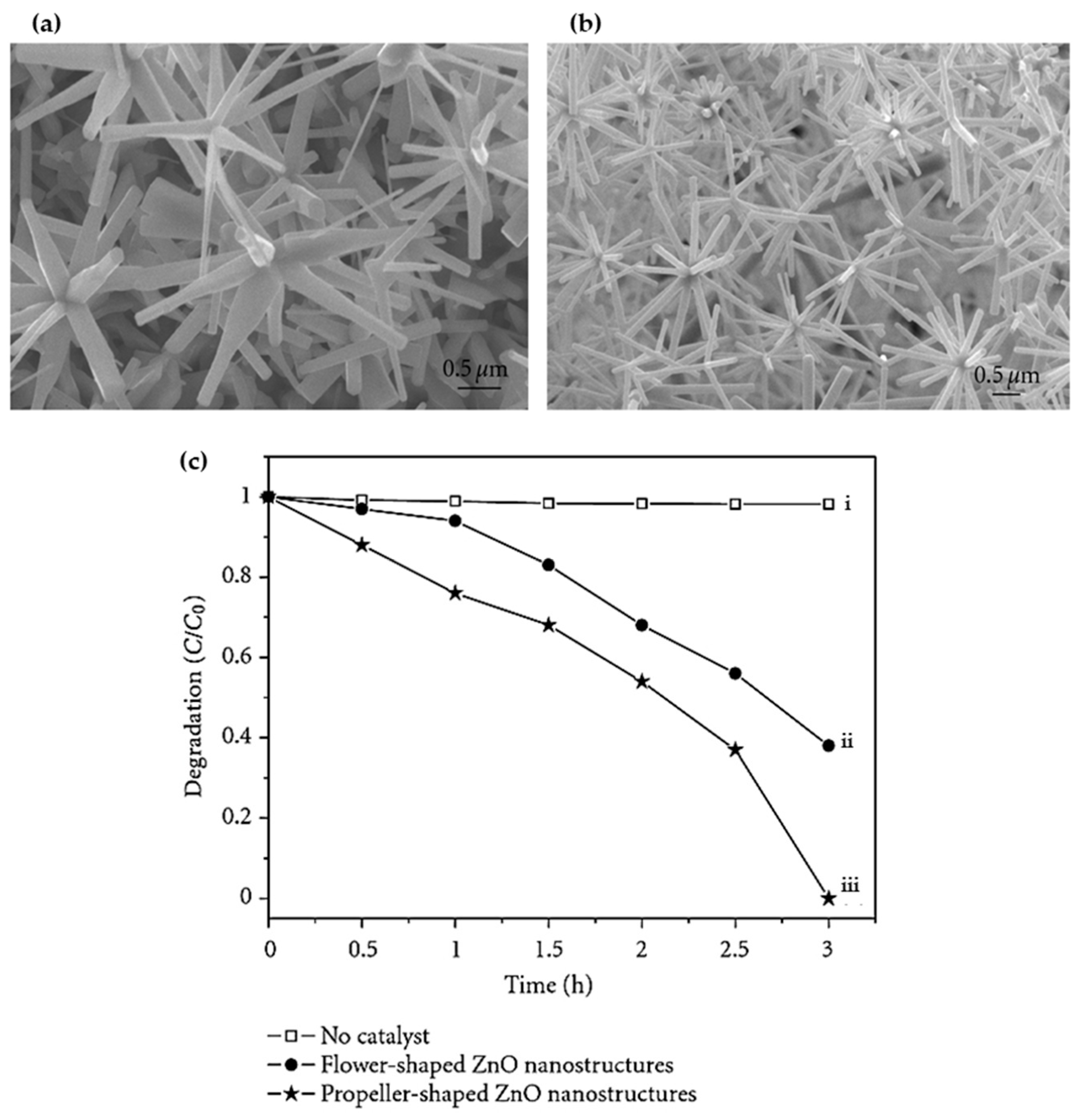
Chemical Vapor Deposition (CVD) is a deposition technique that involves introducing a substrate in contact with vapor-phase precursors and initiating reactions or decomposition, leading to the deposition of the desired molecules. The process removes unwanted by-products through gas flow circulating in the reactor. Variants of CVD include Atmospheric Pressure CVD (APCVD), conducted at atmospheric pressure; Plasma-enhanced CVD (PECVD), enhanced by plasma for room-temperature deposits; and Metalorganic CVD (MOCVD), utilizing metal–organic compounds as precursors. This method can yield a wide range of ZnO nanowire morphologies, sometimes within the same sample. For instance, Wang et al. [111] applied the one-step CVD method to produce distinctive propeller-shaped and flower-shaped ZnO nanostructures on Si substrates (Figure 8a,b). The propeller-shaped ZnO nanostructure consisted of axial nanorods (50 nm tip, 80 nm root, and 1 μm length) surrounded by radial-oriented nanoribbons (20–30 nm in thickness and 1.5 μm in length). Similarly, the flower-shaped ZnO nanostructure replicated the propeller-shaped ZnO morphology, with differences in the shape of the leaves. The nanorod leaves (30 nm in diameter and 1–1.5 μm in length) exhibited a radial alignment and pointed toward a common center. Additionally, the flower-shaped ZnO nanostructures demonstrated sharper and more intense UV emission at 378 nm compared to the propeller-shaped ZnO, indicating superior crystal quality and reduced structural defects. In contrast, the photodegradation of RhB under UV light illumination showed that the propeller-shaped ZnO nanostructures exhibited a higher photocatalytic efficiency relative to the flower-shaped ZnO nanostructures, as depicted in Figure 8c.

4. ZnO as a Photocatalyst

After discussing the generalities of ZnO and the various synthesis methods, we now turn our attention to the role of ZnO as a photocatalyst and explore the mechanism of photocatalysis along with the relationship between synthesis methods and properties, particularly in terms of photocatalytic efficiency. ZnO emerges as a highly efficient photocatalyst, leveraging its exceptional intrinsic qualities to confront modern environmental and energy challenges head-on. This photocatalysis process, driven by light energy to initiate chemical reactions, is pivotal in decomposing organic pollutants and harnessing clean energy resources. The ability of ZnO oxidation processes under standard environmental conditions enhances its practical utility, showcasing its significance in addressing both pollution issues and energy scarcities [112,113]. Upon absorbing photons, ZnO initiates the generation of electron–hole pairs, with electrons transitioning to the conduction band (CB) and holes remaining in the valence band (VB), depicted by Equation (1):
ZnO + h ν     e CB   +   h VB +
These charge carriers migrate to the surface, reacting with adsorbed water and oxygen molecules to form highly reactive species such as hydroxyl radicals (OH) and superoxide anions (O2•−), as depicted in reactions (2) and (3):
h VB + + H 2 O     H + + OH
e CB +   O 2   H + + O 2
These species play a crucial role due to their high reactivity, interacting with pollutant molecules to induce their degradation and discoloration. In addition, the generation of these radicals provides a mechanism to prevent the recombination of electrons and holes, thereby enhancing the photocatalytic efficiency of ZnO [114,115].
The photocatalytic efficiency of ZnO is significantly influenced by the band gap energy and the separation of charge carriers, which are intricately linked to the methods employed in synthesizing ZnO nanostructures. Various studies have demonstrated that different synthesis approaches can tailor the morphology, size, and specific surface area of ZnO nanoparticles, thereby impacting their photocatalytic efficiency [116,117,118,119,120]. The crystalline quality of ZnO is also crucial, as the presence of defect states can either enhance or inhibit photocatalytic activity. Specifically, defects such as oxygen vacancies (VO) and zinc vacancies (VZn) act as traps that prolong the lifetime of excitons, allowing more of them to reach the surface and participate in the creation of ROS [121]. However, surface defects are beneficial only to a certain extent. While surface defects can trap excitons and facilitate the degradation of organic molecules, volume-related defects tend to act as recombination centers, preventing photogenerated electrons from reaching the surface and thus curtailing photocatalytic efficiency. Different strategies have been explored to enhance the photocatalytic performance of ZnO, such as increasing the specific surface area to reduce the prevalence of volume-related defects and doping ZnO with various elements to modify its band gap and improve its electrical and optical properties [122,123,124,125]. Additionally, the introduction of metal co-catalysts can serve as electron traps, discouraging radiative recombination and boosting photocatalytic efficiency [126]. In smaller nanoparticles with a high surface-to-volume ratio, surface defects dominate the material’s properties, whereas, in larger nanoparticles, both surface and volume defects are relevant. Surface defects, particularly VOs, are advantageous as they can adsorb oxide radicals and other molecules, making them active centers for pollutant degradation. When excitons are captured by these vacancies, they are readily transferred to organic molecules adsorbed on the nanoparticle’s surface, enhancing dye degradation efficacy [118,119].
Understanding and optimizing these parameters and intrinsic properties is essential for maximizing the photocatalytic performance of ZnO, making it a promising material for environmental remediation and energy conversion applications.

4.1. Impurity Presence Effect

The organic contaminants in the synthesis process of ZnO can include residual chemicals or impurities from the precursor materials used during the synthesis, such as Polyvinylpyrrolidone (PVP), zinc nitrate hexahydrate, and sodium hydroxide (NaOH) [127]. These contaminants may also originate from the chemical reactions involved in the synthesis process, leading to the incorporation of organic residues on the surface of the ZnO nanoparticles [128]. The presence of these organic contaminants can detrimentally impact the photocatalytic performance of the catalyst by acting as hole traps on the semiconductor surface, which, in turn, leads to increased charge recombination and a subsequent decline in photocatalytic efficiency [30]. In their investigation, Umar et al. [129] explored the influence of annealing temperature on various properties and the photocatalytic efficiencies of ZnO NPs prepared through a facile solution combustion approach, employing zinc nitrate as the precursor and dextrose as the fuel and oxidizer. Notably, their findings revealed that annealing temperature significantly affects the particle size, morphology, bandgap, and photocatalytic performance of ZnO nanomaterials. The variations in key physical parameters for ZnO NPs with respect to annealing temperature are presented in Table 1. Furthermore, the ZnO nanoparticles annealed at different temperatures were effectively employed in the photocatalytic degradation of a water-soluble direct red-23 (DR-23) dye. The outcomes demonstrated that specimens annealed at 400 °C, 500 °C, 700 °C, and 800 °C exhibited photodegradation percentages of 88.48%, 95.49%, 92.63%, and 86.40%, respectively, as depicted in Figure 9. Notably, ZnO nanoparticles annealed at 600 °C achieved nearly complete photodegradation of the DR-23 dye. The data in Table 1 indicate that particle size increased with annealing temperature, leading to a reduction in the bandgap. Additionally, a smaller particle size resulted in an increased surface area of the photocatalyst, both of which contributed to enhancing the photocatalytic activities of ZnO nanoparticles. The heightened crystallinity of ZnO nanoparticles at elevated annealing temperatures also accounted for the improved photocatalytic degradation. Furthermore, the introduction of native defects in the ZnO crystal in the form of neutral (VO), singly charged ( V O + ), or doubly charged ( V O + + ) oxygen vacancies at higher annealing temperatures up to 600 °C was found to play a pivotal role in enhancing the photocatalytic efficiencies of the synthesized ZnO nanoparticles. These defects in the ZnO mitigated electron–hole recombination, increased the quantum yield, and thereby resulted in the superior photocatalytic activities of the ZnO nanoparticles. Moreover, the greater prevalence of these oxygen vacancies on the ZnO surface at higher annealing temperatures also served as traps for electrons from the conduction band. The decline in the photocatalytic activities of the ZnO photocatalyst at even higher annealing temperatures of 700° C and 800 °C may be ascribed to the reduction in surface area due to the agglomeration of ZnO particles; however, it is noteworthy that the morphology of the ZnO NPs remained unchanged in the study.
In their study, Li et al. [130] synthesized ZnO NPs with varying average crystallite sizes (21.3 nm, 38.2 nm, 46.9 nm, 56.5 nm, and 89.8 nm) by calcining the prepared powders at different temperatures ranging from 200 °C to 1000 °C. Their investigation revealed that ZnO prismatic particles calcined at 800 °C exhibited the highest level of photocatalytic activity. Furthermore, Giraldi et al. [131] conducted research on the synthesis of ZnO nanoparticles through a chemical method, investigating the impact of annealing temperature on particle size, crystallinity, and photocatalytic performance in the degradation of RhB. They noted that as the annealing temperature increased from 100 °C to 500 °C, the particle sizes expanded from 13 nm to 28 nm, accompanied by enhanced crystallinity and improved photocatalytic degradation outcomes. Moreover, Franco et al. [132] highlighted the effectiveness of ZnO calcination in removing residual organic residues, emphasizing the crucial role played by the specific surface area in this process. Their findings indicated that an annealing temperature of 500 °C represented the optimal point, beyond which, particle size augmentation and a reduction in specific surface area occurred, subsequently leading to a detrimental impact on photocatalytic activity. This underscores the delicate balance between calcination temperature, particle size, specific surface area, and, ultimately, the photocatalytic performance of ZnO NPs.

4.2. Influence of Specific Surface Area

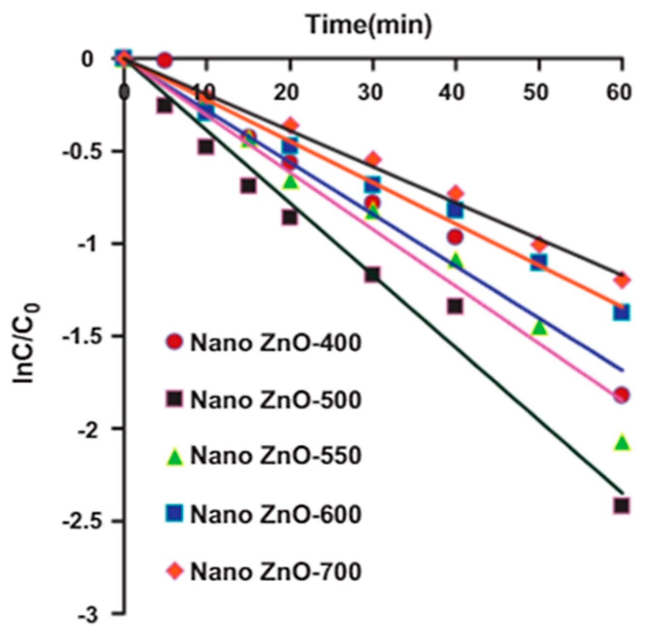
The surface area of a material decreases with an increase in the size of its crystallites, which depends on several factors such as calcination temperature and/or synthesis conditions, as shown in the preceding section. Most often, authors suggest that the photocatalytic activity of ZnO increases with an elevation in specific surface area due to a reduction in the recombination of photo-generated electron–hole pairs [117,122,133,134]. In their investigation, Hayat et al. [135] delved into the impact of calcination temperature on the photocatalytic activity of ZnO for phenol degradation. By varying the calcination temperature from 400 °C to 700 °C, the researchers explored its effect on the morphology and crystallite size of ZnO. The outcomes revealed that a reduction in particle size, leading to an increase in specific surface area, enhanced the photocatalytic efficiency by facilitating the adsorption of more phenol molecules. Employing the sol–gel method, they successfully synthesized ZnO NPs of different sizes by adjusting the calcination temperature. Interestingly, they observed that with increasing calcination temperature, the crystallites tended to agglomerate, resulting in a simultaneous loss of the catalyst’s activity. Notably, nano ZnO synthesized through sol–gel and calcined at 500 °C exhibited superior activity for the photocatalytic oxidation of phenol compared to the other photocatalysts considered in the study (Figure 10). Furthermore, the research highlighted the influence of various operating factors such as catalyst concentration, initial phenol concentration, solution pH, and irradiation intensity on the photocatalytic process. The obtained apparent kinetic rate constants followed the order nano ZnO-500 > nano ZnO-550 > nano ZnO-400 > nano ZnO-600 > nano ZnO-700, indicating the significant impact of these factors on photocatalytic efficiency. The smaller size, better dispersion, and homogeneity of nanoparticles were identified as pivotal factors contributing to the high photonic efficiency and accelerated degradation rate of phenol for nano ZnO-500.
Becker et al. [117] further investigated the impact of particle size on ZnO photocatalysts and highlighted that when ZnO NPs exhibit equivalent levels of crystallinity and morphology, the specific surface area emerges as the predominant factor influencing efficiency. In a similar vein, Flores et al. [122] emphasized the significance of a large specific surface area for augmenting photocatalytic efficiency. Nonetheless, they posited that surface area alone is not the sole crucial factor. Their research underscored the essential role of oxygen defects as a critical parameter for achieving heightened photocatalytic activity. Consistent with this, Wolski et al. [136] presented findings aligning with the notion that surface area does not stand alone as the singular critical factor. Their study indicated that a moderate surface area coupled with excellent crystallinity represents the optimal balance for maximizing performance. Contrarily, alternative studies [123,137] have suggested that a smaller surface area may yield greater effectiveness than a larger one, highlighting the significance of additional parameters such as morphology [123] and the exposure of polar planes [137]. This diverse body of research underscores the multifaceted nature of factors influencing the photocatalytic efficiency of ZnO NPs and the necessity of considering a range of parameters beyond specific surface area alone.

4.3. Influence of Morphology

The impact of morphology on the photocatalytic properties of ZnO is a subject of significant interest, with varied research findings shedding light on the complex relationship between morphology and effectiveness. Saravanan et al. [138] suggest that morphology plays a crucial role in determining photocatalytic efficiency, highlighting that ZnO spherical nanoparticles outperform nanorods despite the latter’s larger surface area. This raises intriguing questions about the genuine significance of morphology in dictating performance. On a different note, Daou et al. [139] found that ZnO with a spherical or hexagonal morphology exhibits higher efficacy in degrading MO compared to rod-shaped structures. In contrast, Li et al. [123] identified ZnO needles as the most effective, showcasing superior performance despite their relatively small surface area. Thakur et al. [125] advocate for nanospheres as optimal for enhanced photocatalytic activity. Similarly, Akir et al. [140] observed that a spherical morphology excels in degrading RhB when compared to nanosheets or hexagonal prismatic forms. Furthermore, spherical ZnO nanoparticles have shown remarkable activity in degrading MB when contrasted with nanorods, attributed to the presence of oxygen defects that mitigate electron–hole recombination and stimulate free radical generation [141]. Notably, Ahumada-Lazo et al. [142] established a significant correlation between the surface morphology of ZnO films and their photocatalytic activity, particularly in the decolorization of orange G dye solutions. Additionally, Jaafar et al. [143] demonstrated the exceptional degradation efficiency of irregular, nano-sized, sphere-like ZnO aggregates, achieving an impressive 87% degradation of phenol under visible light irradiation. Furthermore, Weerathunga et al. [144] underscored the catalytic prowess of pyramidal ZnO, achieving a remarkable 30% conversion rate for benzyl alcohol, surpassing the conversion rates of plate- and rod-shaped ZnO variants. Moreover, Shidpour et al. [145], in their comparison of different ZnO morphologies, including nanowires, nanorods, spheres, and welded nanoparticles, demonstrated the superior degradation capacity of nanowire-shaped particles in the breakdown of MB dye.
The literature emphasizes the importance of polar and non-polar facets and their relevance in photocatalysis [124,137,146,147,148]. However, this phenomenon is also linked to various other parameters such as morphology [147,149], surface [147], and oxygen vacancies [124,141]. Consequently, it is challenging to decisively determine the actual importance of morphology. The morphology and specific surface area of zinc oxide (ZnO) play pivotal roles in determining its photocatalytic performance [145]. Morphological variations, such as nanoparticles, nanorods, nanosheets, and porous structures, significantly influence the active surface area and light absorption properties of ZnO. These structures provide a high surface-to-volume ratio, enhanced directional electron transport, and exposure of reactive crystallographic facets, all of which are crucial for efficient photocatalysis. A higher specific surface area increases the availability of active sites and improves the separation of electron–hole pairs, thereby reducing recombination rates and enhancing photocatalytic efficiency. Optimized morphologies, often achieved through engineering hierarchical and porous structures, synergize these effects to maximize photocatalytic activity [150]. Consequently, ZnO with a tailored morphology and high specific surface area shows exceptional potential in environmental remediation, antibacterial applications, and solar energy conversion, underscoring the importance of precise material design in advancing photocatalytic technologies.

4.4. Impact of Structural Defects

In addition to focusing on the morphology and particle size of ZnO, structural defects have also been widely discussed. The presence of oxygen defects has drawn significant interest in current research concerning photocatalysis. Nandi et al. [151] prepared ZnO NPs and studied the impact of structural defects on photocatalytic efficiency for degrading the dye RhB under UV irradiation. They found that the catalyst with the most defects was the most effective, given that it had the largest specific surface area. Furthermore, ZnO exhibited better efficiency in degrading estrone, despite its smaller surface area compared to Aeroxide® titanium dioxide P25 (TiO2-P25) [152]. The authors attributed this efficacy to the ionic defects existing in the crystalline network of ZnO. They suggested that oxygen and zinc vacancies can trap charge carriers, thus preventing the recombination of electron–hole pairs and potentially enhancing photocatalytic performance.
Xu et al. [153] found that the photocatalytic degradation efficiency of ZnO with a high oxygen defect toward tetracycline was increased to more than 99.9%, about three times that of low-oxygen defect ZnO. Also, ultrathin ZnO/Al2O3 nanosheets with abundant oxygen defects could rapidly degrade tetracycline and RhB, affording 88.4% and 76.9% degradation in 150 min, respectively [154]. Similarly, oxygen defect-rich pencil-like ZnO nanorods possessed better photocatalytic performance for tetracycline degradation, which could reach 96% under optimal conditions [155]. The research of the Singh [156] and Pei [157] groups successively confirmed the conclusion of Li et al. [155] that more oxygen defects result in better photocatalytic performance.
On the other hand, while some publications consider Zn defects important to improve ZnO’s photocatalytic activity [136,152,158,159], a study by Danilenko et al. [160] demonstrated that zinc defects are deleterious to photocatalysis. The appearance of cationic defects in the structure decreased the photocatalytic activity in degrading phenol, while oxygen defects enhanced it, promoting charge separation, consistent with several other publications [161,162,163,164]. Additionally, He et al. [161] indicated that the photocatalytic efficiency of ZnO decreased with an increase in the calcination temperature, a phenomenon that could be attributed to the simultaneous reduction in oxygen defects. These results align with the work of Wang et al. [164], which showed that the concentration of oxygen defects is strongly dependent on the calcination temperature and that oxygen deficits render ZnO more effective. However, most publications highlighting the highest photocatalytic activity attributed to the catalyst with the highest level of oxygen defects do not account for the specific surface area, which increases in parallel with the increase in oxygen defects. Similarly, several other publications working on ZnO-based composite materials, such as Ag3PO4/ZnO [165] and C3N4/ZnO [166], also conclude that oxygen defects enhance photocatalytic efficiency and demonstrate that a higher concentration of defects corresponds to a larger surface. Conversely, contrary to the aforementioned suggestions, several other studies report an unfavorable impact of oxygen and zinc defects on ZnO’s photocatalytic activity, showing that they act as recombination centers, limiting its effectiveness [119,167,168].

5. Enhancing the Photocatalytic Efficiency of ZnO

While ZnO exhibits greater photocatalytic potential compared to its bulk counterparts, it is not without noticeable drawbacks. The primary limitations of bare ZnO include its susceptibility to dissolution at an acidic pH, photo corrosion in alkaline solutions under UV irradiation [169], rapid electron–hole recombination, a tendency toward agglomeration, and poor absorption of visible light [170]. Various strategies have been employed to address these drawbacks and enhance the structural stability of ZnO [171], such as doping with metals and non-metals, integrating with other semiconductors, employing surface deposition of conducting metals, and coupling with carbon materials, which will be further elaborated on in the subsequent subsections.

5.1. Doping

Doping, a method involving the introduction of elements into a semiconductor’s lattice, is a common approach to augment the photocatalytic performance of ZnO. This technique effectively reduces the band gap, modifies the energy level structure, enhances electrical conductivity, and broadens the absorption range to include visible light, thereby improving catalytic activity. However, excessive concentrations of dopants can generate physical defects, diminishing the photocatalytic efficiency of ZnO and altering its structure, thereby negating its advantages as an excellent photocatalyst [116].

5.1.1. Non-Metallic Doping

Non-metallic doping, particularly with nitrogen (N) [172,173,174,175], carbon (C) [176,177], fluorine (F) [178,179], and sulfur (S) [180,181], has gained prominence in the quest to enhance the activity of semiconductor photocatalysts. N-doping, notably, has revolutionized the field, following the pioneering work by Asahi’s team [158] demonstrating its efficacy in harnessing visible light utilization by TiO2. N-doping achieves this by replacing oxygen vacancies with non-metallic ions or inducing defects, thereby reducing the energy band gap of ZnO and significantly expanding its optical response region. Furthermore, N-doping introduces unique characteristics to ZnO, enabling the synthesis of p-type ZnO and effectively narrowing the energy band, making it a notable non-metallic doping element.
Moreover, the incorporation of C into the ZnO lattice has shown remarkable effects, extending carrier lifetime, suppressing recombination rates, improving intrinsic defects, and significantly enhancing visible light capture efficiency. Research by Yu et al. [182] highlighted the effectiveness of C-doping in inhibiting the recombination of photo-generated electron–hole pairs in ZnO, thus providing new active sites and preventing h+ and e complexes. Additionally, the successful preparation of C–ZnO by Islam et al. [183] demonstrated a significant red shift in the UV absorption peak of ZnO post-C doping, enabling the efficient catalytic degradation of pollutants under visible light activation.
In a study by Pan et al. [184], a novel two-step calcination process was introduced, resulting in a porous ZnO structure with exceptional charge transfer and light absorption characteristics, showcasing notable photocatalytic activity. Furthermore, the close match in ionic radii between fluorine (F) and oxygen (O), measuring 1.33 Å and 1.4 Å for F and O2− ions, respectively, enabled the effective substitution of F for O2− in the crystal structure, facilitating the formation of n-type ZnO and the release of free electrons within the semiconductor matrix. Rueda-Salaya’s research team [185] investigated the doping of various F ratios into the ZnO lattice, demonstrating outcomes such as suppressed microcrystal growth, reduced oxygen vacancies, a narrowed band gap, and enhanced efficacy in degrading diclofenac (DCF) in aqueous environments, as depicted in Figure 11.
Moreover, boron (B) [186] and phosphorus (P) [187] have also been explored for ZnO doping, exhibiting remarkable performance in pollutant degradation, pathogen inactivation, and solar hydrogen production, among other applications.

5.1.2. Transition Metal Doping

Transition metal doping alters the ZnO lattice, enhancing its ability to separate charge carriers and boosting its photocatalytic performance [188]. By creating new electronic levels in the band gap, it can absorb visible light and facilitate charge transfer within the material [189].
Among the common dopant elements such as cobalt (Co), manganese (Mn), silver (Ag), iron (Fe), gold (Au), and copper (Cu), Co stands out due to its multiple electronic states and high solubility in the ZnO lattice, making it an ideal candidate for doping.
For instance, Co-doped ZnO quantum dots produced by Liu et al. [190] showed promising results in disinfecting Escherichia coli. Increasing Co doping improved light absorption and charge separation, leading to better photocatalytic activity. In another study, Lu et al. [40] synthesized Co-doped ZnO nanorods using a facile hydrothermal process, achieving a 93% degradation of alizarin red dye in 60 min. Similarly, Xiao et al. [191] demonstrated enhanced photocatalytic activity with Co-doped ZnO, effectively degrading MB dye in 300 min. These studies highlight the importance of defects in ZnO for improved photocatalytic performance.
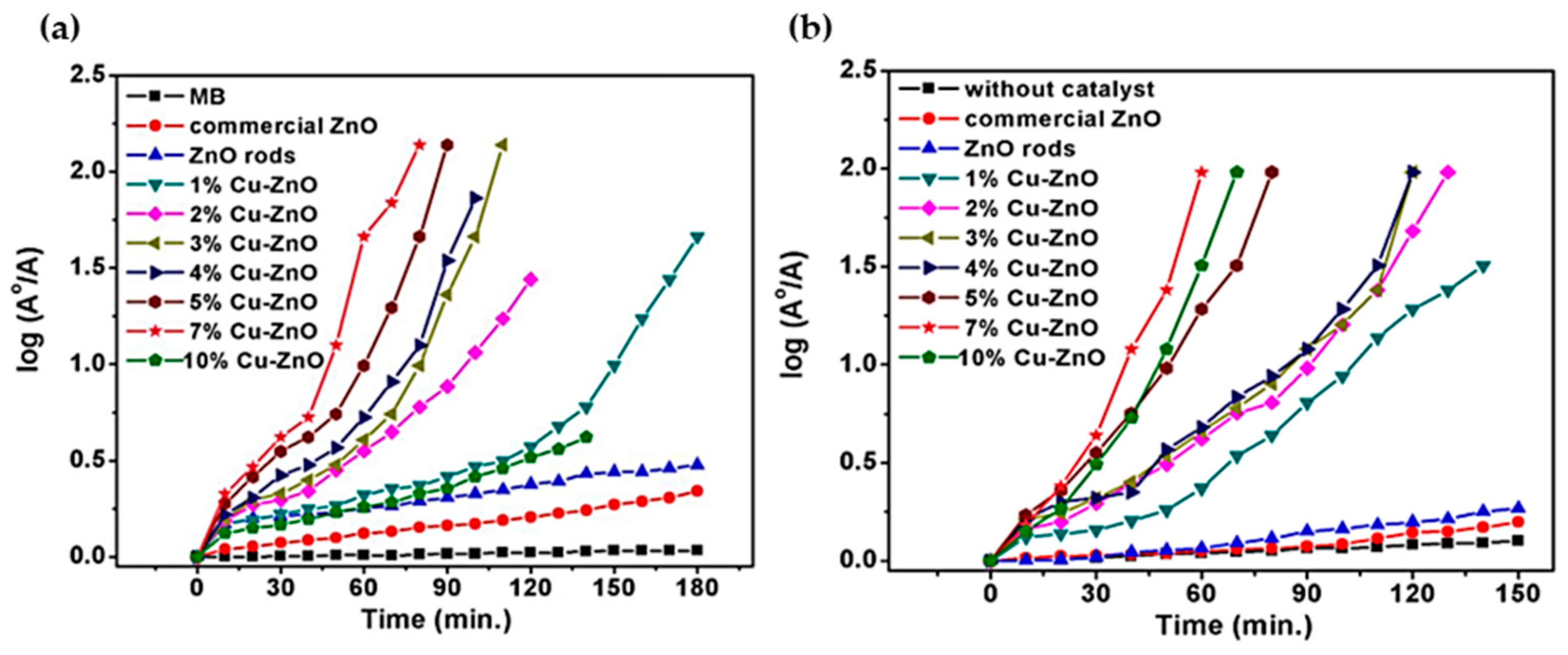
In a different study, researchers developed Cu-doped ZnO through a solvothermal method, showcasing remarkable photocatalytic properties under visible light [192]. The UV–Vis absorption spectrum of Cu-doped ZnO displayed an effective optical performance throughout the visible light spectrum, with a broad tail band spanning approximately 400 nm to 750 nm. Shah’s work [193] on rod-shaped Cu-doped ZnO indicated that increased Cu content narrowed the optical band gap, subsequently enhancing the degradation rate. Cu-doped ZnO nanorods demonstrated efficient photocatalytic activity, with 20 mg showing a photodegradation efficiency of 57.5% for MO and up to 60% for MB. Pawar et al. [194] synthesized Cu-doped ZnO microstructures, with 7% Cu-doped ZnO requiring 50 min for the photocatalytic degradation of MB and RhB dyes (refer to Figure 12). The improved photocatalytic activity was attributed to the presence of (001) polar surfaces, oxygen vacancies, and the increased absorption of visible light. Additionally, Mohan et al. [195] conducted a study on Cu-doped ZnO nanorods synthesized through the vapor transport method, focusing on the UV light-driven photocatalytic degradation of resazurin dye, and attributed the enhancement in photocatalytic activity of Cu-doped ZnO nanorods to the presence of oxygen vacancies.
Furthermore, Li et al. [196] investigated the impact of Ag-doping on the structural, electronic, and optical properties of ZnO NWs using first-principles calculation. The study revealed that Ag dopants preferred substitution at the Zn sites, improving visible photocatalytic performance by introducing an additional absorption peak in the visible region, associated with the excitation of the Ag gap state. This led to a reduction in the gap of ZnO NWs, highlighting the potential application of AgO defects for visible light catalysis.
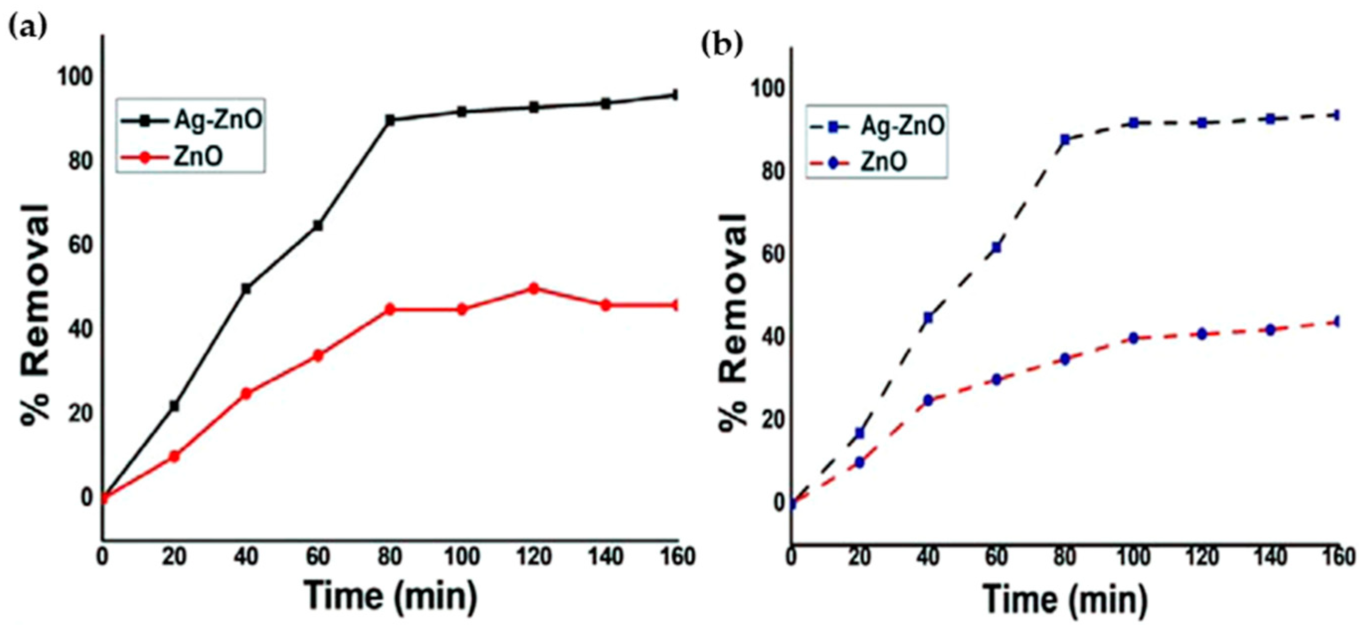
In their study, Chauhan et al. [197] reported on the photocatalytic dye degradation and antimicrobial activity of pure ZnO and Ag-doped ZnO synthesized using a green method involving the use of Cannabis sativa leaf extract as a reducing and stabilizing agent. The findings revealed that the synthesized Ag-ZnO and ZnO NPs exhibited antimicrobial activity against various human pathogenic bacteria (both Gram-positive and Gram-negative) and different fungal strains. Specifically, Ag-doped ZnO NPs demonstrated the maximum inhibition zone against all bacteria, including Escherichia coli, Klebsiella pneumonia, methicillin-resistant Staphylococcus aureus, Pseudomonas aeruginosa, Salmonella typhi, and Staphylococcus aureus, while ZnO NPs exhibited the minimum inhibition zone. Furthermore, both ZnO NPs and Ag-doped ZnO NPs effectively inhibited the growth of two different plant pathogenic fungi, Fusarium spp. and Rosellinia necatrix. Additionally, under solar light, Ag-ZnO and ZnO NPs demonstrated the removal of 96% and 38% of Congo red and 94% and 35% of MO in 80 min, as illustrated in Figure 13.
In our previous work [198], we successfully synthesized the homogeneous transition metal-doped ZnO NWs on a Si substrate via hydrothermal method. The doped samples, including ZnO:Fe 3%, ZnO:Ag 2%, and ZnO:Co 2%, demonstrated enhanced efficiency in degrading MO compared to undoped ZnO. The doped samples achieved complete MO degradation within 180 min or less, while the undoped ZnO achieved only 95% degradation in the same time frame, as shown in Figure 14. This improved performance can be attributed to a reduction in bandgap energy, leading to increased photon absorption and more energy for the reaction, as well as an increased number of oxygen vacancies in the nanowire lattices, acting as electron traps. Additionally, the dopant ions on the nanowire surface helped reduce the recombination rate of electron–hole pairs.
Additionally, the doping of Mg [199], Mn [200], and Au [201] led to the optimization of ZnO’s photocatalytic performance.

5.1.3. Rare Earth Metal Doping

Recent research interest in using rare earth (RE) elements to modify ZnO has focused on their exceptional optical properties. RE ions like (La), europium (Eu), neodymium (Nd), samarium (Sm), dysprosium (Dy), praseodymium (Pr), ytterbium (Yb), and cerium (Ce) have been added to ZnO, sparking discussions regarding their impact on ZnO’s photocatalytic performance. Different views exist on the effects of RE doping. Some suggest it leads to lattice expansion, increasing oxygen vacancies but reducing photogenerated hole and electron formation. Others propose that rare earth ions broaden ZnO’s light absorption range, facilitating efficient photon energy transfer.
Studies on La-doped ZnO, including research by Nguyen et al. [202] and others [203,204,205], have shown excellent performance in gas-sensitive and photocatalytic activities. Research into various La-doping concentrations has offered valuable insights into reducing the band gap and enhancing pollutant degradation. For instance, Nguyen et al.’s study revealed a reduced band gap of ZnO upon La3+ ion doping, enabling efficient MO dye breakdown using La0.1Zn0.9O under visible light [202]. Similarly, Jia et al. [206] found significantly improved photocatalytic efficiency with solvothermally synthesized La3+-doped ZnO materials, identifying 2% La as the optimal concentration for the peak degradation of RhB. Additionally, Kumar et al. [207] reported notable effectiveness in degrading MO, RhB dyes, and picric acid (PA) with 1 mol% La-doped ZnO.
Continuing our discussion on RE element doping in ZnO, further research has shown promising outcomes. For instance, incorporating Eu into ZnO nanoparticles led to enhanced photocatalytic performance, with 1.62% Eu-doped nanoparticles achieving 99.3% degradation of methylene blue in just 60 min [208]. Dash et al. [209] found that the breakdown of MO increased with higher Eu levels, reaching 90% efficiency. Franco et al. [210] noted that Eu-doped ZnO, produced through a supercritical anti-solvent process, exhibited excellent mineralization and discoloration, almost entirely removing eriochrome black-T azo dye after 240 min of UV light exposure. In another investigation, Alam et al. [211] used a straightforward sol–gel approach to create ZnO NPs doped with RE metals (La, Nd, Sm, and Dy) and tested their photocatalytic activity in degrading MB and RhB under UV light. Among these, Nd-doped ZnO displayed the highest efficiency, achieving a degradation rate of 98%. Furthermore, investigations explored the photocatalytic efficacy of pure ZnO and ZnO doped with RE elements (La, Ce, Pr, Er, and Yb) for treating pollutants in water and wastewater. The results revealed that Er-, Ce-, Pr-, and, notably, Yb-doped ZnO exhibited higher photoactivity compared to unmodified ZnO and the established benchmark material TiO2 P25 (refer to Figure 15) [212].

5.2. Co-Doping of ZnO

In addition to single-element doping, the investigation of the simultaneous doping of multiple elements into ZnO has revealed significant potential for enhancing photocatalytic activity. For instance, the simultaneous doping of sulfur (S) and chlorine (Cl) into ZnO resulted in the modification of ZnO with a high density of active sites and reduced charge complexation rate of electron–hole pairs, effectively reducing the optical band gap and substantially increasing the photocatalytic degradation rate compared to pure ZnO [213]. Furthermore, research efforts have highlighted the modification of ZnO’s physicochemical properties through co-doping with rare earth ions. For example, the co-doping of La3+ and Ce3+ led to the contraction of the ZnO band gap energy, shifting it from UV to visible light absorption, thereby enhancing luminescence and antibacterial properties [214]. Similarly, the co-doping of Ce and Yb induced changes in the crystal structure of ZnO, with variations observed in lattice constants based on the concentrations of the co-dopants [215]. Notably, the nature and concentrations of the doping elements in the composites significantly influenced the photocatalytic activity, especially with excessive co-dopant concentrations affecting carrier recombination before reaching the nano photocatalyst surface. These insights underscore the potential of co-doping strategies in tailoring the photocatalytic properties of ZnO for diverse applications [141].

5.3. Noble Metal Deposition on ZnO

The deposition of noble metal nanoparticles (e.g., Ag, Au, Pd) or their oxides onto ZnO represents a powerful strategy to improve ZnO’s photocatalytic activity by inhibiting the recombination of photogenerated h+ and e pairs. Unlike doping, noble metals deposited on ZnO surfaces have distinct Fermi levels, facilitating electron transfer from the semiconductor to the metal. This process creates a space charge layer that provides additional active sites, thereby prolonging redox reactions and generating more h+/e pairs, ultimately enhancing ZnO’s photocatalytic performance. Although effective, the high cost of precious metals necessitates exploration into economical deposition methods for noble metal-modified ZnO. Research has focused significantly on depositing Ag Nps on ZnO due to their affordability and stability. Despite the inherent difficulty in depositing Ag NPs on ZnO surfaces, various research endeavors have explored deposition methodologies at different temperatures and durations [42,216,217]. Moreover, Ag NPs exhibit efficient antibacterial properties against both Gram-negative and Gram-positive bacteria, although concerns about their cytotoxicity have been raised, prompting investigations into controlled release strategies to mitigate adverse effects [218]. Additionally, porous ZnO–Ag composites have demonstrated promising outcomes in ROS generation under visible light, showing potential in biomedical applications such as wound healing. Similarly, palladium (Pd) integration with ZnO has been explored extensively, driven by Pd’s catalytic prowess in converters and fuel cells. Methods like the one-pot synthesis by Bao et al. [43] have successfully yielded Pd-ZnO composites with exceptional catalytic and recyclability properties. Further innovations, such as microwave hydrothermal synthesis, reported by Veerakumar et al. [219], have led to stable Pd-ZnO nanostars capable of degrading hazardous pesticides under visible light, emphasizing their role in environmental sustainability. Chang et al. [220] also advanced Pd-ZnO integration through deposition and lattice doping, enhancing optical properties and catalytic efficiency for improved environmental remediation strategies. These developments highlight the versatile applications of noble metal-modified ZnO in addressing environmental challenges and promoting public health.
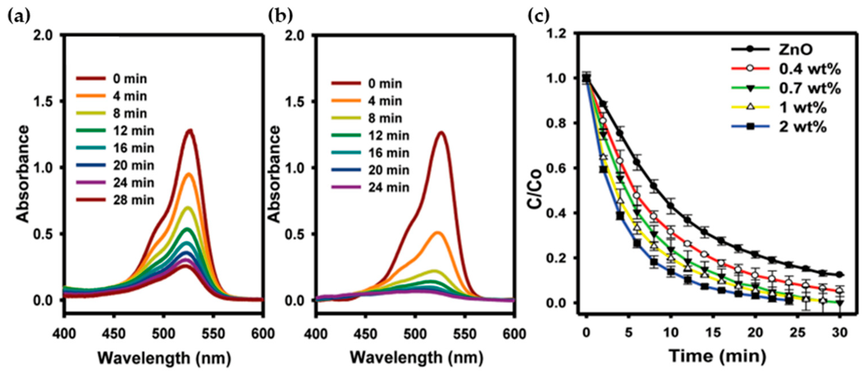
Similarly, the integration of noble metal Au NPs into ZnO has proven highly effective in enhancing its photocatalytic activity through various mechanisms. Au NPs act as effective charge carriers, facilitating interfacial charge transfer and extending light absorption into the visible spectrum, thereby optimizing solar energy utilization [44,221,222]. This has enabled the development of Au-ZnO nanocomposites with enhanced physical and chemical properties suitable for applications such as pollutant degradation (Figure 16), dye-sensitized solar cells, and biosensing, thereby advancing sustainable energy applications [44]. Udawatte et al. [44] demonstrated well-defined Au/ZnO Nps composites by modifying ZnO with preformed Au NPs shielded with bifunctional glutathione ligands. These composites exhibited significantly improved photocatalytic activity, eliminating the need for thermal activation processes in organic substrate breakdown via oxidative and reductive pathways. The photocatalytic efficiency was directly correlated with Au loading, where thiolate-protected Au NPs effectively extracted electrons from photoexcited ZnO, thereby enhancing charge separation and overall catalytic performance. Assessing the degradation of rhodamine 6G (R6G) dye in aqueous media, the absorption profiles during photocatalytic reactions with ZnO and the 2 wt% composite illustrated comparable degradation pathways for R6G (Figure 16a,b). However, the composite achieved complete dye bleaching within 30 min, contrasting with partial decomposition by ZnO alone. Furthermore, the time for 50% degradation of R6G decreased from approximately 8 min with ZnO to 6 min with a 0.4 wt% composite and further to about 2.5 min at 2 wt% Au NPs loading, demonstrating significant enhancement in catalytic activity with increasing Au NPs content. These findings underscore the pivotal role of Au NPs in augmenting the photocatalytic performance of ZnO-based composites, marking a substantial stride in enhancing their practical applicability.
Furthermore, the synergistic effect of bimetallic coupling, as demonstrated by Lee et al. [223] and Zhai et al. [224], showcased enhanced photocatalytic activity compared to monometallic deposition, making it a promising avenue for constructing efficient ZnO-based photocatalysts. These studies highlighted the generation of Schottky barriers at the interfaces of Au–ZnO and Pd–ZnO, resulting in improved photodegradation capabilities.

5.4. Semiconductor Composite

Coupling ZnO with other semiconductors has emerged as a highly promising approach to significantly enhancing its photocatalytic activity. This method involves creating nanocomposites with superior properties, particularly for photocatalysis, by boosting light absorption, suppressing photo-induced electron–hole pair recombination, and promoting increased charge separation. A study by Lin and Chiang showcased how the extended lifetime of charge carriers in nanocomposites, achieved through inter-particle electron transfer between the conduction bands, leads to enhanced photo-degradation reactions [225]. Additionally, the coupling of ZnO with semiconductors of varying band gaps, such as Cds, SnO2, ZnS, TiO2, CuO, α-Fe2O3, and Co3O4, has been extensively investigated, demonstrating a remarkable potential to improve the overall photocatalytic process [45,46,47,48,49,50,51,52,53,54,55,56,226,227,228,229].
For example, Zgura et al. [53] delved into the morphological, structural, optical, and photocatalytic aspects of ZnO and ZnO–CdS composites, uncovering that the introduction of CdS impacts the nucleation growth of ZnO. This was evidenced by the quasi-epitaxial growth of ZnO on the (111) crystal faces of CdS nanoparticles with a cubic crystal structure, resulting in flower-like structures of varying sizes. The intrinsic defect in ZnO–CdS acted as a h+/e trap, leading to a decreased complexation rate of electrons and holes, rendering it significantly more efficient in the degradation of RhB.
Additionally, a study addressing the photodegradation of MO highlighted the favorable performance of nanosized, coupled ZnO/SnO2 photocatalysts, particularly when the Sn content was approximately 33.3 mol%, emphasizing the pivotal role of material composition in coupled semiconductor photocatalysts [229]. These instances collectively depict the valuable potential of coupling ZnO with other semiconductors to enhance its photocatalytic activity, positioning such systems as crucial in advancing environmental remediation technologies.
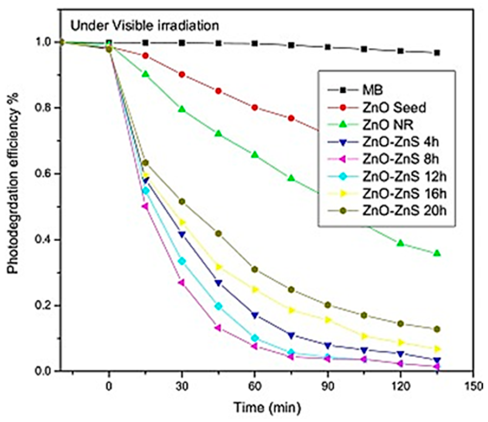
At present, a large number of studies have reported different semiconductor deposition methods to modify ZnO to improve its photocatalytic ability. For example, Ranjith et al. [47] reported vertically aligned core–shell ZnO–ZnS nanocomposites that exhibited high photocatalytic activity and excellent dye degradation stability under light irradiation. Upon visible light irradiation, these ZnO-based nanostructures exhibited varying levels of MB decomposition: 64.4%, 96.3%, 98.7%, 98.3%, 98.21%, 93.9%, and 87.4% for ZnO nanorod, ZnO-ZnS nanorod (4 h), ZnO-ZnS nanorod (8 h), ZnO-ZnS nanorod (12 h), ZnO-ZnS nanorod (16 h), and ZnO-ZnS nanorod (20 h) core–shell morphologies, respectively, within 135 min (Figure 17). This demonstrated a consistent trend in catalytic efficiency with varying shell wall thicknesses under both UV and natural sunlight irradiation. The UV–Vis absorption spectra revealed a blue shift at the absorption edge, attributed to the improved charge separation between ZnS and ZnO, a phenomenon further enhanced by the thickness of the shell wall.
In another work, it was reported that the photoluminescence spectra of CuO-coupled ZnO nanomaterials showed characteristic peaks in the visible range, and the composite material improved the absorption in the visible region of the solar spectrum [55]. The increase in the amount of ROS generated under visible light catalysis was effective in killing methicillin-resistant Staphylococcus aureus [56]. The α-Fe2O3 and ZnO composites also exhibited photocatalytic degradation and antibacterial properties, which provided a new strategy for the photodynamic antibacterial killing of multi-drug resistant bacteria [54]. The unpaired h+ and e within the surface of the Ag/Fe2O3/ZnO heterostructure fabricated by Rahmah et al. [48] using chemical precipitation could generate free radicals at both the CB energy level (superoxide ion) and VB energy level (hydroxyl group), possessing efficient photocatalytic and superhydrophobic properties. ZnO–Ag3PO4-WO3 heterojunctions could improve the effective strategy of photocatalytic activity by improving chemical and structural properties as well as inducing inter-site charge transfer, promoting the utilization of visible light and showing excellent ammonia degradation activity in sunlight [230].

5.5. Coupling with Carbon-Based Materials

Carbon-based materials, including fullerene, carbon nanotubes, graphene materials, and graphitic carbon nitride, play essential roles across various technological applications, particularly in catalysis and water treatment, due to their diverse forms and exceptional properties [231,232]. Among these materials, graphene stands out for its unique hexagonal lattice structure of sp2-bonded carbon atoms, providing high electrical conductivity, mechanical strength, and a large specific surface area. When integrated with ZnO, graphene and its derivatives like graphene oxide (GO) and reduced graphene oxide (RGO) play a pivotal role in enhancing photocatalytic performance by facilitating efficient charge separation at the semiconductor interface [233].
The disparity in Fermi levels between reduced graphene oxide and the oxide semiconductor enables rapid electron transfer to the graphene surface, thereby promoting faster reduction reactions. This mechanism enhances photocatalytic efficiency by improving the separation efficiency of photogenerated electron–hole pairs [234]. Additionally, the π-π stacking interaction between the aromatic rings of graphene and incoming moieties significantly accelerates their adsorption on oxide semiconductors [235].
Recent advancements and research efforts have focused on utilizing various forms of ZnO composites incorporating graphene and GO for organic pollutant degradation. For instance, ZnO/graphene composites prepared via chemical precipitation demonstrated a notable 2.4 times increase in photocatalytic degradation efficiency concerning MB compared to bare ZnO [57]. However, a similar method followed by heat treatment resulted in the formation of multilayer graphene, leading to a reduced surface area and, subsequently, lower photocatalytic activity. On the other hand, graphene loaded onto ZnO nanofibers created via electrospinning with a 0.5% graphene content exhibited excellent photocatalytic efficiency in degrading MB under UV irradiation, attributed to better electron transport facilitated by graphene’s superior electrical conductivity [236]. Moreover, the enhanced photocatalytic activity of ZnO–graphene composites was associated with the robust interaction between ZnO and defect sites of graphene [57]. In another study, a graphene–ZnO composite with a quasi-shell–core structure was proposed for the photocatalytic degradation of RhB [58]. The establishment of an electric field between the ZnO core and the graphene shell layer reduced the charge transfer resistance for photogenerated electrons, enhancing the photocatalytic dye degradation capacity. The composite structure was further modified by coating ZnO nanospheres with reduced graphene oxide, achieved through the electrostatic attraction between positively charged ZnO nanospheres and negatively charged reduced graphene oxide [237]. Notably, approximately 10% reduced graphene oxide exhibited a fivefold enhancement in photoactivity compared to bare ZnO nanospheres, attributed to the decreased recombination of electron/hole pairs and the improved absorption capacity of ultraviolet light. The efficiency of photocatalytic degradation was closely linked to the fate and transfer of photogenerated electrons, with the highly enhanced photoactivity attributed to the substantial interfacial contact between ZnO nanospheres and RGO nanosheets, significantly improving the fate and transfer of photogenerated electron–hole pairs from ZnO.
Based on a study by Nguyen et al. [238], a comparison of the photoactivity of ZnO-rGO with different ZnO morphologies (nanospheres, nanodisks, and nanorods) was conducted for the photocatalytic degradation of dyes (MB and RhB) under UV irradiation. The results indicated that the smallest particle size (15–35 nm) was achieved with ZnO spheres, leading to the rapid transport of excited electrons compared to short rods and disks. Importantly, the ZnO sphere–RGO composites (sZG) exhibited higher adsorption and enhanced photocatalytic degradation of the dyes, as depicted in Figure 18.
In their study, Tayebi et al. [239] also reported how different shapes of ZnO, combined with rGO, affect photocatalytic activity, using MO degradation as a model. They compared ZnO nanoparticles (ZnO NPs), ZnO NP-RGO (GNP), ZnO nanorods (ZnO NRs), and ZnO NR-rGO (GNR) under UV and visible light in Figure 19. The results showed that the GNR composite achieved the highest photocatalytic activity, significantly reducing photo-corrosion to approximately 0.5%, compared to 10%, 35%, and 45% for ZnO/GNP, ZnO NR, and ZnO NP, respectively. This improvement in GNR was attributed to the better movement of photoexcited electron–hole pairs along the ZnO NRs, reducing electron–hole recombination compared to ZnO NPs.
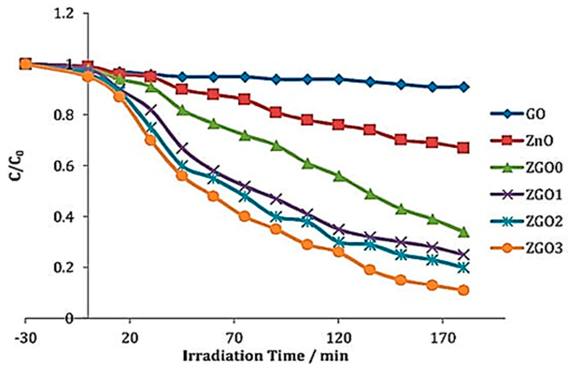
Mohamed et al. [240] synthesized ZnO hollow microsphere–RGO nanocomposites and evaluated their efficacy in degrading 2,4-dichlorophenoxyacetic acid (2,4-D) under direct sunlight irradiation. Their study demonstrated that incorporating varying amounts of graphene oxide (GO) at 0% (ZGO0), 1% (ZGO1), 3% (ZGO2), and 5% (ZGO3) significantly enhanced photocatalytic activity, as depicted in Figure 20. This improvement was attributed to the reduction of electron–hole pair recombination and the modification of ZnO’s bandgap energy from 3.1 eV to 2.7 eV, enabling the absorption of visible light.
In another study, Nenavathu et al. [241] found that adding just 0.09 wt% functionalized graphene oxide nanosheets (FGS) to ZnO significantly boosted photocatalytic activity. They achieved around 94.5% degradation of safranin-T dye under visible light. Figure 21a shows how FGS-ZnO breaks down safranin-T. They tested different amounts of graphene oxide (0.05 wt%, 0.09 wt%, 0.10 wt%, 0.50 wt%, and 5.00 wt%) in FGS-ZnO compared to pristine ZnO nanoparticles for degrading safranin-T, as shown in Figure 21b. The improved performance of the 0.09 wt% FGS-ZnO was because it enhanced dye adsorption, separated charge carriers effectively, and combined FGS and ZnO components well.
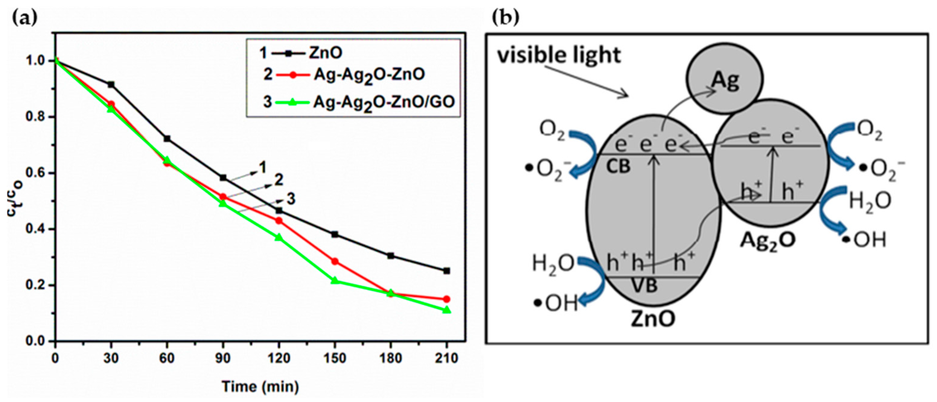
Umukoro et al. [242] used Ag-Ag2O-ZnO/GO nanocomposites as photocatalysts to degrade acid blue 74 dye in synthetic wastewater under visible light. They found that the nanocomposite removed 90% of the dye, compared to 85% with Ag-Ag2O-ZnO and 75% with ZnO alone (Figure 22a). The nanocomposite’s superior performance was due to several factors: Ag’s ability to absorb light and enhance charge separation, reduced electron–hole pair recombination at the Ag2O-ZnO interface, and graphene’s efficient electron mobility, facilitating the effective transfer of photogenerated electrons and holes from ZnO to graphene. Figure 22b illustrates the proposed mechanism of visible light photocatalytic activity with Ag-Ag2O-ZnO/GO.

6. Role of ZnO in Water Treatment

Throughout the preceding sections, our exploration delved into the extraordinary photocatalytic properties of ZnO, meticulously assessing the diverse impacts of various synthesis methods on its catalytic effectiveness. We scrutinized the innovative strategies devised to overcome challenges and enhance ZnO’s photocatalytic prowess. Our in-depth analysis has highlighted the pivotal role of ZnO and ZnO-based photocatalysts in efficiently degrading organic pollutants, notably demonstrated through the impressive degradation of dyes. Moving beyond this foundational role, our focus shifts to illuminating the broader significance of ZnO in water treatment. This section encapsulates the multifaceted contributions of ZnO, from its adept removal of various organic pollutants including agricultural pesticides, pharmaceuticals, and antibiotics to its critical role in water disinfection. By untangling the complex web of ZnO’s involvement in water treatment, we set the stage for a compelling conclusion that magnifies its importance as a cornerstone in the pursuit of sustainable solutions for ensuring access to clean water.

6.1. Removal of Organic Pollutants

The utilization of ZnO in the removal of organic pollutants has emerged as a pivotal strategy in mitigating the severe environmental and health hazards posed by over 80,000 organic–synthetic compounds prevalent in various industrial sectors such as cosmetics, food processing, paper and pulp, textiles, petrochemicals, agrochemicals, and beyond [243]. These pollutants, due to their large-scale production, extensive use, and frequent discharge, significantly elevate the burden on the environment and pose acute and chronic risks to all living things through processes like biomagnification and accumulation [243]. ZnO’s exceptional photophysical properties make it an ideal catalyst for degrading these pollutants, involving a series of mechanisms including the diffusion of pollutants onto ZnO’s surface, their subsequent adsorption, redox reactions in the adsorbed phase, the desorption of the degradation products, and the final elimination of these products from the interface region.
In the context of dye pollutant degradation, ZnO NPs have exhibited exceptional efficacy, as demonstrated in the preceding section of this review. Building upon these foundational findings, Isa et al. [244] introduced an innovative approach by designing ZnO nanoparticles into a micro-flower architecture, resulting in a notable 99% removal rate of 10 mg/L MO within 300 min, utilizing 150 mg of a ZnO catalyst. Similarly, Inderyas et al. [245] reported a significant degradation of fast acid black dye, achieving an 85% removal under UV light in 75 min and a 65% removal under solar irradiation in 240 min.
For agricultural pesticides, ZnO has shown excellence in degrading compounds like phorate, as evidenced by Niranjani and Anchana [246], with degradation occurring within 120 min under solar light. Previous studies have consistently emphasized the significant influence of operational parameters, including pH, catalyst dosage, original pesticide concentration, and the type of synthetic pesticide, on degradation efficiency [247,248,249,250]. For example, Premalatha and Miranda [248] reported the highly efficient ZnO-Bi2O3 composite for the degradation of lambda-cyhalothrin (L-CHT), a harmful pyrethroid pesticide, under visible light irradiation. The proposed mechanism for the photocatalytic degradation of L-CHT using the ZnO-Bi2O3 composite is shown in Figure 23. Compared to bare ZnO, the ZnO-Bi2O3 composite achieved a degradation percentage of 85.7% within 120 min, using a catalyst dosage of 1.2 g/L in a 50 mg/L L-CHT solution. The intermediate products formed during the degradation process were identified as Phthalic acid, butyl 2-ethyl butyl ester, and Diethyl phthalate, with no other hazardous intermediates detected. The researchers investigated the influence of various parameters on the degradation efficiency, including pH, initial L-CHT concentration, and catalyst dosage. The optimum pH for efficient degradation was found to be 7, as higher pH values led to a decrease in degradation due to the repulsion between the negatively charged catalyst surface and the pesticide molecules. Regarding the initial L-CHT concentration, a degradation percentage of 89.91% was achieved with 30 mg/L, which decreased to 62.83% as the concentration increased to 100 mg/L. This was attributed to the increased adsorption of pesticide molecules on the catalyst surface, leading to reduced hydroxyl radical generation and the adsorption of intermediates on the catalyst, reducing the efficiency of active sites. The effect of catalyst dosage was also studied, and the degradation percentage increased linearly with an increase in the catalyst amount from 0.4 g/L to 1.2 g/L but then decreased with a further increase to 1.6 g/L. This was due to the enhanced absorption of photons and adsorption of L-CHT molecules at higher catalyst loadings, which was offset by the increased turbidity of the solution, reducing light penetration. Compared to dark adsorption, photolysis, and ZnO alone, the ZnO-Bi2O3 composite exhibited the highest degradation efficiency of 85.7% under the optimized conditions, demonstrating the synergistic effect of the heterojunction formation between ZnO and Bi2O3. The study highlighted the excellent performance of ZnO-based composites, such as ZnO-Bi2O3, in the photocatalytic degradation of recalcitrant pyrethroid pesticides like lambda-cyhalothrin.
Aulakh et al. [250] reported the synthesis of various ZnO nanostructures, including nanorods, cotton ball-like structures, and nanospheres, as well as their copper-loaded counterparts, for the effective photocatalytic degradation of the pesticide methyl parathion under UV light. The researchers demonstrated that ZnO nanorods proved to be a better catalyst compared to the other synthesized nanostructures, owing to their elongated morphology. The ZnO nanorods were nearly 1.4 times more efficient than the ZnO cotton ball-like structures. Furthermore, the photocatalytic activity was enhanced by the loading of Cu onto the ZnO nanostructures due to the reduced recombination rate of electron–hole pairs. The Cu-loaded ZNR were found to degrade the entire methyl parathion compound by up to 99% within just 80 min, whereas the bare ZnO nanorods took 3 h under similar conditions. This represents a nearly 6-fold increase in efficiency. A plausible mechanism for pesticide degradation proposed in this study is shown in Figure 24.
Furthermore, Khan et al. [247] evaluated the photodegradation activity of Fe-ZnO nanocomposites for the degradation of the organophosphate pesticide chlorpyrifos at different concentrations under UV irradiation. They reported up to 93.5% degradation of 10 ppm chlorpyrifos within 60 min, confirmed by reductions in TOC and Chemical Oxygen Demand (COD) values.
Apart from this, the surface properties of ZnO, such as surface defects and oxygen vacancies, can greatly determine its photocatalytic capability [251]. In a study by Korake et al. [251], the photocatalytic activity of 0.5 mol% La-doped ZnO was investigated for the degradation of the pesticide metasystox. The researchers found that the degradation efficiency of metasystox increased with up to 0.5 mol% La doping but then decreased at higher doping levels. Among the catalysts studied, the 0.5 mol% La-doped ZnO exhibited the highest photocatalytic activity for metasystox degradation. The maximum reduction in metasystox concentration was observed under static conditions at pH 8. Furthermore, a reduction in the COD of metasystox was observed after 150 min of treatment. Cytotoxicological studies on meristematic root tip cells of Allium cepa revealed that the photocatalytically degraded products of metasystox were less toxic compared to the original metasystox compound.
To further elevate the degradation rate, the addition of oxidants, mainly peroxide, can facilitate the dissociation process of pesticides to the safe end-product [252]. Regardless of the reputation of other semiconductors in agricultural pollutant degradation, ZnO has been proposed as the most promising catalyst, even more feasible than its commonly investigated counterpart, TiO2 [253].
Moreover, ZnO’s versatility extends to the degradation of pharmaceuticals, particularly antibiotics and analgesics. The degradation efficiency is significantly enhanced through various modifications such as metal doping, heterostructure formation, or the incorporation of metal–organic frameworks (MOFs), enabling ZnO to effectively operate under solar light conditions [254]. Similar to other organic pollutants, the degradation rate of different antibiotics and analgesics is influenced by factors such as light sources, temperature, pH, and time [255]. Notably, studies have shown the successful degradation of antibiotics and analgesics including ampicillin, ciprofloxacin, diclofenac, ofloxacin, acetaminophen, paracetamol, and ibuprofen using ZnO photocatalysts in research by Mirzaei et al. [254], Eskandari et al. [256], Malakootian et al. [257], Kaur et al. [258], Thi and Lee [205], Choina et al. [259], and Ranjith Kumar et al. [260].
For example, Eskandari et al. [256] investigated the efficacy of utilizing UVC light and ZnO in the photocatalytic degradation of ciprofloxacin (CIP) in polluted water. They identified the optimal conditions for degrading CIP in a solution with 10 mg/L of the compound at a pH level of 5.0 and ZnO concentration of 0.15 g/L, with stirring at 600 rpm and an exposure time of 140 min. The obtained results maintained a relative standard deviation (RSD) below 7%. Through the kinetic analyses, Eskandari et al. determined pseudo-first-order reaction kinetics with a rate constant of 0.032 min−1, underscoring the effectiveness of this method in CIP degradation within aqueous environments.
Additionally, Malakootian et al. [257] conducted a study on the photocatalytic ozonation degradation of CIP using ZnO nanoparticles immobilized on the surface of stones. The research aimed to explore the efficacy of photocatalytic ozonation for eliminating ciprofloxacin from aquatic environments and optimizing the key parameters of the process. The study revealed that the highest removal efficiency of CIP was achieved at specific optimal conditions: pH of 7, reaction time of 30 min, photocatalyst concentration of 3 g/L, and initial CIP concentration of 10 mg/L. The process yielded an impressive removal efficiency of 96% under these optimized conditions. Moreover, linear kinetic models demonstrated that the degradation process followed pseudo-first-order and Langmuir–Hinshelwood kinetics. The study concluded that this method exhibited a high removal efficiency and was well-suited for removing CIP from aquatic environments.
In their study, Kaur et al. [258] investigated the use of silver-modified ZnO nanoplates as an efficient photocatalyst for the degradation of the antibiotic drug ofloxacin under solar irradiation. Their comprehensive experiments demonstrated that silver-modified ZnO at a concentration of 0.25 g/L achieved approximately 98% photocatalytic degradation of ofloxacin (initially at 10 mg/L, pH 7) within 150 min under solar illumination. Notably, the prepared silver-modified ZnO exhibited significantly higher photocatalytic activity compared to bare ZnO. This enhancement in photocatalytic activity could be attributed to efficient charge separation as well as the role of silver nanoparticles as electron sinks on the ZnO surface. Furthermore, the photocatalytic degradation process of ofloxacin conformed well to a pseudo-first-order kinetic model.
Thi and Lee [205] detailed the effective photocatalytic degradation of paracetamol using La-doped ZnO photocatalyst under visible light irradiation. In their study, they applied La-doped ZnO photocatalysts to treat 100 mg/L paracetamol in an aqueous solution under visible light irradiation, achieving a remarkable degradation efficiency of 99% and total organic carbon (TOC) removal of 85% after 3 h. Specifically, the 1.0 wt% La-doped ZnO photocatalyst exhibited the highest photocatalytic activity for paracetamol degradation.
Choina et al. [259] reported the influence of the textural properties of ZnO nanoparticles on the adsorption and photocatalytic remediation of water from pharmaceuticals. Two types of zinc oxide nanoparticle photocatalysts were prepared and characterized. They were highly active in the photocatalytic degradation of tetracycline and ibuprofen. Smaller particles showed a somewhat better performance in the abatement of the drugs due to the increased specific surface area. Although the larger 100 nm sized ZnO nanoparticles had distinctly lower specific surface areas, their activity was relatively high compared to smaller ones. This finding was attributed to improved adsorption properties that facilitated the photocatalytic conversion. However, they deactivated faster at higher drug concentrations. Enhanced adsorption of tetracycline at the catalyst surface was observed at low substrate and catalyst concentrations compared to high concentrations, indicating different photocatalytic behaviors of high and low pollutant concentrations.
Ranjith Kumar et al. [260] delved into the impacts of N ion implantation in vertically aligned ZnO nanorod arrays and its effect on the photocatalytic degradation of acetaminophen. Their analysis of the ZnO nanorods’ X-ray diffraction revealed a wurtzite structure with a prominent (002) diffraction peak that exhibited a slight shift after N-ion implantation. Additionally, field emission scanning electron microscopy images of the NRAs displayed a length of approximately 4 μm and a diameter of roughly 150 nm. Furthermore, UV–visible spectroscopy unveiled that the band gap of pristine ZnO nanorod arrays decreased from 3.2 to 2.18 eV after N-ion implantation. Interestingly, under visible irradiation, the N-ion-implanted ZnO catalyst showcased a significant increase in the photocatalytic degradation of acetaminophen, elevating the degradation efficiency from 60.0% to 98.46% over a span of 120 min.
Additionally, ZnO has demonstrated its capability in degrading stimulants such as ketamine, amphetamine, morphine, and caffeine. For example, Lin et al. [261] reported a near-complete removal of methamphetamine, ketamine, and morphine found in the influent and effluent of domestic treatment plants, as well as in rivers, within 30 min using ZnO. Their study compared the degradation rates of different contaminants under varying light conditions and catalyst loadings, plotting the fitted pseudo-first order rate constants and calculating the half-lives for each target compound. The results presented in Table 2 demonstrate the degradation efficiency of the three drugs under UV light at 254 nm with different ZnO catalyst dosages, revealing that morphine was most rapidly removed, while methamphetamine and ketamine were removed at roughly equal rates. Direct photolytic results further indicated that morphine exhibited the highest removal rate owing to its higher UV light absorption, facilitating more extensive degradation compared to ketamine and methamphetamine. Consequently, morphine showed the highest removal rate both by photocatalysis and photolysis, with ZnO proving to be a more effective catalyst for ketamine degradation than methamphetamine. Calculated times to achieve a 3-log reduction efficiency for each drug, detailed in Table 3, clearly illustrate that morphine’s removal was the most rapid among the three drugs.
In their work, Elhalil et al. [262] presented a detailed study on Mg-ZnO-Al2O3 photocatalysts, achieving a remarkable 98.9% degradation rate of caffeine, one of the most consumed psychoactive substances, in just 70 min of irradiation. They meticulously explored various experimental parameters including doping amount, irradiation time, photocatalyst dosage, initial solution pH, caffeine concentration, and the photocatalyst’s reuse potential. The results indicated that Mg-ZnO-Al2O3 significantly outperformed undoped pure ZnO and commercial Degussa P-25 TiO2 in terms of degradation efficiency. Crucially, the photocatalyst exhibited good stability and maintained its effectiveness after three regeneration cycles, showcasing its reusability and robustness.

6.2. Disinfection of Water

ZnO is one of the nanomaterials that has attracted significant research interest in recent years as an antibacterial agent, given the limitations of conventional disinfection methods such as chlorination, ultraviolet treatment, and ozonation. Chlorination proves ineffective for some highly resistant waterborne pathogens and also has a tendency to form carcinogenic disinfection by-product DBPs when chlorine is added to water [263]. Similarly, while ozonation forms fewer by-products, it is more costly than chemical disinfection and can form harmful bromate when ozone reacts with bromide ions in water. UV treatment also does not leave a residual in treated water, offering no protection against re-infection in the distribution network [264]. Along with other inorganic oxides such as Ag NPs and TiO2, ZnO has attracted interest because it exhibits strong antibacterial activity even in small amounts [265]. ZnO is regarded as a good antibacterial agent because it is stable under harsh processing conditions and is considered safe for humans and animals. Compared to organic materials, inorganic materials like ZnO have greater durability, selectivity, and heat resistance [266].
Several mechanisms have been proposed for the antimicrobial activity of ZnO nanoparticles:
  • Release of Reactive Oxygen Species (ROS): The antimicrobial activity of ZnO nanoparticles involves the release of oxygen species from the surface of ZnO, which cause fatal damage to microorganisms. ROS are known to cause oxidative stress by damaging DNA, cell membranes, and cellular proteins. The rupture of the cell wall is due to the surface activity of ZnO, which causes the decomposition of the cell wall and subsequently the cell membrane, the leakage of cell contents, and, eventually, cell death [266,267].
  • Release of Zn2+ Ions: Another possible mechanism of ZnO antibacterial activity is the release of Zn2+ ions, which can damage the cell membrane and penetrate the intracellular contents. Studies have suggested that the toxicity of nano ZnO against Escherichia coli and Saccharomyces cerevisiae could result from the solubility of the Zn2+ ions in the microorganism-containing medium [268].
  • Direct Contact of Nanoparticles with Cell Membrane: Cell damage does not necessarily result from the entry of the metal oxide particles into the cell. More importantly, the contact between the bacterial cell and the particle causes changes in the microenvironment within the contact area of the organism and particle. This can lead to increased membrane permeability and the subsequent cellular internalization of the nanoparticles [269,270].
Other research has also explored the role of photocatalytic activity in the antimicrobial properties of ZnO. By absorbing UV light, which activates its interaction with bacteria, ZnO nanoparticle suspensions can produce ROS such as H2O2, which have a phototoxic effect on bacteria [271]. However, studies have shown that inhibition could occur even in dark conditions, suggesting that mechanisms other than photocatalysis, such as oxidative stress and the destruction of membrane integrity, may also contribute to the antibacterial activity of ZnO [272]. The size and concentration of ZnO nanoparticles have also been found to play a crucial role in their disinfection efficiency [272,273,274]. Smaller nanoparticles with a higher surface area-to-volume ratio tend to exhibit enhanced antibacterial activity. Additionally, the photocatalytic efficiency of ZnO has been shown to increase with reduced particle size due to the quantum confinement effect [274].
In order to achieve a thorough comprehension of ZnO’s antimicrobial activity, conducting an extensive review of the literature is imperative. Additionally, it is important to note that antimicrobial activity refers to the process by which microbial growth is either inhibited or destroyed.
In our recent groundbreaking research led by Abou Zeid et al. [28], we presented a noteworthy contribution regarding the antibacterial efficacy of ZnO NWs against Pseudomonas putida, a significant Gram-negative bacterium. Our study meticulously investigated the antibacterial activity of ZnO NWs synthesized via hydrothermal methods in both dark and UV light conditions, with detailed insights illustrated in Figure 25a,b. Through comprehensive experimentation, we unequivocally highlighted that the unique morphology of ZnO NWs, achieved through hydrothermal growth, possesses the capability to disrupt bacterial cells, thereby effectively inhibiting bacterial growth. Furthermore, our research highlighted the release of Zn2+ ions from ZnO NWs as a key factor contributing to their antibacterial properties. These released ions demonstrate the potential to disturb the bacterial cell membrane, consequently impeding bacterial growth. Additionally, the generation of ROS under UV light further enhanced the antibacterial activity of the ZnO NWs. The proposed mechanism detailing these intricate interactions is clearly elucidated in Figure 25c, providing a comprehensive understanding of the multifaceted antibacterial mechanisms exhibited by ZnO NWs.
In their study, Baruah et al. [114] conducted a comprehensive examination focusing on the potential applications of photocatalysis as a promising approach for water disinfection. They detailed the growth of ZnO nanorods on a paper support crafted from softwood pulp. The study investigated the photocatalytic activity and antibacterial properties of a paper sheet embedded with ZnO nanorods within its porous matrix. The ZnO nanorods were securely anchored to cellulose fibers, enabling the reuse of the photocatalytic paper samples multiple times with only a nominal decrease in efficiency. The research findings revealed that the paper filled with ZnO nanorods exhibited significant photodegradation, with up to 93% degradation observed for methylene blue upon visible light irradiation at 963 W/m2 for 120 min. Similarly, a photodegradation rate of approximately 35% was noted for MO under the same conditions. Antibacterial tests showcased the inhibitory effect of the photocatalytic paper on the growth of Escherichia coli under ambient lighting conditions. The zone of inhibition, indicating the absence of viable bacterial cells upon visible light exposure, demonstrated the immobilization effect facilitated by photocatalysis. The study observed that bacteria were immobilized on the surface of the photocatalytic paper, preventing their proliferation in the vicinity of the ZnO-treated paper. The results of the antibacterial experiments, depicted in Figure 26, highlighted the efficacy of the photocatalytic paper in inhibiting bacterial growth. The study concluded that photocatalytic paper presents an appealing functional membrane for potential applications in water purification purposes.
In a study by Navale et al. [271], the authors investigated the antimicrobial properties and mechanistic activities of synthesized ZnO NPs. The nanoparticles exhibited an average size of 20–25 nm and were subjected to concentrations ranging from 0 to 100 μg/mL. These concentrations were tested against four representative microorganisms: the bacterial pathogens Staphylococcus aureus (Gram-positive) and Salmonella typhimurium (Gram-negative), as well as the fungal plant pathogens Aspergillus flavus and Aspergillus fumigatus. The minimum inhibitory concentration (MIC), defined as the lowest concentration at which no visible growth of the cells was observed, was determined using a method recommended by the Clinical and Laboratory Standards Institute (formerly the National Committee for Clinical Laboratory Standards). Bacterial cells were incubated at 37 °C for 24 h with various concentrations of ZnO nanoparticles (20–100 μg/mL), and the MIC was found to be 40 μg/mL. The antifungal activity was assessed using the broth dilution method, where the fungal strains were incubated with varying ZnO concentrations for 24–72 h. It was reported that both the antibacterial and antifungal activities, as well as the viability of the microorganisms, increased with the rising ZnO concentration.
Furthermore, Narayanan et al. [275] conducted a study to evaluate the antimicrobial efficacy of ZnO against human pathogens, including Staphylococcus aureus, Escherichia coli, Klebsiella pneumoniae, Enterococcus faecalis, and Pseudomonas aeruginosa, using the well diffusion method. The study focused on determining the zone of inhibition (ZOI) to assess the antibacterial activity of ZnO nanoparticles at varying concentrations (20–100 μg/mL). The results of the study revealed that even at a lower concentration of 20 μg/mL, ZnO nanoparticles demonstrated robust antibacterial activity against all tested pathogens, including both Gram-negative and Gram-positive bacteria. In a study by Tayel et al. [276], the antimicrobial activity of ZnO was investigated on nine bacterial strains, encompassing both Gram-positive and Gram-negative types, with a focus on foodborne pathogenic strains. The research revealed that Gram-positive bacteria exhibited higher susceptibility to ZnO due to their comparatively simpler cell membrane structure. In contrast, the thicker cell membrane of Gram-negative bacteria was found to potentially impede ZnO penetration into the cells and hinder its interaction with internal bacterial components. Among the strains examined, Bacillus cereus displayed the highest sensitivity, while Pseudomonas spp. was identified as the most resistant. Notably, the exposure of Staphylococcus aureus to ZnO nanoparticles resulted in cell explosion or lysis after a 2 h exposure, with complete disappearance occurring after 4 h of exposure. These promising findings led the researchers to recommend ZnO as a preservative agent against foodborne pathogens, with a caveat to confirm its biosafety before implementation. In a study by Barnes et al. [277], the photocatalytic antibacterial activity of ZnO was evaluated on four bacteria, including two Gram-positive species (S. aureus and B. subtilis) and two Gram-negative species (E. coli and P. aeruginosa). The primary objective of the study was to establish a correlation between the antimicrobial action of ZnO and the generation of ROS. The findings revealed that two of the bacterial species exhibited susceptibility to ZnO nanoparticles even under dark conditions, indicating the potent antibacterial properties of ZnO. Furthermore, the study determined that there was no observed association between the type of bacteria cell wall and its inactivation by ZnO, suggesting a broad-spectrum antibacterial effect, irrespective of the bacterial cell wall type.
The antifungal activity of ZnO nanoparticles was studied by He et al. [278] on two pathogenic fungi, Botrytis cinerea and Penicillium expansum, using the agar dilution method at various ZnO concentrations (0, 3, 6, and 12 mM) by measuring fungal colonies. The results indicated that ZnO nanoparticles greater than 3 mM significantly inhibited the growth of the two fungi, and increased with increasing concentration. Compared to B. cinerea, P. expansum was more sensitive to ZnO nanoparticle treatment, which resulted from the difference in the growth morphologies of the two fungi. Further, the results suggested a different mechanism of the inhibitory effect of ZnO nanoparticles on fungi compared to that on bacteria.
Zhang et al. [279] conducted a study on the characteristics, dispersion, and stability of commercial metal oxide nanoparticles, including Fe2O3, ZnO, NiO, and SiO2, in water, as well as their removal by water treatment processes. The investigation revealed that, with the exception of silica, these nanoparticles exhibited rapid aggregation in tap water, and they proved resistant to disaggregation via ultrasound or chemical dispersants. Furthermore, the study demonstrated that alum coagulation was only able to remove less than 80% of the total mass of nanoparticles. Additionally, the research findings indicated that the behavior of metal oxide nanoparticles in water is intricately linked to their physical properties and their interactions with other components present in the water.
In their investigation into the stability of three commercially available metal oxide nanoparticles (TiO2, ZnO, and SiO2) in aqueous solutions, Tso et al. [280] discovered that the nanoparticles rapidly aggregated and settled in pure water. Upon experimentation, ultrasonication emerged as the most effective method for dispersing the nanoparticles in water, thereby overcoming their tendency to aggregate. The study’s findings highlighted the varying stability of nanoparticles under different water conditions. Notably, in samples of lake water and wastewater, the aggregation of nanoparticles occurred at a slower rate compared to pure water. This phenomenon was attributed to the presence of organic colloids such as humic compounds and surfactants, which acted to hinder the rapid aggregation of the nanoparticles. The findings align with the research conducted by Li et al. [62], elucidating the influence of water chemistry on the toxicity of ZnO NPs in aqueous environments. Li and colleagues undertook a comprehensive investigation into the toxicity of ZnO NPs to E. coli, with a specific focus on understanding how water chemistry can modulate this toxicity. Their study aimed to uncover the mechanisms responsible for the toxic effects of ZnO nanoparticles on E. coli and investigate how the composition of the medium might influence these effects. The research findings from Li et al. shed significant light on the intricate and dynamic interactions between ZnO NPs and bacteria under diverse environmental conditions. Notably, the study highlighted the nuanced influence of various medium components on the toxicity of NPs in aqueous systems, providing valuable insights into the complex interplay between water chemistry and the impact of ZnO nanoparticles. Consequently, this work contributed substantially to enhancing the broader understanding of how environmental factors, particularly water chemistry, can dynamically modulate the behavior and impact of ZnO NPs, thereby emphasizing the critical importance of contextual environmental considerations in the assessment and management of nanoparticle toxicity [62].

7. Conclusions and Future Perspectives

In conclusion, this comprehensive review meticulously examined the progress in ZnO-based photocatalysts for water treatment, highlighting their diverse properties and burgeoning trends with the potential to reshape environmental restoration practices. The intricate interplay between ZnO’s inherent characteristics and the innovative enhancements discussed underscores their adaptability and efficacy in addressing the intricate challenges posed by water pollution. The extensive characterization of ZnO presented in this review, spanning structural, optical, electrical, magnetic, mechanical, thermal, photocatalytic, and antibacterial properties, as well as cost-effectiveness and various synthesis methods, collectively demonstrates the versatility of ZnO in enabling efficient and sustainable water treatment across diverse applications.
Moreover, this review delved into strategies that bolster the stability, affordability, and overall sustainability of ZnO-based photocatalysts, charting a course toward the implementation of large-scale solutions for clean water provision. By exploring methodologies to optimize water treatment efficiency, acknowledging the crucial balance between effectiveness and environmental responsibility, and addressing toxicity concerns related to modified ZnO-based materials, this review illuminates a roadmap for the evolution of advanced water treatment technologies. The insightful findings presented not only refine the present comprehension of ZnO-based photocatalysts but also unveil promising future research avenues and practical applications, paving the way for innovative, eco-friendly, and efficient water treatment modalities.
This extended review collects and comments on results reported in the literature, providing the scientific community with an up-to-date understanding of the topic. The collaborative endeavors and forward-thinking research directions articulated herein underscore the transformative potential of leveraging ZnO-based photocatalysts to establish widespread and effective water treatment solutions on a global scale. In essence, this review significantly contributes to the preservation of vital water resources for present and future generations, underscoring the pivotal role of advanced materials in upholding environmental integrity and public health.
Looking ahead, this review sets the stage for future research directions and practical applications in the realm of ZnO-based photocatalysts for water treatment. These perspectives encompass a wide array of opportunities, including innovative synthesis techniques and surface functionalization to tailor the properties of ZnO-based photocatalysts for specific water treatment applications. Additionally, the exploration of advanced composite materials and heterojunctions is crucial for enhancing the efficiency and performance of ZnO-based photocatalysts.
Environmental fate and impact studies are paramount to ensure the safe and sustainable application of ZnO-based photocatalysts, while techno-economic assessments will provide insights into the viability and scalability of these technologies. Collaboration between researchers, policymakers, and regulatory bodies is essential for the development of comprehensive frameworks for the safe implementation of ZnO-based photocatalysts. Engaging communities and stakeholders, integrating these technologies into industrial water treatment systems, and promoting continuous innovation and education are keys to advancing the field of ZnO-based photocatalysts for water treatment.
These future perspectives aim to foster a holistic approach to water treatment, leveraging the transformative potential of ZnO-based photocatalysts to address global water pollution challenges sustainably and effectively. The realization of these research directions holds promise for innovative, eco-friendly, and efficient water treatment solutions that contribute to the preservation of vital water resources and the enhancement of environmental and public health on a global scale.

Author Contributions

The individual contributions for the redaction of this review paper were distributed as follows: conceptualization, Y.L.-W. and S.A.Z.; writing—original draft preparation, S.A.Z.; writing—review and editing Y.L.-W. and S.A.Z.; visualization, Y.L.-W. and S.A.Z.; supervision, Y.L.-W. All authors have read and agreed to the published version of the manuscript.

Funding

This research received no external funding.

Data Availability Statement

No new data were created or analyzed in this study. Data sharing is not applicable to this article.

Conflicts of Interest

The authors declare no conflicts of interest.

References

  1. Fuzil, N.S.; Othman, N.H.; Alias, N.H.; Marpani, F.; Othman, M.H.D.; Ismail, A.F.; Lau, W.J.; Li, K.; Kusworo, T.D.; Ichinose, I.; et al. A Review on Photothermal Material and Its Usage in the Development of Photothermal Membrane for Sustainable Clean Water Production. Desalination 2021, 517, 115259. [Google Scholar] [CrossRef]
  2. Tang, W.; Pei, Y.; Zheng, H.; Zhao, Y.; Shu, L.; Zhang, H. Twenty Years of China’s Water Pollution Control: Experiences and Challenges. Chemosphere 2022, 295, 133875. [Google Scholar] [CrossRef]
  3. Aal, N.A.; Al-Hazmi, F.; Al-Ghamdi, A.A.; Al-Ghamdi, A.A.; El-Tantawy, F.; Yakuphanoglu, F. Novel Rapid Synthesis of Zinc Oxide Nanotubes via Hydrothermal Technique and Antibacterial Properties. Spectrochim. Acta Part A Mol. Biomol. Spectrosc. 2015, 135, 871–877. [Google Scholar] [CrossRef]
  4. Coelho, E.F.; Santos, D.L.; de Lima, L.W.F.; Castricini, A.; Barros, D.L.; Filgueiras, R.; da Cunha, F.F. Water Regimes on Soil Covered with Plastic Film Mulch and Relationships with Soil Water Availability, Yield, and Water Use Efficiency of Papaya Trees. Agric. Water Manag. 2022, 269, 107709. [Google Scholar] [CrossRef]
  5. Uma Maheswari, B.; Sivakumar, V.M.; Thirumarimurugan, M. Sequester Adsorption of Cr(VI) and Pb(II) Ions from Aqueous Solution by Green Synthesized Nanocomposite (OB/ZnO): Characterization, Kinetics, Isotherm and Mechanism. Environ. Nanotechnol. Monit. Manag. 2022, 18, 100680. [Google Scholar] [CrossRef]
  6. Hasan, Z.; Jhung, S.H. Removal of Hazardous Organics from Water Using Metal-Organic Frameworks (MOFs): Plausible Mechanisms for Selective Adsorptions. J. Hazard. Mater. 2015, 283, 329–339. [Google Scholar] [CrossRef]
  7. Yang, T.; Xu, Y.; Huang, Q.; Sun, Y.; Liang, X.; Wang, L.; Qin, X.; Zhao, L. Adsorption Characteristics and the Removal Mechanism of Two Novel Fe-Zn Composite Modified Biochar for Cd(II) in Water. Bioresour. Technol. 2021, 333, 125078. [Google Scholar] [CrossRef] [PubMed]
  8. Asahi, R.; Morikawa, T.; Ohwaki, T.; Aoki, K.; Taga, Y. Visible-Light Photocatalysis in Nitrogen-Doped Titanium Oxides. Science 2001, 293, 269–271. [Google Scholar] [CrossRef] [PubMed]
  9. Chen, X.; Liu, L.; Yu, P.Y.; Mao, S.S. Increasing Solar Absorption for Photocatalysis with Black Hydrogenated Titanium Dioxide Nanocrystals. Science 2011, 331, 746–750. [Google Scholar] [CrossRef]
  10. Li, B.; Liu, T.; Wang, Y.; Wang, Z. ZnO/Graphene-Oxide Nanocomposite with Remarkably Enhanced Visible-Light-Driven Photocatalytic Performance. J. Colloid Interface Sci. 2012, 377, 114–121. [Google Scholar] [CrossRef]
  11. Kumar, N.; Chauhan, N.S.; Mittal, A.; Sharma, S. TiO2 and Its Composites as Promising Biomaterials: A Review. Biometals 2018, 31, 147–159. [Google Scholar] [CrossRef] [PubMed]
  12. Zhang, H.; Lv, X.; Li, Y.; Wang, Y.; Li, J. P25-Graphene Composite as a High Performance Photocatalyst. ACS Nano 2010, 4, 380–386. [Google Scholar] [CrossRef] [PubMed]
  13. Verma, N.; Yadav, S.; Marí, B.; Mittal, A.; Jindal, J. Synthesis and Charcterization of Coupled ZnO/SnO2 Photocatalysts and Their Activity towards Degradation of Cibacron Red Dye. Trans. Indian Ceram. Soc. 2018, 77, 1–7. [Google Scholar] [CrossRef]
  14. Uribe-López, M.C.; Hidalgo-López, M.C.; López-González, R.; Frías-Márquez, D.M.; Núñez-Nogueira, G.; Hernández-Castillo, D.; Alvarez-Lemus, M.A. Photocatalytic Activity of ZnO Nanoparticles and the Role of the Synthesis Method on Their Physical and Chemical Properties. J. Photochem. Photobiol. A Chem. 2021, 404, 112866. [Google Scholar] [CrossRef]
  15. Neri, G.; Bonavita, A.; Milone, C.; Galvagno, S. Role of the Au Oxidation State in the CO Sensing Mechanism of Au/Iron Oxide-Based Gas Sensors. Sens. Actuators B Chem. 2003, 93, 402–408. [Google Scholar] [CrossRef]
  16. Hitam, C.N.C.; Jalil, A.A. A Review on Exploration of Fe2O3 Photocatalyst towards Degradation of Dyes and Organic Contaminants. J. Environ. Manag. 2020, 258, 110050. [Google Scholar] [CrossRef]
  17. Ng, Y.H.; Iwase, A.; Kudo, A.; Amal, R. Reducing Graphene Oxide on a Visible-Light BiVO4 Photocatalyst for an Enhanced Photoelectrochemical Water Splitting. J. Phys. Chem. Lett. 2010, 1, 2607–2612. [Google Scholar] [CrossRef]
  18. Kamble, G.S.; Natarajan, T.S.; Patil, S.S.; Thomas, M.; Chougale, R.K.; Sanadi, P.D.; Siddharth, U.S.; Ling, Y.-C. BiVO4 As a Sustainable and Emerging Photocatalyst: Synthesis Methodologies, Engineering Properties, and Its Volatile Organic Compounds Degradation Efficiency. Nanomaterials 2023, 13, 1528. [Google Scholar] [CrossRef]
  19. Ramanathan, G.; Murali, K.R. Photocatalytic Activity of SnO2 Nanoparticles. J. Appl. Electrochem. 2022, 52, 849–859. [Google Scholar] [CrossRef]
  20. Kwang Benno Park, H.; Kumar, P.; Kebaili, I.; Boukhris, I.; Hwan Joo, Y.; Hyun Sung, T.; Kumar, A. Optimization and Modelling of Magnesium Oxide (MgO) Photocatalytic Degradation of Binary Dyes Using Response Surface Methodology. Sci. Rep. 2024, 14, 9412. [Google Scholar] [CrossRef]
  21. Sun, R.; Chen, Z.; Yang, Y.; Peng, J.; Zheng, T. Effects and Mechanism of SiO2 on Photocatalysis and Super Hydrophilicity of TiO2 Films Prepared by Sol-Gel Method. Mater. Res. Express 2019, 6, 46409. [Google Scholar] [CrossRef]
  22. Cai, J.; Li, D.; Jiang, L.; Yuan, J.; Li, Z.; Li, K. Review on CeO2-Based Photocatalysts for Photocatalytic Reduction of CO2: Progresses and Perspectives. Energy Fuels 2023, 37, 4878–4897. [Google Scholar] [CrossRef]
  23. Liu, X.; Sayed, M.; Bie, C.; Cheng, B.; Hu, B.; Yu, J.; Zhang, L. Hollow CdS-Based Photocatalysts. J. Mater. 2021, 7, 419–439. [Google Scholar] [CrossRef]
  24. Rajaraman, T.S.; Parikh, S.P.; Gandhi, V.G. Black TiO2: A Review of Its Properties and Conflicting Trends. Chem. Eng. J. 2020, 389, 123918. [Google Scholar] [CrossRef]
  25. Zhou, S.; Jiang, L.; Wang, H.; Yang, J.; Yuan, X.; Wang, H.; Liang, J.; Li, X.; Li, H.; Bu, Y. Oxygen Vacancies Modified TiO2/O-Terminated Ti3C2 Composites: Unravelling the Dual Effects between Oxygen Vacancy and High-Work-Function Titanium Carbide. Adv. Funct. Mater. 2023, 33, 2307702. [Google Scholar] [CrossRef]
  26. Şahin, B.; Aydin, R.; Soylu, S.; Türkmen, M.; Kara, M.; Akkaya, A.; Çetin, H.; Ayyıldız, E. The Effect of Thymus Syriacus Plant Extract on the Main Physical and Antibacterial Activities of ZnO Nanoparticles Synthesized by SILAR Method. Inorg. Chem. Commun. 2022, 135, 109088. [Google Scholar] [CrossRef]
  27. Negi, A.; Gangwar, R.; Kumar Vishwakarma, R.; Singh Negi, D. Antibacterial, Antioxidant and Photodegradation Potential of ZnO Nanoparticles Mediated via Roots of Taraxacum Officinale Radix. Mater. Today Proc. 2022, 57, 2435–2443. [Google Scholar] [CrossRef]
  28. Abou Zeid, S.; Perez, A.; Bastide, S.; Le Pivert, M.; Rossano, S.; Remita, H.; Hautière, N.; Leprince-Wang, Y. Antibacterial and Photocatalytic Properties of ZnO Nanostructure Decorated Coatings. Coatings 2024, 14, 41. [Google Scholar] [CrossRef]
  29. Wang, Z.L. Nanostructures of Zinc Oxide. Mater. Today 2004, 7, 26–33. [Google Scholar] [CrossRef]
  30. Hariharan, C. Photocatalytic Degradation of Organic Contaminants in Water by ZnO Nanoparticles: Revisited. Appl. Catal. A Gen. 2006, 304, 55–61. [Google Scholar] [CrossRef]
  31. Akyol, A.; Yatmaz, H.C.; Bayramoglu, M. Photocatalytic Decolorization of Remazol Red RR in Aqueous ZnO Suspensions. Appl. Catal. B Environ. 2004, 54, 19–24. [Google Scholar] [CrossRef]
  32. Colón, G.; Hidalgo, M.C.; Navío, J.A.; Pulido Melián, E.; González Díaz, O.; Doña Rodríguez, J.M. Highly Photoactive ZnO by Amine Capping-Assisted Hydrothermal Treatment. Appl. Catal. B Environ. 2008, 83, 30–38. [Google Scholar] [CrossRef]
  33. Lizama, C.; Freer, J.; Baeza, J.; Mansilla, H.D. Optimized Photodegradation of Reactive Blue 19 on TiO2 and ZnO Suspensions. Catal. Today 2002, 76, 235–246. [Google Scholar] [CrossRef]
  34. Chandiran, A.K.; Abdi-Jalebi, M.; Nazeeruddin, M.K.; Grätzel, M. Analysis of Electron Transfer Properties of ZnO and TiO2 Photoanodes for Dye-Sensitized Solar Cells. ACS Nano 2014, 8, 2261–2268. [Google Scholar] [CrossRef]
  35. Anta, J.A.; Guillén, E.; Tena-Zaera, R. ZnO-Based Dye-Sensitized Solar Cells. J. Phys. Chem. C 2012, 116, 11413–11425. [Google Scholar] [CrossRef]
  36. Daneshvar, N.; Salari, D.; Khataee, A.R. Photocatalytic Degradation of Azo Dye Acid Red 14 in Water on ZnO as an Alternative Catalyst to TiO2. J. Photochem. Photobiol. A Chem. 2004, 162, 317–322. [Google Scholar] [CrossRef]
  37. Kandavelu, V.; Kastien, H.; Thampi, K.R. Photocatalytic Degradation of Isothiazolin-3-Ones in Water and Emulsion Paints Containing Nanocrystalline TiO2 and ZnO Catalysts. Appl. Catal. B Environ. 2004, 48, 101–111. [Google Scholar] [CrossRef]
  38. Zhao, S.-W.; Zuo, H.-F.; Guo, Y.; Pan, Q. Carbon-Doped ZnO Aided by Carboxymethyl Cellulose: Fabrication, Photoluminescence and Photocatalytic Applications. J. Alloys Compd. 2017, 695, 1029–1037. [Google Scholar] [CrossRef]
  39. Rooydell, R.; Brahma, S.; Wang, R.-C.; Modaberi, M.R.; Ebrahimzadeh, F.; Liu, C.-P. Cu Doped ZnO Nanorods with Controllable Cu Content by Using Single Metal Organic Precursors and Their Photocatalytic and Luminescence Properties. J. Alloys Compd. 2017, 691, 936–945. [Google Scholar] [CrossRef]
  40. Lu, Y.; Lin, Y.; Wang, D.; Wang, L.; Xie, T.; Jiang, T. A High Performance Cobalt-Doped ZnO Visible Light Photocatalyst and Its Photogenerated Charge Transfer Properties. Nano Res. 2011, 4, 1144–1152. [Google Scholar] [CrossRef]
  41. Güy, N.; Özacar, M. The Influence of Noble Metals on Photocatalytic Activity of ZnO for Congo Red Degradation. Int. J. Hydrogen Energy 2016, 41, 20100–20112. [Google Scholar] [CrossRef]
  42. Sampaio, M.J.; Lima, M.J.; Baptista, D.L.; Silva, A.M.T.; Silva, C.G.; Faria, J.L. Ag-Loaded ZnO Materials for Photocatalytic Water Treatment. Chem. Eng. J. 2017, 318, 95–102. [Google Scholar] [CrossRef]
  43. Bao, Z.; Yuan, Y.; Leng, C.; Li, L.; Zhao, K.; Sun, Z. One-Pot Synthesis of Noble Metal/Zinc Oxide Composites with Controllable Morphology and High Catalytic Performance. ACS Appl. Mater. Interfaces 2017, 9, 16417–16425. [Google Scholar] [CrossRef] [PubMed]
  44. Udawatte, N.; Lee, M.; Kim, J.; Lee, D. Well-Defined Au/ZnO Nanoparticle Composites Exhibiting Enhanced Photocatalytic Activities. ACS Appl. Mater. Interfaces 2011, 3, 4531–4538. [Google Scholar] [CrossRef] [PubMed]
  45. Moradi, S.; Aberoomand-Azar, P.; Raeis-Farshid, S.; Abedini-Khorrami, S.; Givianrad, M.H. The Effect of Different Molar Ratios of ZnO on Characterization and Photocatalytic Activity of TiO2/ZnO Nanocomposite. J. Saudi Chem. Soc. 2016, 20, 373–378. [Google Scholar] [CrossRef]
  46. Prabhu, Y.T.; Navakoteswara Rao, V.; Shankar, M.V.; Sreedhar, B.; Pal, U. The Facile Hydrothermal Synthesis of CuO@ZnO Heterojunction Nanostructures for Enhanced Photocatalytic Hydrogen Evolution. New J. Chem. 2019, 43, 6794–6805. [Google Scholar] [CrossRef]
  47. Ranjith, K.S.; Castillo, R.B.; Sillanpaa, M.; Rajendra Kumar, R.T. Effective Shell Wall Thickness of Vertically Aligned ZnO-ZnS Core-Shell Nanorod Arrays on Visible Photocatalytic and Photo Sensing Properties. Appl. Catal. B Environ. 2018, 237, 128–139. [Google Scholar] [CrossRef]
  48. Rahmah, M.I.; Sabry, R.S.; Aziz, W.J. Preparation of Superhydrophobic Ag/Fe2O3/ZnO Surfaces with Photocatalytic Activity. Surf. Eng. 2021, 37, 1320–1327. [Google Scholar] [CrossRef]
  49. Gang, R.; Xu, L.; Xia, Y.; Cai, J.; Zhang, L.; Wang, S.; Li, R. Fabrication of MoS2 QDs/ZnO Nanosheet 0D/2D Heterojunction Photocatalysts for Organic Dyes and Gaseous Heavy Metal Removal. J. Colloid Interface Sci. 2020, 579, 853–861. [Google Scholar] [CrossRef]
  50. Xu, P.; Wang, P.; Wang, Q.; Wei, R.; Li, Y.; Xin, Y.; Zheng, T.; Hu, L.; Wang, X.; Zhang, G. Facile Synthesis of Ag2O/ZnO/RGO Heterojunction with Enhanced Photocatalytic Activity under Simulated Solar Light: Kinetics and Mechanism. J. Hazard. Mater. 2021, 403, 124011. [Google Scholar] [CrossRef]
  51. Liu, B.; Bie, C.; Zhang, Y.; Wang, L.; Li, Y.; Yu, J. Hierarchically Porous ZnO/g-C3N4 S-Scheme Heterojunction Photocatalyst for Efficient H2O2 Production. Langmuir 2021, 37, 14114–14124. [Google Scholar] [CrossRef]
  52. Zgura, I.; Preda, N.; Enculescu, M.; Diamandescu, L.; Negrila, C.; Bacalum, M.; Ungureanu, C.; Barbinta-Patrascu, M.E. Cytotoxicity, Antioxidant, Antibacterial, and Photocatalytic Activities of ZnO-CdS Powders. Materials 2020, 13, 182. [Google Scholar] [CrossRef] [PubMed]
  53. Zgura, I.; Preda, N.; Socol, G.; Ghica, C.; Ghica, D.; Enculescu, M.; Negut, I.; Nedelcu, L.; Frunza, L.; Ganea, C.P.; et al. Wet Chemical Synthesis of ZnO-CdS Composites and Their Photocatalytic Activity. Mater. Res. Bull. 2018, 99, 174–181. [Google Scholar] [CrossRef]
  54. Joseph, J.A.; Nair, S.B.; John, S.S.; Shaji, S.; Philip, R.R. α-Fe2O3/ZnO Heterostructure for Enhanced Photocatalytic and Antibacterial Activity. Semicond. Sci. Technol. 2021, 36, 95007. [Google Scholar] [CrossRef]
  55. Taufique, M.F.N.; Haque, A.; Karnati, P.; Ghosh, K. ZnO–CuO Nanocomposites with Improved Photocatalytic Activity for Environmental and Energy Applications. J. Electron. Mater. 2018, 47, 6731–6745. [Google Scholar] [CrossRef]
  56. Jan, T.; Azmat, S.; Mansoor, Q.; Waqas, H.M.; Adil, M.; Ilyas, S.Z.; Ahmad, I.; Ismail, M. Superior Antibacterial Activity of ZnO-CuO Nanocomposite Synthesized by a Chemical Co-Precipitation Approach. Microb. Pathog. 2019, 134, 103579. [Google Scholar] [CrossRef]
  57. Gayathri, S.; Jayabal, P.; Kottaisamy, M.; Ramakrishnan, V. Synthesis of ZnO Decorated Graphene Nanocomposite for Enhanced Photocatalytic Properties. J. Appl. Phys. 2014, 115, 173504. [Google Scholar] [CrossRef]
  58. Bu, Y.; Chen, Z.; Li, W.; Hou, B. Highly Efficient Photocatalytic Performance of Graphene–ZnO Quasi-Shell–Core Composite Material. ACS Appl. Mater. Interfaces 2013, 5, 12361–12368. [Google Scholar] [CrossRef] [PubMed]
  59. Ong, W.-J.; Voon, S.-Y.; Tan, L.-L.; Goh, B.T.; Yong, S.-T.; Chai, S.-P. Enhanced Daylight-Induced Photocatalytic Activity of Solvent Exfoliated Graphene (SEG)/ZnO Hybrid Nanocomposites toward Degradation of Reactive Black 5. Ind. Eng. Chem. Res. 2014, 53, 17333–17344. [Google Scholar] [CrossRef]
  60. Ambika, S.; Sundrarajan, M. Antibacterial Behaviour of Vitex Negundo Extract Assisted ZnO Nanoparticles against Pathogenic Bacteria. J. Photochem. Photobiol. B. 2015, 146, 52–57. [Google Scholar] [CrossRef]
  61. Panigrahi, J.; Behera, D.; Mohanty, I.; Subudhi, U.; Nayak, B.B.; Acharya, B.S. Radio Frequency Plasma Enhanced Chemical Vapor Based ZnO Thin Film Deposition on Glass Substrate: A Novel Approach towards Antibacterial Agent. Appl. Surf. Sci. 2011, 258, 304–311. [Google Scholar] [CrossRef]
  62. Li, M.; Zhu, L.; Lin, D. Toxicity of ZnO Nanoparticles to Escherichia Coli: Mechanism and the Influence of Medium Components. Environ. Sci. Technol. 2011, 45, 1977–1983. [Google Scholar] [CrossRef]
  63. Zhang, L.; Jiang, Y.; Ding, Y.; Daskalakis, N.; Jeuken, L.; Povey, M.; O’Neill, A.J.; York, D.W. Mechanistic Investigation into Antibacterial Behaviour of Suspensions of ZnO Nanoparticles against E. Coli. J. Nanopart. Res. 2010, 12, 1625–1636. [Google Scholar] [CrossRef]
  64. Song, D.; Widenborg, P.; Chin, W.; Aberle, A.G. Investigation of Lateral Parameter Variations of Al-Doped Zinc Oxide Films Prepared on Glass Substrates by Rf Magnetron Sputtering. Sol. Energy Mater. Sol. Cells 2002, 73, 1–20. [Google Scholar] [CrossRef]
  65. Miccoli, I.; Spampinato, R.; Marzo, F.; Prete, P.; Lovergine, N. DC-Magnetron Sputtering of ZnO:Al Films on (00.1)Al2O3 Substrates from Slip-Casting Sintered Ceramic Targets. Appl. Surf. Sci. 2014, 313, 418–423. [Google Scholar] [CrossRef]
  66. Winer, I.; Shter, G.E.; Mann-Lahav, M.; Grader, G.S. Effect of Solvents and Stabilizers on Sol–Gel Deposition of Ga-Doped Zinc Oxide TCO Films. J. Mater. Res. 2011, 26, 1309–1315. [Google Scholar] [CrossRef]
  67. Ferreira, S.H.; Cunha, I.; Pinto, J.V.; Neto, J.P.; Pereira, L.; Fortunato, E.; Martins, R. UV-Responsive Screen-Printed Porous ZnO Nanostructures on Office Paper for Sustainable and Foldable Electronics. Chemosensors 2021, 9, 192. [Google Scholar] [CrossRef]
  68. Banerjee, D.; Lao, J.Y.; Wang, D.Z.; Huang, J.Y.; Ren, Z.F.; Steeves, D.; Kimball, B.; Sennett, M. Large-Quantity Free-Standing ZnO Nanowires. Appl. Phys. Lett. 2003, 83, 2061–2063. [Google Scholar] [CrossRef]
  69. Wahab, R.; Ansari, S.G.; Kim, Y.-S.; Seo, H.-K.; Shin, H.-S. Room Temperature Synthesis of Needle-Shaped ZnO Nanorods via Sonochemical Method. Appl. Surf. Sci. 2007, 253, 7622–7626. [Google Scholar] [CrossRef]
  70. Leprince-Wang, Y.; Martin, N.; Habba, Y.; Le Pivert, M.; Capochichi-Gnambodoe, M. ZnO Nanostructure Based Photocatalysis for Water Purification. NanoWorld J. 2020, 6. [Google Scholar] [CrossRef]
  71. Chiu, W.S.; Khiew, P.S.; Cloke, M.; Isa, D.; Tan, T.K.; Radiman, S.; Abd-Shukor, R.; Hamid, M.A.A.; Huang, N.M.; Lim, H.N.; et al. Photocatalytic Study of Two-Dimensional ZnO Nanopellets in the Decomposition of Methylene Blue. Chem. Eng. J. 2010, 158, 345–352. [Google Scholar] [CrossRef]
  72. Polshettiwar, V.; Baruwati, B.; Varma, R.S. Self-Assembly of Metal Oxides into Three-Dimensional Nanostructures: Synthesis and Application in Catalysis. ACS Nano 2009, 3, 728–736. [Google Scholar] [CrossRef]
  73. Reynolds, D.C.; Look, D.C.; Jogai, B.; Litton, C.W.; Cantwell, G.; Harsch, W.C. Valence-Band Ordering in ZnO. Phys. Rev. B 1999, 60, 2340–2344. [Google Scholar] [CrossRef]
  74. Janotti, A.; Van de Walle, C.G. Fundamentals of Zinc Oxide as a Semiconductor. Rep. Prog. Phys. 2009, 72, 126501. [Google Scholar] [CrossRef]
  75. Coleman, V.A.; Bradby, J.E.; Jagadish, C.; Munroe, P.; Heo, Y.W.; Pearton, S.J.; Norton, D.P.; Inoue, M.; Yano, M. Mechanical Properties of ZnO Epitaxial Layers Grown on A- and c-Axis Sapphire. Appl. Phys. Lett. 2005, 86, 203105. [Google Scholar] [CrossRef]
  76. Dietl, T.; Ohno, H.; Matsukura, F.; Cibert, J.; Ferrand, D. Zener Model Description of Ferromagnetism in Zinc-Blende Magnetic Semiconductors. Science 2000, 287, 1019–1022. [Google Scholar] [CrossRef]
  77. Jagadish, C.; Pearton, S.J. Zinc Oxide Bulk, Thin Films and Nanostructures Processing, Properties, and Applications, 1st ed.; Jagadish, C., Pearton, S.J., Eds.; Elsevier Science: Amsterdam, The Netherlands, 2006; ISBN 9780080447223. [Google Scholar]
  78. Zhang, L.; Li, Y.; Liu, X.; Zhao, L.; Ding, Y.; Povey, M.; Cang, D. The Properties of ZnO Nanofluids and the Role of H2O2 in the Disinfection Activity against Escherichia Coli. Water Res. 2013, 47, 4013–4021. [Google Scholar] [CrossRef]
  79. Elaaraj, I.; Raouan, S.E.R.; Nakkabi, A.; Es-sounni, B.; Koraichi, I.; El Moualij, N.; Fahim, M. Synthesis, Characterization and Antioxidant, Antibacterial Activity Zn2+, Cu2+, Ni2+ and Co2+, Complexes of Ligand [2-(Thiophen-2-Yl)-1-(Thiophen-2-Ylmethyl)-1H-Benzo[d]Imidazole]. J. Indian Chem. Soc. 2022, 99, 100404. [Google Scholar] [CrossRef]
  80. Bellanger, X.; Schneider, R.; Dezanet, C.; Arroua, B.; Balan, L.; Billard, P.; Merlin, C. Zn2+ Leakage and Photo-Induced Reactive Oxidative Species Do Not Explain the Full Toxicity of ZnO Core Quantum Dots. J. Hazard. Mater. 2020, 396, 122616. [Google Scholar] [CrossRef]
  81. Ding, Y.; Kong, X.Y.; Wang, Z.L. Doping and Planar Defects in the Formation of Single-Crystal ZnO Nanorings. Phys. Rev. B 2004, 70, 235408. [Google Scholar] [CrossRef]
  82. Zhou, M.Y.; Qu, L.S.; Gao, H. Synthesis and Photoluminescence Properties of ZnO Nanohelices. IOP Conf. Ser. Mater. Sci. Eng. 2017, 213, 12009. [Google Scholar] [CrossRef]
  83. Peng, D.; Huang, Y.; Yu, K.; Li, L.; Zhu, Z. Synthesis and Field Emission Properties of Hierarchical ZnO Nanostructures. J. Nanomater. 2010, 2010, 153–157. [Google Scholar] [CrossRef]
  84. Zhang, Y.; Heng, L.; Jiang, L. Chemically Controllable Fabrication of One-Dimensional ZnO Nanostructures and Their Applications in Solar Cells. J. Nanosci. Nanotechnol. 2014, 14, 5597–5613. [Google Scholar] [CrossRef]
  85. Abdullah, H.; Ariyanto, N.P.; Shaari, S.; Yuliarto, B.; Junaidi, S. Study of Porous Nanoflake ZnO for Dye-Sensitized Solar Cell Application. Am. J. Eng. Appl. Sci. 2009, 2, 236–240. [Google Scholar] [CrossRef]
  86. Gokarna, A.; Parize, R.; Kadiri, H.; Nomenyo, K.; Patriarche, G.; Miska, P.; Lerondel, G. Highly Crystalline Urchin-like Structures Made of Ultra-Thin Zinc Oxide Nanowires. RSC Adv. 2014, 4, 47234–47239. [Google Scholar] [CrossRef]
  87. Chauhan, M.S.; Kumar, R.; Umar, A.; Chauhan, S.; Kumar, G.; Faisal, M.; Hwang, S.W.; Al-Hajry, A. Utilization of ZnO Nanocones for the Photocatalytic Degradation of Acridine Orange. J. Nanosci. Nanotechnol. 2011, 11, 4061–4066. [Google Scholar] [CrossRef]
  88. Raliya, R.; Tarafdar, J.C. ZnO Nanoparticle Biosynthesis and Its Effect on Phosphorous-Mobilizing Enzyme Secretion and Gum Contents in Clusterbean (Cyamopsis tetragonoloba L.). Agric. Res. 2013, 2, 48–57. [Google Scholar] [CrossRef]
  89. Yu, J.; Yu, X. Hydrothermal Synthesis and Photocatalytic Activity of Zinc Oxide Hollow Spheres. Environ. Sci. Technol. 2008, 42, 4902–4907. [Google Scholar] [CrossRef]
  90. Folawewo, A.D.; Bala, M.D. Nanocomposite Zinc Oxide-Based Photocatalysts: Recent Developments in Their Use for the Treatment of Dye-Polluted Wastewater. Water 2022, 14, 3899. [Google Scholar] [CrossRef]
  91. Ong, C.B.; Ng, L.Y.; Mohammad, A.W. A Review of ZnO Nanoparticles as Solar Photocatalysts: Synthesis, Mechanisms and Applications. Renew. Sustain. Energy Rev. 2018, 81, 536–551. [Google Scholar] [CrossRef]
  92. Chen, X.; Mao, S.S. Titanium Dioxide Nanomaterials:  Synthesis, Properties, Modifications, and Applications. Chem. Rev. 2007, 107, 2891–2959. [Google Scholar] [CrossRef] [PubMed]
  93. Mahlambi, M.M.; Ngila, C.J.; Mamba, B.B. Recent Developments in Environmental Photocatalytic Degradation of Organic Pollutants: The Case of Titanium Dioxide Nanoparticles—A Review. J. Nanomater. 2015, 2015, 790173. [Google Scholar] [CrossRef]
  94. Chen, D.; Jiao, X.; Cheng, G. Hydrothermal Synthesis of Zinc Oxide Powders with Different Morphologies. Solid State Commun. 1999, 113, 363–366. [Google Scholar] [CrossRef]
  95. Ismail, A.A.; El-Midany, A.; Abdel-Aal, E.A.; El-Shall, H. Application of Statistical Design to Optimize the Preparation of ZnO Nanoparticles via Hydrothermal Technique. Mater. Lett. 2005, 59, 1924–1928. [Google Scholar] [CrossRef]
  96. Yoshimura, M.; Byrappa, K. Hydrothermal Processing of Materials: Past, Present and Future. J. Mater. Sci. 2008, 43, 2085–2103. [Google Scholar] [CrossRef]
  97. Chevalier-César, C.; Capochichi-Gnambodoe, M.; Lin, F.; Yu, D.; Leprince-Wang, Y. Effect of Growth Time and Annealing on the Structural Defect Concentration of Hydrothermally Grown ZnO Nanowires. AIMS Mater. Sci. 2016, 3, 562–572. [Google Scholar] [CrossRef]
  98. Chevalier-César, C.; Capochichi-Gnambodoe, M.; Leprince-Wang, Y. Growth Mechanism Studies of ZnO Nanowire Arrays via Hydrothermal Method. Appl. Phys. A 2014, 115, 953–960. [Google Scholar] [CrossRef]
  99. Li, T.; Fan, H.M.; Xue, J.M.; Ding, J. Synthesis of Highly-Textured ZnO Films on Different Substrates by Hydrothermal Route. Thin Solid Films 2010, 518, e114–e117. [Google Scholar] [CrossRef]
  100. Yu, J.; Huang, B.; Qin, X.; Zhang, X.; Wang, Z.; Liu, H. Hydrothermal Synthesis and Characterization of ZnO Films with Different Nanostructures. Appl. Surf. Sci. 2011, 257, 5563–5565. [Google Scholar] [CrossRef]
  101. Holi, A.M.; Zainal, Z.; Talib, Z.A.; Lim, H.-N.; Yap, C.-C.; Chang, S.-K.; Ayal, A.K. Effect of Hydrothermal Growth Time on ZnO Nanorod Arrays Photoelectrode Performance. Optik 2016, 127, 11111–11118. [Google Scholar] [CrossRef]
  102. Shrisha, B.V.; Bhat, S.; Kushavah, D.; Gopalakrishna Naik, K. Hydrothermal Growth and Characterization of Al-Doped ZnO Nanorods. Mater. Today Proc. 2016, 3, 1693–1701. [Google Scholar] [CrossRef]
  103. Wen, X.; Wu, W.; Ding, Y.; Wang, Z.L. Seedless Synthesis of Patterned ZnO Nanowire Arrays on Metal Thin Films (Au, Ag, Cu, Sn) and Their Application for Flexible Electromechanical Sensing. J. Mater. Chem. 2012, 22, 9469–9476. [Google Scholar] [CrossRef]
  104. Lee, C.Y.; Li, S.Y.; Lin, P.; Tseng, T.Y. ZnO Nanowires Hydrothermally Grown on PET Polymer Substrates and Their Characteristics. J. Nanosci. Nanotechnol. 2005, 5, 1088–1094. [Google Scholar] [CrossRef] [PubMed]
  105. Khan, S.B.; Faisal, M.; Rahman, M.M.; Jamal, A. Low-Temperature Growth of ZnO Nanoparticles: Photocatalyst and Acetone Sensor. Talanta 2011, 85, 943–949. [Google Scholar] [CrossRef] [PubMed]
  106. Habba, Y.G.; Capochichi-Gnambodoe, M.; Serairi, L.; Leprince-Wang, Y. Enhanced Photocatalytic Activity of ZnO Nanostructure for Water Purification. Phys. Status Solidi 2016, 253, 1480–1484. [Google Scholar] [CrossRef]
  107. Le Pivert, M.; Martin, N.; Leprince-Wang, Y. Hydrothermally Grown ZnO Nanostructures for Water Purification via Photocatalysis. Crystals 2022, 12, 308. [Google Scholar] [CrossRef]
  108. Wang, Y.; Yang, C.; Liu, Y.; Fan, Y.; Dang, F.; Qiu, Y.; Zhou, H.; Wang, W.; Liu, Y. Solvothermal Synthesis of ZnO Nanoparticles for Photocatalytic Degradation of Methyl Orange and P-Nitrophenol. Water 2021, 13, 3224. [Google Scholar] [CrossRef]
  109. Motelica, L.; Oprea, O.-C.; Vasile, B.-S.; Ficai, A.; Ficai, D.; Andronescu, E.; Holban, A.M. Antibacterial Activity of Solvothermal Obtained ZnO Nanoparticles with Different Morphology and Photocatalytic Activity against a Dye Mixture: Methylene Blue, Rhodamine B and Methyl Orange. Int. J. Mol. Sci. 2023, 24, 5677. [Google Scholar] [CrossRef]
  110. Xu, L.; Hu, Y.-L.; Pelligra, C.; Chen, C.-H.; Jin, L.; Huang, H.; Sithambaram, S.; Aindow, M.; Joesten, R.; Suib, S.L. ZnO with Different Morphologies Synthesized by Solvothermal Methods for Enhanced Photocatalytic Activity. Chem. Mater. 2009, 21, 2875–2885. [Google Scholar] [CrossRef]
  111. Wang, S.L.; Zhu, H.W.; Tang, W.H.; Li, P.G. Propeller-Shaped ZnO Nanostructures Obtained by Chemical Vapor Deposition: Photoluminescence and Photocatalytic Properties. J. Nanomater. 2012, 2012, 594290. [Google Scholar] [CrossRef]
  112. Hezam, A.; Drmosh, Q.A.; Ponnamma, D.; Bajiri, M.A.; Qamar, M.; Namratha, K.; Zare, M.; Nayan, M.B.; Onaizi, S.A.; Byrappa, K. Strategies to Enhance ZnO Photocatalyst’s Performance for Water Treatment: A Comprehensive Review. Chem. Rec. 2022, 22, e202100299. [Google Scholar] [CrossRef] [PubMed]
  113. Chijioke-Okere, M.O.; Okorocha, N.J.; Anukam, B.N.; Oguzie, E.E. Photocatalytic Degradation of a Basic Dye Using Zinc Oxide Nanocatalyst. Int. Lett. Chem. Phys. Astron. 2019, 81, 18–26. [Google Scholar] [CrossRef]
  114. Baruah, S.; Pal, S.K.; Dutta, J. Nanostructured Zinc Oxide for Water Treatment. Nanosci. Nanotechnol.-Asia 2012, 2, 90–102. [Google Scholar] [CrossRef]
  115. Qi, K.; Cheng, B.; Yu, J.; Ho, W. Review on the Improvement of the Photocatalytic and Antibacterial Activities of ZnO. J. Alloys Compd. 2017, 727, 792–820. [Google Scholar] [CrossRef]
  116. Lee, K.M.; Lai, C.W.; Ngai, K.S.; Juan, J.C. Recent Developments of Zinc Oxide Based Photocatalyst in Water Treatment Technology: A Review. Water Res. 2016, 88, 428–448. [Google Scholar] [CrossRef] [PubMed]
  117. Becker, J.; Raghupathi, K.R.; St. Pierre, J.; Zhao, D.; Koodali, R.T. Tuning of the Crystallite and Particle Sizes of ZnO Nanocrystalline Materials in Solvothermal Synthesis and Their Photocatalytic Activity for Dye Degradation. J. Phys. Chem. C 2011, 115, 13844–13850. [Google Scholar] [CrossRef]
  118. Liu, F.; Leung, Y.H.; Djurišić, A.B.; Ng, A.M.C.; Chan, W.K. Native Defects in ZnO: Effect on Dye Adsorption and Photocatalytic Degradation. J. Phys. Chem. C 2013, 117, 12218–12228. [Google Scholar] [CrossRef]
  119. Gurylev, V.; Perng, T.P. Defect Engineering of ZnO: Review on Oxygen and Zinc Vacancies. J. Eur. Ceram. Soc. 2021, 41, 4977–4996. [Google Scholar] [CrossRef]
  120. Nicholas, N.J.; Franks, G.V.; Ducker, W.A. Selective Adsorption to Particular Crystal Faces of ZnO. Langmuir 2012, 28, 7189–7196. [Google Scholar] [CrossRef]
  121. Rao, F.; Zhu, G.; Zhang, W.; Xu, Y.; Cao, B.; Shi, X.; Gao, J.; Huang, Y.; Huang, Y.; Hojamberdiev, M. Maximizing the Formation of Reactive Oxygen Species for Deep Oxidation of NO via Manipulating the Oxygen-Vacancy Defect Position on (BiO)2CO3. ACS Catal. 2021, 11, 7735–7749. [Google Scholar] [CrossRef]
  122. Flores, N.M.; Pal, U.; Galeazzi, R.; Sandoval, A. Effects of Morphology, Surface Area, and Defect Content on the Photocatalytic Dye Degradation Performance of ZnO Nanostructures. RSC Adv. 2014, 4, 41099–41110. [Google Scholar] [CrossRef]
  123. Li, D.; Haneda, H. Morphologies of Zinc Oxide Particles and Their Effects on Photocatalysis. Chemosphere 2003, 51, 129–137. [Google Scholar] [CrossRef]
  124. Li, G.R.; Hu, T.; Pan, G.L.; Yan, T.Y.; Gao, X.P.; Zhu, H.Y. Morphology−Function Relationship of ZnO: Polar Planes, Oxygen Vacancies, and Activity. J. Phys. Chem. C 2008, 112, 11859–11864. [Google Scholar] [CrossRef]
  125. Thakur, S.; Neogi, S.; Ray, A.K. Morphology-Controlled Synthesis of ZnO Nanostructures for Caffeine Degradation and Escherichia Coli Inactivation in Water. Catalysts 2021, 11, 63. [Google Scholar] [CrossRef]
  126. Chen, K.; Xiao, J.; Vequizo, J.J.M.; Hisatomi, T.; Ma, Y.; Nakabayashi, M.; Takata, T.; Yamakata, A.; Shibata, N.; Domen, K. Overall Water Splitting by a SrTaO2N-Based Photocatalyst Decorated with an Ir-Promoted Ru-Based Cocatalyst. J. Am. Chem. Soc. 2023, 145, 3839–3843. [Google Scholar] [CrossRef]
  127. Singh, K.; Nancy; Bhattu, M.; Singh, G.; Mubarak, N.M.; Singh, J. Light-Absorption-Driven Photocatalysis and Antimicrobial Potential of PVP-Capped Zinc Oxide Nanoparticles. Sci. Rep. 2023, 13, 13886. [Google Scholar] [CrossRef]
  128. El Golli, A.; Contreras, S.; Dridi, C. Bio-Synthesized ZnO Nanoparticles and Sunlight-Driven Photocatalysis for Environmentally-Friendly and Sustainable Route of Synthetic Petroleum Refinery Wastewater Treatment. Sci. Rep. 2023, 13, 20809. [Google Scholar] [CrossRef]
  129. Umar, A.; Kumar, R.; Kumar, G.; Algarni, H.; Kim, S.H. Effect of Annealing Temperature on the Properties and Photocatalytic Efficiencies of ZnO Nanoparticles. J. Alloys Compd. 2015, 648, 46–52. [Google Scholar] [CrossRef]
  130. Li, D.; Balek, V.; Ohashi, N.; Mitsuhashi, T.; Hishita, S.; Haneda, H. Self-Assembly Prismatic Aggregates Formed during the Calcination of ZnO Powders: In Situ Monitoring by ETA Technique and Their Photocatalytic Properties. J. Colloid Interface Sci. 2005, 289, 472–478. [Google Scholar] [CrossRef]
  131. Giraldi, T.R.; Santos, G.V.F.; Mendonça, V.R.; Ribeiro, C.; Weber, I.T. Annealing Effects on the Photocatalytic Activity of ZnO Nanoparticles. J. Nanosci. Nanotechnol. 2011, 11, 3635–3640. [Google Scholar] [CrossRef]
  132. Franco, P.; Sacco, O.; De Marco, I.; Vaiano, V. Zinc Oxide Nanoparticles Obtained by Supercritical Antisolvent Precipitation for the Photocatalytic Degradation of Crystal Violet Dye. Catalysts 2019, 9, 346. [Google Scholar] [CrossRef]
  133. Casey, P.S.; Rossouw, C.J.; Boskovic, S.; Lawrence, K.A.; Turney, T.W. Incorporation of Dopants into the Lattice of ZnO Nanoparticles to Control Photoactivity. Superlattices Microstruct. 2006, 39, 97–106. [Google Scholar] [CrossRef]
  134. Jing, L.; Xu, Z.; Sun, X.; Shang, J.; Cai, W. The Surface Properties and Photocatalytic Activities of ZnO Ultrafine Particles. Appl. Surf. Sci. 2001, 180, 308–314. [Google Scholar] [CrossRef]
  135. Hayat, K.; Gondal, M.A.; Khaled, M.M.; Ahmed, S.; Shemsi, A.M. Nano ZnO Synthesis by Modified Sol Gel Method and Its Application in Heterogeneous Photocatalytic Removal of Phenol from Water. Appl. Catal. A Gen. 2011, 393, 122–129. [Google Scholar] [CrossRef]
  136. Wolski, L.; Whitten, J.E.; Sobczak, I.; Ziolek, M. The Effect of the Preparation Procedure on the Morphology, Texture and Photocatalytic Properties of ZnO. Mater. Res. Bull. 2017, 85, 35–46. [Google Scholar] [CrossRef]
  137. Chen, Y.; Zhao, H.; Liu, B.; Yang, H. Charge Separation between Wurtzite ZnO Polar {001} Surfaces and Their Enhanced Photocatalytic Activity. Appl. Catal. B Environ. 2015, 163, 189–197. [Google Scholar] [CrossRef]
  138. Saravanan, R.; Gupta, V.K.; Narayanan, V.; Stephen, A. Comparative Study on Photocatalytic Activity of ZnO Prepared by Different Methods. J. Mol. Liq. 2013, 181, 133–141. [Google Scholar] [CrossRef]
  139. Daou, I.; Zegaoui, O.; Elghazouani, A. Physicochemical and Photocatalytic Properties of the ZnO Particles Synthesized by Two Different Methods Using Three Different Precursors. Comptes Rendus Chim. 2017, 20, 47–54. [Google Scholar] [CrossRef]
  140. Akir, S.; Barras, A.; Coffinier, Y.; Bououdina, M.; Boukherroub, R.; Omrani, A.D. Eco-Friendly Synthesis of ZnO Nanoparticles with Different Morphologies and Their Visible Light Photocatalytic Performance for the Degradation of Rhodamine B. Ceram. Int. 2016, 42, 10259–10265. [Google Scholar] [CrossRef]
  141. Gupta, J.; Barick, K.C.; Bahadur, D. Defect Mediated Photocatalytic Activity in Shape-Controlled ZnO Nanostructures. J. Alloys Compd. 2011, 509, 6725–6730. [Google Scholar] [CrossRef]
  142. Ahumada-Lazo, R.; Torres-Martínez, L.M.; Ruíz-Gómez, M.A.; Vega-Becerra, O.E.; Figueroa-Torres, M.Z. Photocatalytic Efficiency of Reusable ZnO Thin Films Deposited by Sputtering Technique. Appl. Surf. Sci. 2014, 322, 35–40. [Google Scholar] [CrossRef]
  143. Jaafar, N.F.; Najman, A.M.M.; Marfur, A.; Jusoh, N.W.C. Strategies for the Formation of Oxygen Vacancies in Zinc Oxide Nanoparticles Used for Photocatalytic Degradation of Phenol under Visible Light Irradiation. J. Photochem. Photobiol. A Chem. 2020, 388, 112202. [Google Scholar] [CrossRef]
  144. Weerathunga, H.; Tang, C.; Brock, A.J.; Sarina, S.; Wang, T.; Liu, Q.; Zhu, H.-Y.; Du, A.; Waclawik, E.R. Nanostructure Shape-Effects in ZnO Heterogeneous Photocatalysis. J. Colloid Interface Sci. 2022, 606, 588–599. [Google Scholar] [CrossRef] [PubMed]
  145. Shidpour, R.; Simchi, A.; Ghanbari, F.; Vossoughi, M. Photo-Degradation of Organic Dye by Zinc Oxide Nanosystems with Special Defect Structure: Effect of the Morphology and Annealing Temperature. Appl. Catal. A Gen. 2014, 472, 198–204. [Google Scholar] [CrossRef]
  146. Jang, E.S.; Won, J.-H.; Hwang, S.-J.; Choy, J.-H. Fine Tuning of the Face Orientation of ZnO Crystals to Optimize Their Photocatalytic Activity. Adv. Mater. 2006, 18, 3309–3312. [Google Scholar] [CrossRef]
  147. Li, C.; Zhou, H.; Yang, S.; Wei, L.; Han, Z.; Zhang, Y.; Pan, H. Preadsorption of O2 on the Exposed (001) Facets of ZnO Nanostructures for Enhanced Sensing of Gaseous Acetone. ACS Appl. Nano Mater. 2019, 2, 6144–6151. [Google Scholar] [CrossRef]
  148. Yamaguchi, Y.; Yamazaki, M.; Yoshihara, S.; Shirakashi, T. Photocatalytic ZnO Films Prepared by Anodizing. J. Electroanal. Chem. 1998, 442, 1–3. [Google Scholar] [CrossRef]
  149. Zhang, L.; Zhu, Y.-J. ZnO Micro- and Nano-Structures: Microwave-Assisted Solvothermal Synthesis, Morphology Control and Photocatalytic Properties. Appl. Phys. A 2009, 97, 847–852. [Google Scholar] [CrossRef]
  150. Sun, H.; Yu, Y.; Luo, J.; Ahmad, M.; Zhu, J. Morphology-Controlled Synthesis of ZnO 3D Hierarchical Structures and Their Photocatalytic Performance. CrystEngComm 2012, 14, 8626–8632. [Google Scholar] [CrossRef]
  151. Nandi, P.; Das, D. Photocatalytic Degradation of Rhodamine-B Dye by Stable ZnO Nanostructures with Different Calcination Temperature Induced Defects. Appl. Surf. Sci. 2019, 465, 546–556. [Google Scholar] [CrossRef]
  152. Han, J.; Liu, Y.; Singhal, N.; Wang, L.; Gao, W. Comparative Photocatalytic Degradation of Estrone in Water by ZnO and TiO2 under Artificial UVA and Solar Irradiation. Chem. Eng. J. 2012, 213, 150–162. [Google Scholar] [CrossRef]
  153. Xu, Y.; Li, H.; Sun, B.; Qiao, P.; Ren, L.; Tian, G.; Jiang, B.; Pan, K.; Zhou, W. Surface Oxygen Vacancy Defect-Promoted Electron-Hole Separation for Porous Defective ZnO Hexagonal Plates and Enhanced Solar-Driven Photocatalytic Performance. Chem. Eng. J. 2020, 379, 122295. [Google Scholar] [CrossRef]
  154. Nie, Q.; Xie, Y.; Ma, J.; Wang, J.; Zhang, G. High Piezo–Catalytic Activity of ZnO/Al2O3 Nanosheets Utilizing Ultrasonic Energy for Wastewater Treatment. J. Clean. Prod. 2020, 242, 118532. [Google Scholar] [CrossRef]
  155. Li, T.; Liu, Y.; Li, M.; Jiang, J.; Gao, J.; Dong, S. Fabrication of Oxygen Defect-Rich Pencil-like ZnO Nanorods with CDots and Ag Co-Enhanced Photocatalytic Activity for Tetracycline Hydrochloride Degradation. Sep. Purif. Technol. 2021, 266, 118605. [Google Scholar] [CrossRef]
  156. Singh, J.; Juneja, S.; Palsaniya, S.; Manna, A.K.; Soni, R.K.; Bhattacharya, J. Evidence of Oxygen Defects Mediated Enhanced Photocatalytic and Antibacterial Performance of ZnO Nanorods. Colloids Surf. B. Biointerfaces 2019, 184, 110541. [Google Scholar] [CrossRef]
  157. Pei, Z.; Ding, L.; Hu, J.; Weng, S.; Zheng, Z.; Huang, M.; Liu, P. Defect and Its Dominance in ZnO Films: A New Insight into the Role of Defect over Photocatalytic Activity. Appl. Catal. B Environ. 2013, 142–143, 736–743. [Google Scholar] [CrossRef]
  158. Le, T.K.; Nguyen, T.L.; Hoang, C.N.; Nguyen, D.K.A.; Lund, T.; Nguyen, H.K.H.; Huynh, T.K.X. Formation of Surface Defects by Thermal Shock Method for the Improved Photocatalytic Activity of ZnO Nanoparticles. J. Asian Ceram. Soc. 2020, 8, 193–202. [Google Scholar] [CrossRef]
  159. Zhang, H.; Tao, Z.; Xu, W.; Lu, S.; Yuan, F. First-Principles Study of Dopants and Defects in S-Doped ZnO and Its Effect on Photocatalytic Activity. Comput. Mater. Sci. 2012, 58, 119–124. [Google Scholar] [CrossRef]
  160. Danilenko, I.; Gorban, O.; Maksimchuk, P.; Viagin, O.; Malyukin, Y.; Gorban, S.; Volkova, G.; Glasunova, V.; Mendez-Medrano, M.G.; Colbeau-Justin, C.; et al. Photocatalytic Activity of ZnO Nanopowders: The Role of Production Techniques in the Formation of Structural Defects. Catal. Today 2019, 328, 99–104. [Google Scholar] [CrossRef]
  161. He, L.; Tong, Z.; Wang, Z.; Chen, M.; Huang, N.; Zhang, W. Effects of Calcination Temperature and Heating Rate on the Photocatalytic Properties of ZnO Prepared by Pyrolysis. J. Colloid Interface Sci. 2018, 509, 448–456. [Google Scholar] [CrossRef]
  162. Kim, Y.-H.; Kim, S.; Kim, K.; Kim, C.; Jang, J.H.; Kim, Y.-M.; Lee, H. Multiscale Probing of the Influence of the Defect-Induced Variation of Oxygen Vacancies on the Photocatalytic Activity of Doped ZnO Nanoparticles. J. Mater. Chem. A 2020, 8, 25345–25354. [Google Scholar] [CrossRef]
  163. Zhang, Q.; Xu, M.; You, B.; Zhang, Q.; Yuan, H.; Ostrikov, K. Oxygen Vacancy-Mediated ZnO Nanoparticle Photocatalyst for Degradation of Methylene Blue. Appl. Sci. 2018, 8, 353. [Google Scholar] [CrossRef]
  164. Wang, J.; Wang, Z.; Huang, B.; Ma, Y.; Liu, Y.; Qin, X.; Zhang, X.; Dai, Y. Oxygen Vacancy Induced Band-Gap Narrowing and Enhanced Visible Light Photocatalytic Activity of ZnO. ACS Appl. Mater. Interfaces 2012, 4, 4024–4030. [Google Scholar] [CrossRef] [PubMed]
  165. Wang, J.; Xia, Y.; Dong, Y.; Chen, R.; Xiang, L.; Komarneni, S. Defect-Rich ZnO Nanosheets of High Surface Area as an Efficient Visible-Light Photocatalyst. Appl. Catal. B Environ. 2016, 192, 8–16. [Google Scholar] [CrossRef]
  166. Wang, J.; Xia, Y.; Zhao, H.; Wang, G.; Xiang, L.; Xu, J.; Komarneni, S. Oxygen Defects-Mediated Z-Scheme Charge Separation in g-C3N4/ZnO Photocatalysts for Enhanced Visible-Light Degradation of 4-Chlorophenol and Hydrogen Evolution. Appl. Catal. B Environ. 2017, 206, 406–416. [Google Scholar] [CrossRef]
  167. Chen, D.; Wang, Z.; Ren, T.; Ding, H.; Yao, W.; Zong, R.; Zhu, Y. Influence of Defects on the Photocatalytic Activity of ZnO. J. Phys. Chem. C 2014, 118, 15300–15307. [Google Scholar] [CrossRef]
  168. Aggelopoulos, C.A.; Dimitropoulos, M.; Govatsi, A.; Sygellou, L.; Tsakiroglou, C.D.; Yannopoulos, S.N. Influence of the Surface-to-Bulk Defects Ratio of ZnO and TiO2 on Their UV-Mediated Photocatalytic Activity. Appl. Catal. B Environ. 2017, 205, 292–301. [Google Scholar] [CrossRef]
  169. Qamar, M.T.; Aslam, M.; Ismail, I.M.I.; Salah, N.; Hameed, A. The Assessment of the Photocatalytic Activity of Magnetically Retrievable ZnO Coated γ-Fe2O3 in Sunlight Exposure. Chem. Eng. J. 2016, 283, 656–667. [Google Scholar] [CrossRef]
  170. Gomez-Solís, C.; Ballesteros, J.C.; Torres-Martínez, L.M.; Juárez-Ramírez, I.; Díaz Torres, L.A.; Elvira Zarazua-Morin, M.; Lee, S.W. Rapid Synthesis of ZnO Nano-Corncobs from Nital Solution and Its Application in the Photodegradation of Methyl Orange. J. Photochem. Photobiol. A Chem. 2015, 298, 49–54. [Google Scholar] [CrossRef]
  171. Kumar, S.G.; Rao, K.S.R.K. Comparison of Modification Strategies towards Enhanced Charge Carrier Separation and Photocatalytic Degradation Activity of Metal Oxide Semiconductors (TiO2, WO3 and ZnO). Appl. Surf. Sci. 2017, 391, 124–148. [Google Scholar] [CrossRef]
  172. Chakraborty, M.; Thangavel, R.; Asokan, K. N Doped ZnO and ZnO Nanorods Based P-n Homojunction Fabricated by Ion Implantation. AIP Conf. Proc. 2018, 1953, 50047. [Google Scholar] [CrossRef]
  173. Wang, M.; Ren, F.; Zhou, J.; Cai, G.; Cai, L.; Hu, Y.; Wang, D.; Liu, Y.; Guo, L.; Shen, S. N Doping to ZnO Nanorods for Photoelectrochemical Water Splitting under Visible Light: Engineered Impurity Distribution and Terraced Band Structure. Sci. Rep. 2015, 5, 12925. [Google Scholar] [CrossRef] [PubMed]
  174. Panda, N.R.; Acharya, B.S.; Nayak, P. Sonochemical Synthesis of Nitrogen Doped ZnO Nanorods: Effect of Anions on Growth and Optical Properties. J. Mater. Sci. Mater. Electron. 2013, 24, 4043–4049. [Google Scholar] [CrossRef]
  175. Yu, Z.; Yin, L.-C.; Xie, Y.; Liu, G.; Ma, X.; Cheng, H.-M. Crystallinity-Dependent Substitutional Nitrogen Doping in ZnO and Its Improved Visible Light Photocatalytic Activity. J. Colloid Interface Sci. 2013, 400, 18–23. [Google Scholar] [CrossRef] [PubMed]
  176. Duan, X.; Chen, G.; Li, C.; Yin, Y.; Jin, W.; Guo, L.; Ye, H.; Zhu, Y.; Wu, Y. Tailoring the Surface of ZnO Nanorods into Corrugated Nanorods via a Selective Chemical Etch Method. Nanotechnology 2016, 27, 295601. [Google Scholar] [CrossRef] [PubMed]
  177. Perillo, P.M.; Atia, M.N. C-Doped ZnO Nanorods for Photocatalytic Degradation of p-Aminobenzoic Acid under Sunlight. Nano-Struct. Nano-Objects 2017, 10, 125–130. [Google Scholar] [CrossRef]
  178. Ratheesh Kumar, P.M.; Sudha Kartha, C.; Vijayakumar, K.P.; Singh, F.; Avasthi, D.K. Effect of Fluorine Doping on Structural, Electrical and Optical Properties of ZnO Thin Films. Mater. Sci. Eng. B 2005, 117, 307–312. [Google Scholar] [CrossRef]
  179. El Hichou, A.; Bougrine, A.; Bubendorff, J.L.; Ebothé, J.; Addou, M.; Troyon, M. Structural, Optical and Cathodoluminescence Characteristics of Sprayed Undoped and Fluorine-Doped ZnO Thin Films. Semicond. Sci. Technol. 2002, 17, 607. [Google Scholar] [CrossRef]
  180. Mirzaeifard, Z.; Shariatinia, Z.; Jourshabani, M.; Rezaei Darvishi, S.M. ZnO Photocatalyst Revisited: Effective Photocatalytic Degradation of Emerging Contaminants Using S-Doped ZnO Nanoparticles under Visible Light Radiation. Ind. Eng. Chem. Res. 2020, 59, 15894–15911. [Google Scholar] [CrossRef]
  181. Khan, A.; Ahmed, M.I.; Adam, A.; Azad, A.-M.; Qamar, M. A Novel Fabrication Methodology for Sulfur-Doped ZnO Nanorods as an Active Photoanode for Improved Water Oxidation in Visible-Light Regime. Nanotechnology 2017, 28, 55602. [Google Scholar] [CrossRef]
  182. Yu, W.; Zhang, J.; Peng, T. New Insight into the Enhanced Photocatalytic Activity of N-, C- and S-Doped ZnO Photocatalysts. Appl. Catal. B Environ. 2016, 181, 220–227. [Google Scholar] [CrossRef]
  183. Islam, S.E.; Hang, D.-R.; Chen, C.-H.; Chou, M.M.C.; Liang, C.-T.; Sharma, K.H. Rational Design of Hetero-Dimensional C-ZnO/MoS2 Nanocomposite Anchored on 3D Mesoporous Carbon Framework towards Synergistically Enhanced Stability and Efficient Visible-Light-Driven Photocatalytic Activity. Chemosphere 2021, 266, 129148. [Google Scholar] [CrossRef]
  184. Pan, L.; Muhammad, T.; Ma, L.; Huang, Z.-F.; Wang, S.; Wang, L.; Zou, J.-J.; Zhang, X. MOF-Derived C-Doped ZnO Prepared via a Two-Step Calcination for Efficient Photocatalysis. Appl. Catal. B Environ. 2016, 189, 181–191. [Google Scholar] [CrossRef]
  185. Rueda-Salaya, L.; Hernández-Ramírez, A.; Hinojosa-Reyes, L.; Guzmán-Mar, J.L.; Villanueva-Rodríguez, M.; Sánchez-Cervantes, E. Solar Photocatalytic Degradation of Diclofenac Aqueous Solution Using Fluorine Doped Zinc Oxide as Catalyst. J. Photochem. Photobiol. A Chem. 2020, 391, 112364. [Google Scholar] [CrossRef]
  186. Núñez-Salas, R.E.; Rodríguez-Chueca, J.; Hernández-Ramírez, A.; Rodríguez, E.; Maya-Treviño, M. de L. Evaluation of B-ZnO on Photocatalytic Inactivation of Escherichia Coli and Enterococcus Sp. J. Environ. Chem. Eng. 2021, 9, 104940. [Google Scholar] [CrossRef]
  187. Wang, D.; Jiao, S.; Zhang, S.; Li, H.; Gao, S.; Wang, J.; Ren, J.; Yu, Q.J.; Zhang, Y. Growth Mechanism and Optical Properties of Phosphorus-Doped ZnO Nanorods Synthesized by Hydrothermal Method. Phys. Status Solidi 2017, 214, 1600959. [Google Scholar] [CrossRef]
  188. Zheng, A.L.T.; Abdullah, C.A.C.; Chung, E.L.T.; Andou, Y. Recent Progress in Visible Light-Doped ZnO Photocatalyst for Pollution Control. Int. J. Environ. Sci. Technol. 2023, 20, 5753–5772. [Google Scholar] [CrossRef]
  189. Palmisano, L.; Augugliaro, V.; Sclafani, A.; Schiavello, M. Activity of Chromium-Ion-Doped Titania for the Dinitrogen Photoreduction to Ammonia and for the Phenol Photodegradation. J. Phys. Chem. 1988, 92, 6710–6713. [Google Scholar] [CrossRef]
  190. Liu, J.; Wang, Y.; Shen, J.; Liu, H.; Li, J.; Wang, A.; Hui, A.; Munir, H.A. Superoxide Anion: Critical Source of High Performance Antibacterial Activity in Co-Doped ZnO QDs. Ceram. Int. 2020, 46, 15822–15830. [Google Scholar] [CrossRef]
  191. Xiao, Q.; Zhang, J.; Xiao, C.; Tan, X. Photocatalytic Decolorization of Methylene Blue over Zn1−xCoxO under Visible Light Irradiation. Mater. Sci. Eng. B 2007, 142, 121–125. [Google Scholar] [CrossRef]
  192. Wu, C.; Shen, L.; Yu, H.; Zhang, Y.-C.; Huang, Q. Solvothermal Synthesis of Cu-Doped ZnO Nanowires with Visible Light-Driven Photocatalytic Activity. Mater. Lett. 2012, 74, 236–238. [Google Scholar] [CrossRef]
  193. Shah, A.A.; Bhatti, M.A.; Tahira, A.; Chandio, A.D.; Channa, I.A.; Sahito, A.G.; Chalangar, E.; Willander, M.; Nur, O.; Ibupoto, Z.H. Facile Synthesis of Copper Doped ZnO Nanorods for the Efficient Photo Degradation of Methylene Blue and Methyl Orange. Ceram. Int. 2020, 46, 9997–10005. [Google Scholar] [CrossRef]
  194. Pawar, R.C.; Choi, D.-H.; Lee, J.-S.; Lee, C.S. Formation of Polar Surfaces in Microstructured ZnO by Doping with Cu and Applications in Photocatalysis Using Visible Light. Mater. Chem. Phys. 2015, 151, 167–180. [Google Scholar] [CrossRef]
  195. Mohan, R.; Krishnamoorthy, K.; Kim, S.-J. Enhanced Photocatalytic Activity of Cu-Doped ZnO Nanorods. Solid State Commun. 2012, 152, 375–380. [Google Scholar] [CrossRef]
  196. Li, Y.; Zhao, X.; Fan, W. Structural, Electronic, and Optical Properties of Ag-Doped ZnO Nanowires: First Principles Study. J. Phys. Chem. C 2011, 115, 3552–3557. [Google Scholar] [CrossRef]
  197. Chauhan, A.; Verma, R.; Kumari, S.; Sharma, A.; Shandilya, P.; Li, X.; Batoo, K.M.; Imran, A.; Kulshrestha, S.; Kumar, R. Photocatalytic Dye Degradation and Antimicrobial Activities of Pure and Ag-Doped ZnO Using Cannabis Sativa Leaf Extract. Sci. Rep. 2020, 10, 7881. [Google Scholar] [CrossRef]
  198. Martin, N.C.; Capochichi-Gnambodoe, M.; Le Pivert, M.; Leprince-Wang, Y. A Comparative Study on the Photocatalytic Efficiency of ZnO Nanowires Doped by Different Transition Metals. Acta Phys. Pol. A 2019, 135, 471–474. [Google Scholar] [CrossRef]
  199. Adam, R.E.; Alnoor, H.; Pozina, G.; Liu, X.; Willander, M.; Nur, O. Synthesis of Mg-Doped ZnO NPs via a Chemical Low-Temperature Method and Investigation of the Efficient Photocatalytic Activity for the Degradation of Dyes under Solar Light. Solid State Sci. 2020, 99, 106053. [Google Scholar] [CrossRef]
  200. Kadam, S.A.; Thomas, S.A.; Ma, Y.-R.; Maria Jose, L.; Sajan, D.; Aravind, A. Investigation of Adsorption and Photocatalytic Behavior of Manganese Doped Zinc Oxide Nanostructures. Inorg. Chem. Commun. 2021, 134, 108981. [Google Scholar] [CrossRef]
  201. Guo, J.; Wang, S.; Lin, Z.; Liu, L.; Hui, Y. Ultrasensitive Acetone Sensor Based on Holey Zinc Oxide Nanosheets Doped by Gold Nanoparticles. Mater. Lett. 2021, 302, 130443. [Google Scholar] [CrossRef]
  202. Nguyen, L.T.T.; Nguyen, L.T.H.; Duong, A.T.T.; Nguyen, B.D.; Quang Hai, N.; Chu, V.H.; Nguyen, T.D.; Bach, L.G. Preparation, Characterization and Photocatalytic Activity of La-Doped Zinc Oxide Nanoparticles. Materials 2019, 12, 1195. [Google Scholar] [CrossRef]
  203. Shakir, M.; Faraz, M.; Sherwani, M.A.; Al-Resayes, S.I. Photocatalytic Degradation of the Paracetamol Drug Using Lanthanum Doped ZnO Nanoparticles and Their In-Vitro Cytotoxicity Assay. J. Lumin. 2016, 176, 159–167. [Google Scholar] [CrossRef]
  204. Shivananjaiah, H.N.; Sailaja Kumari, K.; Geetha, M.S. Green Mediated Synthesis of Lanthanum Doped Zinc Oxide: Study of Its Structural, Optical and Latent Fingerprint Application. J. Rare Earths 2020, 38, 1281–1287. [Google Scholar] [CrossRef]
  205. Thi, V.H.-T.; Lee, B.-K. Effective Photocatalytic Degradation of Paracetamol Using La-Doped ZnO Photocatalyst under Visible Light Irradiation. Mater. Res. Bull. 2017, 96, 171–182. [Google Scholar] [CrossRef]
  206. Jia, T.; Wang, W.; Long, F.; Fu, Z.; Wang, H.; Zhang, Q. Fabrication, Characterization and Photocatalytic Activity of La-Doped ZnO Nanowires. J. Alloys Compd. 2009, 484, 410–415. [Google Scholar] [CrossRef]
  207. Kumar, M.; Negi, K.; Umar, A.; Chauhan, M.S. Photocatalytic and Fluorescent Chemical Sensing Applications of La-Doped ZnO Nanoparticles. Chem. Pap. 2021, 75, 1555–1566. [Google Scholar] [CrossRef]
  208. Hernández-Carrillo, M.A.; Torres-Ricárdez, R.; García-Mendoza, M.F.; Ramírez-Morales, E.; Rojas-Blanco, L.; Díaz-Flores, L.L.; Sepúlveda-Palacios, G.E.; Paraguay-Delgado, F.; Pérez-Hernández, G. Eu-Modified ZnO Nanoparticles for Applications in Photocatalysis. Catal. Today 2020, 349, 191–197. [Google Scholar] [CrossRef]
  209. Dash, D.; Panda, N.R.; Sahu, D. Photoluminescence and Photocatalytic Properties of Europium Doped ZnO Nanoparticles. Appl. Surf. Sci. 2019, 494, 666–674. [Google Scholar] [CrossRef]
  210. Franco, P.; Sacco, O.; De Marco, I.; Sannino, D.; Vaiano, V. Photocatalytic Degradation of Eriochrome Black-T Azo Dye Using Eu-Doped ZnO Prepared by Supercritical Antisolvent Precipitation Route: A Preliminary Investigation. Top. Catal. 2020, 63, 1193–1205. [Google Scholar] [CrossRef]
  211. Alam, U.; Khan, A.; Ali, D.; Bahnemann, D.; Muneer, M. Comparative Photocatalytic Activity of Sol–Gel Derived Rare Earth Metal (La, Nd, Sm and Dy)-Doped ZnO Photocatalysts for Degradation of Dyes. RSC Adv. 2018, 8, 17582–17594. [Google Scholar] [CrossRef]
  212. Sordello, F.; Berruti, I.; Gionco, C.; Paganini, M.C.; Calza, P.; Minero, C. Photocatalytic Performances of Rare Earth Element-Doped Zinc Oxide toward Pollutant Abatement in Water and Wastewater. Appl. Catal. B Environ. 2019, 245, 159–166. [Google Scholar] [CrossRef]
  213. Ujjan, Z.A.; Bhatti, M.A.; Shah, A.A.; Tahira, A.; Shaikh, N.M.; Kumar, S.; Mugheri, A.Q.; Medany, S.S.; Nafady, A.; Alnjiman, F.; et al. Simultaneous Doping of Sulfur and Chloride Ions into ZnO Nanorods for Improved Photocatalytic Properties towards Degradation of Methylene Blue. Ceram. Int. 2022, 48, 5535–5545. [Google Scholar] [CrossRef]
  214. Iqbal, J.; Liu, X.; Zhu, H.; Wu, Z.B.; Zhang, Y.; Yu, D.; Yu, R. Raman and Highly Ultraviolet Red-Shifted near Band-Edge Properties of LaCe-Co-Doped ZnO Nanoparticles. Acta Mater. 2009, 57, 4790–4796. [Google Scholar] [CrossRef]
  215. Hannachi, E.; Slimani, Y.; Nawaz, M.; Trabelsi, Z.; Yasin, G.; Bilal, M.; Almessiere, M.A.; Baykal, A.; Thakur, A.; Thakur, P. Synthesis, Characterization, and Evaluation of the Photocatalytic Properties of Zinc Oxide Co-Doped with Lanthanides Elements. J. Phys. Chem. Solids 2022, 170, 110910. [Google Scholar] [CrossRef]
  216. Mou, H.; Song, C.; Zhou, Y.; Zhang, B.; Wang, D. Design and Synthesis of Porous Ag/ZnO Nanosheets Assemblies as Super Photocatalysts for Enhanced Visible-Light Degradation of 4-Nitrophenol and Hydrogen Evolution. Appl. Catal. B Environ. 2018, 221, 565–573. [Google Scholar] [CrossRef]
  217. Muñoz-Fernandez, L.; Sierra-Fernandez, A.; Flores-Carrasco, G.; Milošević, O.; Rabanal, M.E. Solvothermal Synthesis of Ag/ZnO Micro/Nanostructures with Different Precursors for Advanced Photocatalytic Applications. Adv. Powder Technol. 2017, 28, 83–92. [Google Scholar] [CrossRef]
  218. Faiz, M.B.; Amal, R.; Marquis, C.P.; Harry, E.J.; Sotiriou, G.A.; Rice, S.A.; Gunawan, C. Nanosilver and the Microbiological Activity of the Particulate Solids versus the Leached Soluble Silver. Nanotoxicology 2018, 12, 263–273. [Google Scholar] [CrossRef]
  219. Veerakumar, P.; Sangili, A.; Saranya, K.; Pandikumar, A.; Lin, K.-C. Palladium and Silver Nanoparticles Embedded on Zinc Oxide Nanostars for Photocatalytic Degradation of Pesticides and Herbicides. Chem. Eng. J. 2021, 410, 128434. [Google Scholar] [CrossRef]
  220. Chang, Y.; Xu, J.; Zhang, Y.; Ma, S.; Xin, L.; Zhu, L.; Xu, C. Optical Properties and Photocatalytic Performances of Pd Modified ZnO Samples. J. Phys. Chem. C 2009, 113, 18761–18767. [Google Scholar] [CrossRef]
  221. Kim, K.-J.; Kreider, P.B.; Chang, C.-H.; Park, C.-M.; Ahn, H.-G. Visible-Light-Sensitive Nanoscale Au-ZnO Photocatalysts. J. Nanopart. Res. 2013, 15, 1606. [Google Scholar] [CrossRef]
  222. Chen, Z.H.; Tang, Y.B.; Liu, C.P.; Leung, Y.H.; Yuan, G.D.; Chen, L.M.; Wang, Y.Q.; Bello, I.; Zapien, J.A.; Zhang, W.J.; et al. Vertically Aligned ZnO Nanorod Arrays Sentisized with Gold Nanoparticles for Schottky Barrier Photovoltaic Cells. J. Phys. Chem. C 2009, 113, 13433–13437. [Google Scholar] [CrossRef]
  223. Lee, S.J.; Jung, H.J.; Koutavarapu, R.; Lee, S.H.; Arumugam, M.; Kim, J.H.; Choi, M.Y. ZnO Supported Au/Pd Bimetallic Nanocomposites for Plasmon Improved Photocatalytic Activity for Methylene Blue Degradation under Visible Light Irradiation. Appl. Surf. Sci. 2019, 496, 143665. [Google Scholar] [CrossRef]
  224. Zhai, H.; Liu, X.; Wang, Z.; Liu, Y.; Zheng, Z.; Qin, X.; Zhang, X.; Wang, P.; Huang, B. ZnO Nanorod Decorated by Au-Ag Alloy with Greatly Increased Activity for Photocatalytic Ethylene Oxidation. Chin. J. Catal. 2020, 41, 1613–1621. [Google Scholar] [CrossRef]
  225. Lin, C.-C.; Chiang, Y.-J. Preparation of Coupled ZnO/SnO2 Photocatalysts Using a Rotating Packed Bed. Chem. Eng. J. 2012, 181–182, 196–205. [Google Scholar] [CrossRef]
  226. Chiang, Y.-J.; Lin, C.-C. Photocatalytic Decolorization of Methylene Blue in Aqueous Solutions Using Coupled ZnO/SnO2 Photocatalysts. Powder Technol. 2013, 246, 137–143. [Google Scholar] [CrossRef]
  227. Yang, G.; Yan, Z.; Xiao, T. Preparation and Characterization of SnO2/ZnO/TiO2 Composite Semiconductor with Enhanced Photocatalytic Activity. Appl. Surf. Sci. 2012, 258, 8704–8712. [Google Scholar] [CrossRef]
  228. Hosseinzadeh, G.; Zinatloo-Ajabshir, S.; Yousefi, A. Innovative Synthesis of a Novel ZnO/ZnBi2O4/Graphene Ternary Heterojunction Nanocomposite Photocatalyst in the Presence of Tragacanth Mucilage as Natural Surfactant. Ceram. Int. 2022, 48, 6078–6086. [Google Scholar] [CrossRef]
  229. Wang, C.; Wang, X.; Xu, B.-Q.; Zhao, J.; Mai, B.; Peng, P.; Sheng, G.; Fu, J. Enhanced Photocatalytic Performance of Nanosized Coupled ZnO/SnO2 Photocatalysts for Methyl Orange Degradation. J. Photochem. Photobiol. A Chem. 2004, 168, 47–52. [Google Scholar] [CrossRef]
  230. Shaveisi, Y.; Sharifnia, S.; Karamian, E. Application of Mixture Experimental Design for Photocatalytic Ammonia Degradation by Sunlight-Driven WO3-Ag3PO4-ZnO Ternary Photocatalysts. Int. J. Energy Res. 2019, 43, 4879–4897. [Google Scholar] [CrossRef]
  231. Tau, O.; Lovergine, N.; Prete, P. Adsorption and Decomposition Steps on Cu(111) of Liquid Aromatic Hydrocarbon Precursors for Low-Temperature CVD of Graphene: A DFT Study. Carbon 2023, 206, 142–149. [Google Scholar] [CrossRef]
  232. Paul, R.; Wang, M.; Roy, A. Transparent Graphene/BN-Graphene Stacked Nanofilms for Electrocatalytic Oxygen Evolution. ACS Appl. Nano Mater. 2020, 3, 10418–10426. [Google Scholar] [CrossRef]
  233. Fung, C.-M.; Tang, J.-Y.; Tan, L.-L.; Mohamed, A.R.; Chai, S.-P. Recent Progress in Two-Dimensional Nanomaterials for Photocatalytic Carbon Dioxide Transformation into Solar Fuels. Mater. Today Sustain. 2020, 9, 100037. [Google Scholar] [CrossRef]
  234. Liu, J.; Bai, H.; Wang, Y.; Liu, Z.; Zhang, X.; Sun, D.D. Self-Assembling TiO2 Nanorods on Large Graphene Oxide Sheets at a Two-Phase Interface and Their Anti-Recombination in Photocatalytic Applications. Adv. Funct. Mater. 2010, 20, 4175–4181. [Google Scholar] [CrossRef]
  235. Zhu, C.; Guo, S.; Wang, P.; Xing, L.; Fang, Y.; Zhai, Y.; Dong, S. One-Pot, Water-Phase Approach to High-Quality Graphene/TiO2 Composite Nanosheets. Chem. Commun. 2010, 46, 7148–7150. [Google Scholar] [CrossRef] [PubMed]
  236. An, S.; Joshi, B.N.; Lee, M.W.; Kim, N.Y.; Yoon, S.S. Electrospun Graphene-ZnO Nanofiber Mats for Photocatalysis Applications. Appl. Surf. Sci. 2014, 294, 24–28. [Google Scholar] [CrossRef]
  237. Weng, B.; Yang, M.-Q.; Zhang, N.; Xu, Y.-J. Toward the Enhanced Photoactivity and Photostability of ZnO Nanospheres via Intimate Surface Coating with Reduced Graphene Oxide. J. Mater. Chem. A 2014, 2, 9380–9389. [Google Scholar] [CrossRef]
  238. Nguyen, V.Q.; Baynosa, M.L.; Nguyen, V.H.; Tuma, D.; Lee, Y.R.; Shim, J.-J. Solvent-Driven Morphology-Controlled Synthesis of Highly Efficient Long-Life ZnO/Graphene Nanocomposite Photocatalysts for the Practical Degradation of Organic Wastewater under Solar Light. Appl. Surf. Sci. 2019, 486, 37–51. [Google Scholar] [CrossRef]
  239. Tayebi, M.; Tayyebi, A.; Masoumi, Z.; Lee, B.-K. Photocorrosion Suppression and Photoelectrochemical (PEC) Enhancement of ZnO via Hybridization with Graphene Nanosheets. Appl. Surf. Sci. 2020, 502, 144189. [Google Scholar] [CrossRef]
  240. Mohamed, H.H. Sonochemical Synthesis of ZnO Hollow Microstructure/Reduced Graphene Oxide for Enhanced Sunlight Photocatalytic Degradation of Organic Pollutants. J. Photochem. Photobiol. A Chem. 2018, 353, 401–408. [Google Scholar] [CrossRef]
  241. Nenavathu, B.P.; Kandula, S.; Verma, S. Visible-Light-Driven Photocatalytic Degradation of Safranin-T Dye Using Functionalized Graphene Oxide Nanosheet (FGS)/ZnO Nanocomposites. RSC Adv. 2018, 8, 19659–19667. [Google Scholar] [CrossRef]
  242. Umukoro, E.H.; Peleyeju, M.G.; Ngila, J.C.; Arotiba, O.A. Photocatalytic Degradation of Acid Blue 74 in Water Using Ag–Ag2O–Zno Nanostuctures Anchored on Graphene Oxide. Solid State Sci. 2016, 51, 66–73. [Google Scholar] [CrossRef]
  243. Geetha, D.; Nagarajan, E.R. Chapter 3—Impact and Issues of Organic Pollutants. In Management of Contaminants of Emerging Concern (CEC) in Environment; Singh, P., Hussain, C.M., Rajkhowa, S.B.T., Eds.; Elsevier: Amsterdam, The Netherlands, 2021; pp. 93–126. ISBN 978-0-12-822263-8. [Google Scholar]
  244. Mohamed Isa, E.D.; Che Jusoh, N.W.; Hazan, R.; Shameli, K. Photocatalytic Degradation of Methyl Orange Using Pullulan-Mediated Porous Zinc Oxide Microflowers. Environ. Sci. Pollut. Res. 2021, 28, 5774–5785. [Google Scholar] [CrossRef] [PubMed]
  245. Inderyas, A.; Bhatti, I.A.; Ashar, A.; Ashraf, M.; Ghani, A.; Yousaf, M.; Mohsin, M.; Ahmad, M.; Rafique, S.; Masood, N.; et al. Synthesis of Immobilized ZnO over Polyurethane and Photocatalytic Activity Evaluation for the Degradation of Azo Dye under UV and Solar Light Irardiation. Mater. Res. Express 2020, 7, 25033. [Google Scholar] [CrossRef]
  246. Niranjani, R.; AnchanaDevi, C. Photocatalytic Degradation of Pesticide Phorate Using Zinc Oxide Nanoparticles. Int. J. Acad. Res. Dev. 2017, 2, 35–40. [Google Scholar]
  247. Khan, S.H.; Pathak, B.; Fulekar, M.H. Synthesis, Characterization and Photocatalytic Degradation of Chlorpyrifos by Novel Fe: ZnO Nanocomposite Material. Nanotechnol. Environ. Eng. 2018, 3, 13. [Google Scholar] [CrossRef]
  248. Premalatha, N.; Rose Miranda, L. Surfactant Modified ZnO–Bi2O3 Nanocomposite for Degradation of Lambda- Cyhalothrin Pesticide in Visible Light: A Study of Reaction Kinetics and Intermediates. J. Environ. Manag. 2019, 246, 259–266. [Google Scholar] [CrossRef] [PubMed]
  249. Yadav, S.; Kumar, N.; Kumari, V.; Mittal, A.; Sharma, S. Photocatalytic Degradation of Triclopyr, a Persistent Pesticide by ZnO/SnO2 Nano-Composities. Mater. Today Proc. 2019, 19, 642–645. [Google Scholar] [CrossRef]
  250. Aulakh, M.K.; Kaur, S.; Pal, B.; Singh, S. Morphological Influence of ZnO Nanostructures and Their Cu Loaded Composites for Effective Photodegradation of Methyl Parathion. Solid State Sci. 2020, 99, 106045. [Google Scholar] [CrossRef]
  251. Korake, P.V.; Sridharkrishna, R.; Hankare, P.P.; Garadkar, K.M. Photocatalytic Degradation of Phosphamidon Using Ag-Doped ZnO Nanorods. Toxicol. Environ. Chem. 2012, 94, 1075–1085. [Google Scholar] [CrossRef]
  252. Devipriya, S.; Yesodharan, S. Photocatalytic Degradation of Pesticide Contaminants in Water. Sol. Energy Mater. Sol. Cells 2005, 86, 309–348. [Google Scholar] [CrossRef]
  253. Khan, S.H.; Pathak, B. Zinc Oxide Based Photocatalytic Degradation of Persistent Pesticides: A Comprehensive Review. Environ. Nanotechnol. Monit. Manag. 2020, 13, 100290. [Google Scholar] [CrossRef]
  254. Mirzaei, A.; Chen, Z.; Haghighat, F.; Yerushalmi, L. Removal of Pharmaceuticals and Endocrine Disrupting Compounds from Water by Zinc Oxide-Based Photocatalytic Degradation: A Review. Sustain. Cities Soc. 2016, 27, 407–418. [Google Scholar] [CrossRef]
  255. Majumder, S.; Chatterjee, S.; Basnet, P.; Mukherjee, J. ZnO Based Nanomaterials for Photocatalytic Degradation of Aqueous Pharmaceutical Waste Solutions—A Contemporary Review. Environ. Nanotechnol. Monit. Manag. 2020, 14, 100386. [Google Scholar] [CrossRef]
  256. Eskandari, M.; Goudarzi, N.; Moussavi, S.G. Application of Low-Voltage UVC Light and Synthetic ZnO Nanoparticles to Photocatalytic Degradation of Ciprofloxacin in Aqueous Sample Solutions. Water Environ. J. 2018, 32, 58–66. [Google Scholar] [CrossRef]
  257. Malakootian, M.; Mahdizadeh, H.; Dehdarirad, A.; Amiri Gharghani, M. Photocatalytic Ozonation Degradation of Ciprofloxacin Using ZnO Nanoparticles Immobilized on the Surface of Stones. J. Dispers. Sci. Technol. 2019, 40, 846–854. [Google Scholar] [CrossRef]
  258. Kaur, A.; Gupta, G.; Ibhadon, A.O.; Salunke, D.B.; Sinha, A.S.K.; Kansal, S.K. A Facile Synthesis of Silver Modified ZnO Nanoplates for Efficient Removal of Ofloxacin Drug in Aqueous Phase under Solar Irradiation. J. Environ. Chem. Eng. 2018, 6, 3621–3630. [Google Scholar] [CrossRef]
  259. Choina, J.; Bagabas, A.; Fischer, C.; Flechsig, G.-U.; Kosslick, H.; Alshammari, A.; Schulz, A. The Influence of the Textural Properties of ZnO Nanoparticles on Adsorption and Photocatalytic Remediation of Water from Pharmaceuticals. Catal. Today 2015, 241, 47–54. [Google Scholar] [CrossRef]
  260. Ranjith Kumar, D.; Ranjith, K.S.; Haldorai, Y.; Kandasami, A.; Rajendra Kumar, R.T. Nitrogen-Implanted ZnO Nanorod Arrays for Visible Light Photocatalytic Degradation of a Pharmaceutical Drug Acetaminophen. ACS Omega 2019, 4, 11973–11979. [Google Scholar] [CrossRef]
  261. Lin, C.-F.; Shiu, Y.-J.; Kuo, C.-S.; Lin, A.Y.-C.; Wu, C.-H.; Hong, P.-K.A. Photocatalytic Degradation of Morphine, Methamphetamine, and Ketamine by Illuminated TiO2 and ZnO. React. Kinet. Mech. Catal. 2013, 110, 559–574. [Google Scholar] [CrossRef]
  262. Elhalil, A.; Elmoubarki, R.; Farnane, M.; Machrouhi, A.; Sadiq, M.; Mahjoubi, F.Z.; Qourzal, S.; Barka, N. Photocatalytic Degradation of Caffeine as a Model Pharmaceutical Pollutant on Mg Doped ZnO-Al2O3 Heterostructure. Environ. Nanotechnol. Monit. Manag. 2018, 10, 63–72. [Google Scholar] [CrossRef]
  263. Motshekga, S.C.; Ray, S.S.; Onyango, M.S.; Momba, M.N.B. Preparation and Antibacterial Activity of Chitosan-Based Nanocomposites Containing Bentonite-Supported Silver and Zinc Oxide Nanoparticles for Water Disinfection. Appl. Clay Sci. 2015, 114, 330–339. [Google Scholar] [CrossRef]
  264. Dimapilis, E.A.S.; Hsu, C.-S.; Mendoza, R.M.O.; Lu, M.-C. Zinc Oxide Nanoparticles for Water Disinfection. Sustain. Environ. Res. 2018, 28, 47–56. [Google Scholar] [CrossRef]
  265. Stoimenov, P.K.; Klinger, R.L.; Marchin, G.L.; Klabunde, K.J. Metal Oxide Nanoparticles as Bactericidal Agents. Langmuir 2002, 18, 6679–6686. [Google Scholar] [CrossRef]
  266. Padmavathy, N.; Vijayaraghavan, R. Enhanced Bioactivity of ZnO Nanoparticles-an Antimicrobial Study. Sci. Technol. Adv. Mater. 2008, 9, 35004. [Google Scholar] [CrossRef]
  267. Sirelkhatim, A.; Mahmud, S.; Seeni, A.; Kaus, N.H.M.; Ann, L.C.; Bakhori, S.K.M.; Hasan, H.; Mohamad, D. Review on Zinc Oxide Nanoparticles: Antibacterial Activity and Toxicity Mechanism. Nano-Micro Lett. 2015, 7, 219–242. [Google Scholar] [CrossRef] [PubMed]
  268. Kasemets, K.; Ivask, A.; Dubourguier, H.-C.; Kahru, A. Toxicity of Nanoparticles of ZnO, CuO and TiO2 to Yeast Saccharomyces Cerevisiae. Toxicol. Vitr. 2009, 23, 1116–1122. [Google Scholar] [CrossRef]
  269. Heinlaan, M.; Ivask, A.; Blinova, I.; Dubourguier, H.-C.; Kahru, A. Toxicity of Nanosized and Bulk ZnO, CuO and TiO2 to Bacteria Vibrio Fischeri and Crustaceans Daphnia Magna and Thamnocephalus Platyurus. Chemosphere 2008, 71, 1308–1316. [Google Scholar] [CrossRef]
  270. Brayner, R.; Ferrari-Iliou, R.; Brivois, N.; Djediat, S.; Benedetti, M.F.; Fiévet, F. Toxicological Impact Studies Based on Escherichia Coli Bacteria in Ultrafine ZnO Nanoparticles Colloidal Medium. Nano Lett. 2006, 6, 866–870. [Google Scholar] [CrossRef]
  271. Navale, G.; Late, D. Antimicrobial Activity of ZnO Nanoparticles against Pathogenic Bacteria and Fungi. JSM Nanotechnol. Nanomed. 2015, 3, 1033. [Google Scholar] [CrossRef]
  272. Adams, L.K.; Lyon, D.Y.; Alvarez, P.J.J. Comparative Eco-Toxicity of Nanoscale TiO2, SiO2, and ZnO Water Suspensions. Water Res. 2006, 40, 3527–3532. [Google Scholar] [CrossRef]
  273. Jones, N.; Ray, B.; Ranjit, K.T.; Manna, A.C. Antibacterial Activity of ZnO Nanoparticle Suspensions on a Broad Spectrum of Microorganisms. FEMS Microbiol. Lett. 2008, 279, 71–76. [Google Scholar] [CrossRef] [PubMed]
  274. Gondal, M.A.; Dastageer, M.A.; Khalil, A.; Hayat, K.; Yamani, Z.H. Nanostructured ZnO Synthesis and Its Application for Effective Disinfection of Escherichia Coli Micro Organism in Water. J. Nanopart. Res. 2011, 13, 3423–3430. [Google Scholar] [CrossRef]
  275. Narayanan, P.M.; Wilson, W.S.; Abraham, A.T.; Sevanan, M. Synthesis, Characterization, and Antimicrobial Activity of Zinc Oxide Nanoparticles Against Human Pathogens. Bionanoscience 2012, 2, 329–335. [Google Scholar] [CrossRef]
  276. Tayel, A.A.; El-Tras, W.F.; Moussa, S.; El-baz, A.F.; Mahrous, H.; Salem, M.F.; Brimer, L. Antibacterial Action of Zinc Oxide Nanoparticles against Foodborne Pathogens. J. Food Saf. 2011, 31, 211–218. [Google Scholar] [CrossRef]
  277. Barnes, R.J.; Molina, R.; Xu, J.; Dobson, P.J.; Thompson, I.P. Comparison of TiO2 and ZnO Nanoparticles for Photocatalytic Degradation of Methylene Blue and the Correlated Inactivation of Gram-Positive and Gram-Negative Bacteria. J. Nanopart. Res. 2013, 15, 1432. [Google Scholar] [CrossRef]
  278. He, L.; Liu, Y.; Mustapha, A.; Lin, M. Antifungal Activity of Zinc Oxide Nanoparticles against Botrytis Cinerea and Penicillium Expansum. Microbiol. Res. 2011, 166, 207–215. [Google Scholar] [CrossRef]
  279. Zhang, Y.; Chen, Y.; Westerhoff, P.; Hristovski, K.; Crittenden, J.C. Stability of Commercial Metal Oxide Nanoparticles in Water. Water Res. 2008, 42, 2204–2212. [Google Scholar] [CrossRef]
  280. Tso, C.; Zhung, C.; Shih, Y.; Tseng, Y.-M.; Wu, S.; Doong, R. Stability of Metal Oxide Nanoparticles in Aqueous Solutions. Water Sci. Technol. 2010, 61, 127–133. [Google Scholar] [CrossRef]
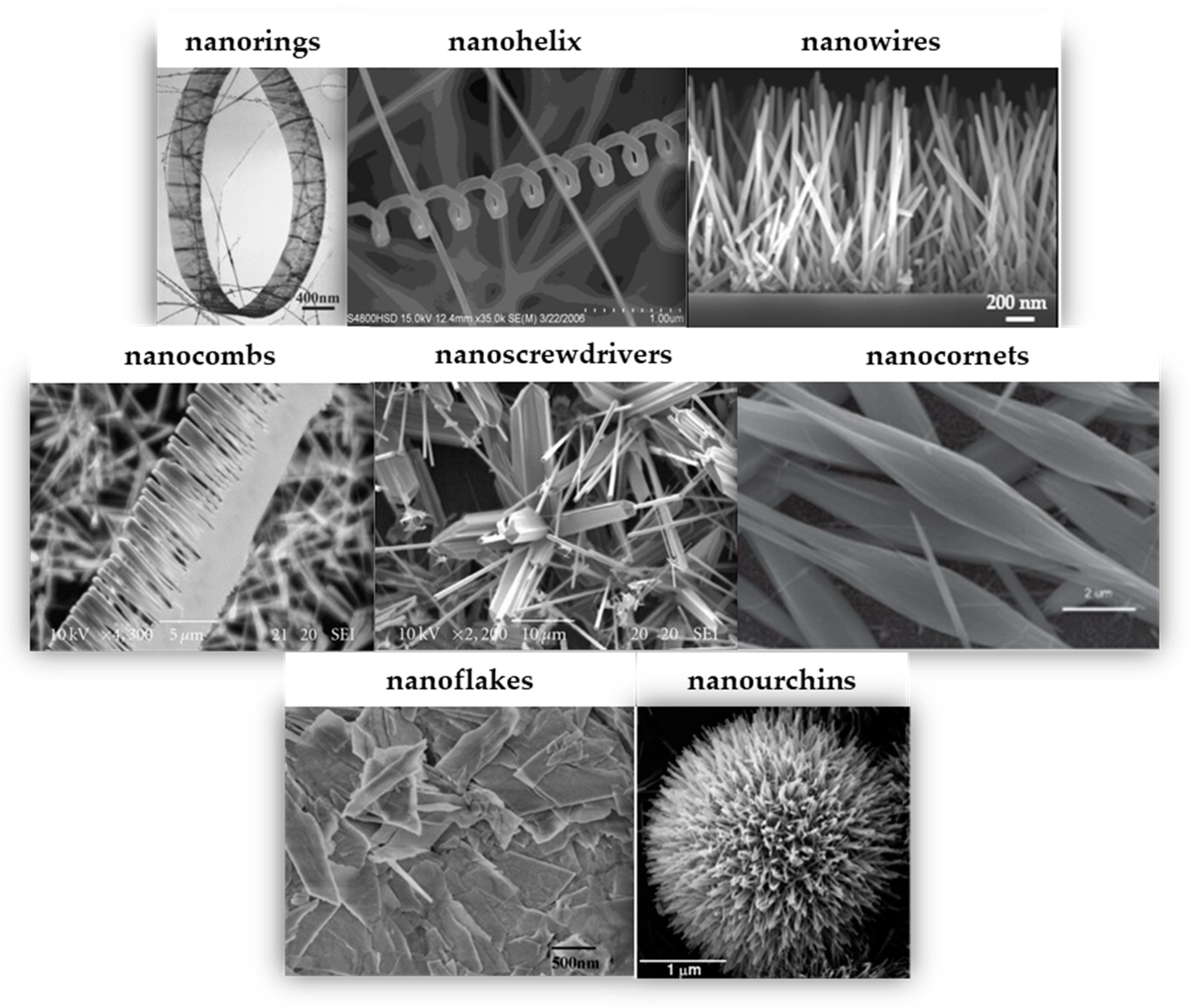
Figure 1. Different structures of ZnO. One-dimensional (1D) ZnO: nanorings (Reprinted with permission from [81]. Copyright © 2004, American Physical Society), nanohelix (Reprinted with permission from [82]. Copyright © 2017, the authors), nanowire (Reprinted with permission from [28]. Copyright © 2023, the authors), nanocombs and nanoscrewdrivers (Reprinted with permission from [83]. Copyright © 2010, the authors), and nanocornets (Reprinted with permission from [84]. Copyright © 2014, American Scientific Publishers). Two-dimensional (2D) ZnO: nanoflakes (Reprinted with permission from [85]. Copyright © 2009, Science Publications). Three-dimensional (3D) ZnO: Nanourchins (Reprinted with permission from [86]. Copyright © 2014, The Royal Society of Chemistry).
Crystals 14 00611 g001
Figure 2. Representative plots showing (a) absorption spectrum variations over time, (b) absorbance changes with irradiation time, (c) percentage degradation over irradiation time, and (d) comparison of degradation rates in AO decomposition. Reprinted with permission from [105]. Copyright © 2011, Elsevier B.V.
Crystals 14 00611 g002
Figure 3. Adsorption spectra of (a,b) MB, (c,d) MO, and (e,f) AR14 solutions in the presence of ZnO NWs under UV exposure. Reprinted with permission from [106]. Copyright © 2016, WILEY-VCH Verlag GmbH & Co. KGaA, Weinheim.
Crystals 14 00611 g003
Figure 4. Illustrations of experimental setup and photodegradation rate evolution for three dye solutions: (a) MB, (b) MO, and (c) AR14 under static and dynamic modes. Reprinted with permission from [70]. Copyright © 2020, the authors.
Crystals 14 00611 g004
Figure 5. Effect of ZnO NP dose on the photocatalytic degradation of (a) MO and (b) PNP. Reprinted with permission from [108]. Copyright © 2021, the authors.
Crystals 14 00611 g005
Figure 6. Degradation efficiency of the ZnO_B sample over five photocatalytic cycles against an MO + RhB + MB mixture. Reprinted with permission from [109]. Copyright © 2023, the authors.
Crystals 14 00611 g006
Figure 7. Performance of ZnO catalysts for phenol photodegradation: (a) THF, (b) decane, (c) H2O, (d) toluene, (e) ethanol, (f) acetone, (g) commercial ZnO, and (h) without catalyst. Reprinted with permission from [110]. Copyright © 2009, American Chemical Society.
Crystals 14 00611 g007
Figure 8. FESEM images of (a) propeller-shaped and (b) flower-shaped ZnO nanostructures. (c) Photodegradation of RhB (1.0 × 10⁻⁵ M, 30 mL) under UV light: (i) no catalyst, (ii) flower-shaped ZnO nanostructures, and (iii) propeller-shaped ZnO nanostructures. C is the concentration of RhB and C0 is the initial concentration. Reprinted with permission from [111]. Copyright © 2012, the authors.
Crystals 14 00611 g008
Figure 9. Variations in percentage photodegradation of DR-23 dye with ZnO nanoparticles annealed at different temperatures for 80 min of irradiation time. Reprinted with permission from [129]. Copyright © 2015, Elsevier B.V.
Crystals 14 00611 g009
Figure 10. Plot of ln C/C0 vs. laser exposure time for an aqueous solution of phenol in the presence of nano ZnO calcined at different temperatures. Experimental conditions: phenol concentration = 1.064 mM, V = 120 mL, nano ZnO concentration = 2.5 g/L, laser energy = 50 mJ, laser exposure time = 60 min. Reprinted with permission from [135]. Copyright © 2010, Elsevier B.V.
Crystals 14 00611 g010
Figure 11. Diclofenac mineralization at varying solution pH values using ZnO materials with catalyst loadings of 0.5, 1.0, and 1.5 g/L: (a) pH 5, (b) pH 6.5, and (c) pH 8. Reprinted with permission from [185]. Copyright © 2020, Elsevier B.V.
Crystals 14 00611 g011
Figure 12. Photodegradation of (a) MB and (b) RhB over time with and without catalysts. Reprinted with permission from [194]. Copyright © 2014, Elsevier B.V.
Crystals 14 00611 g012
Figure 13. Photodegradation of (a) MB and (b) RhB over time with and without catalysts. Reprinted with permission from [197]. Copyright © 2020, the authors.
Crystals 14 00611 g013
Figure 14. Photocatalytic performance of transition metal-doped ZnO nanowires: (a) ZnO:Fe NWs, (b) ZnO:Ag NWs, and (c) ZnO:Co NWs. Reprinted with permission from [198]. Copyright © 2019, Acta Physica Polonica. A.
Crystals 14 00611 g014
Figure 15. Phenol degradation kinetics with various catalysts under UV–A light in Milli-Q water (top), effluent wastewaters (EWW) (middle), and influent wastewaters (IWW) (bottom). Reprinted with permission from [212]. Copyright © 2018, Elsevier B.V.
Crystals 14 00611 g015
Figure 16. Temporal evolution of absorption spectra during photocatalysis of R6G using (a) ZnO, (b) 2 wt% composite, and (c) change in R6G concentration (C/C0) with ZnO and composites containing AuNP loadings (0.4, 0.7, 1, and 2 wt%). R6G concentration was estimated based on absorbance at 525 nm. Reprinted with permission from [44]. Copyright © 2011, American Chemical Society.
Crystals 14 00611 g016
Figure 17. Photodegradation efficiency of as-prepared ZnO nanorods and ZnO-ZnS core–shell nanorods with varying shell wall thicknesses against MB under visible irradiation. Reprinted with permission from [47]. Copyright © 2018, Elsevier B.V.
Crystals 14 00611 g017
Figure 18. Photocatalytic degradation of (a) 10 mg/L MB solution and (b) 10 mg/L RhB solution under UV irradiation with and without RGO, ZnO sphere–RGO composites (sZG), ZnO disk–RGO composites (dZG), and ZnO nanorod–RGO composites (rZG) (0.1 g/L). Reprinted with permission from [238]. Copyright © 2019, Elsevier B.V.
Crystals 14 00611 g018
Figure 19. Photodegradation of MO (a) under UV light and (b) under visible light irradiation using ZnO NPs, ZnO NRs, GNP, and GNR. Reprinted with permission from [239]. Copyright © 2019, Elsevier B.V.
Crystals 14 00611 g019
Figure 20. Photocatalytic degradation of 2,4-D (10 ppm), shown as the variation of C/C0 with irradiation time in the presence of GO, ZnO, ZGO0, ZGO1, ZGO2, and ZGO3. Reprinted with permission from [240]. Copyright © 2017, Elsevier B.V.
Crystals 14 00611 g020
Figure 21. (a) Proposed photocatalytic mechanism for safranin-T dye degradation using FGS/ZnO. (b) Comparison of photocatalytic activity between FGS/ZnO and pristine ZnO NPs. Reprinted with permission from [241]. Copyright © 2018, The Royal Society of Chemistry.
Crystals 14 00611 g021
Figure 22. (a) Photocatalytic degradation plots of 20 ppm acid blue 74 dye using (1) ZnO, (2) AgAg2O-ZnO, and (3) Ag-Ag2O-ZnO/GO. (b) Mechanisms of photocatalytic activity for Ag-Ag2O-ZnO/GO in the degradation of acid blue 74 dye. Reproduced from Umukoro, E.H.; Peleyeju, M.G.; Ngila, J.C.; Arotiba, O.A. Photocatalytic Degradation of Acid Blue 74 in Water Using Ag–Ag2O–Zno Nanostuctures Anchored on Graphene Oxide. Solid State Sci. 2016, 51, 66–73. Copyright© 2015, Elsevier Masson SAS. All rights reserved [242].
Crystals 14 00611 g022
Figure 23. Schematic representation of the degradation mechanism of L-CHT using the ZnO-Bi2O3 composite. Reprinted with permission from [248]. Copyright © 2019, Elsevier Ltd.
Crystals 14 00611 g023
Figure 24. Plausible mechanism for MP degradation with Cu-ZnO NPs. Reproduced from Aulakh, M.K.; Kaur, S.; Pal, B.; Singh, S. Morphological Influence of ZnO Nanostructures and Their Cu Loaded Composites for Effective Photodegradation of Methyl Parathion. Solid State Sci. 2020, 99, 106045. Copyright © 2019 Elsevier Masson SAS. All rights reserved [250].
Crystals 14 00611 g024
Figure 25. Photo images demonstrating the antimicrobial activity against P. putida of the prepared ZnO NWs against bare Si (a) in the dark and (b) under UV light (10 min; 0.17 W/cm2) utilizing the printing method. Reprinted with permission from [28]. Copyright © 2023, the authors. (c) Plausible mechanism for antibacterial efficacy of ZnO NWs against Pseudomonas putida.
Crystals 14 00611 g025
Figure 26. Results of antibacterial experiments conducted using photocatalytic paper (Sample 1) after 48 h of incubation (a) in the dark and (b) under illumination with a tungsten halogen lamp. The inhibition zone increased from 1.7 × 1.7 cm2 in the dark to 2.1 × 2.1 cm2 under visible light illumination. Optical images captured at 1000× magnification (c) outside the inhibition zone and (d) inside the inhibition zone. Reprinted with permission from [114]. Copyright © 2012, Bentham Science Publishers, Ltd.
Crystals 14 00611 g026
Table 1. Variations in physical properties for ZnO NPs with annealing temperature. Reprinted with permission from [129]. Copyright © 2015, Elsevier B.V.
Annealing Temp.λmax. (nm)Band Gap (eV)Particle Size (nm) (from FESEM)Crystallite Size (nm) (from XRD)
400374.43.31740–4524.94
500376.83.29655–6028.16
600378.03.28585–9031.19
700379.23.275100–11037.90
800381.63.254115–12048.53
Table 2. Fitted pseudo-first-order rate constants for contaminant degradation under various conditions. Reprinted with permission from [261]. Copyright © 2013, Springer Nature.
Compound and System Catalyst Dosage (g/L) Removal Efficiency (%) in 30 min Reaction Rate, k (min−1) t1/2 (min)
Ketamine,
UV lamp/ZnO
0.01 98.5 0.14 5.90
0.04 98.7 0.15 5.30
0.05 99.9 0.233.83
0.10 99.9 0.263.11
0.40 99.9 0.432.25
0.70 99.9 0.253.54
1.00 99.9 0.272.72
Methamphetamine,
UV lamp/ZnO
0.01 81.4 0.06 14.76
0.04 98.8 0.15 7.46
0.05 99.9 0.23 6.27
0.10 99.9 0.32 4.70
0.40 99.9 0.38 2.58
0.70 99.9 0.14 5.44
1.00 99.9 0.223.53
Morphine,
UV lamp/ZnO
0.01 99.7 0.19 4.26
0.04 99.9 0.21 3.61
0.05 99.9 0.21 3.39
0.10 99.9 0.37 2.38
0.40 99.9 0.68 1.06
0.70 99.9 0.48 1.52
1.00 99.9 0.60 1.14
Table 3. Estimated time to achieve 3-log destruction efficiency through photocatalysis. Reprinted with permission from [261]. Copyright © 2013, Springer Nature.
System Dosage (g/L) Ketamine (min) Methamphetamine (min) Morphine (min)
UV lamp/ZnO 0.4 17 19 10
UVLED/ZnO 0.4 48 95 34
Disclaimer/Publisher’s Note: The statements, opinions and data contained in all publications are solely those of the individual author(s) and contributor(s) and not of MDPI and/or the editor(s). MDPI and/or the editor(s) disclaim responsibility for any injury to people or property resulting from any ideas, methods, instructions or products referred to in the content.