
Perspective on Nanofiber Electrochemical Sensors: Design of Relative Selectivity Experiments


Abstract
:1. Introduction
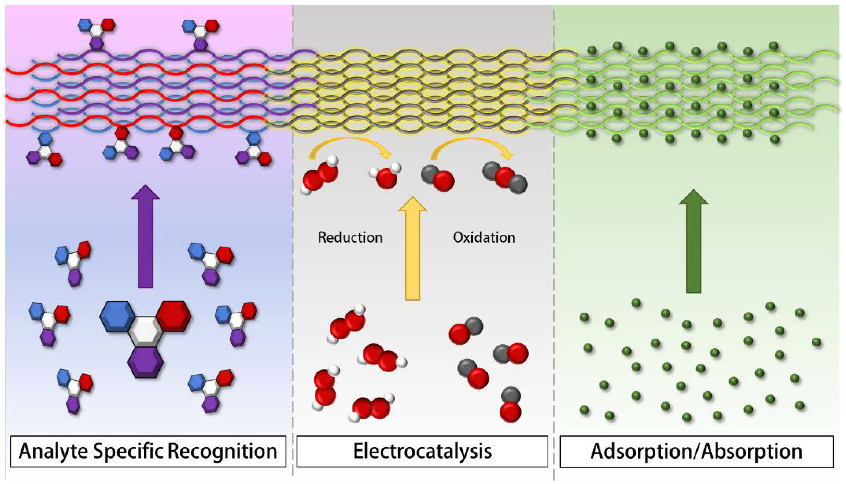
2. Nanofiber Roles in Sensing Mechanisms
2.1. Electrocatalytic Activity
- Metal oxide nanofibers can directly act as electron carriers as the source of a sensor’s electrocatalytic behavior [36,37], functioning largely as class-recognition type sensors (Table 1, #2 and 3). In the case of one biosensor for purine detection (Table 1, #2), CuO nanofibers (CuO NFs) and ZnO nanoparticles (ZnO NPs) were immobilized within a poly-L-cysteine (PLC) matrix [36]. The surface was prepared through simultaneous electropolymerization in a buffered, aqueous solution of L-cysteine (LC), ZnO NPs, and CuO NFs. The CuO-ZnO heterostructures were said to form p-n junctions that greatly enhanced sensitivity. The sensitivity of the sensor increased from 0.353 to 2.66 μA/μM for guanine and from 0.155 to 2.67 μA/μM for adenine when compared to a sensor that uses metal nanoparticles without nanofibers. This improvement was attributed to a synergistic combination of the electrocatalytic behaviors of the two metal oxide nanostructures. The nanofibers also increased the electron transfer capacity of the electrode surface.
- N-doped carbon nanofibers (NCNF) can be used in conjunction with N-doped graphene quantum dots (NGQD), as both NCNFs and NGQDs have catalytic activity toward nitrite and the combination of the two produces much higher sensitivity (Table 1, #7) [41]. The composite with NGQDs was formed by hydrothermal treatment with in situ quantum dot synthesis. The dried NGQD/NCNF was physically adhered to the surface of glassy carbon electrodes. Introduction of the doped nanofibers into the glassy carbon electrode resulted in an increase in the oxidation peak current from 9.914 to 20.56 μA. For nitrite sensing, The NGQD/NCNF composite biosensor showed an improvement in the limit of detection from 8.1 to 3 μM when compared to a porous graphite sensor. The doping of the CNFs with heteroatoms, in this case nitrogen, synergistically working with NGQDs by increasing electron transfer, allowed the fibers to electrocatalyze the oxidation of nitrite.
- An example of a sensor that uses complex surface interactions to achieve redox catalysis is a polymethylene blue (PMB)-decorated Cu-CNF sensor for the oxidation of creatinine (Table 1, #9) [43]. An activated carbon microfiber (ACF) surface was dispersed with Cu(NO3)2 which was used for in situ Cu NP synthesis. CNFs were added to the surface via chemical vapor deposition. The PMB was then synthesized on the surface through an electro-polymerization method. The full construction of the surface was a PMB nanofiber matrix on-top of a copper infused CNF/activated carbon surface mat (PMB-Cu-NF/ACF). The CNF/ACF mat is an adsorptive surface, but the interactions between the PMB-Cu promotes the selective catalysis of creatinine oxidation. Detection, with differential pulse voltammetry and cyclic voltammetry, resulted in an improvement of the detection limit from 56.55 to 0.24 ng/mL when compared to a copper electrode without the PMB NFs or CNFs. This improvement was attributed to the selective nature of the surface interactions promoted by the electrocatalytic activity promoted by the large surface area of the nanofiber matrix.
- Carbon-based nanofibers can be fabricated to have a high density of states for an increased electrocatalytic response. One sensor was fabricated by carbonizing electrospun poly-acrylonitrile onto graphitized fiber carbon paper. The high density of electronic states led to a wide detection range of dopamine oxidation. [71]. The deposition time was varied from 5 h to 33 h, which lead to the dynamic range increasing from 8–9000 μM to 0.2–700,000 μM. Additionally, the limit of detection improved from 5.58 to 0.07 μM. The dynamic range and the limit of detection were improved by orders of magnitude due to the high surface area and edge effects of the nanofibers.
2.2. Adsorptive Behavior
- An atrazine sensor that uses tin (IV) oxide nanofibers to achieve a very low limit of detection by using nanofiber adsorption to enhance atrazine interaction with traditional antibody biorecognition elements was created (Table 1, #10) [44]. Atrazine antibodies were grafted to SnO2 NFs that were predispersed onto a glassy carbon electrode surface. The antibodies promoted specific interaction via traditional biorecognition routes. The absorptive effects of the tin (IV) oxide promoted a lower limit of detection (0.9 zM) which is orders of magnitude smaller than other atrazine sensors (typically around 20 pM) [44].
- Organic polymer nanofibers do not have the same electrocatalytic potential as metal oxides, but due to their tunable size and inexpensive fabrication, are commonly used for their adsorptive effects in combination with electrocatalytic compounds (Table 1, #4, 5, 6, and 8) [38,39,40,42]. A specific example of this is a composite sensor made with polypyrrole (PPy) NFs for the simultaneous electrocatalytic determination of ascorbic acid, dopamine, paracetamol, and tryptophan (Table 1, #4) [38]. ZnO nanosheets and CuxO nanoparticles were electrochemically deposited on PPy NFs to create a 3D CuxO-ZnO NP/PPyNF/RGO structure. The zinc oxide–copper oxide p-n junction heterostructures electrocatalytically oxidize the analytes. The PPy NFs were used to increase the adsorption of the analytes to the surface, which increases sensitivity, as well as to prevent graphene sheet aggregation for an increase in stability. An increase in the linear range from 0.5–20 μM to 0.04–420 μM of dopamine and a decrease the in limit of detection from 0.17 to 0.012 μM of dopamine was observed compared to Ni and CuO modified surfaces without the nanofiber.
- In the aforementioned creatinine sensor, the PMB fibers produce a catalytic effect while the copper dispersed CNF composite promotes adsorption to improve sensor performance. (Table 1, #9) [43]. This is an example of combining two different nanofibers in such a way that they have separate but complementary roles. The CNFs used in this sensor increase the adsorption of creatinine to the surface, resulting in an additional increase of reported sensitivity.
- Another example of a sensor that uses the adsorptive mechanism of nanofibers is a pH and H2O2 sensor that uses a layer-by-layer assembly of PAA/PANI nanofibers [77]. The PANI nanofibers were synthesized using ammonium persulfate chemistry and were deposited onto a cleaned glassy carbon electrode in alternating fashion with PAA. The numbers of layers of PAA and PANI resulted in different adsorptive properties, and therefore, different electrochemical response. After six layers of PAA and PANI, the linear range of the sensor increased from 0.005–0.8 to 0.001–6 mM and the detection limit improved from 1.2 to 0.3 μM. The improvement of these properties was attributed to the high surface area and microporosity of the sensor surface, which can be tuned by changing the modification procedure.
2.3. Analyte-Specific Recognition
- One type of analyte specific interaction occurs between traditional biorecognition elements like antibodies or enzymes. For example, the previously mentioned atrazine sensor uses a traditional biorecognition element, an antibody, integrated into a nanofiber network (Table 1, #10) [44]. The antibody increases the specificity from the specific analyte interaction, and the nanofiber network provides an increase in antibody loading (high surface area) and an increase in the binding kinetics via adsorption. The interfering compound that was found to interact with the sensor the most was melamine, with a 15.6% change in peak current at 1 μM, while atrazine at that same concentration resulted in a 43.5% change at the same concentration. Additionally, a 7.2% interference was found in a 1:1 mixture of atrazine and urea.
- Analyte-specific interactions can also take the form of highly selective chemical interactions with surface lattice structures. The previously mentioned creatinine sensor has a specific binding event between the PMB-Cu nanofiber matrix and creatinine, allowing the PMB-Cu heterostructure to act as a synthetic chemical recognition element (Table 1, #9). [43]. The sensor was tested for multiple interfering compounds in clinically relevant ratios, and some non-specific adsorption was found which could be mitigated by washing. The end result after washing was minimal interference. The sensor was also tested in cerebrospinal fluid, saliva, and blood serum, resulting in an average recovery of 98.3%.
- Specific steric repulsion can occur between elements of a sensor and potential interferents, as is the case between biofouling proteins and poly(ethylene) glycol (PEG), as seen in a polyaniline nanofiber-based DNA sensor (Table 1, #5) [39]. There is a specific steric repulsion between the PANI/PEG composite and potential biofouling proteins. DNA capture probes were attached to a PANI NF/PEG surface, which added an additional specific interaction that improved selectivity, an analyte-specific interaction between DNA capture probes and the DNA analyte. The improvement in selectivity was evaluated by comparing the response of the sensor to DNA with one base pair mismatch from the target in a 10,000-fold concentration. Even at such high concentrations, the mismatched DNA only produced 25% the signal compared to the target DNA.
3. Selectivity Experiments in the Development of Nanofiber Sensors
3.1. Interferant Control Experiments

| # | Analyte | Tested Interferant Compounds | Highest Found Interferant | Ref. |
|---|---|---|---|---|
| 1 | Idarubicin hydrochloride | Ca2+, Mg2+, Fe2+, Cl−, glucose, lactose, fructose, AA, CA, UA, urea, acetaminophen, epirubicin, doxorubicin, daunorubicin, cysteine | 3% from 5-fold cysteine and AA | [35] |
| 2 | Adenine, guanine | Na+, Mg2+, Ca2+, Cu2+, Zn2+, Fe3+, CO32+, NO3−, Cl−, thymine, xanthine, cytosine, tyrosine, tryptophan, aspartic acid, pyridoxine, AA, FA, UA, glucose, alanine, glycine, arginine, L-cysteine | 17.6% from 200-fold tryptophan toward guanine determination 9.0% from 500-fold tryptofan toward adenine determination | [36] |
| 3 | Acetaminophen | UA, DA, AA, glucose | DA * | [37] |
| 4 | AA, DA, Paracetamol, and tryptophan | Cytesine, epinephrine, glucose, UA, FA, and tyrosine | 6.96% from 500-fold FA | [38] |
| 5 | DNA sequence | BSA, HSA, IgG, Hb, base-mismatched DNA | 25% from 10,000-fold single base-pair mismatched DNA | [39] |
| 6 | Hydrogen peroxide | AA, UA, DA | 1-fold AA, UA, and DA * | [40] |
| 7 | Nitrite | K+, Ca2+, Na+, Mg2+, Zn2+, Ag+, NH4+, Cl−, NO3−, CO32−, HCO3−, PO43− | 10% from 100-fold Ag+ and Zn2+ | [41] |
| 8 | H2O2, glucose | AA, UA, DA | AA * | [42] |
| 9 | Creatinine | DA, AA, UA, cholesterol, urea, glucose, glutamine, bilirubinketones, hemoglobin, pyruvic acid | Clinically relevant ratios of all compounds * | [43] |
| 10 | Atrazine | Urea, glucose, antibiotic, BSA, HSA, Na+, melamine | 15.6% from 1-fold melamine | [44] |
| 11 | BCSC | BT-474, HepG2, L02 | 1.5% from 1-fold HepG2 | [45] |
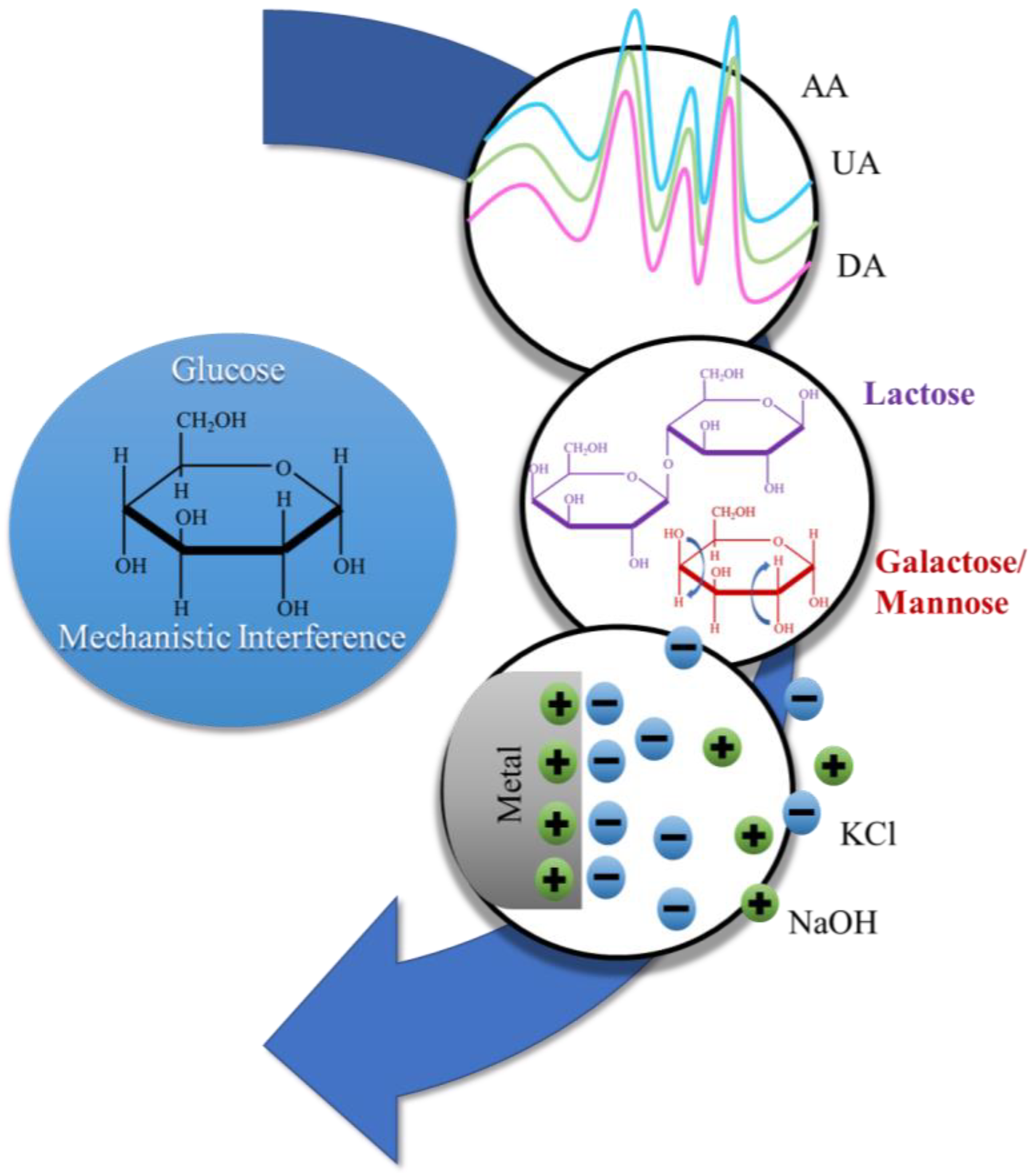
- Electrochemical oxidation peaks have the potential to be misinterpreted when the equilibrium potential of interferants is similar to that of the target analyte. For example, the peaks of AA, UA, DA, and acetaminophen are similar to the oxidation peaks of catalyzed glucose (Figure 4), and can interfere with the interpretation of the data [91]. When a sensor is run potentiometrically, the oxidation and reduction potentials should be investigated in addition to the signal strength. Typically, this will affect interferants with similar electrochemically active functional groups in electrocatalytic modalities, as seen in Table 2 (#2, 3, 4, 6, and 8). Within these examples, the oxidative or reductive overlap was found to have the largest interference.
- Common functional groups between the molecules have the potential to adsorb similar to the target analyte. Galactose and lactose were tested because, as with glucose, they are sugars with a similar chemical structure (Figure 4) [91]. Similar chemical structures are a commonly tested interferant. Testing the adsorption of similar chemical structures is very important in adsorption or analyte-specific recognition sensors. Adsorptive modality disruptions are more common when a sensor runs via an amperometric or impedimetric modality. As seen in Table 2 (#1, 5, 10, and 11), the primary interferant is one of a similar chemical structure.
- Surface interactions through charge-induced adsorption, such as ions, can alter the surface electrolyte double-layer capacitance and influence the electrochemical output. These induced changes can result in an artificially higher or lower concentration measurement via alteration in the electrochemical transduction output. For example, KCl can change the overall charge of the solution, and small changes can result in an altered signal (Figure 4) [91]. Also, NaOH has the potential to lower the acidity of the analyte solution, which means that changes in water ionic charges should be tested as an interferant as well. Ionic interference was only tested in three examples (Table 2, #1, 2, and 7), but was found to be the major interferant in example #7 (Table 2). We would expect ionic changes to be more commonly found to interfere with electrochemical transduction if these interferant control experiments were more widely tested.
3.2. Transitioning to Real Samples and Analysis
- Dilution was a step during sample preparation for four of the reported sensors (Table 3, #1, 2, 4, and 7): the TiO2/CNF sensor for idarubicin hydrochloride [35]; the adenine and guanine sensor that uses a PLC/ZnO-NPs/CuO-NF modified surface [36]; the 3DCuxO-ZnO NP/PPyNF/RGO sensor for simultaneous detection of ascorbic acid, dopamine, paracetamol, and tryptophan [38]; and the NGQD/NCNF sensor for nitrite determination [41]. Simple dilution is relatively easy to perform, and therefore, would be acceptable even for untrained individuals. Dilution is primarily performed to reduce non-specific binding and physisorption of interfering agents in the complex media. It can also help to reduce viscous effects that would prevent efficient diffusion of the analyte to the surface. The detailed sensors that use this sample preparation (Table 3, #1, 2, 4, and 7) have highly adsorbent surfaces that benefit from the dilution step to promote the binding of the analyte.
- Centrifugation and filtration were steps during the sample preparation for two of the sensors (Table 3, #1 and 7): the TiO2/CNF sensor for idarubicin hydrochloride [35] and the NGQD/NCNF sensor for nitrite determination [41]. Centrifugation and filtration, depending on the application, may require special equipment that would limit the point-of-need use of this type of sensor. Centrifugation and filtration are useful in separating large components like proteins, cells, and macromolecules from biological samples. The separation of large macromolecules is important for the idarubicin and nitrite sensors because the large molecules could non-specifically bind to and foul the surface interfering with the adsorption mechanism (Table 3, #1 and 7).
- Ultrasonication was a step in just one of the sensors (Table 3, #10): the SnO2 for the determination of atrazine [44]. Ultrasonication requires the use of specialized equipment such that point-of-need applications are rarely suitable. Ultrasonication is used to break larger molecules down into smaller parts, which minimizes the adsorption of potential interferants compared to the intended analyte. The atrazine sensor (Table 3, #10) uses antibodies as biorecognition elements, and the ultrasonication breaks down larger proteins that could be susceptible to non-specific binding.
- pH adjustment was a sample preparation step in the complex media analysis of the adenine and guanine sensor that uses a PLC/ZnO-NPs/CuO-NF modified surface (Table 3, #2) [36]. The pH adjustment should be performed by a trained individual to ensure the pH adjustment is properly achieved. As previously mentioned, the charge of a solution affects the double layer capacitance of a surface, and therefore, influences the adsorption of the analyte and interfering compounds. Attaining a favorable pH environment is necessary for the selectivity and sensitivity of some sensors. Further, the pH has to be adjusted occasionally to increase the accessibility of the analyte. For example, a DNA sensor (Table 3, #2) uses HCl to digest the DNA to increase the accessibility of adenine and guanine in complex media [36]. However, the designed sensor operates better at neutral pH, which further makes a pH adjustment with NaOH after digestion necessary.
| # | Complex Media Tested | Sample Preparation | Ref. |
|---|---|---|---|
| 1 | Human serum, human urine | Centrifugation, filtration, dilution | [35] |
| 2 | Sturgeon sperm DNA, Human blood DNA, Flavithermus DNA | Digestion in HCl, heating, rapid cooling, neutralization with NaOH, dilution in PBS | [36] |
| 3 | Human serum | None specified | [37] |
| 4 | Human serum | Dilution | [38] |
| 5 | Human serum | None specified | [39] |
| 6 | N/A | N/A | [40] |
| 7 | Sausage, pickle, lake Water, tap water | Sausage and pickle: deproteinization, centrifugation, filtration, dilution with PBS Water: centrifugation, filtration, dilution with PBS | [41] |
| 8 | N/A | N/A | [42] |
| 9 | Human serum, human Cerebral spinofluid, Human saliva | None specified | [43] |
| 10 | Ground water, river Water | Ultra-sonicated | [44] |
| 11 | Fetal bovine serum | None specified | [45] |
3.3. Future Perspectives on Selectivity Experiments
4. Conclusions
Author Contributions
Funding
Data Availability Statement
Acknowledgments
Conflicts of Interest
Abbreviations
References
- Sharma, S.; Sudhakara, P.; Omran, A.A.B.; Singh, J.; Ilyas, R.A. Recent trends and developments in conducting polymer nanocomposites for multifunctional applications. Polymers 2021, 13, 2898. [Google Scholar] [CrossRef] [PubMed]
- Korotcenkov, G. Current trends in nanomaterials for metal oxide-based conductometric gas sensors: Advantages and limitations. part 1: 1D and 2D nanostructures. Nanomaterials 2020, 10, 1392. [Google Scholar] [CrossRef] [PubMed]
- Song, J.; Kim, M.; Lee, H. Recent advances on nanofiber fabrications: Unconventional state-of-the-art spinning techniques. Polymers 2020, 12, 1386. [Google Scholar] [CrossRef]
- Zhao, K.; Kang, S.-X.; Yang, Y.-Y.; Yu, D.-G. Electrospun functional nanofiber membrane for antibiotic removal in water: Review. Polymers 2021, 13, 226. [Google Scholar] [CrossRef] [PubMed]
- Alali, K.T.; Liu, J.; Aljebawi, K.; Liu, Q.; Chen, R.; Yu, J.; Zhang, M.; Wang, J. 3D hybrid Ni-multiwall carbon nanotubes/carbon nanofibers for detecting sarin nerve agent at room temperature. J. Alloys Compd. 2019, 780, 680–689. [Google Scholar] [CrossRef]
- Vilian, A.T.E.; Ranjith, K.S.; Lee, S.J.; Umapathi, R.; Hwang, S.-K.; Oh, C.W.; Huh, Y.S.; Han, Y.-K. Hierarchical dense Ni−Co layered double hydroxide supported carbon nanofibers for the electrochemical determination of metronidazole in biological samples. Electrochim. Acta 2020, 354, 136723. [Google Scholar] [CrossRef]
- Ramakrishna, S.; Zamani, M.; Prabhakaran, M.P. Advances in drug delivery via electrospun and electrosprayed nanomaterials. Int. J. Nanomed. 2013, 8, 2997. [Google Scholar] [CrossRef] [Green Version]
- Matlock-Colangelo, L.; Baeumner, A.J. Recent progress in the design of nanofiber-based biosensing devices. Lab Chip 2012, 12, 2612. [Google Scholar] [CrossRef]
- Fadil, F.; Affandi, N.D.N.; Misnon, M.I.; Bonnia, N.N.; Harun, A.M.; Alam, M.K. Review on electrospun nanofiber-applied products. Polymers 2021, 13, 2087. [Google Scholar] [CrossRef]
- Liu, X.; He, J.-H.; Sakthivel, R.; Chung, R.-J. Rare earth erbium molybdate nanoflakes decorated functionalized carbon nanofibers: An affordable and potential catalytic platform for the electrooxidation of phenothiazine. Electrochim. Acta 2020, 358, 136885. [Google Scholar] [CrossRef]
- Li, X.-X.; Xu, L.-Y.; He, J.-H. Nanofibers membrane for detecting heavy metal ions. Therm. Sci. 2020, 24, 2463–2468. [Google Scholar] [CrossRef]
- Liu, K.; Zhou, Z.; Yan, X.; Meng, X.; Tang, H.; Qu, K.; Gao, Y.; Li, Y.; Yu, J.; Li, L. Polyaniline nanofiber wrapped fabric for high performance flexible pressure sensors. Polymers 2019, 11, 1120. [Google Scholar] [CrossRef] [PubMed] [Green Version]
- Adeola, A.O.; Fapohunda, O.; Jimoh, A.T.; Toluwaloju, T.I.; Ige, A.O.; Ogunyele, A.C. Scientific applications and prospects of nanomaterials: A multidisciplinary review. Afr. J. Biotechnol. 2019, 18, 946–961. [Google Scholar] [CrossRef] [Green Version]
- Atakaramians, S.; Dong, F.Q.; Monro, T.M.; Afshar, S.V. Radiated and guided optical waves of a magnetic dipole-nanofiber system. Sci. Rep. 2019, 9, 3568. [Google Scholar] [CrossRef] [PubMed] [Green Version]
- Li, Z.; Liu, S.; Song, S.; Xu, W.; Sun, Y.; Dai, Y. Porous ceramic nanofibers as new catalysts toward heterogeneous reactions. Compos. Commun. 2019, 15, 168–178. [Google Scholar] [CrossRef]
- Chen, H.; Huang, M.; Liu, Y.; Meng, L.; Ma, M. Functionalized electrospun nanofiber membranes for water treatment: A review. Sci. Total Environ. 2020, 739, 139944. [Google Scholar] [CrossRef] [PubMed]
- Jin, H.; Nayeem, M.O.G.; Lee, S.; Matsuhisa, N.; Inoue, D.; Yokota, T.; Hashizume, D.; Someya, T. Highly durable nanofiber-reinforced elastic conductors for skin-tight electronic textiles. ACS Nano 2019, 13, 7905–7912. [Google Scholar] [CrossRef] [PubMed]
- Sun, X.; Tyagi, P.; Agate, S.; McCord, M.G.; Lucia, L.A.; Pal, L. Highly tunable bioadhesion and optics of 3D printable PNIPAm/cellulose nanofibrils hydrogels. Carbohydr. Polym. 2020, 234, 115898. [Google Scholar] [CrossRef] [PubMed]
- Park, S.J.; Kwon, O.S.; Lee, J.E.; Jang, J.; Yoon, H. Conducting polymer-based nanohybrid transducers: A potential route to high sensitivity and selectivity sensors. Sensors 2014, 14, 3604–3630. [Google Scholar] [CrossRef]
- Ashrafmansouri, S.-S.; Willersinn, S.; Esfahany, M.N.; Bart, H.-J. Influence of silica nanoparticles on mass transfer in a membrane-based micro-contactor. RSC Adv. 2016, 6, 19089–19097. [Google Scholar] [CrossRef] [Green Version]
- Yang, C.X.; Zhu, Q.; Dong, W.P.; Fan, Y.Q.; Wang, W.L. Preparation and characterization of phosphoric acid-modified biochar nanomaterials with highly efficient adsorption and photodegradation ability. Langmuir 2021, 37, 9253–9263. [Google Scholar] [CrossRef] [PubMed]
- Lee, K.; Kim, K.; Yoon, H.; Kim, H. Chemical design of functional polymer structures for biosensors: From nanoscale to macroscale. Polymers 2018, 10, 551. [Google Scholar] [CrossRef] [PubMed] [Green Version]
- Halicka, K.; Cabaj, J. Electrospun nanofibers for sensing and biosensing applications—A review. Int. J. Mol. Sci. 2021, 22, 6357. [Google Scholar] [CrossRef] [PubMed]
- Wang, H.; Zhang, B.; Zeng, X.; Yan, L.; Zheng, J.; Ling, M.; Hou, Y.; Lu, Y.; Liang, C. 3D porous carbon nanofibers with CeO2-decorated as cathode matrix for high performance lithium-sulfur batteries. J. Power Sources 2020, 473, 228588. [Google Scholar] [CrossRef]
- Nathani, A.; Sharma, C.S. Electrospun mesoporous poly(Styrene-Block-Methyl-Methacrylate) nanofibers as biosensing platform: Effect of fibers porosity on sensitivity. Electroanalysis 2019, 31, 2138–2144. [Google Scholar] [CrossRef]
- Horne, J.; McLoughlin, L.; Bridgers, B.; Wujcik, E.K. Recent developments in nanofiber-based sensors for disease detection, immunosensing, and monitoring. Sens. Actuators Rep. 2020, 2, 100005. [Google Scholar] [CrossRef]
- Pumera, M.; Ambrosi, A.; Bonanni, A.; Chng, E.L.K.; Poh, H.L. Graphene for electrochemical sensing and biosensing. TrAC Trends Anal. Chem. 2010, 29, 954–965. [Google Scholar] [CrossRef]
- Ambrosi, A.; Sasaki, T.; Pumera, M. Platelet graphite nanofibers for electrochemical sensing and biosensing: The influence of graphene sheet orientation. Chem.—Asian J. 2010, 5, 266–271. [Google Scholar] [CrossRef] [PubMed]
- Vamvakaki, V.; Tsagaraki, K.; Chaniotakis, N. Carbon nanofiber-based glucose biosensor. Anal. Chem. 2006, 78, 5538–5542. [Google Scholar] [CrossRef]
- Pérez, B.; Del Valle, M.; Alegret, S.; Merkoçi, A. Carbon nanofiber vs. carbon microparticles as modifiers of glassy carbon and gold electrodes applied in electrochemical sensing of NADH. Talanta 2007, 74, 398–404. [Google Scholar] [CrossRef]
- Morales, M.A.; Paiva, W.A.; Marvin, L.; Balog, E.R.M.; Halpern, J.M. Electrochemical characterization of the stimuli-response of surface-immobilized elastin-like polymers. Soft Matter. 2019, 15, 9640–9646. [Google Scholar] [CrossRef] [Green Version]
- Aluri, S.; Pastuszka, M.K.; Moses, A.S.; MacKay, J.A. Elastin-like peptide amphiphiles form nanofibers with tunable length. Biomacromolecules 2012, 13, 2645–2654. [Google Scholar] [CrossRef] [PubMed] [Green Version]
- Varanko, A.K.; Su, J.C.; Chilkoti, A. Elastin-like polypeptides for biomedical applications. Annu. Rev. Biomed. Eng. 2020, 22, 343–369. [Google Scholar] [CrossRef]
- Bao, J.; Hou, C.; Dong, Q.; Ma, X.; Chen, J.; Huo, D.; Yang, M.; El Galil, K.H.A.; Chen, W.; Lei, Y. ELP-OPH/BSA/TiO2 nanofibers/c-MWCNTs based biosensor for sensitive and selective determination of p-nitrophenyl substituted organophosphate pesticides in aqueous system. Biosens. Bioelectron. 2016, 85, 935–942. [Google Scholar] [CrossRef]
- Arkan, E.; Paimard, G.; Moradi, K. A novel electrochemical sensor based on electrospun TiO2 nanoparticles/carbon nanofibers for determination of Idarubicin in biological samples. J. Electroanal. Chem. 2017, 801, 480–487. [Google Scholar] [CrossRef]
- Arvand, M.; Sayyar Ardaki, M. Poly-l-cysteine/electrospun copper oxide nanofibers-zinc oxide nanoparticles nanocomposite as sensing element of an electrochemical sensor for simultaneous determination of adenine and guanine in biological samples and evaluation of damage to dsDNA and DNA purine bases by UV radiation. Anal. Chim. Acta 2017, 986, 25–41. [Google Scholar] [CrossRef] [PubMed]
- Cao, F.; Dong, Q.; Li, C.; Chen, J.; Ma, X.; Huang, Y.; Song, D.; Ji, C.; Lei, Y. Electrochemical sensor for detecting pain reliever/fever reducer drug acetaminophen based on electrospun CeBiO nanofibers modified screen-printed electrode. Sens. Actuators B Chem. 2018, 256, 143–150. [Google Scholar] [CrossRef]
- Ghanbari, K.; Bonyadi, S. An electrochemical sensor based on reduced graphene oxide decorated with polypyrrole nanofibers and zinc oxide–copper oxide p–n junction heterostructures for the simultaneous voltammetric determination of ascorbic acid, dopamine, paracetamol, and tryptoph. New J. Chem. 2018, 42, 8512–8523. [Google Scholar] [CrossRef]
- Hui, N.; Sun, X.; Niu, S.; Luo, X. PEGylated polyaniline nanofibers: Antifouling and conducting biomaterial for electrochemical DNA sensing. ACS Appl. Mater. Interfaces 2017, 9, 2914–2923. [Google Scholar] [CrossRef]
- Li, Y.; Zhang, L.; Li, J.; Su, Z.; Wei, G. Sequence-designed peptide nanofibers bridged conjugation of graphene quantum dots with graphene oxide for high performance electrochemical hydrogen peroxide biosensor. JINoP 2016, 2, 334. [Google Scholar] [CrossRef]
- Li, L.; Liu, D.; Wang, K.; Mao, H.; You, T. Quantitative detection of nitrite with N-doped graphene quantum dots decorated N-doped carbon nanofibers composite-based electrochemical sensor. Sens. Actuators B Chem. 2017, 252, 17–23. [Google Scholar] [CrossRef]
- Liu, T.; Guo, Y.; Zhang, Z.; Miao, Z.; Zhang, X.; Su, Z. Fabrication of hollow CuO/PANI hybrid nanofibers for non-enzymatic electrochemical detection of H2O2 and glucose. Sens. Actuators B Chem. 2019, 286, 370–376. [Google Scholar] [CrossRef]
- Pandey, I.; Bairagi, P.K.; Verma, N. Electrochemically grown polymethylene blue nanofilm on copper-carbon nanofiber nanocomposite: An electrochemical sensor for creatinine. Sens. Actuators B Chem. 2018, 277, 562–570. [Google Scholar] [CrossRef]
- Supraja, P.; Tripathy, S.; Krishna Vanjari, S.R.; Singh, V.; Singh, S.G. Electrospun tin (IV) oxide nanofiber based electrochemical sensor for ultra-sensitive and selective detection of atrazine in water at trace levels. Biosens. Bioelectron. 2019, 141, 111441. [Google Scholar] [CrossRef] [PubMed]
- Tang, Y.; Dai, Y.; Huang, X.; Li, L.; Han, B.; Cao, Y.; Zhao, J. Self-assembling peptide-based multifunctional nanofibers for electrochemical identification of breast cancer stem-like cells. Anal. Chem. 2019, 91, 7531–7537. [Google Scholar] [CrossRef] [PubMed]
- Mane, P.P.; Ambekar, R.S.; Kandasubramanian, B. Electrospun nanofiber-based cancer sensors: A review. Int. J. Pharm. 2020, 583, 119364. [Google Scholar] [CrossRef] [PubMed]
- Liu, Y.; Hao, M.; Chen, Z.; Liu, L.; Liu, Y.; Yang, W.; Ramakrishna, S. A review on recent advances in application of electrospun nanofiber materials as biosensors. Curr. Opin. Biomed. Eng. 2020, 13, 174–189. [Google Scholar] [CrossRef]
- Balasubramanian, P.; Annalakshmi, M.; Chen, S.-M.; Sathesh, T.; Balamurugan, T.S.T. Ultrasonic energy-assisted preparation of β-cyclodextrin-carbon nanofiber composite: Application for electrochemical sensing of nitrofurantoin. Ultrason. Sonochem. 2019, 52, 391–400. [Google Scholar] [CrossRef] [PubMed]
- Guan, H.; Zhang, J.; Liu, Y.; Zhao, Y.; Zhang, B. Rapid quantitative determination of hydrogen peroxide using an electrochemical sensor based on PtNi alloy/CeO2 plates embedded in N-doped carbon nanofibers. Electrochim. Acta 2019, 295, 997–1005. [Google Scholar] [CrossRef]
- Xie, H.; Luo, G.; Niu, Y.; Weng, W.; Zhao, Y.; Ling, Z.; Ruan, C.; Li, G.; Sun, W. Synthesis and utilization of Co3O4 doped carbon nanofiber for fabrication of hemoglobin-based electrochemical sensor. Mater. Sci. Eng. C. 2020, 107, 110209. [Google Scholar] [CrossRef] [PubMed]
- Panahi, Z.; Custer, L.; Halpern, J.M. Recent advances in non-enzymatic electrochemical detection of hydrophobic metabolites in biofluids. Sens. Actuators Rep. 2021, 3, 100051. [Google Scholar] [CrossRef]
- Carneiro, M.C.C.G.; Sousa-Castillo, A.; Correa-Duarte, M.A.; Sales, M.G.F. Dual biorecognition by combining molecularly-imprinted polymer and antibody in SERS detection. Application to carcinoembryonic antigen. Biosens. Bioelectron. 2019, 146, 111761. [Google Scholar] [CrossRef]
- Rozaini, M.N.H.; Kiatkittipong, W.; Saad, B.; Yahaya, N.; Shaharun, M.S.; Sangu, S.S.; Mohamed Saheed, M.S.; Wong, Y.F.; Mohamad, M.; Sambudi, N.S.; et al. Green adsorption–desorption of mixed triclosan, triclocarban, 2-phenylphenol, bisphenol A and 4-tert-octylphenol using MXene encapsulated polypropylene membrane protected micro-solid-phase extraction device in amplifying the HPLC analysis. Microchem. J. 2021, 170, 106695. [Google Scholar] [CrossRef]
- Halpern, J.M.; Wang, B.; Haick, H. Controlling the sensing properties of silicon nanowires via the bonds nearest to the silicon nanowire surface. ACS Appl. Mater. Interfaces 2015, 7, 11315–11321. [Google Scholar] [CrossRef]
- Mukherjee, A.; Rosenwaks, Y. Recent advances in silicon FET devices for gas and volatile organic compound sensing. Chemosensors 2021, 9, 260. [Google Scholar] [CrossRef]
- Paska, Y.; Haick, H. Interactive effect of hysteresis and surface chemistry on gated silicon nanowire gas sensors. ACS Appl. Mater. Interfaces 2012, 4, 2604–2617. [Google Scholar] [CrossRef] [PubMed]
- Karimi-Maleh, H.; Karimi, F.; Alizadeh, M.; Sanati, A.L. Electrochemical sensors, a bright future in the fabrication of portable kits in analytical systems. Chem. Rec. 2020, 20, 682–692. [Google Scholar] [CrossRef]
- Tooley, C.; Gasperoni, C.; Marnoto, S.; Halpern, J. Evaluation of metal oxide surface catalysts for the electrochemical activation of amino acids. Sensors 2018, 18, 3144. [Google Scholar] [CrossRef] [Green Version]
- Drew, C.; Liu, X.; Ziegler, D.; Wang, X.; Bruno, F.F.; Whitten, J.; Samuelson, L.A.; Kumar, J. Metal oxide-coated polymer nanofibers. Nano Lett. 2003, 3, 143–147. [Google Scholar] [CrossRef]
- Wang, J.; Zhao, X.; Li, J.; Kuang, X.; Fan, Y.; Wei, G.; Su, Z. Electrostatic assembly of peptide nanofiber–biomimetic silver nanowires onto graphene for electrochemical sensors. ACS Macro Lett. 2014, 3, 529–533. [Google Scholar] [CrossRef]
- Zhang, W.; Lin, D.; Wang, H.; Li, J.; Nienhaus, G.U.; Su, Z.; Wei, G.; Shang, L. Supramolecular self-assembly bioinspired synthesis of luminescent gold nanocluster-embedded peptide nanofibers for temperature sensing and cellular imaging. Bioconjug. Chem. 2017, 28, 2224–2229. [Google Scholar] [CrossRef] [PubMed] [Green Version]
- Liu, Z.; Lu, Y.; Confer, M.P.; Cui, H.; Li, J.; Li, Y.; Wang, Y.; Street, S.C.; Wujcik, E.K.; Wang, R. Thermally stable RuOx–CeO2 nanofiber catalysts for low-temperature CO oxidation. ACS Appl. Nano Mater. 2020, 3, 8403–8413. [Google Scholar] [CrossRef]
- Zhang, Z.; Wu, X.; Kou, Z.; Song, N.; Nie, G.; Wang, C.; Verpoort, F.; Mu, S. Rational design of electrospun nanofiber-typed electrocatalysts for water splitting: A review. Chem. Eng. J. 2021, 428, 131133. [Google Scholar] [CrossRef]
- Tan, D.; Lee, W.; Kim, Y.E.; Ko, Y.N.; Youn, M.H.; Jeon, Y.E.; Hong, J.; Jeong, S.K.; Park, K.T. SnO2/ZnO composite hollow nanofiber electrocatalyst for efficient CO2 reduction to formate. ACS Sustain. Chem. Eng. 2020, 8, 10639–10645. [Google Scholar] [CrossRef]
- Akula, S.; Sahu, A.K. Structurally modulated graphitic carbon nanofiber and heteroatom (N,F) engineering toward metal-Free ORR electrocatalysts for polymer electrolyte membrane fuel cells. ACS Appl. Mater. Interfaces 2020, 12, 11438–11449. [Google Scholar] [CrossRef]
- Zhu, R.; Chen, F.; Wang, J.; Song, Y.; Cheng, J.; Mao, M.; Ma, H.; Lu, J.; Cheng, Y. Multi-channel V-doped CoP hollow nanofibers as high-performance hydrogen evolution reaction electrocatalysts. Nanoscale 2020, 12, 9144–9151. [Google Scholar] [CrossRef]
- Yao, S.; Zhang, C.; Guo, R.; Majeed, A.; He, Y.; Wang, Y.; Shen, X.; Li, T.; Qin, S. CoS2-decorated cobalt/nitrogen co-doped carbon nanofiber networks as dual functional electrocatalysts for enhancing electrochemical redox kinetics in lithium–sulfur batteries. ACS Sustain. Chem. Eng. 2020, 8, 13600–13609. [Google Scholar] [CrossRef]
- Sebastián, D.; Calderón, J.C.; González-Expósito, J.S.; Pastor, E.; Martínez-Huerta, M.V.; Suelves, I.; Moliner, R.; Lázaro, M.J. Influence of carbon nanofiber properties as electrocatalyst support on the electrochemical performance for PEM fuel cells. Int. J. Hydrogen Energy 2010, 35, 9934–9942. [Google Scholar] [CrossRef]
- Lu, X.; Li, M.; Wang, H.; Wang, C. Advanced electrospun nanomaterials for highly efficient electrocatalysis. Inorg. Chem. Front. 2019, 6, 3012–3040. [Google Scholar] [CrossRef]
- Paton-Carrero, A.; de la Osa, A.R.; Sanchez, P.; Rodriguez-Gomez, A.; Romero, A. Towards new routes to increase the electrocatalytic activity for oxygen reduction reaction of n-doped graphene nanofibers. J. Electroanal. Chem. 2020, 878, 114631. [Google Scholar] [CrossRef]
- Mao, X.; Yang, X.; Rutledge, G.C.; Alan Hatton, T. Ultra-wide-range electrochemical sensing using continuous electrospun carbon nanofibers with high densities of states. ACS Appl. Mater. Interfaces 2014, 6, 3394–3405. [Google Scholar] [CrossRef] [PubMed]
- Al-Dhahebi, A.M.; Gopinath, S.C.B.; Saheed, M.S.M. Graphene impregnated electrospun nanofiber sensing materials: A comprehensive overview on bridging laboratory set-up to industry. Nano Converg. 2020, 7, 27. [Google Scholar] [CrossRef]
- Vipin, A.K.; Fugetsu, B.; Sakata, I.; Isogai, A.; Endo, M.; Li, M.; Dresselhaus, M.S. Cellulose nanofiber backboned Prussian blue nanoparticles as powerful adsorbents for the selective elimination of radioactive cesium. Sci. Rep. 2016, 6, 37009. [Google Scholar] [CrossRef] [PubMed] [Green Version]
- Gong, X.; Yang, D.; Wang, N.; Sun, S.; Nie, J.; Ma, G. Polyethylenimine grafted chitosan nanofiber membrane as adsorbent for selective elimination of anionic dyes. Fibers Polym. 2020, 21, 2231–2238. [Google Scholar] [CrossRef]
- Liang, J.; Song, Q.; Lin, J.; Li, G.; Fang, Y.; Guo, Z.; Huang, Y.; Lee, C.-S.; Tang, C. In situ cu-loaded porous boron nitride nanofiber as an efficient adsorbent for CO2 capture. ACS Sustain. Chem. Eng. 2020, 8, 7454–7462. [Google Scholar] [CrossRef]
- Malik, H.; Qureshi, U.A.; Muqeet, M.; Mahar, R.B.; Ahmed, F.; Khatri, Z. Removal of lead from aqueous solution using polyacrylonitrile/magnetite nanofibers. Environ. Sci. Pollut. Res. 2018, 25, 3557–3564. [Google Scholar] [CrossRef] [PubMed]
- Hu, Z.; Xu, J.; Tian, Y.; Peng, R.; Xian, Y.; Ran, Q.; Jin, L. Layer-by-layer assembly of polyaniline nanofibers/poly(acrylic acid) multilayer film and electrochemical sensing. Electrochim. Acta 2009, 54, 4056–4061. [Google Scholar] [CrossRef]
- Khan, M.; Nagal, V.; Nakate, U.T.; Khan, M.R.; Khosla, A.; Ahmad, R. Engineered CuO nanofibers with boosted non-enzymatic glucose sensing performance. J. Electrochem. Soc. 2021, 168, 067507. [Google Scholar] [CrossRef]
- Abedalwafa, M.A.; Li, Y.; Ni, C.; Yang, G.; Wang, L. Non-enzymatic colorimetric sensor strip based on melamine-functionalized gold nanoparticles assembled on polyamide nanofiber membranes for the detection of metronidazole. Anal. Methods 2019, 11, 3706–3713. [Google Scholar] [CrossRef]
- Shamsabadi, A.S.; Tavanai, H.; Ranjbar, M.; Farnood, A.; Bazarganipour, M. Electrochemical non-enzymatic sensing of glucose by gold nanoparticles incorporated graphene nanofibers. Mater. Today Commun. 2020, 24, 100963. [Google Scholar] [CrossRef]
- Guo, S.; Dong, S.; Wang, E. Polyaniline/Pt hybrid nanofibers: High-efficiency nanoelectrocatalysts for electrochemical devices. Small 2009, 5, 1869–1876. [Google Scholar] [CrossRef] [PubMed]
- Khoshroo, A.; Hosseinzadeh, L.; Sobhani-Nasab, A.; Rahimi-Nasrabadi, M.; Ahmadi, F. Silver nanofibers/ionic liquid nanocomposite based electrochemical sensor for detection of clonazepam via electrochemically amplified detection. Microchem. J. 2019, 145, 1185–1190. [Google Scholar] [CrossRef]
- Yang, T.; Du, M.; Zhu, H.; Zhang, M.; Zou, M. Immobilization of Pt nanoparticles in carbon nanofibers: Bifunctional catalyst for hydrogen evolution and electrochemical sensor. Electrochim. Acta 2015, 167, 48–54. [Google Scholar] [CrossRef]
- Vashist, S.K.; Luong, J.H.T. Recent advances in electrochemical biosensing schemes using graphene and graphene-based nanocomposites. Carbon 2015, 84, 519–550. [Google Scholar] [CrossRef]
- Ghoshdastidar, S.; Gangula, A.; Kainth, J.; Saranathan, S.; Elangovan, A.; Afrasiabi, Z.; Hainsworth, D.P.; Upendran, A.; Kannan, R. Plate-adherent nanosubstrate for improved ELISA of small molecules: A proof of concept study. Anal. Chem. 2020, 92, 10952–10956. [Google Scholar] [CrossRef]
- Gou, L.; Sheng, Y.; Peng, Q.; Ling, J.; Yue, H.; Chen, F.; Tang, H. Ternary nanocube-based “off-on” blinking-type electrochemiluminescence towards enzyme-free detection of hepatitis B virus (HBV)-related DNA. Sens. Actuators B Chem. 2020, 312, 127987. [Google Scholar] [CrossRef]
- Morales, M.A.; Halpern, J.M. Guide to selecting a biorecognition element for biosensors. Bioconjug. Chem. 2018, 29, 3231–3239. [Google Scholar] [CrossRef] [PubMed]
- Huang, J.; Liu, Y.; You, T. Carbon nanofiber based electrochemical biosensors: A review. Anal. Methods 2010, 2, 202. [Google Scholar] [CrossRef]
- Nair, K.G.; Ramakrishnan, V.; Unnathpadi, R.; Karuppanan, K.K.; Pullithadathil, B. Unraveling hydrogen adsorption kinetics of bimetallic Au–Pt nanoisland-functionalized carbon nanofibers for room-temperature gas sensor applications. J. Phys. Chem. C 2020, 124, 7144–7155. [Google Scholar] [CrossRef]
- Promphet, N.; Rattanarat, P.; Rangkupan, R.; Chailapakul, O.; Rodthongkum, N. An electrochemical sensor based on graphene/polyaniline/polystyrene nanoporous fibers modified electrode for simultaneous determination of lead and cadmium. Sens. Actuators B Chem. 2015, 207, 526–534. [Google Scholar] [CrossRef]
- Ye, D.; Liang, G.; Li, H.; Luo, J.; Zhang, S.; Chen, H.; Kong, J. A novel nonenzymatic sensor based on CuO nanoneedle/graphene/carbon nanofiber modified electrode for probing glucose in saliva. Talanta 2013, 116, 223–230. [Google Scholar] [CrossRef]
- Oliveira, V.H.B.; Rechotnek, F.; da Silva, E.P.; de Sousa Marques, V.; Rubira, A.F.; Silva, R.; Lourenço, S.A.; Muniz, E.C. A sensitive electrochemical sensor for Pb2+ ions based on ZnO nanofibers functionalized by L-cysteine. J. Mol. Liq. 2020, 309, 113041. [Google Scholar] [CrossRef]
- Bahrami, G.; Ehzari, H.; Mirzabeigy, S.; Mohammadi, B.; Arkan, E. Fabrication of a sensitive electrochemical sensor based on electrospun magnetic nanofibers for morphine analysis in biological samples. Mater. Sci. Eng. C 2020, 106, 110183. [Google Scholar] [CrossRef] [PubMed]
- Moozarm Nia, P.; Meng, W.P.; Lorestani, F.; Mahmoudian, M.R.; Alias, Y. Electrodeposition of copper oxide/polypyrrole/reduced graphene oxide as a nonenzymatic glucose biosensor. Sens. Actuators B Chem. 2015, 209, 100–108. [Google Scholar] [CrossRef]
- Niu, X.; Yang, W.; Wang, G.; Ren, J.; Guo, H.; Gao, J. A novel electrochemical sensor of bisphenol A based on stacked graphene nanofibers/gold nanoparticles composite modified glassy carbon electrode. Electrochim. Acta 2013, 98, 167–175. [Google Scholar] [CrossRef]


| # | General Material | Nanofiber Material | Analyte Tested | LDL | Precision (RSD) | Sensing Mech. + | Ref. |
|---|---|---|---|---|---|---|---|
| 1 | Carbon | TiO2/CNF | Idarubicin hydrochloride | 3 µM | 2.40% | AD | [35] |
| 2 | Organic polymer/metal oxide | PLC/ZnO-NPs/CuO-NFs | Adenine, guanine | 12.48 nM | 2.3% | EC | [36] |
| Guanine | 1.25 nM | 1.2% | |||||
| 3 | Metal oxide | CeBiOx | Acetaminophen | 0.2 µM | 0.49% | EC | [37] |
| 4 | Organic polymer | 3D CuxO-ZnO NP/PPyNF/RGO | Ascorbic acid | 0.024 µM | 0.67% | AD, EC | [38] |
| Dopamine | 0.012 µM | 0.81% | |||||
| Paracetamol | 0.01 µM | 0.95% | |||||
| Tryptophan | 0.016 µM | 1.14% | |||||
| 5 | Organic polymer/metal oxide | PANI NF/PEG | DNA sequence | 0.0038 pM | 5.80% | AD and ASR | [39] |
| 6 | Peptide | GQD/PNF/GO | Hydrogen Peroxide | 1.056 µM | N/R* | AD, EC | [40] |
| 7 | Carbon | NGQD/NCNF | Nitrite | 3 µM | 4.27% | EC | [41] |
| 8 | Organic polymer/metal oxide | CuO/PANI NF | H2O2 | 0.110 µM | N/R* | AD, EC | [42] |
| Glucose | 0.45 µM | N/R | |||||
| 9 | Carbon/metal/organic polymer | PMB-Cu-NF/ACF | Creatinine | 0.2 ng/mL | 1–2% | EC, AD, and ASR | [43] |
| 10 | Metal oxide | SnO2 | Atrazine | 0.9 zM | 2.5% | AD and ASR | [44] |
| 11 | Peptide | PNF | Breast cancer stem-like cells | 6 cells/mL | Within 10% | ASR | [45] |
Publisher’s Note: MDPI stays neutral with regard to jurisdictional claims in published maps and institutional affiliations. |
© 2021 by the authors. Licensee MDPI, Basel, Switzerland. This article is an open access article distributed under the terms and conditions of the Creative Commons Attribution (CC BY) license (https://creativecommons.org/licenses/by/4.0/).
Share and Cite
Feeney, S.G.; LaFreniere, J.M.J.; Halpern, J.M. Perspective on Nanofiber Electrochemical Sensors: Design of Relative Selectivity Experiments. Polymers 2021, 13, 3706. https://doi.org/10.3390/polym13213706
Feeney SG, LaFreniere JMJ, Halpern JM. Perspective on Nanofiber Electrochemical Sensors: Design of Relative Selectivity Experiments. Polymers. 2021; 13(21):3706. https://doi.org/10.3390/polym13213706
Chicago/Turabian StyleFeeney, Stanley G., Joelle M. J. LaFreniere, and Jeffrey Mark Halpern. 2021. "Perspective on Nanofiber Electrochemical Sensors: Design of Relative Selectivity Experiments" Polymers 13, no. 21: 3706. https://doi.org/10.3390/polym13213706
APA StyleFeeney, S. G., LaFreniere, J. M. J., & Halpern, J. M. (2021). Perspective on Nanofiber Electrochemical Sensors: Design of Relative Selectivity Experiments. Polymers, 13(21), 3706. https://doi.org/10.3390/polym13213706