Article

Fracture Resistance and Microleakage around Direct Restorations in High C-Factor Cavities

1
Department of Operative and Esthetic Dentistry, Faculty of Dentistry, University of Szeged, H-6720 Szeged, Hungary
2
DicomLAB Dental Ltd., H-6726 Szeged, Hungary
3
Schindler Dent Ltd., H-6400 Kiskunhalas, Hungary
4
Department of Food Engineering, Faculty of Engineering, University of Szeged, H-6725 Szeged, Hungary
5
Department of Biomaterials Science and Turku Clinical Biomaterials Center—TCBC, Institute of Dentistry, University of Turku, FI-20520 Turku, Finland
*
Author to whom correspondence should be addressed.
These authors contributed equally to this work.
Polymers 2022, 14(17), 3463; https://doi.org/10.3390/polym14173463
Submission received: 30 July 2022 / Revised: 20 August 2022 / Accepted: 21 August 2022 / Published: 25 August 2022
(This article belongs to the Special Issue Advanced Polymeric Materials for Dental Applications)

Abstract

:
The aim of this research was to evaluate the mechanical impact of different direct restorations in terms of fracture resistance, and subsequent fracture pattern, in occlusal high C-factor cavities. Furthermore, the adaptation of different direct restorations in the form of gap formation was also evaluated. Seventy-two intact mandibular molars were collected and randomly distributed into three groups (n = 24). Class I occlusal cavities with standardized dimensions were prepared in all specimens. After adhesive treatment, the cavities were restored with direct restorations utilizing three different materials. Group 1: layered conventional packable resin composite (Filtek Ultimate), Group 2: bulk-fill resin composite (SDR), Group 3: bulk-fill short fibre-reinforced composite (SFRC; everX Posterior) covered with packable composite occlusally. Half of the restored specimens underwent static load-to fracture testing (n = 12/group), while the rest underwent sectioning and staining for microleakage evaluation and gap formation analysis. Fracture patterns were evaluated visually among the mechanically tested specimens. The layered composite restoration (Group 1) showed significantly lower fracture resistance compared to the bulk fill groups (Group 2, p = 0.005, Group 3, p = 0.008), while there was no difference in fracture resistance between the other groups. In terms of gap formation values, the layered composite restoration (Group 1) produced significantly higher gap formation compared to the bulk-fill groups (Group 2, p = 0.000, Group 3, p = 0.000). Regarding the fracture pattern, SFRC (Group 3) produced the highest number, while SDR (Group 2) produced the lowest number of repairable fractures. The use of bulk-fill resin composite (fibre or non-fibre-reinforced) for occlusal direct restorations in high C-factor cavities showed promising achievements regarding both fracture resistance and microleakage. Furthermore, the use of short fibre-reinforced bulk-fill composite can also improve the fracture pattern of the restoration-tooth unit. Bulk-fill materials provide a simple and effective solution for restoring and reinforcing high C-factor occlusal cavities.

1. Introduction

Today, resin composite materials are the primary choice for direct restorations in the posterior dentition, and clinical studies reported high clinical performance and durability [1,2,3]. Since the introduction of resin composites more than 50 years ago, they have undergone constant development [4]. Some material-related issues, however, still remain, and these lead to problems for both the operator and the patient. One of these issues is the polymerization shrinkage of the restorative material. Polymerization shrinkage generates stress within resin composites, at the interface between the composite restoration and the tooth substance, as well as within the tooth structure [4]. This can lead to internal and marginal gaps, micro-cracking of either the restorative material and tooth structure (or both), and marginal stain cuspal movement [5], which can, in turn, result in restoration loss [6]. Conversely, in anterior restorations, shrinkage issues lead to fewer consequences, although remaining aesthetic concerns [7,8]. One possible solution to minimize these negative effects of polymerization shrinkage and related stress is utilizing an incremental technique when placing the composite material. In this technique, resin composites are applied in horizontal or oblique increments of a maximum thickness of 2 mm. As each increment is light-cured separately, the technique allows adequate light penetration to cure the material [9]. This procedure is thought to reduce the final volumetric shrinkage of the material, and thus minimize internal gap formation [10]. Furthermore, it also decreases the C-factor (the ratio of bonded surface to unbonded free surface) [9,11]. However, the incremental technique is complex, it increases chair time and voids may be included between the increment layers [10,11].
Due to the clear demand for simplified application and reduced application time, so-called ‘‘bulk-fill’’ resin composites have been developed for restoring Class I and Class II posterior cavities [4]. Bulk-fill resin composites can be applied in a single increment of 4–5 mm thickness, depending on the product [12]. The mechanical properties of bulk-fill resin composites promote lower polymerization shrinkage stress, better stress distribution, good adaptation to the cavity walls and high fracture resistance [13,14]. However, this clearly depends on the specific material [5,13]. To ease application and adaptation, flowable bulk-fill composites with lower filler content have been brought to the market. The first marketed flowable bulk-fill composite was SDR (Dentsply Sirona, Wien, Austria), which contains a stress-relieving additive in the monomer matrix [1,15]. So far, several studies have confirmed that the polymerization shrinkage stress of SDR is lower than that of other low-viscosity (conventional and bulk-fill) composites or conventional high-viscosity composites [16,17,18].
The other challenge with resin composite materials is their inadequate fracture toughness compared to dentine’s [19]. Fracture toughness describes the damage tolerance of the material and can be considered as a measure of fatigue resistance, which predicts structural performance [20,21]. Most likely due to this deficiency, a resin composite direct filling is not the best solution to reinforce deep cavities in molar teeth [22,23,24]. A solution to this problem has been proposed to be the use of short fibre-reinforced composites (SFRC) to substitute the missing dentine in direct or indirect restorations [23,25,26]. Incorporation of fibres into dental resin composites have been studied extensively and has shown superior mechanical performance compared to non-fibre containing resin composite restorative materials [27,28,29,30,31,32]. Bijelic-Donova et al. showed that SFRC had a significantly higher fracture toughness (2.4 MPa m1/2) and fatigue limit than conventional PFC resins (range: 0.9–1.1 MPa m1/2) [33]. With its high fracture toughness and other unique features, SFRC can work as a stress-absorbing layer within the restoration [24,34]. Furthermore, as SFRC is quite transparent and short fibres can scatter the light, SFRC can and should be used as a bulk-fill material with a curing depth up to 5 mms [35,36,37]. However, due to its high viscosity, internal adaptation and void formation remain as potential challenges.
The aim of our study was to evaluate the internal adaptation and fracture resistance of direct restorative fibre-reinforced and non-fibre-reinforced composite materials in high C-factor cavities.

2. Materials and Methods

The study procedures were approved by the Regional Human Biomedical Research Ethics Committee of the University of Szeged. The study procedures conformed to the Declaration of Helsinki in all respects.
Seventy-two mandibular molars extracted for periodontal reasons were selected for the investigation. The teeth were randomly divided into three test groups (Groups 1–3, n = 24). The teeth had similar morphological parameters in terms of their coronal dimensions (mesio-distal width, bucco-lingual width, coronal height, allowing a maximum of 10% deviation from the measured mean). The following exclusion criteria were applied: caries, former endodontic procedures, posts or other coronal restorations, any visible crack or fracture. The freshly extracted molar teeth were kept in 5.25% NaOCl for 5 min before being preserved at room temperature in 0.9% saline solution until use. All teeth were used within 8 weeks of extraction. Hand scalers were used to remove the soft tissue covering the root surface.

2.1. Cavity Preparation

Class I occlusal cavities of standardized size were prepared in the central area of the occlusal surface of each tooth, with the following parameters: the length of the cavity was 5 mm (mesio-distal dimension), its width was 4 mm (bucco-lingual dimension) and its depth was also 4 mm.
The preparation was performed under water cooling, using a round end parallel diamond bur (883H.806.314146.544 FG, Hager & Meisinger, Neuss, Germany). The cavity walls were prepared parallel to the axis of the tooth. The cavity dimensions were constantly evaluated with a 15 UNC periodontal probe (American Eagle Probe UNC 15, American Eagle Instruments, Missoula, MT, USA). The depth of the cavity was measured from cusp tip to cavity floor in a way that the entire length of the probe was touching the cavity wall while the measurement was taken. Restoration took place in the same session, after cavity preparation. All used restorative materials with their main physical properties are listed in Table 1 and Table 2.

2.2. Restoration

All specimens underwent the same adhesive treatment. Enamel was selectively acid-etched with 35% phosphoric acid for 15 s, washed with water and air dried. A one-step, self-etch, universal adhesive system (G-Premio Bond, GC Europe, Leuven, Belgium) was used for bonding according to the manufacturer’s instructions. Extra adhesive was eliminated by suction drying (Evacuation Tip, Starryshine, Anaheim, CA, USA) within 0.5 cm from the cavity. The adhesive was light-cured for 20 s with an Elipar Deep Cure-L LED light (3M, St. Paul, MN, USA). The average power density of the light source, measured with a digital radiometer (Jetlite light tester; J. Morita USA Inc. Irvine, CA, USA) prior to the bonding procedure, was 1200 ± 150 mW/cm2. Teeth were restored with three different direct restorative materials and related techniques as follows (Figure 1):
Specimens in Group 1 were restored with a conventional packable composite material (Filtek Ultimate-A2 enamel shade and A3 dentine shade, 3M) layered obliquely (in maximum 2 mm increments). Each increment was light-cured for 20 s.
Group 2 was restored with a flowable bulk-fill composite material (SDR, Dentsply Sirona) as bulk-fill. The material was used according to the manufacturer’s instructions. The material was dispensed directly into the cavity using slow, steady pressure, in a single increment, and light-cured for 20 s.
The cavities of Group 3 were restored with packable SFRC (everX Posterior, GC Europe) applied as bulk-fill. The material was placed in single increment, observing the anatomy of dentine, leaving 1.5 mm occlusally for the final composite layers. The SFRC was light-cured from the occlusal surface for 40 s. The final layer was conventional packable composite material (Filtek Ultimate-A2 enamel shade). This layer was light-cured for 20 s.
All restorations were finished with a fine granular diamond bur (FG 7406-018, Jet Diamonds, Kerr, USA and FG 249-F012, Horico, Germany) and aluminium oxide polishers (One GlossPS Midi, Shofu Dental Gmbh, Ratingen, Germany). After the restorative procedures, mechanical testing was carried out on 12 teeth from each group (n = 36) and a further 12 teeth from each restored group (n = 36) underwent sectioning and microleakage and gap formation analysis.

2.3. Mechanical Testing

Mechanical testing was performed according to the protocol used in our previous studies performed on molar teeth [20,22,23,38]. To simulate the periodontal ligament, the root surface of each tooth was coated with a layer of liquid latex separating material (Rubber-Sep, Kerr, Orange, CA) prior to embedding. The specimens were embedded in methacrylate resin (Technovit 4004, Heraeus-Kulzer, Hanau, Germany) at 2 mm from the cementoenamel junction (CEJ) to simulate the bone level. The restored specimens were then submitted to static load-to-fracture testing (Lloyd R1000, Lloyd Instruments Ltd., Fareham, UK) at a crosshead speed of 2 mm/min. Load was applied using a ball-shaped stainless steel stylus of 6 mm in diameter, positioned at the centre of the occlusal surface of the tooth between the buccal and oral cusps. A force vs. extension curve was dynamically plotted for each specimen. Fracture threshold was defined as the load at which the tooth-restoration complex exhibited the first fracture, resulting in a peak formation on the extension curve, and it was recorded in Newtons (N).

2.4. Microleakage Testing and Gap Formation Evaluation

Preparation of the specimens for microleakage testing was performed according to the protocol of Yamazaki et al. [30]. The specimens were dipped in artificial saliva at 37 °C for 24 h, and the roots were covered with two coats of nail polish till reaching 1 mm around the margins of the restoration After the varnish had dried, the apical end of the tooth was dipped in heated wax. Subsequently, specimens were placed in 1% methylene blue for 24 h. An acrylic resin (Duracryl ™ Plus, Spofa Dental, Kerr, Jičín, Czech Republic) was used for embedding the later stained specimens. The specimens were stored at 35 °C for 30 min at 3 bar pressure using a polymerizer. The teeth were sectioned longitudinally, in a sagittal plane into 2 slabs, along the central fissure with a wet trimmer (MT3-Renfert, Hilzingen, Germany). The sectioned specimens were evaluated using a DCM-310 digital camera (Scopetek, Hangzou, China) attached to a stereo microscope at 40× magnification (Carl Zeiss, JENA, Germany) with Plan Achromat 4×/0.10 objective and ScopePhoto (Scopetek). Each sectioned specimen was divided into 4 sites, and dye penetration along the cavity walls was analysed on the sectioned sample. The degree of microleakage according to the depth of dye penetration was assessed using a 4-grade scale as a modification of previous scoring systems [39] (Table 3).
After dye penetration evaluation, internal gap formation and the actual size of the gap was measured along the cavity wall—restoration interface. In each of the four sites, measurement was taken on 20 points along the interface (80/sectioned specimens).

3. Results

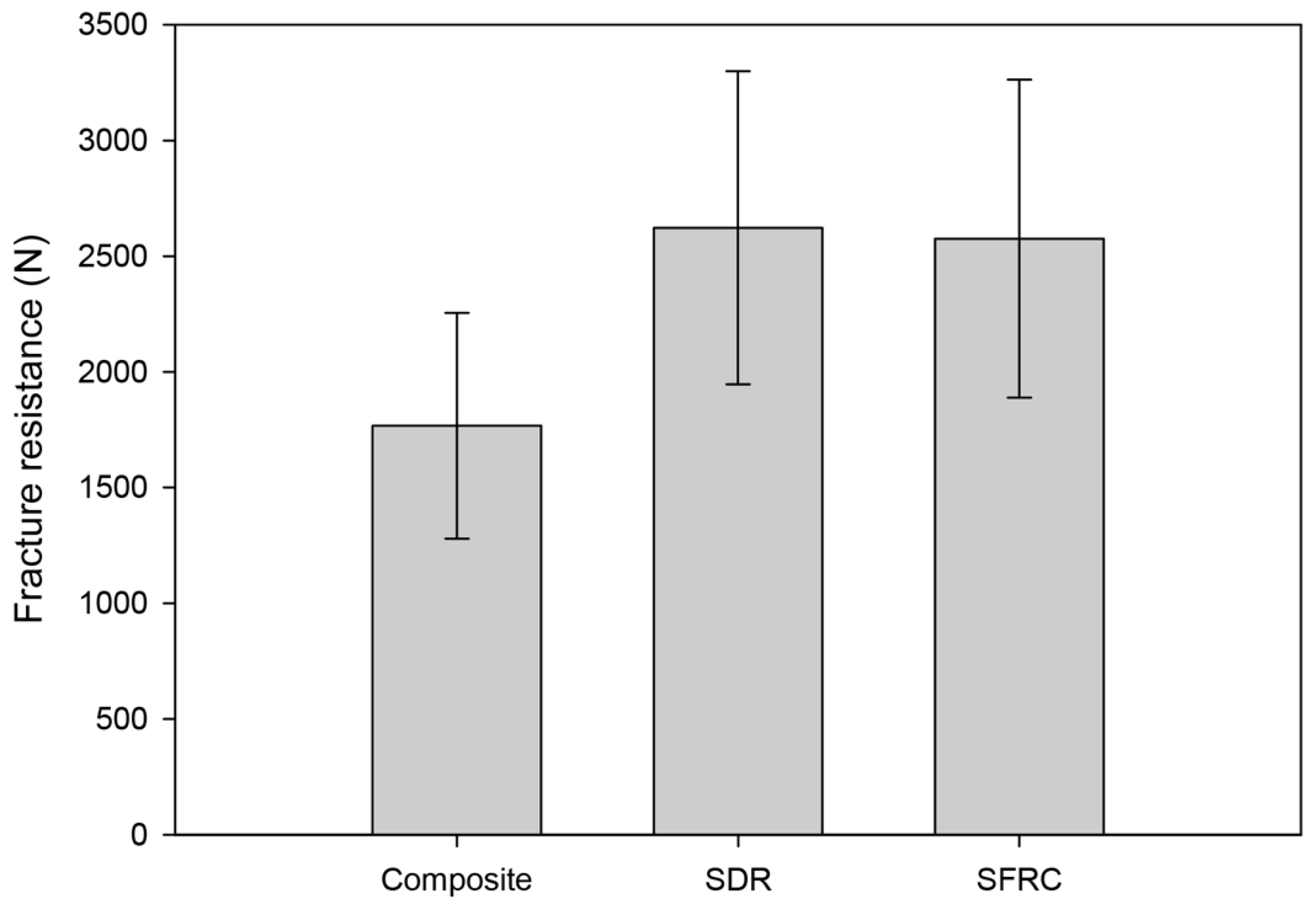
Figure 2 displays the fracture thresholds for the different study groups. The layered composite filling (Group 1) showed significantly lower fracture resistance compared to the bulk-fill groups (Group 2, p = 0.005, Group 3, p = 0.008), while there was no difference in fracture resistance between the other groups (Figure 2). Thus, the first null hypothesis was rejected.
Regarding the fracture pattern, cavities restored with SFRC (Group 3) managed to show the highest number (dominantly restorable), while teeth restored with SDR (Group 2) presented the lowest number (dominantly non-restorable) of repairable fractures (Table 4). Therefore, the null hypothesis regarding fracture patterns was also rejected.
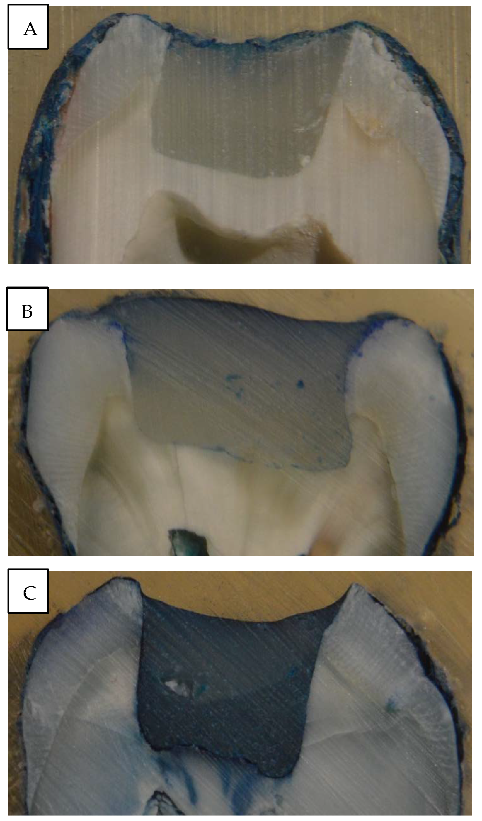
Regarding microleakage testing, Group 2 showed dominantly shallower penetration along the interface, while the rest of the groups (Group 1 and 3) produced dominantly deeper dye penetration reaching deeper parts of the cavity and the interface (Table 5 and Figure 3).
In terms of gap formation values at the bonding interface, layered composite filling (Group 1) produced significantly higher gap formation compared to the bulk-fill groups (Group 2, p = 0.000, Group 3, p = 0.000) (Figure 4). Therefore, the third null hypothesis was also rejected. Again, the two bulk-fill groups (Group 2 and 3) did not differ in gap formation along from each other along the restoration-cavity interface.

4. Discussion

The two main failure types of direct posterior composite restorations in clinical settings are secondary caries and bulk fractures [3,40]. The former is related to early gap formation and subsequent degradation of the interface between the direct restoration and the cavity walls [10]. As already shown by many, polymerization shrinkage and subsequent stress development can be a reason behind marginal gap formation, leading to marginal discoloration, nanoleakage and therefore secondary caries [4,41,42]. The magnitude of the polymerization-related stress depends upon the cavity configuration (C-factor), and the physical characteristics of the composite material, namely its elastic modulus and polymerization conversion rate [43]. When cavities with high C-factor are filled with large amounts of composite material, the integrity of the bond interface is at risk and thus also the longevity of the restoration [44]. This was emphasized by Han et al. who pointed out that in high C-factor cavities the internal adaptation was inferior and the prevalence of imperfect margins was higher compared to low C-factor cavities [45]. The deleterious effects of high C-factor cavities are also visible when measuring microtensile bond strength, compared to low C-factor cavities [46]. Among vital posterior cavities, C-factor is the highest in Class I occlusal cavities. As restoring high C-factor cavities with direct composite restorations could be problematic from a mechanical and gap formation point of view [47], high C-factor Class I occlusal cavities were tested in our study. As the results from the static load-to-fracture tests show, cavities restored with bulk-fill materials (Groups 2 and 3) showed significantly higher load bearing capacity compared to the layered composite filling group (Group 1), at p < 0.001 for both groups. This is in accordance with the findings of Rosatto et al., where SDR was characterized by higher fracture resistance than layered composite fillings [14]. However, our present findings are in contrast with the findings of other studies regarding fracture resistance [48,49,50,51]. Bonilla et al. and Rosa de Lacerda et al. did not manage to show any difference in fracture resistance between teeth restored with a bulk-fill composite and a layered composite filling. Of note, neither of these studies used SDR as a bulk-fill composite [48,49].
Bulk-fill composites may vary according to their composition, the specific monomer and photoinitiator system they use, their consistency, and whether they need to be capped with conventional composite or not. It is thus possible that the difference between the results of the cited studies and our results presented in this study stems from the fact that the other studies used materials of different compositions. De Assis et al. did not find difference in fracture strength when comparing cavities restored with SDR or a layered conventional composite filling [50]. Al-Nahedh et al. even found SDR to be inferior to layered composite fillings in terms of fracture resistance in Class II cavities [51]. The same authors also pointed out that when SDR was capped with conventional composite occlusally, the fracture resistance of the resulting restoration was not different from that of a layered composite restoration. It is important to highlight that due to wear and aesthetic properties, clinically SDR is recommended to be covered with conventional composite except in conservative occlusal cavities.
In our study, SFRC restorations showed significantly higher fracture resistance compared to layered composite fillings in Class I cavities (p = 0.008). This is in line with the findings of Molnár et al. [24]. SFRC is a dental restorative composite intended to be used in high stress-bearing areas as a dentine replacement material [52,53,54]. Mechanical testing has shown that the load-bearing capacity, flexural strength and fracture toughness of SFRC is superior in comparison with conventional composite materials. As SFRC can be light cured up to 5 mm [55], it was used in a bulk-fill manner in our study.
In terms of fracture pattern, SFRC (Group 3) showed the highest number and also showed dominantly repairable fractures. This shows that by the use of SFRC, a more favorable fracture profile can be reached as compared to conventional composite restorative materials, and this is in line with our previous findings [20,23,38]. The explanation most probably lies in the obvious difference in fracture toughness between reinforced and non-reinforced composites [20,25]. Previous studies have shown that SFRC can re-direct and stop crack propagation within materials [21,25,56]. When non-fibre-reinforced composites were used for restoring cavities in the posterior region, both favourable and unfavourable fractures occurred. SDR restorations were predominantly characterised by non-repairable fractures. This is in line with previous studies on the usual fracture pattern when utilising non-fibre-reinforced composites [20,22,23,38]. As shown by Molnár et al. via fractography, the brittleness of the conventional composite materials generates the bulk fracture, which propagates easily through the whole thickness of the restoration [24].
Regarding the adaptation and subsequent gap formation of the restorative materials, bulk-fill materials (Groups 2 and 3) in our study were characterised by significantly smaller gaps at the bonded interphase compared to the layered composite filling (Group 1), at p < 0.001 for both groups. Bulk-fill materials have been developed to eliminate the need for incremental layering [10]. So far, bulk-fill restorations seem to perform like conventional restorations in terms of marginal adaptation, physico-mechanical properties, fatigue resistance and the tendency to induce cuspal deflection [49,57,58,59]. The favorable results for SDR may be explained by SDR generating less polymerization shrinkage stress, possibly as a result of containing a ‘‘polymerization modulator’’ [50,60], which supposedly counteracts polymerization stress through lower polymerization rate [50,61]. Our findings regarding SDR in terms of gap formation are in line with the findings of Peutzfeldt et al. [4] and Al-Nahedh et al. [51]. The explanation for this observation could well be the flowable consistency of SDR [62]. The appearance of flowable bulk-fill materials will probably make the bulk-filling technique even more popular with clinicians as several advantages have been recorded, including low polymerization shrinkage and stress, reduced cuspal deflection and improved self-leveling ability [16,63]. Although flowable composites generally shrink more than conventional paste-like composites, due to their higher amount of resin matrix [64], the subsequent shrinkage stress remains low in comparison [46,65]. Our findings are in contrast with those of Thongbai-On et al. [66] or Park et al. [16] who did not find significant difference between SDR and layered composite filling in terms of gap formation along the interphase. It must be noted that both of the cited studies investigated Class II MO/MOD cavities, which can be an explanation for the different outcomes. In our study, bulk-fill SFRC was characterised by significantly fewer gaps than layered composite restorations (p = 0.000). This might be attributed to the unique fibre content and anisotrophicity of the SFRC material. Garoushi et al. pointed out that the orientation of the reinforcing fibres in anisotropic materials has a major influence on polymerization shrinkage: as it is controlled in the direction of the fibres, it is never homogeneously distributed in all directions [35,55].
Therefore, during polymerization, the material cannot shrink along the length of the fibres and retains its original dimensions horizontally, while the polymer matrix between the fibres can shrink [35]. In many specimens, internal bubble formation within the material could be seen when SFRC was used (Figure 5).
The same was observed by clinicians when using packable SFRC in a bulk manner. As suggested by Fráter and co-workers, these bubbles could be partly a sign of internal stress relief: if shrinkage does not occur at the interface, it might cause internal voids inside the bulk material [67].
The continuity of the developed gaps was measured with dye penetration in this study. Teeth restored with SDR showed shallower dye penetration along the interphase and also showed the highest proportion of perfect margins. According to Peutzfeldt et al., whether marginal gaps are formed depends on an interplay between multiple factors, of which some are related to the resin composite, while others are related to the specific cavity and restorative procedure [4]. In our study, we sought to keep the factors related to the cavity and the restorative procedure constant. Standardized cavities were prepared to ensure that all cavities had a high C-factor. Our finding regarding the superiority of SDR in terms of marginal integrity is in line with the findings of de Assis et al. [50] and Gerula-Szymańska et al. [68], but they contradict the findings of de Dietschi et al. [69]. When looking at the number of continuous gaps in Group 1 (layered composite filling) and Group 3 (SFRC), both groups produced deeper penetration, even many times reaching the floor of the cavity (Figure 5, Table 5).
One of the limitations of our study is that static load-to-fracture testing was used without fatigue testing. While static load-to-fracture tests model a sudden, greater force (such as when trauma occurs), dynamic loading is more appropriate to study the mechanical consequences of the forces that act during ordinary chewing. An accelerated dynamic loading test represents a realistic compromise between the two extremes. In this test, cyclic loading is applied, but the magnitude of the force is not constant for the entire duration of the test: it increases stepwise, always after a given number of cycles have been completed [70]. Thus, fatigue testing should be carried out in the future in the same groups. The other limitation of this study is that the specimens were not thermocycled before the marginal integrity analyses. As shown by Dietschi et al. [69], adaptation can greatly differ before and after thermocycling. Our results should be interpreted with this limitation in mind. Future studies should incorporate thermocycling, fatigue testing and also other bulk-fill materials to better understand the above proposed research topic.

5. Conclusions

Bulk-fill resin composites (SDR and SFRC) have demonstrated encouraging results in terms of fracture resistance when compared to conventional packable composite. Furthermore, these bulk-fill materials showed improved internal adaptation to the cavity walls compared to the gap formation present in case of utilising conventional composite material in a high C-factor cavity. Moreover, SFRC showed more restorable fracture behaviour compared to the other tested materials. Thus, bulk-fill materials provide a simple and effective solution for restoring and reinforcing high C-factor occlusal cavities.

Author Contributions

Conceptualization, E.B., S.G. and M.F.; methodology, E.B., M.S. and Á.S.; software, G.B. and B.S.P.; validation, J.M. and T.S.; formal analysis, G.B. and B.S.P.; investigation, E.B., M.F., M.S. and Á.S.; resources, M.F.; data curation, G.B.; writing—original draft preparation, E.B. and M.F.; writing—review and editing, S.G.; visualization, T.S. and J.M.; supervision, M.F.; project administration, E.B. and M.F.; funding acquisition, M.F. All authors have read and agreed to the published version of the manuscript.

Funding

This study was supported by the Bolyai János Research Scholarship (BO/701/20/5) and by the ÚNKP-21-5-SZTE New National Excellence Program of The Ministry for Innovation and Technology from the Source of National Research, Development and Innovation Fund, Hungary.

Institutional Review Board Statement

The study was conducted according to theguidelines of the Declaration of Helsinki, and approved by the Ethics Committee of the University of Szeged (4029).

Informed Consent Statement

Not applicable.

Data Availability Statement

Data is contained within the article.

Conflicts of Interest

The authors declare no conflict of interest.

References

  1. Haak, R.; Näke, T.; Park, K.J.; Ziebolz, D.; Krause, F.; Schneider, H. Internal and marginal adaptation of high-viscosity bulk-fill composites in class II cavities placed with different adhesive strategies. Odontology 2019, 107, 374–382. [Google Scholar] [CrossRef] [PubMed]
  2. Opdam, N.J.; van de Sande, F.H.; Bronkhorst, E.; Cenci, M.S.; Bottenberg, P.; Pallesen, U.; Gaengler, P.; Lindberg, A.; Huysmans, M.C.; van Dijken, J.W. Longevity of posterior composite restorations: A systematic review and meta-analysis. J. Dent. Res. 2014, 93, 943–949. [Google Scholar] [CrossRef]
  3. Demarco, F.F.; Corrêa, M.B.; Cenci, M.S.; Moraes, R.R.; Opdam, N.J. Longevity of posterior composite restorations: Not only a matter of materials. Dent. Mater. 2012, 28, 87–101. [Google Scholar] [CrossRef] [PubMed]
  4. Peutzfeldt, A.; Mühlebach, S.; Lussi, A.; Flury, S. Marginal Gap Formation in Approximal “Bulk Fill” Resin Composite Restorations After Artificial Ageing. Oper. Dent. 2018, 43, 180–189. [Google Scholar] [CrossRef] [PubMed]
  5. Soares, C.J.; Faria-E-Silva, A.L.; Rodrigues, M.P.; Vilela, A.B.F.; Pfeifer, C.S.; Tantbirojn, D.; Versluis, A. Polymerization shrinkage stress of composite resins and resin cements—What do we need to know? Braz. Oral Res. 2017, 31, e62. [Google Scholar] [CrossRef]
  6. Cardoso, M.V.; de Almeida Neves, A.; Mine, A.; Coutinho, E.; Van Landuyt, K.; De Munck, J.; Van Meerbeek, B. Current aspects on bonding effectiveness and stability in adhesive dentistry. Aust. Dent. J. 2011, 56, 31–44. [Google Scholar] [CrossRef]
  7. Manauta, J.; Salat, A.; Putignano, A.; Devoto, W.; Paolone, G.; Hardan, L.S. Stratification in anterior teeth using one dentine shade and a predefined thickness of enamel: A new concept in composite layering—Part I. Odontostomatol. Trop. 2014, 37, 5–16. [Google Scholar]
  8. Manauta, J.; Salat, A.; Putignano, A.; Devoto, W.; Paolone, G.; Hardan, L.S. Stratification in anterior teeth using one dentine shade and a predefined thickness of enamel: A new concept in composite layering—Part II. Odontostomatol. Trop. 2014, 37, 5–13. [Google Scholar]
  9. Alqudaihi, F.S.; Cook, N.B.; Diefenderfer, K.E.; Bottino, M.C.; Platt, J.A. Comparison of Internal Adaptation of Bulk-fill and Increment-fill Resin Composite Materials. Oper. Dent. 2019, 44, 32–44. [Google Scholar] [CrossRef]
  10. Fronza, B.M.; Rueggeberg, F.A.; Braga, R.R.; Mogilevych, B.; Soares, L.E.; Martin, A.A.; Ambrosano, G.; Giannini, M. Monomer conversion, microhardness, internal marginal adaptation, and shrinkage stress of bulk-fill resin composites. Dent. Mater. 2015, 31, 1542–1551. [Google Scholar] [CrossRef] [PubMed]
  11. Park, J.; Chang, J.; Ferracane, J.; Lee, I.B. How should composite be layered to reduce shrinkage stress: Incremental or bulk filling? Dent. Mater. 2008, 24, 1501–1505. [Google Scholar] [CrossRef] [PubMed]
  12. Flury, S.; Peutzfeldt, A.; Lussi, A. Influence of increment thickness on microhardness and dentin bond strength of bulk fill resin composites. Dent. Mater. 2014, 30, 1104–1112. [Google Scholar] [CrossRef] [PubMed]
  13. Cerda-Rizo, E.R.; de Paula Rodrigues, M.; Vilela, A.; Braga, S.; Oliveira, L.; Garcia-Silva, T.C.; Soares, C.J. Bonding Interaction and Shrinkage Stress of Low-viscosity Bulk Fill Resin Composites with High-viscosity Bulk Fill or Conventional Resin Composites. Oper. Dent. 2019, 44, 625–636. [Google Scholar] [CrossRef] [PubMed]
  14. Rosatto, C.M.; Bicalho, A.A.; Veríssimo, C.; Bragança, G.F.; Rodrigues, M.P.; Tantbirojn, D.; Versluis, A.; Soares, C.J. Mechanical properties, shrinkage stress, cuspal strain and fracture resistance of molars restored with bulk-fill composites and incremental filling technique. J. Dent. 2015, 43, 1519–1528. [Google Scholar] [CrossRef] [PubMed]
  15. Jang, J.H.; Park, S.H.; Hwang, I.N. Polymerization shrinkage and depth of cure of bulk-fill resin composites and highly filled flowable resin. Oper. Dent. 2015, 40, 172–180. [Google Scholar] [CrossRef]
  16. Park, K.J.; Pfeffer, M.; Näke, T.; Schneider, H.; Ziebolz, D.; Haak, R. Evaluation of low-viscosity bulk-fill composites regarding marginal and internal adaptation. Odontology 2021, 109, 139–148. [Google Scholar] [CrossRef]
  17. Kim, R.J.; Kim, Y.J.; Choi, N.S.; Lee, I.B. Polymerization shrinkage, modulus, and shrinkage stress related to tooth-restoration interfacial debonding in bulk-fill composites. J. Dent. 2015, 43, 430–439. [Google Scholar] [CrossRef]
  18. Rizzante, F.A.P.; Mondelli, R.F.L.; Furuse, A.Y.; Borges, A.F.S.; Mendonça, G.; Ishikiriama, S.K. Shrinkage stress and elastic modulus assessment of bulk-fill composites. J. Appl. Oral Sci. 2019, 27, e20180132. [Google Scholar] [CrossRef]
  19. Deliperi, S.; Alleman, D.; Rudo, D. Stress-reduced Direct Composites for the Restoration of Structurally Compromised Teeth: Fiber Design According to the "Wallpapering" Technique. Oper. Dent. 2017, 42, 233–243. [Google Scholar] [CrossRef]
  20. Fráter, M.; Sáry, T.; Vincze-Bandi, E.; Volom, A.; Braunitzer, G.; Szabó, P.B.; Garoushi, S.; Forster, A. Fracture Behavior of Short Fiber-Reinforced Direct Restorations in Large MOD Cavities. Polymers 2021, 13, 2040. [Google Scholar] [CrossRef]
  21. Lassila, L.; Keulemans, F.; Säilynoja, E.; Vallittu, P.K.; Garoushi, S. Mechanical properties and fracture behavior of flowable fiber reinforced composite restorations. Dent. Mater. 2018, 34, 598–606. [Google Scholar] [CrossRef] [PubMed]
  22. Forster, A.; Braunitzer, G.; Tóth, M.; Szabó, P.B.; Fráter, M. In Vitro Fracture Resistance of Adhesively Restored Molar Teeth with Different MOD Cavity Dimensions. J. Prosthodont. 2019, 28, 325–331. [Google Scholar] [CrossRef] [PubMed] [Green Version]
  23. Sáry, T.; Garoushi, S.; Braunitzer, G.; Alleman, D.; Volom, A.; Fráter, M. Fracture behaviour of MOD restorations reinforced by various fibre-reinforced techniques—An in vitro study. J. Mech. Behav. Biomed. Mater. 2019, 98, 348–356. [Google Scholar] [CrossRef] [PubMed]
  24. Molnár, J.; Fráter, M.; Sáry, T.; Braunitzer, G.; Vallittu, P.K.; Lassila, L.; Garoushi, S. Fatigue performance of endodontically treated molars restored with different dentin replacement materials. Dent. Mater. 2022, 38, 83–93. [Google Scholar] [CrossRef] [PubMed]
  25. Lassila, L.; Säilynoja, E.; Prinssi, R.; Vallittu, P.K.; Garoushi, S. Fracture behavior of Bi-structure fiber-reinforced composite restorations. J. Mech. Behav. Biomed. Mater. 2020, 101, 103444. [Google Scholar] [CrossRef] [PubMed]
  26. Garoushi, S.; Sungur, S.; Boz, Y.; Ozkan, P.; Vallittu, P.K.; Uctasli, S.; Lassila, L. Influence of short-fiber composite base on fracture behavior of direct and indirect restorations. Clin. Oral Investig. 2021, 25, 4543–4552. [Google Scholar] [CrossRef]
  27. Zaghloul, M.M.J.; Mohamed, Y.S.; El-Gamal, H. Fatigue and tensile behaviors of fiber-reinforced thermosetting composites embedded with nanoparticles. J. Compos. Mater. 2019, 56, 709–718. [Google Scholar] [CrossRef]
  28. Zaghloul, M.M.J.; Zaghloul, M.Y.M.; Zaghloul, M.M.Y. Experimental and modeling analysis of mechanical-electrical behaviors of polypropylene composites filled with graphite and MWCNT fillers. Polym. Test. 2017, 63, 467–474. [Google Scholar] [CrossRef]
  29. Zaghloul, M.Y.M.; Zaghloul, M.M.Y.; Zaghloul, M.M.Y. Developments in polyester composite materials–An in-depth review on natural fibers and nanofillers. Compos. Struct. 2021, 278, 114698. [Google Scholar] [CrossRef]
  30. Zaghloul, M.M.Y.; Zaghloul, M.M.Y. Influence of flame retardant magnesium hydroxide on the mechanical properties of high density polyethylene composites. J. Reinf. Plast. Compos. 2017, 36, 1802–1816. [Google Scholar] [CrossRef]
  31. Zaghloul, M.Y.; Zaghloul, M.M.Y.; Zaghloul, M.M.Y. Influence of Stress Level and Fibre Volume Fraction on Fatigue Performance of Glass Fibre-Reinforced Polyester Composites. Polymers 2022, 14, 2662. [Google Scholar] [CrossRef] [PubMed]
  32. Fuseini, M.; Zaghloul, M.M.Y. Investigation of Electrophoretic Deposition of PANI Nano fibers as a Manufacturing Technology for corrosion protection. Prog. Org. Coat. 2022, 171, 107015. [Google Scholar] [CrossRef]
  33. Bijelic-Donova, J.; Garoushi, S.; Lassila, L.V.; Keulemans, F.; Vallittu, P.K. Mechanical and structural characterization of discontinuous fiber-reinforced dental resin composite. J. Dent. 2016, 52, 70–78. [Google Scholar] [CrossRef] [PubMed]
  34. Alshabib, A.; Jurado, C.A.; Tsujimoto, A. Short fiber-reinforced resin-based composites (SFRCs); Current status and future perspectives. Dent. Mater. J. 2022, 20, 2022–2080. [Google Scholar] [CrossRef] [PubMed]
  35. Garoushi, S.; Gargoum, A.; Vallittu, P.K.; Lassila, L. Short fiber-reinforced composite restorations: A review of the current literature. J. Investig. Clin. Dent. 2018, 9, 12330. [Google Scholar] [CrossRef] [PubMed]
  36. Lempel, E.; Őri, Z.; Szalma, J.; Lovász, B.V.; Kiss, A.; Tóth, Á.; Kunsági-Máté, S. Effect of exposure time and pre-heating on the conversion degree of conventional, bulk-fill, fiber reinforced and polyacid-modified resin composites. Dent. Mater. 2019, 35, 217–228. [Google Scholar] [CrossRef]
  37. Lempel, E.; Őri, Z.; Kincses, D.; Lovász, B.V.; Kunsági-Máté, S.; Szalma, J. Degree of conversion and in vitro temperature rise of pulp chamber during polymerization of flowable and sculptable conventional, bulk-fill and short-fibre reinforced resin composites. Dent. Mater. 2021, 37, 983–997. [Google Scholar] [CrossRef] [PubMed]
  38. Fráter, M.; Forster, A.; Keresztúri, M.; Braunitzer, G.; Nagy, K. In vitro fracture resistance of molar teeth restored with a short fibre-reinforced composite material. J. Dent. 2014, 42, 1143–1450. [Google Scholar] [CrossRef]
  39. Yamazaki, P.C.; Bedran-Russo, A.K.; Pereira, P.N.; Wsift, E.J., Jr. Microleakage evaluation of a new low-shrinkage composite restorative material. Oper. Dent. 2006, 31, 670–676. [Google Scholar] [CrossRef]
  40. Da Rosa Rodolpho, P.A.; Donassollo, T.A.; Cenci, M.S.; Loguércio, A.D.; Moraes, R.R.; Bronkhorst, E.M.; Opdam, N.J.; Demarco, F.F. 22-Year clinical evaluation of the performance of two posterior composites with different filler characteristics. Dent. Mater. 2011, 27, 955–963. [Google Scholar] [CrossRef]
  41. Manhart, J.; Kunzelmann, K.H.; Chen, H.Y.; Hickel, R. Mechanical properties and wear behavior of light-cured packable composite resins. Dent. Mater. 2000, 16, 33–40. [Google Scholar] [CrossRef]
  42. Behery, H.; El-Mowafy, O.; El-Badrawy, W.; Saleh, B.; Nabih, S. Cuspal Deflection of Premolars Restored with Bulk-Fill Composite Resins. J. Esthet. Restor. Dent. 2016, 28, 122–130. [Google Scholar] [CrossRef] [PubMed]
  43. Van Ende, A.; De Munck, J.; Mine, A.; Lambrechts, P.; Van Meerbeek, B. Does a low-shrinking composite induce less stress at the adhesive interface? Dent. Mater. 2010, 26, 215–222. [Google Scholar] [CrossRef] [PubMed]
  44. de Oliveira, I.L.M.; de Brito, O.F.F.; de Melo Monteiro, L.M.C.G.Q.; Resende Montes, M.A.J. Microtensile Bond Strength of Bulk-fill Resin Composite Restorations in High C-factor Cavities. J. Contemp. Dent. Pract. 2020, 21, 626–631. [Google Scholar] [CrossRef]
  45. Han, S.H.; Sadr, A.; Tagami, J.; Park, S.H. Internal adaptation of resin composites at two configurations: Influence of polymerization shrinkage and stress. Dent. Mater. 2016, 32, 1085–1094. [Google Scholar] [CrossRef]
  46. Van Ende, A.; De Munck, J.; Van Landuyt, K.L.; Poitevin, A.; Peumans, M.; Van Meerbeek, B. Bulk-filling of high C-factor posterior cavities: Effect on adhesion to cavity-bottom dentin. Dent. Mater. 2013, 29, 269–277. [Google Scholar] [CrossRef] [PubMed]
  47. Han, S.H.; Park, S.H. Incremental and Bulk-fill Techniques with Bulk-fill Resin Composite in Different Cavity Configurations. Oper. Dent. 2018, 43, 631–641. [Google Scholar] [CrossRef]
  48. Bonilla, E.D.; Hayashi, M.; Pameijer, C.H.; Le, N.V.; Morrow, B.R.; Garcia-Godoy, F. The effect of two composite placement techniques on fracture resistance of MOD restorations with various resin composites. J. Dent. 2020, 101, 103348. [Google Scholar] [CrossRef]
  49. Rosa de Lacerda, L.; Bossardi, M.; Silveira Mitterhofer, W.J.; Galbiatti de Carvalho, F.; Carlo, H.L.; Piva, E.; Münchow, E.A. New generation bulk-fill resin composites: Effects on mechanical strength and fracture reliability. J. Mech. Behav. Biomed. Mater. 2019, 96, 214–218. [Google Scholar] [CrossRef]
  50. de Assis, F.S.; Lima, S.N.; Tonetto, M.R.; Bhandi, S.H.; Pinto, S.C.; Malaquias, P.; Loguercio, A.D.; Bandéca, M.C. Evaluation of Bond Strength, Marginal Integrity, and Fracture Strength of Bulk- vs Incrementally-filled Restorations. J. Adhes. Dent. 2016, 18, 317–323. [Google Scholar] [CrossRef]
  51. Al-Nahedh, H.N.; Alawami, Z. Fracture Resistance and Marginal Adaptation of Capped and Uncapped Bulk-fill Resin-based Materials. Oper. Dent. 2020, 45, 43–56. [Google Scholar] [CrossRef] [PubMed]
  52. Garoushi, S.; Mangoush, E.; Vallittu, P.K.; Lassila, L. Short fiber reinforced composite: A new alternative for direct onlay restorations. Open Dent. J. 2013, 7, 181–185. [Google Scholar] [CrossRef] [PubMed] [Green Version]
  53. Fennis, W.M.; Kreulen, C.M.; Tezvergil, A.; Lassila, L.; Vallittu, P.K.; Creugers, N.H. In vitro repair of fractured fiber-reinforced cusp-replacing composite restorations. Int. J. Dent. 2011, 2011, 165938. [Google Scholar] [CrossRef]
  54. Fráter, M.; Lassila, L.; Braunitzer, G.; Vallittu, P.K.; Garoushi, S. Fracture resistance and marginal gap formation of post-core restorations: Influence of different fiber-reinforced composites. Clin. Oral Investig. 2020, 24, 265–276. [Google Scholar] [CrossRef] [PubMed]
  55. Garoushi, S.; Säilynoja, E.; Vallittu, P.K.; Lassila, L. Physical properties and depth of cure of a new short fiber reinforced composite. Dent. Mater. 2013, 29, 835–841. [Google Scholar] [CrossRef]
  56. Fráter, M.; Sáry, T.; Jókai, B.; Braunitzer, G.; Säilynoja, E.; Vallittu, P.K.; Lassila, L.; Garoushi, S. Fatigue behavior of endodontically treated premolars restored with different fiber-reinforced designs. Dent. Mater. 2021, 37, 391–402. [Google Scholar] [CrossRef] [PubMed]
  57. Fronza, B.M.; Makishi, P.; Sadr, A.; Shimada, Y.; Sumi, Y.; Tagami, J.; Giannini, M. Evaluation of bulk-fill systems: Microtensile bond strength and non-destructive imaging of marginal adaptation. Braz. Oral Res. 2018, 32, 80. [Google Scholar] [CrossRef]
  58. Sousa-Lima, R.X.; Silva, L.; Chaves, L.; Geraldeli, S.; Alonso, R.; Borges, B. Extensive Assessment of the Physical, Mechanical, and Adhesion Behavior of a Low-viscosity Bulk Fill Composite and a Traditional Resin Composite in Tooth Cavities. Oper. Dent. 2017, 42, 159–166. [Google Scholar] [CrossRef]
  59. Tsujimoto, A.; Nagura, Y.; Barkmeier, W.W.; Watanabe, H.; Johnson, W.W.; Takamizawa, T.; Latta, M.A.; Miyazaki, M. Simulated cuspal deflection and flexural properties of high viscosity bulk-fill and conventional resin composites. J. Mech. Behav. Biomed. Mater. 2018, 87, 111–118. [Google Scholar] [CrossRef]
  60. Burgess, J.; Cakir, D. Comparative properties of low-shrinkage composite resins. Compend. Contin. Educ. Dent. 2010, 31, 10–15. [Google Scholar] [PubMed]
  61. Ilie, N.; Hickel, R. Investigations on a methacrylate-based flowable composite based on the SDR™ technology. Dent. Mater. 2011, 27, 348–355. [Google Scholar] [CrossRef] [PubMed]
  62. Orłowski, M.; Tarczydło, B.; Chałas, R. Evaluation of marginal integrity of four bulk-fill dental composite materials: In vitro study. Sci. World J. 2015, 2015, 701262. [Google Scholar] [CrossRef] [PubMed] [Green Version]
  63. Tarle, Z.; Attin, T.; Marovic, D.; Andermatt, L.; Ristic, M.; Tauböck, T.T. Influence of irradiation time on subsurface degree of conversion and microhardness of high-viscosity bulk-fill resin composites. Clin. Oral Investig. 2015, 19, 831–840. [Google Scholar] [CrossRef] [PubMed]
  64. Tauböck, T.T.; Jäger, F.; Attin, T. Polymerization shrinkage and shrinkage force kinetics of high- and low-viscosity dimethacrylate- and ormocer-based bulk-fill resin composites. Odontology 2019, 107, 103–110. [Google Scholar] [CrossRef] [PubMed]
  65. Braga, R.R.; Hilton, T.J.; Ferracane, J.L. Contraction stress of flowable composite materials and their efficacy as stress-relieving layers. J. Am. Dent. Assoc. 2003, 134, 721–728. [Google Scholar] [CrossRef] [PubMed]
  66. Thongbai-On, N.; Chotvorrarak, K.; Banomyong, D.; Burrow, M.F.; Osiri, S.; Pattaravisitsate, N. Fracture resistance, gap and void formation in root-filled mandibular molars restored with bulk-fill resin composites and glass-ionomer cement base. J. Investig. Clin. Dent. 2019, 10, 12435. [Google Scholar] [CrossRef]
  67. Fráter, M.; Sáry, T.; Néma, V.; Braunitzer, G.; Vallittu, P.; Lassila, L.; Garoushi, S. Fatigue failure load of immature anterior teeth: Influence of different fiber post-core systems. Odontology 2021, 109, 222–230. [Google Scholar] [CrossRef]
  68. Gerula-Szymańska, A.; Kaczor, K.; Lewusz-Butkiewicz, K.; Nowicka, A. Marginal integrity of flowable and packable bulk fill materials used for class II restorations -A systematic review and meta-analysis of in vitro studies. Dent. Mater. J. 2020, 39, 335–344. [Google Scholar] [CrossRef]
  69. Dietschi, D.; Curto, F.D.; Di Bella, E.; Krejci, I.; Ardu, S. In vitro evaluation of marginal adaptation in medium- and large size direct class II restorations using a bulk-fill or layering technique. J. Dent. 2021, 115, 103828. [Google Scholar] [CrossRef]
  70. Szabó, V.T.; Szabó, B.; Tarjányi, T.; Trenyik, S.Z.E.; Szabó, P.B.; Fráter, M. Analog and digital modelling of sound and impaired periodontal supporting tissues during mechanical testing. Analecta Tech. Szeged. 2021, 15, 2. [Google Scholar] [CrossRef]
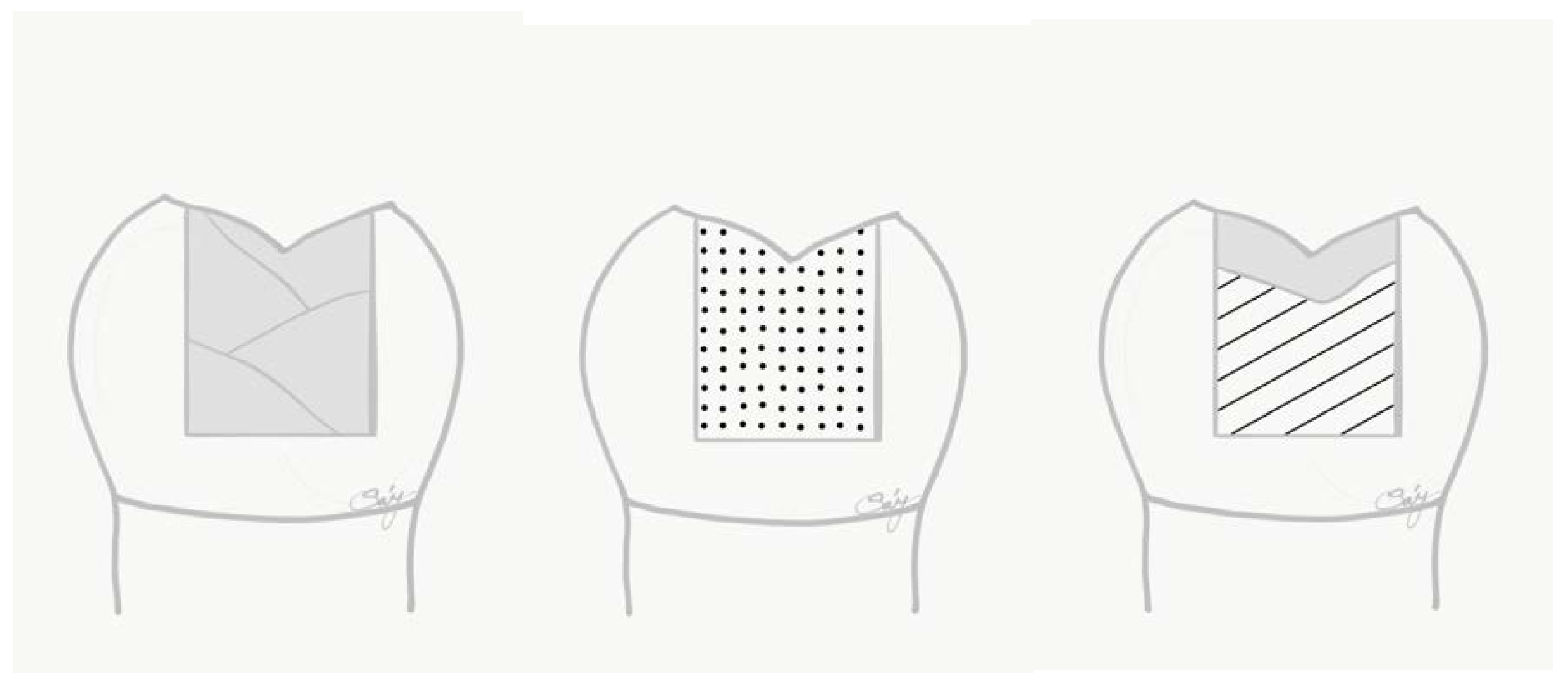
Figure 1. Schematic figure representing the test groups (from left to right). Group 1: layered conventional packable composite filling; Group 2: bulk-fill composite filling; Group 3: bulk-fill SFRC and conventional packable composite occlusal coverage.
Polymers 14 03463 g001
Figure 2. Column plots of fracture resistance by group (group mean +/− SD).
Polymers 14 03463 g002
Figure 3. Pictures (AC) of the sectioned specimen after dye penetration analysis. Picture (A) showed minimal to no marginal staining, while the other pictures show moderate (Picture (B)) to deep (Picture (C)) penetration of the dye, starting from the margins.
Polymers 14 03463 g003
Figure 4. Column plots of gap size by group (group mean +/− SD).
Polymers 14 03463 g004
Figure 5. Internal air-bubble formation within the cured SFRC material.
Polymers 14 03463 g005
Table 1. Restorative materials used during the study.
CategoryLOT NumberMaterialManufacturerComposition
ConditionerBFDJ8Ultra-EtchUltradent35% phosphoric acid, water, cobalt aluminate blue, spinel, glycol, siloxan
Adhesive1703031G-Premio BondGC10-MDP (5–10%), 4-MET, dimethacrylate
(10–20%), dimethacrylate component
(1–5%), photo-initiator (1–5%), butylated
hydroxytoluene (<1%), acetone (25–50%),
water (24%)
CompositeN841976Filtek Ultimate Composite Resin3MBis-GMA, UDMA, TEGDMA, Bis-EMA, 20 nm silica and 4–11 nm zirconia filler, camphorquinone, accelerators, pigments and others.
SFR composite1212261EverX PosteriorGCBis-GMA, PMMA, TEGDMA, 74.2 wt%, 53.6 vol%
Short E-glass fibre filler,
barium glass
Bulk-fill composite1202174Surefil SDRDentsplyTEGDMA, EBADMA, 68 wt%, 44 vol%,
Barium borosilicate glass
SFR: Short-fibre reinforced; Bis-GMA: 2,2-bis[p-(2-hydroxy-3-methacryloxy propoxy)phenyl]propane; MDP:10-methacryloyloxydecyl dihydrogen phosphate; 4-MET: 4-methacryloyloxyethyl trimellitate; MEPS: methacryloyloxyalkyl thiophosphate methylmethacrylate; TEGDMA: triethylene glycol dimethacrylate; UDMA: urethane dimethacrylate; PMMA: polymethyl methacrylate, PEGDMA: poly (ethylene glycol) dimethacrylate; EBPADMA: Ethoxylated bisphenol A dimethacrylate.
Table 2. Physical properties of the restorative materials, according to the manufacturers.
MaterialVolumetric Shrinkage (%)Fracture Toughness (MPa m1/2)Flexural Strength (MPa)
Filtek Ultimate Composite Resin2.01.22160 ± 20
EverX Posterior2.92.61153 ± 9
Surefil SDR3.51.25120 ± 13
Table 3. Four grade scale showing degree of microleakage.
ScoreContent
0No Microleakage
1Dye penetration within the occlusal half of the axial cavity wall
2Dye penetration extending into the lower half of the axial cavity wall
3Dye penetration spreading along cavity floor
Table 4. The distribution of fracture patterns among the tested groups (n = 12).
Study GroupRestorable Fracture Non-Restorable Fracture
Group 1 (Composite)75
Group 2 (SDR)210
Group 3 (SFRC)120
Table 5. The microleakage score among the tested groups (n = 12).
Dye Penetration ScoreGroup 1 (Composite)Group 2 (SDR)Group 3 (SFRC)
0142
1473
2615
3102
Publisher’s Note: MDPI stays neutral with regard to jurisdictional claims in published maps and institutional affiliations.