

Assessment of the Micro-Tensile Bond Strength of a Novel Bioactive Dental Restorative Material (Surefil One)


Abstract
:1. Introduction
- There are no differences in the micro-tensile bond strength between Surefil one and RIVA.
- Different dentin conditions have no effect on Surefil one’s micro-tensile bond strength.
2. Materials and Methods
2.1. Specimens and Sampling Technique
- Inclusion criteria: Teeth with sound and caries-free dentine; molar teeth.
- Exclusion criteria: Endodontically treated teeth; badly decayed teeth; premolar and anterior teeth.
- Surefil one no-treatment group (NTG): Surefil one was applied directly to the dentine surface and light-cured for 20 s.
- Surefil one acid etch group (EG): Dentin was acid etched using 37% phosphoric acid for 15 s before the application of Surefil one and light-cured for 20 s.
- Surefil one adhesive group (EAG): Dentin was acid etched using 37% phosphoric acid for 15 s; then, universal adhesive was applied using a micro brush and spread gently with air and light-cured for 20 s, followed by Surefil one application.
- Resin-modified glass ionomer control group (RIVA): The material was applied directly to the dentine surface and light-cured for 20 s.
2.2. Specimen Preparation
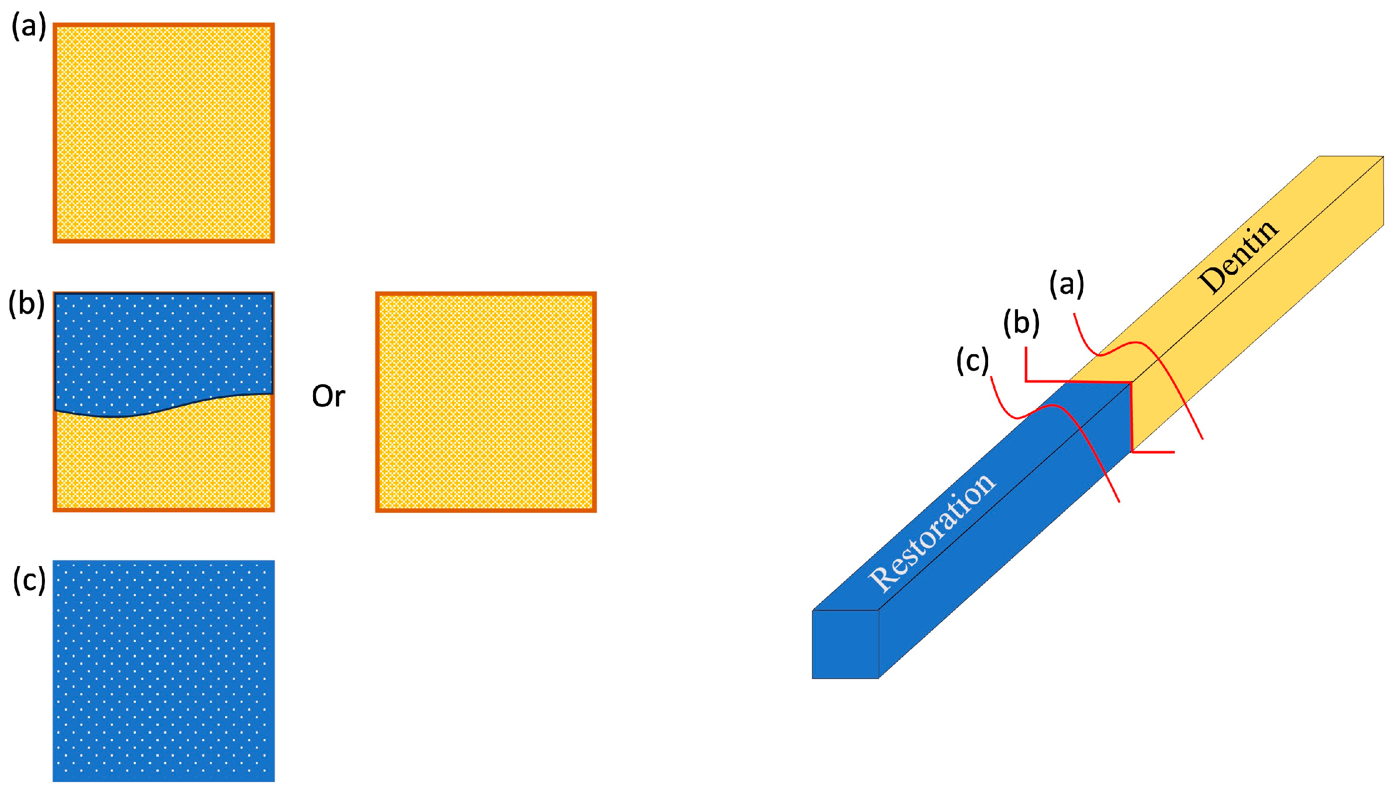
2.3. Micro-Tensile Strength Measurements and Failure Modes
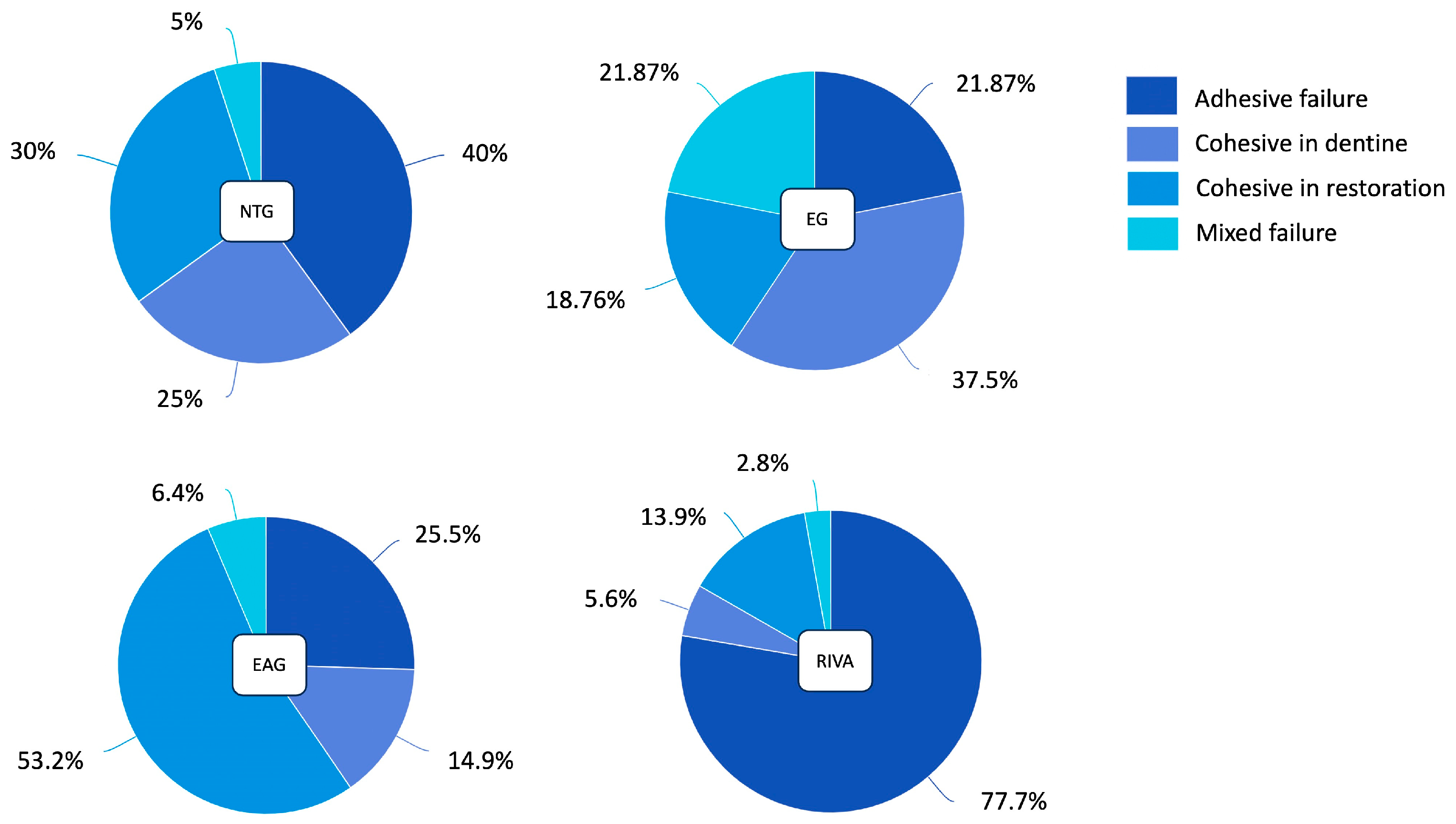
- Adhesive: Interfacial bond failure between the restoration and dentine.
- Cohesive in the restoration: When a fracture allows a layer of restoration to remain on both surfaces.
- Cohesive in the dentin: When a fracture allows a layer of dentine to remain on both surfaces.
- Mixed failure: If the failure shows more than one substrate [13].
2.4. Statistical Analysis
3. Results
4. Discussion
5. Conclusions
- -
- Surefil one has a higher micro-tensile bond strength than RIVA.
- -
- Different dentin conditions did not significantly affect Surefil one’s μTBS performance.
- -
- Surefil one could potentially be used as a substitute for RMGI restorative material, especially in cases of long-term temporary restorations, since it has a better tensile bond and fluoride release, and there is no need for added complicated steps.
- -
- The restoration is not technique-sensitive and is less time-consuming.
- -
- Further long-term clinical and laboratory studies are needed.
Author Contributions
Funding
Institutional Review Board Statement
Data Availability Statement
Acknowledgments
Conflicts of Interest
References
- Ilie, N.; Hickel, R. Resin composite restorative materials. Aust. Dent. J. 2011, 56, 59–66. [Google Scholar] [CrossRef] [PubMed]
- Berg, J.H. Glass ionomer cements. Pediatr. Dent. 2002, 24, 430–438. [Google Scholar] [CrossRef] [PubMed]
- Łagocka, R.; Skoczyk-Jaworska, M.; Mazurek-Mochol, M. Self-adhesive, bulk-fill bioactive materials as an alternative to silver amalgam in restorative dentistry. Pomeranian J. Life Sci. 2022, 68, 36–44. [Google Scholar]
- Berzins, D.W.; Abey, S.; Costache, M.C.; Wilkie, C.A.; Roberts, H.W. Resin-modified glass-ionomer setting reaction competition. J. Dent. Res. 2010, 89, 82–86. [Google Scholar] [CrossRef] [PubMed]
- Frankenberger, R.; Dudek, M.-C.; Winter, J.; Braun, A.; Krämer, N.; von Stein-Lausnitz, M.; Roggendorf, M.J. Amalgam Alternatives Critically Evaluated: Effect of Long-term Thermomechanical Loading on Marginal Quality, Wear, and Fracture Behavior. J. Adhes. Dent. 2020, 22, 107–116. [Google Scholar] [CrossRef]
- Lohbauer, U.; Belli, R. The mechanical performance of a novel self-adhesive restorative material. J. Adhes. Dent. 2020, 22, 47–58. [Google Scholar] [CrossRef] [PubMed]
- Rathke, A.; Pfefferkorn, F.; McGuire, M.K.; Heard, R.H.; Seemann, R. One-year clinical results of restorations using a novel self-adhesive resin-based bulk-fill restorative. Sci. Rep. 2022, 12, 3934. [Google Scholar] [CrossRef]
- François, P.; Remadi, A.; Le Goff, S.; Abdel-Gawad, S.; Attal, J.P.; Dursun, E. Flexural properties and dentin adhesion in recently developed self-adhesive bulk-fill materials. J. Oral Sci. 2021, 63, 139–144. [Google Scholar] [CrossRef]
- Aldowsari, M.K.; Alfawzan, F.; Alhaidari, A.; Alhogail, N.; Alshargi, R.; Bin Saleh, S.; Sulimany, A.M.; Alturki, M. Comparison of Shear Bond Strength of Three Types of Adhesive Materials Used in the Restoration of Permanent Molars after Treatment with Silver Diamine Fluoride: An In Vitro Study. Materials 2023, 16, 6831. [Google Scholar] [CrossRef] [PubMed]
- Albelasy, E.H.; Hamama, H.H.; Chew, H.P.; Montasser, M.; Mahmoud, S.H. Clinical performance of two ion-releasing bulk-fill composites in class I and class II restorations: A two-year evaluation. J. Esthet. Restor. Dent. 2024, 36, 723–736. [Google Scholar] [CrossRef]
- Maghaireh, G.A.; Albashaireh, Z.S.; Allouz, H.A. Postoperative sensitivity in posterior restorations restored with self-adhesive and conventional bulk-fill resin composites: A randomized clinical split-mouth trial. J. Dent. 2023, 137, 104655. [Google Scholar] [CrossRef] [PubMed]
- Watanabe, I.; Nakabayashi, N. Measurement methods for adhesion to dentine: The current status in Japan. J. Dent. 1994, 22, 67–72. [Google Scholar] [CrossRef] [PubMed]
- Armstrong, S.; Breschi, L.; Özcan, M.; Pfefferkorn, F.; Ferrari, M.; Van Meerbeek, B. Academy of Dental Materials guidance on in vitro testing of dental composite bonding effectiveness to dentin/enamel using micro-tensile bond strength (μTBS) approach. Dent. Mater. 2017, 33, 133–143. [Google Scholar] [CrossRef]
- Opdam, N.J.M.; Van De Sande, F.H.; Bronkhorst, E.; Cenci, M.S.; Bottenberg, P.; Pallesen, U.; Gaengler, P.; Lindberg, A.; Huysmans, M.C.D.N.J.M.; Van Dijken, J.W. Longevity of posterior composite restorations: A systematic review and meta-analysis. J. Dent. Res. 2014, 93, 943–949. [Google Scholar] [CrossRef] [PubMed]
- Latta, M.A.; Tsujimoto, A.; Takamizawa, T.; Barkmeier, W.W. Enamel and dentin bond durability of self-adhesive restorative materials. J. Adhes. Dent. 2020, 22, 99–105. [Google Scholar] [CrossRef] [PubMed]
- Sulaiman, T.A.; Abdulmajeed, A.A.; Altitinchi, A.; Ahmed, S.N.; Donovan, T.E. Physical Properties, Film Thickness, and Bond Strengths of Resin-Modified Glass Ionomer Cements According to Their Delivery Method. J. Prosthodont. 2019, 28, 85–90. [Google Scholar] [CrossRef] [PubMed]
- Li, X.J.; Zhao, S.J.; Niu, L.N.; Tay, F.R.; Jiao, K.; Gao, Y.; Chen, J.H. Effect of luting cement and thermomechanical loading on retention of glass fibre posts in root canals. J. Dent. 2014, 42, 75–83. [Google Scholar] [CrossRef]
- Zhao, Z.; Wang, Q.; Zhao, J.; Zhao, B.; Ma, Z.; Zhang, C. Adhesion of Teeth. Front. Mater. 2021, 7, 615225. [Google Scholar] [CrossRef]
- Francois, P.; Fouquet, V.; Attal, J.P.; Dursun, E. Commercially available fluoride-releasing restorative materials: A review and a proposal for classification. Materials 2020, 13, 2313. [Google Scholar] [CrossRef]
- Yao, C.; Ahmed, M.H.; Okazaki, Y.; Van Landuyt, K.L.; Huang, C.; Van Meerbeek, B. Bonding Efficacy of a New Self-Adhesive Restorative onto Flat Dentin vs Class-I Cavity-bottom Dentin. J. Adhes. Dent. 2020, 22, 65–77. [Google Scholar] [CrossRef]
- Elraggal, A.; Raheem, A.; Alhotan, A. Bond Strength, Microleakage, Microgaps, and Marginal Adaptation of Self-adhesive Resin Composites to Tooth Substrates with and without Preconditioning with Universal Adhesives. J. Adhes. Dent. 2024, 26, 53–64. [Google Scholar] [CrossRef] [PubMed]
- Eichler, E.; Vach, K.; Schlueter, N.; Jacker-Guhr, S.; Luehrs, A.K. Dentin adhesion of bulk-fill composites and universal adhesives in class I-cavities with high C-factor. J. Dent. 2024, 142, 104852. [Google Scholar] [CrossRef] [PubMed]
- Coutinho, E.; Yoshida, Y.; Inoue, S.; Fukuda, R.; Snauwaert, J.; Nakayama, Y.; De Munck, J.; Lambrechts, P.; Suzuki, K.; Van Meerbeek, B. Gel phase formation at resin-modified glass-ionomer/tooth interfaces. J. Dent. Res. 2007, 86, 656–661. [Google Scholar] [CrossRef] [PubMed]


| Material, Manufacturer | Composition | Type | Application Technique |
|---|---|---|---|
| Surefil one, Dentsply Sirona, Charlotte, NC, USA | Modified polyacid (MOPOS), bifunctional acrylate (BADEP), acrylic acid, reactive glass filler, water, non-reactive glass filler, initiator, and stabilizer | Self-adhesive bulk Fill | Activate the capsule by pressing it onto a stable surface; then, mix it in the amalgamator for 10 s; then, place it by using a capsule dispenser in the deepest part of the cavity; then, light cure for 20 s |
| RIVA resin-modified glass ionomer, SDI, Victoria, Australia | Ion glass filler, fluoride, strontium ions photo-initiators, polyacrylic acid, water, and water-soluble methacrylate monomer | Self-adhesive RMGI | Activate the capsule by pressing it onto a stable surface; then, mix it in the amalgamator for 10 s; then, place it by using a capsule dispenser in the deepest part of the cavity; then, light cure for 20 s (use the layering technique for cavities deeper than 2 mm) |
| Prime&Bond active, Dentsply Sirona, Charlotte, NC, USA | Bi- and multi-functional acrylate, modified phosphoric acid, acrylate resin, initiator, stabilizer, and isopropanol | Universal adhesive | Apply a bonding agent to all surfaces and slightly agitate for 20 s; then, evaporate the solvent using air for at least 5 s; then, light cure for 10–20 s depending on the power output: ≥500 mW/cm2 =20 s ≥800 mW/cm2 = 10 s |
| Material, Manufacturer | Composition | Instruction | Usage |
|---|---|---|---|
| Formaline 10%, Thermo Fisher Scientific Inc., Waltham, MA, USA. | Paraformaldehyde, RO water, sodium hydroxide, and HCL | 25 g Para-formaldehyde mixed with 250 mL/RO water and 3 full drops/sodium hydroxide. Stir with heat until clear and then buffer to neutral pH with HCl | To sterilize the teeth and as storage media |
| Zapit glue, Dental Ventures of America Inc., Corona, CA, USA. | Ethyl-2 Cyanoacrylate: Poly (Methyl Methacrylate) Hydroquinone | Place a small amount of glue on each side of the specimen and then spray the accelerator to speed up the setting time | To glue the specimens into the micro-tensile jigs |
| 37% Phosphoric acid, FGM, Joinville, Brazil. | Water-based gel containing 37% phosphoric acid | Etch the enamel and dentin for 15 s; then, wash the surface abundantly with water and dry the cavity | To remove the smear layer |
| Micro-Tensile, MPa | ||||
|---|---|---|---|---|
| Restoration | Dentin Condition | N | Mean | Standard Deviation |
| Riva control | Control | 36 | 7.83 | ±7.41 |
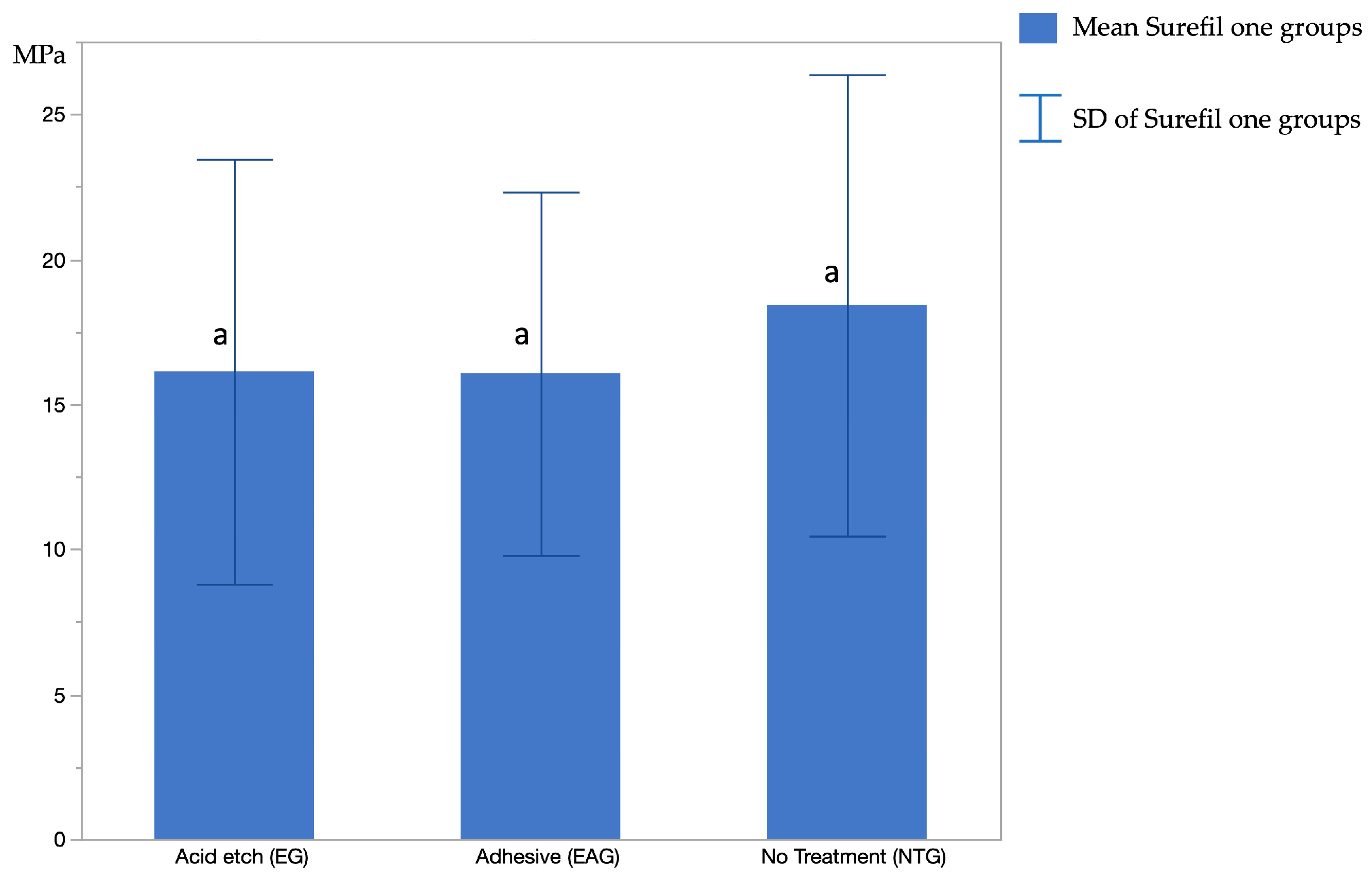
| Surefil one | Acid etch (EG) | 32 | 16.13 | ±7.33 |
| Adhesive (EAG) | 47 | 16.07 | ±6.27 | |
| No treatment (NTG) | 40 | 18.42 | ±7.96 | |
| Restoration | Dentin Condition | Restoration | Dentin Condition | Prob > |t| |
|---|---|---|---|---|
| RIVA | Control | Surefil one | Adhesive | <0.0001 * |
| RIVA | Control | Surefil one | No treatment | <0.0001 * |
| RIVA | Control | Surefil one | Acid etch | <0.0001 * |
| Surefil one | Adhesive | Surefil one | No treatment | 0.4302 |
| Surefil one | Adhesive | Surefil one | Acid etch | 1.0000 |
| Surefil one | No treatment | Surefil one | Acid etch | 0.5402 |
Disclaimer/Publisher’s Note: The statements, opinions and data contained in all publications are solely those of the individual author(s) and contributor(s) and not of MDPI and/or the editor(s). MDPI and/or the editor(s) disclaim responsibility for any injury to people or property resulting from any ideas, methods, instructions or products referred to in the content. |
© 2024 by the authors. Licensee MDPI, Basel, Switzerland. This article is an open access article distributed under the terms and conditions of the Creative Commons Attribution (CC BY) license (https://creativecommons.org/licenses/by/4.0/).
Share and Cite
Alghamdi, A.A.; Athamh, S.; Alzhrani, R.; Filemban, H. Assessment of the Micro-Tensile Bond Strength of a Novel Bioactive Dental Restorative Material (Surefil One). Polymers 2024, 16, 1558. https://doi.org/10.3390/polym16111558
Alghamdi AA, Athamh S, Alzhrani R, Filemban H. Assessment of the Micro-Tensile Bond Strength of a Novel Bioactive Dental Restorative Material (Surefil One). Polymers. 2024; 16(11):1558. https://doi.org/10.3390/polym16111558
Chicago/Turabian StyleAlghamdi, Abdulrahman A., Smaher Athamh, Reham Alzhrani, and Hanan Filemban. 2024. "Assessment of the Micro-Tensile Bond Strength of a Novel Bioactive Dental Restorative Material (Surefil One)" Polymers 16, no. 11: 1558. https://doi.org/10.3390/polym16111558