Article

Physicochemical Characterization and Biological Properties of Polysaccharides from Alpiniae oxyphyllae Fructus

by
Risi Wang
1,3,
Xinmei Ruan
3,
Jun Chen
3,
Lizhen Deng
3,
Wei Zhou
4,
Xixiang Shuai
2,3,*,
Ruihong Liang
3 and
Taotao Dai
3,*
1
School of Food Science and Engineering, Jiangxi Agricultural University, Nanchang 330045, China
2
Key Laboratory of Tropical Fruit Biology of Ministry of Agriculture and Rural Affairs, South Subtropical Crop Research Institute, China Academy of Tropical Agricultural Sciences, Zhanjiang 524091, China
3
State Key Laboratory of Food Science and Technology, Nanchang University, Nanchang 330047, China
4
Key Laboratory of Tropical Crop Products Processing of Ministry of Agriculture and Rural Affairs, Agricultural Products Processing Research Institute, Chinese Academy of Tropical Agricultural Sciences, Zhanjiang 524001, China
*
Authors to whom correspondence should be addressed.
Polymers 2024, 16(12), 1705; https://doi.org/10.3390/polym16121705
Submission received: 13 May 2024 / Revised: 2 June 2024 / Accepted: 6 June 2024 / Published: 14 June 2024
(This article belongs to the Special Issue Polymer Composite Analysis and Characterization II)

Abstract

:
Polysaccharides (AOPs) were extracted from Alpiniae oxyphyllae fructus using three distinct methods: hot water (AOP-HW), hydrochloric acid (AOP-AC), and NaOH/NaBH4 (AOP-AL). This study systematically investigated and compared the physicochemical properties, structural characteristics, antioxidant activities, and α-amylase inhibitory activities of the extracted polysaccharides. Among the three AOPs, AOP-AC exhibited the highest yield (13.76%) and neutral sugar content (80.57%), but had the lowest molecular weight (121.28 kDa). Conversely, AOP-HW had the lowest yield (4.54%) but the highest molecular weight (385.42 kDa). AOP-AL was predominantly composed of arabinose (28.42 mol%), galacturonic acid (17.61 mol%), and galactose (17.09 mol%), while glucose was the major sugar in both AOP-HW (52.31 mol%) and AOP-AC (94.77 mol%). Functionally, AOP-AL demonstrated superior scavenging activities against DPPH, hydroxyl, and ABTS radicals, whereas AOP-AC exhibited the strongest inhibitory effect on α-amylase. These findings indicate that the extraction solvent significantly influences the physicochemical and biological properties of AOPs, thus guiding the selection of appropriate extraction methods for specific applications. The results of this study have broad implications for industries seeking natural polysaccharides with antioxidant and enzymatic inhibitory properties.

Graphical Abstract

1. Introduction

Alpiniae oxyphyllae Miq., belonging to the species of Zingiberaceae, is an herbal medicine distributed between the Yangtze River and the Nan Mountains in China. The dried mature fruit of Alpiniae oxyphyllae (A. oxyphyllae) Miq. was considered to have the potential to warm the kidney, reduce the urine, and store essential substances in traditional Chinese medicine theory [1,2,3]. In China, A. oxyphyllae fructus has been used on a large scale as a Chinese medicine material. A. oxyphyllae fructus mainly contains polysaccharide, essential oil, terpene, flavonoid, and heptane derivatives [4]. Modern pharmacological studies have proved that A. oxyphyllae fructus polysaccharide (AOP) shows a good efficacy in urinary incontinence, anti-neuro inflammatory activity, antiviral activity, and immunoregulation [1,5,6,7,8]. Yang et al. characterized the structure of A. oxyphyllae fructus polysaccharide and found that it could significantly increase the phagocytic activity of RAW264.7 macrophages [9]. Generally, the bioactivity of polysaccharides depends on the chemical composition and structural features, which varies with a range of factors, especially the extraction solvent employed [10,11,12].
Extraction conditions, including temperature, pH, and the type and concentration of the extracting solvents, can directly influence the structure of the obtained polysaccharide. Solvents have significant effects on the biological activities and physicochemical features of polysaccharides [13,14]. Water, acid, and alkali are common extracting agents, selected based on the nature of the polysaccharide. Hot water extraction is widely utilized for polysaccharide extraction because it is simple, low-cost, safe, and environment-friendly [15,16]. Acidic and alkali solutions are also usually used to extract polysaccharides. It has been reported that acidic solvents are effective in cleaving glycoside linkages within polysaccharides, leading to increased yields of small-molecule polysaccharides [17,18]. Moreover, several relevant studies have found that alkali-extracted polysaccharides possess higher bioactivities, such as anti-inflammatory and antioxidant properties [19,20]. Zhang, Zhu, Zhang, Yang, Ni, Zhang, Liu, and Zhang [21] extracted polysaccharides from green tea leaf using different extraction solvents and found that alkali-extracted polysaccharide showed a higher viscosity and better gelling properties than hot-water- and acid-extracted polysaccharides. Therefore, it is necessary to compare the physicochemical and biological properties of polysaccharides with different solvents. Thus far, the research about polysaccharides from A. oxyphyllae fructus has all involved water extraction [1,5,6]. The effect of different extraction solvents on the physicochemical and biological properties of polysaccharides of A. oxyphyllae fructus is still unclear.
In summary, the objective of this study was to evaluate the effects of different solvents (hot water, hydrochloric acid, and NaOH/NaBH4) on the physicochemical (chemical composition, molecular weight (Mw), morphology, and crystal structure) and biological (antioxidant and α-amylase inhibitory ability) properties of polysaccharides extracted from A. oxyphyllae fructus. This work might provide valuable information for the utilization of AOPs in the food and dietary supplements industries.

2. Result and Discussion

2.1. Extraction Yields and Physicochemical Properties of AOPs

2.1.1. Extraction Yields

In general, the yield of polysaccharides from different sources depends on the extraction parameters and raw materials. As shown in Table 1, the yield of AOPs varies with the extraction solvent. AOP-AC had the highest yield (13.76%), followed by AOP-AL (8.88%) and AOP-HW (4.54%). The higher yields of AOP-AC and AOP-AL may result from the disruption of the cell wall structure under acidic and alkali conditions, which accelerated the release of soluble polysaccharides into the extract [22]. During the extraction process, hot water only dissolves long-chain soluble polysaccharides, resulting in the lowest extraction yield of AOP-HW. Zhang, Zhu, Zhang, Yang, Ni, Zhang, Liu, and Zhang [21] also found that the yield of green tea leaf polysaccharides extracted by alkali and acid solution was higher than that by hydrothermal extraction. In the acidic extract, the glycosidic bond between biopolymers was disrupted by H+, and the soluble molecules quickly dissolved towards the extracellular space under the electrostatic stress, resulting in a significant increase in the production of polysaccharides [23].

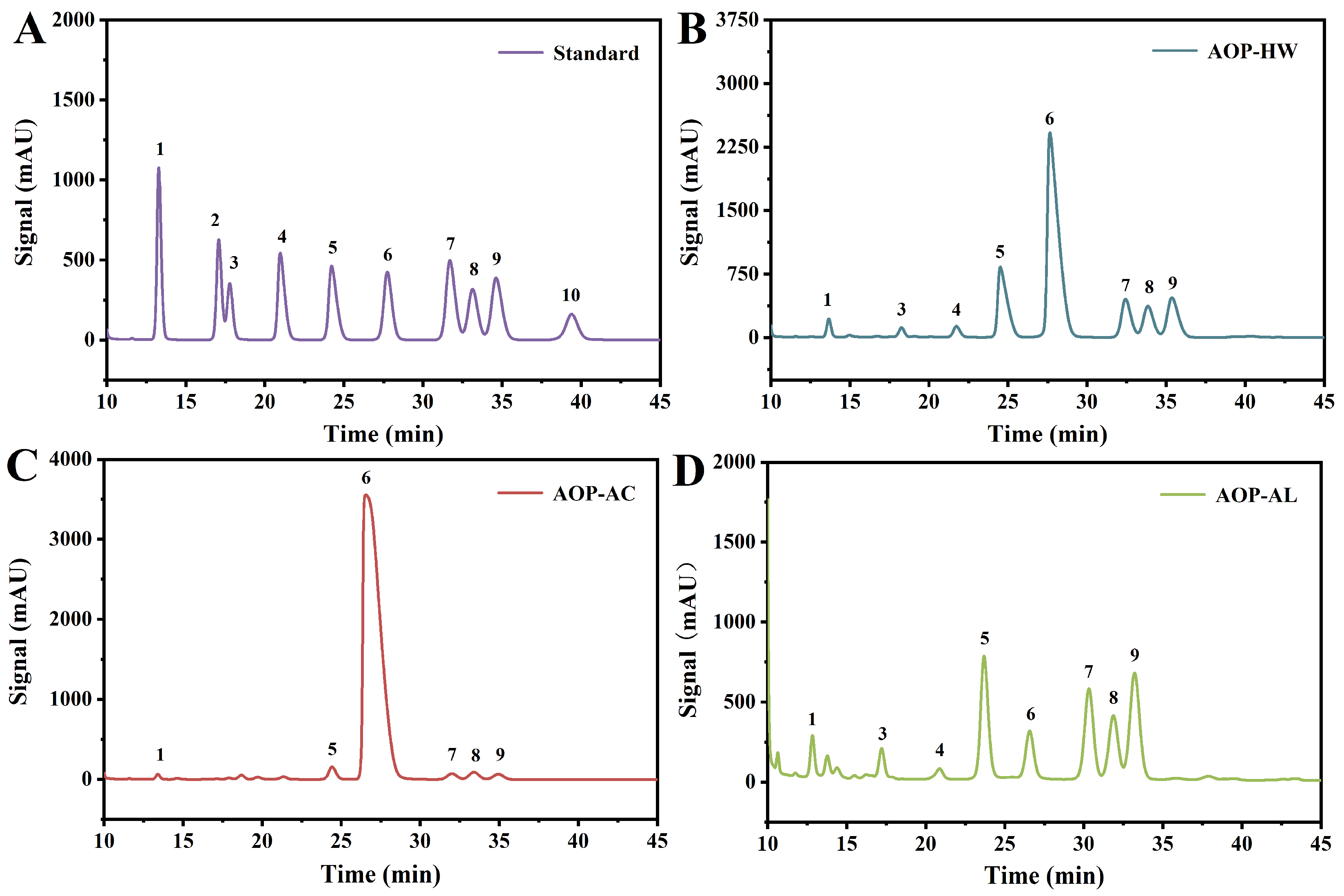
2.1.2. Chemical Composition
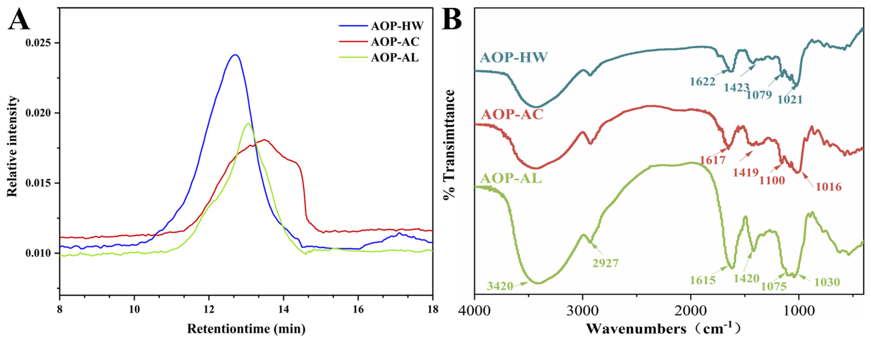
The uronic acid, neutral sugar, and protein contents of the three AOPs are shown in Table 1. The neutral sugar contents of AOP-AC and AOP-HW were 80.57% and 69.62%, respectively, significantly higher than that of AOP-AL (28.28%). This result may be interpreted as the fact that the glycosidic bonds in the AOPs are more sensitive, and the acidic environment promotes its hydrolysis [22]. The uronic acid contents of AOP-AL, AOP-HW, and AOP-AC were 63.13%, 19.51%, and 13.15%, which indicated that the three AOPs were all anionic polysaccharides. This result also indicated that AOP-AL was an acidic polysaccharide. This finding may be due to the fact that alkaline conditions were conducive to the release of uronic acid, thus increasing the content of uronic acid in the AOP-AL. All AOPs were contaminated with protein, but the protein content was low (1.12–3.37%). It was possible that the obtained polysaccharides were deproteinized by the Sevag reagent and the remaining protein was closely connected to the framework of polysaccharides.
The monosaccharide composition results of the three AOPs were presented in Table 2 and Figure 1. There were certain differences in the relative abundance of each polysaccharide. Glucose was the predominant sugar found in AOP-HW (52.31 mol%) and AOP-AC (94.77 mol%), which was in line with Han’s study (96.8 mol%) [1]. Glucose was found to account for an exclusively high proportion of the hot-water-extracted polysaccharides from the same Zingiberaceae family, such as Kaempferia galanga L (34.10 mol%) and Zingiber officinale (96.00 mol%) [24,25]. However, the main monosaccharides of AOP-AL were determined as arabinose (28.42 mol%), galactose (17.64 mol%), and galacturonic acid (17.09 mol%), indicating that AOP-AL was likely to be hemicellulose [26]. And the monosaccharides of the AOP-HW sample mainly included arabinose, glucose, and xylose, which was speculated to be araboxylan [27]. As the glucose content of AOP-AC exceeds 90%, it was presumed to be glucan. This finding was aligned with a previous study which showed that the polysaccharides extracted from Alpiniae oxyphyllae fructus were composed of glucose alone [9]. Their exact structure has yet to be further resolved by means such as nuclear magnetic resonance. The above results indicated that extraction solvents affected the chemical composition of the AOPs.

2.1.3. Mw of AOPs

The Mw of AOPs detected by high-performance size exclusion chromatography is displayed in Table 1 and Figure 2A. The molecular weight profiles of AOP-HW, AOP-AC, and AOP-AL all contained a single RI peak, and their Mw values were 385.42, 121.28, and 232.40 kDa, respectively. The lower average Mw of AOP-AL and AOP-AC indicated that polysaccharides were degraded during the extraction process due to harsh extraction conditions. A similar trend was found in the previous study; the Mw decreased with the increase in H+/OH- concentration, and polysaccharides gradually degraded from large molecules to small-size polymers [28]. Both AOP-HW and AOP-AL exhibited symmetrical peaks, while AOP-AC had a broad molecular mass distribution. The relatively wide Mw distribution of AOP-AC might be due to the cleavage of glycosidic bonds and intramolecular hydrogen bonds of AOP-AC to form a variety of glucose polymers during the extraction process, inferring this polysaccharide may have a highly branched and heterogeneous structure [19].

2.1.4. FT-IR Spectroscopy Analysis

The FT-IR spectra of AOP-HW, AOP-AC, and AOP-AL are depicted in Figure 2B. The absorption peak between 3100 and 3700 cm−1 corresponds to the O−H stretching vibration caused by inter- and intramolecular hydrogen bonds [29]. The band at 2800–3000 cm−1 is associated with the stretching vibration of C−H in unconstrained carbohydrates [5]. The strong absorption peak at 1615–1653 cm−1 is accounted for by the C=O asymmetric stretching vibration of the carbonyl group, while the weak band at 1420 cm−1 reflects the C−O stretching vibration of the carbonyl group [30]. The peak at 1090 cm−1 indicates the existence of a pyranose ring in the main chain [25]. The characteristic absorption at 1020 cm−1 might represent an α-1,6 linkage [30,31]. The three AOPs by FT-IR analysis demonstrated the typical absorption peaks of polysaccharides. A small shoulder at ca. 1730 cm–1 is visible for AOP-HW which is accounted for by the C=O stretching vibration of the methyl esterified carboxylic group. However, the C=O stretching vibration of the methyl esterified carboxylic group is not shown in the FT-IR profile of AOP-AC and AOP-AL, which indicates that de-esterification possibly occurs during acidic or alkali extraction.

2.2. Morphological Properties
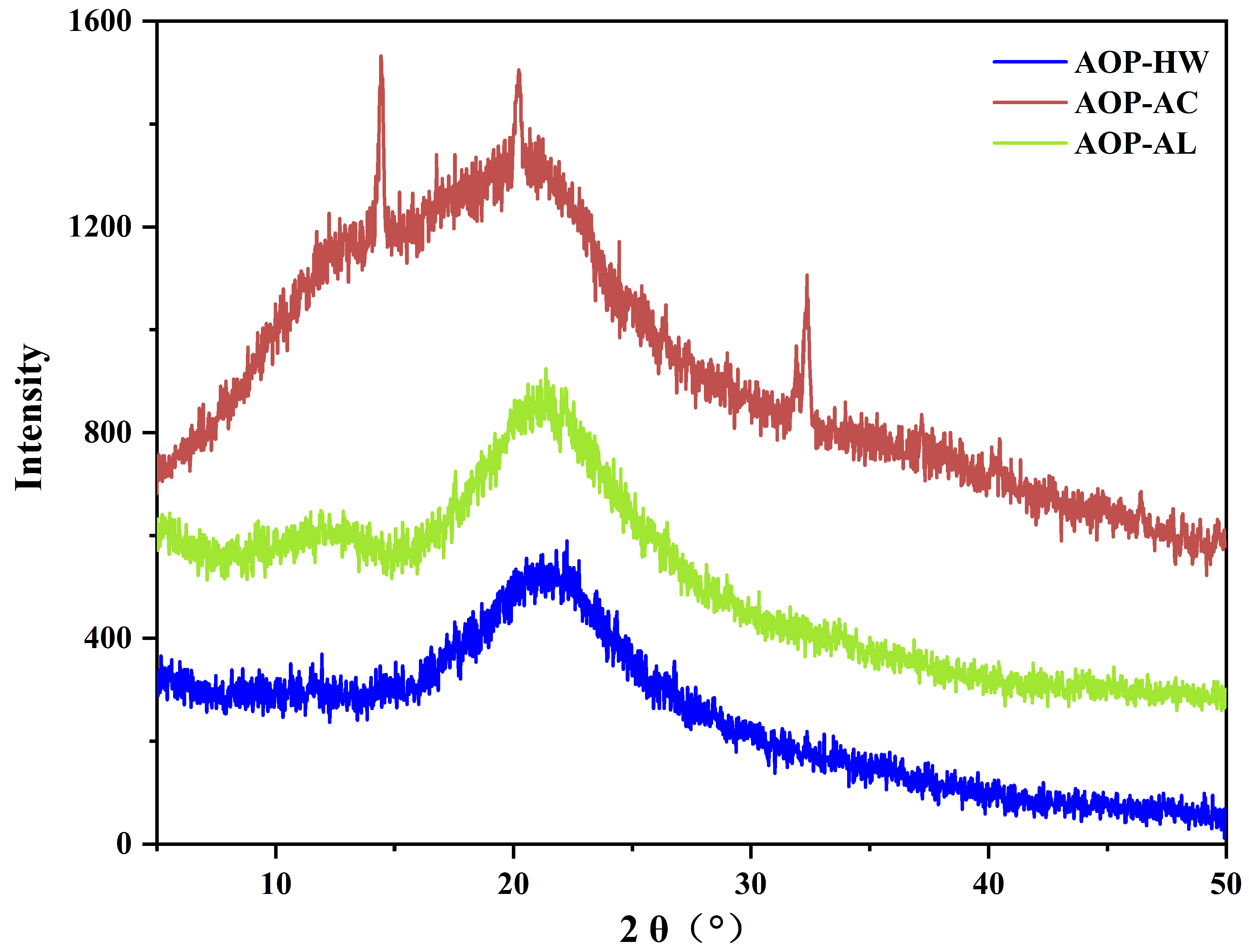
The SEM images provide the surface morphological properties of the AOPs. The morphologies of the three AOPs presented significant differences in shapes and sizes (Figure 3). The microstructure of AOP-HW appeared an irregular and slightly rough layer with uneven dimensions (Figure 3A), which was similar to that reported by Shi, Zhong, Zhang, and Yan [5]. AOP-AL possessed many thick and smooth slice patches, and some filamentous connections were found on the surface (Figure 3C). However, AOP-AC exhibited a block appearance with uneven dimensions, which was composed of many corrosive hill-like and irregular pores (Figure 3B). Based on the principle of hydrochloric acid extraction, it could be inferred that the polysaccharide molecules were effectively decomposed into small segments by H+ [32], which was in line with the abovementioned Mw distribution and yield of AOP-AC.

2.3. XRD Analysis

XRD was applied to examine the crystalline structure of the three AOPs. Generally, sharp and narrow peaks reflect crystalline structures, while broad peaks indicate amorphous structures. As shown in Figure 4, AOP-HW and AOP-AL displayed no particularly obvious diffraction peaks between 5° and 50°, indicating that these two polysaccharides were amorphous polymers. Most studies have reported that water-extracted polysaccharides from plants are amorphous structures without obvious crystallization peaks [33,34]. Meanwhile, AOP-AL was likely to be derived from hemicellulose, because it contained a high content of arabinose, galactose, and galacturonic acid. Soluble hemicellulose is a group of complex plant polysaccharides arranged in disorder [35], thus AOP-AL was an amorphous structure. However, AOP-AC was a crystalline structure with major diffraction peaks at 14.45°, 26.25°, and 32.38°. These diffraction peaks might be related to the complex and distinctive molecular organization of cellulose, which corresponds to the elevated abundance of glucose in the monosaccharide composition of AOP-AC. These observations suggest that the extraction process was closely related to the structure of the polysaccharides.

2.4. In vitro Antioxidant Activity

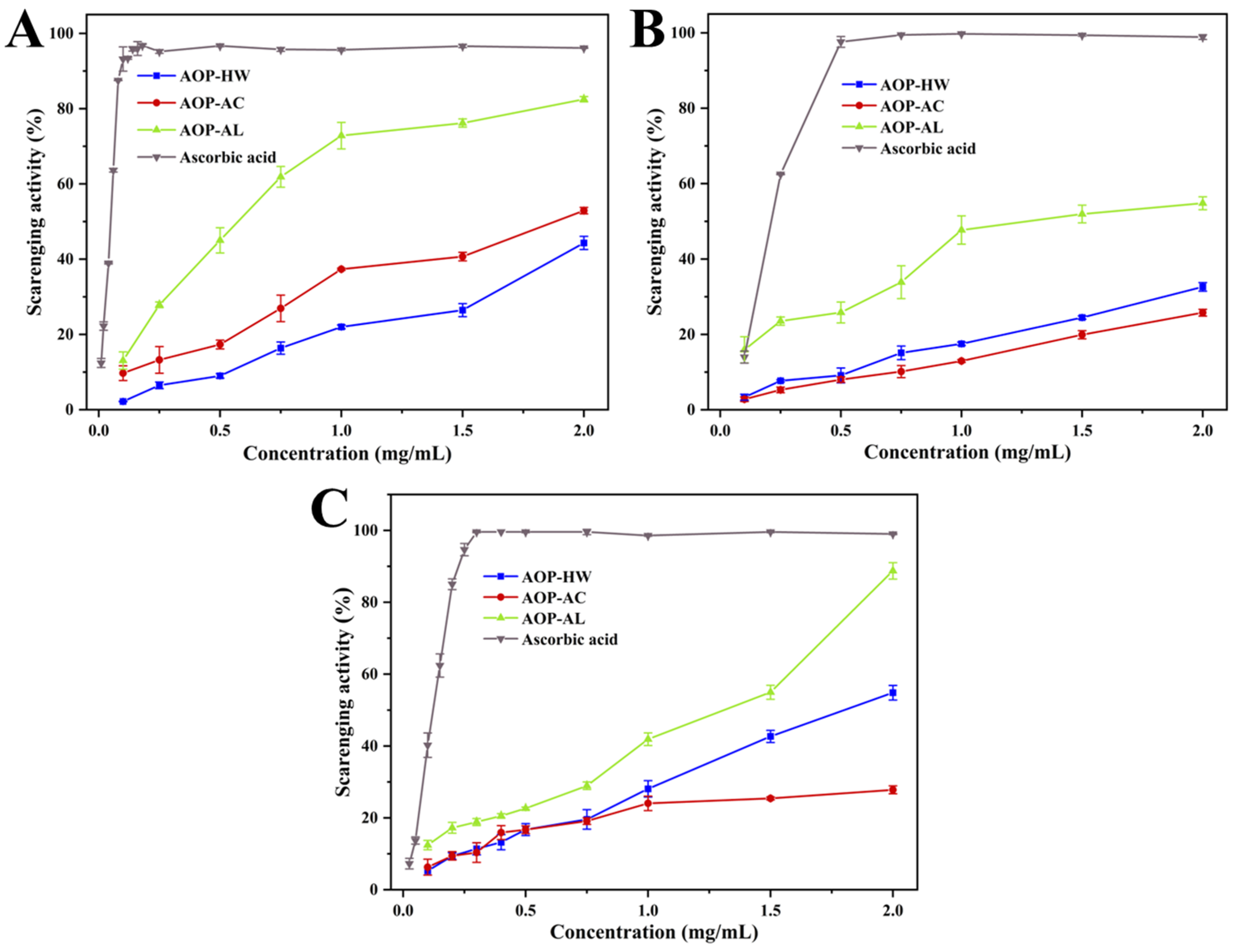
2.4.1. DPPH Radical Scavenging Activity

The DPPH radical, a kind of stable free radical source, has been widely used in the evaluation of natural active compounds [36,37]. Figure 5A shows the scavenging activity of the three AOPs against DPPH radicals. The DPPH radical scavenging activities of AOP-HW, AOP-AC, and AOP-AL increased with concentration, achieving peak values of 44.33%, 52.90%, and 82.51% at a concentration of 2.0 mg/mL, respectively. AOP-AL displayed a remarkably higher scavenging activity against DPPH radicals (IC50 of 0.56 mg/mL) than AOP-HW (IC50 of 2.16 mg/mL) and AOP-AC (IC50 of 1.74 mg/mL) (Table 3). These findings suggested that AOP-AL is a potential scavenger of DPPH radicals.

2.4.2. Hydroxyl Radical Scavenging Activity

The hydroxyl radical is a physiological free radical with the greatest lethality on biological components [38]. As shown in Figure 5B, three AOPs exhibited a moderate scavenging ability of the hydroxyl radical. At the concentration of 2.0 mg/mL, the hydroxyl radical scavenging rate of AOP-AL was 1.84 and 2.33 times that of AOP-HW and AOP-AC, respectively. AOP-AL was a considerably effective hydroxyl radical scavenger with an IC50 value of 1.07 mg/mL, which was stronger than the alkali-extracted Laminaria japonica polysaccharide (IC50 of 1.75 mg/mL) [18] and Zingiber officinale Roscoe stem and leaf polysaccharide (IC50 of 2.426 mg/mL) [39]. These results revealed that AOP-AL exhibited an excellent scavenging activity on hydroxyl radicals, which was consistent with the results of the DPPH radical scavenging activity.

2.4.3. ABTS Radical Scavenging Activity

The ABTS radical, a well-known nitrogen-centered synthetic radical, has been widely used to evaluate the antioxidant capacity of polysaccharides [22,40]. At the concentration of 2.0 mg/mL, the scavenging rates of AOP-HW, AOP-AC, and AOP-AL were 54.84%, 27.81%, and 88.76%, respectively (as illustrated in Figure 5C). Among the three AOPs, AOP-AL showed the lowest IC50 value (1.40 mg/mL) on the ABTS radical scavenging ability, followed by AOP-HW (2.10 mg/mL) and AOP-AC (3.63 mg/mL), which were stronger than those of the results reported by Amamou, Lazreg, Hafsa, Majdoub, Rihouey, Le Cerf, and Achour [41].
Generally speaking, the types of antioxidants can be summarized as hydrogen atom transfer and single electron transfer [42]. The antioxidant potential of polysaccharides is closely related to their structure characteristics, including chemical composition, type of glycosidic linkage, and monosaccharide composition and conformation [33]. It was suggested that polysaccharides containing a high uronic acid content were effective antioxidants, because their electrophilic groups could promote the release of hydrogen from the O−H bond [43]. In this study, AOP-AL exhibited the best effective antioxidant activity, which was probably due to it having the highest content of uronic acid. Moreover, AOP-HW demonstrated a superior scavenging activity compared to AOP-AC in both the ABTS and hydroxyl radical models. A possible explanation is that AOP-HW had a higher galactose content than AOP-AC. Previous studies have reported that the antioxidant activities of polysaccharides may be explained by their abundant galactose content [44,45].

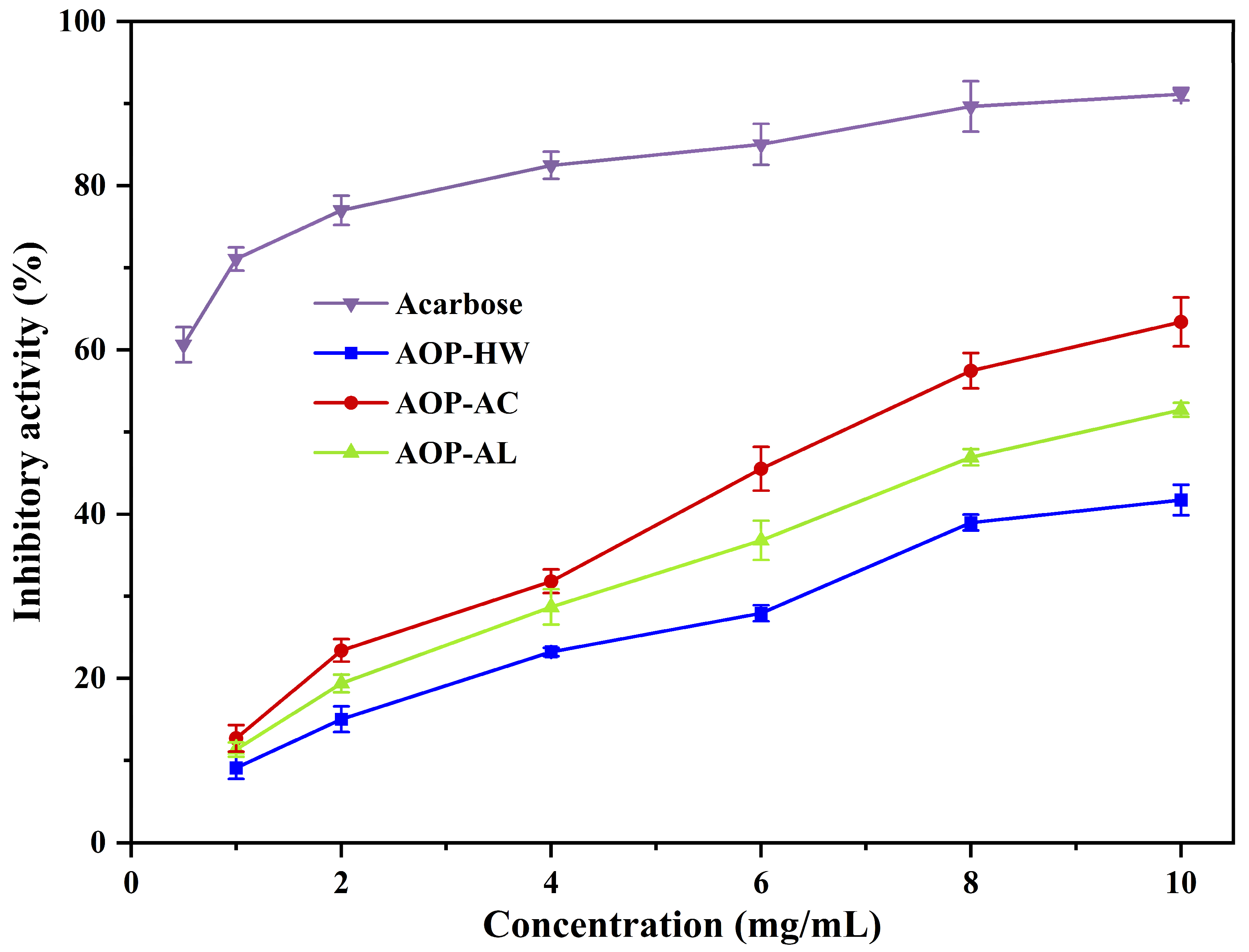
2.5. Inhibitory Effects on α-Amylase Activity

The rise of postprandial blood glucose is related to the hydrolysis of starch by α-amylase. Therefore, the α-amylase inhibition assay can be considered as a rapid and simple strategy to evaluate the in vitro hypoglycemic activity [46].
As depicted in Figure 6, the inhibitory effects of the three AOPs on α-amylase activity intensified with higher concentrations. At the concentration of 10 mg/mL, the inhibitory abilities of AOP-AC, AOP-AL, and AOP-HW on α-amylase were 63.41%, 52.71%, and 41.73%, respectively, which were higher than those of polysaccharides extracted from Trifolium pretense L. and comfrey root (20–35%) [37,47]. AOP-AC possessed the strongest inhibitory effect on α-amylase activity among the three AOPs. Amamou, Lazreg, Hafsa, Majdoub, Rihouey, Le Cerf, and Achour [41] reported that anti-hyperglycemic activity might be related to the high amounts of glucose in the polysaccharide structure. Meanwhile, the high content of uronic acid could also inhibit the enzyme, because the amino acid residues of α-amylase easily interact with free carboxyl groups [13], which verified that AOP-AL had a stronger α-amylase inhibitory activity than AOP-HW despite the low glucose content.

3. Materials and Methods

3.1. Materials and Reagents

Alpiniae oxyphyllae fructus was collected from Guangxi Renjitang Pharmaceutical Co. (Guangxi, China). In total, 10 monosaccharide standards including galacturonic acid, xylose, mannose, arabinose, rhamnose, ribose, glucose, galactose, glucuronic acid, and fucose were provided by Yuanye Biotechnology Co., LTD. (Shanghai, China). Acarbose, α-amylase (3700 U/g), 1,1-diphenyl-2-picrylhydrazyl (DPPH), and dextran standards were purchased from Sigma-Aldrich Co. (Shanghai, China). All other chemicals were obtained from Solarbio Science & Technology Co., LTD. (Beijing, China).

3.2. Polysaccharide Extraction and Isolation

Alpiniae oxyphyllae fructus were separately extracted using an acid solution, hot water, and alkaline solution. The specific extraction conditions were chosen based on a combination of referenced literature [48] and our preliminary experimental findings. In brief, 100.0 g of A. oxyphyllae fructus powder was degreased with n-hexane (1:5 w/v) for 6 h, and then extracted with 3 L of distilled water at 90 °C for 2 h, 3 L of hydrochloric acid (pH 1.5) at 90 °C for 2 h, and 3 L of 0.1 mol/L NaOH/20 mmol/L NaBH4 solution at 25 °C for 2 h, respectively. After centrifugation (5000 rpm, 5 min), the supernatant was neutralized and concentrated with a rotary evaporator. The crude polysaccharide was isolated by adding four volumes of ethanol, allowing the mixture to stand for 12 h, and then centrifuging it at 5000 rpm for 20 min. The polysaccharide solution was then deproteinized with Sevag reagent and dialyzed (8–14 kDa) for 3 days. The three dehydrated polysaccharides were obtained by freeze-drying (FD5 series, SIM International Group, LA, USA), namely, AOP-HW, AOP-AC, and AOP-AL.
The extraction yield of crude A. oxyphyllae fructus polysaccharide was calculated using Formula (1):
Y i e l d ( % ) = [ ( C × V × d ) / m ] × 100
where C represents concentration of polysaccharides (mg/mL), V represents volume of extraction solution (mL), d denotes dilution ratio, and m represents weight of A. oxyphyllae fructus powders (g).

3.3. Determination of Chemical Composition

The contents of neutral sugar, uronic acid, and protein were determined according to the phenol–sulfuric method [49], carbazole–sulfuric acid method (galacturonic acid as the standard) [49], and spectrophotometric method (bovine serum albumin as the standard) [50], respectively.

3.4. Monosaccharide Composition Analysis

The monosaccharide composition of AOPs was analyzed using an HPLC system (1260 series, Agilent Technologies, USA) equipped with a diode array detector and a Thermo ODS-2 column (4.6 × 250 mm, 5 μm) [17]. In brief, a 2.0 mg polysaccharide sample was hydrolyzed in trifluoroacetic acid (2 mol/L) for 2 h (120 °C). After removing the trifluoroacetic acid by nitrogen flushing, 0.5 mL of 1-phenyl-3-methyl-5-pyrazolone methanol solution (0.5 mol/L) and 0.5 mL of NaOH solution (0.3 mol/L) were added to the sample. The mixture was heated at 70 °C for 30 min and neutralized with hydrochloric acid. Subsequently, 0.5 mL of chloroform was added, and the extraction was repeated three times. The aqueous layers were combined for HPLC analysis. The chromatographic conditions were as follows: UV detection wavelength (254 nm), mobile phase = phosphate buffer (0.1 mol/L, pH 7.0) and acetonitrile (82:18, v/v), column temperature = 25 °C, flow rate = 1.0 mL/min, injection volume = 20 μL [1].

3.5. Determination of Mw

The Mw of AOPs was analyzed by high-performance size exclusion chromatography (Agilent 1260, Agilent Technologies, Santa Clara, CA, USA) equipped with an RI detector and a PL aquagel-OH mixed-M column (7.5 × 300 mm, 8 μm) [51]. The polysaccharide was dissolved in ultrapure water containing 0.02% NaN3 and filtered through a 0.45 μm syringe filter. The chromatographic analysis was conducted under the following conditions: column temperature set at 30 °C, flow rate maintained at 0.5 mL/min, injection volume of 20 μL. In addition, calibration curves were established using dextran standards with known molecular weights (Mw: 4.32, 12.6, 60.6, 110, and 496 kDa).

3.6. FT-IR Spectroscopy

The functional groups of AOPs were analyzed by FT-IR spectroscopy [52]. The sample was fully ground with dried KBr powder and then tabled into a transparent sheet. A Nicolet 5700 spectrometer (Thermo Nicolet Co., Waltham, MA, USA) was used to obtain spectra in the wavelength range of 4000–400 cm−1.

3.7. Scanning Electron Microscopy Analysis (SEM)

The dried polysaccharide powder was pasted on a conductive pile and sputtered with a platinum layer [53]. The SEM image was observed using a scanning electron microscope (JEOL Ltd., Akishima, Tokyo, Japan). The detailed measurement parameters were set to the acceleration voltage of 5 kV and the magnification of 5000-fold.

3.8. X-ray Diffraction Analysis (XRD)

The crystal structure of AOPs were analyzed using an X-ray diffractometer (D8 Advance, Bruker, Germany) [54]. The measurement was carried out between 5° and 50° (2θ). The counting time and step size were set to 1 s/step and 0.02°, respectively.

3.9. Antioxidant Qctivities

3.9.1. DPPH Radical Scavenging Activity

The antioxidant activities of AOPs were assessed using the methodology outlined in a previous literature [33]. Specifically, a mixture containing 1 mL of 0.2 mmol/L DPPH solution (dissolved in 50% ethanol) and 1 mL of polysaccharide solution (at concentrations ranging from 0 to 2 mg/mL) was observed at 25 °C in darkness for 30 min. Subsequently, the absorbance was measured at 517 nm using a TU-1810 spectrophotometer, with distilled water serving as the blank control. The results were computed based on Formula (2):
Scavenging   activity   ( % ) = ( 1 A 1 / A 0 )   ×   100 %
where A0 represents the absorbance of 1 mL distilled water plus 1 mL DPPH solution, and A1 shows the absorbance of 1 mL polysaccharide solution to be measured plus 1 mL DPPH solution.

3.9.2. Hydroxyl Radical Scavenging Activity

The hydroxyl radical scavenging activities were analyzed according to the method reported by Wei and Zhang [38]. In summary, an aliquot of 1 mL containing 2 mmol/L FeSO4 and another aliquot of 1 mL with 6 mmol/L ethanol–salicylic acid were introduced to 1 mL of a polysaccharide solution. Subsequently, this amalgamation was combined with 1 mL of 6 mmol/L H2O2 while being continuously agitated. Following this, the resulting mixture was subjected to incubation for a duration of 30 minutes at a temperature of 37 °C. The optical density was then measured at a wavelength of 510 nm using a TU-1810 spectrophotometer. The hydroxyl radical scavenging activity was calculated according to Formula (3):
Scavenging   activity   ( % ) =   [ 1 ( A 2 A 1 ) / A 0 ]   ×   100 %
where A0 represents the absorbance of the reaction system containing 1 mL FeSO4, 1 mL ethanol–salicylic acid solution, 1 mL H2O, and 1 mL H2O2; A1 shows the absorbance of the reaction system containing 1 mL FeSO4, 1 mL ethanol–salicylic acid solution, 1 mL polysaccharide solution, and 1 mL H2O; and A2 denotes the absorbance of the reaction system containing 1 mL FeSO4, 1 mL ethanol–salicylic acid solution, 1 mL polysaccharide solution, and 1 mL H2O2.

3.9.3. ABTS Radical Scavenging Activity

The ABTS radical scavenging activity was measured in accordance with the procedures outlined in the literature [31] with certain adjustments. Initially, the ABTS stock solution was formulated following the documented protocol and subsequently diluted using phosphate buffer (pH 7.0) to achieve an absorbance of 0.700 ± 0.030 at 734 nm. Subsequently, a mixture of 30 μL of polysaccharide solution and 970 μL of ABTS diluent was prepared in a 48-well microplate. Following a 6 min incubation period in darkness, the absorbance was measured at 734 nm using a microplate reader. The ABTS radical scavenging activity was estimated by Formula (4):
Scavenging   activity   ( % )   = [ 1 ( A 0 A 1 ) / A 0 ]   ×   100 %  
where A0 denotes the absorbance value of the blank control, while A1 indicates the absorbance value of the sample.

3.10. α-Amylase Inhibitory Activity

The α-amylase inhibitory activity was performed as reported by Duan, Shang, Chen, Li, and Wu [42]. Briefly, α-amylase solution (100 μL, 0.5 mg/mL) and phosphate buffer (100 μL, 0.2 mol/L, pH 6.9) were mixed with 100 μL of polysaccharides solution at 37 °C for 10 min. Then, a soluble starch solution (200 μL, 1.0%) was added and incubated for 10 min at 37 °C. The reaction was suspended by adding dinitrosalicylic acid reagent (200 μL) and heating in boiling water for 10 min. Finally, the absorbance was observed at 540 nm by a TU-1810 spectrophotometer. The α-amylase inhibitory activity was calculated according to Formula (5):
  α - amylase   inhibitory   activity   ( % )   = [ 1 A 0 A 1 / A 0 ]   ×   100 %
where A0 represents the absorbance of the blank control, and A1 shows the absorbance of the sample.

3.11. Statistical Analysis

All experiments and analyses were replicated thrice, and the results were displayed as the mean ± standard deviation. The origin 2021 (OriginLab, Northampton, MA, USA) and SPSS 26.0 (SPSS Inc., Chicago, IL, USA) software were used to conduct draw the graph and one-way ANOVA, respectively. The p < 0.05 represents a significance difference between samples.

4. Conclusions

This study demonstrated the successful extraction of polysaccharides (AOPs) from Alpiniae oxyphyllae fructus using hot water, hydrochloric acid, and NaOH/NaBH4. The choice of extraction solvent significantly influenced the yield, molecular weight, surface morphology, monosaccharide composition, and crystal structure of the AOPs. Specifically, AOP-HW and AOP-AC were rich in glucose, while AOP-AL predominantly comprised arabinose. Functionally, AOP-AL exhibited a superior antioxidant activity, making it a potential candidate for health supplements and pharmaceuticals targeting oxidative stress. In contrast, AOP-AC showed the strongest α-amylase inhibitory activity, suggesting its use in managing hyperglycemia and as a functional ingredient in diabetic-friendly foods. These findings provide crucial insights into selecting appropriate extraction methods based on desired functional properties, underscoring the potential for tailoring AOPs’ production to specific industrial applications such as natural antioxidants or enzymatic inhibitors.

Author Contributions

R.W.: Writing—original draft. X.R.: Investigation, conceptualization. J.C.: Formal analysis. W.Z.: Resources, validation. T.D.: Resources, project administration. R.L.: Methodology. L.D.: Software. X.S.: Writing—review and editing, funding acquisition, project administration. All authors have read and agreed to the published version of the manuscript.

Funding

This research was funded by the National Natural Science Foundation of China [Grant No. 32202206, 32101948], Central Government Guide Local Special Fund Project for Scientific and Technological Development of Jiangxi Province [Grant No. 20212ZDD02008 and No. 20221ZDD02001], Key Research and Development Plan of Jiangxi Province [Grant No. 20212BBF61008], Research Program of State Key Laboratory of Food Science and Technology, Nanchang University [Grant No. SKLF-ZZB-202114 and SKLF-ZZA-202208], and Foundation of the Key Laboratory of Tropical Crop Products’ Processing of Ministry of Agriculture and Rural Affairs, P.R. China [Grant No. KFKT201901].

Institutional Review Board Statement

Not applicable for studies not involving humans or animals.

Informed Consent Statement

Not applicable.

Data Availability Statement

The original contributions presented in the study are included in the article, further inquiries can be directed to the corresponding authors.

Acknowledgments

The authors would like to thank the South Subtropical Crop Research Institute, China Academy of Tropical Agricultural Sciences, and Key Laboratory of Hainan Province for Postharvest Physiology and Technology of Tropical Horticultural Products (Zhanjiang) for their expert technical assistance.

Conflicts of Interest

The authors declare no conflicts of interest.

References

  1. Han, Y.; Wu, J.; Liu, Y.; Qi, J.; Wang, C.; Yu, T.; Xia, Y.; Li, H. Therapeutic effect and mechanism of polysaccharide from Alpiniae oxyphyllae fructus on urinary incontinence. Int. J. Biol. Macromol. 2019, 128, 804–813. [Google Scholar] [CrossRef] [PubMed]
  2. Zhou, S.; Liu, L.; Zhang, Y.; Zhang, Z.; Li, H.; Fan, F.; He, J.; Kang, J.; Zuo, L. Integrated untargeted and targeted metabolomics to reveal therapeutic effect and mechanism of Alpiniae oxyphyllae fructus on Alzheimer’s disease in APP/PS1 mice. Front. Pharmacol. 2023, 13, 1104954. [Google Scholar] [CrossRef] [PubMed]
  3. Chen, C.M.; Lin, C.Y.; Chung, Y.P.; Liu, C.H.; Huang, K.T.; Guan, S.S.; Wu, C.T.; Liu, S.H. Protective effects of nootkatone on renal inflammation, apoptosis, and fibrosis in a unilateral ureteral obstructive mouse model. Nutrients 2021, 13, 3921. [Google Scholar] [CrossRef] [PubMed]
  4. Zhang, Q.; Zheng, Y.; Hu, X.; Hu, X.; Lv, W.; Lv, D.; Chen, J.; Wu, M.; Song, Q.; Shentu, J. Ethnopharmacological uses, phytochemistry, biological activities, and therapeutic applications of Alpinia oxyphylla Miguel: A review. J. Ethnopharmacol. 2018, 224, 149–168. [Google Scholar] [CrossRef]
  5. Shi, W.; Zhong, J.; Zhang, Q.; Yan, C. Structural characterization and antineuroinflammatory activity of a novel heteropolysaccharide obtained from the fruits of Alpinia oxyphylla. Carbohyd Polym. 2020, 229, 115405. [Google Scholar] [CrossRef] [PubMed]
  6. Yang, X.; Yang, Y.; Chen, H.; Xu, T.; Li, C.; Zhou, R.; Gao, L.; Han, M.; He, X.; Chen, Y. Extraction, isolation, immunoregulatory activity, and characterization of Alpiniae oxyphyllae fructus polysaccharides. Int. J. Biol. Macromol. 2020, 155, 927–937. [Google Scholar] [CrossRef]
  7. Luo, Q.; Zhang, C.; Chen, Y.; Chen, H.; Yang, Y. Alpiniae oxyphyllae fructus polysaccharide 3 inhibits porcine epidemic diarrhea virus entry into IPEC-J2 cells. Res. Vet. Sci. 2022, 152, 434–441. [Google Scholar] [CrossRef] [PubMed]
  8. Chen, Y.; Zhang, Y.; Luo, Q.; Zhu, Y.; Du, H.; Liao, S.; Yang, Y.; Chen, H. Inhibition of porcine epidemic diarrhea virus by Alpiniae oxyphyllae fructus polysaccharide 3. Res. Vet. Sci. 2021, 141, 146–155. [Google Scholar] [CrossRef]
  9. Yang, X.; Zhou, S.; Li, H.; An, J.; Li, C.; Zhou, R.; Teng, L.; Zhu, Y.; Liao, S.; Yang, Y.; et al. Structural characterization of Alpiniae oxyphyllae fructus polysaccharide 2 and its activation effects on RAW264.7 macrophages. Int. Immunopharmacol. 2021, 97, 107708. [Google Scholar] [CrossRef]
  10. Bian, Y.; Lei, C.; Li, N.; Xu, H.; Gong, W.; Gao, M.; Hu, Q.; Jia, L. Chemical characterization and bioactivity analysis of polysaccharides extracted from Ginkgo Folium residue with different solvents. Chromatographia 2023, 86, 375–385. [Google Scholar] [CrossRef]
  11. Dou, Z.M.; Chen, C.; Huang, Q.; Fu, X. Comparative study on the effect of extraction solvent on the physicochemical properties and bioactivity of blackberry fruit polysaccharides. Int. J. Biol. Macromol. 2021, 183, 1548–1559. [Google Scholar] [CrossRef] [PubMed]
  12. Ma, X.; Li, L.; Deng, D.; Tan, Y.; Wu, Y.; Ming, G.; Zhang, Q.; He, X.; Li, Y.; Wang, K. Protective effect of Alpinia oxyphylla fructus polysaccharides extracted with different solvents on dextran sulfate sodium-induced ulcerative colitis in mice. J. Food Biochem. 2023, 217, 1–19. [Google Scholar] [CrossRef]
  13. Fang, C.; Chen, G.; Kan, J. Comparison on characterization and biological activities of Mentha haplocalyx polysaccharides at different solvent extractions. Int. J. Biol. Macromol. 2020, 154, 916–928. [Google Scholar] [CrossRef] [PubMed]
  14. Wang, Z.B.; Pei, J.J.; Ma, H.L.; Cai, P.F.; Yan, J.K. Effect of extraction media on preliminary characterizations and antioxidant activities of Phellinus linteus polysaccharides. Carbohyd Polym. 2014, 109, 49–55. [Google Scholar] [CrossRef] [PubMed]
  15. Meng, M.; Sun, Y.; Qi, Y.; Xu, J.; Sun, J.; Bai, Y.; Han, L.; Han, R.; Hou, L.; Sun, H. Structural characterization and induction of tumor cell apoptosis of polysaccharide from purple sweet potato (Ipomoea batatas (L.) Lam). Int. J. Biol. Macromol. 2023, 235, 123799. [Google Scholar] [CrossRef] [PubMed]
  16. Chen, Y.; Cai, Y.; Zhao, Z.; Yang, D.; Xu, X. Optimization of extraction process, preliminary characterization and safety study of crude polysaccharides from Morindae Officinalis Radix. Foods 2023, 12, 1590. [Google Scholar] [CrossRef] [PubMed]
  17. Gao, J.; Lin, L.; Sun, B.; Zhao, M. A comparison study on polysaccharides extracted from Laminaria japonica using different methods: Structural characterization and bile acid-binding capacity. Food Funct. 2017, 8, 3043–3052. [Google Scholar] [CrossRef] [PubMed]
  18. Sun, Y.; Hou, S.; Song, S.; Zhang, B.; Ai, C.; Chen, X.; Liu, N. Impact of acidic, water and alkaline extraction on structural features, antioxidant activities of Laminaria japonica polysaccharides. Int. J. Biol. Macromol. 2018, 112, 985–995. [Google Scholar] [CrossRef] [PubMed]
  19. Campestrini, L.H.; Rasera, G.B.; de Camargo, A.C.; Franchin, M.; Nani, B.D.; Rosalen, P.L.; Canniatti-Brazaca, S.G.; Telles Biasoto, A.C.; Shahidi, F.; Alencar, S.M. Alkaline conditions better extract anti-inflammatory polysaccharides from winemaking by-products. Food Res. Int. 2020, 131, 108532. [Google Scholar] [CrossRef]
  20. Chen, L.; Liu, J.; Zhang, Y.; Dai, B.; An, Y.; Yu, L. Structural, thermal, and anti-inflammatory properties of a novel pectic polysaccharide from Alfalfa (Medicago sativa L.) Stem. J. Agric. Food Chem. 2015, 63, 3219–3228. [Google Scholar] [CrossRef]
  21. Zhang, C.; Zhu, X.; Zhang, F.; Yang, X.; Ni, L.; Zhang, W.; Liu, Z.; Zhang, Y. Improving viscosity and gelling properties of leaf pectin by comparing five pectin extraction methods using green tea leaf as a model material. Food Hydrocoll. 2020, 98, 105246. [Google Scholar] [CrossRef]
  22. Peasura, N.; Laohakunjit, N.; Kerdchoechuen, O.; Wanlapa, S. Characteristics and antioxidant of Ulva intestinalis sulphated polysaccharides extracted with different solvents. Int. J. Biol. Macromol. 2015, 81, 912–919. [Google Scholar] [CrossRef] [PubMed]
  23. Ye, J.; Hua, X.; Wang, M.; Zhang, W.; Yang, R. Effect of extraction pH on the yield and physicochemical properties of polysaccharides extracts from peanut sediment of aqueous extraction process. LWT-Food Sci. Technol. 2019, 106, 137–144. [Google Scholar] [CrossRef]
  24. Liao, D.W.; Cheng, C.; Liu, J.P.; Zhao, L.Y.; Huang, D.C.; Chen, G.T. Characterization and antitumor activities of polysaccharides obtained from ginger (Zingiber officinale) by different extraction methods. Int. J. Biol. Macromol. 2020, 152, 894–903. [Google Scholar] [CrossRef] [PubMed]
  25. Yang, X.; Ji, H.; Feng, Y.; Yu, J.; Liu, A. Structural characterization and antitumor activity of polysaccharides from Kaempferia galanga L. Oxid. Med. Cell Longev. 2018, 2018, 9579262. [Google Scholar] [CrossRef]
  26. Qiu, S.; Yadav, P.M.; Chau, K.H.; Yin, L.J. Physicochemical characterization and rheological behavior of hemicelluloses isolated from sorghum bran, sorghum bagasse and sorghum biomass. Food Hydrocoll. 2020, 100, 105382. [Google Scholar] [CrossRef]
  27. Yin, J.; Chen, H.; Lin, H.; Xie, M.; Nie, S. Structural features of alkaline extracted polysaccharide from the seeds of Plantago asiatica L. and its rheological properties. Molecules 2016, 21, 1181. [Google Scholar] [CrossRef]
  28. Bai, L.; Zhu, P.; Wang, W.; Wang, M. The influence of extraction pH on the chemical compositions, macromolecular characteristics, and rheological properties of polysaccharide: The case of okra polysaccharide. Food Hydrocoll. 2020, 102, 105586. [Google Scholar] [CrossRef]
  29. Peng, Z.; Tian, S.; Li, H.; Zhu, L.; Zhao, Z.; Zheng, G.; Wen, Q.; Tian, H.; Yang, D. Extraction, characterization, and antioxidant properties of cell wall polysaccharides from the pericarp of Citrus Reticulata cv. Chachiensis. Food Hydrocoll. 2023, 136, 108237. [Google Scholar] [CrossRef]
  30. Huang, Y.; Xie, W.; Tang, T.; Chen, H.; Zhou, X. Structural characteristics, antioxidant and hypoglycemic activities of polysaccharides from Mori Fructus based on different extraction methods. Front. Nutr. 2023, 10, 1125831. [Google Scholar] [CrossRef]
  31. Cui, Y.; Wang, R.; Cao, S.; Ismael, M.; Wang, X.; Lue, X. A galacturonic acid-rich polysaccharide from Diospyros kaki peel: Isolation, characterization, rheological properties and antioxidant activities in vitro. Food Chem. 2023, 416, 135781. [Google Scholar] [CrossRef] [PubMed]
  32. Yan, J.K.; Ding, Z.C.; Gao, X.; Wang, Y.Y.; Yang, Y.; Wu, D.; Zhang, H.N. Comparative study of physicochemical properties and bioactivity of Hericium erinaceus polysaccharides at different solvent extractions. Carbohyd Polym. 2018, 193, 373–382. [Google Scholar] [CrossRef] [PubMed]
  33. Jiang, L.; Wang, W.; Wen, P.; Shen, M.; Li, H.; Ren, Y.; Xiao, Y.; Song, Q.; Chen, Y.; Yu, Q.; et al. Two water-soluble polysaccharides from mung bean skin: Physicochemical characterization, antioxidant and antibacterial activities. Food Hydrocoll. 2020, 100, 105412. [Google Scholar] [CrossRef]
  34. Li, X.; Wang, L. Effect of extraction method on structure and antioxidant activity of Hohenbuehelia serotina polysaccharides. Int. J. Biol. Macromol. 2016, 83, 270–276. [Google Scholar] [CrossRef] [PubMed]
  35. Huang, L.Z.; Ma, M.G.; Ji, X.X.; Choi, S.E.; Si, C. Recent developments and applications of hemicellulose from wheat straw: A review. Front. Bioeng. Biotech. 2021, 9, 690773. [Google Scholar] [CrossRef] [PubMed]
  36. Abuduaini, M.; Li, J.; Ruan, J.H.; Zhao, Y.X.; Maitinuer, M.; Aisa, H.A. Bioassay-guided preparation of antioxidant, anti-inflammatory active fraction from crabapples (Malus prunifolia (Willd.) Borkh.). Food Chem. 2023, 406, 135091. [Google Scholar] [CrossRef] [PubMed]
  37. Zhang, H.; Zhao, J.; Shang, H.; Guo, Y.; Chen, S. Extraction, purification, hypoglycemic and antioxidant activities of red clover (Trifolium pratense L.) polysaccharides. Int. J. Biol. Macromol. 2020, 148, 750–760. [Google Scholar] [CrossRef]
  38. Wei, Q.; Zhang, Y.H. Ultrasound-assisted polysaccharide extraction from Cercis chinensis and properites, antioxidant activity of polysaccharide. Ultrason. Sonochem. 2023, 96, 106422. [Google Scholar] [CrossRef]
  39. Chen, X.; Chen, G.; Wang, Z.; Kan, J. A comparison of a polysaccharide extracted from ginger (Zingiber officinale) stems and leaves using different methods: Preparation, structure characteristics, and biological activities. Int. J. Biol. Macromol. 2020, 151, 635–649. [Google Scholar] [CrossRef]
  40. Zhang, Z.; Chen, Z.; Zhang, C.; Kang, W. Physicochemical properties and biological activities of Tremella hydrocolloids. Food Chem. 2023, 407, 135164. [Google Scholar] [CrossRef]
  41. Amamou, S.; Lazreg, H.; Hafsa, J.; Majdoub, H.; Rihouey, C.; Le Cerf, D.; Achour, L. Effect of extraction condition on the antioxidant, antiglycation and α-amylase inhibitory activities of Opuntia macrorhiza fruit peels polysaccharides. LWT-Food Sci. Technol. 2020, 127, 109411. [Google Scholar] [CrossRef]
  42. Wang, J.; Hu, S.; Nie, S.; Yu, Q.; Xie, M. Reviews on mechanisms of in vitro antioxidant activity of polysaccharides. Oxid. Med. Cell Longev. 2016, 2016, 5692852. [Google Scholar] [CrossRef] [PubMed]
  43. Wang, X.Y.; Zhang, D.D.; Yin, J.Y.; Nie, S.P.; Xie, M.Y. Recent developments in Hericium erinaceus polysaccharides: Extraction, purification, structural characteristics and biological activities. Crit. Rev. Food Sci. 2019, 59, S96–S115. [Google Scholar] [CrossRef] [PubMed]
  44. Cai, L.; Zou, S.; Liang, D.; Luan, L. Structural characterization, antioxidant and hepatoprotective activities of polysaccharides from Sophorae tonkinensis Radix. Carbohyd Polym. 2018, 184, 354–365. [Google Scholar] [CrossRef] [PubMed]
  45. Fan, J.; Wu, Z.; Zhao, T.; Sun, Y.; Ye, H.; Xu, R.; Zeng, X. Characterization, antioxidant and hepatoprotective activities of polysaccharides from Ilex latifolia Thunb. Carbohyd Polym. 2014, 101, 990–997. [Google Scholar] [CrossRef] [PubMed]
  46. Duan, M.; Shang, H.; Chen, S.; Li, R.; Wu, H. Physicochemical properties and activities of comfrey polysaccharides extracted by different techniques. Int. J. Biol. Macromol. 2018, 115, 876–882. [Google Scholar] [CrossRef] [PubMed]
  47. Chen, S.; Shang, H.; Yang, J.; Li, R.; Wu, H. Effects of different extraction techniques on physicochemical properties and activities of polysaccharides from comfrey (Symphytum officinale L.) root. Ind. Crop Prod. 2018, 121, 18–25. [Google Scholar] [CrossRef]
  48. Zhou, S.Y.; Rahman, A.; Li, J.H.; Wei, C.Y.; Chen, J.L.; Linhardt, R.J.; Ye, X.Q.; Chen, S.G. Extraction methods affect the structure of Goji (Lycium barbarum) polysaccharides. Molecules 2020, 25, 936. [Google Scholar] [CrossRef] [PubMed]
  49. Hamilton, J.K.; Smith, F. Reduction of the products of periodate oxidation of carbohydrates. III. The constitution of amylopectin1. J. Am. Chem. Soc. 1956, 78, 5910–5912. [Google Scholar] [CrossRef]
  50. Bitter, T.; Muir, H.M. A modified uronic acid carbazole reaction. Anal. Biochem. 1962, 4, 330–334. [Google Scholar] [CrossRef]
  51. Compton, S.J.; Jones, C.G. Mechanism of dye response and interference in the bradford protein assay. Anal. Biochem. 1985, 151, 369–374. [Google Scholar] [CrossRef] [PubMed]
  52. von Hunolstein, C.; Parisi, L.; Bottaro, D. Simple and rapid technique for monitoring the quality of meningococcal polysaccharides by high performance size-exclusion chromatography. J. Biochem. Biophys. Methods. 2003, 56, 291–296. [Google Scholar] [CrossRef] [PubMed]
  53. Chen, J.; Niu, X.; Dai, T.; Hua, H.; Feng, S.; Liu, C.; McClements, D.J.; Liang, R. Amino acid-amidated pectin: Preparation and characterization. Food Chem. 2020, 309, 125768. [Google Scholar] [CrossRef] [PubMed]
  54. Qian, J.Y.; Chen, W.; Zhang, W.M.; Zhang, H. Adulteration identification of some fungal polysaccharides with SEM, XRD, IR and optical rotation: A primary approach. Carbohyd Polym. 2009, 78, 620–625. [Google Scholar] [CrossRef]
Figure 1. HPLC spectra of monosaccharide composition. (A) Monosaccharide standards, (B) AOP-HW, (C) AOP-AC, and (D) AOP-AL. Peaks: (1) mannose, (2) ribose, (3) rhamnose, (4) glucuronic acid, (5) galacturonic acid, (6) glucose, (7) galactose, (8) xylose, (9) arabinose, and (10) fucose. The samples AOP-HW, AOP-AC, and AOP-AL represent polysaccharides extracted from A. oxyphyllae fructus using hot water, hydrochloric acid, and NaOH/NaBH4, respectively.
Polymers 16 01705 g001
Figure 2. Molecular weight curves (A) and FT-IR spectra (B) of three polysaccharides extracted from A. oxyphyllae fructus with different extraction methods. The samples AOP-HW, AOP-AC, and AOP-AL represent polysaccharides extracted from A. oxyphyllae fructus using hot water, hydrochloric acid, and NaOH/NaBH4, respectively.
Polymers 16 01705 g002
Figure 3. Scanning electron micrographs of (A) AOP-HW, (B) AOP-AC, and (C) AOP-AL at 5000-fold magnification. The samples AOP-HW, AOP-AC, and AOP-AL represent polysaccharides extracted from A. oxyphyllae fructus using hot water, hydrochloric acid, and NaOH/NaBH4, respectively.
Polymers 16 01705 g003
Figure 4. X-ray diffraction patterns of three polysaccharides extracted from A. oxyphyllae fructus with different extraction methods. The samples AOP-HW, AOP-AC, and AOP-AL represent polysaccharides extracted from A. oxyphyllae fructus using hot water, hydrochloric acid, and NaOH/NaBH4, respectively.
Polymers 16 01705 g004
Figure 5. The antioxidant activities of three polysaccharides extracted from A. oxyphyllae fructus with different extraction methods. (A) DPPH radical scavenging activity, (B) hydroxyl radical scavenging activity, (C) ABTS radical scavenging activity. The samples AOP-HW, AOP-AC, and AOP-AL represent polysaccharides extracted from A. oxyphyllae fructus using hot water, hydrochloric acid, and NaOH/NaBH4, respectively.
Polymers 16 01705 g005
Figure 6. Inhibitory effects of three polysaccharides extracted from A. oxyphyllae fructus with different extraction methods on the α-amylase activity. The samples AOP-HW, AOP-AC, and AOP-AL represent polysaccharides extracted from A. oxyphyllae fructus using hot water, hydrochloric acid, and NaOH/NaBH4, respectively.
Polymers 16 01705 g006
Table 1. Yield (%), neutral sugar content (%), uronic acid content (%), protein content (%), and molecular weight (kDa) of A. oxyphyllae fructus polysaccharides extracted using different solvents.
SampleYield (%)Neutral Sugar (%)Uronic Acid (%)Protein (%)Mw (kDa)
AOP-HW4.54 ± 0.46 c69.62 ± 2.11 b19.51 ± 1.21 b1.12 ± 0.95 b385.42 ± 5.64 a
AOP-AC13.76 ± 0.29 a80.57 ± 0.72 a13.15 ± 1.10 c1.24 ± 0.76 b121.28 ± 5.18 c
AOP-AL8.88 ± 0.21 b28.28 ± 1.14 c63.13 ± 2.58 a3.37 ± 1.30 a232.40 ± 3.93 b
The samples AOP-HW, AOP-AC, and AOP-AL represent polysaccharides extracted from A. oxyphyllae fructus using hot water, hydrochloric acid, and NaOH/NaBH4, respectively. Different letters in the same column represent significant differences (p < 0.05). Mw, molecular weight.
Table 2. Monosaccharide ratio of three polysaccharides extracted from A. oxyphyllae fructus with different extraction methods (mol%).
SamplesManRhaGlcAGalAGlcGalXylAra
AOP-HW1.52 ± 0.05 b2.59 ± 0.03 b1.65 ± 0.04 b13.54 ± 0.13 b52.31 ± 0.44 b6.86 ± 0.10 b10.08 ± 0.06 a10.81 ± 0.16 b
AOP-AC0.30 ± 0.01 cN.D.N.D.1.42 ± 0.04 c94.77 ± 0.36 a0.78 ± 0.06 c1.74 ± 0.02 b1.00 ± 0.04 c
AOP-AL3.45 ± 0.11 a9.84 ± 0.23 a3.24 ± 0.02 a17.09 ± 0.21 a10.33 ± 0.19 c17.64 ± 0.14 a10.00 ± 0.07 a28.42 ± 0.21 a
Samples AOP-HW, AOP-AC, and AOP-AL represent polysaccharides extracted from A. oxyphyllae fructus using hot water, hydrochloric acid, and NaOH/NaBH4, respectively. Different letters in the same column represent significant differences (p < 0.05). Man, mannose; Rha, rhamnose; GlcA, glucuronic acid; GalA, galacturonic acid; Glc, glucose; Gal, galactose; Xyl, xylose; Ara, arabinose; N.D., not detected.
Table 3. Scavenging activity of three polysaccharides extracted from A. oxyphyllae fructus with different extraction methods for DPPH, hydroxyl, and ABTS radicals.
SamplesIC50 (mg/mL)
DPPH RadicalHydroxyl RadicalABTS Radical
AOP-HW2.16 ± 0.06 a2.29 ± 0.14 b2.10 ± 0.07 b
AOP-AC1.74 ± 0.08 b2.79 ± 0.12 a3.63 ± 0.10 a
AOP-AL0.56 ± 0.04 c1.07 ± 0.06 c1.40 ± 0.08 c
Ascorbic acid0.05 ± 0.00 d0.32 ± 0.02 d0.12 ± 0.03 d
The samples AOP-HW, AOP-AC, and AOP-AL represent polysaccharides extracted from A. oxyphyllae fructus using hot water, hydrochloric acid, and NaOH/NaBH4, respectively. Different letters in the same column represent significant differences (p < 0.05).
Disclaimer/Publisher’s Note: The statements, opinions and data contained in all publications are solely those of the individual author(s) and contributor(s) and not of MDPI and/or the editor(s). MDPI and/or the editor(s) disclaim responsibility for any injury to people or property resulting from any ideas, methods, instructions or products referred to in the content.

