Article

Analysis of Cytosine Methylation in Genomic DNA of Solanum × michoacanum (+) S. tuberosum Somatic Hybrids

by
Paulina Smyda-Dajmund
1,*,
Jadwiga Śliwka
1,*,
Clizia Villano
2,
Marta Janiszewska
1,
Riccardo Aversano
2,
Piotr Tomasz Bednarek
3,
Domenico Carputo
2 and
Ewa Zimnoch-Guzowska
1
1
Młochów Research Center, Plant Breeding and Acclimatization Institute—National Research Institute, Platanowa 19, 05-831 Młochów, Poland
2
Department of Agricultural Sciences, University of Naples Federico II, via Università 100, 80055 Portici, Italy
3
Department of Plant Physiology and Biochemistry, Plant Breeding and Acclimatization Institute—National Research Institute, Radzików, 05-870 Błonie, Poland
*
Authors to whom correspondence should be addressed.
Agronomy 2021, 11(5), 845; https://doi.org/10.3390/agronomy11050845
Submission received: 29 March 2021 / Revised: 15 April 2021 / Accepted: 23 April 2021 / Published: 26 April 2021
(This article belongs to the Section Crop Breeding and Genetics)

Abstract

:
Interspecific somatic hybridization is a noteworthy breeding strategy that allows the production of novel genetic variability when crossing barriers exist between two parental species. Although the genetic consequences of somatic hybridization have been well documented, little is known on its impact at the epigenetic level. The objective of our research was to investigate the epigenetic changes, in particular DNA methylation, occurring in a population of potato somatic hybrids. The analysis of 96 Solanum × michoacanum (+) S. tuberosum somatic hybrids from five fusion combinations and their parents was carried out by methylation-sensitive amplified polymorphism (MSAP) and high-performance liquid chromatography (HPLC) methods. Six MSAP primer combinations generated 622 unique bands, of which 295 were fully methylated. HPLC analysis showed from 15.5% to 16.9% total cytosine methylation within the parental forms. Overall, the MSAP and HPLC methods indicated an increase in DNA methylation in the somatic hybrids in comparison to their parents. Among the latter, a lower degree of DNA methylation in the wild S. × michoacanum species than S. tuberosum was found. Our findings indicated that somatic hybridization changed the level of cytosine methylation in the studied potato somatic hybrids.

1. Introduction

Somatic fusion is a useful tool in plant breeding to overcome crossing barriers and enable the transfer of important traits. The obtained hybrids, named somatic hybrids, are artificially produced polyploids (allopolyploids) containing a proper mixture of parental nuclear and organellar genomes able to determine their survival, growth and development. Relationships between nuclear genomes as well as between nuclear and organellar genomes are re-established after somatic fusion. Somatic hybridization results in changes in the structure and sequence of DNA and also affects the epigenetic regulation of gene expression [1,2]. The DNA methylation, histone modifications and RNA interference are epigenetic mechanisms that regulate the gene expression with no changes in the DNA sequence [3,4,5]. The most studied epigenetic mechanism in plants is the alteration of DNA methylation, which involves the addition of a methyl group to the 5′-carbon of the pyrimidine ring of the cytosine nucleotides [6] by DNA methyltransferases [7]. The methyl group added by covalent bonds provides the basis to which various protein structures are attached and modifies gene expression [8]. DNA methylation affects plant development and has an important role in plant responses to abiotic and biotic stresses [5], e.g., drought and salinity [3,9,10], low temperature [11,12,13], drugs [14], maintenance of plants in long-term storage [15,16] and pathogens [17,18,19]. In plants, genomic DNA methylation is not accidental, and it covers cytosines located at the coding and noncoding regions in three DNA sequence motifs: CG, CHG and CHH, where H means T, A or C [7,20]. The levels of methylation are diverse among various plant species and depend on the type of plant tissue [21]. The highest methylation level in plants is always in the CG motif [22]. Changes in the epigenome may be reversible or stable and are transmitted during meiosis and mitosis [16,23,24]. The methylation pattern can also change during the growth and development of plants [25]. This aspect is particularly important in polyploid crops, such as coffee, cotton, potato, peanut, sugarcane and wheat [21]. Indeed, compared to diploids, there is a higher copy number of repetitive sequences in the genomes of polyploids; because of this, a higher methylation level is required to silence such elements and ensure the normal development of polyploid plants [20,26]. Little is known about such changes in methylation patterns in somatic hybrids.
Methods of methylation analysis are dedicated to whole genome methylation profiling (global methods) or to searching for differentially methylated regions of the genome or specific genes (specific methods) [27,28,29]. In recent years, various methodologies have been developed and applied in different species [30]. Among them, the methylation-sensitive amplified polymorphism (MSAP) analysis is the most popular one and has been successfully used in various plant species [17,31,32,33,34,35,36,37,38], including potato [13,39,40]. This method is based on the use of methylation-sensitive restriction enzymes, which recognize and cleave 5′-CCGG-3′ sites based on differentially located methylated cytosine residues [41,42]. Another global technique frequently used for cytosine methylation analysis in plants is HPLC [14,43]. In this technique, the level of methylated and unmethylated cytosine is determined and the proportion of methylated cytosine is calculated with the appropriate formula [37]. MSAP and HPLC techniques provide a general picture of the methylation status and are relatively inexpensive and simple to perform. For these reasons, both techniques have been selected in the present study for investigation of methylation changes occurring in S.× michoacanum (+) S. tuberosum (mch (+) tbr) somatic hybrids. Mch (+) tbr somatic hybrids were obtained in our laboratory to transfer resistance to Phytophthora infestans from mch into the tbr genome [44]. Two somatic hybrids from the 97 tested were resistant to P. infestans [45]. Previous analysis of the genetic composition of mch (+) tbr somatic hybrids, evaluated by diversity arrays technology (DArT) markers, indicated that 13.9 to 29.6% of the markers that were present in the fusion parents were lost in the hybrids [45]. Analysis showed losses of single DArT markers spread over the whole length of every chromosome of each somatic hybrid. The explanation of this phenomenon may be the change in the methylation pattern in genomes of the mch (+) tbr hybrids due to the somatic hybridization process. Digestion of genomic DNA in DArT technology is carried out with a methylation-sensitive PstI restriction enzyme. Changes in methylation pattern in somatic hybrids could change PstI enzyme digestion, resulting in different DArT patterns than in their parental forms.
Our hypothesis is that the somatic fusion process generates changes in the level of methylation in the genome of somatic hybrids compared to their parents. Moreover, epigenetic changes have a role in plant resistance responses by regulating the expression of the genes related to resistance to pathogens [19], which may also result in a lack of expression of resistance to P. infestans and may explain the low frequency of obtained resistant hybrids [44,45]. The aim of this research was to determine the methylation levels of mch (+) tbr somatic hybrids and their parental forms based on MSAP and HPLC methods.

2. Materials and Methods

2.1. Plant Material

We used 96 interspecific mch (+) tbr somatic hybrids (MS1–MS31, MS33–MS95 and MS97–MS98) from five fusion combinations and their five parents: diploid (2n = 2x = 24) clones of S. × michoacanum mch/8 and mch/39, an interspecific potato hybrid; DG 81-68, a dihaploid clone dHBard (2n = 2x = 24) derived from cv. Bard; and a tetraploid (2n = 4x = 48) cultivar Rywal. Somatic hybrids from combinations mch/8 (+) dHBard, mch/39 (+) DG 81-68 and mch/39 (+) dHBard are all tetraploid, and plants from mch/8 (+) cv. Rywal and mch/39 (+) cv. Rywal combinations have a ploidy higher than 4x. Further details on the somatic hybrids and their parents are described in [44,45].

2.2. DNA Isolation

Genomic DNA was extracted from 200 mg fresh, young leaves of greenhouse-grown plants with the Qiagen DNeasy Plant Mini Kit (Qiagen, Hilden, Germany). The DNA concentration and quality were determined with a NanoDrop Lite Spectrophotometer (Thermo Fischer Scientific, Waltham, MA, USA). The DNA quality was also checked on 1% agarose gels, stained with ethidium bromide, separated during electrophoresis in 1 × TBE (Tris-Borate-EDTA) buffer followed by visualization under UV light.

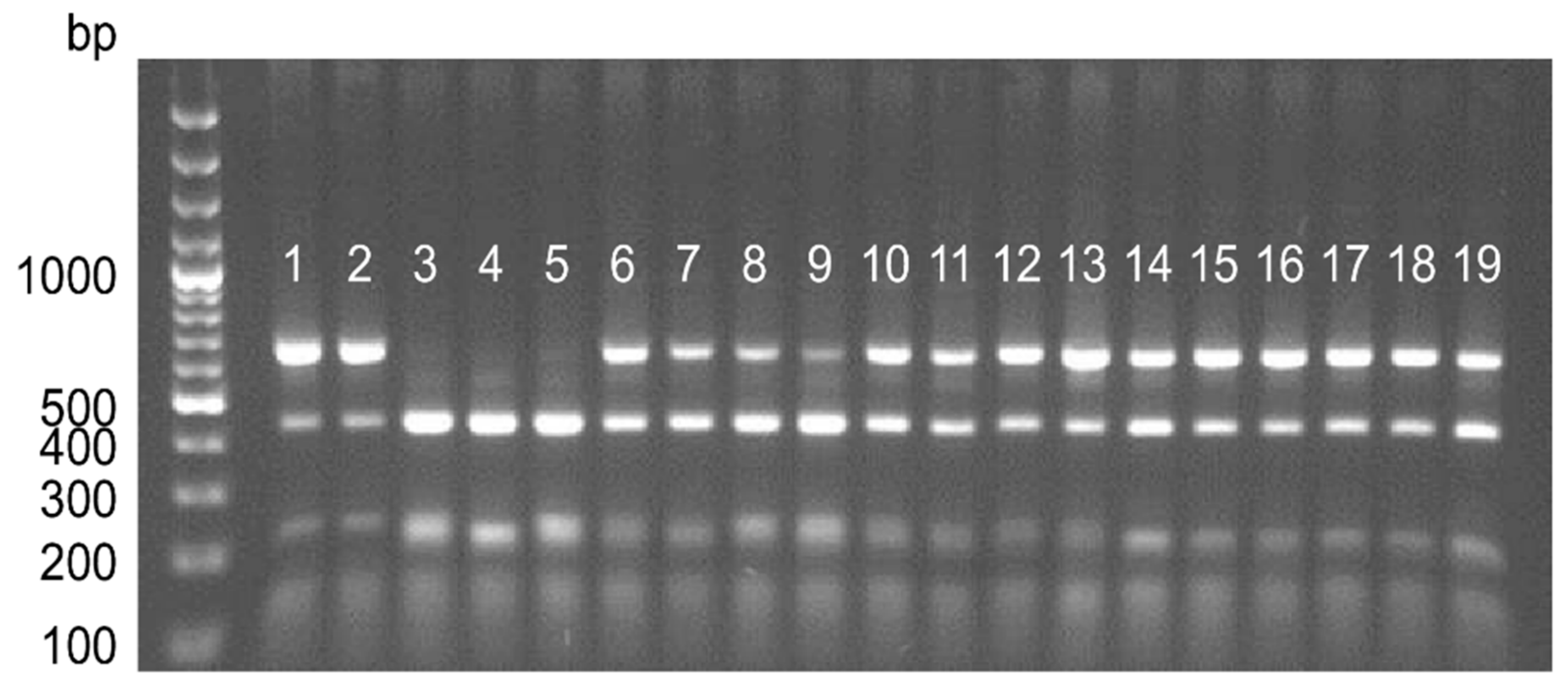
2.3. Conversion of DArT Markers and PCR Analysis

In total, 18 DArT markers were chosen to develop the PCR markers. The names of the selected DArT markers, converted to PCR markers, their chromosomal localization and primer sequences are given in Table S1. The selection of markers was carried out based on previous research by [45]. Ten of the selected markers were present in mch parental forms but absent in tbr parents. Another eight were absent in mch and present in tbr. All 18 markers were absent in somatic hybrids. PCR primers were designed based on the DNA sequence of the DM 1-3 reference potato genome in regions that included sequences of the selected DArT markers. PCR primers were designed using Primer3 software [46]. PCR was performed in a T3000 thermocycler (Biometra GmbH, Göttingen, Germany) in a 20 µL volume reaction mixture containing 2 µL 10 × buffer including 20 mM MgCl2 (Fermentas Life Sciences, Thermo Fischer Scientific, Waltham, MA, USA), 0.5 mM of each dNTP, 0.4 µM each primer, 1 U/µL DreamTaq polymerase (Fermentas Life Sciences, Thermo Fischer Scientific, Waltham, MA, USA) and 30 ng DNA template. The PCR conditions were as follows: one cycle at 95 °C for 10 min followed by 35 cycles at 94 °C for 30 s, 60 °C for 30 s, 72 °C for 60 s and one final extension at 72 °C for 5 min. The PCR products showing no polymorphism at the parental level were digested with 12 different restriction enzymes (MseI; TaqI; RsaI; AluI; DraI; XapI; BsuRI; HindIII; EcoRI; MvaI; BamHI; MwoI) (Fermentas Life Sciences, Thermo Fischer Scientific, Waltham, MA, USA) to obtain differences between the mch and tbr parents. Digestion of the amplicons with selected restriction endonucleases was performed according to the manufacturer’s protocol at 65 °C for 3 h (MseI and TaqI) or 37 °C for 3 h (RsaI; AluI; DraI; XapI; BsuRI; HindIII; EcoRI; MvaI; BamHI; MwoI). The PCR products were separated in 1.5% agarose gels (EURx, Gdańsk, Poland), stained with ethidium bromide and assessed under UV light after electrophoresis in 1 × TBE buffer (Tris-Borate-EDTA). A 100-bp DNA marker (Fermentas Life Sciences, Thermo Fischer Scientific, Waltham, MA, USA) was used to determine the product size.

2.4. Methylation-Sensitive Amplified Polymorphism Analysis

MSAP reactions and analyses were performed as described by [47], with modifications. Sequences of the adapters and pre- and selective amplification primers are shown in Table S2. Restriction and ligation were performed in the same step. For each genotype, two separate restriction/ligation reactions with different combinations of restriction enzymes were performed. The restriction/ligation mix with 350 ng of genomic DNA was incubated overnight at 37 °C in a 50 µL reaction volume. Preamplification PCR was prepared in a 50 µL reaction volume with 0.5 µM of both EcoRI- and MspI/HpaII + 1 primer, 0.2 mM dNTPs, 1 × PCR buffer, 1.5 mM MgCl2 and Taq DNA polymerase at a concentration of 1 U µL−1. The reaction was carried out as follows: one cycle at 72 °C for 1 min, one cycle of 94 °C for 45 s, 65 °C for 30 s, 72 °C for 1 min, 94 °C for 30 s and a reduction of 0.7 °C per cycle from 64.3 °C to 56.6 °C for 30 s and 72 °C for 1 min, followed by 20 cycles of 94 °C for 30 s, 55.9 °C for 30 s, 72 °C for 1 min and one cycle of elongation at 72 °C for 30 s. PCR products were checked on 1% agarose gel. Selective amplification was performed in a 50 µL reaction volume with 0.5 µM EcoRI primer and 0.5 µM MspI/HpaII primer, 0.2 mM dNTPs, 1 × PCR buffer, 2 mM MgCl2 and Taq DNA polymerase at a concentration of 0.4 U µL−1. PCR was performed under the same conditions described for preamplification, except for an initial step at 72 °C for 1 min. Analysis of PCR fragments was performed in a mixture of 1 µL of PCR product, 9 µL Hi-DiTM Formamide (Life Technologies Polska Ltd. Warszawa, Poland) and 0.5 µL GeneScanTM 600 LIZ® Size Standard v2.0 (Life Technologies Polska Ltd. Warszawa, Poland) on an ABI3500 DNA Analyser with 50-cm capillaries and Polymer POP-7 (Life Technologies Polska Ltd. Warszawa, Poland) using GeneMapper v.4.0 software (Life Technologies Polska Ltd. Warszawa, Poland). All PCRs were repeated twice, and reproducible and good quality peaks were scored for analysis. The presence of the DNA fragment was marked as (1), and the absence was (0). The profile of bands of genomic DNA digested with a combination of both MspI and EcoRI was compared to DNA digested with HpaII and EcoRI. By comparing the methylation pattern of every single band of a given size, four MSAP pattern types (MspI/HpaII = 1/1; 1/0; 0/1; 0/0) were expected. The obtained results for somatic hybrids and their parents were transformed into two groups: A (MspI/HpaII = 1/1—unmethylated) and B (MspI/HpaII = 1/0; 0/1; 0/0—methylated). Somatic hybrids possessed eight different methylation patterns in comparison to their parents. The methylation changes observed in the genomes of somatic hybrids are presented below:
mchparenttbrparentsomatic hybridmethylation changes in somatic hybrid
AAANo change
AABHyper-methylation of the somatic hybrid
ABAMethylation pattern of the mch parent
BAAMethylation pattern of the tbr parent
BBBNo change
BBAHypo-methylation of the somatic hybrid
BABMethylation pattern of the mch parent
ABBMethylation pattern of the tbr parent
The analysis of the DNA methylation level of the somatic hybrids and their parental forms consisted of two parts: (1) quantitative analysis, in which the percentage of unmethylated and methylated CCGG sites was determined, and (2) qualitative analysis, in which the percentage of bands in various methylation patterns was determined.

2.5. High-Performance Liquid Chromatography Analysis

DNA samples (4 μg) were dried, dissolved in deionized water (100 μL), denatured at 100 °C for 2 min, and transferred to ice for 5 min. To each sample, 5 μL of 10 mM ZnSO4 and 10 μL of 1.0 U mL−1 nuclease P1 in 30 mM NaOAc (pH 5.4) were added and incubated at 37 °C for 17 h. In the next step, 10 μL of 0.5 M Tris at pH 8.3 and 10 μL of 10.0 U mL−1 alkaline phosphatase in 2.5 M (NH4)2SO4 were added, mixed carefully, and incubated at 37 °C for 2 h. After incubation, prepared samples were centrifuged for 5 min at 12 × 103 rpm. HPLC analysis was performed using a Waters 625 LC System Synergy Max-RP C12 (250 × 4.6 mm, 4u, Phenomenex, Torrance, CA, USA) column combined with a Synergy Max-RP C12 precolumn according to [48,49]. Buffer ‘A’, consisting of 0.5% v/v methanol in 10 mM KH2PO4 at pH 3.7, and buffer ‘B’, consisting of 10% v/v methanol in 10 mM KH2PO4 at pH 3.7, were used for the separation of nucleosides. The gradient used for separation consisted of 100% of buffer ‘A’ to 100% of buffer ‘B’ for 10 min, then 10–25 min of 100% of buffer ‘B’ and 5 min of 100% of buffer ‘A’ with the flow rate 1 mL per min at 30 °C and UV detection at a wavelength of 280 nm. The external standards were (1) DNA (0.5–50 µM), (2) RNA nucleosides (1.5–150 µM) and (3) 5-methyl-2′-deoxycytidine (5 mdC) dissolved in deionized water. Peaks of 2′-deoxycytidine (dC) and 5 mdC had retention times of 6.5 min and 9.3 min, respectively. The 5 mdC component was defined based on Millennium 32 v. 4.0 software (Waters Corporation, Milford, MA, USA). The estimate of nucleosides was determined on the automatically integrated surface areas (μV s−1) of the chromatograph peaks. The amount of cytidine (dC) and 5-methyl-2′-deoxycytidine (5 mdC) in relation to all nucleosides was evaluated using the following formula: dC = dC/(dC + 5 mdC + dG + dT + dA) × 100 and 5 mdC = 5 mdC/(dC + 5 mdC + dG + dT + dA) × 100. The level of global DNA methylation was calculated as the concentration of 5-methyl-2′-deoxycytidine (5 mdC) in relation to the whole amount of cytidine according to the following formula: 5 mdC/(5 mdC + dC) × 100.

3. Results

PCR analysis revealed the presence of all 18 markers converted from the missing DArT markers in the genomes of all 30 selected somatic hybrids from the five fusion combinations. The selected DArT markers were located on various potato chromosomes (I, II, III, IV, V, VI, VII, VIII and X) (Table S1). The known chromosomal assignment allowed the checking of different points in the genome of the somatic hybrids. For example, DArT marker 656,549 (assigned to chromosome I) was present in mch, absent in the tbr parent, and missing within somatic hybrids from all fusion combinations in the DArT analyses, as a PCR marker was detected in all the selected somatic hybrids (Figure 1).
MSAP analysis was applied to check the methylation changes induced by somatic hybridization in random CCGG sites. Overall, out of 14 primer combinations tested (Table S3), six (EHM_2; EHM_3; EHM_8; EHM_9; EHM_12; EHM_13) were polymorphic between the parental forms of the somatic hybrids and generated a total of 622 fragments, ranging from 75 (EHM_3) to 137 (EHM_2) bands per combination (Table 1). The sizes of the analyzed bands were between 51 bp and 630 bp. Although the total number of methylated CCGG sites was 295, the frequency changed by combination, with EHM_12 being the least polymorphic (38.3%) and EHM_9 the most (56.4%) (Table 1). Cytosine methylation levels differed among the parental forms (Table 2). On average, 182.8 bands were analyzed among them, with mch/39 and cv. Rywal showing the lowest and the highest number (148 and 213, respectively). Most of the CCGG sites were unmethylated in all parental forms, except for dHBard, for which 54.3% of the polymorphic sites were otherwise methylated. The CCGG methylation levels of the wild parents mch/8 and mch/39 were lower than those of the tbr (DG 81-68; dHBard and cv. Rywal) parents based on MSAP analysis.
Between 236 (mch/39 (+) DG 81-68) and 272 (mch/8 (+) cv. Rywal) bands per fusion combination were analyzed (Table 3). The number of methylated CCGG sites differed between the fusion populations. Regarding the single fusion combinations, the highest mean percentage of methylated CCGG sites per individual occurred in the mch/39 (+) cv. Rywal combination (53.2%) and the lowest within mch/8 (+) dHBard (45.1%). No new bands appeared among the somatic hybrids, which were not observed in the parents. However, we observed a higher percentage of CCGG methylated motifs relative to the methylation level of the parental forms for fusion combinations mch/8 (+) cv. Rywal, mch/39 (+) DG 81-68 and mch/39 (+) cv. Rywal based on MSAP analysis. The methylation levels of the combinations mch/8 (+) dHBard and mch/39 (+) dHBard were lower than those of their parental forms.
Besides the quantitative analysis of the methylation changes triggered by somatic hybridization, a qualitative analysis was also performed (Table 4). Three major groups of banding patterns were identified (Table 4). The first one comprised those patterns that were unchanged across the parents and their somatic hybrids. Most of them (20%) belonged to the “B-pattern”, which is linked to the methylated status of the CCGG sites. In the second group, the methylation changes occurring in the somatic hybrids were determined. On average, 6.1% of the analyzed MSAP bands were hypermethylated (AAB pattern), and the mch/39 (+) cv. Rywal combinations were the most affected (7.1%). By contrast, 14.8% of the analyzed bands were hypomethylated (BBA, ABA and BAA patterns), with means ranging from 7.2% (mch/8 (+) cv. Rywal) to 19.5% (mch/39 (+) dHBard). Lastly, many of the analyzed somatic hybrids resembled the methylation patterns of one of their parents. Thus, most of the somatic hybrids (mch/8 (+) cv. Rywal, mch/39 (+) DG 81-68, mch/39 (+) cv. Rywal) inherited the banding pattern from the tbr parents, whereas the mch/8 (+) dHBard and mch/39 (+) dHBard banding patterns resembled those of the wild parent. Among the two P. infestans-resistant somatic hybrids, 7.2% (MS21) and 7.5% (MS52) of the analyzed MSAP bands showed hypermethylation (AAB), while the hypomethylated (BBA, ABA and BAA) sites accounted for 45% and 48% of the total sites in MS21 and MS52, respectively.
Regarding the HPLC analysis, the data highlighted variation in the total cytosine methylation, ranging from 15.5% (mch/39) to 16.9% (cv. Rywal) (Table 2). The total cytosine methylation levels of the wild parents, mch/8 and mch/39, were lower compared to the DG 81-68 and cv. Rywal parents, which was in line with the results obtained in the MSAP analysis. There was a one exception, dHBard, which had slightly lower total cytosine methylation level than wild mch/8. Based on the HPLC system, the highest mean percentage of methylated cytosine per individual was 19.4% for the mch/39 (+) DG 81-68 combination, and the lowest 15.9% for the mch/39 (+) cv. Rywal combination. The HPLC results indicated that the levels of cytosine methylation for the somatic hybrids from four combinations were higher than the level of methylation of the parental forms. In combination mch/39 (+) cv. Rywal, a lower level was noticed compared to the cv. Rywal parent (Table 2 and Table 3). The mean percentage of total cytosine methylation for the two late blight-resistant somatic hybrids was 20% and 19.04% for MS21 and MS52, respectively.

4. Discussion

The somatic fusion process is an ‘extreme’ procedure that is complicated and stressful for cells [24]. Somatic hybrids are obtained from electrofused protoplasts; they develop through calluses and are maintained under in vitro conditions. They consist of two different genomes, with increased ploidy levels and new nuclear–cytoplasmic interactions. In the present study, most of the mch (+) tbr somatic hybrids had higher mean CCGG and total cytosine methylation levels than their parents, as shown by the MSAP and HPLC results. It should be pointed out that MSAP detects changes in cytosine methylation in the CCGG motifs, while HPLC detects all the methylated cytosines in the whole genome. In the case of somatic hybrids, the methylation pattern can be changed to a greater extent than in sexual hybrids. It is known that during the plant tissue culture process, epigenetic changes are noted [50], and the DNA methylation level can either increase or decrease [24]. For example, the long-term tissue culture of potato generated 12.56 to 26.13% cytosine methylation changes, as evaluated by MSAP analysis [15]. Changes in the level of cytosine methylation determined by MSAP were also observed in other plant species, such as Cymbidium hybridum [51], Freesia hybrida [52] and Hordeum brevisubulatum [53]. Epigenetic changes can indirectly lead to changes in the DNA sequence by inducing point mutations or activating transposable elements [54].
A few examples of epigenetic changes induced by somatic hybridization have been described in potato. In S. tuberosum (+) S. etuberosum somatic hybrids, 2–6% of the total methylation patterns changed compared to those of their mother plants maintained in tissue culture, as evaluated by MSAP analysis [55]. Changes ranging from 3.2 to 8.5% were reported for S. tuberosum ‘C-13′ (+) S. pinnatisectum [16]. Besides potato, increased methylation levels also have been observed in somatic hybrids of cabbage [56] and citrus [57], as well as in asymmetric somatic hybrids between tall fescue and wheat [58]. Analysis of the methylation of the somatic hybrid Ipomoea batatas (+) I. triloba indicated a reduction in methylation levels compared to the parents [59]. Another factor that may have contributed to the methylation changes observed in our study is the increase in the ploidy level of our hybrids compared to the parental diploid genotypes. A previous study by [60] demonstrated that the allotetraploid form of cotton has a higher methylation level than its ancestral diploid genome and diploid hybrid. In S. commersonii, a wild potato species, the authors of [40,61] compared the level of methylation of the synthesized autotetraploids vs. their diploid parent. The authors noticed an increase in methylation in the synthetic polyploids. In our previous work [45], the genetic composition of the mch (+) tbr hybrids was determined using DArT markers. The analysis provided evidence that significant numbers of DArT markers present in the parental genotypes were missing in the hybrids. The PstI enzyme used in DArT technology is a methylation-sensitive rare cutter that recognizes the CTGCAG sites, and their digestion is blocked by methylation of cytosine at position 5mCTGCAG [62,63]. This makes differential methylation a likely explanation for the loss of DArT markers in our somatic hybrids. We confirmed the presence of DArT DNA sequences missing in the genomes of hybrid forms according to the DArT analysis for 18 selected markers converted into methylation-insensitive PCR markers. These results are consistent with our hypothesis that the methylation pattern in somatic hybrids changes in comparison to their parents. Thus, DArT markers were not detected in the hybrids not because of deletion, chromosome loss or genome rearrangement, but most likely due to methylated restriction of the PstI sites. The authors of [62] noted that 1.2% of the DArT markers could not be grouped into a genetic linkage map of the barley lines because of their non-Mendelian behavior. This phenomenon has been explained by the formation of new, unstable cytosine methylations. DArT technology offers a high-throughput whole-genome genotyping method that can be used for the analysis of genetic diversity, population structure and gene mapping in different organisms. DArT markers have been applied with success to crop plants, including rice [64], barley [62,65], wheat [66], potato [67,68] and many others. These markers can also be used to study the genetic composition of hybrids obtained through somatic hybridization and to dissect recombination patterns, but it should be taken into account that the analysis of the mch (+) tbr somatic hybrids with changed methylation levels induced by the somatic hybridization process may slightly affect the obtained DArT results.
Epigenetic modifications have an impact on the expression of genes. They may cause silencing or activation of some genes, leading to changes in the phenotype [59]. Qualitative analysis of the mch (+) tbr hybrids indicated not only an increase in the methylation level in their genomes but also the hypomethylation of some CCGG sites. A decrease in methylation may cause the expression of silenced genes, chromosome recombination and the activation of transposable elements [21,59]. The purpose of creating somatic hybrids is to transfer important traits, including resistance to biotic and abiotic stresses, into cultivated forms. An important problem associated with somatic hybridization is obtaining a low number of somatic hybrids expressing the desired trait. In our previous research, only two somatic hybrids were resistant to P. infestans, whereas the majority of the tested hybrids were classified as susceptible to late blight. Among the resistant somatic hybrids, a higher percentage of hyper- and hypomethylated CCGG sites was observed compared with the average percentage of the hyper- and hypomethylated CCGG sites for their fusion combinations. HPLC also indicated their higher mean percentage of methylated cytosines compared to the means of their fusion combinations. Both MSAP and HPLC methods did not allow us to accurately determine in which chromosome or chromosome region methylation changed, which could affect the resistance to P. infestans. Therefore, further research is needed. A reduced or a lack of expression of resistance to P. infestans in somatic hybrids has also been reported in S. pinnatisectum (+) S. tuberosum hybrids [69] and in S.× michoacanum (+) S. tuberosum hybrids [70]. One plausible hypothesis to explain these results is the change in the methylation status in the genome of somatic hybrids.

5. Conclusions

Our results confirmed changes in DNA methylation in the genomes of the mch (+) tbr hybrids. The impact of the DNA methylation level of the somatic hybrids on the expression of the desired trait, such as resistance to P. infestans, requires further detailed studies. Comprehensive characteristics of the somatic hybrids of various plant species, including potato, will widen the knowledge of the process of somatic hybridization and will allow for effective use of the obtained somatic hybrids in plant breeding.

Supplementary Materials

The following are available online at https://www.mdpi.com/article/10.3390/agronomy11050845/s1, Table S1: Selected DArT markers converted to PCR markers. DArT markers were present in the mch/tbr genome but absent in the genome of somatic hybrids; Table S2: Sequences of adapters and primers used in methylation-sensitive amplified polymorphism (MSAP) analyses; Table S3: Combinations of selective amplification primers. The primer combinations used for analysis of the level of cytosine methylation in somatic hybrids are shown in bold.

Author Contributions

Conceptualization, P.S.-D., J.Ś. and E.Z.-G.; methodology, P.S.-D., C.V., M.J. and P.T.B.; investigation, P.S.-D., P.T.B.; data curation, P.S.-D., M.J.; writing—original draft preparation, P.S.-D.; writing—review and editing, P.S.-D., J.Ś., C.V., D.C., R.A., P.T.B., E.Z.-G.; supervision, J.Ś., D.C. and E.Z.-G.; project administration, E.Z.-G.; funding acquisition, E.Z.-G. All authors have read and agreed to the published version of the manuscript.

Funding

The research was financed by the National Science Centre in Poland, grant number UMO-2012/07/B/NZ9/01901 and statutory subvention received by Plant Breeding and Acclimatization Institute—National Research Institute from Polish Ministry of Education and Science. P.S.-D. was supported by a young scientist statutory grant in 2021.

Institutional Review Board Statement

Not applicable.

Informed Consent Statement

Not applicable.

Data Availability Statement

The data presented in this study are available in the article and supplementary materials.

Acknowledgments

The authors thank Emil Stefańczyk (Plant Breeding and Acclimatization Institute—National Research Institute, Poland) for help with bioinformatics.

Conflicts of Interest

The authors declare no conflict of interest.

References

  1. Orczyk, W.; Przetakiewicz, J.; Nadolska-Orczyk, A. Somatic hybrids of Solanum tuberosum—Application to genetics and breeding. Plant Cell Tissue Organ. Cult. 2003, 74, 1–13. [Google Scholar] [CrossRef]
  2. Liu, S.; Li, F.; Kong, L.; Sun, Y.; Qin, L.; Chen, S.; Cui, H.; Huang, Y.; Xia, G. Genetic and epigenetic changes in somatic hybrid introgression lines between wheat and tall wheat grass. Genetics 2015, 199, 1035–1045. [Google Scholar] [CrossRef] [PubMed]
  3. Banerjee, A.; Roychoudhury, A. Epigenetic regulation during salinity and drought stress in plants: Histone modification and DNA methylation. Plant Gene 2017, 11, 199–204. [Google Scholar] [CrossRef]
  4. Chwialkowska, K.; Korotko, U.; Kosinska, J.; Szarejko, I.; Kwasniewski, M. Methylation sensitive amplification polymorphism sequencing (MSAP-Seq)—A method for high—Throughput analysis of differentially methylated CCGG sites in plants with large genomes. Front. Plant Sci. 2017, 8, 2056. [Google Scholar] [CrossRef] [Green Version]
  5. Zhang, H.; Lang, Z.; Zhu, J.K. Dynamics and function of DNA methylation in plants. Nat. Rev. Mol. Cell Biol. 2018, 19, 489–506. [Google Scholar] [CrossRef] [PubMed]
  6. Kumar, S.; Chinnusamy, V.; Mohapatra, T. Epigenetics of Modified DNA Bases:5-Methylcytosine and Beyond. Front. Genet. 2018, 9, 640. [Google Scholar] [CrossRef] [PubMed] [Green Version]
  7. Alonso, C.; Pérez, R.; Bazaga, P.; Herrera, C.M. Global DNA cytosine methylation as an evolving trait: Phylogenetic signal and correlated evolution with genome size in angiosperms. Front. Genet. 2015, 6, 4. [Google Scholar] [CrossRef] [Green Version]
  8. Singroha, G.; Sharma, P. Epigenetic Modifications in Plants under Abiotic Stress, Epigenetics; Meccariello, R., Ed.; IntechOpen: London, UK, 2019; Available online: https://www.intechopen.com/books/epigenetics/epigenetic-modifications-in-plants-under-abiotic-stress (accessed on 26 April 2021). [CrossRef] [Green Version]
  9. Sapna, H.; Ashwini, N.; Ramesh, S.; Nataraja, K. Assessment of DNA methylation pattern under drought stress using methylation-sensitive randomly amplified polymorphism analysis in rice. Plant Genet. Resour. 2020, 18, 222–230. [Google Scholar] [CrossRef]
  10. Garg, R.; Chevala, V.V.S.N.; Shankar, R.; Jain, M. Divergent DNA methylation patterns associated with gene expression in rice cultivars with contrasting drought and salinity stress response. Sci. Rep. 2015, 14922. [Google Scholar] [CrossRef] [Green Version]
  11. Steward, N.; Kusano, T.; Sano, H. Expression of ZmMET1, a gene encoding a DNA methyltransferase from maize, is associated not only with DNA replication in actively proliferating cells, but also with altered DNA methylation status in cold-stressed quiescent cells. Nucleic Acids Res. 2000, 28, 3250–3259. [Google Scholar] [CrossRef] [PubMed] [Green Version]
  12. Shan, X.; Wang, X.; Yang, G.; Wu, Y.; Su, S.; Li, S.; Liu, H.; Yuan, Y. Analysis of the DNA methylation of maize (Zea mays L.) in response to cold stress based on methylation- sensitive amplified polymorphisms. J. Plant Biol. 2013, 56, 32–38. [Google Scholar] [CrossRef]
  13. Xin, C.; Hou, R.; Wu, F.; Zhao, Y.; Xiao, H.; Si, W.; Ali, M.E.; Cai, L.; Guo, J. Analysis of cytosine methylation status in potato by methylation-sensitive amplified polymorphisms under low-temperature stress. J. Plant Biol. 2015, 58, 383–390. [Google Scholar] [CrossRef]
  14. Gao, W.; Li, S.; Huang, Y.; Deng, C.; Lu, L. Detection of genome DNA methylation change in spinach induced by 5-azaC. Mol. Cell Probes 2014, 28, 163–166. [Google Scholar] [CrossRef] [PubMed]
  15. Dann, A.L.; Wilson, C.R. Comparative assessment of genetic and epigenetic variation among regenerants of potato (Solanum tuberosum) derived from long-term nodal tissue-culture and cell selection. Plant Cell Rep. 2011, 30, 631–639. [Google Scholar] [CrossRef]
  16. Tiwari, J.K.; Saurabh, S.; Chandel, P.; Singh, B.P.; Bhardwaj, V. Analysis of genetic and epigenetic variation in in vitro propagated potato somatic hybrid by AFLP and MSAP markers. Electron. J. Biotechnol. 2013, 16, 5. [Google Scholar] [CrossRef]
  17. Da, K.; Nowak, J.; Flinn, B. Potato cytosine methylation and gene expression changes induced by a beneficial bacterial endophyte, Burkholderia phytofirmans strain PsJN. Plant Physiol. Biochem. 2012, 50, 24–34. [Google Scholar] [CrossRef]
  18. Le, T.N.; Schumann, U.; Smith, N.A.; Tiwari, S.; Au, P.C.K.; Zhu, Q.H.; Taylor, J.M.; Kazan, K.; Llewellyn, D.J.; Zhang, R.; et al. DNA demethylases target promoter transposable elements to positively regulate stress responsive genes in Arabidopsis. Genome Biol. 2014, 15, 458. [Google Scholar] [CrossRef] [PubMed]
  19. Tirnaz, S.; Batley, J. DNA methylation: Toward crop disease resistance improvement. Trends Plant Sci. 2019, 12, 1149. [Google Scholar] [CrossRef]
  20. Pikaard, C.S.; Scheid, O.M. Epigenetic regulation in plants. Cold Spring Harb. Perspect. Biol. 2014, 6, a019315. [Google Scholar] [CrossRef] [PubMed]
  21. Bartels, A.; Han, Q.; Nair, P.; Stacey, L.; Gaynier, H.; Mosley, M.; Huang, Q.Q.; Pearson, J.K.; Hsieh, T.F.; An, Y.C.; et al. Dynamic DNA methylation in plant growth and development. Int. J. Mol. Sci. 2018, 19, 2144. [Google Scholar] [CrossRef] [PubMed] [Green Version]
  22. Zemach, A.; McDaniel, I.E.; Silva, P.; Zilberman, D. Genome-wide evolutionary analysis of eukaryotic DNA methylation. Science 2010, 328, 916–919. [Google Scholar] [CrossRef] [PubMed] [Green Version]
  23. Brettell, R.I.S.; Dennis, E.S. Reactivation of a silent Ac following tissue culture is associated with heritable alterations in its methylation pattern. Mol. Gen. Genet. 1991, 229, 365–372. [Google Scholar] [CrossRef]
  24. Smulders, M.J.M.; de Klerk, G.J. Epigenetics in plant tissue culture. Plant Growth Regul. 2011, 63, 137–146. [Google Scholar] [CrossRef] [Green Version]
  25. Niederhuth, C.E.; Bewick, A.J.; Ji, L.; Alabady, M.S.; Kim, K.D.; Li, Q.; Rohr, N.A.; Rambani, A.; Burke, J.M.; Udall, J.A.; et al. Widespread natural variation of DNA methylation within angiosperms. Genome Biol. 2016, 17, 194. [Google Scholar] [CrossRef] [Green Version]
  26. Jackson, S.; Chen, Z.J. Genomic and expression plasticity of polyploidy. Curr. Opin. Plant Biol. 2010, 13, 153–159. [Google Scholar] [CrossRef] [Green Version]
  27. Laird, P.W. Principles and challenges of genome-wide DNA methylation analysis. Nat. Rev. Genet. 2010, 11, 191–203. [Google Scholar] [CrossRef]
  28. Kurdyukov, S.; Bullock, M. DNA methylation analysis: Choosing the right method. Biology 2016, 5, 3. [Google Scholar] [CrossRef]
  29. Marconi, G.; Capomaccio, S.; Comino, C.; Acquadro, A.; Portis, E.; Porceddu, A.; Albertini, E. Methylation content sensitive enzyme ddRAD (MCSeEd): A reference-free, whole genome profiling system to address cytosine/adenine methylation changes. Sci. Rep. 2019, 9, 14864. [Google Scholar] [CrossRef] [PubMed] [Green Version]
  30. Pajares, M.J.; Palanca-Ballester, C.; Urtasun, R.; Alemany-Cosme, E.; Lahoz, A.; Sandoval, J. Methods for analysis of specific DNA methylation status. Methods 2021, 187, 3–12. [Google Scholar] [CrossRef]
  31. Li, X.L.; Lin, Z.X.; Nie, Y.C.; Guo, X.P.; Zhang, X.L. Methylation-sensitive amplification polymorphism of epigenetic changes in cotton under salt stress. Acta Agron. Sin. 2009, 35, 588–596. [Google Scholar] [CrossRef]
  32. Marconi, G.; Pace, R.; Traini, A.; Raggi, L.; Lutts, S.; Chiusano, M.; Guiducci, M.; Falcinelli, M.; Benincasa, P.; Albertini, E. Use of MSAP markers to analyse the effects of salt stress on DNA methylation in rapeseed (Brassica napus var. oleifera). PLoS ONE 2013, 8, e75597. [Google Scholar] [CrossRef] [Green Version]
  33. Wang, B.; Zhang, M.; Fu, R.; Qian, X.; Rong, P.; Zhang, Y.; Jiang, P.; Wang, J.; Lu, X.; Wang, D.; et al. Epigenetic mechanisms of salt tolerance and heterosis in Upland cotton (Gossypium hirsutum L.) revealed by methylation-sensitive amplified polymorphism analysis. Euphytica 2016, 208, 477–491. [Google Scholar] [CrossRef]
  34. Gautam, M.; Dang, Y.; Ge, X.; Shao, Y.; Li, Z. Genetic and epigenetic changes in oilseed rape (Brassica napus L.) extracted from intergeneric allopolyploid and additions with Orychophragmus. Front. Plant Sci. 2016, 7, 438. [Google Scholar] [CrossRef] [Green Version]
  35. Abid, G.; Mingeot, D.; Muhovski, Y.; Mergeai, G.; Aouida, M.; Abdelkarim, S.; Aroua, I.; El Ayed, M.; Mhamdi, M.; Sassi, K.; et al. Analysis of DNA methylation patterns associated with drought stress response in faba bean (Vicia faba L.) using methylation-sensitive amplification polymorphism (MSAP). Environ. Exp. Bot. 2017, 142, 34–44. [Google Scholar] [CrossRef]
  36. Li, S.; Xia, Q.; Wang, F.; Yu, X.; Ma, J.; Kou, H.; Lin, X.; Gao, X.; Liu, B. Laser irradiation-induced DNA methylation changes are heritable and accompanied with transpositional activation of mPing in rice. Front. Plant Sci. 2017, 8, 363. [Google Scholar] [CrossRef] [Green Version]
  37. Wang, M.; Qin, L.; Xie, C.; Li, W.; Yuan, J.; Kong, L.; Yu, W.; Xia, G.; Liu, S. Induced and constitutive DNA methylation in a salinity-tolerant wheat introgression line. Plant Cell Physiol. 2014, 55, 1354–1365. [Google Scholar] [CrossRef] [PubMed] [Green Version]
  38. Wang, Y.; Rong, H.; Xie, T.; Jiang, J.; Wu, J.; Wang, Y. Comparison of DNA methylation in the developing seeds of yellow- and black-seeded Brassica napus through MSAP analysis. Euphytica 2016, 209, 157–169. [Google Scholar] [CrossRef]
  39. Marfil, C.F.; Camadro, E.L.; Masuelli, R.W. Phenotypic instability and epigenetic variability in a diploid potato of hybrid origin, Solanum ruiz-lealii. BMC Plant Biol. 2009, 9, 21. [Google Scholar] [CrossRef] [PubMed] [Green Version]
  40. Aversano, R.; Caruso, I.; Aronne, G.; De Micco, V.; Scognamiglio, N.; Carputo, D. Stochastic changes affect Solanum wild species following autopolyploidization. J. Exp. Bot. 2013, 64, 625–635. [Google Scholar] [CrossRef] [PubMed]
  41. Fulneček, J.; Kovařík, A. How to interpret Methylation Sensitive Amplified Polymorphism (MSAP) profiles? BMC Genet. 2014, 15, 2. [Google Scholar] [CrossRef] [Green Version]
  42. González-Benito, M.E.; Ibáñez, A.; Pirredda, M.; Mira, S.; Martin, C. Application of the MSAP technique to evaluate epigenetic changes in plant conservation. Int. J. Mol. Sci. 2020, 21, 7459. [Google Scholar] [CrossRef]
  43. Duan, H.; Li, J.; Zhu, Y.; Jia, W.; Wang, H.; Jiang, L.; Zhou, Y. Responsive changes of DNA methylation in wheat (Triticum aestivum) under water deficit. Sci. Rep. 2020, 10, 7938. [Google Scholar] [CrossRef]
  44. Smyda, P.; Jakuczun, H.; Dębski, K.; Śliwka, J.; Thieme, R.; Nachtigall, M.; Wasilewicz-Flis, I.; Zimnoch-Guzowska, E. Development of somatic hybrids Solanum × michoacanum Bitter. (Rydb.) (+) S. tuberosum L. and autofused 4x S. × michoacanum plants as potential sources of late blight resistance for potato breeding. Plant Cell Rep. 2013, 32, 1231–1241. [Google Scholar] [CrossRef] [Green Version]
  45. Smyda-Dajmund, P.; Śliwka, J.; Wasilewicz-Flis, I.; Jakuczun, H.; Zimnoch-Guzowska, E. Genetic composition of interspecific potato somatic hybrids and autofused 4x plants evaluated by DArT and cytoplasmic DNA markers. Plant Cell Rep. 2016, 35, 1345–1358. [Google Scholar] [CrossRef] [PubMed] [Green Version]
  46. Untergasser, A.; Cutcutache, I.; Koressaar, T.; Ye, J.; Faircloth, B.C.; Remm, M.; Rozen, S.G. Primer3—New capabilities and interfaces. Nucleic Acids Res. 2012, 40, e115. [Google Scholar] [CrossRef] [Green Version]
  47. Albertini, E.; Marconi, G. Methylation-Sensitive Amplified Polymorphism (MSAP) Marker to investigate drought-stress response in Montepulciano and Sangiovese grape cultivars. In Plant Epigenetics and Epigenomics; Spillane, C., McKeown, P., Eds.; Humana Press: Totowa, NJ, USA, 2014; Volume 1112, pp. 151–164. [Google Scholar] [CrossRef]
  48. Havliš, J.; Trbušek, M. 5-Methylcytosine as a marker for the monitoring of DNA methylation. J. Chromatogr. A 2002, 781, 373–392. [Google Scholar] [CrossRef]
  49. Johnston, J.W.; Harding, K. HPLC analysis of plant DNA methylation: A study of critical methodological factors. Plant Physiol. Biochem. 2005, 43, 844–853. [Google Scholar] [CrossRef] [PubMed]
  50. Bednarek, P.T.; Orłowska, R. Plant tissue culture environment as a switch-key of (epi)genetic changes. Plant Cell Tissue Cult. 2019, 140, 245–257. [Google Scholar] [CrossRef] [Green Version]
  51. Chen, X.; Ma, Y.; Chen, F.; Song, W.; Zhang, L. Analysis of DNA methylation patterns of PLBs derived from Cymbidium hybridium based on MSAP. Plant Cell Tissue Organ. Cult. 2009, 98, 67–77. [Google Scholar] [CrossRef]
  52. Gao, X.; Yang, D.; Cao, D.; Ao, M.; Sui, X.; Wang, Q.; Kimatu, J.N.; Wang, L. In Vitro Micropropagation of Freesia hybrida and the Assessment of Genetic and Epigenetic Stability in Regenerated Plantlets. J. Plant Growth Regul. 2010, 29, 257–267. [Google Scholar] [CrossRef]
  53. Li, X.; Yu, X.; Wang, N.; Feng, Q.; Dong, Z.; Liu, L.; Shen, J.; Liu, B. Genetic and epigenetic instabilities induced by tissue culture in wild barley (Hordeum brevisubulatum (Trin.) Link). Plant Cell Tissue Organ. Cult. 2007, 90, 153–168. [Google Scholar] [CrossRef]
  54. Phillips, R.L.; Kaeppler, S.; Olhoft, P. Genetic instability of plant tissue cultures: Breakdown of normal controls. Proc. Natl. Acad. Sci. USA 1994, 91, 5222–5226. [Google Scholar] [CrossRef] [PubMed] [Green Version]
  55. Tiwari, J.K.; Saurabh, S.; Chandel, P.; Devi, S.; Ali, N.; Bist, C.M.; Singh, B.P. Analysis of genetic and epigenetic changes in potato somatic hybrids between Solanum tuberosum and S. etuberosum by AFLP and MSAP markers. Agric. Res. 2015, 4, 339–346. [Google Scholar] [CrossRef]
  56. Sheng, X.G.; Zhao, Z.Q.; Yu, H.F.; Wang, J.S.; Gu, H.H. Rapid alterations of DNA sequence and cytosine methylation induced by somatic hybridization between Brassica oleracea L. var. italica and Brassica nigra (L.) Koch. Plant Cell Tissue Organ. Cult. 2013, 115, 395–405. [Google Scholar] [CrossRef]
  57. Xu, S.X.; Cai, D.F.; Tan, F.Q.; Fang, Y.N.; Xie, K.D.; Grosser, J.W.; Guo, W.W. Citrus somatic hybrid: An alternative system to study rapid structural and epigenetic reorganization in allotetraploid genomes. Plant Cell Tissue Organ. Cult. 2014, 119, 511–522. [Google Scholar] [CrossRef]
  58. Cai, Y.F.; Xiang, F.N.; Zhi, D.Y.; Liu, H.; Xia, G.M. Genotyping of somatic hybrids between Festuca arundinacea Schreb. and Triticum aestivum L. Plant Cell Rep. 2007, 26, 1809–1819. [Google Scholar] [CrossRef]
  59. Jia, L.; Zhai, H.; He, S.; Yang, Y.; Liu, Q. Analysis of drought tolerance and genetic and epigenetic variations in a somatic hybrid between Ipomoea batatas (L.) Lam. and I. triloba L. J. Integr. Agric. 2017, 16, 36–46. [Google Scholar] [CrossRef]
  60. Jackson, S.A. Epigenomics: Dissecting hybridization and polyploidization. Genome Biol. 2017, 18, 117. [Google Scholar] [CrossRef] [Green Version]
  61. Aversano, R.; Scarano, M.T.; Aronne, G.; Caruso, I.; D’Amelia, V.; De Micco, V.; Fasano, C.; Termolino, P.; Carputo, D. Genotype-specific changes associated to early synthesis of autotetraploids in wild potato species. Euphytica 2015, 202, 307–316. [Google Scholar] [CrossRef]
  62. Wenzl, P.; Carling, J.; Kudrna, D.; Jaccoud, D.; Huttner, E.; Kleinhofs, A.; Kilian, A. Diversity Arrays Technology (DArT) for whole-genome profiling of barley. Proc. Natl. Acad. Sci. USA 2004, 101, 9915–9920. [Google Scholar] [CrossRef] [Green Version]
  63. Pootakham, W.; Sonthirod, C.; Naktang, C.; Jomchai, N.; Sangsrakru, D.; Tangphatsornruang, S. Effects of methylation-sensitive enzymes on the enrichment of genic SNPs and the degree of genome complexity reduction in a two-enzyme genotyping-by-sequencing (GBS) approach: A case study in oil palm (Elaeis guineensis). Mol. Breed. 2016, 36, 154. [Google Scholar] [CrossRef] [PubMed] [Green Version]
  64. Jaccoud, D.; Peng, K.; Feinstein, D.; Kilian, A. Diversity Arrays: A solid-state technology for sequence information independent genotyping. Nucleic Acids Res. 2001, 29, 2–7. [Google Scholar] [CrossRef] [Green Version]
  65. Li, H.; Vaillancourt, R.; Mendham, N.; Zhou, M. Comparative mapping of quantitative trait loci associated with waterlogging tolerance in barley (Hordeum vulgare L.). BMC Genom. 2008, 9, 401. [Google Scholar] [CrossRef] [Green Version]
  66. Crossa, J.; Burgueño, J.; Dreisigacker, S.; Vargas, M.; Herrera-Foessel, S.A.; Lillemo, M.; Singh, R.P.; Trethowan, R.; Warburton, M.; Franco, J.; et al. Association analysis of historical bread wheat germplasm using additive genetic covariance of relatives and population structure. Genetics 2007, 177, 1889–1913. [Google Scholar] [CrossRef] [PubMed] [Green Version]
  67. Sołtys-Kalina, D.; Szajko, K.; Sierocka, I.; Śliwka, J.; Strzelczyk-Żyta, D.; Wasilewicz-Flis, I.; Jakuczun, H.; Szweykowska-Kulinska, Z.; Marczewski, W. Novel candidate genes AuxRP and Hsp90 influence the chip color of potato tubers. Mol. Breed. 2015, 35, 224. [Google Scholar] [CrossRef] [PubMed] [Green Version]
  68. Śliwka, J.; Sołtys-Kalina, D.; Szajko, K.; Wasilewicz-Flis, I.; Strzelczyk-Żyta, D.; Zimnoch-Guzowska, E.; Jakuczun, H.; Marczewski, W. Mapping of quantitative trait loci for tuber starch and leaf sucrose contents in diploid potato. Theor. Appl. Genet. 2016, 129, 131–140. [Google Scholar] [CrossRef] [Green Version]
  69. Thieme, R.; Darsow, U.; Gavrilenko, T.; Dorokhov, D.; Tiemann, H. Production of somatic hybrids between S. tuberosum L. and late blight resistant Mexican wild potato species. Euphytica 1997, 97, 189–200. [Google Scholar] [CrossRef]
  70. Szczerbakowa, A.; Tarwacka, J.; Oskiera, M.; Jakuczun, H.; Wielgat, B. Somatic hybridization between the diploids of S. x michoacanum and S. tuberosum. Acta Physiol. Plant 2010, 32, 867–873. [Google Scholar] [CrossRef]
Figure 1. PCR marker converted from DArT marker 656549, digested by XapI. 1—mch/8; 2—mch/39; 3—DG 81-68; 4—dHBard; 5—cv. Rywal; 6–19—somatic hybrids from combinations: 6–7—mch/8 (+) dHBard, 8–9—mch/8 (+) cv. Rywal, 10–13—mch/39 (+) DG 81-68 and 14–19—mch/39 (+) dHBard.
Agronomy 11 00845 g001
Table 1. Sum of the scored MSAP bands amplified by six selected primer combinations in five parental forms.
Name of CombinationPrimer CombinationNo. of Bands ScoredBand Size Range (bp)No. of Methylated CCGG Sites *% of Methylated CCGG Sites
EHM_2fam_EcoRI+3_CCA (+)
HpaII/MspI+3_AAC
13752–5925842.3
EHM_3fam_EcoRI+3_CCA (+) HpaII/MspI+3_ACA7554–5843648
EHM_8fam_EcoRI+3_CCA (+) HpaII/MspI+3_ACT8654–5674451.1
EHM_9fam_EcoRI+3_CCA (+) HpaII/MspI+3_AGC8551–5884856.4
EHM_12fam_EcoRI+3_CAA (+) HpaII/MspI+3_AGC12051–6004638.3
EHM_13fam_EcoRI+3_CAA (+) HpaII/MspI+3_AGG11951–6306352.9
Total 622 295
* In at least one parental form.
Table 2. Differences in the methylation of the CCGG sites detected by six MSAP primers and in the total cytosine methylation based on the HPLC technique in five parental forms of somatic hybrids.
MSAPHPLC
GenotypeTotal no. of BandsUnmethylated CCGG Sites (%)Methylated CCGG Sites (%)% of Cytosine DNA MethylationRange of % of Single Readings
mch/8190123 (64.7)67 (35.3)15.815.7–15.9
mch/39148105 (70.9)43 (29.1)15.515.4–15.6
DG 81-6817790 (50.8)87 (49.2)15.915.9–16.0
dHBard18685 (45.7)101 (54.3)15.715.6–15.9
cv. Rywal213121 (56.8)92 (43.2)16.916.7–17.0
Mean182.8104.878
Table 3. Differences in the methylation of the CCGG sites detected by six MSAP primers and in the total cytosine methylation based on the HPLC technique in five populations of somatic hybrids.
MSAPHPLC
Fusion CombinationNumber of IndividualsTotal No. of Analysed BandsMean Unmethylated CCGG Sites Per Individual (%)Mean Methylated CCGG Sites Per Individual (%)Mean % of Cytosine DNA Methylation Per IndividualMean Range of % of Cytosine Methylation Per Individual
mch/8 (+) dHBard4261140.5 (54.9)118 (45.1)18.816.1–21.5
mch/8 (+) cv. Rywal2272134.5 (52)130.5 (48)18.317.5–19.0
mch/39 (+) DG 81-6838236117.7 (50.6)116.6 (49.4)19.43.9–22.5
mch/39 (+) dHBard44240127.3 (53)112.7 (47)17.313.1–19.7
mch/39 (+) cv. Rywal8251117.4 (46.8)133.6 (53.2)15.914.0–19.2
Total96
Table 4. Mean percentage of the analyzed MSAP bands in eight different methylation patterns in five populations of somatic hybrids.
Mean Percentage of the Analyzed Bands in the Fusion Combinations
Parent mchParent tbrSomatic hybridsmch/8 (+) dHBardmch/8 (+) cv. Rywalmch/39 (+) DG 81-68mch/39 (+) dHBardmch/39 (+) cv. RywalMean
No changes
AAA10.415.57.06.18.49.5
BBB20.524.518.517.518.920.0
14.7
Changes in somatic hybrids
AAB5.75.95.75.67.66.1
BBA17.07.217.319.512.914.8
10.4
Parent-like banding patterns
ABA19.411.812.215.110.813.9
BAB7.94.212.411.115.010.1
BAA8.218.312.812.214.813.3
ABB10.812.514.212.811.612.4
Publisher’s Note: MDPI stays neutral with regard to jurisdictional claims in published maps and institutional affiliations.

