Article

Phenotypic Plasticity in Morphological Traits of Abelmoschus esculentus L. Induced by Histone Deacetylase Inhibitor, Trichostatin A

by
Sasipriya Sasikumar
1,*,
Banur Marulasiddappa Dushyanthakumar
1,
Shankarappa Sridhara
2,
Nagarajappa Adivappar
3,
Harish Babu Bheemanapalli Nagraja
4,
Ahmed M. El-Shehawi
5,
Salman Aloufi
5,
Mohammed Alqurashi
5,
Hosam O. Elansary
6,*,
Khalid M. Elhindi
6,7 and
Eman A. Mahmoud
8
1
Department of Genetics and Plant Breeding, College of Agriculture, Shivamogga, Keladi Shivappa Nayaka University of Agricultural and Horticultural Sciences, Shivamogga 577 201, India
2
Center for Climate Resilient Agriculture, Keladi Shivappa Nayaka University of Agricultural and Horticultural Sciences, Shivamogga 577 201, India
3
Department of Horticulture, College of Agriculture, Shivamogga, Keladi Shivappa Nayaka University of Agricultural and Horticultural Sciences, Shivamogga 577 201, India
4
AICRP on Groundnut, Zonal Agricultural & Horticultural Research Station, Hiriyur 572 143, India
5
Department of Biotechnology, College of Science, Taif University, P.O. Box 11099, Taif 21944, Saudi Arabia
6
Plant Production Department, College of Food & Agriculture Sciences, King Saud University, Riyadh 11451, Saudi Arabia
7
Department of Vegetable and Floriculture, Faculty of Agriculture, Mansoura University, Mansoura 35516, Egypt
8
Department of Food Industries, Faculty of Agriculture, Damietta University, Damietta 34511, Egypt
*
Authors to whom correspondence should be addressed.
Agronomy 2022, 12(10), 2247; https://doi.org/10.3390/agronomy12102247
Submission received: 29 July 2022 / Revised: 6 September 2022 / Accepted: 14 September 2022 / Published: 20 September 2022

Abstract

:
Epigenetic changes such as DNA methylation and histone modifications, when meiotically inherited across generations, can act as a stable evolutionary force that is independent of any accompanying DNA mutations. Certain histone deacetylase (HDAC)-inhibiting chemicals such as Trichostatin A (TSA) and sodium butyrate are known to regulate the total acetylated histones in the genome, which is important for regulating the expression of various traits in all organisms. This study investigated all morphological variations in traits of Abelmoschus esculentus L. (okra) induced by different doses of Trichostatin A in a popular okra variety, Arka Abhay. Two sets of seeds were treated with two doses (0.4 µM and 1.2 µM) of TSA and were incubated in the chemical for three and five days, respectively, to record the effects of dose and incubation periods on various agronomic characters of okra. The treatment of TSA had a negative impact on the majority of the characters under evaluation. Total seedlings emerged, and mean shoot and root length were retarded following the TSA treatment. Extremely dwarfed plants with malformed leaves and flowers were a common observation. Pollen sterility combined with distortion of the reproductive whorls of the flowers were particularly pronounced at high doses with a prolonged incubation period. Treated plants had a significantly delayed first flowering and produced short fruits with altered morphology. Variations in seeds with respect to the number, colour and size were also recorded. Total reduction with respect to seedling parameters, total pollen production, the number of fertile pollens, plant height and other damaging effects on leaves, flowers, fruits and seeds increased as the dose and incubation period increased. Statistical analysis revealed the significant negative effect of TSA treatments on plant height, the number of ridges and locules per fruit, number of seeds per fruit and test weight. The treatment, 1.2 µM Trichostatin A incubated for three days, showed a remarkable difference as traits such as total field emergence, seedling parameters and days to first flowering plant height, number of seeds per fruit and test weight deviated from the expected trend of decreasing growth parameters as the dose and incubation period increased. The study further revealed that the treatment (1.2 µM TSA incubated for three days) can be suggested for use in okra to induce epigenetic variations without significantly compromising the growth and vigour parameters of okra.

1. Introduction

Okra (Abelmoschus esculentus L. Moench) is the sixth most important vegetable crop, widely grown all over the world during spring, summer and rainy seasons [1]. The fruits are a vital source of fat, fibre, calcium, phosphorous, iron, ascorbic acid, carotene, vitamins [2], proteins (20–24%), carbohydrates [3] and edible oil [4]. It is used as a protective food additive in Asian medicine for irritating and inflammatory gastric diseases in humans [5]. Despite its importance, the cultivation of okra is challenging due to various reasons e.g., occurrence of yellow vein mosaic disease, lack of improved varieties able to mitigate climate change, edaphic factors and other biotic and abiotic stresses. The exploited genetic variability especially in the Indian subcontinent [6], the presence of chromosomal differences and improper meiotic division during gene transfer from wild relatives [7] are major limiting factors of crop improvement in okra. The insufficient variations and narrow genetic base demands novel crop improvement techniques such induced mutagenesis and epigenetics in okra.
Epigenetic changes in the genetic material, such as DNA methylation, demethylation and post-translational histone modifications, are known to impart visual variations to many organisms. An organism’s capacity for adaptation and phenotypic plasticity are both supported by naturally born epigenetic diversity [1]. Many biologists have identified that naturally born epigenetic variations are mitotically and meiotically stable and are transferrable across progeny generations [2]. Studies revealed that epigenetically induced phenotypic variations in organisms can be one of the major factors for visible polymorphism. Recent studies also suggest that underlying epigenetic variation may contribute to heritable variance in ecologically significant traits [3].
Researchers are now focussing on artificial induction and stabilization of desirable epigenetic mutations using various epigens on different model plants. Out of different epigenetic regulatory mechanisms, the histone modifications involving the control of acetylation levels are gaining importance [4]. Various enzymes, such as histone acetylases and histone deacetylases, can add or remove the acetyl group from the amino terminal of the histone proteins. Trichostatin A is one such organic compound that interferes with gene expression by actively hampering the removal of acetyl groups in the histones and thereby increasing the levels of the acetylated histones in the genome.
The biomolecule Trichostatin A was isolated from the bacterium Streptomyces hygroscopicus for the first time as an antifungal antibiotic [5]. Later, the potentiality of the chemical in inducing cell arrest, cell differentiation processes and HDAC activities was identified [6]. Trichostatin-A-induced epigenetic variations are reported in many model crops. Alterations in plant physiological processes, such as a reduced mitotic index [7], delayed embryo development and maturation, inhibition of seed germination and improper seedling development, have been observed [8]. Accidental epigenetic alterations in plants are mitotically and meiotically stable [9] and can be transmitted to subsequent generations, resulting in the creation of novel epigenetic alleles (epialleles) [10,11]. Recent studies also suggest that underlying epigenetic variation may contribute to heritable variance in ecologically significant traits [12]. Among different epigenetic regulatory mechanisms, the histone modifications involving the control of acetylation levels are gaining importance [13]. Various enzymes, such as histone acetylases and histone deacetylases, can add or remove the acetyl group from the amino terminal of the histone proteins. HDAC inhibitors have been shown to have both favourable and unfavourable effects on a variety of plant species. Treatments of HDAC inhibitors have been shown to enhance plant regeneration in common wheat [14], improve salinity tolerance [15,16], favours adventitious root formation in tobacco callei [17] and endorse embryogenic development in Arabidopsis [18]. In contrast, multiple investigations have found that HDAC inhibitors impede seed germination and seedling development in several plant species [19,20] and have detrimental effects on embryo maturation in conifer species [21].
Specific and exclusive studies pertaining to the effects of Trichostatin A on different metabolic processes and pathways of different crop plants, such as pollen embryogenesis in barley [22] and plant regeneration in wheat [14], are available in detail. The potentiality of the chemical in inducing cell arrest, cell differentiation processes and HDAC activities was also identified [23]. Alterations in plant physiological processes, such as a reduced mitotic index [24] delayed embryo development and maturation, inhibition of seed germination, improper seedling development [25] and regulation of circadian rhythms [26], were documented in plants on treatment with different concentrations of TSA. Yet, a comprehensive analysis of all the phenotypical alterations induced by TSA in many crops is still lacking. Studies on the impact of HDAC inhibitors on the characteristics of okra are limited and have not yet been fully described. Hence, here we have made an effort to compile a comprehensive list of all the obvious alterations brought about by TSA in an important vegetable crop, okra.

2. Materials and Methods

2.1. Plant Material and Experimental Site

The study was carried out at Zonal Agricultural and Horticultural Station (ZAHRS), Shivamoga, Karnataka, India, during 2020–2021. Genetically pure, fresh and healthy seeds of the okra variety Arka Abhay were collected from the ICAR—Indian Institute of Horticultural Research, Hesaraghatta, Bangalore, India. Trichostatin A (molecular weight 302.37 g Sigma-Aldrich, St. Louis, USA, CAT No: T8552) was selected for the induction of epigenetic mutation in okra. The doses were decided based on a preliminary study (not published) conducted at Birla Institute, Hyderabad, and the reference study detailing plant development in Arabidopsis thaliana [27] with respect to histone modifications.

2.2. Preparation of Mutagenic Solution

Freshly prepared solutions of Trichostatin A were used to treat the seeds. Inside a laminar flow chamber, a stock solution of 3.31 mM Trichostatin A was prepared by dissolving 1 mg of Trichostatin A in 1 mL of dimethyl sulfoxide (DMSO). The working solutions of 0.4 µM and 1.2 µM were calculated using the dilution equation and prepared by diluting with double-distilled water. The stock and working solutions were filter-sterilized before seed treatment.
The seeds obtained were treated with sodium hypochlorite (0.5%) to prevent fungal contamination during incubation and then were pre-soaked for 30 min in sterile water to enhance the imbibition of chemicals. The control seeds were also treated and immersed in distilled water for the same duration. Two sets of 100 seeds of Arka Abhay were treated with 0.4 µM and 1.2 µM Trichostatin A. One set treated with 0.4 µM and 1.2 µM was incubated for 3 days and another treated set was incubated for 5 days in the dark (Plate 4). A control plate was maintained for each treatment, which was treated with DMSO (Table 1).
Previously autoclaved large-sized Petri plates (150 mm × 25 mm) lined with sterile Whatman filter paper which had been soaked in the respective concentration of TSA were used for the incubation of seeds. The lid was closed and sealed with Micropore tape (3M Micropore tape with 1-inch width). After the incubated period, the etiolated seedlings were washed gently using distilled water and carefully transferred to soil in pots under greenhouse conditions. Need-based measures were practiced to obtain a healthy crop.

2.3. Recording Field Observations

Total field emergence of various treatments and the control was recorded on the 14th day after transplanting. Shoot and root length were measured for ten randomly selected plants on the 14th day and were averaged to obtain the treatment means. Several individual normal-looking plants excluding the macro-mutants were randomly tagged from each treatment to analyze the direction and spectrum of mutations induced by different concentrations of Trichostatin A. Special attention was given to plants showing highly desirable traits. Macro-mutations on leaves (size and shape), flowers, fruits (colour, number of ridges and locules, ridge shape) and seeds (size and colour) were scored visually. Observations (including photographs) for various agronomic traits (non-destructive methods) and days to first flowering were documented.

2.4. Assessment of Pollen Sterility

Twenty random plants from each treatment were selected and tagged for the pollen sterility test. From each plant, two flowers that had bloomed on different days were chosen, and the pollen sterility test of each flower was replicated twice. Okra flowers expected to be bloomed the next morning were bagged using butter paper covers in the previous evening to avoid pollen contamination. Pollens from bagged flowers were collected after the anthesis in the morning. Pollens were transferred onto a microscopic slide and suspended in a drop of prepared acetocarmine stain (1 g of acetocarmine in 100 mL of 45% acetic acid), heated using a spirit-lamp flame and observed under the microscope after 5–10 min. Pollens with deep stains were considered fertile, and those which did not have a bright stain were considered sterile. The sterile ones were counted and expressed in percent pollen sterility out of total pollen counted in a microscopic field.

3. Results

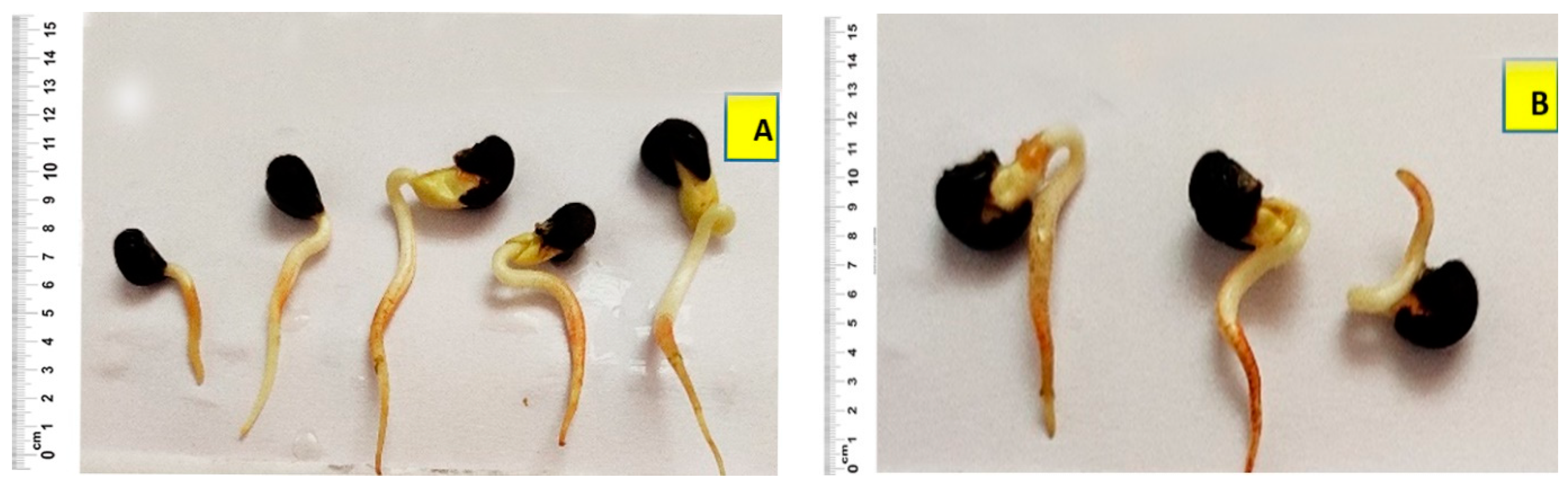
3.1. Seedling Traits among TSA Treatments

Trichostatin A (TSA) treatment suppressed the total field emergence and extremely impaired the root and shoot development among the okra seedlings. Significant reductions with respect to the seedlings’ emergence and total population stand were recorded in all the treatments, especially for those treated with 1.2 µM TSA. Severe root impairment and red pigmented roots were observed among treatments incubated for five days irrespective of the dose of the chemicals, but the intensity of pigmentation found differed (Figure 1). The mean root length and shoot length were found to be significantly reduced in the TSA treatments (Table 2). Control seedlings were normal with respect to root development, and they lacked any pigmentation. A trend of the decreased shoot and root length was observed among the treatments as the dose of the chemical and incubation period increased. However, a slight deviation from this trend was noticed for the treatment T3, wherein unexpected significant increases in the field emergence (51%), mean shoot length (17.25 cm) and mean root length (10.85 cm) were recorded.

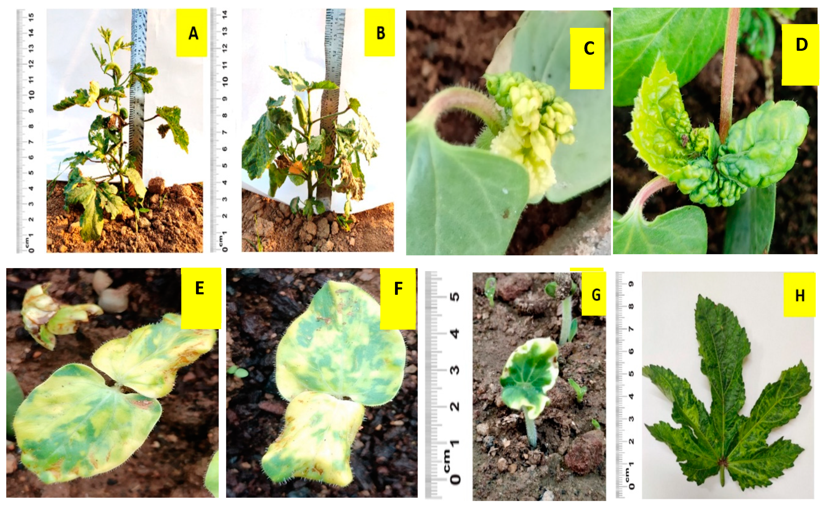
3.2. Plant Stature and Leaf Morphology

The field-transplanted seedlings of all the treatments were dwarfed and exhibited shorter internodes (Figure 2). Apical dominance was found to be repressed as plants developed more axillary shoots and multiple branches combined with a remarkable reduction in the main shoot length. Shoot and leaves developed severe abnormalities, and reductions in total leaf size and number were noticed. Altered leaf morphology with severe crinkling, upward curling and serration of leaves were observed at the higher dose (1.2 µM TSA) (Figure 2). Simultaneously, plants with filamentous leaves with long and narrow blades were noticed among the 0.4 µM TSA treatments. In addition, yellowish patches on the cotyledon leaves and primary leaves were evident in the case of 0.4 µM-TSA-treated seedlings (Figure 2).

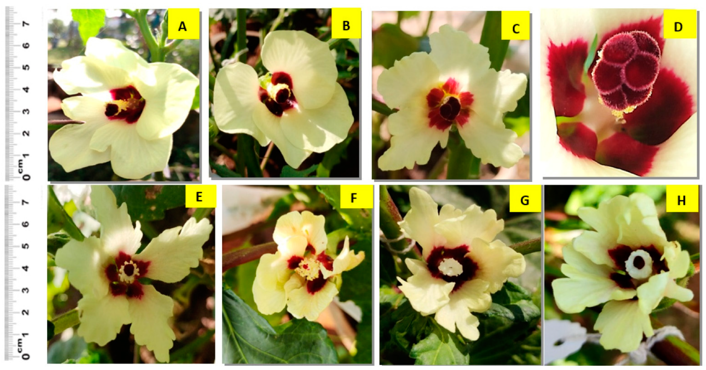
3.3. Flower Morphology and Delay in Flowering

Noticeable differences in flower morphology were evident between different treatments. Variations in the petal shape and size, flowers with fewer or more than five petals and an increased as well as a decreased number of stigmata were observed in most of the treatments. Complete absence of female flower parts, impaired androecium development, defective anther and filament structure, lack/minimal pollen production and complete absence of male organs were common among the TSA treatment with 1.2 µM incubated for five days (Figure 3). On the other hand, though malformations of flowers and sterility of the flower parts were noticed in T1 and T2 treatments compared to control plants, these were comparatively less damaging than in T3 and T4 treatments.
Another important observation among the treatments was regarding the significant delay in the first flowering. The number of days taken to first blooming increased proportionally to the increased dose and incubation period of the treatments (Table 2). The percent increase in days to first flowering was recorded as 18.38 in T4 treatment followed by 13.30% in T2. The treatment T3 exhibited an unusual percent reduction in the number of days for flowering (5.30%) and deviated from the trend. All four treatments took statistically more days to produce the first flower compared to their control plants (flowered on the 49th day).

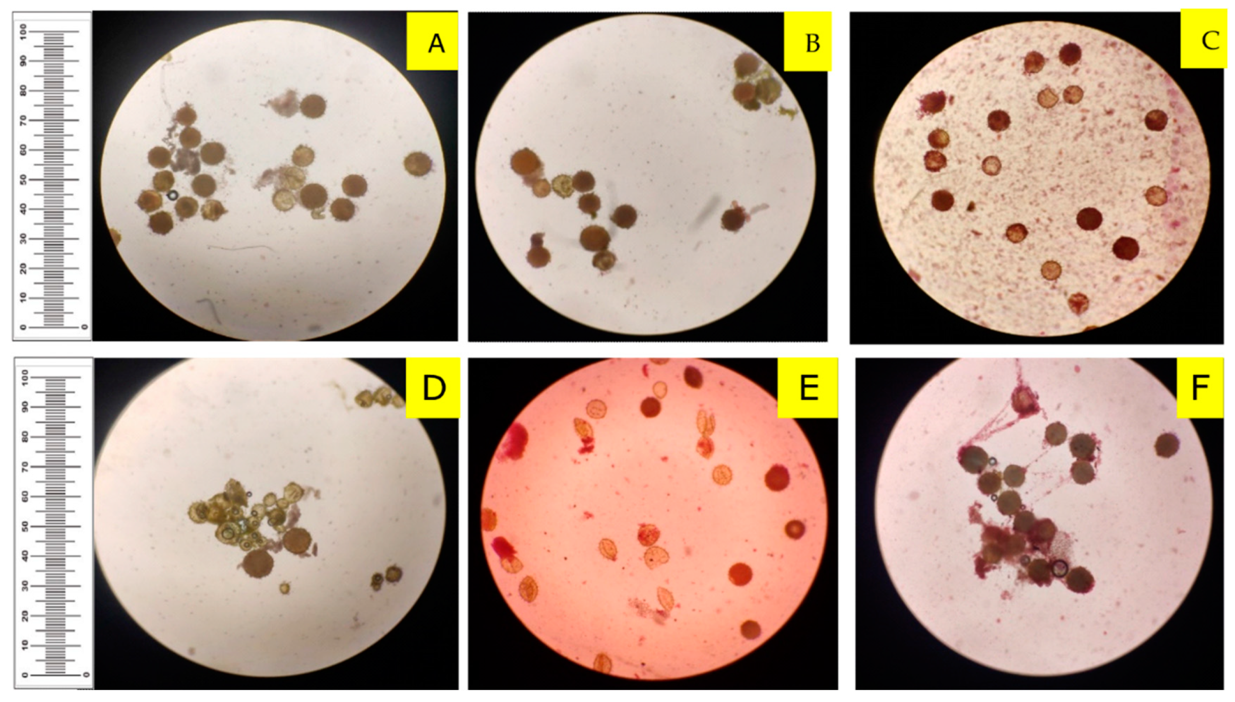
3.4. Pollen Production and Sterility

Pollen morphology was found to be altered after the treatment with TSA. Pollens from non-treated plants were large, spheroidal and had many pores; on the other hand, small, shrunken and mostly long, slender pollens were noticed in the treatment with 1.2 µM TSA incubated for five days (Figure 4).
The total number of pollens produced was found to be lower in all the treatments compared to the control. Number of pollens produced was reduced drastically in treatments T3 and T4, which accounted for more than a 40% reduction (Table 3). Pollen sterility was also observed to be significant in the treatments. The highest mean number of sterile pollens was counted in the extreme dose with prolonged days of incubation (1.2 µM incubated for five days). Comparatively, higher pollen sterility and less pollen production were observed for 1.2 µM TSA treatments than 0.4 µM TSA treatments. Among the 0.4 µM TSA treatments, T1 (incubated for three days) treatment had a lower pollen sterility percentage (25.00%) compared to T2 (incubated for five days) at 27.53%.
Similarly, an increased incubation period among 1.2 µM TSA treatments reported the highest pollen sterility percentage (68.75%) in comparison with the minimal incubated treatment (61.11%). Pollen sterility of the control (1.06%) was also noticed but found negligible. There was a significant reduction in the total number of pollen grains and the number of fertile pollens produced and significantly enhanced pollen sterility in all the TSA treatments compared to the control.

3.5. Fruit and Seed Morphology

Fruits failed to develop in the treatment T4 upon self-pollination, as the sterility of pollen grains was high. The flowers after pollination either withered or the small capsules developed were dropped after three to four days. Considerable sterility of the female and male reproductive parts restricted the crossing of these plants with the control plants. Partial to complete sterility of the plants was noticed in the T4 treatment (Figure 5). Control plants flowered and developed fruit normally. The fruits were light-greenish with a uniform texture and had five ridges and locules. On the other hand, the fruits produced after TSA treatments were shorter and malformed. An increased number of ridges and locules, altered ridge type and shapes and varied shades of green and yellow colour on the fruits were also observed (Figure 5).
Variations concerning the length and weight of the fruits were also visible. The treatment T4 led to seed abortion in few plants or production of minute and malformed seeds. A total reduction in the number of seeds produced per fruit and test weight was seen among treatments, and they also varied in size and shape. Seeds which are extremely small and relatively big with an increased diameter and weight were evident in different treatments. The seeds of control plants were light grey; in contrast, the treated plants exhibited differences in seed colours, including grey, dark grey, brown and black (Figure 5).
The statistical comparison presented in Table 4 indicates that the plant height (cm), number of ridges per fruit, number of locules per fruit, number of seeds per fruit and test weight (g) were influenced by the different doses and incubation period of Trichostatin A treatments. The plant height was significantly reduced with the increase in dose and incubation periods. The highest plant height was recorded in the control group (67.92 cm) followed by T3, T2 and T1 treatments, and the lowest was found in the T1 treatment. The treatment severely affected the fruit and seed formation in the treated plants. A slight but a significant difference in the number of ridges per fruit and number of locules per fruit between the control and treatments were observed, except for the group T3.
The maximum average number of ridges (7.11) and locules (6.88) was noticed in treatment T4, in which most of the okra fruits were malformed. Trichostatin A treatment significantly affected the total number of seeds produced per fruit and weight of 100 seeds in okra. The total number of seeds and test weight of seeds were lower in all treatments in comparison to the control (56.0 and 8.15 g, respectively). Among the treatments, the T3 group produced the highest number of seeds (50.87) and scored the highest test weight of seeds (6.84 g). The least mean number of seeds (37.86) and test weight (3.91 g) was recorded in the T4 group, which was treated with 1.2 µM TSA incubated for five days. A change in the pattern of reduced scores of the phenotypic traits was noticed in the treatment T3 for most of the traits. The plants in the T3 group better performed compared to the other treatments for plant height, number of seeds and test weight.

4. Discussion

HDACs are chemicals with multifaceted effects on a plant’s metabolic processes, mostly connected to the pathways for amino acids, lipids and carbohydrates [20]. One of the most important stages in plant development is germination, which involves several processes associated with the degree of chromatin condensation and reprogramming of gene expression [28]. Roles of histone deacetylases in the normal process of germination and development of roots are already understood in Arabidopsis [21], Medicago [20] and maize [29]. In accordance with previous research findings [13,27,30], the present work revealed that the two different doses and incubation periods of TSA treatments negatively impacted field germination percentage and mean shoot and root lengths of okra seedlings. When examining both the speed and percentage of germination, no significant effect on seed germination was found in a different study employing petunia. These differences suggest the species-specific differences in the structure and physiology of seeds as the Malvaceae species (okra) has seeds with single-layered endosperm covered with layers of testa and tegmen [31], while in the Solanaceae species, (petunia) the embryo is surrounded by three to five thick layers of endosperm cells [32]. The severe root abnormalities and reduced root length noticed in this experiment might account for the reduced plant emergence in the field. TSA treatments are known to increase the total acetylation levels in the roots and shoots, leading to the changed root and shoot cellular patterning, roots with severe developmental abnormalities and induced hair cell development in Arabidopsis seedlings [33]. Higher concentrations of the histone deacetylating agent sodium butyrate inhibited the growth of petunia seedlings, as seen by the shorter seedling length and greater DNA damage [34]. Recently, it was revealed that the use of HDAC inhibitors (such as TSA and NaB) is known to lead to accumulated DNA damage during the early stages of seedling development [20,35] and thus poor plant establishment. Hormones [36] and substances such as NaCl [37] applied exogenously might have a negative impact on the development and growth of the plant root cells. Histone deacetylating agents can interfere with cell division and elongation processes in the meristematic and root elongation zones, leading to malformations in roots [38].
In this study, it was typical to see extremely dwarfed plants with shorter internodes and more axillary shoots. Treatment of Trichostatin A represses the apical dominance and enhances axillary growth in plants. This might account for the development of shorter plants with decreased internodal length and multiple branches. Down-regulation of four rice HDACs (OsHDA704, OsHDA710 and OsHDT703, OsHDA702) using RNAi technology has been reported to produce severe morphological defects [39], such as considerably dwarfed plants with suppressed growth and thin stems. The Arabidopsis mutant plant expressing the antisense AtHD1 also supports the findings of this study. The reduced endogenous AtHD1 transcription resulting in the accumulation of acetylated histones in the mutant plants was associated with various developmental abnormalities, early senescence of the plants, homeotic changes, suppression of apical dominance and induction of axillary branches [40]. On the other hand, the effects of overexpression of HDACs on the plant architecture and development also expound the outcomes of this study. For example, a study focussing on the overexpression of the rice HD OsHDAC1 gene, reported its ability to enhance the overall growth rate among the lines and positively alter the root and shoot development and morphology [41], indicating the prime role of histone deacetylase in governing plant architecture and development.
Plants with multiple fruiting branches and severe deformities of leaves and shoots were observed among the treatments in this study. Chlorosis in the leaves, narrowed leaf blades and deformities such as crinkling and puckering were evident at leaf development stages. The reactivation of certain silent genes governing leaf growth and development is observed in histone deacetylation. The ectopic expression of these genes may reward lethal and unstable phenotypes in the plants during early developmental stages. Arabidopsis mutant plants with enhanced levels of acetylated histones have been reported to repress the expression of certain genes, such as the serrate gene controlling the leaf margin serration [40]. Mutant maize plants developed by HDA108 gene knockout were reported to be correlated with many developmental defects, including a significant reduction in the plant height and alterations in shoot and leaf development combined with internodal shortening. A reduced number of leaves, alterations in leaf morphology, twisting of leaves, leaf knots, leaf blade length reduction and disorganized differentiation of the blade–sheath boundary were evident from the investigation [42]. Studies with ZmHDA101 antisense maize mutants [43] support the origin of the generation of severe developmental and phenotypical abnormalities in transgenic maize plants with altered plant architecture, leaf shapes and sizes. TSA treatment is known to cause altered polarity of leaves and change young leaves from the adaxial side to the abaxial form in Arabidopsis by inhibiting HDAC activity [44]. The development of filamentous leaves having a considerable range of upward curling was prominent, as noticed in our study. An alteration in flag leaf morphology linked with the production of narrow leaves was documented in rice HDAC mutants.
Flowers with a high percentage of ill-formed reproductive parts were prevalent, and the treatments also delayed the flowering onset. The corolla shape and size varied among the treatments, and deformities in the normal development of pistils and stamens were noticeable. A heterochronic shift towards juvenility causes a prolonged vegetative phase and thereby a delay in flowering associated with various flower defects and male and female sterility. Increased levels of tetra-acetylated H4 histone protein resulted in ectopic expression of tissue-specific genes such as SUPERMAN (controls the boundary between carpel and stamen development in flowers) linked with the formation of abnormal flowers lacking sepals and petals or any reproductive organs [40]. Inhibiting histone deacetylation is also known to down-regulate some additional genes (e.g., B-function genes) required for the development of flowers with normal sepals and petals. Association of a MADS-box transcription factor FLOWERING LOCUS C (FLC) acts as a hindrance to the transition from the vegetative to reproductive phase in Arabidopsis. The FLOWERING LOCUS D (FLD), which is a plant homolog of histone deacetylase of mammals, upon a point mutation, contributes to the hyperacetylation of FLC chromatin. It can lead to the up-regulated expression of FLC to cause an extreme delay in flowering [45]. HDAC-inhibited transgenic maize [43] and rice [39] mutants are reported to bloom late, with various inflorescence defects, imperfect differentiation, reduced peduncle elongation and fertility. Genes such as liguleless 2 (lg2), knotted 1 (kn1) and rough sheath 2 (rs2) are found to be impaired, as a result of which the phase transition from vegetative to reproductive was blocked in rice transgenic plants. FVE was found to directly interact with FLOWERING LOCUS C and down-regulate the functions by forming a complex with Arabidopsis HDA6 [46]. Lesions in FVE have resulted in enhanced levels of histone acetylation of the FLC locus, causing the repressed expression of the locus [45]. Therefore, it is evident that HDACs are involved in the regulation of flowering time via forming protein complexes (such as LSD and FVE), which are then recruited to the target genes controlling flower development. This interaction results in deacetylation of target loci such as FLC, leading to the repressed/silenced activity of the genes.
At the same time, overexpression of HDACs in rice (OsHDT1) leading to decreased acetylation levels was observed to generate early flowering in hybrid rice [47], and the reason for this was assumed to be the regulation of key flowering repressors, such as Heading date 1 (Hd1) and OsGigantea 1 (OsG1). Similarly, in an Arabidopsis HDA6 mutant, axe1-5 plants displayed delayed flowering, whereas the axe1-5/flc-3 double mutant was an early-flowering phenotype, indicating the functional role of histone deacetylase in governing flowering time [48]. These research outcomes suggest the importance of histone deacetylases in different plant processes, such as the transition from vegetative growth to the reproductive phase, flowering control and subsequent reproductive organ development.
The TSA-treated okra plants were unable to self-pollinate as they encountered significant pollen sterility, aberrant pollen production and anomalies in the development of the anthers. An hda108 mutation in maize altered microgametogenesis in the anthers and plant reproduction by generating pollens with significant pollen degeneration, which are shrunken in nature [42]. It is known that anther formation, maturation and dehiscence is a multistage, complex process involving various enzymes, such as polygalacturonases (PGs), b-1,4-glucanases, pectin methylesterases and expansins, [49,50]. The proper production and dehiscence of anthers can be hampered by an imbalance between all of these enzymes caused by down-regulating HDACs [42]. Antisense transgenic maize plants with down-regulated HDAC activity generated inflorescence with a limited pollen number, abnormal anther morphology and dehiscence, reduced fertility and lower kernel weight and quality [43]. Similar to the preceding results, the study also revealed the formation of malformed fruits among treated plants, which significantly differed in the number of ridges and locules per fruit. The total number of fertile seeds and the test weight of seeds were also found to be substantially reduced upon TSA treatment. The inevitable role of HDACs in seed formation was confirmed by silencing the expression of AtHD2A, causing abnormal seed development in Arabidopsis [51].

5. Conclusions

The present study in okra using an HDAC inhibitor, Trichostatin A, was found to induce a spectrum of morphological alterations in all the treatments. The treated plants were inferior to the control plants in all the early-growth measures, such as total field emergence, mean shoot and root length. The plant height, leaf, flower and fruit morphology of the control plants were normal, whereas severely dwarfed plants with deformed leaves and flowers were prevalent among TSA treatments. Pollen sterility, deformation of the reproductive whorls of the flower and considerable delay in the commencement of flowering were recorded. Fruits were significantly shorter, with an increased number of ridges and locules and a decrease in the seed number and weight. The experiment clearly indicates that treatments with epigenetic mutagens can lead to the development of phenotypic plasticity in plants. The plants that underwent the T4 treatment performed better among all the treatments with a wide spectrum of morphological changes. Thus, the dose, 1.2 µM incubated for three days, can be recommended for use in other okra varieties for creating epigenetic variations without major deformities. The stability and validation of the induced epigenetic effects in these treatments have to be established through molecular assessment, which is currently in progress.

Author Contributions

Conceptualization, B.M.D., S.S. (Sasipriya Sasikumar) and S.S. (Shankarappa Sridhara); methodology, B.M.D., S.S. (Sasipriya Sasikumar) N.A. and H.B.B.N.; software, B.M.D. and S.S. (Sasipriya Sasikumar); formal analysis, B.M.D., S.S. (Sasipriya Sasikumar), N.A. and H.B.B.N.; investigation, S.S. (Sasipriya Sasikumar); data curation, S.S. (Sasipriya Sasikumar); writing—original draft preparation, S.S. (Sasipriya Sasikumar), B.M.D., N.A., S.S. (Shankarappa Sridhara), A.M.E.-S., S.A., M.A., K.M.E., H.O.E., E.A.M. and H.B.B.N.; writing—review and editing, S.S. (Sasipriya S), S.S. (Shankarappa Sridhara) B.M.D., N.A., A.M.E.-S., S.A., M.A., K.M.E., H.O.E. and E.A.M.; visualization, N.A.; supervision, B.M.D. and S.S. (Shankarappa Sridhara). All authors have read and agreed to the published version of the manuscript.

Funding

The current work was funded by Taif University Researchers Supporting Project number (TURSP—2020/75), Taif University, Taif, Saudi Arabia.

Data Availability Statement

The data presented in this study are available on request from the corresponding author.

Acknowledgments

The authors are thankful to the Director of Research, KSNUAHS Shivamogga, for providing research facilities. The first author is also thankful to ICAR for providing a Junior/Senior Research Fellowship for his Ph.D. studies.

Conflicts of Interest

The authors declare no conflict of interest.

References

  1. Wammanda, D.T.; Kadams, A.M.; Jonah, P.M. Combining ability analysis and heterosis in a diallel cross of okra (Abelmoschus esculentus (L.) Moench). Afr. J. Agric. Res. 2010, 5, 2108–2115. [Google Scholar]
  2. Benchasri, S. Okra (Abelmoschus esculentus (L.) Moench) as a valuable vegetable of the world. Ratarstvo Povrtarstvo 2012, 49, 105–112. [Google Scholar]
  3. Kumar, S.; Yadav, Y.C. Correlation coefficient and path analysis studies in okra [Abelmoschus esculentus (L.) Moench]. Ann. Hortic. 2009, 2, 166–170. [Google Scholar]
  4. Sheikh, M.; Safiuddin, S.; Khan, Z.; Mahmood, I. Effect of Bhendi Yellow Vein Mosaic Virus on Yield Components of Okra Plants. Eur. J. Plant Pathol. 2013, 95, 391–393. [Google Scholar] [CrossRef]
  5. Lengsfeld, C.; Titgemeyer, F.; Faller, G.; Hensel, A. Glycosylated Compounds from Okra Inhibit Adhesion of Helicobacter pylori to Human Gastric Mucosa. J. Agric. Food Chem. 2004, 52, 1495–1503. [Google Scholar] [CrossRef] [PubMed]
  6. Jagajanantham, N.; Dhanavel, D.; Pavadai, P.; Chidambaram, A. Growth and yield parameters using gamma rays in bhendi (Abelmoschus esculentus (L.) Moench) var. Arka Anamika. Int. J. Res. Plant Sci. 2012, 2, 56–58. [Google Scholar]
  7. Kumar, S.; Choudhary, S. Okra Breeding: Recent Approaches and Constraint. Ann. Biol. 2019, 35, 55–60. [Google Scholar]
  8. Sahu, P.P.; Pandey, G.; Sharma, N.; Puranik, S.; Muthamilarasan, M.; Prasad, M. Epigenetic mechanisms of plant stress responses and adaptation. Plant Cell Rep. 2013, 32, 1151–1159. [Google Scholar] [CrossRef]
  9. Cubas, P.; Vincent, C.; Coen, E. An epigenetic mutation responsible for natural variation in floral symmetry. Nature 1999, 401, 157–161. [Google Scholar] [CrossRef]
  10. Weigel, D.; Colot, V. Epialleles in plant evolution. Genome Biol. 2012, 13, 249. [Google Scholar] [CrossRef]
  11. Johannes, F.; Schmitz, R.J. Spontaneous epimutations in plants. New Phytol. 2018, 221, 1253–1259. [Google Scholar] [CrossRef] [PubMed]
  12. Richards, C.L.; Wendel, J.F. The hairy problem of epigenetics in evolution. New Phytol. 2011, 191, 7–9. [Google Scholar] [CrossRef] [PubMed]
  13. Jones, P.A.; Takai, D. The Role of DNA Methylation in Mammalian Epigenetics. Science 2001, 293, 1068–1070. [Google Scholar] [CrossRef] [PubMed]
  14. Bie, X.M.; Dong, L.; Li, X.H.; Wang, H.; Gao, X.-Q.; Li, X.G. Trichostatin A and sodium butyrate promotes plant regeneration in common wheat. Plant Signal. Behav. 2020, 15, 1820681. [Google Scholar] [CrossRef]
  15. Sako, K.; Kim, J.M.; Matsui, A.; Nakamura, K.; Tanaka, M.; Kobayashi, M.; Saito, K.; Nishino, N.; Kusano, N.; Taji, T.; et al. Ky-2, a Histone deacetylase inhibitor, enhances high-salinity stress tolerance in Arabidopsis thaliana. Plant Cell Physiol. 2016, 57, 776–783. [Google Scholar] [CrossRef] [Green Version]
  16. Nguyen, H.M.; Sako, K.; Matsui, A.; Ueda, M.; Tanaka, M.; Ito, A.; Nishino, N.; Yoshida, M.; Seki, M. Transcriptomic analysis of Arabidopsis thaliana plants treated with the Ky-9 and Ky-72 histone deacetylase inhibitors. Plant Signal. Behav. 2018, 13, e1448333. [Google Scholar] [CrossRef]
  17. Lee, M.H.; Lee, J.; Jie, E.Y.; Choi, S.H.; Jiang, L.; Ahn, W.S.; Kim, C.Y.; Kim, S.W. Temporal and Spatial Expression Analysis of Shoot-Regeneration Regulatory Genes during the Adventitious Shoot Formation in Hypocotyl and Cotyledon Explants of Tomato (CV. Micro-Tom). Int. J. Mol. Sci. 2020, 21, 5309. [Google Scholar] [CrossRef]
  18. Li, H.; Soriano, M.; Cordewener, J.; Muino, J.M.; Riksen, T.; Fukuoka, H.; Angenent, G.C.; Boutilier, K. The Histone Deacetylase Inhibitor Trichostatin A Promotes Totipotency in the Male Gametophyte. Plant Cell 2014, 26, 195–209. [Google Scholar] [CrossRef]
  19. Tanaka, M.; Kikuchi, A.; Kamada, H. The Arabidopsis Histone Deacetylases HDA6 and HDA19 Contribute to the Repression of Embryonic Properties after Germination. Plant Physiol. 2007, 146, 149–161. [Google Scholar] [CrossRef]
  20. Pagano, A.; de Sousa Araújo, S.; Macovei, A.; Dondi, D.; Lazzaroni, S.; Balestrazzi, A. Metabolic and gene expression hallmarks of seed germination uncovered by sodium butyrate in Medicago truncatula. Plant Cell Environ. 2019, 42, 259–269. [Google Scholar] [CrossRef]
  21. Uddenberg, D.; Valladares, S.; Abrahamsson, M.; Sundström, J.F.; Sundås-Larsson, A.; Von Arnold, S. Embryogenic potential and expression of embryogenesis-related genes in conifers are affected by treatment with a histone deacetylase inhibitor. Planta 2011, 234, 527–539. [Google Scholar] [CrossRef] [PubMed]
  22. Pandey, P.; Daghma, D.S.; Houben, A.; Kumlehn, J.; Melzer, M.; Rutten, T. Dynamics of post-translationally modified histones during barley pollen embryogenesis in the presence or absence of the epi-drug trichostatin A. Plant Reprod. 2017, 30, 95–105. [Google Scholar] [CrossRef] [PubMed]
  23. Yoshida, M.; Horinouchi, S.; Beppu, T. Trichostatin A and trapoxin: Novel chemical probes for the role of histone acetylation in chromatin structure and function. BioEssays 1995, 17, 423–430. [Google Scholar] [CrossRef] [PubMed]
  24. Yang, F.; Zhang, L.; Li, J.; Huang, J.; Wen, R.; Ma, L.; Zhou, D.; Li, L. Trichostatin A and 5-azacytidine both cause an increase in global histone H4 acetylation and a decrease in global DNA and H3K9 methylation during mitosis in maize. BMC Plant Biol. 2010, 10, 178. [Google Scholar] [CrossRef]
  25. Ma, X.; Lv, S.; Zhang, C.; Yang, C. Histone deacetylases and their functions in plants. Plant Cell Rep. 2013, 32, 465–478. [Google Scholar] [CrossRef]
  26. Perales, M.; Más, P. A Functional Link between Rhythmic Changes in Chromatin Structure and the Arabidopsis Biological Clock. Plant Cell 2007, 19, 2111–2123. [Google Scholar] [CrossRef]
  27. Lochmanová, G.; Ihnatová, I.; Kuchaříková, H.; Brabencová, S.; Zachová, D.; Fajkus, J.; Zdráhal, Z.; Fojtová, M. Different modes of action of genetic and chemical downregulation of histone deacetylases with respect to plant development and histone modifications. Int. J. Mol. Sci. 2019, 20, 5093. [Google Scholar] [CrossRef]
  28. Macovei, A.; Pagano, A.; Leonetti, P.; Carbonera, D.; Balestrazzi, A.; Araújo, S.S. Systems biology and genome-wide approaches to unveil the molecular players involved in the pre-germinative metabolism: Implications on seed technology traits. Plant Cell Rep. 2016, 36, 669–688. [Google Scholar] [CrossRef]
  29. Hou, H.; Wang, P.; Zhang, H.; Wen, H.; Gao, F.; Ma, N.; Wang, Q.; Li, L. Histone Acetylation Is Involved in Gibberellin-Regulated sodCp Gene Expression in Maize Aleurone Layers. Plant Cell Physiol. 2015, 56, 2139–2149. [Google Scholar] [CrossRef]
  30. Colville, A.; Alhattab, R.; Hu, M.; Labbé, H.; Xing, T.; Miki, B. Role of HD2 genes in seed germination and early seedling growth in Arabidopsis. Plant Cell Rep. 2011, 30, 1969–1979. [Google Scholar]
  31. Singh, R.P.; Chauhan, M.P.S. Seed Structure and Systematic Position of Hampea nutricia (Malvaceae). Plant Syst. Evol. 1984, 147, 55–61. [Google Scholar] [CrossRef]
  32. Steinbrecher, T.; Leubner-Metzger, G. The biomechanics of seed germination. J. Exp. Bot. 2017, 68, 765–783. [Google Scholar] [CrossRef] [PubMed]
  33. Xu, C.-R.; Liu, C.; Wang, Y.-L.; Li, L.-C.; Chen, W.-Q.; Xu, Z.-H.; Bai, S.-N. Histone acetylation affects expression of cellular patterning genes in the Arabidopsis root epidermis. Proc. Natl. Acad. Sci. USA 2005, 102, 14469–14474. [Google Scholar] [CrossRef] [PubMed]
  34. Pagano, A.; Andolina, A.; Sabatini, M.E.; Araújo, S.S.; Balestrazzi, A.; Macovei, A. Sodium butyrate induces genotoxic stress in function of photoperiod variations and differentially modulates the expression of genes involved in chromatin modification and DNA repair in Petunia hybrida seedlings. Planta 2020, 251, 102. [Google Scholar] [CrossRef]
  35. Pagano, A.; Araújo, S.; Macovei, A.; Leonetti, P.; Balestrazzi, A. The Seed Repair Response during Germination: Disclosing Correlations between DNA Repair, Antioxidant Response, and Chromatin Remodeling in Medicago truncatula. Front. Plant Sci. 2017, 8, 1972. [Google Scholar] [CrossRef]
  36. Benková, E.; Hejátko, J. Hormone interactions at the root apical meristem. Plant Mol. Biol. 2008, 69, 383–396. [Google Scholar] [CrossRef]
  37. West, G.; Inzé, D.; Beemster, G.T. Cell Cycle Modulation in the Response of the Primary Root of Arabidopsis to Salt Stress. Plant Physiol. 2004, 135, 1050–1058. [Google Scholar] [CrossRef]
  38. Zhang, Q.; Wang, P.; Hou, H.; Zhang, H.; Tan, J.; Huang, Y.; Li, Y.; Wu, J.; Qiu, Z.; Li, L. Histone acetylation and reactive oxygen species are involved in the preprophase arrest induced by sodium butyrate in maize roots. Protoplasma 2016, 254, 167–179. [Google Scholar] [CrossRef]
  39. Hu, Y.; Qin, F.; Huang, L.; Sun, Q.; Li, C.; Zhao, Y.; Zhou, D.-X. Rice histone deacetylase genes display specific expression patterns and developmental functions. Biochem. Biophys. Res. Commun. 2009, 388, 266–271. [Google Scholar] [CrossRef]
  40. Tian, L.; Chen, Z.J. Blocking histone deacetylation in Arabidopsis induces pleiotropic effects on plant gene regulation and development. Proc. Natl. Acad. Sci. USA 2000, 98, 200–205. [Google Scholar] [CrossRef]
  41. Jang, I.-C.; Pahk, Y.-M.; Song, S.I.; Kwon, H.J.; Nahm, B.H.; Kim, J.-K. Structure and expression of the rice class-I type histone deacetylase genes OsHDAC1-3: OsHDAC1 overexpression in transgenic plants leads to increased growth rate and altered architecture. Plant J. 2003, 33, 531–541. [Google Scholar] [CrossRef] [PubMed]
  42. Forestan, C.; Farinati, S.; Rouster, J.; Lassagne, H.; Lauria, M.; Ferro, N.D.; Varotto, S. Control of Maize Vegetative and Reproductive Development, Fertility, and rRNAs Silencing by HISTONE DEACETYLASE 108. Genetics 2018, 208, 1443–1466. [Google Scholar] [CrossRef] [PubMed]
  43. Rossi, V.; Locatelli, S.; Varotto, S.; Donn, G.; Pirona, R.; Henderson, D.A.; Hartings, H.; Motto, M. Maize Histone Deacetylase hda101 Is Involved in Plant Development, Gene Transcription, and Sequence-Specific Modulation of Histone Modification of Genes and Repeats. Plant Cell 2007, 19, 1145–1162. [Google Scholar] [CrossRef] [PubMed]
  44. Ueno, Y.; Ishikawa, T.; Watanabe, K.; Terakura, S.; Iwakawa, H.; Okada, K.; Machida, C.; Machida, Y. Histone Deacetylases and ASYMMETRIC LEAVES2 Are Involved in the Establishment of Polarity in Leaves of Arabidopsis. Plant Cell 2007, 19, 445–457. [Google Scholar] [CrossRef]
  45. He, Y.; Michaels, S.D.; Amasino, R.M. Regulation of Flowering Time by Histone Acetylation in Arabidopsis. Science 2003, 302, 1751–1754. [Google Scholar] [CrossRef]
  46. Gu, X.; Jiang, D.; Yang, W.; Jacob, Y.; Michaels, S.D.; He, Y. Arabidopsis homologs of retinoblastoma-associated protein 46/48 associate with a histone deacetylase to act redundantly in chromatin silencing. PLoS Genet. 2011, 7, e1002366. [Google Scholar] [CrossRef] [Green Version]
  47. Li, C.; Huang, L.; Xu, C.; Zhao, Y.; Zhou, D.-X. Altered Levels of Histone Deacetylase OsHDT1 Affect Differential Gene Expression Patterns in Hybrid Rice. PLoS ONE 2011, 6, e21789. [Google Scholar] [CrossRef]
  48. Yu, C.-W.; Liu, X.; Luo, M.; Chen, C.; Lin, X.; Tian, G.; Lu, Q.; Cui, Y.; Wu, K. HISTONE DEACETYLASE6 Interacts with FLOWERING LOCUS D and Regulates Flowering in Arabidopsis. Plant Physiol. 2011, 156, 173–184. [Google Scholar] [CrossRef]
  49. Futamura, N.; Mori, H.; Kouchi, H.; Shinohara, K. Male Flower-Specific Expression of Genes for Polygalacturonase, Pectin Methylesterase and -1,3-Glucanase in a Dioecious Willow (Salix gilgiana Seemen). Plant Cell Physiol. 2000, 41, 16–26. [Google Scholar] [CrossRef]
  50. Wilson, Z.A.; Song, J.; Taylor, B.; Yang, C. The final split: The regulation of anther dehiscence. J. Exp. Bot. 2011, 62, 1633–1649. [Google Scholar] [CrossRef]
  51. Wu, C.-T.; Morris, J.R. Genes, Genetics, and Epigenetics: A Correspondence. Science 2001, 293, 1103–1105. [Google Scholar] [CrossRef] [PubMed] [Green Version]
Figure 1. Root impairment and pigmentation for 0.4 µM TSA (A) and 1.2 µM TSA (B) incubated for five days, respectively.
Agronomy 12 02247 g001
Figure 2. Plant stature and leaf morphology of TSA treatments. Dwarf plants with shorter internodes and multiple branches (A,B), severe leaf crinkling of younger leaves (C,D), yellow patches on cotyledon leaves (EG), crinkling and yellow patches on mature leaves (H).
Agronomy 12 02247 g002
Figure 3. Flower morphology and sterility of TSA treatments. Alteration of petal numbers and shapes (AC), increased number of stigmata (D), Sterility of reproductive parts (EH).
Agronomy 12 02247 g003
Figure 4. Pollen sterility of TSA treatments: 0.4 µM TSA incubated for three and five days, respectively (A,B), 1.2 µM TSA incubated for three days (C), 1.2 µM TSA incubated for five days (D,E) and control (F).
Agronomy 12 02247 g004
Figure 5. Fruit and seed morphology of TSA treatments. Failure of fruit setting (A,B), deformed fruits with yellow patches (C), increased number of ridges and locules in the fruits (D) and colour and size differences in the seeds (E,F).
Agronomy 12 02247 g005
Table 1. Trichostatin A (TSA) treatment details.
TreatmentDescription of the Treatments
T1 (Control)Control: pre-soaking and incubation in DMSO
T2Pre-soaking for 1 h followed by 3 days of incubation in 0.4 µM TSA.
T3Pre-soaking for 1 h followed by 5 days of incubation in 0.4 µM TSA.
T4Pre-soaking for 1 h followed by 3 days of incubation in 1.2 µM TSA.
T5Pre-soaking for 1 h followed by 5 days of incubation in 1.2 µM TSA.
Table 2. Early-growth parameters of TSA treatments.
TreatmentsFG (%)Mean Root Length (cm)Mean Shoot Length (cm)DFFs% Increase in DFFs
Control8812.7719.1549.220.00
T1619.5113.2351.584.79
T2438.1210.0655.7713.30
T35110.8517.2551.835.30
T4375.667.0258.2718.38
CD (p = 0.05)4.901.323.051.11
FG: field germination, DFFs: days to first flowering.
Table 3. Pollen production and sterility among TSA treatments.
TreatmentsAverage Number of Pollens/Microscopic Slide% Decrease in Total Pollen ProductionAverage Number of Fertile PollensAverage Number of Sterile PollensAverage Pollen Sterility (%)
T18014.89602025.00
T26926.59401927.53
T35442.55332161.11
T44848.93153368.75
Control940.009311.06
CD2.52-1.480.870.08
Table 4. Statistical analysis of agronomic characters in okra treated with TSA.
TreatmentPHNRNLNSTWT
T150.505.455.3631.555.74
T239.386.115.7045.774.77
T355.915.405.1150.876.84
T449.337.116.8837.863.91
Control767.925.005.0056.08.15
CD (p = 0.05)8.502.041.773.761.31
PH: plant height, NR: number of ridges per fruit, NL: number of locules per fruit, NS: number of seeds per fruit, TWT: test weight, CD at 5% level of significance.
Publisher’s Note: MDPI stays neutral with regard to jurisdictional claims in published maps and institutional affiliations.

