Article

Morphological, Cytological, and Molecular Comparison between Diploid and Induced Autotetraploids of Callisia fragrans (Lindl.) Woodson

by
Kateřina Beranová
1,†,
Rohit Bharati
1,†,
Jana Žiarovská
2,
Jana Bilčíková
3,
Kateřina Hamouzová
4,
Miroslav Klíma
5 and
Eloy Fernández-Cusimamani
1,*
1
Faculty of Tropical AgriSciences, Czech University of Life Sciences Prague, Kamýcká 129, Prague 6, 16500 Prague, Czech Republic
2
Faculty of Agrobiology and Food Resources, Slovak University of Agriculture in Nitra, Tr. A. Hlinku 2, 94976 Nitra, Slovakia
3
Research Centre AgroBioTech, Slovak University of Agriculture in Nitra, Tr. A. Hlinku 2, 94976 Nitra, Slovakia
4
Faculty of Agrobiology, Food and Natural Resources, Czech University of Life Sciences Prague, Kamýcká 129, Prague 6, 16500 Prague, Czech Republic
5
Division of Crop Genetics and Breeding, Crop Research Institute, Drnovská 507/73, Praha 6, 16106 Prague, Czech Republic
*
Author to whom correspondence should be addressed.
These authors contributed equally to this work.
Agronomy 2022, 12(10), 2520; https://doi.org/10.3390/agronomy12102520
Submission received: 30 August 2022 / Revised: 11 October 2022 / Accepted: 13 October 2022 / Published: 15 October 2022
(This article belongs to the Section Crop Breeding and Genetics)

Abstract

:
The objective of the current study was to assess the efficiency of oryzalin in inducing polyploids in Callisia fragrans (Lindl.) Woodson by in vitro polyploidization. Shoot tips were subjected to Murashige and Skoog (MS) medium containing oryzalin at concentrations 1, 5, and 10 μM for 4 and 8 weeks. Further, the ploidy levels of the plants were confirmed using flow cytometry and chromosome counting. Among all treatments, six tetraploid plants (2n = 4x = 24) were obtained after 8 weeks in MS medium containing 5 μM oryzalin. Upon ex vitro transfer, tetraploid plants were morphologically distinct compared to diploid plants. The size of the leaf and flower increased significantly and nearly doubled when compared to the mother diploid plant. Further, inductively coupled plasma–optical emission spectrometry showed that tetraploid plants exhibited significantly higher sodium, iron, and calcium content, and the potassium content was increased by 100%. Molecular analysis utilizing iPBS and CDDP markers was tested for the first time in C. fragrans to assess the variation between tetraploid and diploid genotypes. Both the markers generated three major clusters, indicating a clear distinction between diploid, tetraploid, and the mixoploid genotypes. In conclusion, in vitro polyploidization using oryzalin could effectively induce polyploids in this and related species. Additionally, the results obtained in this study will provide a basis for future breeding opportunities in this species.

1. Introduction

Callisia fragrans (Lindl.) Woodson (basket plant; 2n = 2x = 12) is a perennial plant belonging to the Commelinaceae family along with other 500 species. This monocotyledonous plant originated in Mexico and is now widely used across the globe for its medicinal and ornamental properties [1,2,3]. In folk medicine, it is used to treat various conditions such as joint disorders, inflammation, burns, wounds, etc. [3,4]. For instance, leaf extract infused with ethanol is often used to improve immunity and treat joint disorders [4]. Additionally, the ethanolic extract of C. fragrans has also recently been reported to possess anti-inflammatory and antioxidant activities [5]. Moreover, a study concluded that C. fragrans contains various bioactive components, such as phenolic compounds, amino acids, carbohydrates, flavonoids, coumarins, vitamins, etc. [1]. Studies suggest that these compounds are responsible for the treatment of illnesses such as oncological diseases, cardiovascular problems, tuberculosis, asthma, and gastrointestinal disorders, and the healing of burns and wounds [1,3,6,7].
Medicinal plant breeding focuses on obtaining new genotypes with higher and novel content of secondary metabolites. Several methods are used for plant breeding and genetic improvements such as hybridization, heterogenic breeding, mutation induction, polyploid induction, and genetic engineering. One of the most common methods used for breeding medicinal plants is in vitro somatic polyploidization. It is a fast, reliable, efficient, and cheap method of producing novel genotypes with superior agronomical traits. This method is based on the manipulation of somatic chromosomes induced by a wide range of antimitotic agents [8,9,10]. The most commonly utilized antimitotic agent in polyploid induction is colchicine [11,12], although colchicine has various side effects, such as sterility, and toxicity [13]. Hence, recently oryzalin has become an alternative for its effectiveness and fewer side effects [13,14,15]. Polyploidization (chromosome doubling) often causes useful changes such as higher biomass, a higher amount of bioactive compounds, and better environmental (stress) adaptability [9,13,16]. For example, polyploidization increased essential oil content in Thymus vulgaris L. and Chamaemelum nobile All. [14,17]. Similarly, a higher phenolic acid content (cichoric acid, caffeic acid, chlorogenic acid, caftaric acid, and 1,5-dicaffeoyl quinic acid) in tetraploid Echinacea purpurea (L.) Moench [18], total phenolic and flavonoid content in Salvia officinalis L. [19], and increased levels of carotenoid content in Physsalis alkekengi L. [20] and Zingiber officinale Roscoe were observed after chromosome doubling [21].
To date, a few breeding attempts have been made to develop new genotypes in the Commelinaceae family. For instance, studies have utilized gamma irradiations and colchicine to obtain novel genotypes in Commelina benghalensis L. [22,23]. Particularly in C. fragrans, few studies have tested colchicine, caffeine, and hydroquinone for their efficacy in producing novel genotypes. However, oryzalin has never been tested for its efficiency in inducing polyploids in this species or plants from this family. Moreover, an in vitro approach to polyploidization has also not been tested in the species of study. Hence, the aim of this study was to obtain autopolyploid plants from diploid plants (2n = 2x = 12) via in vitro polyploidization using oryzalin as an antimitotic agent and, additionally, to develop an optimized protocol for in vitro polyploidization in this and related species. In newly obtained genotypes, various novel morphological, cytological, and molecular variances could be observed.

2. Materials and Methods

2.1. Plant Material Acquisition, Surface Sterilization, and Culture Establishment

Callisia fragrans diploid plants (2n = 2x = 12) were grown in greenhouse conditions (average temperature 22.5 °C; relative air humidity 70–80%) in plastic containers (5 × 5 cm). After 4 weeks of cultivation, the stolons started to appear. At that development stage, plants were transplanted into larger pots (15 cm in diameter). Over the next eight weeks of cultivation, the stolons grew up to 60 cm in length. After eight weeks, nodal segments were collected and subjected to surface sterilization. Firstly, nodal segments were rinsed with running tap water for 10 min followed by treatment with 70% ethanol (v/v) for 2 min. Further, nodal segments were immersed in a solution containing 1% sodium hypochlorite (v/v) and two drops of Tween 20 for 10 min. Finally, the nodal segments were rinsed three times using autoclaved double distilled water. A rotatory shaker was used for all the rinses. The sterile nodal segments were then introduced into in vitro conditions and then propagated on MS medium [24] supplemented with mg*L−1 6-benzylaminopurine (BAP) and 0.1 mg*L−1 indole-3-acetic acid (NAA) in Erlenmeyer flasks. Explants were cultivated under controlled conditions (day/night temperature 25 °C/20 °C and day/night photoperiod 16 h/8 h with the light intensity of 57.5 μmoL/s/m2).

2.2. Polyploidy Induction
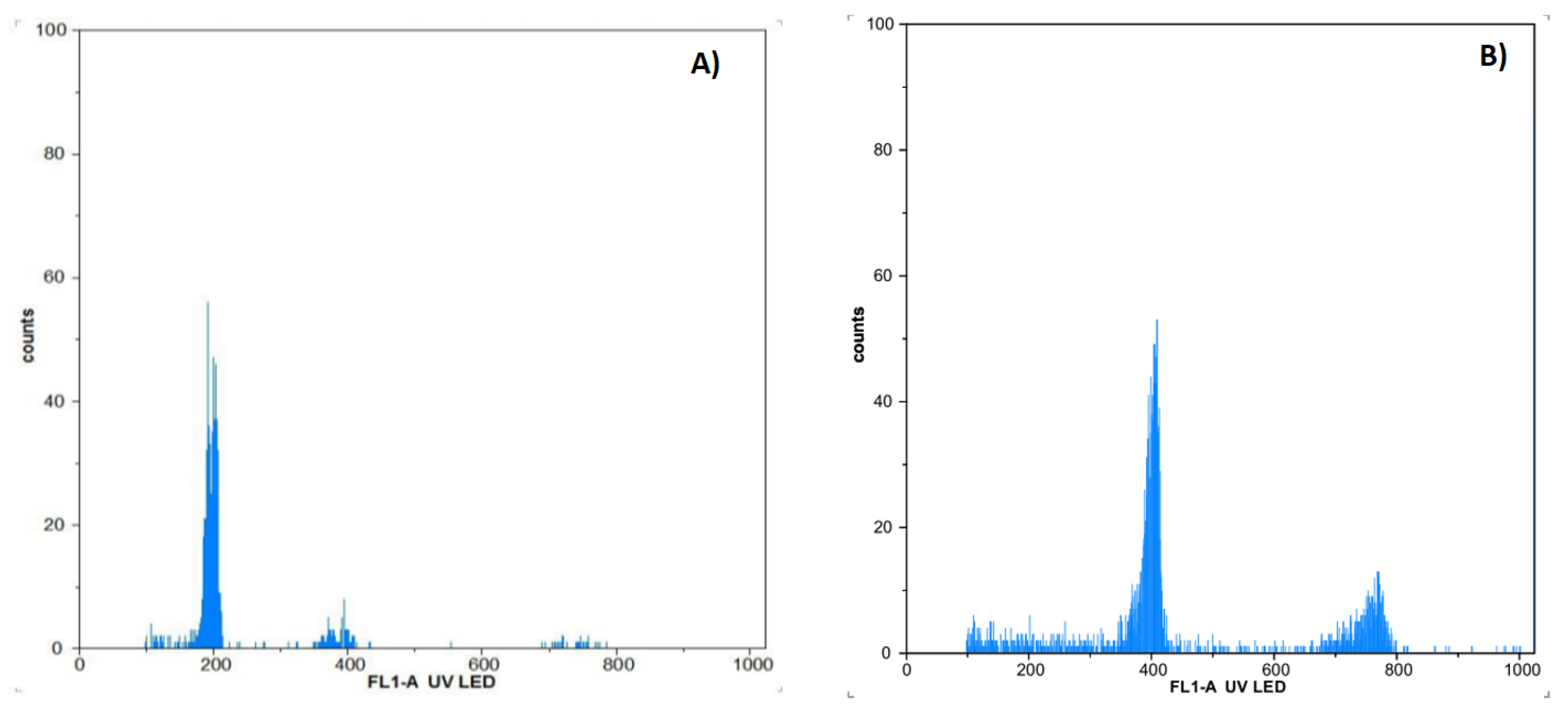
A total of 180 plants (five plants per flask) were exposed to three oryzalin (Sigma-Aldrich, St. Louis, MO, USA) concentrations (1, 5, and 10 µM) and two treatment durations (4 and 8 weeks) on MS media. Thirty plants were used for each variant (concentration × treatment time). Upon the treatment, plants were removed from the oryzalin-containing media and washed with sterile double distilled water under a laminar flow box, and then transferred to oryzalin-free MS media for further propagation. These treated plants were maintained on oryzalin-free MS media for 60 days before ploidy level determination.

2.3. Flow Cytometry

Fresh leaf samples of approx. 1 cm2 were used for the ploidy level determination according to the protocol previously reported [25]. The sample was placed in a Petri dish and chopped with a sharp razor blade by adding 1 mL of Otto I buffer (0.5% Tween 20 (v/v), 0.1 M C6H8O7) into small fragments, creating a suspension of disrupted leaf tissues. The suspension was then filtered through a nylon filter (50 µm). Further, 1 mL Otto II buffer + 4′,6-diamidino-2-phenylindole solution (DAPI, 2 μg/mL, (w/v)) was added to the suspension. The prepared solution was then run through a Partec PAS flow cytometer (Partec, Münster, Germany) and a histogram of the relative DNA content was generated using the Flomax software package (Partec, Nürnberg, Germany) (Version 2.3). The control mother plant was used as the control.

2.4. Plant Transfer into Ex Vitro Conditions

Treated and control plants were propagated on MS media. Plants with well-developed root systems were removed from flasks, and washed under running lukewarm tap water to remove residues of media, and transferred into pots (9 × 9 cm) filled with Agro profi RS 1 modified peat substrate (composition: 70% white peat (w/w); 30% black peat (w/w); 20 kg/m3 bentonite; 1.1 kg/m3 N, P, K (14%, 16%, 18%) (w/w); 150 g/m3 Micromax Premium ('s-Gravenzande, Netherlands)) with the addition of Osmocote Exact standard fertilizer (3 kg/m3, gradual release 8–9 months). The potted plants were covered with plastic foil for one week to ensure sufficient moisture. The plants were then grown under greenhouse conditions (an average temperature of 22.5 °C and natural light). Once a week, the morphological parameters were recorded.

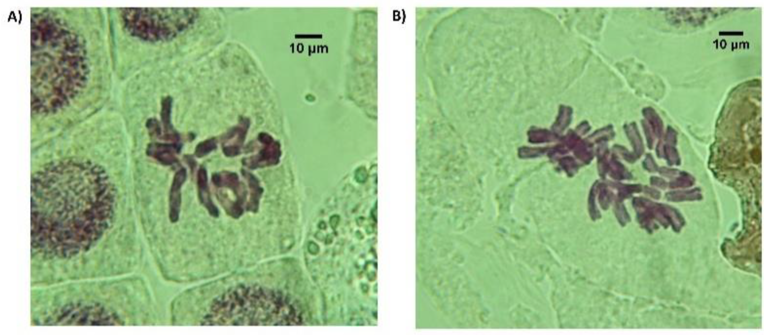
2.5. Chromosome Counting

Fresh root tips (1 cm approx.) of diploid and tetraploid plants were taken from 7–8 o’clock in the morning and kept submerged in a saturated solution of paradichlorobenzene for 3 h. Thereafter, the root tips were washed 3 times using distilled water and immediately put into a freshly prepared solution of ethanol (96%) and acetic acid (99%) (3:1) for 1 h at room temperature. The root tips were rinsed again three times using distilled water. For hydrolysis and staining, the tips were incubated in 1N HCl at 60 °C for 15 min followed by washing three times and staining using Schiff reagent for 1 hour. Roots tips were removed from the Schiff reagent and washed again. The tips of the roots were dissected (approx. 0.2 cm) and placed on a glass slide, then a drop of 2% orcein-acetic was dropped onto the root tip and visualized under a BX51 Olympus light microscope (Olympus Optical Co., Tokyo, Japan) at 100× magnification [26].

2.6. DNA Extraction

Total genomic DNA of all the genotypes was extracted from the young leaves of the plants. Two plants were used to obtain young leaves for DNA extraction. A mixture of leaf tissue was prepared from 10 randomly chosen discs with 1 cm diameter, obtained from at least 5 leaves. The surface of the discs was disinfected slightly with a 20% Savo solution (commercial bleach) (1% NaClO) (v/v). DNA was extracted by a GeneJET plant DNA purification kit (ThermoScientific, Waltham, MA, USA) according to the instructions of the manufacturer. The quantity and quality of extracted DNA were measured by a NanoDrop P500 (Implen GmbH, München, Germany).

2.7. iPBS Analysis

Six different primers previously reported for inter-primer binding site polymorphism analysis (iPBS) were firstly tested for C. fragrans fingerprints [27]. PCRs were prepared by EliZyme Robust HS (Elizabeth Pharmacon, Brno, Czech Republic) together with 800 nM of iPBS primer and 50 ng of DNA in each reaction. PCR conditions of the iPBS were as follows: 95 °C for 5 min; 45 cycles of 95 °C for 1 min, 55 °C for 1 min, 72 °C for 2 min; and 72 °C for 10 min. The PCRs were repeated twice.

2.8. CDDP Analysis

The conserved DNA-derived polymorphism (CDDP) method was used to analyze the variability of C. fragrans control plants with the polyploid genotypes generated utilizing five previously reported primer combinations (WRKY-F + WRKY-R1, WRKY-F + WRKY-R2a, WRKY-F + WRKY-R2b, WRKY-F + WRKY-R3, WRKY-F + WRKY-R3b) [28]. PCRs were prepared by EliZyme Robust HS (Elizabeth Pharmacon) together with 400 nM of each primer and 50 ng of DNA in each reaction. Time and temperature profiling of the PCRs was as follow: 95 °C for 5 min; 40 cycles of 95 °C for 45 s, 54 °C for 45 s, 72 °C for 90 s; and 72 °C for 10 min. The PCRs were repeated twice.

2.9. DNA Fingerprinting

The obtained iPBS and CDDP fingerprint data from 2% agarose electrophoresis (w/v) were analyzed, and the generated profiles were converted into 0–1 binary matrices. The unweighted pair group method with arithmetic mean (UPGMA) analysis was performed, and the Jaccard coefficient of genetic similarity (1945) was used for dendrogram construction. The dendrograms were prepared in the free DendroUPGMA software (http://genomes.urv.cat/UPGMA/ (accessed on 20 July 2022)).

2.10. Mineralization and ICP-OES Elemental Analysis of Micro- and Macronutrients

Approximately 0.25 g of each sample was weighed on an ABT-120/5DW analytical balance (Kern & Sohn, Balingen, Germany) and transferred to polytetrafluoroethylene (PTFE) mineralization tubes. Mineralization was performed with pressure microwave digestion on EthosOne (Milestone, Sorisole, Italy) in 5 mL concentrated nitric acid 69% (v/v) and 1 mL of 30% hydrogen peroxide with 2 mL of ddH2O. The obtained digestate was filtered through Filtrak 390 filter paper. Elemental analysis was performed on an Agilent 720 ICP-OES spectrometer (Agilent Technologies Inc., Santa Clara, CA, USA) with axial plasma configuration and with an SPS-3 autosampler (Agilent Technologies Inc., CA, USA). Three replications of each genotype were carried out.

2.11. Statistical Analysis

Statistical analysis of the data on growth parameters obtained was performed using the statistical software Statistica® (version 13.6). The Kruskal–Wallis test was used to compare the differences between treatments. For the test, differences were considered as significant at p < 0.05.

3. Results

3.1. Chromosome Doubling

A total of 180 plants were exposed to oryzalin at three concentrations and two treatment durations. The survival rate was 100% in all treatments tested. During cultivation on media with oryzalin, the plants generated offshoots (on average 1.7 per plant). In total, 306 plants were regenerated from all treatments. All treated plants were tested by flow cytometry (Figure 1). In total, six autotetraploid and two mixoploid plants were obtained from diploid mother plants across all the treatments. All autotetraploid genotypes were obtained from treatment of 5 µM oryzalin for 8 weeks, and mixoploid plants were obtained from treatment of oryzalin at 5 µM for 4 weeks and 10 µM for 8 weeks. To validate the results from flow cytometry, direct chromosome counting was performed. It was confirmed that the diploid plantlets were 2n = 2x = 12, and after successful polyploid induction, autotetraploid plantlets had 2n = 4x = 24 chromosomes in the somatic cells (Figure 2A,B). Results are summarized in Table 1. The newly obtained six tetraploid genotypes were marked as P1–P6 and two mixoploids as M1–M2. Genotype P6 was not viable, exhibited slower growth, and was unable to create offshoots; hence, it was excluded from the study. All genotypes were transferred into ex vitro conditions with a 100% survival rate.

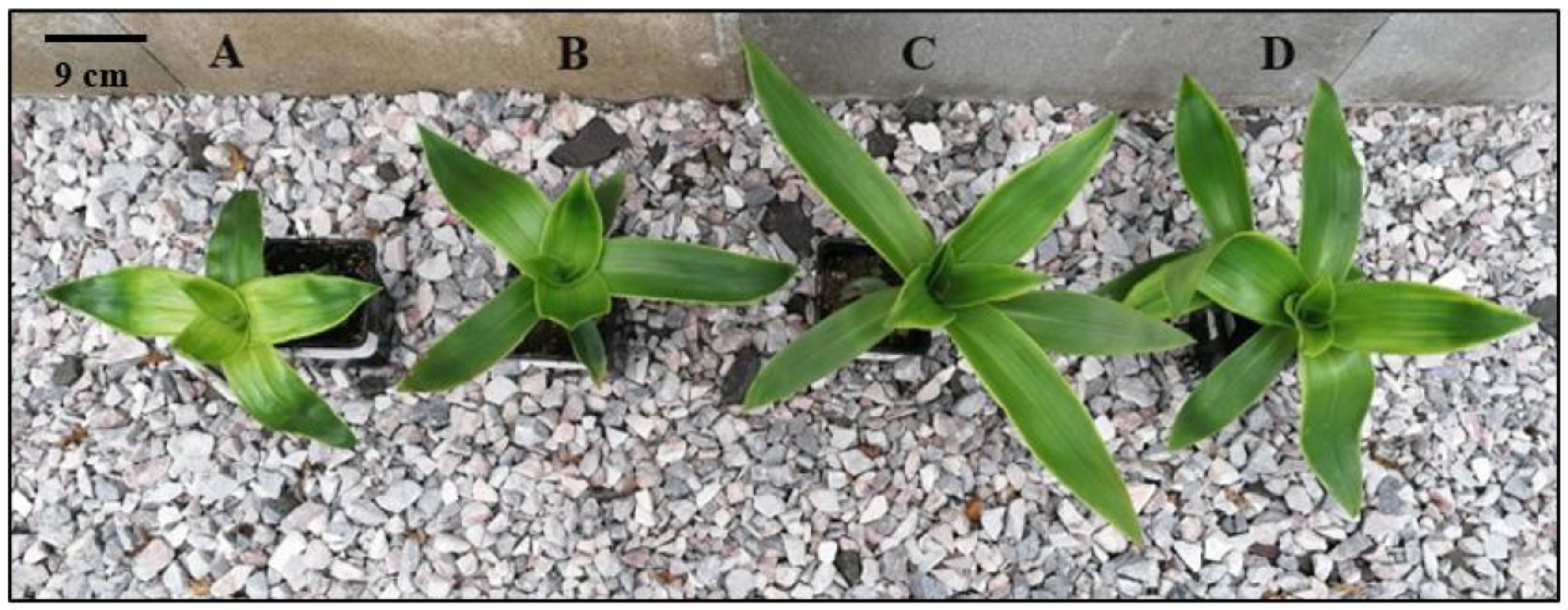
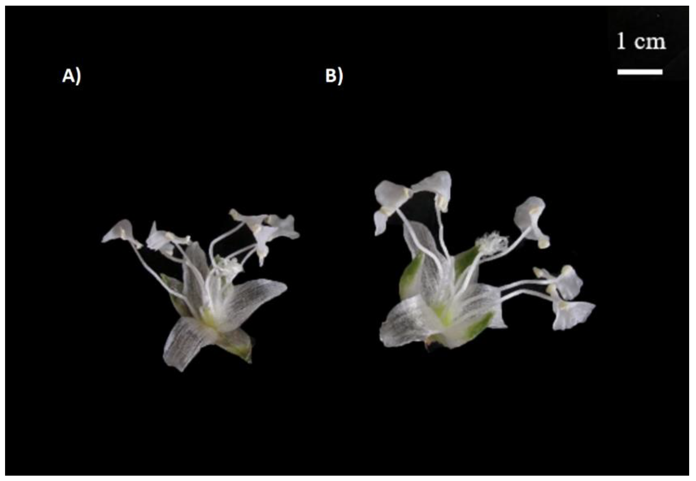
3.2. Morphological Differences between Diploid and the Autotetraploid Genotypes

Polyploid plants exhibited notable morphological differences in comparison to the control genotype. The evaluation of growth variability is summarized in Table 2 and Table 3. Significantly reduced numbers of leaves were detected in P3, P4, M1, and M2 genotypes when compared to the diploid genotype. However, the average leaf length of all polyploid genotypes (P1–P4, M1, M2) increased significantly (5 cm on average), except genotype P5 which exhibited significantly shorter leaves (22.35 ± 0.94 cm) among all the genotypes.
With respect to the height of the plants, the autotetraploid plants did not exhibit any significant difference from the diploid plants although the mixoploids grew shorter and were characterized by more compact growth compared to control plants (46 ± 7.76 and 46 ± 7.75 cm). Similarly, polyploid genotypes did not differ in the average number of internodes except for P4 and mixoploids (M1 and M2) for which it was less than that of diploid plants (Table 2, Figure 3).
All the induced genotypes produced shorter stolons compared to the control genotype. The longest and shortest stolons were observed in the control (76.1 ± 10.04 a) and P5 genotypes, respectively (Table 2). However, the average number of stolons per plant remained unchanged across all the genotypes as no statistically significant differences were observed.
The polyploid plants were characterized by a larger flower size. For example, tetraploid P3, when compared with diploids, showed a significant increase in diameter (66.88%). The P3 genotype grew longer and wider flower petals (Figure 4, Table 3). However, the longest flower petals were measured in the P1 genotype (6.045 ± 1.01 mm). Genotypes P2, M1, and M2 did not bloom at all. Interestingly, the control genotype, P1, and P3 started flowering two weeks earlier than P4 and P5.

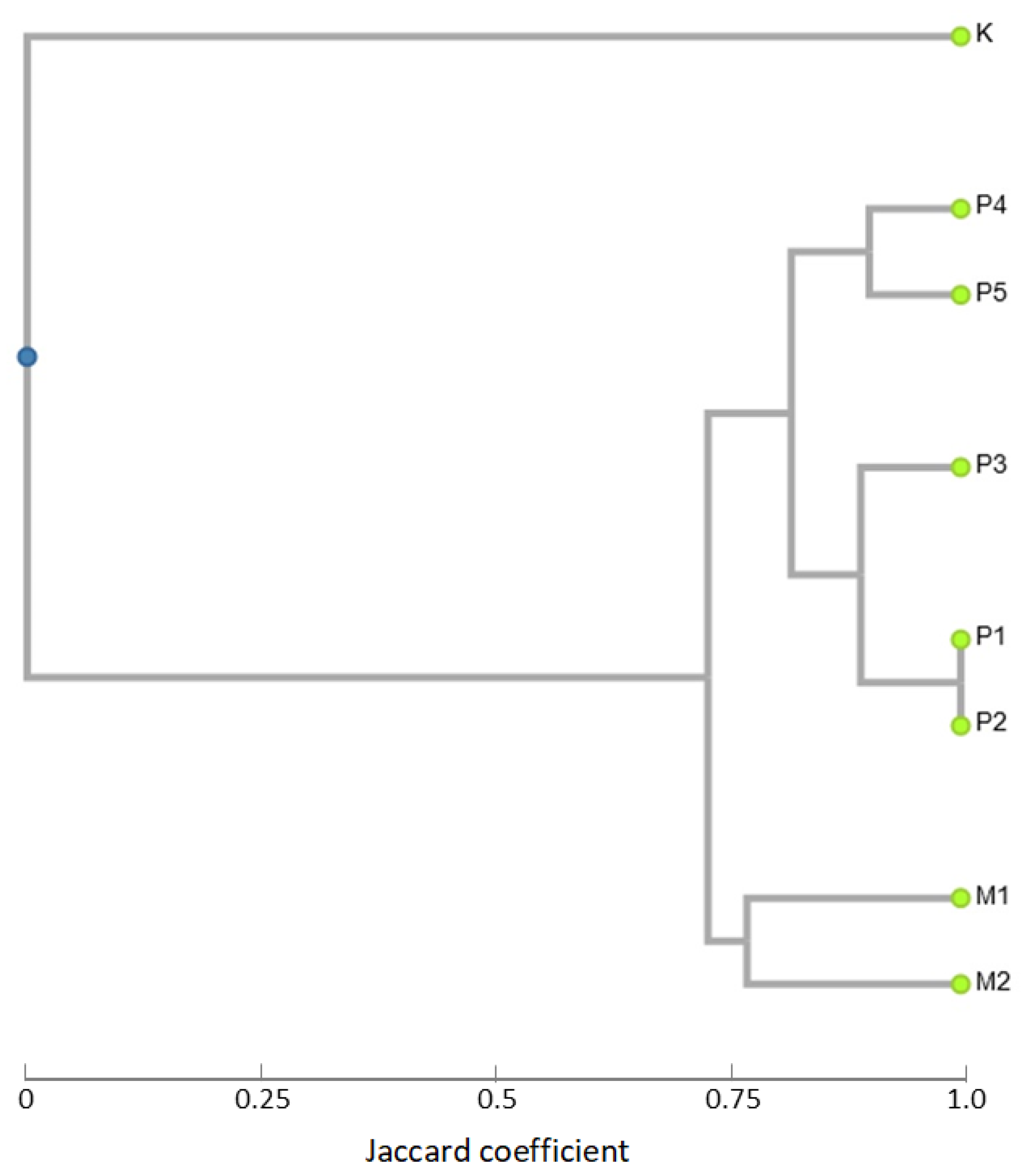
3.3. iPBS Analysis (Inter-Primer Binding Site Polymorphism)

Six previously published iPBS primers were used for the analysis (primers: 1838, 1846, 1882, 2270, 1897, and Frodo2) [27]. Primer 1882 generated a monomorphic profile for all of the analyzed polyploid plants of C. fragrans accessions (P1–P4, M1–M2) with two (80 bp plus 90 bp) amplicon deletions in the control plant. In the control plant, nine fragments were amplified, and in the polyploid accessions, 11 iPBS fragments were amplified. A completely monomorphic iPBS profile was obtained for the primer 1897 with seven generated amplicons across all the genotypes. Primer Frodo2 generated a different amplified iPBS profile for the accession M2, where deletions of short fragments occurred, but all of the other samples have 12 iPBS fragments per accession. Three of the remaining iPBS primers yielded polymorphic profiles among the analyzed C. fragrans plants. Primer 1838 generated 88% polymorphism and amplicons varied from three (control) up to eight (P4) per accession. Insertion of the 260 bp fragment was achieved for the M2 and P4 plants and a 500 bp insertion was obtained for the plants P1–P4. The length of the generated amplicons varied from 45 bp up to 990 bp. Primer 2270 was characterized by 55% polymorphism but provided a different iPBS profile for the control C. fragrans plant, where two deletions occurred. Profiles of M1–M2 and P1–P5 were monomorphic. The number of generated amplicons was five for control plants and seven for polyploid plants. Primer 1846 provided 87.5% polymorphism and was the most variable among the used iPBS primers. No specific pattern was found between the polyploid genotypes and the control plant of C. fragrans. Control plants were not especially different in their iPBS profile, but here, the most amplicons were obtained (11) and the rest of the samples provided from four up to nine amplicons in iPBS. Genotypes M1, P2, and P4 have similar, but not the same, profiles and plants P1 and P5 generated the same iPBS profiles. A dendrogram (Figure 5) was constructed for the analyzed plant variants based on the results of iPBS fingerprinting. Three main branches were generated where the control plant was clearly separated from the treated polyploid plants. Further, two branches separated the tetraploids (P1–P5) from the mixoploids (M1–M2). The P1–P5 plants were divided into two smaller subclusters with the same profile for P1 and P2.

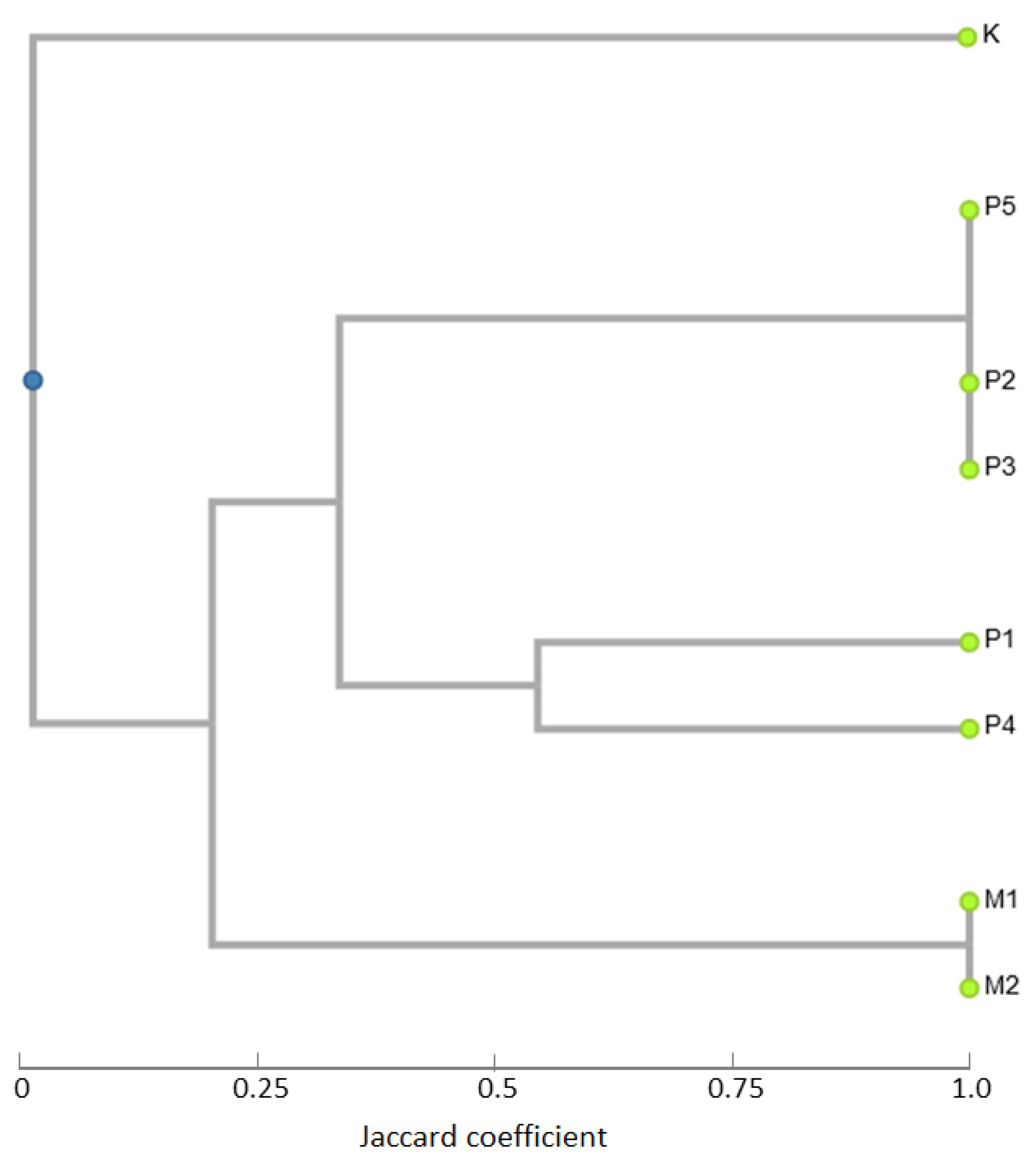
3.4. Conserved DNA-Derived Polymorphism (CDDP) Analysis

In this analysis, five standard primer combinations were used to generate CDDP fingerprint profiles of the coding regions of the C. fragrans genome. In four of them, the control Callisia plant was differentiated by this technique by a specific locus amplification pattern.
Primer combination WRKY-F + WRKY-R1 provided a 430 bp insertion in the profile of the control plant, but the other amplicons correspond fully with the profile of M1 and M2 plants. In the case of P1–P5 genotypes, variability was obtained in the short fragments, deletion of a 490 bp locus was generated, and a 510 bp locus has stronger amplification when compared with the other analyzed C. fragrans accessions. Primer combination WRKY-F + WRKY-R2a resulted in the monomorphic profiles of M and P plants and the generated CDDP profile of the control plant differs in the insertion of one locus of 490 bp and two loci of approximately 130 bp. Primer combination WRKY-F + WRKY-R2b provided differentiated fingerprints for the control plant and P1. The control plant was typical by insertion of a 240 bp locus and deletion of a 260 bp locus. Mixoploid plants and P2–P5 plants have monomorphic profiles and the P1 plant is different from them by the shift of the CDDP fingerprints and insertion of 130 bp and 140 bp loci. Primer combination WRKY-F + WRKY-R3 generated monomorphic profiles for both M and P plants, too. The control plant is different from them by insertion of 150 bp and 370 bp loci. Primer combination WRKY-F + WRKY-R3b provided a completely monomorphic profile of all analyzed C. fragrans plants (new genotypes and control plants).
A dendrogram (Figure 6) was constructed for the analyzed C. fragrans plants based on the results of CDDP fingerprinting. Three main branches were generated with the CDDP profile from the most distinct control plant in comparison with the rest of the analyzed plants. The P1–P5 genotypes were divided into two smaller subclusters with the same profile for P2, P3, and P5. Mixoploid genotypes have the same profiles.
Both of used marker techniques generated fingerprints that were able to distinguish different polyploidy backgrounds of analyzed accessions. The individual characteristics of both are summarized in Table 4.

3.5. Nutrient Content

The nutrient content analysis detected variance in the content of macro- and micronutrients among the studied genotypes. Potassium content showed an almost 100% increase in all polyploid genotypes. The calcium content increased only in the P1 genotype (10.97%) compared to the diploid. Other polyploid and mixoploid genotypes had calcium content reduced in comparison with diploid plants (Table 5). The average sodium content in all the induced genotypes was observed to increase approximately four to six times compared to the control genotype. The summary of nutrient content is given in Table 5.

4. Discussion

There is no information about in vitro induced somatic polyploidy in C. fragrans nor in other species from the Callisia genus. The experiment of in vitro induced somatic polyploidy in C. fragrans using oryzalin was carried out for the first time in the current study. Recent articles report that in vitro induced somatic polyploidy is often used for breeding medicinal plants from other genera and families such as Allium cepa L. [29], Moringa oleifera L. [30], C. nobile, [17], and E. purpurea [18]. For this purpose, oryzalin and colchicine are effectively used as antimitotic agents for obtaining new genotypes. Nevertheless, in medicinal plants, it is reported that oryzalin has become a standard preference over colchicine due to its lower toxicity [10,31]. Moreover, it has been extensively used in the breeding of numerous medicinal plants, for example, Calendula officinalis [11] and Cnidium officinale [32]. Additionally, oryzalin also worked effectively for the chromosome doubling of Scutellaria barbata D. [33], T. vulgaris [14], and Rubus sanctus Schreb. [34]. Interestingly, a study reported that by using oryzalin for polyploidization, the efficiency of obtaining polyploids was raised by 5% in Spathiphyllum wallisii Regel [35]. Similarly, oryzalin has also been found to be more effective than colchicine in genome doubling of Smallanthus sonchifolius (Poepp.) H. [36], Solanum species [37], and Agastache foeniculum (Pursh) Kuntze [12].
From the results of the current study, oryzalin added into media appears as an effective antimitotic agent for polyploidization of C. fragrans. The addition of 5 µM of oryzalin directly into the media for 8 weeks of treatment time was found to be the most efficient treatment for inducing polyploidy in C. fragrans. Despite the fact that the higher concentration was not toxic for the plants, there were no obtained tetraploid genotypes from the higher concentrations. Similar to the current study, the addition of oryzalin in media was successful for polyploidization of Anemone sylvestris L. [25] and S. wallisii [35]. Polyploidization in Anemone sylvestris was significantly higher at medium concentrations of 5 µM and 10 µM compared to the higher concentrations studied. The highest concentration (15 µM) for the 12 weeks’ duration was toxic for all treated plants. The shorter time was sufficient for the induction of tetraploid plants (8 weeks) with increasing concentrations of oryzalin [25]. The lower concentrations were also successfully used for inducing polyploidy in M. oleifera where tetraploid plants were obtained in the treatment of seeds for 1 day with 15 µM and 60 µM of oryzalin [30].
Callisia fragrans plants were well adapted to ex vitro conditions. The survival rate of transferred plants was 100% for all the variants. As previously reported, the autotetraploid plants of Anemone sylvestris grown in field conditions had a 100% survival rate in the next year, while the diploids had a survival rate of only 58% [25].
The newly obtained genotypes in the current study have significant morphological differences. The number of leaves decreased in tetraploid plants of C. fragrans. Although a previous study reported that despite the number of leaves of tetraploid plants of Centella asiatica L. decreasing, the fresh weight of tetraploid plants increased by more than 77% [38]. A similar increase in biomass by polyploidization was also reported in Salvia miltiorrhiza and Scutellaria baicalensis Georgi [39,40]. All genotypes of induced autotetraploid plants of C. fragrans exhibited an increase in leaf length except P5. Similar results were obtained in T. vulgaris [14], S. miltiorrhiza [39], and R. persicus [34]. Induced tetraploid genotypes of M. oleifera also generated larger leaflets [30]. Similarly, autotetraploid genotypes of C. fragrans also generated significantly larger inflorescences with larger petals; only one genotype (P5) generated flowers smaller than the control plant. Previously, a significant increase in flower size was also observed in A. sylvestris [25] and C. nobile [17]. By polyploidization of Rubus spp., the crown diameter increased by 100% in Rubus caesius L. and, on the contrary, Rubus hirtus Rchb. exhibited a decrease in flower size [34].
A variance in the content of macro- and micronutrients was detected. Potassium showed an almost 100% increase in all polyploid and mixoploid genotypes. The calcium content increased only in the P2 genotype. The other genotypes had their calcium content reduced in comparison with diploid plants. Similarly, a previous study reported that upon successful polyploid induction in Moringa oleifera, protein content increased by 20%, fat content increased up to 34%, and calcium content increased up to 20% [30]. Additionally, the amount of chemical compounds has been reported to increase in several medicinal plants such as T. vulgaris [14] and S. miltiorrhiza [39]. Similarly, content of matrine and oxymatrine increased by polyploidy induction of the Chinese medicine plant Sophora tonkinensis Gagnep., where the oxymatrine content was increased by 107.1% in comparison with the control plant [41]. The biochemical compounds of tetraploid C. fragrans and their comparison with the diploid genotype need to be a part of further research.
In both of the DNA-based fingerprints methods used, control plants were separated from polyploid ones. Polyploidization in its natural or in vitro conditions results in genome duplication and is an inevitable part of plant evolution and speciation. Different marker techniques have been used to analyze artificially produced polyploid plants. For instance, microsatellite-based markers were used to characterize the fingerprint profiles of polyploid and aneuploid seedlings from seven diploid Malus populations [42] and Aronia melanocarpa genetic structure, among the native polyploidy populations, was analyzed by randomly amplified polymorphic DNA (RAPD) [43].
Currently, there are no specific DNA markers reported for C. fragrans, therefore nonspecific markers can be utilized for the analysis of its genome variability. By iPBS markers, the variability of the retrotransposon insertion patterns was analyzed in the diploid and tetraploid plants of C. fragrans. Here, iPBS analysis was applied for the first time to test the stability of induced polyploid plants of C. fragrans. A completely monomorphic iPBS profile was obtained for the primer 1897 which generated seven amplicons. The iPBS fingerprinting showed variability between all genotypes and the control plant, thus the control plant was evidently different from all other genotypes. However, P1 and P2 showed the same profile by iPBS markers. To date, many plant species have been analyzed by iPBS markers, such as Liparis loeselii L. [44], Saussurea esthonica Baer ex Rupr. [45], and Prunus armeniaca [46].
Similar to the iPBS, CDDP was applied for the first time to analyze the C. fragrans genome. The used primer combinations did not yield any complete monomorphic profile across the genotype, although some monomorphic bands were obtained for polyploid plants. Three main branches were generated with the CDDP profile, and the control plant-generated fingerprint was clearly separated.
Both of the used marker techniques were effective in distinguishing the control and the polyploid plants and there were only a few differences among them in generated polymorphism as well as in the amplified length of loci. The most important difference was in the similarity coefficients, where the iPBS technique provided a wider individual range of this coefficient and CDDP was narrower in the generated fingerprints.
Different marker techniques have been used to analyze the polymorphism generated through the coding parts of the plant genomes, such as SCoT [47], CDDP [48], TRAP [49], or PBA [50]. Additionally, there are lots of methods using DNA markers for the detection of genetic changes such as inter-simple sequence repeat (ISSR) [51], simple sequence repeat (SSR) [52], randomly amplified polymorphic DNA (RAPD) [53], and amplified fragment length polymorphism (AFLP) [54]. All of them were reported to be reliable [27,55,56], and could be alternatives for analysis of genotype stability of C. fragrans in future studies besides the iPBS and CDDP markers used in this study.

5. Conclusions

This is the first report of the successful in vitro induction of autotetraploids (2n = 4x = 24) in Callisia fragrans (Lindl.) Woodson using oryzalin. Oryzalin was found to be effective in inducing polyploidy in this species under in vitro conditions. The newly obtained genotypes are morphologically different from the diploid genotype. Additionally, the polyploids exhibited a different nutrient content profile than the diploid genotype. DNA-based analysis of polymorphism was performed by iPBS and CDDP markers, which were applied for the first time for C. fragrans. Hence, the in vitro oryzalin-induced polyploidization could be a valuable breeding strategy for C. fragrans to produce improved clones with new features. Additionally, the developed protocol might be effective for polyploid induction in plants from the same or a similar family.

Author Contributions

Conceptualization, K.B. and E.F.-C.; methodology, K.B., R.B., J.Ž., J.B., K.H. and M.K.; formal analysis, R.B., E.F.-C. and M.K.; investigation, K.B., R.B., J.Ž., J.B., K.H. and M.K.; writing—original draft preparation, K.B., E.F.-C. and R.B.; writing—review and editing, R.B., E.F.-C. and M.K.; supervision, E.F.-C.; project administration, E.F.-C.; funding acquisition, E.F.-C. All authors have read and agreed to the published version of the manuscript.

Funding

This research was funded by the Internal Grant Agency of FTA, grant number 20223101, Czech University of Life Sciences Prague, the Czech Republic.

Institutional Review Board Statement

Not applicable.

Informed Consent Statement

Not applicable.

Data Availability Statement

The datasets generated during and/or analyzed during the current study are available from the corresponding author on request.

Conflicts of Interest

The authors declare no conflict of interest.

References

  1. Chernenko, T.V.; Ul’chenko, N.T.; Glushenkova, A.I.; Redzhepov, D. Chemical Investigation of Callisia Fragrans. Chem. Nat. Compd. 2007, 43, 253–255. [Google Scholar] [CrossRef]
  2. Saensouk, S.; Saensouk, P. A Karyological Study of Six Commelinaceae Species from Thailand. Cytologia 2020, 85, 57–62. [Google Scholar] [CrossRef]
  3. Malakyan, M.H. Radioprotective activity of Callisia fragrans grown in soilless (hydroponics) and soil culture conditions. Electron. J. Nat. Sci. 2015, 25, 20–23. [Google Scholar] [CrossRef] [Green Version]
  4. Pranskuniene, Z.; Ratkeviciute, K.; Simaitiene, Z.; Pranskunas, A.; Bernatoniene, J. Ethnobotanical Study of Cultivated Plants in Kaišiadorys District, Lithuania: Possible Trends for New Herbal Based Medicines. Evid.-Based Complement. Altern. Med. 2019, 2019, 3940397. [Google Scholar] [CrossRef] [PubMed] [Green Version]
  5. El Sohafy, S.M.; Nassra, R.A.; D’Urso, G.; Piacente, S.; Sallam, S.M. Chemical Profiling and Biological Screening with Potential Anti-Inflammatory Activity of Callisia fragrans Grown in Egypt. Nat. Prod. Res. 2021, 35, 5521–5524. [Google Scholar] [CrossRef] [PubMed]
  6. Karapetyan, A.S.; Mairapetyan, S.K. Quantitative Determination of Ascorbic Acid in Callisia fragrans under Open-Air Hydroponic Conditions. Bulg. Chem. Commun. 2020, 52, 30–32. [Google Scholar]
  7. Phan, L.T.M.; Nguyen, K.T.P.; Vuong, H.T.; Tran, D.D.; Nguyen, T.X.P.; Hoang, M.N.; Mai, T.P.; Nguyen, H.H. Supercritical Fluid Extraction of Polyphenols from Vietnamese Callisia fragrans Leaves and Antioxidant Activity of the Extract. J. Chem. 2020, 2020, 9548401. [Google Scholar] [CrossRef]
  8. Eng, W.-H.; Ho, W.-S. Polyploidization Using Colchicine in Horticultural Plants: A Review. Sci. Hortic. 2019, 246, 604–617. [Google Scholar] [CrossRef]
  9. Dehghan, E.; Reed, D.W.; Covello, P.S.; Hasanpour, Z.; Palazon, J.; Oksman-Caldentey, K.-M.; Ahmadi, F.S. Genetically Engineered Hairy Root Cultures of Hyoscyamus senecionis and H. muticus: Ploidy as a Promising Parameter in the Metabolic Engineering of Tropane Alkaloids. Plant Cell Rep. 2017, 36, 1615–1626. [Google Scholar] [CrossRef] [Green Version]
  10. Niazian, M.; Nalousi, A.M. Artificial Polyploidy Induction for Improvement of Ornamental and Medicinal Plants. Plant Cell Tissue Organ Cult. 2020, 142, 447–469. [Google Scholar] [CrossRef]
  11. Esmaeili, G.; Van Laere, K.; Muylle, H.; Leus, L. Artificial Chromosome Doubling in Allotetraploid Calendula officinalis. Front. Plant Sci. 2020, 11, 622. [Google Scholar] [CrossRef] [PubMed]
  12. Talebi, S.F.; Saharkhiz, M.J.; Kermani, M.J.; Sharafi, Y.; Raouf Fard, F. Effect of Different Antimitotic Agents on Polyploid Induction of Anise Hyssop (Agastache foeniculum L.). Caryologia 2017, 70, 184–193. [Google Scholar] [CrossRef]
  13. Dhooghe, E.; Van Laere, K.; Eeckhaut, T.; Leus, L.; Van Huylenbroeck, J. Mitotic Chromosome Doubling of Plant Tissues In Vitro. Plant Cell Tissue Organ Cult. 2011, 104, 359–373. [Google Scholar] [CrossRef]
  14. Homaidan Shmeit, Y.; Fernandez, E.; Novy, P.; Kloucek, P.; Orosz, M.; Kokoska, L. Autopolyploidy Effect on Morphological Variation and Essential Oil Content in Thymus vulgaris L. Sci. Hortic. 2020, 263, 109095. [Google Scholar] [CrossRef]
  15. Parsons, J.L.; Martin, S.L.; James, T.; Golenia, G.; Boudko, E.A.; Hepworth, S.R. Polyploidization for the Genetic Improvement of Cannabis sativa. Front. Plant Sci. 2019, 10, 476. [Google Scholar] [CrossRef] [PubMed]
  16. Sabzehzari, M.; Hoveidamanesh, S.; Modarresi, M.; Mohammadi, V. Morphological, Anatomical, Physiological, and Cytological Studies in Diploid and Tetraploid Plants of Plantago Psyllium. Plant Cell Tissue Organ Cult. 2019, 139, 131–137. [Google Scholar] [CrossRef]
  17. Tsuro, M.; Kondo, N.; Noda, M.; Ota, K.; Nakao, Y.; Asada, S. In Vitro Induction of Autotetraploid of Roman Chamomile (Chamaemelum nobile L.) by Colchicine Treatment and Essential Oil Productivity of Its Capitulum. In Vitro Cell. Dev. Biol.-Plant 2016, 52, 479–483. [Google Scholar] [CrossRef]
  18. Mei, B.; Xie, H.; Xing, H.; Kong, D.; Pan, X.; Li, Y.; Wu, H. Changes of Phenolic Acids and Antioxidant Activities in Diploid and Tetraploid Echinacea purpurea at Different Growth Stages. Rev. Bras. Farmacogn. 2020, 30, 510–518. [Google Scholar] [CrossRef]
  19. Hassanzadeh, F.; Zakaria, R.A.; Azad, N.H. Polyploidy Induction in Salvia officinalis L. and Its Effects on Some Morphological and Physiological Characteristics. Cytologia 2020, 85, 157–162. [Google Scholar] [CrossRef]
  20. Santos, G.C.; Cardoso, F.P.; Martins, A.D.; Rodrigues, F.A.; Pasqual, M.; Cossani, P.; Magno Queiroz Luz, J.; Rezende, R.A.L.S.; Soares, J.D.R. Polyploidy Induction in Physalis alkekengi. Biosci. J. 2020, 36, 827–835. [Google Scholar] [CrossRef]
  21. Zhou, J.; Guo, F.; Fu, J.; Xiao, Y.; Wu, J. In Vitro Polyploid Induction Using Colchicine for Zingiber officinale Roscoe Cv. ‘Fengtou’ Ginger. Plant Cell Tissue Organ Cult. 2020, 142, 87–94. [Google Scholar] [CrossRef]
  22. Shaikh, N.; Verma, R.C.; Dass, P.; Khah, M.A. Meiotic Investigation in Reciprocal Translocation Heterozygotes of Commelina benghalensis L. (Commelinaceae) Induced by Gamma Irradiations. Cytologia 2020, 85, 213–217. [Google Scholar] [CrossRef]
  23. Shaikh, N.; Verma, R.C.; Khah, M.A.; Filimban, F.Z.; Albaz, A.A.; Alam, Q. Morphological and Chromosomal Characterization of Diploid and Colchicine Induced Autotetraploid Plants of Commelina benghalensis L. (Commelinaceae). Cytologia 2021, 86, 167–173. [Google Scholar] [CrossRef]
  24. Murashige, T.; Skoog, F. A Revised Medium for Rapid Growth and Bio Assays with Tobacco Tissue Cultures. Physiol. Plant. 1962, 15, 473–497. [Google Scholar] [CrossRef]
  25. Zahumenická, P.; Fernández, E.; Šedivá, J.; Žiarovská, J.; Ros-Santaella, J.L.; Martínez-Fernández, D.; Russo, D.; Milella, L. Morphological, Physiological and Genomic Comparisons between Diploids and Induced Tetraploids in Anemone sylvestris L. Plant Cell Tissue Organ Cult. 2018, 132, 317–327. [Google Scholar] [CrossRef]
  26. Tomaskova, D. Karyological Methods of Ploidy Level Determination in Sugar Beet Varieties. Genet. Slechteni 1996, 31, 161–170. [Google Scholar]
  27. Kalendar, R.; Antonius, K.; Smýkal, P.; Schulman, A.H. IPBS: A Universal Method for DNA Fingerprinting and Retrotransposon Isolation. Theor. Appl. Genet. 2010, 121, 1419–1430. [Google Scholar] [CrossRef] [PubMed]
  28. Collard, B.C.Y.; Mackill, D.J. Conserved DNA-Derived Polymorphism (CDDP): A Simple and Novel Method for Generating DNA Markers in Plants. Plant Mol. Biol. Rep. 2009, 27, 558–562. [Google Scholar] [CrossRef]
  29. Yun, R.Q.A.; Dinarti, D.; Husni, A.; Kosmiatin, M. Callus Regeneration and Polyploidy Induction of Allium cepa L Var. Bima Brebes Using Oryzalin. IOP Conf. Ser. Earth Environ. Sci. 2021, 948, 012043. [Google Scholar] [CrossRef]
  30. Ridwan, R.; Witjaksono, W. Induction of Autotetraploid Moringa Plant (Moringa oleifera) Using Oryzalin. Biodiversitas 2020, 21, 4086–4093. [Google Scholar] [CrossRef]
  31. Salma, U.; Kundu, S.; Mandal, N. Artificial Polyploidy in Medicinal Plants: Advancement in the Last Two Decades and Impending Prospects. J. Crop Sci. Biotechnol. 2017, 20, 9–19. [Google Scholar] [CrossRef]
  32. Kim, H.-E.; Han, J.-E.; Lee, H.; Kim, J.-H.; Kim, H.-H.; Lee, K.-Y.; Shin, J.-H.; Kim, H.-K.; Park, S.-Y. Tetraploidization Increases the Contents of Functional Metabolites in Cnidium officinale. Agronomy 2021, 11, 1561. [Google Scholar] [CrossRef]
  33. Yue, M.; Chen, C.; Liao, J.; Sun, J.; Shen, W. Oryzalin-induced chromosome doubling of Scutellaria barbata. Zhong Yao Cai 2011, 34, 4–8. [Google Scholar] [PubMed]
  34. Sabooni, N.; Gharaghani, A.; Jowkar, A.; Eshghi, S. Successful Polyploidy Induction and Detection in Blackberry Species by Using an In Vitro Protocol. Sci. Hortic. 2022, 295, 110850. [Google Scholar] [CrossRef]
  35. Eeckhaut, T.G.R.; Werbrouck, S.P.O.; Leus, L.W.H.; Van Bockstaele, E.J.; Debergh, P.C. Chemically Induced Polyploidization in Spathiphyllum wallisii Regel through Somatic Embryogenesis. Plant Cell Tissue Organ Cult. 2004, 78, 241–246. [Google Scholar] [CrossRef]
  36. Viehmannová, I.; Cusimamani, E.F.; Bechyne, M.; Vyvadilová, M.; Greplová, M. In Vitro Induction of Polyploidy in Yacon (Smallanthus sonchifolius). Plant Cell Tissue Organ Cult. 2009, 97, 21–25. [Google Scholar] [CrossRef]
  37. Greplova, M.; Polzerova, H.; Domkářová, J. Intra- and Inter-Specific Crosses of Solanum Materials after Mitotic Polyploidization In Vitro. Plant Breed. 2009, 128, 651–657. [Google Scholar] [CrossRef]
  38. Kaensaksiri, T.; Soontornchainaksaeng, P.; Soonthornchareonnon, N.; Prathanturarug, S. In Vitro Induction of Polyploidy in Centella asiatica (L.) Urban. Plant Cell Tissue Organ Cult. 2011, 107, 187–194. [Google Scholar] [CrossRef]
  39. Gao, S.L.; Zhu, D.N.; Cai, Z.H.; Xu, D.R. Autotetraploid Plants from Colchicine-Treated Bud Culture of Salvia miltiorrhiza Bge. Plant Cell Tissue Organ Cult. 1996, 47, 73–77. [Google Scholar] [CrossRef]
  40. Gao, S.L.; Chen, B.J.; Zhu, D.N. In Vitro Production and Identification of Autotetraploids of Scutellaria baicalensis. Plant Cell Tissue Organ Cult. 2002, 70, 289–293. [Google Scholar] [CrossRef]
  41. Wei, K.; Xu, J.; Li, L.; Cai, J.; Miao, J.; Li, M. In Vitro Induction and Generation of Tetraploid Plants of Sophora tonkinensis Gapnep. Pharmacogn. Mag. 2018, 14, 149. [Google Scholar] [CrossRef]
  42. Considine, M.J.; Wan, Y.; D’Antuono, M.F.; Zhou, Q.; Han, M.; Gao, H.; Wang, M. Molecular Genetic Features of Polyploidization and Aneuploidization Reveal Unique Patterns for Genome Duplication in Diploid Malus. PLoS ONE 2012, 7, e29449. [Google Scholar] [CrossRef] [PubMed]
  43. Persson Hovmalm, A.H.; Jeppsson, N.; Bartish, V.I.; Nybom, H. RAPD Analysis of Diploid and Tetraploid Populations of Aronia Points to Different Reproductive Strategies within the Genus: RAPD Analysis of Populations of Aronia. Hereditas 2005, 141, 301–312. [Google Scholar] [CrossRef] [PubMed]
  44. Belogrudova, I.; Grauda, D.; Jakobsone, G.; Rashal, I. Usability of Retrotransposone-Based Molecular Marker System to Assess Genetic Diversity of Liparis loeselii. Acta Biol. Univ. Daugavp. 2012, 12, 4. [Google Scholar]
  45. Gailite, A.; Rungis, D. An Initial Investigation of the Taxonomic Status of Saussurea esthonica Baer Ex Rupr. Utilising DNA Markers and Sequencing. Plant Syst. Evol. 2012, 298, 913–919. [Google Scholar] [CrossRef]
  46. Baránek, M.; Meszáros, M.; Sochorová, J.; Čechová, J.; Raddová, J. Utility of Retrotransposon-Derived Marker Systems for Differentiation of Presumed Clones of the Apricot Cultivar Velkopavlovická. Sci. Hortic. 2012, 143, 1–6. [Google Scholar] [CrossRef]
  47. Petrovičová, L.; Balážová, Ž.; Vivodí k, M.; Gálová, Z. Detection Genetic Variability of Secale cereale L. by Scot Markers. Potravinarstvo 2017, 11, 197–202. [Google Scholar] [CrossRef] [Green Version]
  48. Jiang, L.; Zang, D. Analysis of Genetic Relationships in Rosa rugosa Using Conserved DNA-Derived Polymorphism Markers. Biotechnol. Biotechnol. Equip. 2018, 32, 88–94. [Google Scholar] [CrossRef] [Green Version]
  49. Vivodí k, M.; Gálová, Z.; Balážová, Ž. Genetic Divergence in Tunisian Castor Bean Genotypes Based on Trap Markers. Potravinarstvo 2020, 14, 510–518. [Google Scholar] [CrossRef]
  50. Ravi, R.S.D.; Siril, E.A.; Nair, B.R. The Efficiency of Cytochrome P450 Gene-Based Markers in Accessing Genetic Variability of Drumstick (Moringa oleifera Lam.) Accessions. Mol. Biol. Rep. 2020, 47, 2929–2939. [Google Scholar] [CrossRef]
  51. Stefunova, V.; Bezo, M.; Senkova, M. Genetic Analysis of Three Amaranth Species Using ISSR Markers. Emir. J. Food Agric. 2014, 26, 35. [Google Scholar] [CrossRef]
  52. Balážová, Å.; Gálová, Z.; Vivodík, M. Application of rye SSR markers for detection of genetic diversity in triticale. J. Microbiol. Biotechnol. Food Sci. 2016, 5, 623–626. [Google Scholar] [CrossRef] [Green Version]
  53. Kuťka Hlozáková, T.; Gálová, Z.; Gregová, E.; Vivodí k, M.; Balážová, Ž.; Miháliková, D. RAPD Analysis of the Genetic Polymorphism in European Wheat Genotypes. Potravinarstvo 2016, 10, 1–6. [Google Scholar] [CrossRef]
  54. Iarovsk, J.; Labajov, K. Using of Inter Microsatellite Polymorphism to Evaluate Gamma-Irradiated Amaranth Mutants. Emir. J. Food Agric. 2013, 25, 673. [Google Scholar] [CrossRef]
  55. Kalendar, R.; Schulman, A.H. Transposon-Based Tagging: IRAP, REMAP, and IPBS. In Molecular Plant Taxonomy; Methods in Molecular Biology; Besse, P., Ed.; Humana Press: Totowa, NJ, USA, 2014; Volume 1115, pp. 233–255. [Google Scholar] [CrossRef] [Green Version]
  56. Tiwari, J.K.; Chandel, P.; Gupta, S.; Gopal, J.; Singh, B.P.; Bhardwaj, V. Analysis of Genetic Stability of In Vitro Propagated Potato Microtubers Using DNA Markers. Physiol. Mol. Biol. Plants 2013, 19, 587–595. [Google Scholar] [CrossRef] [PubMed]
Figure 1. Histogram of flow cytometry analysis from C. fragrans: (A) histogram of control plant (diploid) and (B) polyploid plants (tetraploid).
Agronomy 12 02520 g001
Figure 2. Chromosome counting of C. fragrans. (A) Chromosome numbers in diploid C. fragrans (2n = 2x = 12). (B) Chromosome number in induced tetraploid C. fragrans (2n = 4x = 24) under 100× magnification.
Agronomy 12 02520 g002
Figure 3. Two-week-old C. fragrans plants in ex vitro conditions: (A) control plant; (B) tetraploid genotype P2; (C) tetraploid genotype P3; (D) mixoploid genotype M1.
Agronomy 12 02520 g003
Figure 4. Morphological variation between diploid (A) and induced tetraploid (B) flowers of C. fragrans cultivated in greenhouse conditions.
Agronomy 12 02520 g004
Figure 5. Dendrogram of analyzed C. fragrans genotypes based on iPBS markers.
Agronomy 12 02520 g005
Figure 6. Dendrogram of analyzed C. fragrans genotypes based on CDDP results.
Agronomy 12 02520 g006
Table 1. Effect of oryzalin treatment on polyploid induction of Callisia fragrans.
Concentration (µM)Duration (weeks)No. of PlantsNo. of Plants after TreatmentSurvival Rate (%)Mixoploid PlantsTetraploid PlantsEfficiency Rate (%)
143034100000
83066100000
543047100102.13
83063100069.52
1043038100000
83058100101.72
Total 18030610026
Table 2. Morphological characteristics of diploid, mixoploid, and autotetraploid plants cultivated in a greenhouse.
GenotypeAverage Number of InternodesAverage Number of StolonsAverage Number of LeavesAverage Height (cm)Average Length of Stolons (cm)Average Numberof Internodes on StolonAverage Leaf Length (cm)
Control16.3 ± 1.7 a5.5 ± 1.35 a23.7 ± 2.26 a64 ± 6.58 a76.1 ± 10.04 a10.33 ± 0.92 a24.47 ± 1.48 c
P114.8 ± 2.15 a4.4 ± 1.17 a22 ± 1.76 a67.5 ± 10.61 a54.95 ± 10.8 b9.51 ± 0.86 a30.87 ± 0.42 a
P214.3 ± 1.16 a4.3 ± 1.25 a20.9 ± 2.08 a62.8 ± 6.09 a57.012 ±16.87 b6.92 ± 2.3 c28.69 ± 0.88 b
P314.7 ± 0.68 a5.6 ± 0.52 a17.9 ± 0.87 b61.9 ± 3.,48 a64.133 ±5.64 b9.33 ± 0.54 a31.86 ± 1.57 a
P411.9 ± 1.37 b4.5 ± 0.53 a17 ± 0.94 b62 ± 10.33 a61.48 ± 9.92 b8.19 ± 0.94 b31.34 ± 0.82 a
P517.3 ± 1.34 a4.5 ± 0.85 a21.3 ± 1.49 a64.5 ± 5.93 a48.835 ± 5.69 c8.3 ± 2.76 a22.35 ± 0.94 c
M112.3 ± 1.9 b5.2 ± 0.42 a18.8 ± 3.36 b46 ± 7.76 b56.86 ± 15.48 b8.62 ± 1.35 a32.7 ± 1.14 a
M212.2 ± 1.81 b6 ± 1.25 a18.2 ± 2.49 b46 ± 7.75 b57.38 ± 13.73 b8.82 ± 1.21 a32.73 ± 1.61 a
Different superscript letters within the same column differ significantly (Kruskal–Wallis test, p < 0.05).
Table 3. Morphological characteristics of flowers of diploid and autotetraploid plants.
GenotypeAverage Diameter (cm)Petal Length (mm)Petal Width (mm)
K2.08 ± 0.18 b3.54 ± 0.41 b1.94 ± 0.37 b
P12.08 ± 0.16 c6.045 ± 1.01 a2.33 ± 0.88 b
P33.11 ± 0.10 a5.96 ± 0.58 a2.68 ± 0.55 a
P42.22 ± 0.16 b5.97 ± 0.99 a2.42 ± 0.54 b
P51.92 ± 0.09 c4.94 ± 0.55 b2.41 ± 0.77 b
Different superscript letters within the same column differ significantly (Kruskal–Wallis test, p < 0.05).
Table 4. Characteristics of fingerprints obtained by iPBS and CDDP markers.
TechniquePrimer/Primer CombinationPolymorphism (%)Min Length of Generated FragmentsMax Length of Generated FragmentsAverage Dice Coefficient of Genetic Similarity
iPBS18461006911900.52
2270100687890.61
188298608200.95
185499649880.65
1867997118500.89
18681009012300.88
CDDPWRKY-F + R196555600.52
WRKY-F + R2a95827800.45
WRKY-F + R2b99709300.36
WRKY-F + R399505200.49
WRKY-F + R3b93639000.58
Table 5. Macronutrient, micronutrient, and fragment compounds analyzed from leaves of diploid, tetraploid, and mixoploid C. fragrans plants (mg.kg−1).
NutrientControl (K)P1P2P3P4P5M1M2
K15,772.00 ± 174.00 g31,040.35 ± 92.75 d37,369.05 ± 68.15 b30,066.50 ± 39.50 e39,041.80 ± 9.40 a39,047.45 ± 15.05 a33,313.35 ± 5.95 c26,340.25 ± 10.05 f
Ca23,253.40 ± 105.50 b25,777.90 ± 89.40 a8862.80 ± 4.70 g13,685.25 ± 75.05 e13,122.80 ± 10.30 f17,231.55 ± 5.65 c17,334.80 ± 11.60 c14,798.30 ± 10.90 d
Mg12,866.10 ± 120.50 b14,295.50 ± 15.00 a4868.00 ± 10.00 h8039.85 ± 13.35 f6882.75 ± 5.55 g10,316.95 ± 6.25 c9353.75 ± 3.05 d8517.55 ± 4.15 e
Na195.95 ± 1.55 h1368.15 ± 2.95 de1758.25 ± 12.85 a1305.35 ± 7.85 f1706.50 ± 3.80 b1404.50 ± 4.70 cd1351.85 ± 2.55 e922.20 ± 1.50 g
Fe82.80 ± 0.88 d73.88 ± 0.41 e90.95 ± 0.15 c57.97 ± 0.00 f73.97 ± 0.37 e82.03 ± 0.05 d92.98 ± 0.35 bc93.74 ± 0.00 ab
Mn102.40 ± 0.70 e107.15 ± 0.25 e142.90 ± 0.30 b90.25 ± 0.55 f118.90 ± 0.30 d79.05 ± 0.25 g188.75 ± 2.55 a135.22 ± 0.22 c
Zn48.96 ± 0.45 c43.20 ± 0.25 d53.42 ± 0.46 b36.97 ± 0.10 f56.13 ± 0.20 a44.01 ± 0.15 d50.91 ± 0.25 c39.86 ± 0.41 e
Cu26.76 ± 0.22 d34.73 ± 0.35 c54.16 ± 0.00 a26.05 ± 0.25 d33.12 ± 0.77 c25.07 ± 0.08 d43.32 ± 0.14 b41.93 ± 0.10 b
Cr0.15 ± 0.00 f0.17 ± 0.00 cdef0.16 ± 0.00 ef0.12 ± 0.01 g0.18 ± 0.00 bcde0.08 ± 0.00 h0.17 ± 0.01 def0.21 ± 0.01 a
Mo0.40 ± 0.01 f0.52 ± 0.01 de0.46 ± 0.01 ef1.02 ± 0.01 b0.15 ± 0.00 gNDh0.70 ± 0.02 c1.39 ± 0.02 a
CoNDe0.29 ± 0.00 bNDe0.20 ± 0.00 cNDe0.12 ± 0.01 d0.35 ± 0.01 aNDe
AgNDa0.07 ± 0.00 aNDaNDaNDa0.07 ± 0.00 a0.11 ± 0.00 a0.02 ± 0.00 a
Sr49.07 ± 0.10 d43.65 ± 0.40 f54.03 ± 0.15 b38.05 ± 0.10 g56.07 ± 0.14 a46.08 ± 0.14 ce50.95 ± 0.29 c38.56 ± 0.40 g
SbNDf1.98 ± 0.05 a1.90 ± 0.01 a1.72 ± 0.01 b1.44 ± 0.01 c0.91 ± 0.01 d0.47 ± 0.00 eNDf
Li12.04 ± 0.15 a8.07 ± 0.05 c2.44 ± 0.03 g7.68 ± 0.04 c4.04 ± 0.01 f4.85 ± 0.02 e6.22 ± 0.09 d9.76 ± 0.08 b
Ba8.77 ± 0.35 a7.25 ± 0.10 b3.10 ± 0.03 d5.31 ± 0.02 c4.97 ± 0.01 c5.28 ± 0.04 c5.36 ± 0.06 c4.75 ± 0.02 c
Al36.12 ± 0.20 a11.20 ± 0.23 e17.14 ± 0.21 c16.23 ± 0.04 c13.93 ± 0.05 d13.98 ± 0.15 d19.05 ± 0.07 b13.63 ± 0.11 d
OthersAs 2.21
Pb 0.44
As 1.23
Cd 0.11
Ni 0.45
As 1.76
Ni 0.26
Pb 0.74
Pb 0.67NDNDCd 0.06
Ni 0.23
Pb 0.28
ND: Not detected and were considered zero for statistical analysis; nutrients in row “others” were not included in the statistical analysis. Different superscript letters within the same row differ significantly (Kruskal–Wallis test, p < 0.05).
Publisher’s Note: MDPI stays neutral with regard to jurisdictional claims in published maps and institutional affiliations.

