Article

A Novel Route for Double-Layered Encapsulation of Streptomyces fulvissimus Uts22 by Alginate–Arabic Gum for Controlling of Pythium aphanidermatum in Cucumber

by
Roohallah Saberi Riseh
1,*,
Mojde Moradi Pour
1 and
Essaid Ait Barka
2,*
1
Department of Plant Protection, Faculty of Agriculture, Vali-e-Asr University of Rafsanjan, Rafsanjan 7718897111, Iran
2
Induced Resistance and Plant BioProtection Research Unit, UFR Sciences, UPRES EA 4707-USC INRAeE1488, University of Reims Champagne-Ardenne, 51687 Reims, France
*
Authors to whom correspondence should be addressed.
Agronomy 2022, 12(3), 655; https://doi.org/10.3390/agronomy12030655
Submission received: 25 January 2022 / Revised: 1 March 2022 / Accepted: 4 March 2022 / Published: 8 March 2022
(This article belongs to the Special Issue Biotechnology of Microorganisms in the Agriculture Environment)

Abstract

:
Damping-off disease due to Pythium aphanidermatum is one of the most harmful diseases of cucumber. One of the critical issues in the field of biological control is the establishment of a link between the beneficial bacteria screened in the laboratory and its industrial application. Therefore, when developing biocontrol agents, it is necessary to study the optimization of mass production conditions and to select a suitable carrier for their final formulation. In this study, an attempt was made to provide a suitable formulation for a Streptomyces fulvissimus Uts22 strain based on alginate–Arabic gum and nanoparticles (SiO2 and TiO2) with a layer-by-layer technique. X-ray diffraction (XRD) and Fourier transform infrared spectroscopy (FTIR) studies showed that when Arabic gum was added to the composition, an electrostatic interaction occurred between alginate and Arabic gum. The scanning electron microscope image of beads show a cubic shape and good dispersion of microcapsules. The encapsulation efficiency in the prepared formulation was reported to be 94%. The maximum release of bacteria from the capsule was recorded on the 35th day of storage, about 109 CFU/gr. The greenhouse experiments showed that encapsulated bacteria resulted in a 95% reduction in damping-off disease of cucumber and showed more potential effects on increasing plant growth traits than free bacteria. This encapsulation strategy can be considered as a suitable alternative for future applications in the agricultural field.

1. Introduction

The damping-off disease of cucumber seedlings (Cucumis sativus) caused by Pythium aphanidermatum is one of the most harmful diseases of this crop, which causes more damage in hot regions [1]. In addition to the rapid death of seedlings, this species also causes root and crown rot [2]. Since the disease is also significant in most of the cucumber growing areas of Iran, it seems important to prevent and control it. The use of fungicides is the most common method of controlling soil-borne pathogens in the field and greenhouse [3]. However, these chemicals continue to present numerous challenges, such as environmental pollution, emergence of resistance of plant pathogenic fungi to fungicide, and residual toxins on the product [4]. The low effect of chemical methods to control various soil-borne pathogens, as well as their economic cost on one hand and environmental concerns on the other, have made achieving healthier and cheaper methods a serious challenge for researchers. The use of biofertilizers to reach sustainable agriculture and expand the production of non-toxic products has been proposed as an alternative to chemical fertilizers to increase soil fertility in agricultural production [5].
Biological control using microbial antagonists has emerged as an effective strategy to control post-harvest soil-borne diseases. In addition, without harming the plant and the environment, these beneficial microbial agents also increase crop yield [6]. Streptomyces bacteria present in the rhizosphere increase plant growth directly and/or indirectly. Direct plant growth stimulation occurs when a bacterial-synthesized compound is delivered to the plant, or when the bacteria improve absorption of soil nutrients by plants [7]. Potential contributions of this type would include nitrogen fixation, synthesis of siderophores, synthesis of phytohormones and solubilization of the minerals to make them accessible for absorption and utilization by the plants. The indirect promotion happens when they suppress the harmful actions of one or more damaging microorganisms. The production of antibiotics, siderophores, hydrogen cyanide, and various enzymes such as protease, lipase, chitinase, cellulase, etc., are considered as the most important mechanisms in the biological control of plant pathogens by these bacteria [8]. Usually, shortly after their inoculation into the soil, the bacteria population decreases quickly for most plant growth-promoting rhizobacteria (PGPR) when provided without a suitable carrier [9]. The natural heterogeneity of the soil microbiota is the key barrier to the establishment of PGPR agents, since sometimes bacteria cannot find an empty niche to inhabit in the soil [10]. To better-adapt and increase their survival rate, PGPR must compete with the soil microflora and enhance their survival against adverse environmental conditions. The bacterial formulation for use at the field scale should be designed to deliver a reliable source of bacteria that will survive in the soil and become accessible to crops, if necessary [11]. Therefore, a major role of the bacterial formulation is to provide an appropriate environment, consisting of physical protection, for a long period, to prevent a quick reduction of the bacterial population. Inoculant formulation must be stable during production, storage, and transport, especially when the main component is live and sensitive to changes, compared to farm chemicals [12]. The encapsulation of bacteria in a biodegradable matrix is a novel method of bacteria formulation in agricultural science. It preserves bacterial agents in adverse environmental conditions and increases their efficiency in increasing plant growth and controlling plant pathogens [13]. Further, one of the most important advantages of the formulation is the gradual release of bacteria from beads. As a result, colonizing plant roots with a desired strain is better [9].
Currently, the biopolymers complex is one of the most widely used encapsulation methods [14]. The process of encapsulation is based on the attractive interaction between two polymers with two types of different electric charges at a certain pH, leading to a phase separation in the process and therefore resulting in the formation of microcapsules. The capsule wall is conceived to protect against the release of microorganisms to the external environment until the perfect moment. There are several biodegradable polymeric matrices, which have been investigated for bacterial encapsulation, including alginate [15], chitosan [16], starch systems, carboxymethyl cellulose [17], protein–polysaccharide mixtures [18], and poly-lactic acid and poly-lactic-co-glycolic acid [19]. Among them, alginate (ALG), Arabic gum (AG), gelatin, chitosan, and gellan gum are the most frequently found in research reports.
Among the main materials of choice for encapsulating microorganisms [20], ALG-based formulations have the advantages of having non-toxic nature, are accessible at reasonable costs, are biodegradable, and control the release of the entrapped core material into the soil [21]. Further, ALG can be amplified with various secondary polymers to increase their mechanical resistance and stability to develop degradation time in soil, if needed. AG is an arabinogalactan polysaccharide–protein complex anionic compound of three separate sections with different protein levels (2–4%) and molecular weights. This polymer, according to the presence of peptide fragments and residual charged groups, has an effective surface activity and cold solubility [22].
Microencapsulation by complex biopolymers has been reported as more efficient for various bacteria, such as Bacillus salmalaya 139SI [23], Pseudomonas putida Rs-198 [24], B. subtilis VRU1 [25], and Pseudomonas fluorescens VUPF5 [21]. Titanium dioxide nanoparticles (Nano TiO2), one of the main nanomaterials in the agricultural sector, have the interesting feature of increasing the percentage of bacterial adhesion to the plant roots and thus increasing bacterial colonization. Saberi-Riseh and Moradi-Pour et al. [25] showed that Nano TiO2 increased the adhesion of B. subtilis VRU1 on bean root surfaces and enhanced the colonization process. With the occurrence of environmental stresses, SiO2 nanoparticles play an important role in increasing resistance to biotic and abiotic stresses (such as drought, cold, heat, etc.) in plants by triggering oxidative antioxidant defense, reducing oxidative damage to membrane molecules, and maintaining many physiological and photosynthetic processes in plant cells.
Because of the positive effects of PGPRs in agriculture, our goal is to increase the efficiency of beneficial bacteria to improve plant growth, to increase the viability of bacteria during formulation, storage and transport, and to further control the delivery of bacteria in the soil. Therefore, the present study was performed to microencapsulate probiotic cells of Streptomyces fulvissimus Uts22 in double polymers with the layer-by-layer technique using ALG and AG as matrixes enriched with TiO2 and SiO2 nanoparticles. Then, the moisture content, swelling ratio, morphology and other characteristics of microcapsules was investigated. Moreover, the survival rate of bacterial cells was evaluated during in vitro condition, as well as after 60 days of storage at 28 °C. Then, the effect of the prepared formulation on increasing the growth of the cucumber plant and evaluated damping-off disease control efficacy were investigated.

2. Materials and Methods

Streptomyces fulvissimus Uts22 was prepared from the Tehran University strain library while Pythium aphanidermatum isolate was obtained from the University of Jiroft.

2.1. In Vitro Antifungal Assay

S. fulvissimus Uts22 was investigated in an antagonistic evaluation using the dual-culture technique. One plug of P. aphanidermatum (5 mm in diameter) was placed on the center of the PDA medium plate and S. fulvissimus Uts22 was cultured on the edge of a Petri dish. Plates were incubated at 28 °C for 4 days and the antifungal activity of this bacterial was scored according to the scale described by Alfredo and Aleli [26]. The diameter of the mycelium growing out from the fungi plug was measured and was compared to the control samples. To obtain the percentage inhibition of fungal radial growth (PIRG %) the following formula was used [27]:
PIRG = [(R 1 − R 2)/R 1] × 100
with
R 1 = Radial growth of P. aphanidermatum (mm) in the control plates
R 2 = Radial growth of P. aphanidermatum interacting with S. fulvissimus Uts22 (mm)
In each treatment, an average of three replicates was reported.

2.2. Screening of Plant Growth Promoting Activities

2.2.1. Siderophore Production

Siderophore production of the desired bacterial strain was evaluated on Chrome Azurol S (CAS) agar plates [28]. Bacterial culture was spotted on these plates and incubated for three days at 28 °C. The appearance of a yellow to orange-colored zone around bacterial colonies is evidence of the siderophore production.

2.2.2. Auxin Production

The S. fulvissimus Uts22 suspension (1010 CFU/mL) was added to 25 mL of TSB (Tryptic Soy Broth) medium containing L-tryptophan in 50 mL Erlenmeyer flasks and incubated at 28 °C. After 72 h of growth, bacterial culture was centrifuged, and 2 mL of supernatant phase was mixed with 4 mL of Salkowski’s reagent (2 mL 0.5 M FeCl3 + 98 mL 35% HClO4) and kept at room temperature for 30 min for color development. Then, light absorbance was evaluated by spectrophotometer at 535 nm [29].

2.3. Hydrolytic Enzyme Production

2.3.1. Cellulase Assay

Cellulase assay was performed according to the method of Kasana et al. [30]. Bacterium was cultured on CMC agar medium (carboxymethyl cellulose 0.5 gL−1, K2HPO4 0.1 g/L, NaNO3 0.1 g/L, yeast extract 0.05 g/L, MgSO4 0.05 g/L, agar 15 g/L) and incubated at 28 °C for 5 days, then flooded with 1% Congo Red solution and washed twice with distilled water. A clear zone around the colony represents the production of cellulase by this bacterium.

2.3.2. Protease Production

Protease activity with S. fulvissimus Uts22 was evaluated on skim milk agar: yeast extract (2.5 g/L), glucose (1 g/L), skim milk (7%) and agar (15 g/L), pH = 7.0. The S. fulvissimus Uts22 culture was grown for five days at 28 °C. Proteolytic activity was indicated by the clear zone around the cell [31].

2.3.3. Production of Lipase

The production of lipase by S. fulvissimus Uts22 was measured according to the method of Omidvari [32] in medium containing NaCl 5 g/L, CaCl2 0.1 g/L, peptone 10 g/L, Agar 15 g/L, and Tween 80. Bacterium was cultured on this medium and incubated at 28 °C. After three-day depositions, the bacterial colonies showed lipase production.

2.4. Preparation and Characterization of Microcapsules

The formation of the microcapsules in the first stage, by addition of the S. fulvissimus Uts22 suspension and nanoparticles (SiO2 and TiO2) in sodium ALG solution (2%), was performed according to the method of Krasaekoopt et al. [33]. After stirring (10 min) the mixture was dropped, using a 0.3 mm needle, in a calcium chloride 0.1 M solution. After 30 min at room temperature, beads were mixed with AG (4%) to allow its complete gelation. Then, samples were washed with 0.9% saline solution and dried in the oven at 45 °C.

2.4.1. Surface Morphology of Microcapsules

The morphology of microcapsules was characterized by scanning electron microscopy (SEM) with a gold sputter method. The beads were vacuum-dried, then covered with gold and observed microscopically.

2.4.2. FTIR Analysis

The FTIR analysis was performed to investigate the chemical structure of biopolymer powders and their interactions. Samples were prepared by the potassium bromide tablet method. The sample powder was pressed into a small tablet about 1 mm thick. The FTIR spectroscopy was performed using the Nicolt IS10, 60625–1, American FT-IR a spectrometer in the range of 4000 to 400 cm.

2.4.3. X-ray Diffraction

The XRD analysis was performed on powders of pure wall materials and powder coatings prepared with the X-ray device of Agar Scientific, which is equipped with a radiation source of CuKα nm = 0.154 k at 40 kV and 30 mA. The samples were scanned in the diffraction angle range of 10° to 80° (2θ) at an angular speed of 1° (2θ) min−1 at room temperature.

2.4.4. In Vitro Release of S. fulvissimus Uts22 from Microcapsules

In vitro release of the S. fulvissimus Uts22 from microcapsules was quantified using a dialysis bag [34]. One gram of microcapsules was dumped in the dialysis bag, which was sealed and added into phosphate solution buffer, kept at 28 °C and magnetically stirred at 200 rpm. At intervals of five days, the viable bacteria were counted by serial dilution colony count.

2.4.5. Determination of S. fulvissimus Uts22 in the Cucumber Root

To determine the colonization percentage of S. fulvissimus Uts22 microcapsules, S. fulvissimus Uts22 capsules without nanoparticles and free S. fulvissimus Uts22 on the cucumber roots were taken out at 5, 10, 15, 20, 25, 30, 35, 40 and 45 days after inoculation. Afterward, one gram of roots in each treatment was measured, washed, and dissolved in phosphate buffer, then stirred for one hour and cultured on NA medium. The number of bacteria was counted after 48 h incubated at 28 °C [24].

2.5. Greenhouse Experiments

The greenhouse conditions were evaluated using the soil treatment method. The impact of free and formulated S. fulvissimus Uts22 was evaluated on the growth of cucumber plants in the absence of the P. aphanidermatum. Further, the effect of the bacterium and its formulation on cucumber mortality by P. aphanidermatum was also evaluated. To prepare the pathogen inoculum, wheat seeds were used as P. aphanidermatum carrier for inoculation. First, wheat seeds were autoclaved twice for 45 min at 125 °C and then inoculated with mycelium of the P. aphanidermatum in a sterile 250 mL Erlenmeyer flask. The Erlenmeyer flask was stored for two weeks at 28–30 °C. To assess the S. fulvissimus Uts22 potential on cucumber, free or encapsulated S. fulvissimus suspensions were provided as described in Table 1.
In each pot, for each treatment, six cucumber seeds were planted, with four replicates for each treatment. After fifty days, three plants from each treatment were randomly harvested, dry and fresh weight were measured, and disease severity was calculated.

2.6. Statistical Analysis

The measurement of viability, the colonization percentage and growth factor amount were analyzed in one-way ANOVA. The SAS 9.1 (SAS Institute, Inc., Cary, NC, USA) was used to analyze the data and compare means. Each test was performed at least with three replications.

3. Results and Discussion

3.1. In Vitro Antifungal Assay

After seven days, the diameter of the growth inhibition halo was measured, and the results indicated that the presence of S. fulvissimus Uts22 prevented the growth of fungi causing the disease (Figure 1A).

3.2. Screening of Plant Growth Promoting Activities

3.2.1. Siderophore Production

The microorganisms release siderophores that chelate iron and later internalize the subsequent composites into their cytoplasm. Siderophores are low-weight molecules with high affinity to bind and complex Fe (III). The S. fulvissimus Uts22 was able to produce siderophores, as shown by the appearance of an orange halo around the bacterial colony (Figure 1B). Iron uptake by microorganisms with the aid of siderophores contributes to plant nutrition under limiting conditions, by facilitating their access to natural iron sources found in soils. For the first time, Klopper et al. [35] recognized the importance of siderophores as one of the essential antagonistic mechanisms of bacteria against plant pathogens. Streptomyces members are well recognized for their potential to generate multiple siderophores that are separately regulated and continuously acting to be more environmentally competitive [36].

Auxin Production

Among the different hormones, auxins have a key control function, by controlling the majority of plant processes either directly or indirectly, and therefore can be thought as controlling the majority of plant developmental pathways [37,38]. Our results show that the S. fulvissimus Uts22 strain produced 2.36 μg/mL of auxin. In accordance with this result, most rhizospheric beneficial bacteria are capable of producing IAA, which is not only in charge of reinforcing the plant relationship with microorganisms but is also an important factor that positively promotes the plants’ growth and development [39]. A notable feature is the modification of the root morphology, therefore resulting in more nutrient uptake and development [40]. In this way, the production potential of bacterial auxin is believed to be beneficial in decreasing the harmful effects of chemical fertilizers to the ecosystem and can be applied to enhance higher yields and growth [41].

3.2.2. Hydrolytic Enzyme Production

Hydrolytic enzymes have been studied primarily in connection with plant pathogens and their colonization patterns, including the breakdown of plant tissues [42]. Nevertheless, the beneficial rhizospheric community produces a variety of hydrolytic enzymes that are involved in the degradation of the glycosidic bonds present in the cell wall polysaccharides of plant pathogens. In line with this, the S. fulvissimus Uts22 strain showed protease enzyme production on the SMA culture medium (Figure 1C). The appearance of sediments around the bacterial colony in the lipase test indicates the ability of this strain to produce lipase (Figure 1E). Extracellular lytic enzymes such as proteases, cellulases and lipases can be effective in the cell wall of plant pathogens. The production of the protease enzyme is one of the effective mechanisms in the biological control of plant pathogens by bacteria [43]. The cellulase activity of S. fulvissimus Uts22 with the appearance of a halo around the bacterial colony was shown after staining with Congo Red (Figure 1D). Therefore, the ability of S. fulvissimus Uts22 to produce protease, cellulase and lipase (Figure 1) may suggest its ability to degrade cell walls of plant pathogens or plant tissues.

3.3. Preparation and Characterization of Microcapsules

3.3.1. Encapsulation Efficiency

The carrying materials used for the microbial inoculants provide protection from diverse stressors and extend shelf life. In this study, microencapsulation approaches were employed to address the problems related to the release of specific chemicals and probiotics. Encapsulation efficiency is one of the main factors to confirm the encapsulation process [44]. However, the encapsulation efficiency (e.g., in terms of survival rate or release) depends on the technique used. Layer-by-layer, one of the encapsulation techniques, achieves relatively high viability of bacterial cells. According to our results, ALG–AG microcapsules exhibited a high encapsulation efficiency of approximately 94%.

3.3.2. Surface Morphology of Microcapsules

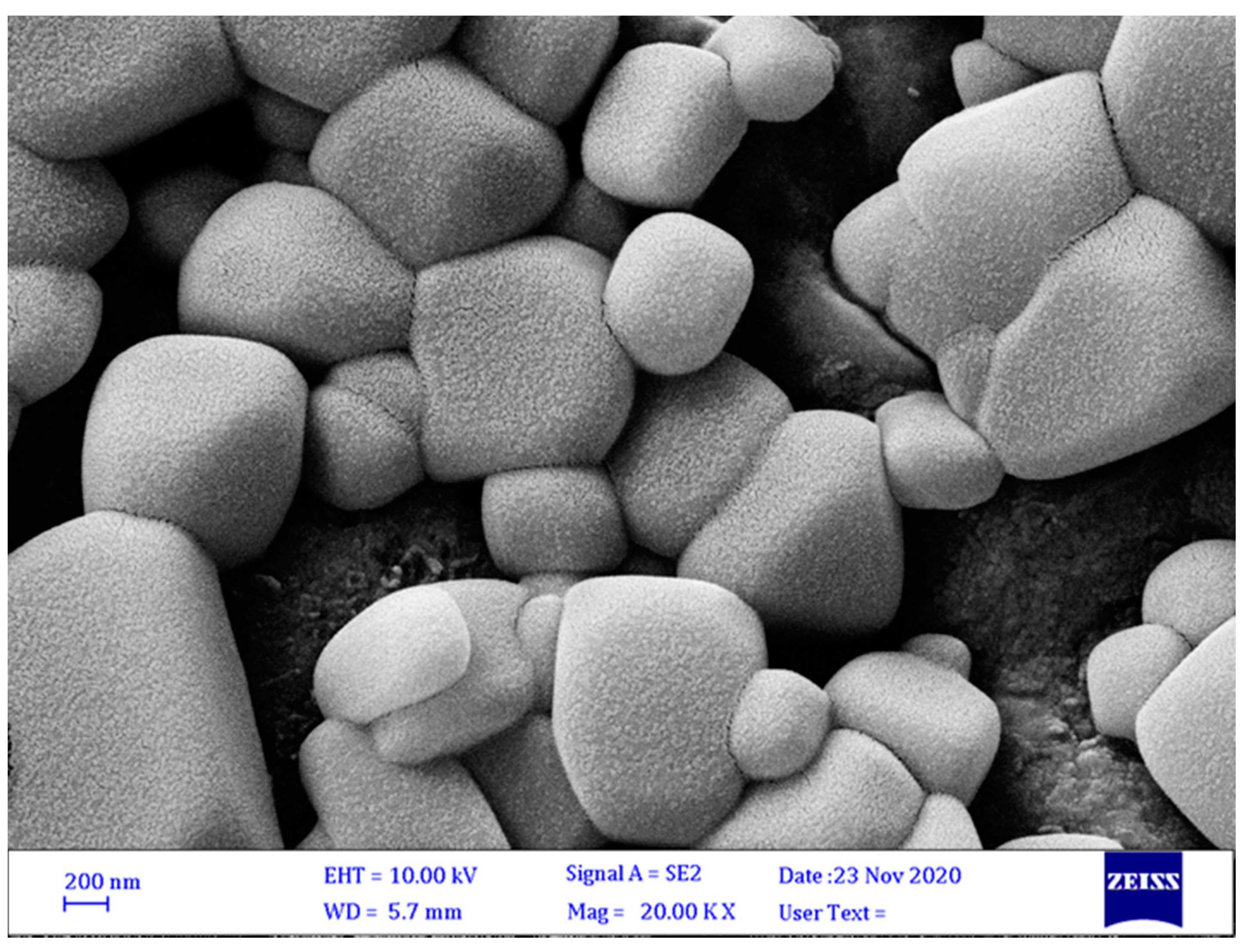
The average size of microcapsules may be dependent on the efficiency and stability of microencapsulation. Macrocapsules usually protect core material better than microcapsules but are less efficient at dispersing the final product. Therefore, it is essential to achieve a formulation with an almost uniform size with high efficiency. The optical microscopy images showed that ALG/AG microcapsules were fully cubic in shape with some irregularities, which can be effective in entrapping and releasing bacteria. The size of microcapsules was reported to be about 140–150 µm (Figure 2).

3.3.3. FTIR Analysis

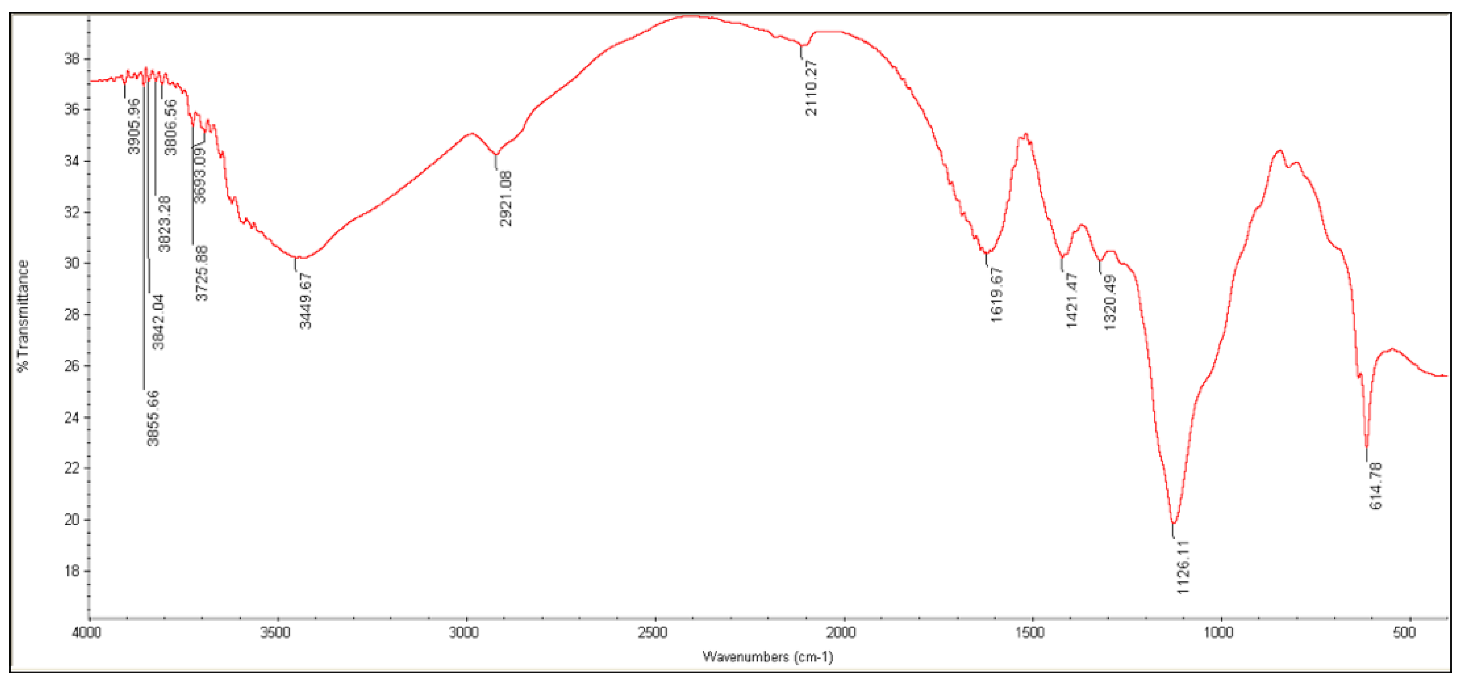
Each Fourier transform (FT-IR) spectrum is a unique physical property and characteristic of the molecule that includes the vibrational patterns of the molecule [45]. The FTIR spectroscopy was performed to identify the various ingredients of the microcapsules. The FTIR spectra obtained are presented for ALG, AG, and complex ALG/AG, respectively, in Figure 3, Figure 4 and Figure 5, while Table 2 shows the peaks and their band assignments between 400 to 4000 cm−1 for each ingredient used in the encapsulation process. The ALG spectra were determined as a reference, which revealed the characteristic presence bands at 1619 and 3449 cm−1, corresponding to C=O and O-H stretching vibrations, respectively.
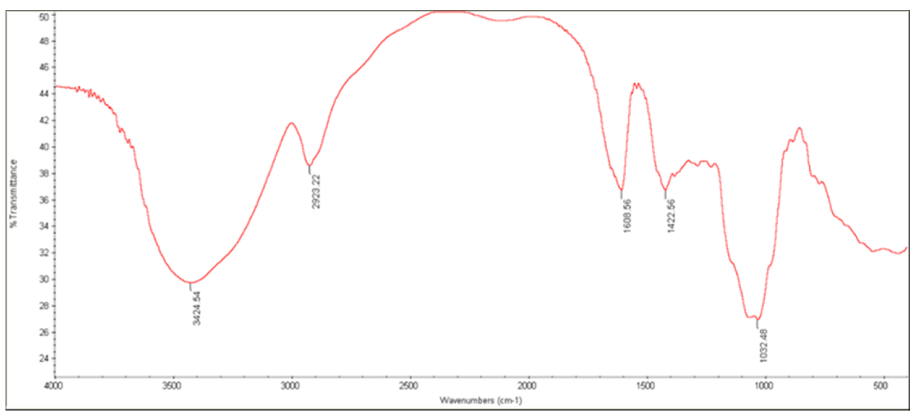
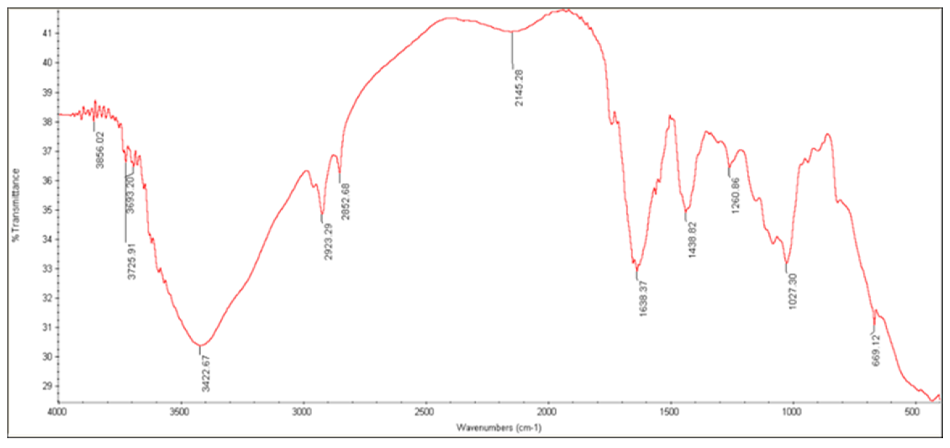
AG has shown characteristic peaks at 3424 cm−1 and 2923 cm−1, which is related to the -OH and -CH2, -CH3 aliphatic groups. Asymmetric and symmetric stretching of the COO− group was observed at 1608 cm−1. The peak at 1422 cm−1 is related to the -OH group bending of the acid group. In summary, the FT-IR spectra of ALG–AG microcapsules showed the significant characteristics of ALG and AG without any major deviation. The peaks near 1400 and 1600 cm−1 related to –COO (asymmetric) and –COO (symmetric) stretching, respectively, were mixed and showed new stronger peaks at the same regions. Therefore, ALG and AG are suitable complexes for bacteria encapsulation.

3.3.4. X-ray Diffraction

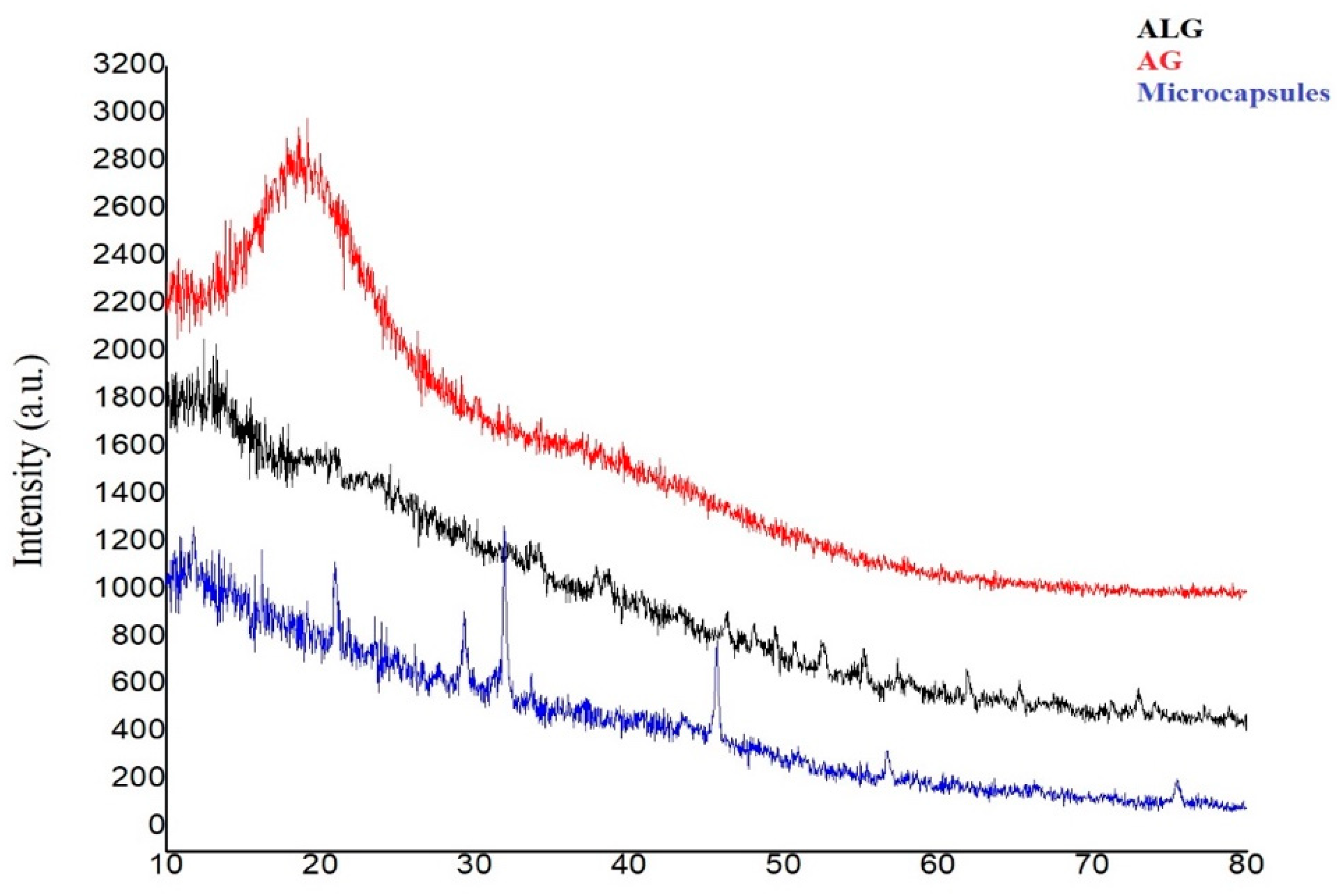
The XRD patterns of the ALG, AG and ALG–AG formulation are presented in Figure 6. The AG is amorphous with no distinct peak while the ALG exhibited no sharp peak. Meantime, for microcapsules we observed three peaks at 2θ = 20.92°, 2θ = 31.89° and 45.65°, which are related to SiO2 nanoparticles, and the peak observed at 2θ = 29.32° is due to the presence of TiO2 nanoparticles in the formulation. The XRD pattern and FTIR spectrum of ALG-AG no incompatible interaction showed. Therefore, this complex might be reported as appropriate materials for bacterial coating.

3.3.5. In Vitro Release of S. fulvissimus Uts22 from Microcapsules

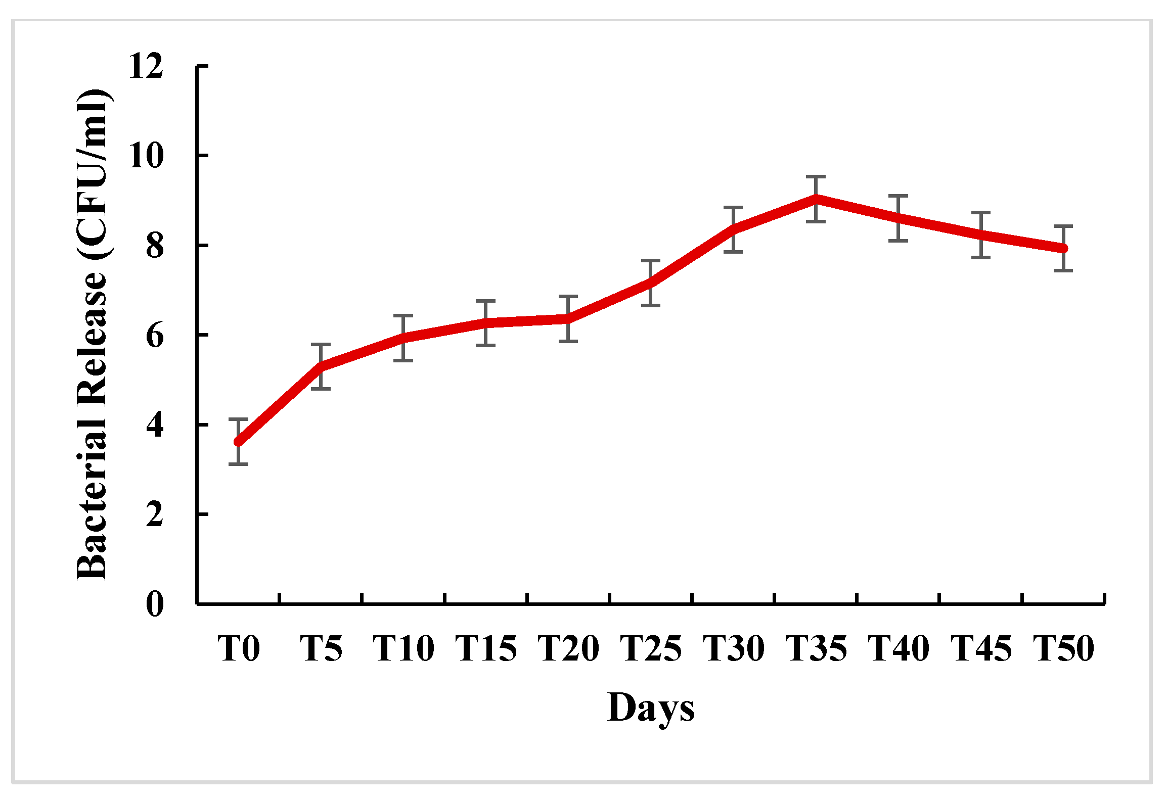
PGPR encapsulation has been extensively applied in agriculture to provide a protective and functionalized surrounding structure for microorganisms. Further, it ensures their gradual and prolonged release. The inoculants should be stored under room temperature but avoiding extreme temperature oscillations. Under these circumstances, the shelf life of liquid inoculants is extremely short and bacterial viability declines dramatically. Additionally, after introduction in the soil, the liquid inoculum has an instantaneous and very rapid release and the rhizobacteria are only delivered at the initial time of plant growth. In this study, we showed that bacterial release increased upwards until day 35, where the highest bacterial population was recorded (109 CFU/g) (Figure 7). Therefore, bioencapsulation allows a niche where rhizobacteria are preserved from soil stress. Beyond the 35th day, though remaining high, the population started to decrease gradually, likely due to environmental factors and the onset of the bacterial death rate.

3.3.6. Determination of S. fulvissimus Uts22 in the Cucumber Root

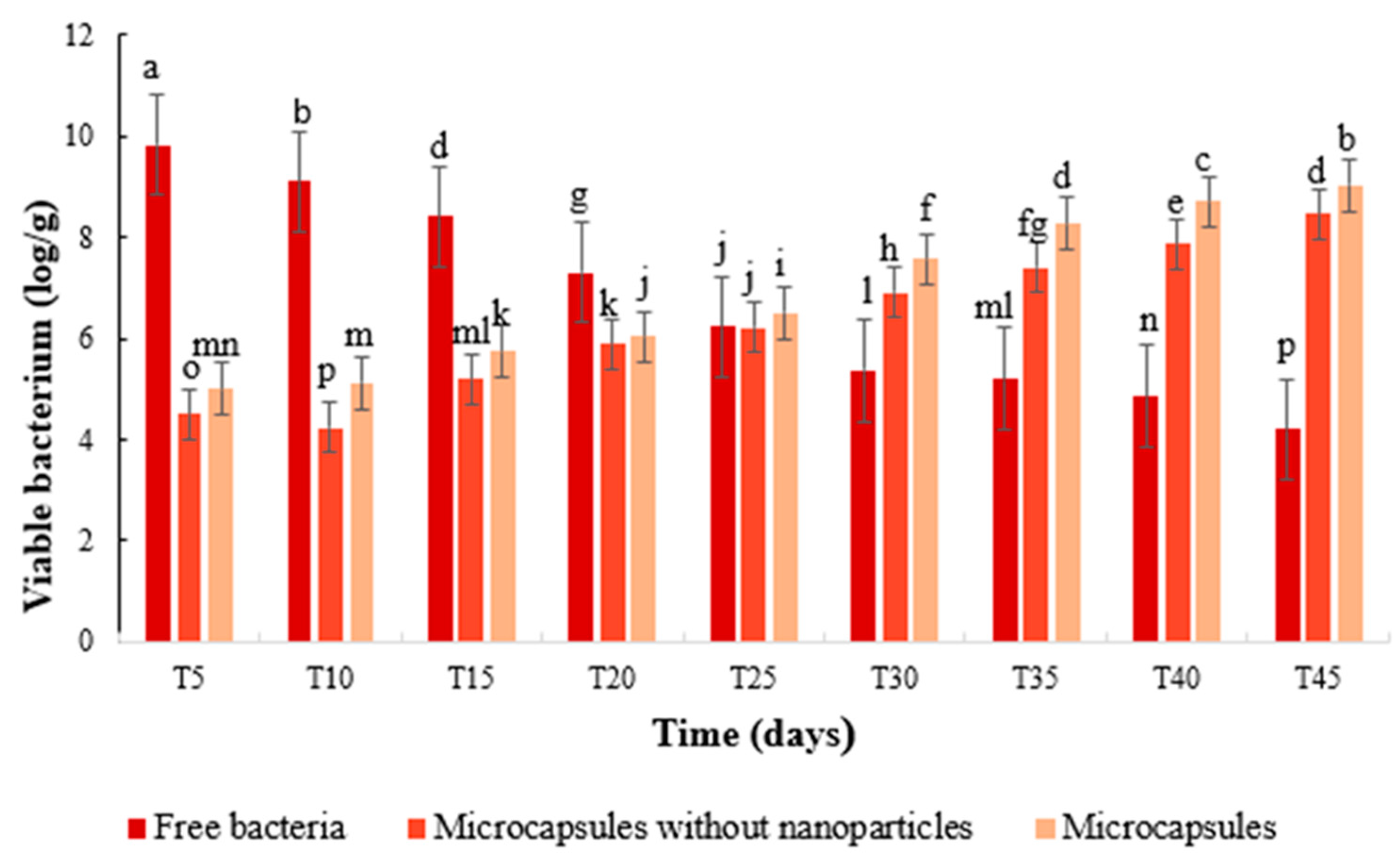
Root colonization by bacterial agents plays an important role in its ability to survive [25]. Therefore, the percentage of root colonization of the cucumber plant with free S. fulvissimus Uts22, encapsulated S. fulvissimus Uts22, microcapsules without nanoparticles, and microcapsules was investigated. Our results show that, from 5 to 20 days, the colonization rate by encapsulated S. fulvissimus Uts22 was lower than that of free S. fulvissimus Uts22 (Figure 8). Then, the rate of colonization became similar in treatments containing microcapsules and uncoated bacteria on day 25. However, the amount of colonization in the rhizosphere became significantly higher in cucumber plants inoculated with microencapsulated S. fulvissimus Uts22 compared to plants treated with free S. fulvissimus Uts22 or encapsulated Uts22 without nanoparticles starting from 30th days and up to 45th days. The S. fulvissimus Uts22 trapped in microcapsules was gradually and continuously released into the soil, leading to the colonization of the cucumber root. Further, according to our research, it was shown that cucumber root colonization by S. fulvissimus Uts22 microcapsules could occur at high levels at 45 days.

3.4. Greenhouse Experiments

By investigating the effect of the different treatments on all studied indicators (reduction of disease severity, dry and fresh weight of shoots and roots), the treatment with microencapsulated S. fulvissimus Uts22 showed the most excellent impact either in the presence or absence of the pathogen. The growth of cucumber seedlings, treated with S. fulvissimus Uts22 microcapsules, showed a significant (p ˂ 0.05) increase compared with cucumbers treated with other treatments. However, the growth of the inoculated cucumbers with the S. fulvissimus Uts22 microcapsules was less than plants treated with free bacteria during the first phase of the developmental growth. This is probably due to the fact that bacteria are gradually released from the microcapsules. Inoculation of cucumber plants with microcapsules and free bacteria indicated a suitable reaction compared with control plants. Shoot length of cucumbers treated with S. fulvissimus Uts22 microcapsules showed a significant (p ˂ 0.05) enhancement compared to the cucumbers treated with uncoated bacteria (Table 3). In addition, dry and fresh weights of shoots and roots treated with microencapsulated S. fulvissimus Uts22 were higher than treatment by uncoated bacteria or free microcapsules.
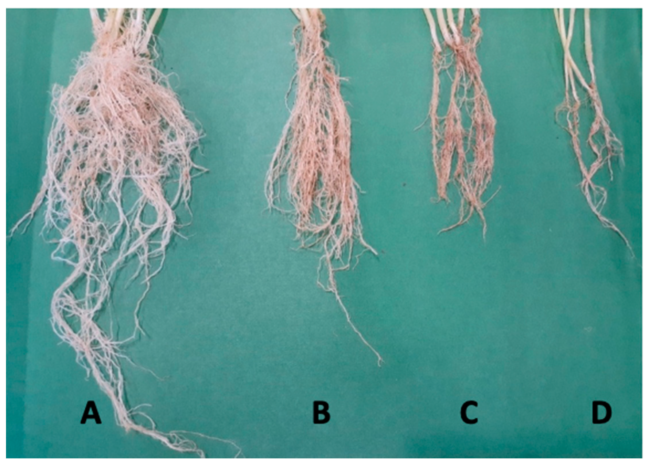
The results of the analysis of variance show a significant difference between treatments at the level of 5%. The addition of P. aphanidermatum in the soil decreases the root dry weights of cucumber plants to 69%. When bacterial microcapsules and free bacteria were added to the soil, these treatments showed a 70% and 40% increase in root dry weights, respectively (Figure 9). Inoculated plants treated with bacterial microcapsules had significantly larger root systems, compared to the Pythium-inoculated, free bacteria and control treatments. These results can be correlated with the IAA produced by S. fulvissimus Uts22 since IAA is an important factor that positively promotes the plants’ growth and development [39]. S. fulvissimus Uts22 microcapsules showed high potency (93.66%) to decrease the P. aphanidermatum infection, while the percent of disease control in cucumber treated with free bacteria and microcapsules without bacteria were 75% and 45%, respectively. According to ANOVA analysis, the results showed that the use of S. fulvissimus Uts22 microcapsules on cucumber plants in the greenhouse increased growth factors and controls the cucumber seedlings damping-off disease.
Streptomycetes are among most skilled chemists in nature and generate an amazing array and variety of bioactive secondary metabolites [46]. The literature is well documented on the mechanisms used by Streptomyces sp. to suppress pathogens in the soil, namely antibiosis, competition for nutrients, production of degradative enzymes, production of nitrous oxide, siderophores, quorum quenching and volatile metabolites [46]. Their ability to acclimate to different environmental circumstances in the rhizosphere makes them valuable competitors. The production of siderophores, which are iron chelators, deplete other microorganisms of this important micronutrient. In accordance with our results, siderophore production by S. griseorubiginouse was effective in the control of banana wilt caused by Fusarium oxysporum f. sp. Cubenese [47]. Further, as has been described in several previous studies, the S. fulvissimus Uts22 is able to secrete several fungal cell wall degrading enzymes, explaining partly the induced observed resistance of cucumber plantlets against P. aphanidermatum [46,48,49].
Biological control is a key component of plant pest and disease management system approaches [50]. In contrast to chemical pesticides, biological control does not proceed quickly. However, in successful cases, biological control has a more lasting effect than chemical pesticides. In this way, the effective role of Streptomyces isolates in the production of volatile compounds, siderophores, regulation of enzymes effective in plant growth, and positive effect on plant growth indices and microbial antagonism has been confirmed in several experiments [46,51,52].
The emerging application of nanotechnology in plant science is one of the current topics in the world that is in its infancy and is still expanding, and where many effects and functions of nanomaterials on physiological and metabolic mechanisms of plants are still unknown. The use of nanotechnology in recent decades has created extensive changes in all fields of science, and agricultural science is no exception to this rule [53]. Silicon is one of the many elements in the earth’s crust but most of its forms are not absorbable by plants. This element has positive effects on increasing water use efficiency and also on plant growth and yield, and increases production and product quality, reduces evapotranspiration, increases resistance to stresses such as drought and toxicity of heavy metals and the production of some antioxidant enzymes, and reduces fungal diseases in the plant [54,55]. TiO2 nanoparticles significantly increased the photosynthetic pigment content under cold stress conditions [56]. The combined effect of SiO2 and TiO2 nanoparticles on the germination and growth of soybean induced an increase in the percentage of soybean germination and growth [57].
As indicated earlier, streptomyces is a very valuable microorganism in various fields including agriculture and medicine. These bacteria are introduced in agriculture as rhizosphere colonizing bacteria and control agents of plant pathogenic fungi and also stimulate plant growth [46,58]. The study of the characteristics of biocontrol agents, and survival and protection of the biological characteristics of this microbial group, are important factors in their development for use in pathogen management. This is especially important in mass production and formulation [59]. The combined use of biocontrol agents and functional nanoparticles such as SiO2 and TiO2 in intelligent formulation can be a new approach in increasing plant growth and controlling plant diseases.

4. Conclusions

Due to the importance of microbial formulation and the effect of nanoparticles in improving plant growth conditions, in this study, we prepared a new formulation based on biopolymers and nanoparticles. Encapsulation of microbial agents, in addition to protecting the trapped agent in adverse environmental conditions, also increases the percentage of colonization by gradual release and improves its biocontrol function. By combining biological control and nanotechnology, the formulation presented in this study showed extraordinary performance in increasing plant growth and controlling damping-off diseases in cucumber seedlings. The results of greenhouse experiments showed that encapsulation of bacterial agents can play an important role in the control of plant pathogens. Based on the results, it was observed that both treatments of uncoated bacteria and encapsulated bacteria have a significant effect on increasing plant growth factors, however, with a significant advantage for encapsulated bacteria. The formulation provided in this study is economically very important for biocontrol producers and farmers and can pave the way for the development of biological controls.

Author Contributions

M.M.P.: Conceptualization, investigation, methodology and writing—original draft preparation. R.S.R.: Project administration, supervision, investigation, methodology and writing—review and editing. E.A.B.: Writing—review and editing. All authors have read and agreed to the published version of the manuscript.

Funding

This research was supported by the Vali-E-Asr University of Rafsanjan, Rafsanjan, Iran.

Institutional Review Board Statement

Not applicable.

Informed Consent Statement

Not applicable.

Data Availability Statement

Not applicable.

Acknowledgments

This research (AGR99PP11084) was funded by the research adjutancy of Vali-e-Asr University of Rafsanjan.

Conflicts of Interest

The authors declare no conflict of interest.

References

  1. Ni, L.; Punja, Z.K. Management of Fungal Diseases on Cucumber (Cucumis sativus L.) and Tomato (Solanum lycopersicum L.) Crops in Greenhouses Using Bacillus subtilis. In Bacilli and Agrobiotechnology: Phytostimulation and Biocontrol. Bacilli in Climate Resilient Agriculture and Bioprospecting; Islam, M., Rahman, M., Pandey, P., Boehme, M., Haesaert, G., Eds.; Springer: Cham, Switzerland, 2019. [Google Scholar]
  2. Halo, B.A.; Al-Yahyai, R.A.; Al-Sadi, A.M. Biological control of Pythium aphanidermatum-induced cucumber and radish damping-off by an endophytic fungus, Cladosporium omanense isolate 31R. Biocontrol. Sci. Technol. 2020, 31, 235–251. [Google Scholar] [CrossRef]
  3. Noble, R.; Coventry, E. Suppression of soil-borne plant diseases with composts: A review. Biocontrol. Sci. Technol. 2005, 15, 3–20. [Google Scholar] [CrossRef]
  4. Nega, A. Review on Concepts in Biological Control of Plant Pathogens. J. Biol. Agric. Healthc. 2014, 4, 33–55. [Google Scholar]
  5. Kaur, P.; Purewal, S.S. Biofertilizers and Their Role in Sustainable Agriculture. In Biofertilizers for Sustainable Agriculture and Environment; Giri, B., Prasad, R., Wu, Q.S., Varma, A., Eds.; Springer: Cham, Switzerland, 2019; Volume 55. [Google Scholar]
  6. Adnan, M.; Islam, W.; Shabbir, A.; Khan, K.; Ghramh, H.A.; Huang, Z.; Chen, H.Y.; Lu, G.-D. Plant defense against fungal pathogens by antagonistic fungi with Trichoderma in focus. Microb. Pathog. 2019, 129, 7–18. [Google Scholar] [CrossRef]
  7. Hu, D.; Li, S.; Li, Y.; Peng, J.; Wei, X.; Ma, J.; Zhang, C.; Jia, N.; Wang, E.; Wang, Z. Streptomyces sp. strain TOR3209: A rhizosphere bacterium promoting growth of tomato by affecting the rhizosphere microbial community. Sci. Rep. 2020, 10, 20132. [Google Scholar] [CrossRef]
  8. Chaiharn, M.; Theantana, T.; Pathom-Aree, W. Evaluation of Biocontrol Activities of Streptomyces spp. against Rice Blast Disease Fungi. Pathogens 2020, 9, 126. [Google Scholar] [CrossRef] [Green Version]
  9. Lobo, C.B.; Tomas, M.S.; Viruel, E.; Ferrero, M.A.; Lucca, M.E. Development of low-cost formulations of plant growth-promoting bacteria to be used as inoculants in beneficial agricultural technologies. Microbiol. Res. 2018, 219, 12–25. [Google Scholar] [CrossRef]
  10. Saberi-Riseh, R.; Moradi-Pour, M.; Mohammadinejad, R.; Thakur, V. Biopolymers for Biological Control of Plant Pathogens: Advances in Microencapsulation of Beneficial Microorganisms. Polymers 2021, 13, 1938. [Google Scholar] [CrossRef]
  11. Pour, M.M.; Saberi-Riseh, R.; Mohammadinejad, R.; Hosseini, A. Nano-Encapsulation of Plant Growth-Promoting Rhizobacteria and Their Metabolites Using Alginate-Silica Nanoparticles and Carbon Nanotube Improves UCB1 Pistachio Micropropagation. J. Microbiol. Biotechnol. 2019, 29, 1096–1103. [Google Scholar] [CrossRef]
  12. Berninger, T.; López, Ó.G.; Bejarano, A.; Preininger, C.; Sessitsch, A. Maintenance and assessment of cell viability in formulation of non-sporulating bacterial inoculants. Microb. Biotechnol. 2018, 11, 277–301. [Google Scholar] [CrossRef] [Green Version]
  13. Schoebitz, M.; López, M.D.; Roldán, A. Bioencapsulation of microbial inoculants for better soil-plant fertilization. A review. Agron. Sustain. Dev. 2013, 33, 751–765. [Google Scholar] [CrossRef]
  14. Riseh, R.S.; Ebrahimi-Zarandi, M.; Tamanadar, E.; Pour, M.M.; Thakur, V.K. Salinity Stress: Toward Sustainable Plant Strategies and Using Plant Growth-Promoting Rhizobacteria Encapsulation for Reducing It. Sustainability 2021, 13, 12758. [Google Scholar] [CrossRef]
  15. Albadran, H.A.; Chatzifragkou, A.; Khutoryanskiy, V.V.; Charalampopoulos, D. Stability of probiotic Lactobacillus plantarum in dry microcapsules under accelerated storage conditions. Food Res. Int. 2015, 74, 208–216. [Google Scholar] [CrossRef] [PubMed]
  16. D’Orazio, G.; Di Gennaro, P.; Boccarusso, M.; Presti, I.; Bizzaro, G.; Giardina, S.; Michelotti, A.; Labra, M.; La Ferla, B. Microencapsulation of new probiotic formulations for gastrointestinal delivery: In vitro study to assess viability and biological properties. Appl. Microbiol. Biotechnol. 2015, 99, 9779–9789. [Google Scholar] [CrossRef]
  17. Ke, Y.; Liu, G.S.; Wang, J.H.; Xue, W.; Du, C.; Wu, G. Preparation of carboxymethyl cellulose based microgels for cell encapsulation. Express Polym. Lett. 2014, 8, 841–849. [Google Scholar] [CrossRef]
  18. Keskin, N.O.S.; Celebioglu, A.; Sarioglu, O.F.; Uyar, T.; Tekinay, T. Encapsulation of living bacteria in electrospun cyclodextrin ultrathin fibers for bioremediation of heavy metals and reactive dye from wastewater. Colloids Surf. B Biointerfaces 2018, 161, 169–176. [Google Scholar] [CrossRef] [PubMed] [Green Version]
  19. Sarioglu, O.F.; Keskin, N.O.S.; Celebioglu, A.; Tekinay, T.; Uyar, T. Bacteria immobilized electrospun polycaprolactone and polylactic acid fibrous webs for remediation of textile dyes in water. Chemosphere 2017, 184, 393–399. [Google Scholar] [CrossRef] [PubMed] [Green Version]
  20. Riseh, R.S.; Skorik, Y.A.; Thakur, V.K.; Pour, M.M.; Tamanadar, E.; Noghabi, S.S. Encapsulation of Plant Biocontrol Bacteria with Alginate as a Main Polymer Material. Int. J. Mol. Sci. 2021, 22, 11165. [Google Scholar] [CrossRef]
  21. Pour, M.M.; Saberi-Riseh, R.; Mohammadinejad, R.; Hosseini, A. Investigating the formulation of alginate- gelatin encapsulated Pseudomonas fluorescens (VUPF5 and T17-4 strains) for controlling Fusarium solani on potato. Int. J. Biol. Macromol. 2019, 133, 603–613. [Google Scholar] [CrossRef]
  22. Li, X.Y.; Chen, X.G.; Cha, D.S.; Park, H.J.; Liu, C. Microencapsulation of a probiotic bacteria with alginate–gelatin and its properties. J. Microencapsul. 2008, 26, 315–324. [Google Scholar] [CrossRef]
  23. Vejan, P.; Abdullah, R.; Khadiran, T.; Ismail, S. Encapsulation of Bacillus salmalaya 139SI using Double Coating Biopolymer Technique. Lett. Appl. Microbiol. 2019, 68, 56–63. [Google Scholar] [CrossRef] [Green Version]
  24. He, Y.; Wu, Z.; Ye, B.-C.; Wang, J.; Guan, X.; Zhang, J. Viability evaluation of alginate-encapsulated Pseudomonas putida Rs-198 under simulated salt-stress conditions and its effect on cotton growth. Eur. J. Soil Biol. 2016, 75, 135–141. [Google Scholar] [CrossRef]
  25. Saberi Riseh, R.; Moradi Pour, M. The effect of Bacillus subtilis Vru1 encapsulated in –bentonite coating enriched with titanium nanoparticles against Rhizoctonia solani on bean. Int. J. Biol. Macromol. 2020, 152, 1089–1097. [Google Scholar] [CrossRef] [PubMed]
  26. Alfredo, M.S.; Aleli Cornelia, R.P. Biological control of sheath blight of upland rice with Trichoderma species. J. Trop. Plant Pathol. 2011, 69, 1–9. [Google Scholar]
  27. Lee, J.; Tan, W.; Ting, A. Revealing the antimicrobial and enzymatic potentials of culturable fungal endophytes from tropical pitcher plants (Nepenthes spp.). Mycosphere 2014, 5, 364–377. [Google Scholar] [CrossRef]
  28. Alexander, D.B.; Zuberer, D.A. Use of chrome azurol S reagents to evaluate siderophore production by rhizosphere bacteria. Biol. Fertil. Soils 1991, 12, 39–45. [Google Scholar] [CrossRef]
  29. Patten, C.L.; Glick, B.R. Bacterial biosynthesis of indole-3-acetic acid. Can. J. Microbiol. 1996, 42, 207–220. [Google Scholar] [CrossRef] [PubMed]
  30. Kasana, R.C.; Salwan, R.; Dhar, H.; Dutt, S.; Gulati, A. A Rapid and Easy Method for the Detection of Microbial Cellulases on Agar Plates Using Gram’s Iodine. Curr. Microbiol. 2008, 57, 503–507. [Google Scholar] [CrossRef]
  31. Maurhofer, M.; Keel, C.; Haas, D.; Défago, G. Influence of plant species on disease suppression by Pseudomonas fluorescens strain CHAO with enhanced antibiotic production. Plant Pathol. 1995, 44, 40–50. [Google Scholar] [CrossRef]
  32. Omidvari, M. Biological Control of Fusarium solani, the Causal Agent of Damping off, by Fluorescent Pseudomonads and Studying Some of Their Antifungal Metabolite Productions on It. Master’s Thesis, Tehran University, Tehran, Iran, 2015; p. 94. (In Persian). [Google Scholar]
  33. Krasaekoopt, W.; Bhandari, B.; Deeth, H.C. Survival of probiotics encapsulated in chitosan-coated alginate beads in yoghurt from UHT- and conventionally treated milk during storage. LWT 2006, 39, 177–183. [Google Scholar] [CrossRef]
  34. Saberi-Riseh, R.; Moradi-Pour, M. A novel encapsulation of Streptomyces fulvissimus Uts22 by spray drying and its biocontrol efficiency against Gaeumannomyces graminis, the causal agent of take-all disease in wheat. Pest Manag. Sci. 2021, 77, 4357–4364. [Google Scholar] [CrossRef]
  35. Kloepper, J.W.; Leong, J.; Teintze, M.; Schroth, M.N. Enhanced plant growth by siderophores produced by plant growth-promoting rhizobacteria. Nature 1980, 286, 885–886. [Google Scholar] [CrossRef]
  36. Challis, G.L.; Hopwood, D.A. Synergy and contingency as driving forces for the evolution of multiple secondary metabolite production by Streptomyces species. Proc. Natl. Acad. Sci. USA 2003, 100, 14555–14561. [Google Scholar] [CrossRef] [PubMed] [Green Version]
  37. Shih-Yung, H. IAA Production by Streptomyces Scabies and Its Role in Plant Microbe Interaction. Master’s Thesis, Cornell University, Ithaca, NY, USA, 2010. [Google Scholar]
  38. Vejan, P.; Abdullah, R.; Khadiran, T.; Ismail, S.; Boyce, A.N. Role of Plant Growth Promoting Rhizobacteria in Agricultural Sustainability—A Review. Molecules 2016, 21, 573. [Google Scholar] [CrossRef] [PubMed]
  39. Egamberdieva, D. Indole-acetic acid production by root associated bacteria and its role in plant growth and development. In Auxins: Structure, Biosynthesis and Functions; Keller, A.H., Fallon, M.D., Eds.; Nova Science Publishers: Hauppauge, NY, USA, 2011; pp. 1–14. [Google Scholar]
  40. Datta, C.; Basu, P. Indole acetic acid production by a Rhizobium species from root nodules of a leguminous shrub Cajanus cojan. Microbiol. Res. 2000, 155, 123–127. [Google Scholar] [CrossRef]
  41. Ahmed, A.; Hasanian, S. Auxins as one of the factors of plant growth improvement by plant growth promoting rhizobacteria. Pol. J. Microbiol. 2014, 63, 261–266. [Google Scholar] [CrossRef]
  42. Yarullina, L.G.; Akhatova, A.R.; Kasimova, R.I. Hydrolytic enzymes and their proteinaceous inhibitors in regulation of plant–pathogen interactions. Russ. J. Plant Physiol. 2016, 63, 193–203. [Google Scholar] [CrossRef]
  43. Keel, C.; Defago, G. Interactions between beneficial soil bacteria and root pathogens: Mechanisms and ecological impact. In Multitrophic Interactions in Terrestrial System; Gange, A.C., Brown, V.K., Eds.; Blackwell Science Oxford, UK: 1997; pp. 27–47.
  44. Çabuk, B.; Tellioglu, H. Protection of Lactobacillus acidophilus NRRL-B 4495 under in vitro gastrointestinal conditions with whey protein/pullulan microcapsules. J. Biosci. Bioeng. 2015, 120, 650–656. [Google Scholar] [CrossRef] [Green Version]
  45. Coates, J. Interpretation of Infrared Spectra, a Practical Approach. In Encyclopedia of Analytical Chemistry; Meyers, R.A., Ed.; John Wiley & Sons Ltd.: Chichester, UK, 2000; pp. 10815–10837. [Google Scholar]
  46. Barka, E.A.; Vatsa, P.; Sanchez, L.; Gaveau-Vaillant, N.; Jacquard, C.; Klenk, H.-P.; Clément, C.; Ouhdouch, Y.; van Wezel, G.-P. The Actinobacteria: Taxonomy, Physiology and their natural products. Microbiol. Mol. Biol. Rev. 2016, 80, 1–43. [Google Scholar] [CrossRef] [Green Version]
  47. Getha, K.; Vikineswary, S.; Wong, W.H.; Seki, T.; Ward, A.; Goodfellow, M. Evaluation of Streptomyces sp. strain g10 for suppression of Fusarium wilt and rhizosphere colonization in pot-grown banana plantlets. J. Ind. Microbiol. Biotechnol. 2005, 32, 24–32. [Google Scholar] [CrossRef]
  48. Errakhi, R.; Bouteau, F.; Lebrihi, A.; Barakate, M. Evidences of biological control capacities of Streptomyces spp. against Sclerotium rolfsii responsible for damping-off disease in sugar beet (Beta vulgaris L.). World J. Microbiol. Biotechnol. 2007, 23, 1503–1509. [Google Scholar] [CrossRef]
  49. El-Tarabily, K.A.; Sivasithamparam, K. Non-streptomycete actinomycetes as biocontrol agents of soil-borne fungal plant pathogens and as plant growth promoters. Soil Biol. Biochem. 2006, 38, 1505–1520. [Google Scholar] [CrossRef]
  50. Sarwar, A.; Latif, Z.; Zhang, S.; Zhu, J.; Zechel, D.L.; Bechthold, A. Biological Control of Potato Common Scab With Rare Isatropolone C Compound Produced by Plant Growth Promoting Streptomyces A1RT. Front. Microbiol. 2018, 9, 1126. [Google Scholar] [CrossRef] [PubMed] [Green Version]
  51. Dias, M.P.; Bastos, M.S.; Xavier, V.B.; Cassel, E.; Astarita, L.V.; Santarém, E.R. Plant growth and resistance promoted by Streptomyces spp. in tomato. Plant Physiol. Biochem. 2017, 118, 479–493. [Google Scholar] [CrossRef] [PubMed]
  52. Cordovez, V.; Carrion, V.J.; Etalo, D.W.; Mumm, R.; Zhu, H.; van Wezel, G.P. Diversity and functions of volatile organic compounds produced by Streptomyces from a disease-suppressive soil. Front. Microbiol. 2015, 6, 1081. [Google Scholar] [CrossRef] [PubMed] [Green Version]
  53. Peyvandi, M.; Mirza, M.; Jamakani, Z.K. The effect of nano Fe chelate and Fe chelate on the growth and activity of some antioxidant. New Cell. Mol. Biotechnol. 2011, 2, 25–32. (In Farsi) [Google Scholar]
  54. Ma, J.F. Role of silicon in enhancing the resistance of plants to biotic and abiotic stresses. Soil Sci. Plant Nutr. 2004, 50, 11–18. [Google Scholar] [CrossRef]
  55. Gao, X.; Zou, C.; Wang, L.; Zhang, F. Silicon Decreases Transpiration Rate and Conductance from Stomata of Maize Plants. J. Plant Nutr. 2006, 29, 1637–1647. [Google Scholar] [CrossRef]
  56. Kardavan, G.; Karamian., R. The effects of titanium dioxide nanoparticles and spermine on physiological responses of licorice (Glycyrrhiza glabra L.) to cold stress. J. Plant Physiol. Breed. 2020, 10, 93–110. [Google Scholar]
  57. Lu, C.M.; Zhang, C.Y.; Wen, J.Q.; Wu, G.R.; Tao, M.Z. Research of the effect of nanometer materials on germination and growth enhancement of glycine max and its mechanism. Soybean Sci. 2002, 21, 168–171. [Google Scholar]
  58. Hastuti, R.D.; Lestari, Y.; Saraswati, R.; Suwanto, A. Chaerani Capability of Streptomyces spp. in Controlling Bacterial Leaf Blight Disease in Rice Plants. Am. J. Agric. Biol. Sci. 2012, 7, 217–223. [Google Scholar] [CrossRef] [Green Version]
  59. Junaid, J.M.; Dar, N.A.; Bhat, T.A.; Bhat, A.H.; Bhat, M.A. Commercial Biocontrol Agents and Their Mechanism of Action in the Management of Plant Pathogens. Int. J. Mod. Plant Anim. Sci. 2013, 1, 39–57. [Google Scholar]
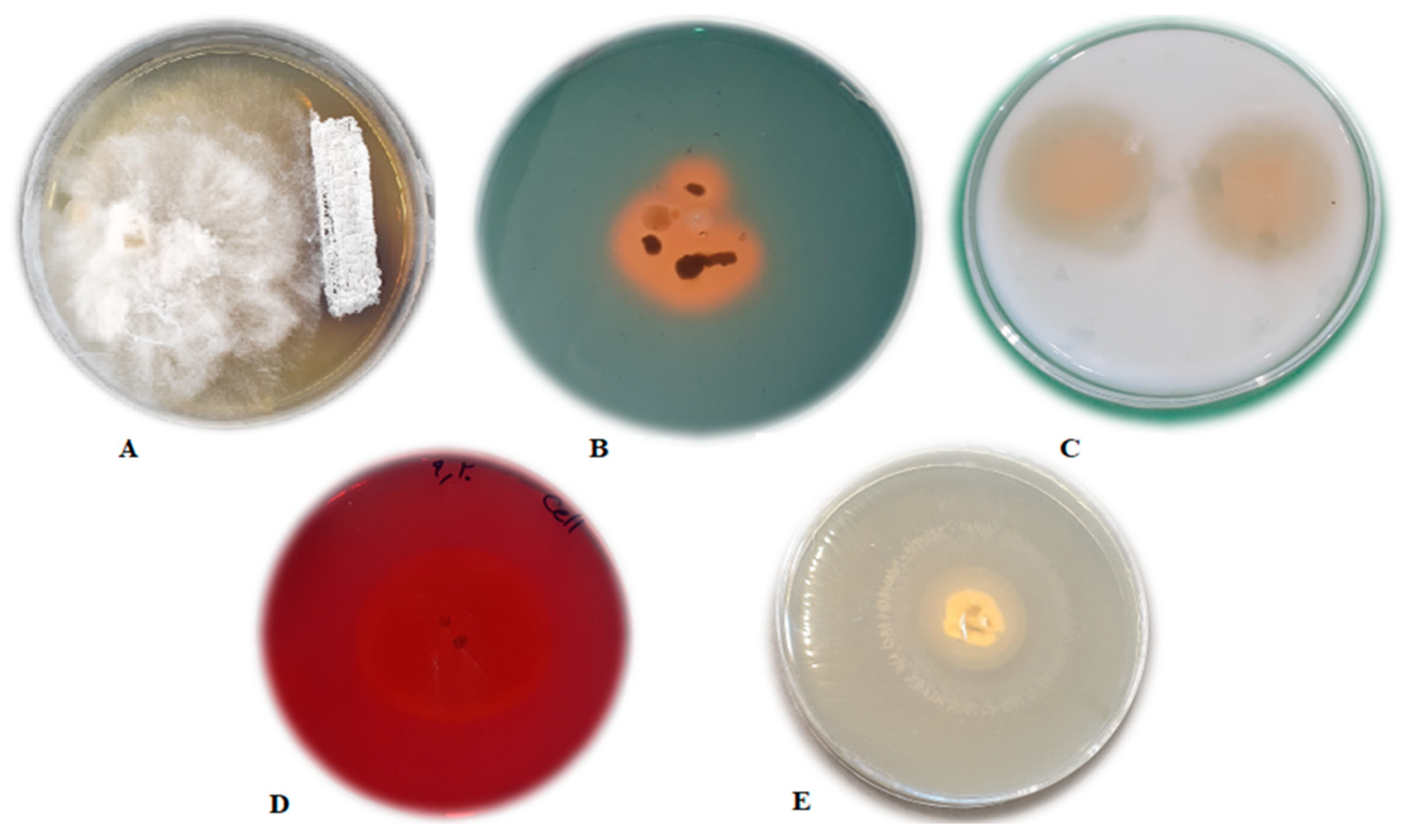
Figure 1. (A) Inhibition zone; (B) production of siderophores; (C) protease production; (D) cellulase production; (E) lipase production.
Agronomy 12 00655 g001
Figure 2. Morphology of microcapsules under the scanning electron microscope.
Agronomy 12 00655 g002
Figure 3. FTIR spectra of alginate.
Agronomy 12 00655 g003
Figure 4. FTIR spectra of Arabic gum.
Agronomy 12 00655 g004
Figure 5. FTIR spectra of ALG–AG complex.
Agronomy 12 00655 g005
Figure 6. XRD patterns of alginate, Arabic gum and alginate–Arabic gum microcapsules.
Agronomy 12 00655 g006
Figure 7. Release of S. fulvissimus Uts22 from microcapsules in phosphate buffer.
Agronomy 12 00655 g007
Figure 8. Changes in the viable amount of S. fulvissimus Uts22 colonizing on the cucumber root by free bacteria, microcapsules without nanoparticles and microcapsules treatments. (X-axis is 5th to 45th days after the start of bacterial release from the capsule and the Y-axis is logarithm of the viable bacteria has been released). Different letters indicate significant differences among treatments.
Agronomy 12 00655 g008
Figure 9. The effect of S. fulvissimus Uts22 strain and its microcapsules on increasing the root in cucumber plants: (A) S. fulvissimus Uts22 microcapsules; (B) S. fulvissimus Uts22 bacteria; (C) control; (D) P. aphanidermatum.
Agronomy 12 00655 g009
Table 1. Experimental protocol followed for obtaining suspension of S. fulvissimus Uts22 to study their potential on cucumber.
Treatments
S. fulvissimus Uts22
FreeEncapsulatedWheat Seeds with
P. aphanidermatum
T110 mL00 g
T210 mL 4 g
T3 10 g0 g
T4 10 g4 g
T5 4 g
T6Microcapsules without bacterium
T7Control
Table 2. Positions of main FTIR stretching bands and their relative assignments for ALG, AG and ALG–AG complex.
Characteristic GroupWavelength (cm−1)
ALGAG
C—O stretch11261022
C—O—C stretch1320-
C=C stretch--
C=O stretch16191608
C—H stretch29212923
O—H stretch34493424
C(=O)—O stretch14211618
Table 3. Efficacy of S. fulvissimus Uts22 bacteria and its microcapsules on growth parameters in cucumber plants. Shoot fresh weight (SF), shoot dry weight (SD), root fresh weight (RF), root dry weight (RD).
TreatmentsSF (g)SD (g)RF (g)RD (g)
T1S. fulvissimus Uts22 bacteria15.76 c5.27 c14.28 c4.31 c
T2S. fulvissimus Uts22 bacteria + P. aphanidermatum15.16 d5.27 d14.28 c4.31 c
T3S. fulvissimus Uts22 microcapsules17.6 a6.24 a15.36 a5.28 a
T4S. fulvissimus Uts22 microcapsules + P. aphanidermatum16.11 b5.98 b15.08 b5.06 b
T5P. aphanidermatum8.11 g1.82 g9.11 f2.15 g
T6Microcapsule without bacteria14.23 e3.95 e13.94 d3.29 e
T7Control12.08 f3.06 f11.19 e3.11 f
Numbers with different letters show significant differences at p ≤ 0.05 (LSD).
Publisher’s Note: MDPI stays neutral with regard to jurisdictional claims in published maps and institutional affiliations.