Article

Sodium Selenate: An Environmental-Friendly Means to Control Tomato Bacterial Speck Disease

Department of Agricultural, Food and Environmental Sciences, University of Perugia, 06121 Perugia, Italy
*
Author to whom correspondence should be addressed.
Agronomy 2022, 12(6), 1351; https://doi.org/10.3390/agronomy12061351
Submission received: 23 April 2022 / Revised: 26 May 2022 / Accepted: 30 May 2022 / Published: 1 June 2022

Abstract

:
Selenium (Se) is a micronutrient which plays a beneficial role in plants. Its adsorption at low doses can stimulate plant growth and enhance the plant’s capacity to respond to abiotic stresses, such as salinity, drought, cold and hot temperature, probably due to its antioxidant properties. Here, the effect of selenium supplied in soil-drench treatments as sodium selenate (Na2SeO4) at the dose of 4 mg L−1 (21.17 µM) per plant was studied on tomato (Solanum lycopersicum L.; cv. Rio Grande) against Pseudomonas syringae pv. tomato (Pst), the causal agent of tomato bacterial speck. Sodium selenate treated tomato plants challenged with Pst showed a reduction in disease severity expressed as percentage of diseased area and number of lesions per leaf. Furthermore, Pst bacterial cells were unable to proliferate in treated tomato plants. The effect of sodium selenate against Pst was also assessed in vitro, demonstrating that the growth of the bacterium was affected in a dose-dependent manner (EC50 = 42 ppm). It is notable that in tomato plants treated with sodium selenate at the above reported dose, a marked callose deposition was observed as well as the expression of the salicylic-acid-responsive tomato ‘pathogenesis-related protein 1b1’ (PR1b1) but not of the jasmonate-mediated ‘proteinase inhibitor 2’ (PIN2) genes. Induced defence responses and direct antimicrobial activity protect treated tomato plants against Pst attacks, suggesting the potential of sodium selanate as an environmentally friendly and effective bacterial control means. Moreover, the increased Se content in treated tomatoes offers an effective approach to reduce Se deficiency problems in human diets.

1. Introduction

Public concern over the widespread use of synthetic pesticides and the needs for alternative control strategies requires the development of new methods for pathogen control that are able to improve food safety and quality and are acceptable to the public. This is a very sensitive issue that underlies the EU Directive 2009/128/EC (https://ec.europa.eu/food/plants/pesticides/sustainable-use-pesticides) (accessed on 28 January 2022). Among the most promising alternatives to chemical pesticides is the use of biochemical pesticides (i.e., substances found in animals, microbes, minerals or plants) that can specifically target pathogens, reducing adverse impacts on food safety, non-target organisms and the environment.
For a long time considered toxic for living organisms, selenium (Se) has been known to be an essential element since 1957 [1], with a very narrow range between deficiency and toxicity [2,3]. Selenium deficiency occurs in several countries, especially where its concentration in soil and food crops is very low [4]. The World Health Organization (WHO) and the United States Department of Agriculture (USDA) recommend a human dietary intake of 50 µg/day of Se as it acts as a cofactor for antioxidant enzymes and regulates thyroid hormone metabolism and immune functions. Additionally, it helps prevent Keshan and Kashin-Beck diseases [5], cancer and male sterility [6,7,8,9,10] and weakening of the immune defence system [11,12]. Since plant foods are the major dietary sources of Se in most countries around the world, increasing the Se content in food crops offers an effective approach to reducing Se deficiency problems for humans and animals [13,14]. Furthermore, Se is used in biomedicine, biochemistry and environmental science [15,16,17,18,19,20]. It has been demonstrated that, in plants, Se is accumulated and transformed into bioactive molecules which have nutraceutical values [21,22]. Hence, Se is a beneficial non-essential element for plants which promotes plant growth [3], enhances the ability to resist abiotic stresses such as heavy metal contamination and improves drought tolerance [15,23]. In addition, it protects crops against ultraviolet B, salt, senescence, cold, high temperature, and desiccation [23]. Treatments with appropriate Se concentrations inhibit the growth and spread of many phytopathogenic fungi and bacteria [24,25,26,27,28]; therefore, Se represents a feasible alternative for plant disease control, especially for those caused by phytopathogenic bacteria for which preventive and curative control means are limited.
Tomato (Solanum lycopersicum L.) is one of the most important horticultural crops with a world production of 38.7 million tonnes in 2021 [29]. Tomato production in Europe, in particular in the Mediterranean countries, is increasing and it is second to China (https://www.atlasbig.com/) (accessed on 28 January 2022). Tomato cultivation is subject, in all environments, to numerous adversities with varying aetiologies, both biotic and abiotic. Concerning biotic adversities, tomato is susceptible to various viruses, bacteria and fungi which can cause very severe diseases and are limiting factors to tomato yield and quality. Bacterial speck disease, caused by Pseudomonas syringae pv. tomato (Pst) (Okabe) Young, Dye & Wilkie, is one of the most common and often serious diseases affecting tomatoes. Currently, effective methods for containing the pathogen and suppressing losses are prevention and the use of copper fungicides in conjunction with maneb. However, there is an urgent need to reduce the risks associated with chemical products of pollution in the environment and human health, and with the development of copper-resistant strains of Pst [30], it is necessary to develop alternative control strategies for disease management that are both effective and environmentally friendly.
In this study, the effect of sodium selenate on bacterial speck disease severity, Pst growth in vitro and in planta was tested. Furthermore, the induction of morphological (i.e., callose deposition) and biochemical defence responses (i.e., PR1b1 and PIN2 gene expression) in tomato plants was studied.

2. Materials and Methods

2.1. Plant Material and Treatments

Seeds of tomato plants (Solanum lycopersicum L.; cv. Rio Grande) were grown in a seedbed containing modular tray substrate (Klasmann-Deilmann, GmbH; Geeste, Germany) in a growth chamber programmed for a 12-h day at 25 °C and 12-h night at 18 °C with 70 ± 90% RH. Cool white fluorescent lamps provided 240 µE m−2 s−1 illumination. At the 2nd to 3rd true leaf stage, plants were transplanted into 9 × 9 × 12.5 cm pots (one plant per pot) containing the modular tray substrate (140 g per pot) indicated above. To evaluate the ability of sodium selenate (Na2SeO4) to protect tomato plants against Pst attacks, a solution of sodium selenate (4 ppm; 21.17 µM) or, for comparison, acibenzolar-S-methyl (ASM) (benzo-(1,2,3)-thiadiazole-7-carbothioic acid S-methyl ester) (50% water dispersible granule; 45 mg active ingredient L−1; 213.98 µM) were applied to the transplanted plants as soil drench (100 mL per pot). In a preliminary study, we have verified that this sodium selenate dose is non-phytotoxic (data not shown). According to [31,32,33], a water solution of ASM 2.1 × 10⁴ mol L−1 was applied. One hundred ml of water were applied to the control plants. Starting from 2 days after the treatments, all plants were irrigated with 50 mL of water per pot every two days until the end of the experiment.

2.2. Pathogen, Inoculation and Disease Evaluation

The strain DAPP-PG 215 (race 0) of Pseudomonas syringae pv. tomato (Pst) from the phytopathogenic bacterial collection of the Plant Protection Section of the Department of Agricultural, Food and Environmental Sciences, University of Perugia (Italy) was used in the present study [34]. The strain was maintained at −80 °C in vials containing 15% glycerol.
To prepare the inoculum, Pst was grown on nutrient agar (NA, Thermo Fisher Scientific, Waltham, MA, USA) plates for 24 h at 27 °C in the dark and the bacterial culture suspended in sterile deionised water until reaching about 108 cells mL−1, corresponding to an optical density (OD) of 0.06 at 660 nm.
Fourteen days post treatment (dpt), all tomato plants were adaxially and abaxially spray-inoculated with a Pst suspension using an airbrush (Airstar 200/hp 1,5/24 lt). After inoculation, plants were covered with plastic bags for the first 2 days. Disease severity, expressed as percentage of diseased area, was determined on the 3rd, 4th and 5th leaves, 14 days post inoculation (dpi). For this purpose, inoculated leaves were detached and photographed with the Nikon digital camera D90. The diseased area was calculated on the images using the Assess software (Image Analysis Software for Plant Disease Quantification; APS Press, St. Paul, MN, USA; [35]).

2.3. Pathogen Growth in Planta

The growth of Pst in planta was determined at 6 dpi in treated and untreated tomato plants. Three leaf discs (1 cm in diameter) collected from the 3rd to the 5th leaves with a cork borer were homogenized in a mortar in 1 mL deionized sterile water; the homogenates were tenfold diluted in sterile deionized water and 0.1 mL of each dilution spread on the surface of NA plates (9 cm diameter) with a Drigalski spatula. Twenty-four hours after the incubation at 27 °C in the dark, the number of Pst colonies were determined using a digital colony counter (Galaxy 230, Rocker Scientific, New Taipei, Taiwan).

2.4. The Effect of Sodium Selenate on In Vitro Pst Growth

The effect of increasing concentrations of sodium selenate (0–84 ppm) on the growth of Pst in vitro was evaluated in microplates, measuring changes in OD630 every hour, from 0 to 24 h at 27 °C with the Thermo Scientific MultiSkan EX microplate reader (Thermo Fisher Scientific, Waltham, MA, USA), set with 5 sec shaking every min. Each well of the 96-well microplates was filled with 160 µL of King’s B (KB) medium [36], 20 µL of a Pst suspension (108 cells mL−1), prepared as reported in Section 2.2, and increasing concentrations of sodium selenate (0, 12, 24, 36, 48, 60, 72 and 84 ppm), reaching the final volume of 200 µL. The effective concentrations (EC) were calculated in the exponential phase of the bacterial growth (15 h), as reported in the statistical analysis (see Section 2.7).

2.5. Determination of Selenium in Tomato Leaves

Total Se concentrations in leaves (3rd, 4th and 5th) of untreated tomato plants (control) or plants treated with sodium selenate or ASM (see Section 2.1) were determined following the US-EPA Method 3052B at 28 dpt, corresponding to 14 dpi, i.e., the time of disease evaluation.
The leaf samples (0.25 g) were microwave-digested (ETHOS One high-performance microwave digestion system; Milestone Inc., Sorisole, Bergamo, Italy) with a mixture of HNO3 (Carlo Erba Reagents S.r.l., Milan, Italy) and H2O2 (9:1, v/v; Carlo Erba Reagents S.r.l., Milan, Italy) for 30 min at 1.000 W and 200 °C. After cooling, the samples were diluted up to 20 mL with Milli-Water (18.2 MΩ) and filtered with a 0.22 µm filter. The determination of the Se in the digested materials was accomplished by using an atomic absorption spectrophotometer, equipped with a graphite furnish and a deuterium lamp (Shimadzu AA-6800, GF-AAS, “Shimadzu Corp.”, Tokyo, Japan). The background correction was done using a matrix modifier [Pd(NO3)2, 0.5 mol M in HNO3]. The standard solutions of total Se were used by diluting the corresponding stock solutions (Selenium standard 1000 mg L−1 for AAS TraceCert Sigma Aldrich, St. Louis, MO, USA).
The difference between the mean concentrations in samples with and without Se application was tested by the Student t test for paired samples for each stage.
Each experiment was independently repeated three times with three replicates per treatment.

2.6. Plant Induced Resistance Markers

To verify whether the treatment of tomato plants with sodium selenate was able to induce plant resistance, callose deposition and the expression of the salicylic-acid-responsive tomato ‘pathogenesis-related protein 1b1’ (PR1b1) and of the jasmonate-mediated ‘proteinase inhibitor 2’ (PIN2) genes were assayed.
Callose depositions in leaf tissues were determined at 2 dpt as described by [37] with minor modifications. Briefly, leaves pieces (5 × 5 mm), cut from the first leaflet of the third true leaf of treated or untreated tomato plants, were cleared in 96% ethanol at 80 °C for 10 min, rinsed in phosphate buffer (0.07 M, pH 9), and stained for 1 h and 30 min in the same buffer with added 0.01% aniline-blue (Carlo Erba Reagents S.r.l., Milan, Italy). On the stained sample, callose was quantified using an epifluorescence microscope equipped with UV filters (excitation, BP 365–395; barrier, LP 420) and ImageJ software [38]. Each experiment was independently repeated three times with three plants per treatment and three pieces of leaf per plant. One photo was taken at random for each piece for a total of nine replicates (photos) per treatment.
For the determination of the PR1b1 and PIN2 gene expression, tomato leaves from treated and untreated plants were collected at 24, 48, 72 and 96 h post treatments (hpt) and snap-frozen in liquid nitrogen. Total RNA was extracted from the leaf samples using the PureLinkTM RNA Mini Kit (Ambion, Life Technologies, Carlsbad, CA, USA) according to the manufacturer’s protocol. To digest possible contaminating genomic DNA, DNase treatment was performed using PureLink® DNase Set (Ambion, Life Technologies, Carlsbad, CA, USA) according to the manufacturer’s instructions. Five hundred ng of total RNA was retro-transcribed using iScriptTM cDNA Synthesis Kit (Bio-Rad Laboratories Inc., Hercules, CA, USA) according to the manufacturer’s protocol. Leaf cDNA was diluted 1:20 in RNA-free water. RealTime Quantitative Reverse Transcription PCR (qRT-PCR) was carried out using SsoFastTM EvaGreen® Supermix (Bio-Rad Laboratories Inc., CA, USA) in 20 μL reactions. Primer sequences (Table S1) for PR1b1 and PIN2 genes were obtained from [39,40], respectively. The primers were used in 400 nM reaction concentration each. Amplification was performed for 40 cycles at the specific annealing temperature for each primer set (Table S1) in CFX-96 real-time PC detection system (Bio-Rad Laboratories Inc., CA, USA). A dissociation curve analysis was carried out at the end of each reaction to verify potential primer dimers and non-specific amplification products. Threshold cycles (Cq) were used to measure the relative gene expression ratio of target gene vs. reference gene(s) according to the method of [41] and modified by [42] to account for two reference genes. In detail, for target or reference genes, raw Cq values were transformed to relative quantities using the delta-Cq formula Q = EΔCq, where in our case, we adopted a static efficiency of 2, and ΔCq is the difference between the Cq of a control sample and the Cq value of the sample in question. The housekeeping genes actin and elongation factor-1α (EF1α) (Table S1) were used for normalization of qRT-PCR data. The normalised expression was log + 1 transformed before plotting and analysis. Each experiment was repeated independently twice, with three biological and technical replicates.

2.7. Statistical Analysis

All data analyses are reported as mean ± standard error (SE) of 3–10 replicates of three independent experiments (see figure legends for details). One way analysis of variance (ANOVA) was carried out using EXCEL macro DSAASTAT version 1.514 [43] to analyze separately the effects of selenate treatment on disease severity and bacterial growth in planta, callose deposition assay and gene expression.
The data related to the Pst in vitro growth were analyzed using a nonlinear regression dose–response model proposed by [44], according to Equation (1):
Y = C + D C 1 + exp { b [ log ( X ) log ( a ) ] }
where
-
Y is the bacterial growth reduction as a function of sodium selenate concentration,
-
D is the upper asymptote (positive control response),
-
C is the lower asymptote (response of the highest concentration tested),
-
a is the sodium selenate concentration that gives the intermediate response between the upper and the lower asymptotes,
-
b is the slope at the point of inflection.
The resulting dose–response curve was used to calculate the effective concentrations as EC10, EC50 and EC90, which indicate the concentrations of sodium selenate corresponding to bacterial growth reductions of 10%, 50% and 90%, respectively, with respect to the untreated control [45].

3. Results

3.1. Bacterial Speck Disease Severity and Pathogen Growth in Leaves of Sodium Selenate Treated Tomato Plants

Soil drench treatment with sodium selenate (4 ppm; 21.17 µM), systemically protected tomato leaves against Pst DAPP-PG 215 attack (Figure 1), significantly reducing the disease severity expressed both as percentage of diseased leaf area and number of lesions per leaf (Figure 2A,B). In particular, at 14 dpi (28 dpt), the reduction in the mean diseased area for sodium selenate treatments was 0.71% and it was not significantly different to that for the ASM treatments (0.81%) (Figure 2A). Furthermore, when compared to control plants, a significant reduction in the mean of number of lesions per leaf was detected both in sodium selenate- and ASM-treated plants; in the ASM treated plants the reduction was even more marked (3.22 lesions per leaf) than that observed in the sodium selenate treated plants (13.92 lesions per leaf) (Figure 2B).
At six dpi, in sodium selenate treated plants a significant reduction (6.7%) of in planta growth of Pst was observed compared to control plants (Figure 2C). A greater but not statistically different reduction (10.26%) was observed in ASM treated plants (Figure 2C).
The in vitro activity of different concentrations of sodium selenate on Pst growth was expressed as OD630 relative to the control (%) and fitted well with the log-logistic curve reported in Equation (2) (Figure 3):
y = 100 { 1 + exp   [ 6.62   ( log ( x ) log ( 42.0 ) ] }
Based on this curve, the calculated effective concentrations for the sodium selenate treatments are 42.0 ± 0.64 ppm (EC50), 49.6 ± 1.52 ppm (EC75) and 58.5 ± 2.85 ppm (EC90).

3.2. Level of Selenium in Leaves of Sodium Selenate Treated Tomato Plants

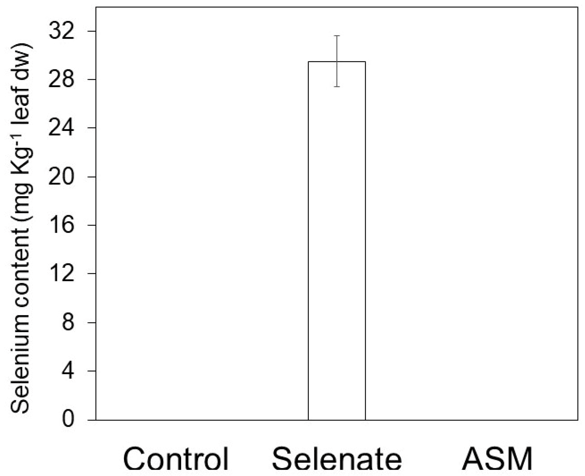
In sodium selenate treated tomato plants, at 28 dpi (14 dpi), the selenium content was equal to 28 ppb leaf dry weight, whereas it was not detectable in the controls or ASM-treated plants (Figure 4).

3.3. Callose Deposition in Leaves of Sodium Selenate Treated Tomato Plants

At 48 hpt, callose deposition was significantly higher in leaves of sodium selenate treated tomato plants, compared with the control (Figure 5). In contrast, callose deposition was not significantly different among leaves of sodium selenate and ASM treated plants, used as positive control (Figure 5).

3.4. PR-1 and PIN2 Genes Expression in Leaves of Sodium Selenate Treated Tomato Plants

In sodium selenate and ASM treated tomato plants, significant expression of the salicylic-acid-responsive PR1b1 gene was observed at 24, 48 and 72 htp, with the maximum of expression at 48 htp (Figure 6A). At 96 hpt, PR1b1 induction by ASM was also significant. Conversely, a significant reduction was observed for the jasmonate-mediated PIN2 gene expression in sodium selenate treated tomato plants but not for ASM (Figure 6B).

4. Discussion

Tomato is an economically important crop for the world market that is threatened by several bacterial diseases. European Union policy is directed towards significant reductions in pesticide use as indiscriminate utilization of pesticides in crop protection leads to numerous side effects and environmental pollution. Therefore, it is crucial to develop effective crop health management strategies that are highly sustainable and environment-friendly. A great opportunity to defend plants against pathogens is offered by Se. Here, we demonstrated that Se-treated tomato plants are protected against Pst, and a significant reduction of in planta bacterial growth was observed. The in planta Pst growth reduction, similar to that observed in ASM-treated tomato plants, and the high level of Se (28 ppb leaf dry weight) in sodium selenate-treated plants, demonstrates the direct antimicrobial activity of this beneficial element. In many pathosystems, it has been demonstrated that the protective effect of Se is almost completely attributed to its toxicity since Se inhibited the growth of Alternaria brassicicola and Fusarium sp. [46,47], the spore germination and germ tube elongation of Botrytis cinerea [26], mycelial spread of Phanerochaete chrysosporium and Penicillium expansum in cultural medium [27,48], and it interfered with Sclerotinia sclerotiorum metabolism [24,49,50]. Se toxicity against Pst was verified in vitro using different concentrations of sodium selenate and an EC50 of 42 ppm was determined. It has been demonstrated that Se 0.5 mg L−1 damages mycelial structures and osmoregulation of S. sclerotiorum [49] and that addition of Se 24 mg L−1 to a medium caused destruction of B. cinerea conidia plasma membrane and loss of cytoplasmic materials from mycelia [26]. Furthermore, [51] demonstrated that addition of Se 0.05 mg L−1 impairs the viability of Fusarium cells. Selenium nanoparticles, applied against bacteria and fungi, exert their antimicrobial action at doses ranging from 5–25 µg L−1 (Staphylococcus aureus and Escherichia coli) up to 1000 mg L−1 (Sclerospora graminicola) [22]. The above reported results demonstrated that Se toxicity depends on the dose and the chemical form in which it is supplied.
In Se-treated tomato plants we also observed that the protective effect expressed as a percentage of the diseased area is comparable to that of ASM, but when expressed as the number of lesions per leaf, the protective effect of sodium selenate was less evident with respect to ASM, and therefore, in sodium selenate treated plants, the diameter of the lesions was lower than those for ASM treated tomato plants. ASM is a functional analogue of salicylic acid and its action is mediated by the activation of pathogenesis related proteins (PR-proteins). It also contains sulfur and it has been demonstrated that sulfur plays a key role in plant defence [52,53]. In the periodic table, Se is the element immediately below sulfur and they are very similar; hence, many organisms use Se instead of sulfur in several metabolic pathways [54]. Therefore, Se can also contribute to the activation of resistance. [25] reported that Se accumulation in oilseed rape plants leads to a decrease in sclerotinia stem rot disease incidence.
To significantly reduce bacterial speck disease severity, copper-based formulations can be used preventively [55]. However, the efficacy of these compounds is reduced by the frequent onset of copper-resistant Pst strains [56,57].
In our tomato plants, the induced resistance phenomenon also seems to be involved in the protective effect against Pst. In fact, the Se treatment of tomato plants at non-toxic concentrations strongly increased the cytological (i.e., callose accumulation in plant cell wall) and molecular (i.e., PR1b1 gene expression) markers of induced resistance. Similar results were obtained with zinc phosphate soil treatment [32]. Zinc phosphate inorganic salt protects tomato plants against Pst attack through the induction of morphological and biochemical plant defence responses and direct antimicrobial activity [32]. The antimicrobial activity of zinc phosphate is greater than that of sodium selenate, and in zinc phosphate treated plants, at the early treatment, a reduction of PIN2 gene expression was observed. In sodium selenate treated plants, the PIN2 gene was not expressed. [58] demonstrated that in Arabidopsis thaliana plants treated with selenate or selenite, production of a reactive oxygen species occurred, which triggered the expression of genes related to calcium signaling. Changes in cytosolic calcium concentrations determined the enhancement of NADPH oxidase activity that stimulated the production of ethylene, jasmonic acid and salycilic acid [58,59]. Recently, it has been demonstrated that Se applied in the soil significantly protects oilseed rape leaves against S. sclerotiorum attack by activating the salicylic acid, jasmonic acid and ethylene defence pathways [28]. Similar results were reported in tomato plants treated with Se nanoparticles, which showed a decrease in the severity of symptoms caused by Alternaria solani and an induction of the activity of enzymes related to multiple plant defence pathways that participate in the defence response of plant cells [60].
Our findings have demonstrated that sodium selenate treated tomato plants showed a significant accumulation of callose that serves as a cellular defence mechanism against pathogens attacks as well as an induced resistance marker. Thus, histochemical deposition and increased expression of the PR1b1 gene indicate the potential of Se to induce resistance. Similar results were reported by [61] in the tomato-Phytopohthora infestans pathosystem. The authors reported that Se nanoparticles induced resistance mechanisms at cellular, biochemical and gene expression levels and they suggest to use them as nano-biostimulant fungicide to protect tomato plants.
Although we have explored the efficacy of sodium selenate in protecting tomato plants in the worldwide cultivated cultivar RioGrande, which is very susceptible to the predominant race 0 of Pst, it would be interesting to extend our study to other tomato cultivars and Pst strains.
In conclusion, we have demonstrated that Se can be used in plant protection both as an antimicrobial agent and a resistance inducer, making it an alternative and ecofriendly tool for the treatment and control of bacterial plant diseases. Modern consumers are increasingly aware about the nutritional aspects of foods and look for products which guarantee a high intake of health-promoting components, such as vitamins, carotenoids and phenolic compounds. Several studies have highlighted the potential of Se treatments for biofortification (i.e., the increase in nutritional value) of vegetable commodities; therefore, its application could benefit the human diet [62]. Treatment with Se delays tomato fruit ripening, due to the repression of ethylene-synthetic genes [63]. Se foliar treatments on tomato plants before fruit development increased the level of antioxidant compounds, enhanced the maintenance of fruit quality during storage and controlled the development of grey mold rot caused by Botrytis cinerea, as a result of a general Se-mediated stimulation of the antioxidant defence system [64]. Furthermore, Se foliar treatments led to an increase in soluble sugars, amino acids and bioactive compounds such as flavonoids, glutathione, vitamins C and E in pink tomatoes [65,66].
Se fertilization can have health promoting effects on plants and humans, and has the additional advantage of producing Se-fortified food useful for preventing human health problems caused by a lack of Se intake.

Supplementary Materials

The following supporting information can be downloaded at: https://www.mdpi.com/article/10.3390/agronomy12061351/s1, Table S1: Primers used in the qRT-PCR assays.

Author Contributions

Conceptualization, C.M. and R.B.; Methodology, C.M., M.B., M.Q. and B.O.; Software, M.B.; Validation, C.M., M.B., M.Q. and B.O.; Formal Analysis, M.B., M.Q. and B.O.; Investigation, C.M., M.B., M.Q. and B.O.; Data Curation, C.M., R.B., M.B., M.Q., B.O. and D.B.; Writing—Original Draft Preparation, C.M.; Writing—Review and Editing, C.M. and R.B.; Visualization, C.M. and R.B.; Supervision C.M., R.B. and D.B.; Project Administration C.M. and R.B.; Funding Acquisition, C.M., R.B. and D.B. All authors have read and agreed to the published version of the manuscript.

Funding

This study was supported through funding from POR-FESR 2014–2020 Asse 1—Ricerca e Innovazione: Valorizzazione degli scarti di biomasse destinate alla filiera agroalimentare.

Acknowledgments

We thank Luca Bonciarelli and Maurizio Orfei for technical assistance.

Conflicts of Interest

The authors declare that they have no competing financial interest or personal relationships that could have influenced the work reported in this paper.

References

  1. Schwarz, K.; Foltz, C.M. Selenium as an integral part of factor 3 against dietary necrotic liver degeneration. J. Am. Chem. Soc 1957, 79, 3292–3293. [Google Scholar] [CrossRef]
  2. Chauhan, R.; Awasthi, S.; Srivastava, S.; Dwivedi, S.; Pilon-Smits, E.A.; Dhankher, O.P.; Tripathi, R.D. Understanding selenium metabolism in plants and its role as a beneficial element. Crit. Rev. Environ. Sci. 2019, 49, 1937–1958. [Google Scholar] [CrossRef]
  3. Schiavon, M.; Pilon-Smits, E.A. The fascinating facets of plant selenium accumulation–biochemistry, physiology, evolution and ecology. New Phytol. 2017, 213, 1582–1596. [Google Scholar] [CrossRef] [PubMed] [Green Version]
  4. Rayman, M.P. The importance of selenium to human health. Lancet 2000, 356, 233–241. [Google Scholar] [CrossRef] [Green Version]
  5. Liu, H.; Yu, F.; Shao, W.; Ding, D.; Yu, Z.; Chen, F.; Geng, D.; Tan, X.; Lammi, M.J.; Guo, X. Associations between selenium content in hair and Kashin-Beck disease/Keshan Disease in children in northwestern China: A prospective cohort study. Biol. Trace Elem. Res. 2018, 184, 16–23. [Google Scholar] [CrossRef]
  6. Husen, A.; Siddiqi, K.S. Plants and microbes assisted selenium nanoparticles: Characterization and application. J. Nanobiotechnol. 2014, 12, 1–10. [Google Scholar] [CrossRef] [PubMed] [Green Version]
  7. Rayman, M.P. Selenium and human health. Lancet 2012, 379, 1256–1268. [Google Scholar] [CrossRef]
  8. Roman, M.; Jitaru, P.; Barbante, C. Selenium biochemistry and its role for human health. Metallomics 2014, 6, 25–54. [Google Scholar] [CrossRef]
  9. Srivastava, N.; Mukhopadhyay, M. Biosynthesis and structural characterization of selenium nanoparticles mediated by Zooglea ramigera. Powder Technol. 2013, 244, 26–29. [Google Scholar] [CrossRef]
  10. Wu, M.; Cong, X.; Li, M.; Rao, S.; Liu, Y.; Guo, J.; Zhu, S.; Chen, S.; Xu, F.; Cheng, S. Effects of different exogenous selenium on Se accumulation, nutrition quality, elements uptake, and antioxidant response in the hyperaccumulation plant Cardamine violifolia. Ecotox. Environ. Saf. 2020, 204, 111045. [Google Scholar] [CrossRef]
  11. Khurana, A.; Tekula, S.; Saifi, M.A.; Venkatesh, P.; Godugu, C. Therapeutic applications of selenium nanoparticles. Biomed. Pharmacother. 2019, 111, 802–812. [Google Scholar] [CrossRef] [PubMed]
  12. Sonkusre, P. Specificity of biogenic selenium nanoparticles for prostate cancer therapy with reduced risk of toxicity: An in vitro and in vivo study. Front. Oncol. 2020, 9, 1541. [Google Scholar] [CrossRef] [Green Version]
  13. Hatfield, D.L.; Carlson, B.A.; Tsuji, P.A.; Tobe, R.; Gladyshev, V.N. Selenium and cancer. In Molecular, Genetic, and Nutritional Aspects of Major and Trace Minerals; Elsevier: Amsterdam, The Netherlands, 2017; pp. 463–473. [Google Scholar]
  14. Hatfield, D.L.; Tsuji, P.A.; Carlson, B.A.; Gladyshev, V.N. Selenium and selenocysteine: Roles in cancer, health, and development. Trends Biochem. Sci. 2014, 39, 112–120. [Google Scholar] [CrossRef] [Green Version]
  15. Ahmad, R.; Waraich, E.A.; Nawaz, F.; Ashraf, M.Y.; Khalid, M. Selenium (Se) improves drought tolerance in crop plants—A myth or fact? J. Sci. Food Agric. 2016, 96, 372–380. [Google Scholar] [CrossRef]
  16. Banuelos, G. Phyto-products may be essential for sustainability and implementation of phytoremediation. Environ. Pollut. 2006, 144, 19–23. [Google Scholar] [CrossRef] [PubMed]
  17. Doucha, J.; Lívanský, K.; Kotrbáček, V.; Zachleder, V. Production of Chlorella biomass enriched by selenium and its use in animal nutrition: A review. Appl. Microbiol. Biotechnol. 2009, 83, 1001–1008. [Google Scholar] [CrossRef] [PubMed]
  18. Tapiero, H.; Townsend, D.; Tew, K. The antioxidant role of selenium and seleno-compounds. Biomed. Pharmacother. 2003, 57, 134–144. [Google Scholar] [CrossRef]
  19. Winkel, L.H.; Johnson, C.A.; Lenz, M.; Grundl, T.; Leupin, O.X.; Amini, M.; Charlet, L. Environmental selenium research: From microscopic processes to global understanding. Environ. Sci. Technol. 2012, 46, 571–579. [Google Scholar] [CrossRef]
  20. Yuan, L.; Zhu, Y.; Lin, Z.-Q.; Banuelos, G.; Li, W.; Yin, X. A novel selenocystine-accumulating plant in selenium-mine drainage area in Enshi, China. PLoS ONE 2013, 8, e65615. [Google Scholar] [CrossRef]
  21. Ellis, D.R.; Salt, D.E. Plants, selenium and human health. Curr. Opin. Plant Biol. 2003, 6, 273–279. [Google Scholar] [CrossRef]
  22. Garza-García, J.J.O.; Hernández-Díaz, J.A.; Zamudio-Ojeda, A.; León-Morales, J.M.; Guerrero-Guzmán, A.; Sánchez-Chiprés, D.R.; López-Velázquez, J.C.; García-Morales, S. The Role of selenium nanoparticles in agriculture and food technology. Biol. Trace Elem. Res. 2021, 200, 2528–2548. [Google Scholar] [CrossRef] [PubMed]
  23. Feng, R.; Wei, C.; Tu, S. The roles of selenium in protecting plants against abiotic stresses. Environ. Exp. Bot. 2013, 87, 58–68. [Google Scholar] [CrossRef]
  24. Cheng, Q.; Hu, C.; Jia, W.; Cai, M.; Zhao, Y.; Tang, Y.; Yang, D.; Zhou, Y.; Sun, X.; Zhao, X. Selenium reduces the pathogenicity of Sclerotinia sclerotiorum by inhibiting sclerotial formation and germination. Ecotox. Environ. Saf. 2019, 183, 109503. [Google Scholar] [CrossRef] [PubMed]
  25. Liu, K.; Cai, M.; Hu, C.; Sun, X.; Cheng, Q.; Jia, W.; Yang, T.; Nie, M.; Zhao, X. Selenium (Se) reduces Sclerotinia stem rot disease incidence of oilseed rape by increasing plant Se concentration and shifting soil microbial community and functional profiles. Environ. Pollut. 2019, 254, 113051. [Google Scholar] [CrossRef]
  26. Wu, Z.; Yin, X.; Bañuelos, G.S.; Lin, Z.-Q.; Zhu, Z.; Liu, Y.; Yuan, L.; Li, M. Effect of selenium on control of postharvest gray mold of tomato fruit and the possible mechanisms involved. Front. Microbiol. 2016, 6, 1441. [Google Scholar] [CrossRef] [PubMed]
  27. Wu, Z.-L.; Yin, X.-B.; Lin, Z.-Q.; Banuelos, G.S.; Yuan, L.-X.; Liu, Y.; Li, M. Inhibitory effect of selenium against Penicillium expansum and its possible mechanisms of action. Curr. Microbiol. 2014, 69, 192–201. [Google Scholar] [CrossRef] [PubMed]
  28. Xu, J.; Jia, W.; Hu, C.; Nie, M.; Ming, J.; Cheng, Q.; Cai, M.; Sun, X.; Li, X.; Zheng, X. Selenium as a potential fungicide could protect oilseed rape leaves from Sclerotinia sclerotiorum infection. Environ. Pollut. 2020, 257, 113495. [Google Scholar] [CrossRef]
  29. WPTC. Tomato News. 2021. Available online: https://www.tomatonews.com/en (accessed on 28 January 2022).
  30. Alexander, S.; Kim, S.; Waldenmaier, C. First report of copper-tolerant Pseudomonas syringae pv. tomato in Virginia. Plant Dis. 1999, 83, 964. [Google Scholar] [CrossRef]
  31. Molinari, S. Systemic acquired resistance activation in Solanaceous crops as a management strategy against root-knot nematodes. Pest Manag. Sci. 2016, 72, 888–896. [Google Scholar] [CrossRef]
  32. Quaglia, M.; Bocchini, M.; Orfei, B.; D’Amato, R.; Famiani, F.; Moretti, C.; Buonaurio, R. Zinc phosphate protects tomato plants against Pseudomonas syringae pv. tomato. J. Plant Dis. Prot. 2021, 128, 989–998. [Google Scholar] [CrossRef]
  33. Scarponi, L.; Buonaurio, R.; Martinetti, L. Persistence and translocation of a benzothiadiazole derivative in tomato plants in relation to systemic acquired resistance against Pseudomonas syringae pv tomato. Pest Manag. Sci. 2001, 57, 262–268. [Google Scholar] [CrossRef] [PubMed]
  34. Buonaurio, R.; Stravato, V.; Cappelli, C. Occurrence of Pseudomonas syringae pv tomato race 1 in Italy on Pto gene-bearing tomato plants. J. Phytopathol. 1996, 144, 437–440. [Google Scholar] [CrossRef]
  35. Lamari, L. Assess: Image Analysis Software for Plant Disease Quantification; APS Press: St. Paul, MN, USA, 2002. [Google Scholar]
  36. King, E.O.; Ward, M.K.; Raney, D.E. Two simple media for the demonstration of pyocyanin and fluorescin. J. Lab. Clin. Med. 1954, 44, 301–307. [Google Scholar] [PubMed]
  37. Luna, E.; Pastor, V.; Robert, J.; Flors, V.; Mauch-Mani, B.; Ton, J. Callose deposition: A multifaceted plant defense response. MPMI 2011, 24, 183–193. [Google Scholar] [CrossRef] [PubMed] [Green Version]
  38. Quaglia, M.; Baglivo, F.; Moretti, C. Postharvest β-aminobutyric-acid–primed resistance is not effective in the control of Penicillium expansum Link. on ‘Golden delicious’ apple fruit. J. Crop Prot. 2017, 102, 43–48. [Google Scholar] [CrossRef]
  39. Goyal, R.K.; Fatima, T.; Topuz, M.; Bernadec, A.; Sicher, R.; Handa, A.K.; Mattoo, A.K. Pathogenesis-related protein 1b1 (PR1b1) is a major tomato fruit protein responsive to chilling temperature and upregulated in high polyamine transgenic genotypes. Front. Plant Sci. 2016, 7, 901. [Google Scholar] [CrossRef] [PubMed] [Green Version]
  40. Castagna, A.; Ederli, L.; Pasqualini, S.; Mensuali-Sodi, A.; Baldan, B.; Donnini, S.; Ranieri, A. The tomato ethylene receptor LE-ETR3 (NR) is not involved in mediating ozone sensitivity: Causal relationships among ethylene emission, oxidative burst and tissue damage. New Phytol. 2007, 174, 342–356. [Google Scholar] [CrossRef]
  41. Pfaffl, M.W. A new mathematical model for relative quantification in real-time RT–PCR. Nucleic Acids Res. 2001, 29, e45. [Google Scholar] [CrossRef]
  42. Vandesompele, J.; De Preter, K.; Pattyn, F.; Poppe, B.; Van Roy, N.; De Paepe, A.; Speleman, F. Accurate normalization of real-time quantitative RT-PCR data by geometric averaging of multiple internal control genes. Genome Biol. 2002, 3, 1–12. [Google Scholar] [CrossRef] [Green Version]
  43. Onofri, A.; Pannacci, E. Spreadsheet tools for biometry classes in crop science programmes. Commun. Biometry Crop. Sci. 2014, 9, 43–53. [Google Scholar]
  44. Streibig, J.C.; Kudsk, P. Dose-response curves and statistical models. In Herbicide Bioassay; CRC Press: Boca Raton, FL, USA, 1993. [Google Scholar]
  45. Pestemer, W.; Günther, P. Growth inhibition of plants as a bioassay for herbicide analysis. In Analysis of Pesticides in Ground and Surface Water I; Springer: Berlin/Heidelberg, Germany, 1995; pp. 219–231. [Google Scholar]
  46. Companioni, B.; Medrano, J.; Torres, J.; Flores, A.; Rodríguez, E.; Benavides, A. Protective action of sodium selenite against Fusarium wilt in tomato: Total protein contents, levels of phenolic compounds and changes in antioxidant potential. In Proceedings of the II International Symposium on Soilless Culture and Hydroponics 947, Puebla, Mexico, 15–19 May 2011; pp. 321–327. [Google Scholar]
  47. Hanson, B.; Garifullina, G.F.; Lindblom, S.D.; Wangeline, A.; Ackley, A.; Kramer, K.; Norton, A.P.; Lawrence, C.B.; Pilon-Smits, E.A. Selenium accumulation protects Brassica juncea from invertebrate herbivory and fungal infection. New Phytol. 2003, 159, 461–469. [Google Scholar] [CrossRef] [PubMed] [Green Version]
  48. Espinosa-Ortiz, E.J.; Gonzalez-Gil, G.; Saikaly, P.E.; van Hullebusch, E.D.; Lens, P.N. Effects of selenium oxyanions on the white-rot fungus Phanerochaete chrysosporium. Appl. Microbiol. Biotechnol. 2015, 99, 2405–2418. [Google Scholar] [CrossRef] [PubMed] [Green Version]
  49. Jia, W.; Hu, C.; Ming, J.; Zhao, Y.; Xin, J.; Sun, X.; Zhao, X. Action of selenium against Sclerotinia sclerotiorum: Damaging membrane system and interfering with metabolism. Pestic. Biochem. Phys. 2018, 150, 10–16. [Google Scholar] [CrossRef] [PubMed]
  50. Jia, W.; Hu, C.; Xu, J.; Ming, J.; Zhao, Y.; Cai, M.; Sun, X.; Liu, X.; Zhao, X. Dissolved organic matter derived from rape straw pretreated with selenium in soil improves the inhibition of Sclerotinia sclerotiorum growth. J. Hazard. Mater. 2019, 369, 601–610. [Google Scholar] [CrossRef]
  51. Yin, H.; Zhang, Y.; Zhang, F.; Hu, J.T.; Zhao, Y.M.; Cheng, B.L. Effects of selenium on Fusarium growth and associated fermentation products and the relationship with chondrocyte viability. Biomed. Environ. Sci. 2017, 30, 134–138. [Google Scholar]
  52. Künstler, A.; Gullner, G.; Ádám, A.L.; Kolozsváriné Nagy, J.; Király, L. The versatile roles of sulfur-containing biomolecules in plant defense—A road to disease resistance. Plants 2020, 9, 1705. [Google Scholar] [CrossRef]
  53. Nwachukwu, I.D.; Slusarenko, A.J.; Gruhlke, M.C.H. Sulfur and sulfur compounds in plant defence. Nat. Prod. Commun. 2012, 7, 395–400. [Google Scholar] [CrossRef] [Green Version]
  54. Sekowska, A.; Kung, H.F.; Danchin, A. Sulfur metabolism in Escherichia coli and related bacteria: Facts and fiction. J. Mol. Microbiol. Biotechnol. 2000, 2, 145–177. [Google Scholar]
  55. Yunis, H.; Bashan, Y.; Okon, Y.; Henis, Y. Weather dependence, yield losses, and control of bacterial speck of tomato caused by Pseudomonas tomato. Plant Dis. 1980, 64, 937–939. [Google Scholar] [CrossRef]
  56. Bender, C.L.; Cooksey, D.A. Indigenous plasmids in Pseudomonas syringae pv. tomato: Conjugative transfer and role on copper resistance. J. Bacteriol. 1986, 165, 534–541. [Google Scholar] [CrossRef] [Green Version]
  57. Cha, J.S.; Cooksey, D. Copper resistance in Pseudomonas syringae mediated by periplasmic and outer membrane proteins. Proc. Natl. Acad. Sci. USA 1991, 88, 8915–8919. [Google Scholar] [CrossRef] [PubMed] [Green Version]
  58. Van Hoewyk, D.; Takahashi, H.; Inoue, E.; Hess, A.; Tamaoki, M.; Pilon-Smits, E.A. Transcriptome analyses give insights into selenium-stress responses and selenium tolerance mechanisms in Arabidopsis. Physiol. Plant. 2008, 132, 236–253. [Google Scholar] [CrossRef]
  59. Tamaoki, M.; Maruyama-Nakashita, A. Molecular mechanisms of selenium responses and resistance in plants. In Selenium in Plants: Molecular, Physiological, Ecological and Evolutionary Aspects; Pilon-Smits, E.A.H., Winkel, L.H.E., Lin, Z.-Q., Eds.; Springer International Publishing: Cham, Switzerland, 2017; pp. 35–51. [Google Scholar]
  60. Quiterio-Gutiérrez, T.; Ortega-Ortiz, H.; Cadenas-Pliego, G.; Hernández-Fuentes, A.D.; Sandoval-Rangel, A.; Benavides-Mendoza, A.; Cabrera-de la Fuente, M.; Juárez-Maldonado, A. The application of selenium and copper nanoparticles modifies the biochemical responses of tomato plants under stress by Alternaria solani. Int. J. Mol. Sci. 2019, 20, 1950. [Google Scholar] [CrossRef] [PubMed] [Green Version]
  61. Joshi, S.M.; De Britto, S.; Jogaiah, S. Myco-engineered selenium nanoparticles elicit resistance against tomato late blight disease by regulating differential expression of cellular, biochemical and defense responsive genes. J. Biotech. 2021, 325, 196–206. [Google Scholar] [CrossRef] [PubMed]
  62. El-Ramady, H.; Faizy, S.E.-D.; Abdalla, N.; Taha, H.; Domokos-Szabolcsy, É.; Fari, M.; Elsakhawy, T.; Omara, A.E.-D.; Shalaby, T.; Bayoumi, Y. Selenium and nano-selenium biofortification for human health: Opportunities and challenges. Soil Syst. 2020, 4, 57. [Google Scholar] [CrossRef]
  63. Zhu, Z.; Chen, Y.; Shi, G.; Zhang, X. Selenium delays tomato fruit ripening by inhibiting ethylene biosynthesis and enhancing the antioxidant defense system. Food Chem. 2017, 219, 179–184. [Google Scholar] [CrossRef]
  64. Zhu, Z.; Chen, Y.; Zhang, X.; Li, M. Effect of foliar treatment of sodium selenate on postharvest decay and quality of tomato fruits. Sci. Hortic. 2016, 198, 304–310. [Google Scholar] [CrossRef]
  65. Zhu, Z.; Zhang, Y.; Liu, J.; Chen, Y.; Zhang, X. Exploring the effects of selenium treatment on the nutritional quality of tomato fruit. Food Chem. 2018, 252, 9–15. [Google Scholar] [CrossRef]
  66. Fowler, J.H.; Narváez-Vásquez, J.; Aromdee, D.N.; Pautot, V.; Holzer, F.M.; Walling, L.L. Leucine aminopeptidase regulates defense and wound signalling in tomato downstream of jasmonic acid. Plant Cell 2009, 21, 1239–1251. [Google Scholar] [CrossRef] [Green Version]
Figure 1. Protective effect of sodium selenate (4 ppm; 21.17 µM) on tomato leaves against Pseudomonas syringae pv. tomato 14 days after the inoculation. Tomato leaves of control sodium selenate- and acibenzolar-S-methyl- (ASM) treated plants.
Agronomy 12 01351 g001
Figure 2. Protective effect of sodium selenate (4 ppm; 21.17 µM), and acibenzolar-S-methyl (213.98 µM; ASM) for comparison, on disease severity of Pseudomonas syringae pv. tomato (Pst) on tomato plants, expressed as % of leaf diseased area (A), and number of lesions per leaf at 14 days post-inoculation (B), as well as Pst growth in planta at 6 days post-inoculation (C). Data for each column represent the means of 10 (A,B) and 4 (C) replicates ± SE. Data of each plot followed by the same letters are not significantly different according to the Duncan’s multiple range test at p = 0.01 (A,B) and p = 0.05 (C).
Agronomy 12 01351 g002
Figure 3. Sodium selenate activity on in vitro growth of Pseudomonas syringae pv tomato (Pst). Dose-response curve on Pst growth versus concentration of sodium selenate (0, 6, 12, 18, 24, 30, 36 and 42 ppm) applied. Bacterial growth data were obtained by spectrophotometer readings (OD630 nm) every hour for 24 h and expressed here as percent of the control. Closed circles represent the observed data (n = 8), and the line is the log-logistic curve with the equation y = 100/{1 + exp [6.62 (log(x) − log(42.0))]}. The standard error of the mean is 0.701. Eight replicates for each concentration were tested and three independent experiments were carried out.
Agronomy 12 01351 g003
Figure 4. Selenium content in tomato leaves treated with sodium selenite. Selenium content (mg kg−1 dry weigh) in tomato (cv. Rio Grande) leaves at 28 days post treatment (dpt) with water (control), sodium selenate (4 ppm; 21.17 µM) or acibenzolar-S-methyl (213.98 µM; ASM). Data are means of three independent experiments ± SE. Columns with different letters are significantly different (p ≤ 0.01; Duncan’s multiple range test). d.w., dry weight.
Agronomy 12 01351 g004
Figure 5. Callose accumulation in tomato leaves at 48 h post-treatment (hpt) with water (control), sodium selenate (4 ppm; 21.17 µM) and acibenzolar-S-methyl (213.98 µM; ASM). Callose was visualized by aniline blue staining and epifluorescence. Quantification was carried out by determining the percentage (%) of blue pixels on digitalized photos. Each experiment was independently repeated three times, with three plants per treatments, three pieces from the first leaflet of the 2nd true leaf of the tomato plants and one photograph taken at random for each piece. Data from the three experiments were submitted to one factor (treatment) analysis of variance (ANOVA). Each column represents the mean of 27 replicate photos per treatment ± SE. Different letters indicate significant differences (p ≤ 0.01, Duncan’s tests). Top: representative digitalized photograph areas at 10× magnification of blue pixels corresponding with each bar.
Agronomy 12 01351 g005
Figure 6. Effect of sodium selenite treatments on defence gene expression. Relative expression levels of the salicylic-acid-responsive tomato ‘pathogenesis-related protein 1b1’ (PR1b1) (A) and the jasmonate-mediated ‘proteinase inhibitor 2’ (PIN2) (B) genes in tomato (cv. Rio Grande) leaves at 24, 48, 72 and 96 h post treatment (hpt) with water (control), sodium selenate (4 ppm; 21.17 µM) and acibenzolar-S-methyl (213.98 µM; ASM). Data are means of three biological replicates (each consisting of the average value of three technical replications) ± SE. Columns with different letters are significantly different (p ≤ 0.05) according to ANOVA followed by Duncan’s multiple range tests.
Agronomy 12 01351 g006
Publisher’s Note: MDPI stays neutral with regard to jurisdictional claims in published maps and institutional affiliations.