Article

Effect of Artificial Light Treatment on the Physiological Property and Biological Activity of the Aerial and Underground Parts of Atractylodes macrocephala

1
Interdisciplinary Program in Smart Science, Kangwon National University, Chuncheon 24341, Korea
2
Department of Herb Crop Resources, NIHHS, RDA, Eumseong 27709, Korea
3
Division of Bioresource Sciences, Kangwon National University, Chuncheon 24341, Korea
*
Author to whom correspondence should be addressed.
Agronomy 2022, 12(7), 1485; https://doi.org/10.3390/agronomy12071485
Submission received: 17 May 2022 / Revised: 14 June 2022 / Accepted: 17 June 2022 / Published: 21 June 2022

Abstract

:
Atractylodes macrocephala Koidz. is primarily used as a raw material in herbal medicine to treat digestive diseases. To improve the functionality of A. macrocephala, its growth patterns under artificial light were studied. A. macrocephala grew better under MEL light, with the highest chlorophyll content (57.07 ± 0.65 SPAD), than under other artificial light sources. The DPPH free radical scavenging activity of 2000 μg·mL−1 underground extract treated with LED-red light was the highest (95.3 ± 1.1%). Furthermore, the total phenol and flavonoid contents of underground extract treated with LED-green light were the highest at 24.93 ± 0.3 mg GAE·g−1 and 11.2 ± 0.3 mg QE·g−1, respectively. Moreover, in the analysis of whitening activity, the tyrosinase inhibition rate of 5000 μg·mL −1 extract treated with LED-red light was the highest (84.6 ± 2.9%). In anti-inflammatory activity assay, LPS- induced RAW 264.7 cells exposed to 100 μg·mL−1 extract treated with fluorescent light showed the lowest NO levels (2.97 ± 0.14%). Finally, the expression of iNOS and COX-2, which are related to anti-inflammatory activity, was suppressed in cells exposed to artificial light-treated extract compared with that in controls, indicating potent anti-inflammatory activity. Therefore, growth under artificial light can improve the various biological functions of A. macrocephala.

1. Introduction

Atractylodes macrocephala Koidz. of the Compositae family presents better productivity than Atractylodes japonica Koidz. It is a cultivated crop [1]. Atractylodis macrocephalae Rhizoma is called “Baekchul” in Korean herbal medicine [2,3], and it has been recognized and included in the prescriptions of representative oriental medicines, such as Sipjeondaebotang [1].
Recently, indoor cultivation methods for plant production have been widely applied to avoid the adverse impacts of climate change and pests [4]. During indoor cultivation, artificial light sources, including fluorescent lamps, metal halide lamps, sodium lamps, and light-emitting diodes (LEDs), are deployed. LEDs are known to be artificial light sources with the highest efficiency owing to their higher photosynthetic photon flux density (PPFD) relative to power consumption [5]. In addition, microwave electrodeless light (MEL) generates plasma light by charging gas and has a longer lifespan than other light sources that depend on electrode lifespan. Recently, the effects of MEL on the growth of Salvia miltiorrhiza—a medicinal plant—were confirmed; however, only a few studies have examined the cultivation and characteristics of other plants grown under MEL worldwide [6].
In both humans and plants, excess reactive oxygen species (ROS) production damages the antioxidant defense system, thereby reducing resistance to diseases and pathogens [7]. Comprehensive research on natural antioxidants that can eliminate free radicals, which induce ROS generation, is underway [8]. Plant polyphenols are high- molecular-weight compounds containing phenols with hydroxyl groups (-OH) in the benzene ring as functional groups. They can readily combine with various compounds and inhibit ROS [9]. Furthermore, the antioxidant and anticancer activities of these compounds have been documented. Among polyphenols, flavonoids present a flavone structure, in which the benzene ring is connected with three carbons as a basic structure. In addition, flavonoids are often colored, conferring plant leaves or fruits with characteristic colors. Furthermore, they play pivotal roles in protecting plants from UV rays. Similar to polyphenols, flavonoids possess antioxidant, antimicrobial, and anticancer properties. Representative flavonoids include anthocyanins, quercetin, and catechin. Among these, quercetin can reduce the occurrence of stomach cancer [10].
Melanin is a pigment component conferring dark color to the human skin and hair as well as causing browning reaction in plants. Melanin is produced through the reaction of tyrosine, tyrosinase, and tyrosinase-related proteins (TRP-1 and TRP-2) in melanocytes in the basal layer of the skin epidermis and protects the skin from UV rays and other external stimuli [11]. Recently, with the depletion of the ozone layer, the level of UV rays in sunlight has increased, leading to excessive melanin production in skin exposed to sunlight for prolonged periods. To prevent such problems from the perspective of esthetics, efforts are underway to find inhibitors of tyrosinase involved in melanin production. Currently available whitening substances include kojic acid, arbutin, and hydroquinone, which produce a strong inhibitory effect on tyrosinase and act as antibacterial substances. However, owing to the cytotoxicity, skin inflammation, and mutagenic potential of these ingredients, safety concerns have been raised [12,13]. Therefore, demand for safe whitening substances derived from natural products with fewer side effects has increased and attempts to discover natural substances with whitening activity from various plants are ongoing.
Following bacterial infection or allergen stimulation, inflammatory reactions occur in the body. Although this inflammatory response is primarily elicited as a recovery response to the regeneration of damaged areas, along with a protective action against pathogens, excess of this response leads to chronic inflammation, ultimately causing diseases, such as arthritis and arteriosclerosis. Lipopolysaccharide (LPS), one of the substances inducing inflammation, is present in the cell walls of some gram-negative bacteria. Nuclear factor kappa B (NF-κB) is translocated into the nucleus, and the expression of genes encoding proteins involved in inflammatory reactions, such as inducible nitric oxide synthase (iNOS) and cyclooxygenase-2 (COX-2), is enhanced. Finally, macrophages secrete nitric oxide (NO) and inflammatory cytokines [14,15], which contribute to the activation of innate immunity and adaptive immune response through the recognition of foreign antigens; however, when produced in excess, these components cause an inflammatory reaction in the body, which can lead to fever, edema, tissue damage, or mutations [16]. Furthermore, certain plant components can inhibit such an inflammatory response. For instance, atractylenolide I and III in the rhizomes of native seedlings suppressed LPS-induced TNF-α expression in macrophages [17]. Atractylodes macrocephala Koidz. has been studied to affect the treatment-related inflammatory response, amino acid synthesis, and energy metabolism in chronic gastritis [18]. Three compounds isolated from Atractylodes macrocephala, 2-[(2E)-3,7-dimethyl-2,6-octadienyl]-6-methyl-2, 5-cyclohexadiene-1, 4-dione; 1-acetoxy-tetradeca-6E,12E-diene-8, 10-diyne-3-ol; 1,3-diacetoxy-tetradeca-6E, 12E-diene-8, 10-diyne are effective therapeutic substances for the treatment of inflammatory diseases [19].
Previous studies on A. macrocephala have reported the physiological activity of roots; however, there has been no research on the smart farming of this plant to improve the content of active ingredients. To this end, in the present study, the aim is to search for an artificial light source with increased functionality by investigating the growth characteristics, chlorophyll content, antioxidant activity, total phenol and flavonoid contents, whitening activity, and anti-inflammatory activity of plants in A. macrocephala. Our work provides fundamental data for the application of smart cultivation technology in other medicinal plants.

2. Materials and Methods

2.1. Plant Materials and Artificial Light Treatment

A. macrocephala was obtained from the Department of Herb Crop Resources, NIHHS, RDA, Eumseong, South Korea. Seedlings grown from seedstock at room temperature for 1 month were used. Based on the general greenhouse cultivation, the culture room seedling period was also set to 2 weeks because the condition after 2 weeks of cultivation was stable to some extent in both the aerial and underground parts, making it suitable for transplanting into the open field. For the number of individuals, six plants per treatment group were used. Following transfer to pots containing sterile soil, the grafts were maintained at 25 ± 1 °C for 2 weeks in a culture room. The following artificial light sources were used in the experiment (16 h light/8 h dark): fluorescent light (FL: Continuous spectrum, 24 µmol/m2·s), LED-red (630 nm, 22 µmol/m2·s), LED-blue (450 nm, 14 µmol/m2·s), LED-green (520 nm, 12 µmol/m2·s), and MEL (continuous spectrum, 52 µmol/m2·s). In addition, the sun light used as a control is that of general sunlight.

2.2. Measurement of Plant Growth Characteristics and Chlorophyll Content

To assess growth characteristics, after 2 weeks of light treatment in each group, plant height, leaf length, leaf width, leaf number, and dry weight of the aerial parts as well as the total length and dry weight of the underground part were measured. Leaf chlorophyl content was measured using the SPAD-502 plus chlorophyll meter (Konica Minolta Co., Ltd., Tokyo, Japan). The SPAD value was determined based on measurements on the top three leaves of each plant.

2.3. Extraction and Concentration

Three harvested A. macrocephala materials were separated into aerial and underground parts and dried for 3 days in a freeze dryer (OPR-FDB-5003 Freeze Dryer, OPERON Co., Ltd., Kimpo, Korea). After crushing, the dried plants were extracted for 3 days using 100% MeOH, and the extract was then filtered using a filter paper (Tokyu Roshi Kaisha Ltd., Tokyo, Japan). The filtered extract was concentrated using a rotary evaporator (YELA N-1000; Tokyo Rikakikai Co., Ltd., Tokyo, Japan) at 45 °C.

2.4. DPPH Readical Scavenging Assay

Briefly, 100 µL of 0.15 mM DPPH (1,1-diphenyl-2-picrylhydrazyl, Alfa Aesar Co., Ltd., Ward Hill, MA USA) was added to 100 μL of extract diluted to concentrations of 500 μg·mL−1, 1000 μg·mL−1, and 2000 μg·mL−1 and incubated in the dark at room temperature. After reacting for 30 min, absorbance was measured at 519 nm using a UV–Vis spectrophotometer (Multiskan FC Microplate Photometer, Thermo Fisher Scientific Inc., cleveland, OH, USA) [20]. The inhibition rate was calculated as follows:
Inhibition rate (%) = (absorbance of control group − absorbance of sample addition group)/(absorbance of control group) × 100

2.5. Total Phenol Content

Briefly, 50 μL of Folin–Ciocalteau reagent (Sigma-Aldrich Co., Ltd., St. Louis, MO, USA) was added to 100 μL of extract diluted to a concentration of 1 mg·mL1. After reaction for 5 min, 20% Na2CO3 (Junsei Chemicals Co., Ltd., Tokyo, Japan) was added and allowed to react for 15 min. After adding 1 mL of distilled water, absorbance was measured at 740 nm using a UV–Vis spectrophotometer (Multiskan FC Microplate Photometer, Thermo Fisher Scientific Inc., Ltd., cleveland, OH, USA) [21].

2.6. Total Flavonoid Content

Briefly, 100 μL of 10% aluminum nitrate (Yakuri Co., Ltd., Kyoto, Japan) and 100 μL of 1 M potassium acetate (Mallinckrodt Co., Ltd., Tokyo, Japan) were added to 500 μL of extract diluted to a concentration of 1 mg·mL−1. After reacting for 40 min, absorbance was measured at 414 nm using a UV–Vis spectrophotometer (Thermo Fisher Scientific Inc., cleveland, OH, USA) [22].

2.7. Antimicrobial Activity Assay

Antimicrobial activity assays were performed using the two-fold serial dilution method [23]. The microorganisms used in the assay were Bacillus subtilis (KCTC 1021), Staphylococcus aureus (KCTC 1916), Escherichia coli (KCTC 1924), Salmonella typhimurium (KCTC 1925), Pseudomonas aeruginosa (KCTC 2742), and Vibrio litoralis (KCTC 13228). Each microorganism was cultured in a suspension medium at an optimal temperature. Each microbial suspension was diluted 200-fold using LB medium, distributed into a 96-well microplate, and dispensed at a two-fold dilution until the sample concentration reached from 1000 μg·mL−1 to approximately 7.8 μg·mL−1. After culturing the plate in the dark for 24 h, the minimum inhibitory concentration (MIC) was determined by visual observation. A sample containing only solvent (MeOH) was used as a negative control and tetracycline as a positive control.

2.8. Tyrosinase Inhibitory Activity Assay

The tyrosinase inhibition activity was measured according to the Bernard and Berthon method [24]. To 40 μL of extract, 40 μL of 125 U mushroom tyrosinase (Sigma-Aldrich Co., Ltd., St. Louis, MO, USA) and 120 μL of 10 mM L-DOPA (3,4-dihydroxy-L-phenyalanine, Sigma-Aldrich Co., Ltd., St. Louis, MO, USA) were added. After reacting at 37 °C in the dark for 30 min, absorbance was measured at 519 nm using a UV–Vis spectrophotometer (Thermo Fisher Scientific Inc., cleveland, OH, USA).

2.9. MTT Analysis

RAW 264.7 cells were purchased from the Korean Cell Line Bank. The cells were cultured in DMEM supplemented with 10% FBS (Hyclone Laboratories Inc., Omaha, NE, USA) and 1% penicillin (Lonza Walkersville Inc., Walkersville, MD, USA) (100 U·mL−1) at 37 °C in a 5% CO2 incubator (Snayo Co., Ltd., MCO-19AIC, Fujioka, Japan). For cytotoxicity screening, the cells were aliquoted into 96-well plates at a density of 105 cells·well−1 and cultured at 37 °C in a 5% CO2 incubator for 24 h. Thereafter, the supernatant was removed, and 100 μL of each sample was treated with plant extract at concentrations of 10 μg·mL−1, 100 μg·mL−1, or 200 μg·mL−1 [25]. After 24 h, the medium was removed, and the MTT reagent diluted to a concentration of 500 μg·mL−1 was added. After 4 h, the supernatant was removed, and the cells were treated with 100 μL DMSO (Duchefa Biochemie Inc., Haarlem, Netherlands). After 20 min, absorbance was measured at 620 nm using a UV–vis spectrophotometer (Thermo Fisher Scientific Inc., cleveland, OH, USA).

2.10. Nitric Oxide (NO) Production Rate and Expression of Inflammatory Genes in LPS-Treated Raw 264.7 Cells

After removing the supernatant of the RAW 264.7 cell culture solution, 100 μL of 10 μg·mL−1, 50 μg·mL−1, or 100 μg·mL−1 extract was added, and the cells were cultured for 24 h such that the ratio of LPS to extract was 1:1. Next, 50 μL of the supernatant was removed, and the remaining medium was treated with 1% sulfanilamide (Sigma-Aldrich Co., Ltd., St. Louis, MO, USA) and 50 μL of Griess reagent [5% phosphoric acid (Wako Chemicals Inc., Richmond, VA, USA) + 0.1% N-(1-naphthyl)-ethylenediamine dihydrochloride) (Sigma-Aldrich Co., Ltd., St. Louis, MO, USA)]. After reacting for 20 min, absorbance was measured at 519 nm using a UV–Vis spectrophotometer (Thermo Fisher Scientific Inc., cleveland, OH, USA) [26].
Reverse transcriptase-polymerase chain reaction (RT-PCR) was performed to confirm the transcript levels of inflammation-related genes in RAW 264.7 cells. RAW 264.7 cells were aliquoted in a 96-well plate at a density of 105 cells·well−1 and then cultured at 37 °C in a 5% CO2 incubator (Snayo Co., Ltd., MCO-19AIC, Fujioka, Japan) for 24 h. The extract and the LPS-added medium were replaced, and the cells were incubated for an additional 24 h in a 5% CO2 incubator. RNA was extracted from the cultured cells using a total RNA extraction kit (Cat. No. 17211, Intron Co., Ltd., Seoul, Korea), and cDNA was synthesized. The following primers for inflammation-related genes were used: β-actin (F) 5′- TGACGGGGTCACCCACACTGTGCCCATCTA-3′, β-actin (R) 5′- CTAGAAGCATTTGCGGTGGACGATGGAGGG-3′, iNOS (F) 5′- CCCTTCCGAAGTTTCTGGCAGCAGC-3′, iNOS (R) 5′- GGCTGTCAGAGCCTCGTGGCTTTGG -3′, COX-2(F) 5′-CACTACATCCTGACCCACTT-3′, COX-2(R) 5′-ATGCTCCTGCTTGAGTATGT-3′, TNF-α(F) 5′-TTGACCTCAGCGCTGAGTTG- 3′, and TNF-α (R) 5′-CCTGTAGCCCACGTCGTAGC-3′. RT-PCR was performed with initial denaturation of 5 min at 94 °C, followed by 30 cycles at 94 °C for 1 min, 55 °C for 1 min, and 72 °C for 1 min, and final extension at 72 °C for 10 min. The produced PCR products were electrophoresed on 1% agarose gel, and the expression levels were measured using the Gel Doc XR+ Gel Documentation System (Bio-Rad Inc., Hercules, CA, USA).

2.11. Statistical Processing Analysis

IBM SPSS Statistics v24 program (SPSS, International Business Machines Co. Ltd., Warwick, NY, USA) was used to test the significance of the data that was repeated three times per each experiment. The significance level was 0.05 to Duncan’s Multiple Range Test (DMRT). validated (p < 0.05).

3. Results and Discussion

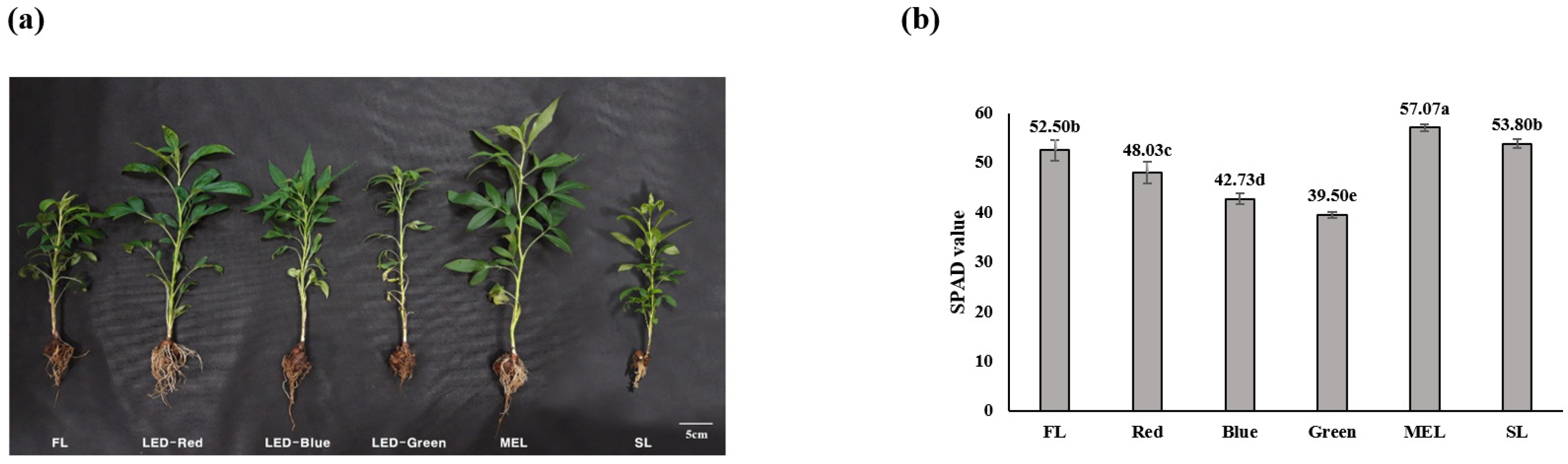
3.1. Growth Characteristics and Chlorophyll Content of A. macrocephala Grown under Artificial Light Source

In the growth investigation of aerial parts of A. macrocephala treated with artificial light, the highest plant length (37.30 ± 5.90 cm) was recorded in the MEL treatment (Figure 1a and Table 1). The highest leaf length (10.77 ± 1.15 cm) was recorded in the LED-red light treatment, and the highest leaf width and leaf number were measured (10.30 ± 2.35 cm and 21.33 ± 4.93 cm, respectively) were recorded in the MEL treatment. The highest dry weight of both aerial and underground parts was noted under sunlight (2.35 ± 0.06 cm and 0.33 ± 0.08 cm, respectively). Regarding underground parts, root growth did not significantly differ among the artificial light sources. The highest chlorophyll content (57.07 ± 0.65 SPAD) was recorded in the MEL treatment (Figure 1b).
In lettuce grown under artificial light sources, the closed plant production system, plant height, fresh weight, and dry weight were the highest under fluorescent light [27]. In Salvia, leaf area increased under red or mixed light [28]. In the present study, plant length, leaf width, and leaf number increased under MEL because it comprises various wavelengths as opposed to other light sources. Furthermore, a higher chlorophyll content implies efficient light use. Even under the same light conditions, light-harvesting complex II selectively and efficiently receives external light to promote photosynthetic metabolism and reduce sugars in the body. The supply is more active, and both exposure time and intensity of light affect plant photosynthesis [29,30]. In the present study, chlorophyll content was the highest under MEL, perhaps because the effective wavelength range for photosynthesis was diverse, increasing its efficiency.

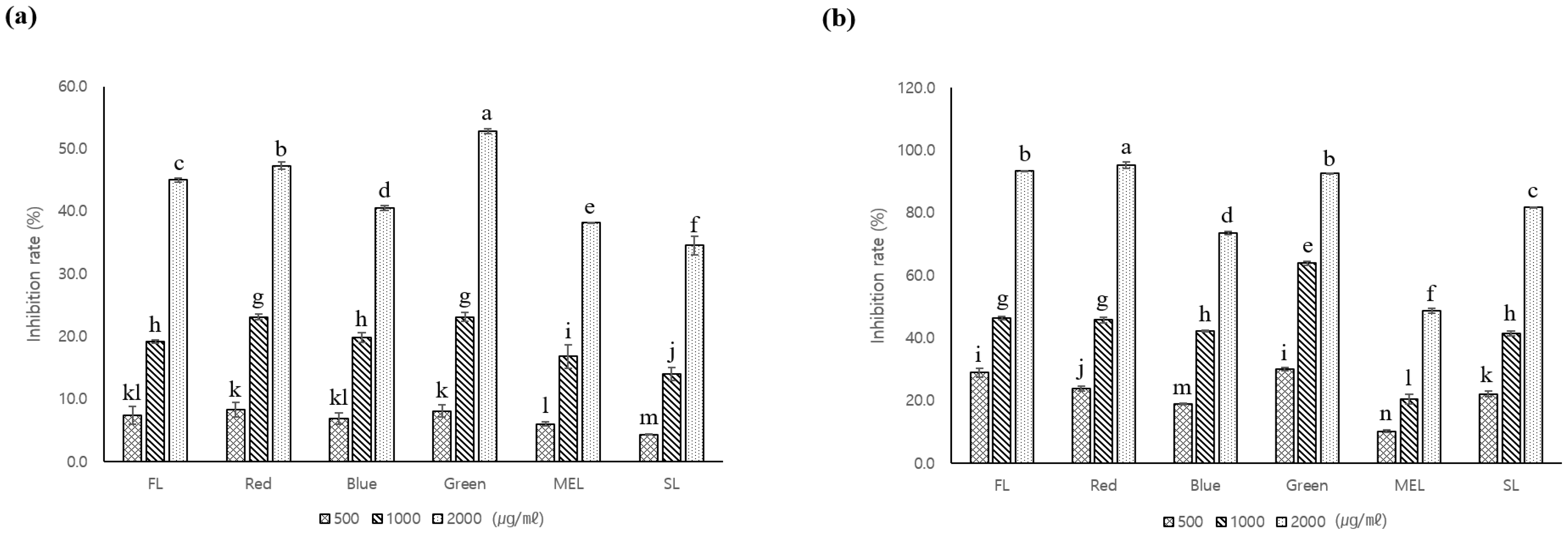
3.2. Antioxidant and Whitening Activity of A. macrocephala Grown under Artificial Light

The free radical scavenging ability of A. macrocephala grown under artificial light was analyzed using the DPPH assay. The difference in free radical scavenging ability between the aerial and underground parts was large among the artificial light sources. Underground parts showed a much higher free radical scavenging ability than aerial parts (Figure 2). In the case of aerial parts, the free radical scavenging ability of plants treated with LED-green light was the highest at 52.9 ± 0.4%. In the case of underground parts, the free radical scavenging ability of plants treated with LED-red light was the highest at 95.3 ± 1.1%. The DPPH assay measures the ability of a compound to act as a free radical scavenger or hydrogen donor and its antioxidant capacity [18]. In Salvia miltiorrhiza grown under artificial light, the free radical scavenging activity of aerial part extract was in the order of fluorescent light, MEL, blue light, green light, and red light, while that of underground part extract was in the order of blue light, green light, red light, MEL, and fluorescent light [6]. These trends can be attributed to the fact that the availability of artificial light differs between the aerial and underground parts, as evidenced in the present experiment.
The highest total phenol content (24.93 ± 0.3 mg GAE·g−1) was recorded in the underground parts of A. macrocephala grown under LED-green light (Figure 3a,b). In the case of aerial parts, the highest total phenol content (14.0 ± 0.3 mg GAE·g−1) was noted in plants grown under LED-blue light. However, there was no significant difference in the utilization of artificial light among aerial parts. The total flavonoid content of aerial and underground part extracts was the highest (37.6 ± 0.5 mg QE·g−1 and 11.2 ± 0.3 mg QE·g−1, respectively) under LED-red and LED-green light, respectively, with significant differences compared to values under other light sources (Figure 3c,d). In Salvia miltiorrhiza grown under artificial light, the total phenol content of both aerial and underground parts was higher under LED-blue light [6], consistent with our results for aerial parts. In addition, the total flavonoid content of Salvia miltiorrhiza was the highest under LED-red light in the aboveground part and under LED-green light in the underground part, consistent with the results of the present study. In the leaves and roots of Rehmannia glutinosa grown under artificial light, the total phenol content was in the order of blue, red, and fluorescent light and flavonoid content was in the order of red, blue, and fluorescent light [31]. Thus, the usability of artificial light wavelengths varies across plant species.

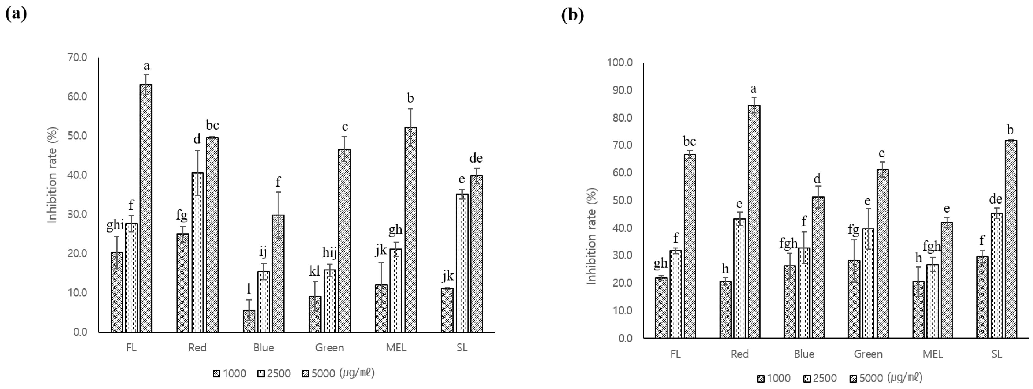
3.3. Tyrosinase Inhibitory Activity of A. macrocephala Grown under Artificial Light

To investigate the whitening activity of A. macrocephala grown under artificial light source, the samples were diluted to three concentrations (1000 μg·mL−1, 2500 μg·mL−1, and 5000 μg·mL−1) and tyrosinase inhibitory activity was measured. Tyrosinase inhibitory activity increased with increasing concentration of aerial or underground part extract. In particular, at the concentration of 5000 μg·mL−1, the difference in activity of extracts across artificial light sources was the most significant. Specifically, the aerial part extract showed the highest activity (63.1 ± 2.6%) under fluorescent light, while the underground part extract showed the highest activity (84.6 ± 2.9%) under LED-red light (Figure 4). In another study on each part of Perilla grown under various artificial light sources, the tyrosinase inhibitory activity increased with increase in concentration, with the highest activity recorded under LEP [32]. Similarly, in another study on Astragalus membranaceus grown under artificial light, the highest tyrosinase inhibitory activity (64.49%) was recorded under LEP [33]. In various plants and fungi, such as Drynaria sp., Asphodelus microcarpus, and Tremella fuciformis, many studies have reported that the higher the content of polyphenols and flavonoids, the higher the antioxidant potential and pigmentation relief [34,35,36].

3.4. Antimicrobial Activity of A. macrocephala Grown under Artificial Light

Antimicrobial activity of A. macrocephala grown under artificial light against V. litoralis, E. coli, B. subtilis, S. aureus, S. typhimurium and P. aeruginosa was tested (Table 2). The underground part extract showed antimicrobial activity against V. litoralis but the aerial part extract did not. All extracts, except those from plants grown under MEL, inhibited V. litoralis at a concentration of 0.25 μg·mL−1 or less. Both aerial and underground part extracts showed antimicrobial activity against E. coli. At a concentration of 1 μg·mL−1 or less, the aerial part extract showed antimicrobial activity against P. aeruginosa, but the underground part extract did not. In a previous study on Saliva miltiorrhiza in which antimicrobial activity was tested using the same method, the aerial part extract showed no inhibitory activity, but the root extract showed minimal inhibitory activity against all microorganisms [6]. Thus, the antimicrobial effect on P. aeruginosa observed in the present study is somewhat different from the reported minimum inhibitory activity of aerial part extracts. Antimicrobial activity may vary depending on complex factors, such as the type of microorganism, wavelength of artificial light, and plant species.

3.5. Anti-Inflammatory Activity of A. macrocephala Grown under Artificial Light

To determine the degree of anti-inflammatory activity of the extracts in LPS-induced RAW 264.7 cells, a cytotoxicity test was performed. In cytotoxicity assays at concentrations of 10 μg·mL−1, 100 μg·mL−1, and 200 μg·mL−1, cellular activity decreased in a concentration dependent manner. At the concentration of 100 μg·mL−1 or less, 70% or more cell activity was verified; thus, anti-inflammatory activity in LPS-induced cells was investigated at a concentration of 100 μg·mL−1 or less (Figure 5). The anti-inflammatory activity of the underground part extract was higher than that of the aerial part extract. Both aerial and underground part extracts inhibited NO production in a concentration-dependent manner. In the aerial parts, at the concentration of 100 μg·mL−1, extract of plants grown under LED-blue light showed the highest inhibitory activity against NO production (64.77 ± 1.16%), followed by that of plants grown under LED-green light (75.42 ± 4.07%). In the underground parts, the extract of plants grown under fluorescent light showed the highest inhibitory activity against NO production (2.97 ± 0.14%), followed by extracts of plants grown under LED-red light (7.89 ± 2.46%) and LED-green light (6.35 ± 4.64%).
Based on these results, the expression levels of inflammation-inducing genes were assessed in cells treated with extracts of plants grown under fluorescent light and LED. Excellent anti-inflammatory effects were noted in cells treated with extract of A. macrocephala grown under artificial light (Figure 6). Specifically, the expression of iNOS, a gene that induces NO production, was suppressed in both treatment groups compared with that in the control group. In addition, the expression of COX-2 gene, which interferes with immunity by inducing inflammation, was suppressed in both treatment groups.
In RAW 264.7 cells, NO plays an important role in blood coagulation, blood pressure regulation, and immune function against cancer cells; however, when present in excess, NO produces detrimental effects on the human body, causing cellular damage and inflammation [37]. A. macrocephala was reported to be related to the anti-inflammatory control mechanism of COX-2, IL-17, TNF, and the C-type lectin receptor signal pathway (Yang et al., 2020). For this reason, it has already been found that A. macrocephala is a material that can be provided as a raw material that can be used in various fields. To further enhance the functionality of these materials, A. macrocephala using an artificial light source was used as a breeding material. Extract of A. macrocephala grown under artificial light effectively inhibited NO production; therefore, its bioactive components may be involved in the regulation of immune function by suppressing the inflammatory response. Furthermore, COX-2 produced by NO synthesizes PGE2 from arachidonic acid by metabolizing phospholipids during the inflammatory process, and PGE2 is known to cause vasodilation, pain, and fever [38]. Since COX-2 plays an important role in inflammation and immune function in RAW 264.7 cells, the extract of A. macrocephala grown under artificial light likely suppresses the inflammatory response by effectively inhibiting COX-2.

4. Conclusions

Artificial light altered the growth characteristics, chlorophyll content, antioxidant activity, whitening activity, and anti-inflammatory activity of A. macrocephala. MEL produced excellent growth characteristics and chlorophyll content, while LED-red and LED-green light increased antioxidant activity and total phenol and flavonoid contents. Furthermore, extracts of A. macrocephala grown under artificial light exhibited antimicrobial activity against V. litoralis, E. coli, and P. aeruginosa. LED-red light improved whitening activity, while fluorescent, LED-red, and LED-green lights enhanced anti- inflammatory activity. Since the functionality of A. macrocephala as a biomaterial varies depending on the type of artificial light source under which it is grown, artificial light appears to be suitable for improving the bioactivity of this medicinal plant. Since the production of crops may gradually decrease in the future due to environmental disasters caused by global warming, the cultivation area for creating an artificial environment should be expanded. In particular, if an artificial light source is used as a plant breeding material, the original functionality will be higher, and it is thought that it can be used for cultivation of smart farms that can be produced year-round. In addition, these results will help to improve the utility value to enable the production of A. macrocephala through plant factory by utilizing LEDs with low energy consumption.

Author Contributions

Conceptualization, E.S.S. and M.J.K.; methodology, M.H.H. and J.W.S.; resources, K.J.H.; supervision, E.S.S.; investigation, E.S.S.; writing, E.S.S. All authors have read and agreed to the published version of the manuscript.

Funding

This research received no external funding.

Institutional Review Board Statement

Not applicable.

Informed Consent Statement

Not applicable.

Data Availability Statement

The data presented in this study are contained within the article.

Acknowledgments

This study was supported by the Bioherb Research Institute, Kangwon National University, Korea.

Conflicts of Interest

The authors declare no conflict of interest.

References

  1. Ministry of Agriculture, Food and Rural Affairs (MAFRA). Production Results of Special Crops; Ministry of Agriculture, Food and Rural Affairs: Sejong, Korea, 2019; pp. 41, 60.
  2. Ministry of Food and Drug Safety (MFDS). The Korea Pharmacopoeia; Ministry of Food and Drug Safety: Seoul, Korea, 2019; p. 44.
  3. Lee, J.H.; Kim, Y.K.; Hong, S.P.; Kim, C.S. Study of Taxonomic Origins of Atractylodis Rhizoma Alba and Atractylodis Rhizoma. Korean J. Orient. Med. 2002, 8, 55–63. [Google Scholar]
  4. Kozai, T.; Niu, G.; Takagaki, M. Plant Factory: An Indoor Vertical Farming System for Efficient Quality Food Production; Academic Press: Cambridge, MA, USA, 2019. [Google Scholar]
  5. Yoon, C.; Choi, H. A Study on the Various Light Source Radiation Conditions and Use of LED Illumination for Plant Factory. J. Korean Ins. Illum. Electric. Installat. Eng. 2011, 25, 14–22. [Google Scholar]
  6. Choi, H.L.; Seo, J.W.; Hwang, M.H.; Lee, H.I.; Kim, M.J.; Yu, C.Y. Growth Characteristics and Functional Analysis of Salvia miltiorrhiza Bunge by Artificial Light Sources. Korean J. Med. Crop Sci. 2020, 28, 200–208. [Google Scholar] [CrossRef]
  7. Duan, Y.; Kim, G.H.; Seong, J.H.; Chung, H.S.; Kim, H.S. Antioxidant Activities of n-Butanol and Ethyl Acetate Extracts from Yam (Dioscorea batatas Decne). J. Korean Oil Chem. Soc. 2015, 32, 599–606. [Google Scholar] [CrossRef]
  8. Yi, M.R.; Kang, C.H.; Bu, H.J. Antioxidant and Antiinflammatory Activity of Extracts from Kohlrabi(Brassica oleracea var. Gonglodes). J. Korean Oil Chem. Soc. 2017, 34, 189–202. [Google Scholar]
  9. Lu, Y.; Foo, L.Y. Antioxidant and Radical Scavenging Activities of Polyphenols from Aapple Pomace. Food Chem. 2000, 68, 81–85. [Google Scholar] [CrossRef]
  10. Ekström, A.; Serafini, M.; Nyren, O.; Wolk, A.; Bosetti, C.; Bellocco, R. Dietary Quercetin Intake and Risk of Gastric Cancer: Results from A Population-based Study in Sweden. Annal. Oncol. 2011, 22, 438–443. [Google Scholar] [CrossRef]
  11. Del Marmol, V.; Beermann, F. Tyrosinase and Related Proteins in Mammalian Pigmentation. FEBS Lett. 1996, 381, 165–168. [Google Scholar] [CrossRef]
  12. Desmedt, B.; Ates, G.; Courselle, P.; De Beer, J.O.; Rogiers, V.; Hendrickx, B.; Deconinck, E.; De Paepe, K. In Vitro Dermal Absorption of Hydroquinone: Protocol Validation and Applicability on Illegal Skin-Whitening Cosmetics. Skin Pharmacol. Physiol. 2016, 29, 300–308. [Google Scholar] [CrossRef]
  13. Sonthalia, S.; Daulatabad, D.; Sarkar, R. Glutathione as A Skin Whitening Agent: Facts, Myths, Evidence and Controversies. Ind. J. Dermatol. Venereol. Leprol. 2016, 82, 262–272. [Google Scholar] [CrossRef]
  14. Pestka, J.; Zhou, H. Toll-like Receptor Priming Sensitizes Macrophages to Proinflammatory Cytokine Gene Induction by Deoxynivalenol and Other Toxicants. Toxicol. Sci. 2006, 92, 445–455. [Google Scholar] [CrossRef] [PubMed]
  15. Sarkar, D.; Saha, P.; Gamre, S.; Bhattacharjee, S.; Hariharan, C.; Ganguly, S.; Sen, R.; Mandal, G.; Chattopadhyay, S.; Majumdar, S.; et al. Anti-inflammatory Effect of Allylpyrocatechol in LPS-induced Macrophages Is Mediated by Suppression of iNOS and COX-2 via the NF-κB Pathway. Int. Immunopharm. 2008, 8, 1264–1271. [Google Scholar] [CrossRef] [PubMed]
  16. Tak, P.P.; Firestein, G.S. NF-kappaB: A Key Role in Inflammatory Diseases. J. Clin. Investig. 2001, 107, 7–11. [Google Scholar] [CrossRef] [PubMed]
  17. Li, C.; He, L.; Dong, H.; Jin, J. Screening for the Anti-inflammatory Activity of Fractions and Compounds from Atractylodes macrocephala Koidz. J. Ethnopharmacol. 2007, 114, 212–217. [Google Scholar] [CrossRef] [PubMed]
  18. Yang, S.; Zhang, J.; Yan, Y.; Yang, M.; Li, C.; Li, J.; Zhong, L.; Gong, Q.; Yu, H. Network Pharmacology-Based Strategy to Investigate the Pharmacologic Mechanisms of Atractylodes macrocephala Koidz. for the Treatment of Chronic Gastritis. Front. Pharmacol. 2020, 10, 1629. [Google Scholar] [CrossRef] [Green Version]
  19. Jeong, D.; Dong, G.Z.; Lee, H.J.; Ryu, J.H. Anti-Inflammatory Compounds from Atractylodes macrocephala. Molecules 2019, 24, 1859. [Google Scholar] [CrossRef] [Green Version]
  20. Kedare, S.B.; Singh, R.P. Genesis and Development of DPPH Method of Antioxidant Assay. J. Food Sci. Technol. 2011, 48, 412–422. [Google Scholar] [CrossRef] [Green Version]
  21. Sato, M.; Ramarathnam, N.; Suzuki, Y.; Ohkubo, T.; Takeuchi, M.; Ochi, H. Varietal Differences in the Phenolic Content and Superoxide Radical Scavenging Potential of Wines from Different Sources. J. Agric. Food Chem. 1996, 44, 37–41. [Google Scholar] [CrossRef]
  22. Moreno, M.I.N.; Isla, M.I.; Sampietro, A.R.; Vattuone, M.A. Comparison of the Free Radical-Scavenging Activity of Propolis from Several Regions of Argentina. J. Ethnopharmacol. 2000, 71, 109–114. [Google Scholar] [CrossRef]
  23. Irith, W.; Kai, H.; Robert, E.W.H. Agar and Broth Dilution Methods to Determine the Minimal Inhibitory Concentration (MIC) of Antimicrobial Substances. Nat. Protoc. 2008, 3, 163–175. [Google Scholar]
  24. Bernard, P.; Berthon, J.Y. Resveratrol: An Original Mechanism on Tyrosinase Inhibition. Inter. J. Cosmet. Sci. 2000, 22, 219–226. [Google Scholar] [CrossRef] [PubMed]
  25. Mosmann, T. Rapid Colorimetric Assay for Cellular Growth and Survival: Application to Proliferation and Cytotoxicity Assays. J. Immunol. Methods 1983, 65, 55–63. [Google Scholar] [CrossRef]
  26. Luis, A.J.M.; Julien, M.; Simona, M.C.; Frans, J.M.H.; Elena, P. Methods of nitric oxide detection in plants: A commentary. Plant Sci. 2011, 181, 509–519. [Google Scholar]
  27. Park, J.E.; Park, Y.G.; Jeong, B.R.; Hwang, S.J. Growth and Anthocyanin Content of Lettuce as Affected by Artificial Light Source and Photoperiod in a Closed-type Plant Production System. Korean J. Hort. Sci. Technol. 2012, 30, 673–679. [Google Scholar]
  28. Heo, J.W.; Lee, Y.B.; Lee, D.B.; Chun, C.H. Light Quality Affects Growth, Net Photosynthetic Rate, and Ethylene Production of Ageratum, African Marigold, and Salvia seedlings. Korean J. Hort. Sci. Technol. 2009, 27, 187–193. [Google Scholar]
  29. Sicora, C.; Zoltan, M.; Imre, V. The Interaction of Visible and UV-B Light During Photodamage and Repair of Photosystem II. Photosyn. Res. 2003, 75, 127–137. [Google Scholar] [CrossRef]
  30. Kang, S.B.; Jang, H.I.; Lee, I.B.; Park, J.M.; Moon, D.K. Effect of Waterlogging Condition on the Photosynthesis of ‘Campbell Early’ Grapevine. Korean J. Hort. Sci. Technol. 2008, 26, 372–379. [Google Scholar]
  31. Manivannan, A.; Soundararajan, P.; Halimah, N.; Ko, C.H.; Jeong, B.R. Blue LED light enhances growth, phytochemical contents, and antioxidant enzyme activities of Rehmannia glutinosa cultured in vitro. Hort. Environ. Biotech. 2015, 56, 105–113. [Google Scholar] [CrossRef]
  32. Yoo, J.H.; Choi, J.H.; Kang, B.J.; Jeon, M.R.; Lee, C.O.; Kim, C.H.; Seong, E.S.; Heo, K.; Yu, C.Y.; Choi, S.K. Antioxidant and Tyrosinase Inhibition Activity Promoting Effects of Perilla by the Light Emitting Plasma. Korean J. Med. Crop Sci. 2017, 25, 37–44. [Google Scholar] [CrossRef]
  33. Choi, J.H.; Seong, E.S.; Yoo, J.H.; Choi, S.K.; Lee, J.G.; Lim, J.D.; Na, J.K.; Yu, C.Y. Enhancement of Growth Characteristics and Biological Activities in Astragalus membranaceus Using Artificial Light Sources. Russ. J. Plant Physiol. 2018, 65, 732–739. [Google Scholar] [CrossRef]
  34. Tan, J.B.L.; Lim, Y.Y. Antioxidant and Tyrosinase Inhibition Activity of the Fertile Fronds and Rhizomes of Three Different Drynaria species. BMC Res. Notes 2015, 8, 468. [Google Scholar] [CrossRef] [PubMed] [Green Version]
  35. Petrillo, A.D.; González-Paramás, A.M.; Era, B.; Medda, R.; Pintus, F.; Santos-Buelga, C.; Fais, A. Tyrosinase Inhibition and Antioxidant Properties of Asphodelus microcarpus Extracts. BMC Complement. Alternat. Med. 2016, 16, 453. [Google Scholar] [CrossRef] [PubMed] [Green Version]
  36. Lee, K.H.; Park, H.S.; Yoon, I.J.; Shin, Y.B.; Baik, Y.C.; Kooh, D.H.; Kim, S.K.; Jung, H.K.; Sim, M.O.; Cho, H.W.; et al. Whitening and Anti-wrinkle Effects of Tremella fuciformis Extracts. Korean J. Med. Crop Sci. 2016, 24, 38–46. [Google Scholar] [CrossRef]
  37. Thong, H.Y.; Maibach, H.I. Irritant Dermatitis as A Model of Inflammation. Drug Discov. Today Dis. Mech. 2008, 5, 85. [Google Scholar] [CrossRef]
  38. You, S.H.; Pyo, Y.H. Antioxidant and Anti-inflammatory Activities of Ethanol Extracts from Wheat Sprout. J. Investig. Cosmetol. 2015, 11, 231–238. [Google Scholar]
Figure 1. Growth characteristics (a) and chlorophyll contents (b) of A. macrocephala under different light sources. Values represent mean of data obtained from three independent experiments (p < 0.05). Significance was indicated with different letters according to statistical analysis.
Agronomy 12 01485 g001
Figure 2. The inhibition rate of DPPH free radical scavenging activities of aerial (a) and underground (b) parts from extracts of A. macrocephala under different light sources. Values represent mean of data obtained from three independent experiments (p < 0.05). Significance was indicated with different letters according to statistical analysis.
Agronomy 12 01485 g002
Figure 3. Total phenolic (a,b) and flavonoid (c,d) contents of aerial and underground parts from extracts of A. macrocephala under different light sources. (1) GAE; gallic acid equivalents. (2) QE; quercetin equivalents. Values represent mean of data obtained from three independent experiments (p < 0.05). Significance was indicated with different letters according to statistical analysis.
Agronomy 12 01485 g003
Figure 4. The inhibition rate of tyrosinase of aerial (a) and underground (b) parts from extracts of A. macrocephala under different light sources. Significance was indicated with different letters according to statistical analysis.
Agronomy 12 01485 g004
Figure 5. Cell viability (a,b) by MTT assay and NO production (c,d) in LPS-induced Raw 264.7 cell with Dachul extracts. (a,c) aerial part of A. macrocephala, (b,d) underground part of A. macrocephala. Significance was indicated with different letters according to statistical analysis.
Agronomy 12 01485 g005
Figure 6. Transcriptional expression levels of iNOS and COX-2 genes in LPS-induced Raw 264.7 cells with underground parts extracts of A. macrocephala. LPS-Con; Control, D-B; Dachul grown under LED-Blue light.
Agronomy 12 01485 g006
Table 1. Growth characteristics by parts of aerial and underground in A. macrocephala treated with artificial light sources for 2 weeks.
Light SourceAerial PartUnderground Part
Plant Length
(cm)
Leaf Length
(cm)
Leaf Width
(cm)
Number of LeavesDry Weight
(g)
Root Length
(cm)
Dry Weight
(g)
FL (1)27.77 ± 0.50 c8.27 ± 1.08 bc7.73 ± 0.25 bc21.67 ± 0.58 a1.16 ± 0.15 cd14.23 ± 3.88 a0.29 ± 0.03 ab
LED-Red35.17 ± 0.98 ab10.77 ± 1.15 a8.80 ± 1.28 abc19.33 ± 1.53 ab1.81 ± 0.13 b14.73 ± 1.94 a0.29 ± 0.09 ab
LED-Blue33.57 ± 4.39 abc9.60 ± 0.75 ab9.40 ± 0.40 ab21.67 ± 4.73 a1.29 ± 0.16 c12.90 ± 1.31 a0.26 ± 0.07 ab
LED-Green30.97 ± 2.95 bc7.70 ± 0.26 c7.57 ± 1.10 bc13.67 ± 2.52 b0.96 ± 0.05 d10.97 ± 1.29 a0.18 ± 0.04 b
MEL (2)37.30 ± 5.90 a9.73 ± 0.23 ab10.30 ± 2.35 a21.33 ± 4.93 a2.03 ± 0.15 b12.50 ± 0.89 a0.17 ± 0.07 b
SL (3)28.90 ± 1.31 bc7.60 ± 0.56 c6.57 ± 0.59 c14.33 ± 1.53 b2.35 ± 0.06 a12.60 ± 1.47 a0.33 ± 0.08 a
(1) FL; fluorescent light, (2) MEL; microwave electrodeless light. (3) SL; sun light. Values represent mean ± S.D. of data obtained from three independent experiments. Duncan’s Multiple Range Test at 5% level (DMRT, p < 0.05). Significance was indicated with different letters according to statistical analysis.
Table 2. Antimicrobial activity by parts of aerial and underground from extracts of A. macrocephala treated with artificial light sources for 2 weeks.
Minimal Inhibitory Concentration (mg/mL)
DachulS. aureusV. litoralisB. subtilisE. coliS. typhimuriumP. aeruginosa
Light SourcePlant Parts
FLAerialND (1)NDND≥1ND≥1
UndergroundND≥1ND≥1NDND
LED-RedAerialNDNDND≥1ND≥1
UndergroundND≥0.5ND≥0.5NDND
LED-BlueAerialNDNDND≥1ND≥1
UndergroundND≥1ND≥0.5NDND
LED-GreenAerialNDNDND≥1ND≥1
UndergroundND≥0.25ND≥0.25NDND
MELAerialNDNDND≥1ND≥1
UndergroundND≥0.5ND≥0.5NDND
SLShootNDNDND≥1ND≥1
UndergroundND≥0.5ND≥0.5NDND
Tetracycline≥0.007≥0.007≥0.007≥0.007≥0.007≥0.007
The MIC values against bacteria were determined by the serial 2-fold dilution method. (1) ND; not detected.
Publisher’s Note: MDPI stays neutral with regard to jurisdictional claims in published maps and institutional affiliations.

