Article

In-Field LAMP Detection of Flavescence Dorée Phytoplasma in Crude Extracts of the Scaphoideus titanus Vector

1
Institute for Sustainable Plant Protection, National Research Council of Italy (IPSP-CNR), 10135 Turin, Italy
2
Department of Agricultural, Forestry and Food Sciences (DISAFA), University of Torino, 10095 Grugliasco, Italy
3
Department of Agricultural, Food and Forest Sciences, University of Palermo, 90128 Palermo, Italy
*
Authors to whom correspondence should be addressed.
Agronomy 2022, 12(7), 1645; https://doi.org/10.3390/agronomy12071645
Submission received: 6 June 2022 / Revised: 5 July 2022 / Accepted: 8 July 2022 / Published: 8 July 2022
(This article belongs to the Special Issue Genetics and Molecular Biology of Pathogens in Agricultural Crops)

Abstract

:
One of the most destructive diseases affecting grapevine in Europe is caused by Flavescence Dorée phytoplasma (FDp), which belongs to the 16Sr-V group and is a European Union quarantine pathogen. Although many molecular techniques such as loop-mediated isothermal amplification (LAMP) are widely used for the rapid detection of FDp in infected grapevine plants, there is no developed isothermal amplification assay for FDp detection in the insect vectors that are fundamental for the spread of the disease. For this reason, a simple in-field real-time LAMP protocol was optimized and developed for the specific detection of FDp in the insect vector Scaphoideus titanus. The LAMP assay was optimized to work with crude insect extracts obtained by manually shaking a single insect in a buffer for 5 min. Such a simple, sensitive, specific, economic, and user-friendly LAMP assay allowed the detection of FDp in S. titanus in less than half an hour, directly in the field. The developed insect tissue preparation procedure, combined with the LAMP protocol, promptly revealed the presence of FDp in infected S. titanus directly in the vineyards, allowing for monitoring of the spread of the pathogen in the field and to apply timely strategies required for the mandatory control of this pathogen.

Graphical Abstract

1. Introduction

Over the years, vegetatively propagated crops, including grapevine, have undergone a remarkable deterioration of the sanitary status worldwide due to obligate-parasite infections [1]. Among woody crops, the grapevine is affected by numerous diseases impacting the production of fresh grapes and wine. Flavescence dorée (FD) is one of the main diseases affecting grapevine in Europe and is caused by Flavescence dorée phytoplasma (FDp). FDp belongs to the elm yellows group (16Sr-V), which includes FDp-C and FDp-D subgroups and various map and vmpA genotypes [2]. FDp caused significant damage in several European countries, including Northern Italy, leading to the loss of several thousand hectares of vineyards and a strong reduction (>50%) in berry production [3,4,5,6,7,8,9].
FDp is persistently transmitted across grapevine plants by the phloem-feeding activity of the monophagous leafhopper Scaphoideus titanus Ball (Homoptera Cicadellidae) [10,11,12]. In Vitis vinifera, FD typical symptoms include the desiccation of inflorescences, berry shrivel, leaf discolouration, the reduction of growth, and the irregular ripening of wood. In Italy, FDp was reported for the first time in 1973 [5,13] and, following the destructive outbreaks often recorded, it was included in the list of European Union quarantine pests with mandatory disease control strategies [14,15].
There are still no effective control methods against this disease and the management is mainly based on prevention, including the use of certified healthy propagation material and cultivation in areas where the FDp vector is not present. Thermotherapy (50 °C for 45 min) of dormant woody plant material of both scions and rootstocks is also a recommended practice to eliminate FDp [16]. However, when the disease is already established, the only way to control it relies on constant monitoring and on mandatory control measures, including insecticide treatments against the pathogen vectors, the eradication of infected plants, and diagnostic procedures to support vigilance and phytosanitary controls [17].
The mandatory control of FD must be supported by an adequate and efficient diagnosis of phytoplasmas. Molecular diagnostic techniques for the FDp detection in infected plants are based on conventional PCR [18,19,20], nested PCR [21,22], real-time PCR [23,24], loop-mediated isothermal amplification (LAMP; with four primers), and accelerated LAMP (with six primers including the loop primers) [25,26]. Furthermore, end-point PCR, nested PCR, and real-time PCR tests are available for FDp detection in insect vectors [20,22,24,27].
Among these techniques, LAMP [28] is widely used in plant pathology thanks to the speed of performance and the rapidity in obtaining results [29]. Furthermore, its sensitivity is higher or comparable to other molecular techniques, allowing the detection of the target DNA in quantities ranging from picograms to femtograms, depending on the protocol [30,31].
Moreover, LAMP assays can be performed directly in the field thanks to the use of a device equipped with a rechargeable battery, thus overcoming the insurmountable obstacle associated with other molecular techniques (conventional PCR and real-time PCR) that require current-powered instruments in the laboratory. Finally, thanks to the high resilience of the enzymatic components used in the LAMP technique towards reaction inhibitors [32], it is not necessary to purify the nucleic acids from the sample. Indeed, it is possible to use crude extracts obtained directly with an alkaline buffer and metal beads, facilitating the mechanical disruption of the tissues [33].
Although the diagnosis of phytoplasma is generally difficult due to the irregular distribution in the host organism and the low concentration in the winter–spring seasons [34], the FDp detection in infected grapevine plants has been successfully carried out with the LAMP and accelerated LAMP assays in the laboratory or directly in the field [25], using crude leaf extracts and achieving results in just 30–60 min [25,35]. Different grapevine plant tissues (leaves, flowers, berries, and peduncles) have proven to be reliable for the timely and specific diagnosis of FDp. Meanwhile, various companies (OptiGene, Horsham, UK; Agdia, Elkhart, IN, USA) developed specific commercial isothermal amplification kits that are now available on the market.
LAMP has been already applied to identify other phytopathogens or symbiont microorganisms in different insect vectors, such as rice viruses in plant hopper vectors, the Wolbachia bacterium in various insect species, Xylella fastidiosa in Auchenorryncha, and the phytoplasma of coconut syndrome in Lophops saccharicida (Kirkaldy), Zophiuma pupillata (Stål), Taparella amata (Walker), and Colgar sp. [36,37,38,39].
Besides the LAMP, another widely used diagnostic method is the isothermal recombinase polymerase amplification (RPA) technique. RPA is similar to the LAMP assay in terms of sensitivity, specificity, speed, constant amplification temperature, possibility of real-time in-field detection, and does not require nucleic acid extraction. RPA is performed with different enzymes and the reaction works with only two primers, while the LAMP assay requires four (simple LAMP) or six primers (accelerated LAMP) [25,26,40,41].
Isothermal amplification techniques are widely used in the rapid detection of FDp in infected vines [25,35] and their application has recently started for the detection of other plant pathogens in insects [37,38]. However, no isothermal amplification assay has been developed so far for the detection of FDp in its insect vectors. This study intends to compare the isothermal amplification protocols available for FDp in plants and to select the best performing procedure to specifically identify FDp in S. titanus. Another objective of this study is to develop a quick insect tissue extraction procedure, avoiding the laborious DNA purification steps and allowing a direct in-field application.

2. Materials and Methods

2.1. Insect Rearing and Plant Cultivation

S. titanus rearing were maintained on healthy grapevine plants grown outdoors in natural conditions at the Department of Agricultural, Forest and Food Sciences (University of Torino, Grugliasco, Italy). Third instar nymphs were periodically collected and transferred on FDp-C(16SrV-C)-infected broad bean (Vicia faba L.) seedlings or on FDp-D(16SrV-D)-infected grapevine plants for the acquisition. After one week of acquisition, all insects were moved to healthy grapevines to complete the latency period. All infected broad bean seedlings and grapevine plants (healthy and infected) were maintained under an anti-insect net in the greenhouse and outdoors, respectively. Healthy insects were directly collected from the rearing, maintained on healthy grapevine, and used as a negative control. Infected (FDp-C or -D) or healthy laboratory-reared nymphs (4th–5th instar) and adults were collected and used for the experiments.

2.2. DNA Extraction

Total DNA was extracted from 36 isolates (Table 1) with cetyltrimethylammonium bromide (CTAB) buffer-based protocol [42], using either single insects or 100 mg of fresh leaf veins dissected from FDp-C- and FDp-D-infected or healthy grapevine plants. Lysis was carried out after grinding the plant or insect material in CTAB buffer (2.5% w/v CTAB, 1.4 M NaCl, 20 mM EDTA pH 8.0, 100 mM Tris-HCl pH 8.0 and 0.2% β-mercaptoethanol) and an incubation at 60 °C for 30 min. Following chloroform/isoamyl alcohol phase extraction, the DNA was precipitated with cold isopropanol and centrifuged for 15 min at 15,000 g at 4 °C. The DNA was washed with cold 70% ethanol by centrifugation for 5 min at 15,000 g at 4 °C. The pellet was then dried at room temperature and resuspended in 100 µL sterile water. DNA quality and concentration were determined using a NanoDrop 2000 Spectrophotometer (Thermo Fisher Scientific, Wilmington, DE, USA), and DNA was then adjusted to a final concentration of 20 ng/µL.

2.3. Isothermal Amplification Assays

To evaluate the best performing isothermal amplification technique for FDp detection in insects, three different isothermal amplification protocols available for FDp diagnosis in grapevine were assessed: (i) LAMP assay targeting the 23 rRNA gene of FDp, using the FD-specific primers and isothermal master mix (both from OptiGene, Horsham, UK), following manufacturer’s instructions (hereafter called LAMP 1); (ii) accelerated LAMP assay targeting the 16 rRNA gene of FDp using primers and conditions described by Kogovšek et al. [25] (hereafter called LAMP 2); (iii) RPA assay using the AmplifyRP® XRT for Phytoplasma vitis FD-kit (Agdia, Elkhart, IN, USA), according to manufacturer’s instructions with some modifications (hereafter called RPA).
Regarding LAMP 1, the 25 μL-LAMP reaction mixture consisted of 15 μL Flavescence dorée LAMP Master Mix, 5 μL LAMP primers mix, and 5 μL DNA extracts. LAMP 1 was performed at 62, 65, and 67 °C for 40 min. For LAMP 2, the reaction was carried out in a 25 μL-LAMP mix containing 2× Isothermal Master Mix ISO-004® (OptiGene, Horsham, UK), 2 μM of each internal primer (FIP and BIP), 0.2 μM of each external primer (F3 and B3), 1 μM of each loop primer (BL and FL primer), and 5 μL DNA. LAMP 2 was conducted at 62, 65, and 67 °C for 40 min. The RPA mix was prepared by adding 5 μL DNA and 20 μL sterile water to the reaction pellet of the AmplifyRP® XRT for Phytoplasma vitis FD-kit (Agdia, Elkhart, IN, USA). RPA was carried out at 40, 42, and 45 °C for 20 min. For each assay, 20, 2, and 0.2 ng/µL of DNA was used (indicated as initial concentration). All primer sets used for isothermal amplification tests are listed in Table S1. Melting curves for LAMP 1 and LAMP 2 assays were generated from 60 °C to 95 °C (ramp speed 0.05 °C/s, with plate readings every 15 s). The analyses were performed using two instruments, the CFX96 Real-Time PCR Detection system (Bio-Rad, Hercules, CA, USA) and bCube2 (Hyris, London, UK).
According to EPPO [17], the analytical specificity of the isothermal amplification assays was tested against the target organism (FDp in 10 isolates of laboratory-reared infected S. titanus and 10 isolates of infected grapevine plants) and non-target organisms ‘Candidatus Phytoplasma (Ca. P.) solani’ and ‘Ca. P. mali’, isolated from three Hyalesthes obsoletus Signoret specimens captured on grapevine plants and three Cacospsylla melanoneura (Förster) specimens captured on apple plants, respectively. Moreover, the selectivity of the assays was determined by assessing the matrix effect (non-infected S. titanus and grapevine plants; 5 isolates each) on the test performance (Table 1). Assay repeatability was evaluated considering the accordance between the results of each replicate of the same sample obtained under the same conditions. The reproducibility of the assays was determined as the concordance between the results of a single test including aliquots of the same sample tested under different conditions (time, instruments, and operators). Three technical replicates for each biological replicate were included in the assay. The best performing isothermal amplification assay was selected and used for further experiments using crude insect and plant extracts, in both laboratory and in field assays.

2.4. Analytic Sensitivity of the Selected Assay

Analytical sensitivity as ‘the limit of detection (LOD)’ of the selected isothermal amplification assay (LAMP 1) was compared to that of the real-time PCR, performed according to Pelletier et al. [24]. The assay sensitivity was tested on DNA from four S. titanus specimens infected by FDp using ten-fold serial dilutions, starting from 10 to 1 × 10⁻⁴ ng/μL, tested in three replicates. DNA extracts were diluted in DNA extracted from healthy S. titanus until reaching the LOD and used as templates for LAMP and real-time PCR assays. [43]. The real-time PCR amplicons of four infected S. titanus specimens were checked on agarose gel for correct DNA size and then purified using a QIAquick PCR Purification Kit (Qiagen, Hilden, Germany) according to the manufacturer’s instructions. Purified DNA was quantified using a NanoDrop 2000 Spectrophotometer (Thermo Fisher Scientific, Wilmington, DE, USA) and used for the calculation of the number of copies (NC) following formula: NC = (amount of DNA in nanograms × 6.022 × 10²³)/(DNA template length × 1 × 109 × 650).

2.5. Development of a Crude Extract Preparation Method for LAMP

Crude extracts were obtained from laboratory-reared S. titanus, using different buffers and homogenization procedures. Insects were processed individually and placed in 2-mL Eppendorf tubes containing two tungsten beads and a 500 µL extraction buffer. Three different extraction buffers were evaluated: (i) Tris-EDTA-Triton X-100 (TET) buffer (1% Triton X-100, 20 mM Tris-HCl pH 8, 20 mM EDTA [38]); (ii) ELISA extraction buffer (1× phosphate-buffered saline, pH 8.2; 0.05% Tween 20, 1% PEG MW 6000, 2% polyvinylpyrrolidone MW 24,000; (iii) OptiGene lysis buffer (OptiGene, Horsham, UK). Following vigorous manual shaking for 2–5 min, the homogenate was either denatured at 95 °C for 10 min and centrifuged at 13,000 rpm for 1 min to pellet insect debris or used directly. In the case of preparing grapevine crude extracts, the same procedure described above was used, using 100 mg of leaf midrib tissue per 500 µL of extraction buffer. Finally, denatured or non-denatured insect homogenates were added to the isothermal amplification mixture, either undiluted or diluted 1:5, 1:7.5, and 1:10.
The best combination of extraction buffer, homogenization treatment, and extract dilution was selected and used for further assays on either S. titanus specimens collected in vineyards or infected grapevine plants.

2.6. LAMP and Real-Time PCR from Field-Captured Insects

2.6.1. Laboratory Assay

In order to assess the in-field performances of the LAMP analysis, S. titanus individuals were collected in August–September 2021 in Canale and Giaglione vineyards (Piedmont region), known for their high incidence of FDp. In addition, leaves of symptomatic and non-symptomatic grapevine plants were collected in the same vineyards (Supplementary Figure S1). In preliminary experiments, samples were transported immediately into the laboratory and prepared as described for LAMP analyses (Section 2.5). In detail, 5 μL of 1:5 diluted crude extracts were added to 15 μL of Flavescence dorée LAMP Master Mix from OptiGene (Horsham, UK), together with 5 μL of the LAMP primer mix, carrying out the reaction in the bCube2 instrument (Hyris), as described in Section 2.3.
The same crude insect and plant extract samples were used in a parallel real-time PCR assay using the FD-specific primers and probe (mapFD) designed by Pelletier et al. [24]. In order to prove the correct homogenization and preparation of the samples and to reduce the risk of false-negative results, we used primers and probe for the detection of S. titanus partial mitochondrial 16S DNA [27] or of grapevine chloroplast tRNAL-F spacer DNA [24], as reaction control. All primer sets for real-time PCR tests are listed in Table S1.

2.6.2. In-Field LAMP Assay

Crude extracts from single insects and leaf veins sampled in the same infected vineyards (Section 2.6.1) were prepared directly in the field, using the TET buffer, without denaturation and centrifugation. Laboratory-prepared crude extracts of infected or healthy insects and plants were used as positive and negative controls adopting the same LAMP conditions described above. In this case, the bCube2 instrument was supplied with a battery.

3. Results

3.1. Selection of the Best Isothermal Amplification Protocol for FDp Detection in S. titanus

In this work, we evaluated the suitability of three different protocols for FDp detection in S. titanus. Both LAMP assays (LAMP 1 and LAMP 2) showed the best performance at 65 °C with the fastest time-to-positive results (data not shown). The LAMP 1 protocol showed higher sensitivity, allowing the FDp amplification in the most diluted S. titanus DNA extracts (corresponding to 40 pg/μL of the reaction mix) with the most rapid positive signals (8.1 to 9.3 min) (Table 1). Moreover, LAMP 1 proved to be specific for FDp, giving no reaction with insects harbouring other phytoplasmas (‘Ca. P. solani’ and ‘Ca. P. mali’) and selective as well since no reaction occurred with non-infected insects and plant extracts. On the other hand, LAMP 2 gave non-specific reactions with ‘Ca. P. solani’ and ‘Ca. P. mali’, as well as with non-infected S. titanus and healthy plants (Table 1). Thus, the melting temperatures of both assays had similar values in FDp-infected samples (84.4–85.2 °C in LAMP 1 and 84.05–85.83 °C in LAMP 2), but LAMP 2 still showed the melting peaks in insect samples infected by ‘Ca. P. solani’ and ‘Ca. P. mali’ (82.4–86.0 °C) and in healthy insects and grapevine plants (81.2–85.6 °C) (Table 1). Moreover, LAMP 1 provided similar results in sensitivity and specificity using FDp-C or FDp-D insect-positive samples, while the results of LAMP 2 were more discordant. Furthermore, LAMP 1 showed 97% repeatability for all DNA extracts tested (either undiluted or diluted) testing nine replicates of the same samples, a value much greater than the 79% obtained with LAMP 2.
Nonetheless, both protocols showed 100% reproducibility in three independent experiments, carried out by three different operators, and two different instruments, i.e., the CFX96 Real-Time PCR Detection system and bCube2. Regarding RPA, no amplification could be obtained with any of the FDp-containing DNA extracts obtained by the CTAB extraction procedure, at any of the temperature tested, while the positive controls from leaf crude extracts containing FDp-C or FDp-D reacted at 42 °C at 8.60 and 8.87 min, respectively (data not shown).
The LAMP 1 was shown to be the most specific and sensitive protocol in detecting FDp in DNA extracts and was therefore adopted to carry out further experiments.

3.2. Analytical Sensitivity of the LAMP 1 Protocol

When the analytical sensitivity of LAMP 1 and real-time PCR were compared, LAMP 1 allowed for detection up to 2.33 FDp copies, with a sensitivity 28 times higher than real-time PCR, detecting up to 65.7 FDp copies. LAMP 1 positive signals were obtained using up to 1 × 10⁻⁴ dilutions of the DNA sample within 30 min of amplification (Table 2).

3.3. Selection of the Best Method for Obtaining Crude S. titanus Extracts for LAMP

When we evaluated the suitability of the three different extraction buffers to obtain a crude sample preparation to test with the LAMP 1 assay, we noticed that a higher number of adult insect samples extracted with the TET buffer resulted positive for FDp (Figure 1). Moreover, the amplification times obtained with the TET buffer were shorter than those obtained with the other two extraction buffers (Table 3 and Figure 1).
Specifically, for all the three buffers the shortest amplification signals were obtained when crude extracts were diluted five-fold, while no amplification signals were visualized with undiluted samples. No significant differences in sensitivity were observed when extracts were subjected to the denaturation/centrifugation step (Figure 1).
Moreover, no significant differences in terms of detection time were observed with the extracts from different S. titanus developmental stages (Table S2). Somewhat earlier detection times were obtained with adults harbouring either of the two FDp subgroups (6.5 min for FDp-C and 6.8 min for FDp-D), while longer detection times were observed with the 5th and 4th nymphal stages, i.e., 7.9 min for FDp-D and 10.9 min for FDp-C at the 5th nymphal stage, and 7.8 min for FDp-D and 11.55 min for FDp-C at the 4th nymphal stage.
Noteworthy, all LAMP results were confirmed using real-time PCR. The LAMP 1 assay allowed to detect FDp even in three samples found negative in real-time PCR (Table S2), thus confirming its higher sensitivity. Nonetheless, we noticed that FDp-specific real-time PCR also worked with insect crude extracts obtained with the TET buffer and not only with purified DNA extracts. This result was consistent with the positive amplification signals obtained with the endogenous plant and insect genes used as the control, in both infected and healthy samples (data not shown).
Based on these results, all further experiments were conducted with crude extracts obtained with the TET buffer, without the denaturation/centrifugation step, and following a five-fold dilution.

3.4. Evaluation of S. titanus Infectivity in the Field

S. titanus individuals, either adults or at the 4th and 5th nymphal instars, were captured in the field during August–September 2021 in vineyards of Piedmont region with clear FD symptoms (Figure S1). Insects were captured mainly from symptomatic plants and all S. titanus samples were found generally in the basal parts of plants, on the lower leaf page. Symptoms observed on grapevine plants during the surveys consisted of leaf reddening or red stripes along the leaf veins in red grapevine cultivars, and leaf yellowing or yellow veins in white grapevine cultivars (Figure S1).
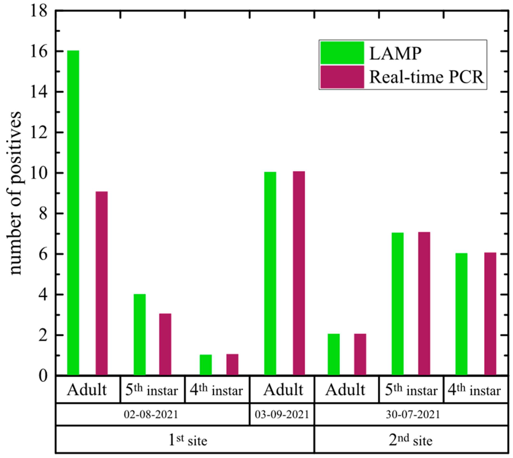
Performing the selected LAMP 1 assay in the laboratory, 46 out of 94 S. titanus specimens resulted as infected by FDp (48.94%). Ninety percent of symptomatic leaf samples gave positive signals, and about 40% of asymptomatic samples also resulted as positive, while crude extracts of healthy grapevine plants were always negative (Figure 2, Table S3). The LAMP results were confirmed by real-time PCR; in addition, LAMP detected FDp in a higher number of samples (i.e., eight) than real-time PCR.
When we compared results from laboratory-reared and field-collected S. titanus (Tables S2 and S3, respectively), we observed that insects from the laboratory showed shorter mean detection times compared with field-collected individuals (adults: 6.7 min vs. 14.5 min; 5th instar: 9.4 min vs. 11.7 min; 4th instar: 9.7 min vs. 11. 1, respectively). Nonetheless, FDp could be detected in field-collected insects for all the insect instars within 16 min.

3.5. Application of the LAMP Assay in the Field

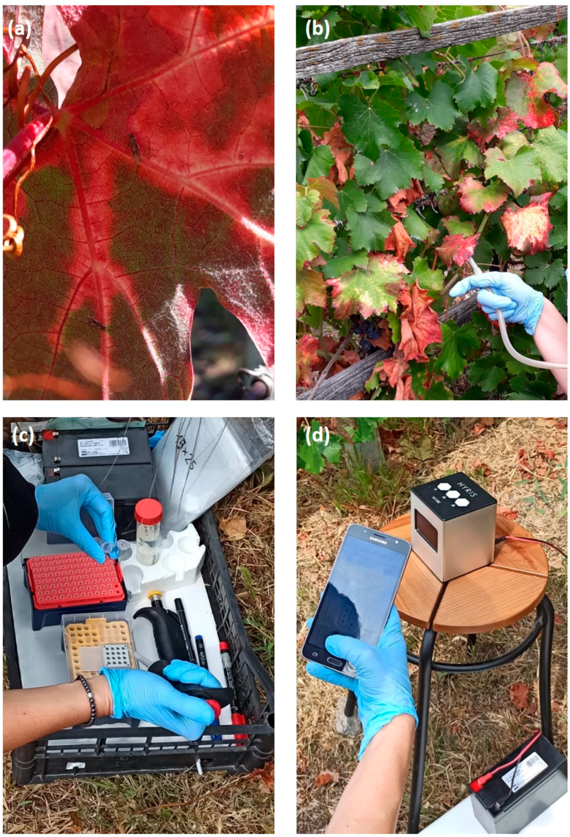
Crude homogenised insect extracts obtained without denaturation and centrifugation were suitable for a specific and sensitive LAMP assay directly in the field, giving results in less than 30 min (Table 4, Figure 3 and Figure 4). Out of 22 tested S. titanus adults, 45.45% resulted as infected by FDp, with similar LAMP detection times performed in the field or in the laboratory (Table S3 and Table 4; 12.9 min for laboratory tests vs. 15.48 min for in-field tests). Moreover, the in-field LAMP assay was able to detect FDp in a higher number of samples (two) than the in-laboratory LAMP. These results confirmed that the optimized LAMP 1 protocol was successful in the detection of the FDp from infected S. titanus insects in-field.

4. Discussion

This study demonstrated the potential of the LAMP assay for the detection of FDp in S. titanus reared in the laboratory or collected in the field. To the best of our knowledge, no molecular isothermal amplification methods were developed for FDp detection in insect vectors. In order to select the best performing procedure for phytoplasma detection in S. titanus, different protocols were tested. The evaluation of the three isothermal amplification assays (LAMP 1, LAMP 2, and RPA) allowed us to identify the protocol LAMP 1 as the best detection method (specificity 100%) (Table 1). Differently, the lower values obtained using the LAMP 2 protocol may have been influenced by the occurrence of a signal in all healthy insect samples, even those reared in the laboratory on healthy plants. Accordingly, Kogovšek et al. [25] also reported positive signals with healthy plant material, thus limiting the usefulness of the LAMP 2 protocol. Noteworthy, no amplification signals were obtained with the RPA assay using DNA extracts from insects or from plants; this negative result is possibly due to the solubilization of DNA in water rather than in the PD1 buffer provided in the kit, or to the high-salt CTAB method used for the DNA extraction that could inhibit downstream enzymatic reactions, as already reported [44,45]. Based on the specificity and sensitivity results obtained, the LAMP 1 assay was selected for all further experiments conducted in this work.
Afterwards, we tested with LAMP 1 crude extracts from insects reared in laboratory conditions or collected in the field. The earlier detection times obtained with laboratory-reared insects compared to field-collected insects are consistent with the controlled conditions of the phytoplasma acquisition by nymphs, left for one week on infected plants; this possibly resulted in a more uniform FDp distribution in the vector population. Moreover, the shortest detection times observed with adults compared to nymphs could result from a higher phytoplasma load in adults due to the completion of the latency period.
Normally, the FDp diagnosis in S. titanus requires to transfer insects collected from vineyards to qualified laboratories for further DNA extraction procedures and PCR assays [24,27]. These operations are laborious, expensive, and time-consuming, requiring about 7 h to be accomplished. For these reasons and due to the lack of timely FDp diagnostic protocols in the vectors, we set up a quick LAMP assay to successfully detect FDp in S. titanus within 35 min, i.e., 5 min for preparing crude insect extracts and 30 min for running the LAMP assay.
During the optimization of the LAMP assay with DNA extracted from FDp-infected insects, we observed that the amplification time at 65 °C could be reduced from 45 min to 30 min, since all the samples analysed tested positive within this period of time. Such a real-time LAMP assay for FDp showed a higher sensitivity than the real-time PCR, revealing not only a faster detection time (12 vs. 90 min), but also a higher sensitivity (2 vs. 66 FDp copy number).
Obtaining timely and repeatable results among laboratories and operators is of primary importance for the early and effective containment of quarantine plant pathogens such as FDp. The LAMP 1 protocol proved to be reliable both in terms of repeatability and reproducibility, confirming previous reports obtained in grapevine [25].
Interestingly, this study revealed that the routinely adopted CTAB protocol to extract DNA from insects [42] can be successfully substituted by an “insect crude extract” preparation. This result was achieved by comparing three different extraction buffers, evaluating the importance to denature crude extracts and testing the dilution effect. The best insect crude extract preparation was achieved with the TET buffer, which was also successful for the in-field LAMP detection of Xylella fastidiosa in “spy insects” on olive trees [38]. The better performance of this buffer can be attributed to the presence of a higher concentration of non-ionic detergent and of a chelating agent inhibiting DNases. Conversely, when all tested insect crude extracts were used undiluted in the LAMP assay, the reaction was inhibited, in line with previous reports [25,37,46]. Therefore, the quick insect crude extract preparation can be a useful alternative to the conventional DNA extraction methods [47], avoiding the use of expensive kits and laboratory facilities. Such ease of sample preparation may facilitate future applications of LAMP assays for real-time and on-site detection of FDp and for epidemiological studies, allowing a substantial reduction of the costs of the analysis.
PCR-based results can be affected by different inhibitory substances, such as polyphenols that bind to DNA and polysaccharides that inhibit Taq enzymatic activity [48,49]. In this study, we showed that crude insect extract can be used not only for LAMP analysis of FDp, but also for real-time PCR, demonstrating the lack of Taq inhibitors in these samples. This suggests that the LAMP results could be confirmed by real-time PCR directly in the field with portable LAMP instruments able to perform real-time PCR cycling programs.
Moreover, the possibility to carry out LAMP assays directly in the field, avoiding the transfer of insect and plant material from vineyards to laboratories, strongly reduces the risk of DNA degradation and accidental dispersion of S. titanus and of the hosted phytoplasma in FDp-free areas. Since FDp may also be transmitted by other leafhoppers such as Orientus ishidae (Matsumura), Oncopisis alni (Schrank), Dictyophara europaea (L.), Allygus modestus (F.), and Allygus mixtus (F.) [2,50,51,52,53], it would be useful to optimize the LAMP assay in these insects. Nonetheless, in the light of the continuous updating of the taxonomic classification of FDp, it would be interesting to test whether the selected LAMP assay can detect the various FD genotypes, especially those responsible for epidemic outbreaks. In this case, it will be necessary to design new LAMP genotype-specific primers targeting, for example, the house-keeping map gene and/or the epidemiologically related vmpA gene [2], combining the results with the currently optimized LAMP protocol that targets the 23 rRNA.
Importantly, this study contributes to extending the applicability of LAMP for detecting plant pathogens in insect organisms. So far, there are more than 200 reports of LAMP detection of pathogens in plants, while only about one-tenth refers to insects. Here, we demonstrated that, in spite of the presence of chitin and polysaccharides, our crude insect extracts are not inhibitory towards the Bacillus subtilis polymerase present in the LAMP reaction, paving the way to a wider application of such a procedure in LAMP-based assays in the future.

5. Conclusions

In conclusion, we developed a protocol for the rapid, sensitive, and specific detection of FDp in the vector S. titanus, strongly accelerating the diagnosis of this pathogen. This method has a great potential for large-scale assays and analysis of both insect and plant material directly in the field, and for new investigations regarding the presence and epidemiology of FDp. The prompt LAMP identification of FDp in the vectors allows rapid evaluation of the disease spread and of new outbreaks in the vineyards and the application of timely control strategies.

Supplementary Materials

The following supporting information can be downloaded at: https://www.mdpi.com/article/10.3390/agronomy12071645/s1, Figure S1: S. titanus and symptomatology observed on grapevine plants infected with FDp. (A) S. titanus instars: 5th nymphal instar (1) adult insect (2). (B) FD symptoms on red grapevine cultivars: red bands along the leaf veins in cv. Bequét (1), leaf reddening in cvs. Barbera (2) and Neretta cuneese (3), and yellow leaf veins on white grapevine cultivar Arneis (4). S. titanus insects (4th instar to adult) and grapevine leaf material were used for fast crude sap preparation for laboratory and in field testing by the LAMP assay.; Table S1: List of the primers and probes used for LAMP, RPA, and real-time PCR.; Table S2: Flavescence dorée phytoplasma (FDp) detection in laboratory-reared Scaphoideus titanus crude extracts by LAMP and real-time PCR assays.; Table S3: In laboratory detection of Flavescence dorée phytoplasma (FDp) from crude extracts of field-captured Scaphoideus titanus and infected grapevine plants by LAMP and real-time PCR assays.

Author Contributions

Conceptualization, S.M. and R.T.; methodology, V.C., C.D., R.P., E.N., S.P. and S.D.; software, C.D., S.P. and S.D.; validation, V.C. and C.D.; writing—original draft preparation, S.M. and R.T.; writing—review and editing, S.M., R.T., V.C. and E.N.; funding acquisition, S.M. and R.T. All authors have read and agreed to the published version of the manuscript.

Funding

This research was funded by Fondazione Cassa di Risparmio di Torino, Project FD_LAMPvett, Ref. No. 2020.2066.

Institutional Review Board Statement

Not applicable.

Informed Consent Statement

Not applicable.

Data Availability Statement

All the data are available in the manuscript.

Acknowledgments

The authors wish to thank Matteo Monchiero, Gian Paolo Accotto, and Daniele Marian for technical and scientific support.

Conflicts of Interest

The authors declare no conflict of interest.

References

  1. Loebenstein, G.; Katis, N.I. Advances in Virus Research. Control of Plant Virus Diseases: Vegetatively-Propagated Crops; Elsevier: Waltham, MA, USA, 2015; Volume 91. [Google Scholar]
  2. Malembic-Maher, S.; Desqué, D.; Khalil, D.; Salar, P.; Bergey, B.; Danet, J.-L.; Duret, S.; Dubrana-Ourabah, M.-P.; Beven, L.; Ember, I.; et al. When a Palearctic bacterium meets a Nearctic insect vector: Genetic and ecological insights into the emergence of the grapevine Flavescence dorée epidemics in Europe. PLOS Pathog. 2020, 16, e1007967. [Google Scholar] [CrossRef] [Green Version]
  3. Morone, C.; Boveri, M.; Giosuè, S.; Gotta, P.; Rossi, V.; Scapin, I.; Marzachì, C. Epidemiology of flavescence dorée in vineyards in northwestern Italy. Phytopathology 2007, 97, 1422–1427. [Google Scholar] [CrossRef] [Green Version]
  4. Duduk, B.; Botti, S.; Ivanović, M.; Krstić, B.; Dukić, N.; Bertaccini, A. Identification of Phytoplasmas Associated with Grapevine Yellows in Serbia. J. Phytopathol. 2004, 152, 575–579. [Google Scholar] [CrossRef]
  5. Belli, G.; Bianco, P.A.; Conti, M. Grapevine yellows in Italy: Past, present and future. J. Plant Pathol. 2010, 92, 303–326. [Google Scholar]
  6. Mehle, N.; Kogovšek, P.; Constable, F.; De Jonghe, K.; Loiseau, M.; Veratti, F.; Marzachi, C.; Ferretti, L.; Angelini, E.; Filippin, L.; et al. Test performance study of isothermal amplification tests for the detection of Grapevine flavescence dorée phytoplasma and ‘Candidatus Phytoplasma solani’. Bull. OEPP 2017, 47, 18–23. [Google Scholar] [CrossRef]
  7. Foissac, X.; Maixner, M. Spread of grapevine phytoplasma diseases in Europe. Phytopathogenic Mollicutes 2013, 3, 47–50. [Google Scholar] [CrossRef]
  8. Plavec, J.; Križanac, I.; Budinščak, Ž.; Škorić, D.; Musić, M.Š. Distribution and epidemiology of Flavescence doree phytoplasma in Croatia. Glas. Biljn. Zaštite 2013, 13, 385–390. [Google Scholar]
  9. Oliveira, M.J.R.A.; Castro, S.; Paltrinieri, S.; Bertaccini, A.; Sottomayor, M.; Santos, C.S.; Vasconcelos, M.W.; Carvalho, S.M.P. “Flavescence dorée” impacts growth, productivity and ultrastructure of Vitis vinifera plants in Portuguese “Vinhos Verdes” region. Sci. Hortic. 2020, 261, 108742. [Google Scholar] [CrossRef]
  10. Mori, N.; Bressan, A.; Martini, M.; Guadagnini, M.; Girolami, V.; Bertaccini, A. Experimental transmission by Scaphoideus titanus Ball of two Flavescence doree-type phytoplasmas. Vitis 2002, 41, 99–102. [Google Scholar]
  11. Chuche, J.; Thiéry, D. Biology and ecology of the Flavescence dorée vector Scaphoideus titanus: A review. Agron. Sustain. Dev. 2014, 34, 381–403. [Google Scholar] [CrossRef] [Green Version]
  12. Alma, A.; Tedeschi, R.; Lessio, F.; Picciau, L.; Gonella, E.; Ferracini, C. Insect vectors of plant pathogenic Mollicutes in the Euro-Mediterranean region. Phytopathogenic Mollicutes 2015, 5, 53. [Google Scholar] [CrossRef]
  13. Belli, G.; Fortusini, A.; Osler, R.; Amici, A. Presenza di una malattia del tipo “Flavescence dorée” in vigneti dell’Oltrepò pavese. Riv. Patol. Veg. 1973, 9, 50–56. [Google Scholar]
  14. European Commission. Commission Implementing Regulation (EU) 2019/2072 of 28 November 2019 Establishing Uniform Conditions for the Implementation of Regulation (EU) 2016/2031 of the European Parliament and the Council, as Regards Protective Measures against Pests of Plants, and Repealing Commission Regulation (EC) No 690/2008 and Amending Commission Implementing Regulation (EU) 2018/2019. Off. J. Eur. Union 2019, 1, 1–279. [Google Scholar]
  15. OEPP/EPPO. PM 1/2 (30), EPPO A1 and A2 Lists of Pests Recommended for Regulation as Quarantine Pests. Available online: https://gd.eppo.int/standards/PM1/ (accessed on 25 March 2022).
  16. OEPP/EPPO. Hot water treatment of grapevine to control Grapevine flavescence doree phytoplasma. Bull. OEPP 2012, 42, 490–492. [Google Scholar]
  17. OEPP/EPPO. PM 7/079 (2) Grapevine flavescence dorée phytoplasma. Bull. OEPP 2016, 46, 78–93. [Google Scholar]
  18. Deng, S.; Hiruki, C. Amplification of 16S rRNA genes from culturable and nonculturable mollicutes. J. Microbiol. Methods 1991, 14, 53–61. [Google Scholar] [CrossRef]
  19. Schneider, B.; Cousin, M.T.; Klinkong, S.; Seemüller, E. Taxonomic relatedness and phylogenetic positions of phytoplasmas associated with diseases of faba bean, sunnhemp, sesame, soybean, and eggplant. Z. Pflanzenkrankh. Pflanzenschutz 1995, 102, 225–232. [Google Scholar]
  20. Daire, X.; Clair, D.; Reinert, W.; Boudon-Padieu, E. Detection and differentiation of grapevine yellows phytoplasmas belonging to the elm yellows group and to the stolbur subgroup by PCR amplification of non-ribosomal DNA. Eur. J. Plant Pathol. 1997, 103, 507–514. [Google Scholar] [CrossRef]
  21. Lee, I.-M.; Gundersen, D.E.; Hammond, R.W.; Davis, R.E. Use of mycoplasmalike organism (MLO) group-specific oligonucleotide primers for nested-PCR assays to detect mixed-MLO infections in a single host plant. Phytopathology 1994, 84, 559–566. [Google Scholar] [CrossRef]
  22. Clair, D.; Larrue, J.; Aubert, G.; Gillet, J.; Cloquemin, G.; Boudon-Padieu, E. A multiplex nested-PCR assay for sensitive and simultaneous detection and direct identification of phytoplasma in the Elm yellows group and Stolbur group and its use in survey of grapevine yellows in France. Vitis 2003, 42, 151–157. [Google Scholar]
  23. Hren, M.; Boben, J.; Rotter, A.; Kralj, P.; Gruden, K.; Ravnikar, M. Real-time PCR detection systems for Flavescence dorée and Bois noir phytoplasmas in grapevine: Comparison with conventional PCR detection and application in diagnostics. Plant Pathol. 2007, 56, 785–796. [Google Scholar] [CrossRef]
  24. Pelletier, C.; Salar, P.; Gillet, J.; Cloquemin, G.; Very, P.; Foissac, X.; Malembic-Maher, S. Triplex real-time PCR assay for sensitive and simultaneous detection of grapevine phytoplasmas of the 16SrV and 16SrXII-A groups with an endogenous analytical control. Vitis 2009, 48, 87–95. [Google Scholar]
  25. Kogovšek, P.; Hodgetts, J.; Hall, J.; Prezelj, N.; Nikolić, P.; Mehle, N.; Lenarčič, R.; Rotter, A.; Dickinson, M.; Boonham, N.; et al. LAMP assay and rapid sample preparation method for on-site detection of flavescence dorée phytoplasma in grapevine. Plant Pathol. 2015, 64, 286–296. [Google Scholar] [CrossRef]
  26. Nagamine, K.; Hase, T.; Notomi, T. Accelerated reaction by loop-mediated isothermal amplification using loop primers. Mol. Cell. Probes 2002, 16, 223–229. [Google Scholar] [CrossRef]
  27. Gallmetzer, A.; Innerebner, G.; Roschatt, C.; Reyes-Dominguez, Y. Dynamics of Scaphoideus titanus population in southern South Tyrol (Italy) and detection of Grapevine Flavescence Dorée phytoplasma in the insect with a multiplex real-time PCR. Vitis 2020, 59, 85–89. [Google Scholar]
  28. Notomi, T.; Okayama, H.; Masubuchi, H.; Yonekawa, T.; Watanabe, K.; Amino, N.; Hase, T. Loop-mediated isothermal amplification of DNA. Nucleic Acids Res. 2000, 28, e63. [Google Scholar] [CrossRef] [Green Version]
  29. Panno, S.; Matić, S.; Tiberini, A.; Caruso, A.G.; Bella, P.; Torta, L.; Stassi, R.; Davino, S. Loop Mediated Isothermal Amplification: Principles and Applications in Plant Virology. Plants 2020, 9, 461. [Google Scholar] [CrossRef] [Green Version]
  30. Ortega, S.F.; Matic, S.; Spadaro, D. Impiego di nuove tecniche molecolari per la diagnosi delle malattie. Prot. Colt. 2018, 2, 9–14. [Google Scholar]
  31. Bertacca, S.; Caruso, A.G.; Trippa, D.; Marchese, A.; Giovino, A.; Matic, S.; Noris, E.; Ambrosio, M.I.F.S.; Alfaro, A.; Panno, S.; et al. Development of a Real-Time Loop-Mediated Isothermal Amplification Assay for the Rapid Detection of Olea Europaea Geminivirus. Plants 2022, 11, 660. [Google Scholar] [CrossRef]
  32. Francois, P.; Tangomo, M.; Hibbs, J.; Bonetti, E.-J.; Boehme, C.C.; Notomi, T.; Perkins, M.D.; Schrenzel, J. Robustness of a loop-mediated isothermal amplification reaction for diagnostic applications. FEMS Immunol. Med. Microbiol. 2011, 62, 41–48. [Google Scholar] [CrossRef] [Green Version]
  33. Tomlinson, J.A.; Ostoja-Starzewska, S.; Webb, K.; Cole, J.A.; Barnes, A.V.; Dickinson, M.; Boonham, N. A loop-mediated isothermal amplification-based method for confirmation of Guignardia citricarpa in citrus black spot lesions. Eur. J. Plant Pathol. 2013, 136, 217–224. [Google Scholar] [CrossRef]
  34. Constable, F.E.; Gibb, K.S.; Symons, R.H. Seasonal distribution of phytoplasmas in Australian grapevines. Plant Pathol. 2003, 52, 267–276. [Google Scholar] [CrossRef]
  35. Kogovšek, P.; Mehle, N.; Pugelj, A.; Jakomin, T.; Schroers, H.-J.; Ravnikar, M.; Dermastia, M. Rapid loop-mediated isothermal amplification assays for grapevine yellows phytoplasmas on crude leaf-vein homogenate has the same performance as qPCR. Eur. J. Plant Pathol. 2017, 148, 75–84. [Google Scholar] [CrossRef] [Green Version]
  36. Gonçalves, D.D.S.; Cassimiro, A.P.A.; de Oliveira, C.D.; Rodrigues, N.B.; Moreira, L.A. Wolbachia detection in insects through LAMP: Loop mediated isothermal amplification. Parasites Vectors 2014, 7, 228. [Google Scholar] [CrossRef] [Green Version]
  37. Sasaya, T. Detection Methods for Rice Viruses by a Reverse-Transcription Loop-Mediated Isothermal Amplification (RT-LAMP). In Plant Virology Protocols; Methods in Molecular Biology (Methods and, Protocols); Uyeda, I., Masuta, C., Eds.; Humana Press: New York, NY, USA, 2015. [Google Scholar]
  38. Yaseen, T.; Drago, S.; Valentini, F.; Elbeaino, T.; Stampone, G.; Digiaro, M.; D’Onghia, A.M. On-site detection of Xylella fastidiosa in host plants and in “spy insects” using the real-time loop-mediated isothermal amplification method. Phytopathol. Mediterr. 2015, 54, 488–496. [Google Scholar]
  39. Lu, H.; Wilson, B.A.L.; Ash, G.J.; Woruba, S.B.; Fletcher, M.J.; You, M.; Yang, G.; Gurr, G.M. Determining putative vectors of the Bogia Coconut Syndrome phytoplasma using loop-mediated isothermal amplification of single-insect feeding media. Sci. Rep. 2016, 6, 35801. [Google Scholar] [CrossRef] [Green Version]
  40. Lobato, I.M.; O’Sullivan, C.K. Recombinase polymerase amplification: Basics, applications and recent advances. TrAC Trends Anal. Chem. 2018, 98, 19–35. [Google Scholar] [CrossRef]
  41. Babu, B.; Ochoa-Corona, F.M.; Paret, M.L. Recombinase polymerase amplification applied to plant virus detection and potential implications. Anal. Biochem. 2018, 546, 72–77. [Google Scholar] [CrossRef]
  42. Bertin, S.; Bosco, D. Molecular identification of phytoplasma vector species. In Phytoplasma Methods and Protocols; Dickinson, M., Hodgetts, J., Eds.; Humana Press: Totowa, NJ, USA, 2013; pp. 87–108. [Google Scholar]
  43. Hibbert, D.B.; Gooding, J. Data Analysis for Chemistry: An Introductory Guide for Students and Laboratory Scientists; Oxford University Press: Oxford, UK, 2005. [Google Scholar]
  44. Arseneau, J.-R.; Steeves, R.; Laflamme, M. Modified low-salt CTAB extraction of high-quality DNA from contaminant-rich tissues. Mol. Ecol. Resour. 2016, 17, 686–693. [Google Scholar] [CrossRef]
  45. Inglis, P.W.; Pappas, M.D.C.R.; Resende, L.V.; Grattapaglia, D. Fast and inexpensive protocols for consistent extraction of high quality DNA and RNA from challenging plant and fungal samples for high-throughput SNP genotyping and sequencing applications. PLoS ONE 2018, 13, e0206085. [Google Scholar] [CrossRef]
  46. Romero, J.L.R.; Carver, G.D.; Johnson, P.A.; Perry, K.L.; Thompson, J.R. A rapid, sensitive and inexpensive method for detection of grapevine red blotch virus without tissue extraction using loop-mediated isothermal amplification. Arch. Virol. 2019, 164, 1453–1457. [Google Scholar] [CrossRef]
  47. Boonham, N.; Kreuze, J.; Winter, S.; van der Vlugt, R.; Bergervoet, J.; Tomlinson, J.; Mumford, R. Methods in virus diagnostics: From ELISA to next generation sequencing. Virus Res. 2014, 186, 20–31. [Google Scholar] [CrossRef]
  48. John, M.E. An efficient method for isolation of RNA and DNA from plants containing polyphenolics. Nucleic Acids Res. 1992, 20, 2381. [Google Scholar] [CrossRef] [Green Version]
  49. Fang, G.; Hammar, S.; Grumet, R. A quick and inexpensive method for removing polysaccharides from plant genomic DNA. BioTechniques 1992, 13, 52–54. [Google Scholar]
  50. Maixner, M.; Reinert, W.; Darimont, H. Transmission of grapevine yellows by Oncopsis alni (Schrank) (Auchenorrhyncha: Macropsinae). Vitis 2000, 39, 83–84. [Google Scholar]
  51. Lessio, F.; Picciau, L.; Gonella, E.; Tota, F.; Mandrioli, M.; Alma, A. The mosaic leafhopper Orientus ishidae: Host plants, spatial distribution, infectivity, and transmission of 16SrV phytoplasmas to vines. Bull. Insectology 2016, 69, 277–289. [Google Scholar]
  52. Belgeri, E.; Rizzoli, A.; Jermini, M.; Angelini, E.; Filippin, L.; Rigamonti, I.E. First report of Flavescence dorée phytoplasma identification and characterization in three species of leafhoppers. J. Plant Pathol. 2022, 104, 375–379. [Google Scholar] [CrossRef]
  53. Krstic, O.; Cvrković, T.; Marinković, S.; Jakovljević, M.; Mitrović, M.; Toševski, I.; Jović, J. Genetic Diversity of Flavescence Doree Phytoplasmas in Vineyards of Serbia: From the Widespread Occurrence of Autochthonous Map-M51 to the Emergence of Endemic Map-FD2 (Vectotype II) and New Map-FD3 (Vectotype III) Epidemic Genotypes. Agronomy 2022, 12, 448. [Google Scholar] [CrossRef]
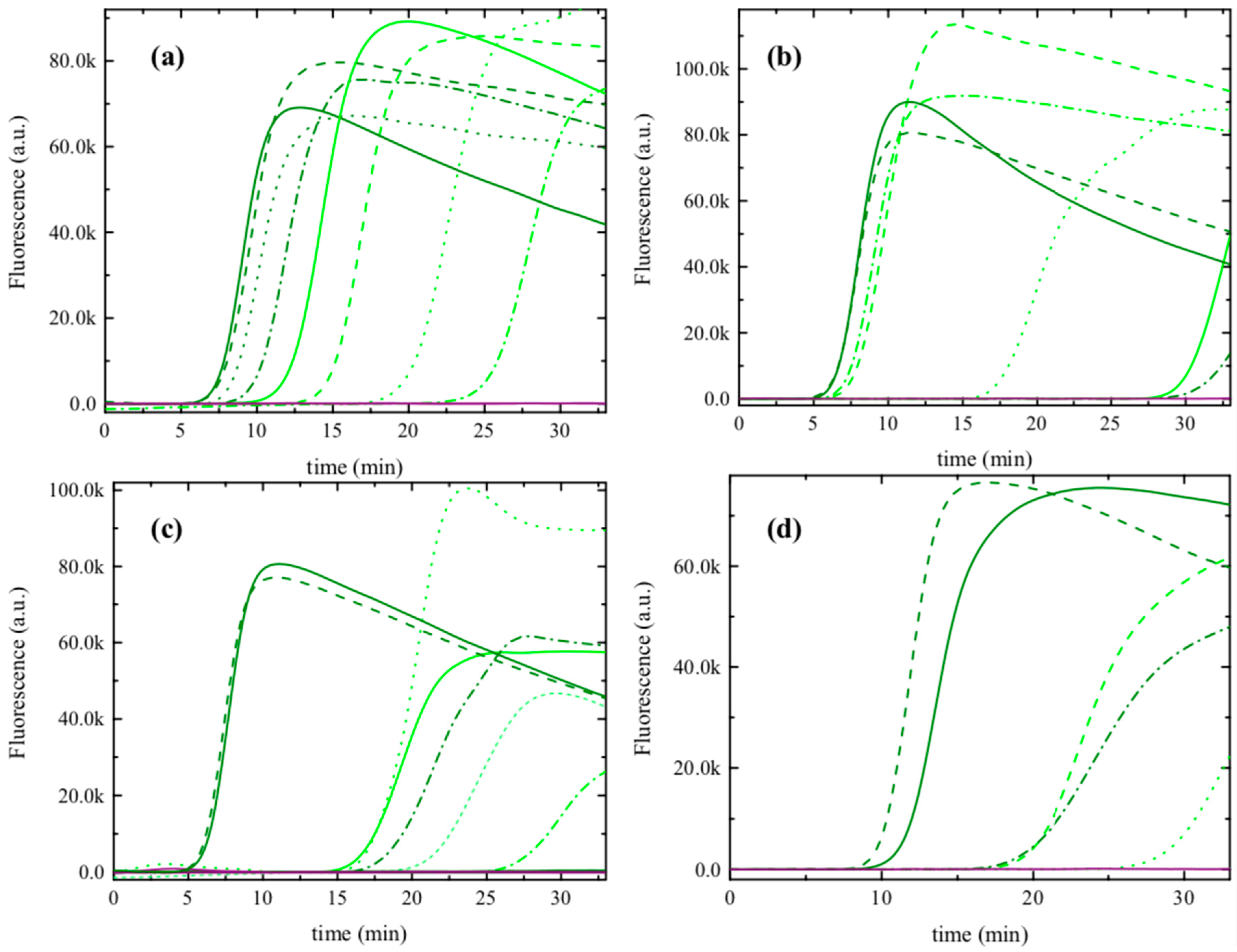
Figure 1. Comparison of different crude extract preparation buffers for the detection of Flavescence dorée phytoplasma (FDp) by LAMP in Scaphoideus titanus: (a) Tris-EDTA-Triton X-100 buffer without denaturation at 95 °C, (b) Tris-EDTA-Triton X-100 buffer with denaturation at 95 °C, (c) ELISA extraction buffer, and (d) OptiGene lysis buffer. The extraction with two latter buffers (c,d) does not include a denaturation at 95 °C. Infected samples are indicated by green colour (each sample with the same line type) and healthy samples are shown in purple colour in all panels.
Agronomy 12 01645 g001
Figure 2. Comparison of in laboratory detection of Flavescence dorée phytoplasma (FDp) from crude extracts of Scaphoideus titanus (adults, 5th, and 4th instars) captured from two vineyards by LAMP and real-time PCR assays.
Agronomy 12 01645 g002
Figure 3. In-field detection of Flavescence dorée phytoplasma (FDp) in Scaphoideus titanus using real-time LAMP assay: (a) adult of S. titanus on infected grapevine leaves; (b) capture of S. titanus; (c) preparation of insect crude extracts and the LAMP reaction mixture; and (d) set up of the LAMP assay by smart phone and bCube2 instrument running in the vineyard.
Agronomy 12 01645 g003
Figure 4. Real-time LAMP results for the detection of Flavescence dorée phytoplasma (FDp) in Scaphoideus titanus crude extracts in field. The results are visualized on the mobile phone as: (a) amplification results; (b) melting curve results. Infected samples are indicated by green colour (each sample with the same colour gradation) and healthy samples are shown in purple colour in both panels.
Agronomy 12 01645 g004
Table 1. Comparison of three isothermal amplification protocols for the specific detection of Flavescence dorée phytoplasma (FDp) in insects and plants.
Host/PathogenNo. of samplesLAMP 1LAMP 2
DNADNA
4 ng/µL *0.4 ng/µL0.04 ng/µL4 ng/µL0.4 ng/µL0.04 ng/µL
Scaphoideus titanus insects infected with FDp-D57.3 ± 0.8(85.10 ± 0.10)8.8 ± 1.0(85.05 ± 0.21)8.1 ± 2.0(84.90 ± 0.00)5.8 ± 1.3(84.90 ± 0.10)6.8 ± 1.8(85.00 ± 0.14)13 ± 3(84.95 ± 0.07)
S. titanus insects infected with FDp-C58.4 ± 1.4(85.20 ± 0.14)8.87 ± 0.23(85.10 ± 0.00)9.3 ± 2.0(85.00 ± 0.14)9.2 ± 0.8(85.15 ± 0.07)7.1 ± 0.3(85.83 ± 0.29)18.4 ± 5.1(85.15 ± 0.07)
Grapevine plants infected with FDp-D59.6 ± 0.8(84.50 ± 0.00)9.0 ± 0.9(85.0 ± 0.10)10.5 ± 0.485.20 ± 0.148.3 ± 1.3(84.05 ± 0.11)12.1 ± 1.0(84.70 ± 0.14)22 ± 5(85.0 ± 1.3)
Grapevine plants infected with FDp-C59.6 ± 1.4(85.15 ± 0.07)8.0 ± 1.1(84.4 ± 0.4)12.4 ± 1.484.80 ± 0.288.3 ± 2.1(84.80 ± 0.25)9.9 ± 1.7(85.2 ± 0.4)18.4 ± 1.4(85.4 ± 0.8)
Hyalesthes obsoletus insects infected with 16Sr-XII-A (‘Ca. P. solani’)3nd **(nd)nd(nd)nd(nd)18.2 ± 2.5(85.50 ± 0.25)25 ± 3(82.4 ± 0.5)32.2 ± 0.5(83.30 ± 0.00)
Cacopsylla melanoneura insects infected with 16Sr-X-A (‘Ca. P. mali’)3nd(nd)nd(nd)nd(nd)26.8 ± 1.6(86.00 ± 0.00)29 ± 3(85.8 ± 0.4)37.4 ± 2.4(85.8 ± 0.4)
Healthy S. titanus5nd(nd)nd(nd)nd(nd)20 ± 4(84.0 ± 0.8)22 ± 3(81.2 ± 1.2)34 ± 4(85.6 ± 0.4)
Healthy grapevine5nd(nd)nd(nd)nd(nd)18 ± 4(81.45 ± 0.21)27 ± 4(84.8 ± 0.4)33 ± 3(84 ± 3)
* The first value for each DNA concentration is referred to as ‘Time to positive’ (Tp) ± standard deviation (SD), while the value in parenthesis indicates the melting temperature (Tmelt) ± SD. ** nd = not determined.
Table 2. Comparison of the sensitivity of the LAMP assay (LAMP 1) and real-time PCR on serial dilutions of DNA extracted from Scaphoideus titanus infected by Flavescence dorée phytoplasma (FDp).
Insect DNA DilutionEstimated Copy Number of FDp DNALAMP
Tp (min ± SD) *
Real-Time PCR
Ct ± SD **
1 × 10126,013,887.617.6 ± 0.521.8 ± 0.4
1 × 10−12,473,463.128.6 ± 0.824.27 ± 0.15
1 × 10−256,098.859.4 ± 0.929.8 ± 0.8
1 × 10−365.7611.7 ± 0.435.38 ± 0.23
1 × 10−42.3326.3 ± 1.3nd ***
1 × 10⁻⁵ndndnd
* Tp = Time to positive; SD = standard deviation; ** Ct = threshold cycle; *** nd = not determined.
Table 3. Comparison of the Flavescence dorée phytoplasma (FDp) detection in Scaphoideus titanus vectors from undiluted and diluted crude extracts obtained with different extraction buffers.
Host/PathogenTime/
Temperature
TET BufferELISA
Extraction Buffer
OptiGene Lysis
Buffer
Undiluted1:51:7.51:10Undiluted1:51:7.51:10Undiluted1:51:7.51:10
Scaphoideus titanus insects infected by FDp-DTp (min ± SD) *nd **6.2 ± 1.111.8 ± 2.418 ± 3nd8 ± 314.6 ± 1.921 ± 8nd11 ± 321.4 ± 1.830 ± 7
Tmelt ± SD ***nd84.60 ± 0.0785.0 ± 0.585.5 ± 0.4nd85.05 ± 0.0785.1 ± 0.885.0 ± 0.5nd85.2 ± 0.484.8 ± 0.884 ± 4
S. titanus insects infected by FDp-CTp (min ± SD)nd6.6 ± 1.514.1 ± 1.922 ± 3nd8.1 ± 2.020 ± 622 ± 6nd10 ± 424 ± 332 ± 6
Tmelt ± SDnd84.70 ± 0.1485.30 ± 0.2784.4 ± 1.9nd84.90 ± 0.1484.7 ± 0.485.6 ± 0.4nd85.8 ± 0.585.1 ± 0.484 ± 3
Healthy S. titanus insectsTp (min ± SD)ndndndndndndndndndndndnd
Tmelt ± SDndndndndndndndndndndndnd
* Tp = Time to positive; SD = standard deviation; ** nd = not determined; *** Tmelt = melting temperature.
Table 4. In-field detection of Flavescence dorée phytoplasma (FDp) using crude extracts of field-captured Scaphoideus titanus and infected grapevine plants by LAMP.
IsolateLAMP
Tp (Min) *Tmelt (Min) **
St_F1nd ***nd
St_F222.5584.34
St_F315.4184.54
St_F4ndnd
St_F5ndnd
St_F613.4984.54
St_F714.2784.71
St_F8ndnd
St_F916.3684.38
St_F10ndnd
St_F1118.0184.35
St_F1226.5584.24
St_F13ndnd
St_F1412.2684.39
St_F1511.3384.56
St_F16ndnd
St_F17ndnd
St_F18ndnd
St_F19ndnd
St_F20ndnd
St_F2110.1885.04
St_F229.2684.72
Mean value ± SD15.4884.53
* Tp = Time to positive; ** Tmelt = melting temperature; *** nd = not determined.
Publisher’s Note: MDPI stays neutral with regard to jurisdictional claims in published maps and institutional affiliations.