Article

Harnessing the Eco-Friendly Potential of Asparagus racemosus Leaf Extract Fabricated Ni/Ni(OH)2 Nanoparticles for Sustainable Seed Germination and Seedling Growth of Vigna radiata

1
Amity Institute of Biotechnology, Amity University Uttar Pradesh, Lucknow Campus, Lucknow 226028, India
2
Department of Botany, Bankim Sardar College, Tangrakhali 743329, India
3
College of Agriculture, Central Agricultural University, Imphal 795004, India
4
Department of Biotechnology, School of Life Sciences, Mahatma Gandhi Central University, Motihari 845401, India
5
Department of Forestry, Mizoram University, Aizawl 796004, India
6
Department of Agronomy, Faculty of Agriculture and Biotechnology, Bydgoszcz University of Science and Technology, Al. Prof. S. Kaliskiego 7, 85-796 Bydgoszcz, Poland
7
Faculty of Agriculture, University of Life Sciences “King Mihai I” from Timisoara, 300645 Timisoara, Romania
*
Authors to whom correspondence should be addressed.
Agronomy 2023, 13(8), 2073; https://doi.org/10.3390/agronomy13082073
Submission received: 30 June 2023 / Revised: 3 August 2023 / Accepted: 4 August 2023 / Published: 7 August 2023

Abstract

:
The increasing utilization of nanoparticles (NPs) in agricultural practices has led to a surge in demand for nano-based products. Herein, we investigate the dose-dependent impacts of nickel hydroxide (Ni(OH)2)/nickel (Ni) NPs, synthesized using Asparagus racemosus Linn. leaf extract, on the seed germination and growth of Vigna radiata (Linn.) Wilczek. In all seed samples, 100% germination was observed in Treatment 1 (2.74 mg mL−1) and Treatment 2 (5.48 mg mL−1) of Ni/Ni(OH)2 NPs. However, in Treatment 3 (8.22 mg mL−1) and Treatment 4 (10.96 mg mL−1), the germination percentage was lower, reaching 80%. Further, Treatment 5 (13.70 mg mL−1) of Ni/Ni(OH)2 NPs showed a reduced germination rate of 60%, indicating a prolonged germination process at higher concentrations. Remarkably, the length of seedlings showed a significant increase in all experimental treatments compared to the control group, which received 5 mL of distilled water. Among the investigated parameters, Treatment 2 demonstrated the most promising outcomes, exhibiting the highest chlorophyll stability index (23.73%) and membrane stability index (67.89%) values, as well as the lowest root ion leakage (24.75%). These findings indicate that Ni/Ni(OH)2 has the capacity to enhance seed germination and foster seedling growth.

1. Introduction

The agricultural industry, despite undergoing revolutionary transformations, continues to face challenges posed by climate change and the increasing demands for food security [1,2]. In this context, nanotechnology has emerged as a promising avenue to enhance agricultural productivity by improving the management and application of inputs in plant production.
Numerous investigations have explored nickel (Ni) bioavailability in soil and plants, as its applications span from everyday household products to industrial settings. Ni is found in very minute amounts in the air, water, and soil and is subsequently absorbed by plants through their roots after it becomes accessible in the soil [3]. It is a vital nutrient for healthy plant development and growth and is necessary for activating various enzymes, including urease and glyoxalase-I. Numerous physiological functions, including nitrogen metabolism, photosynthesis, reproductive and vegetative development, and seed germination depend on the mineral Ni. As a result, plants require sufficient Ni supplies to complete their life cycle [3]. However, elevated levels of Ni can stimulate the generation of reactive oxygen species (ROS), which negatively impact various biochemical and physiological processes, including mineral uptake, transpiration, and photosynthesis, posing potential hazards to plant health. ROS production accelerates lipid peroxidation, leading to plasma membrane deterioration and the inhibition of essential enzyme activities [3].
Nanofertilizers offer several advantages, including their nanoscale size, which allows for enhanced permeability within plant systems, increased surface area providing more reaction sites, and the potential for lower dosages [2,4]. One approach to regulate and slow-release various micronutrients and macronutrients into the soil, while minimizing build-up and pollution of natural resources, involves replacing traditional fertilization techniques with nanofertilizers. Nanofertilizer technology necessitates an integrated approach as it has the potential to outperform conventional fertilizers. These nanofertilizers can comprise mineral emulsions or nanoparticles (NPs) enclosed by nanomaterials or protected by thin coatings [2,5]. The unique physicochemical characteristics of NPs enable them to interact with plants and induce a range of morphological and physiological alterations [2,6]. Nanofertilizers, classified into three types, i.e., nanoparticulate, micronutrient, and macronutrient nanofertilizers, have emerged as promising tools for enhancing plant growth and development [7]. Micronutrient nanofertilizers encompass essential elements such as zinc (Zn), manganese (Mn), nickel (Ni), iron (Fe), copper (Cu), and molybdenum (Mo) packaged within NPs. Recent studies have highlighted the superior benefits of organically fabricated micronutrient nanofertilizers compared to chemically synthesized NPs, emphasizing their positive impact on plant development [8]. The demand for ecologically sound and energy-efficient routes to produce metallic NPs like Ni NPs has propelled both scientific research and commercial industries to invest in green synthesis technologies [9]. Exploiting plant products, extracts, parts, and organisms for the eco-friendly fabrication of Ni NPs yields products that are safe for the ecosystem and the environment [10]. Utilizing plant extracts as reducing agents in the green synthesis of metallic NPs offers both commercial and environmental advantages [11].
In comparison to other magnetic NPs, Ni NPs exhibit greater potential as catalysts in chemical processes, as well as in propellants and sintering additives for fibers, polymers, and coatings [12]. The abundance of Ni in the Earth’s crust makes it a more cost-effective option compared to many other metals utilized as catalysts [13]. Ni NPs can be employed in various forms, including nanofluids with different degrees of purity, such as ultra-high purity, high purity, dispersed, coated, and passivated configurations [14].
The utilization of Ni-based NPs holds significant potential for enhancing agricultural productivity and meeting the growing demands of the modern era’s food requirements. In this context, Chaudhary et al. [15] conducted a study investigating the impact of nickel oxide (NiO) nanodisks on germination using Vigna radiata (Linn.) Wilczek seeds. The results revealed strong antiproliferative activity, with growth inhibition values ranging from 12.6% to 46%. Singh et al. [16] investigated the production of NiO-based NPs using the cell-free extract of Spirogyra sp. The study demonstrated a positive effect at lower concentrations, with an increase in the V. radiata seedling’s length. However, at higher concentrations, the seed germination and seedling growth of V. radiata were adversely affected. Understanding the intricate interactions between Ni NPs and plants is crucial for harnessing their potential in agricultural applications and optimizing their usage for sustainable crop production. Furthermore, research in this field is necessary to fully elucidate the mechanisms underlying these effects and to explore the broader implications of Ni NPs utilization in agricultural systems.
The fabrication, characterization, and evaluation of in vitro catalytic, antibacterial, and antioxidant properties of Ni/Ni(OH)2 nanoparticles using A. racemosus have been previously documented [17]. However, further investigation on the dose-dependent effects of plant-derived Ni-based NPs on seed germination, seedling growth, and other related factors remains unexplored, presenting an opportunity for novel insights and applications. Therefore, to address this gap, the present study investigates the dose-dependent impacts of A. racemosus leaf extract-fabricated Ni/Ni(OH)2 NPs on the seed germination and growth of mung bean V. radiata. Moreover, physiological parameters such as membrane stability index (MSI), chlorophyll stability index (CSI), and root ion leakage (RIL) were also evaluated to report the effects on V. radiata. Thus, the present study contributes to assessing the potentials of A. racemosus-derived Ni/Ni(OH)2 NPs in the germination and growth of V. radiata seeds. This study also presents a viable alternative path for plant breeding methods that emphasize environmentally friendly practices.

2. Materials and Methods

2.1. Biofabrication of Ni/Ni(OH)2 NPs

The Ni/Ni(OH)2 NPs were successfully synthesized through the utilization of cell-free leaf extracts obtained from A. racemosus Linn. Concurrently, nickel sulfate hexahydrate (NiSO4·6H2O) (HiMedia Laboratory Pvt. Ltd., Thane, India) served as the precursor in accordance with the previously described methodology [17]. The resultant solution of NPs demonstrated a concentration of 13.70 mg mL−1 of Ni/Ni(OH)2 NPs. The integration of A. racemosus leaf extracts as a reducing, stabilizing, and capping agent, in conjunction with the NiSO4·6H2O precursor, facilitated the efficient fabrication of the intended Ni/Ni(OH)2 NPs.

2.2. Seeds Collection and Treatment

The mung bean (V. radiata) seeds were procured from the Seed Testing Laboratory, Uttar Pradesh Beej Vikas Nigam, Lucknow, India. Prior to experimentation, the seeds underwent a meticulous cleansing process, involving immersion in double distilled water for a duration of 2 h, followed by surface sterilization utilizing a 10% sodium hypochlorite solution (HiMedia Laboratory Pvt. Ltd., Thane, India). Experiments were performed in triplicate and repeated twice. For each set, a moist paper towel was prepared, onto which seeds were distributed, ensuring minimal overlap. The other half of the paper towel was subsequently folded over the seeds, thus creating a moist environment on both sides of the seeds. The prepared paper towels were then rolled into a cylindrical shape, securing each with a rubber band placed approximately one inch from the top. Accordingly, to prevent desiccation and facilitate the experimental conditions, the prepared seed-containing paper towel was placed inside a transparent container kept inside the airtight growth chamber, while partially submerged in a solution of Ni/Ni(OH)2 NPs. The experimental design encompassed five treatment groups, employing varying concentrations of Ni/Ni(OH)2 NPs in a dose-dependent manner, alongside one control group (used only 5 mL distilled water), wherein each group comprised five seeds. Treatment 1 [1 mL of Ni/Ni(OH)2 NPs + 4 mL distilled water] involved the incubation of seeds with 2.74 mg mL−1 concentration of Ni/Ni(OH)2 NPs; Treatment 2 [2 mL of Ni/Ni(OH)2 NPs + 3 mL distilled water] with a concentration of 5.48 mg mL−1; Treatment 3 [3 mL of Ni/Ni(OH)2 NPs + 2 mL distilled water] with a concentration of 8.22 mg mL−1; Treatment 4 [4 mL of Ni/Ni(OH)2 NPs + 1 mL distilled water] with a concentration of 10.96 mg mL−1; and Treatment 5 encompassed the exposure of seeds to 5 mL of Ni/Ni(OH)2 NPs with a concentration of 13.70 mg mL−1. In contrast, the control group involved the incubation of seeds solely in 5 mL of distilled water. To assess the germination progress of the treated seeds, a visual inspection of the paper towel was conducted after 24 h (day 1), 48 h (day 2), and 72 h (day 3). Additionally, the seedling growth data were recorded until day 8. Furthermore, other parameters were also analyzed to obtain comprehensive results.

2.3. Estimation of Morphological Parameters

2.3.1. Germination

At predetermined time intervals of day 1 (24 h), day 2 (48 h), and day 3 (72 h) post-incubation, the seeds were carefully inspected, and the count of germinated seeds was recorded. This process was repeated iteratively until all the seeds had undergone germination. To ensure accuracy, the assessment of seed germination was performed within a thoroughly sanitized environment. The occurrence of germination was determined by the visible rupture of the seed coat by the emerging radicle. The calculation of the germination percentage was executed using the following equation [18]:
Seed   gemination   %   = Number   of   germinated   seeds Total   number   of   seeds × 100

2.3.2. Germinated Seedlings Length

The length of germinated seedlings was measured using a thirty-centimeter scale. This measurement encompassed the sum of both the shoot length and root length of each seedling, and it was recorded by determining the distance from the apex of the seedling to the tip of its root [18,19].

2.3.3. Germinated Seedlings Biomass

Fresh seedlings were thoroughly washed to eliminate any external contaminants. The washed seedlings were then weighed using a precise measuring scale, and the measurements were recorded. To remove moisture content, the samples were subjected to drying in a hot-air oven maintained at a temperature of 60 °C. After achieving a state of complete dryness, the seedlings were weighed once again, yielding the dry weight (DW) expressed in grams. The ratio of dry weight to fresh weight was determined by dividing the dry weight (DW) by the corresponding fresh weight (FW) of the seedlings [18,20].

2.4. Estimation of Biochemical Parameters

2.4.1. Chlorophyll Estimation

Approximately 300 mg (300,000 µg) of fresh leaf samples were subjected to a partial drying process by immersing them in 95% ethanol (SD Fine-Chem Ltd., Mumbai, India) for a duration of 5 min. This step was performed to facilitate the subsequent estimation of chlorophyll content. Subsequently, the leaves were finely chopped into small pieces and further pulverized using a mortar and pestle. The resulting leaf material was combined with 5 mL of 80% acetone (v/v) (Sigma-Aldrich Chemicals Pvt. Ltd., Bangalore, India) in a 50 mL centrifuge tube, followed by an incubation period in darkness lasting 30 min. After the incubation, the tubes were subjected to centrifugation at 847× g and 4 °C for 15 min. The resulting supernatant, containing the extracted green pigments, was carefully preserved in a dark environment. The quantification of chlorophyll content was carried out using an Ultraviolet–Visible (UV–Vis) Spectrophotometer (Shimadzu 4650; Model: UV-1800; Shimadzu Corporation, Kyoto, Japan), measuring the absorbance at wavelengths of 645, 652, and 663 nm. The chlorophyll a, chlorophyll b, and total chlorophyll contents were determined using the provided equations [21]:
Chl   a   ( mg   g 1   fw ) = [ 12.7 × D 663 2.69 × D 645 ] × V 1000 × W
Chl   b   ( mg   g 1   fw ) = [ 22.9 × D 645 4.68 × D 663 ] × V 1000 × W
Total   Chl   ( mg   g 1   fw ) = D 652 × 1000 × V 1000 × W
where V = final volume of 80% acetone; D = optical density; W = weight of sample; and fw = fresh weight of the sample.

2.4.2. Chlorophyll Stability Index

To assess the chlorophyll stability index (CSI) of the leaves, spectroscopy techniques were employed, following the methodology proposed by Kaloyereas [22]. The CSI was determined by comparing the percentage of light transmission in leaf samples subjected to different treatments, in accordance with the prescribed formula given below. For each treatment, two test tubes were prepared, each containing 10 mL of distilled water and 0.25 g of leaf sample. One of the test tubes served as the control, maintained at room temperature, while the other was subjected to a temperature of 55 °C within a water bath for a duration of 60 min. Subsequently, the optical density (OD) of the extracted pigments from both test tubes was measured at 652 nm using a UV–Vis spectroscopy instrument.
CSI   % = OD   at   652   nm   of   treated   sample OD   at   652   nm   of   control × 100

2.4.3. Membrane Stability Index

The calculation of the membrane stability index (MSI) was carried out following the methodology described by Premachandra et al. [23], with modifications as outlined by Sairam [24]. A meticulously cleaned test tube was selected for the experiment. A precisely weighed amount of 0.1 g of clean leaf material was combined with 10 mL of double distilled water. Subsequently, the test tube was subjected to a controlled temperature of 40 °C within a water bath for a duration of 30 min. To assess the electrical conductivity (C1) of the water, a high-quality conductivity meter (HM Digital AP-2) (HM Digital India Pvt. Ltd., Alwar, India) was employed. The same sample was then exposed to a temperature of 100 °C in a water bath for 10 min, followed by a measurement of its conductivity (C2). The membrane stability index (MSI) was determined utilizing the following formula:
MSI = [(C1 − C2)/C1] × 100
This index served as a valuable indicator of the membrane integrity and stability in the leaf samples, providing insights into their adaptability and resistance to stress conditions.

2.4.4. Root Ion Leakage

The assessment of root ion leakage (RIL) in the plant samples was conducted following the established protocol introduced by Lutts et al. [25]. Meticulously collected root samples (0.3 g) were carefully placed into separate test tubes for both control and treated plants. Subsequently, 10 mL of double distilled water was added to each test tube, creating a sample mixture. The test tubes were then subjected to an incubation period of approximately 5 min at a temperature of 25 °C. The electrical conductivity (EC0) of the sample solutions was determined using a highly accurate conductivity meter. After the initial measurement, the test tubes were incubated for a duration of 12 h. Following this incubation period, the electrical conductivity (EC1) of the sample solutions was measured once again. To further analyze root ion leakage, the test tubes were placed in a water bath and heated for 30 min. Subsequently, the electrical conductivity (EC2) of the sample solutions was re-evaluated once the samples had cooled down to room temperature. The calculation of relative conductivity (RC) was carried out utilizing the following equation, which serves as an indicator of root ion leakage:
Relative conductivity (RC) = [(EC1 − EC0)/EC2] × 100
This measure provides valuable insights into the extent of root ion leakage, highlighting the potential impact of treatments on membrane integrity and ion regulation within the root system.

2.5. Statistical Analysis

All experiments were conducted in triplicate to ensure the reproducibility of the results, and to validate the outcomes/results, they were repeated twice. In each set of experiments, five seeds were inoculated/utilized, and their average value was treated as a single reading, designated as the number of replicates (n) = 1 for each set. Consequently, for each experiment, three independent sets were employed, leading to a total of n = 3. Considering the repetition of the experiments twice in triplicate, the overall number of replicates becomes n = 6. Furthermore, the statistical evaluation of the data was carried out using IBM SPSS Statistics software (version 22, SPSS, Chicago, IL, USA). For data analysis, one-way ANOVA (analysis of variance) was employed to assess the significance of variations among the experimental groups. To identify any notable differences between the groups, Duncan’s new multiple-range test was applied. Furthermore, Microsoft Excel Version 2307 (Microsoft Corporation (I) Pvt. Ltd., Gurgaon, India) was utilized to generate graphical representations, aiding in the comprehensive visualization and interpretation of the obtained results. These statistical analyses and data visualization techniques enhanced our understanding and facilitated the effective communication of the experimental outcomes.

3. Results and Discussion

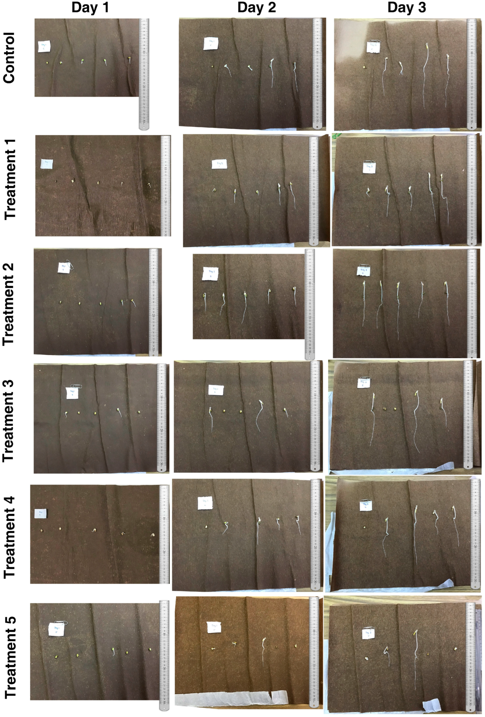
3.1. Germination of V. radiata Seeds

The germination of V. radiata seeds in the presence of five different concentrations of A. racemosus leaf extract-synthesized Ni/Ni(OH)2 NPs (Treatments 1–5) and control conditions on day 1 (24 h), day 2 (48 h), and day 3 (72 h) of treatments are shown in Figure 1, and further seedling growth from day 4 to day 8 of treatments is shown in Figure 2. The germination % of V. radiata seeds decreased at the higher concentration of Ni/Ni(OH)2 NPs in comparison to control conditions, as depicted in Figure 1 and Figure 2, suggesting a prolonged gemination. After 24 h (1 day) of incubation, 60% of seeds were found to germinate under Treatment 1 (NP concentration of 2.74 mg mL−1), Treatment 3 (NP concentration of 8.22 mg mL−1), and Treatment 4 (NP concentration of 10.96 mg mL−1) as well as only 20% under Treatment 5 (NP concentration of 13.70 mg mL−1), whereas in case of Treatment 2 (NP concentration of 5.48 mg mL−1) and control conditions it was 80%. However, after 2 days (48 h) of incubation, it was found that 100% of seeds were germinated under Treatment 1 and Treatment 2, followed by 80% under Treatment 4 and trailed by 60 and 40% under Treatment 3 and Treatment 5, respectively. Moreover, no change was observed under control conditions after 2 days (48 h) and 3 days (72 h) of incubation as compared to the 1 day (24 h) of incubation. Similarly, after 3 days (72 h) of incubation, no changes were observed under Treatment 1, Treatment 2, and Treatment 4 as compared to the 2 days (48 h) of incubation. Furthermore, the germination % increased up to 80% and 60% under Treatments 3 and 5, respectively (Table 1). As the concentration of Ni/Ni(OH)2 NPs increases, the germination % of seedlings decreases in comparison to the control conditions. In low concentrations, Ni has been shown to have a crucial function in enzymes like ascorbate peroxidase (APX) [26], which might be responsible for a higher germination % as compared to the control conditions. However, at higher concentrations, prolonged germination in the seeds by Ni caused by a disruption in biochemical metabolism may be due to the suppressed activities of protease and α-amylase [27]. This probably reduces the availability of sugars for the creation of metabolic energy as well as necessary amino acids to produce enzymes and proteins essential for the developing embryo in Vigna sp. [27].

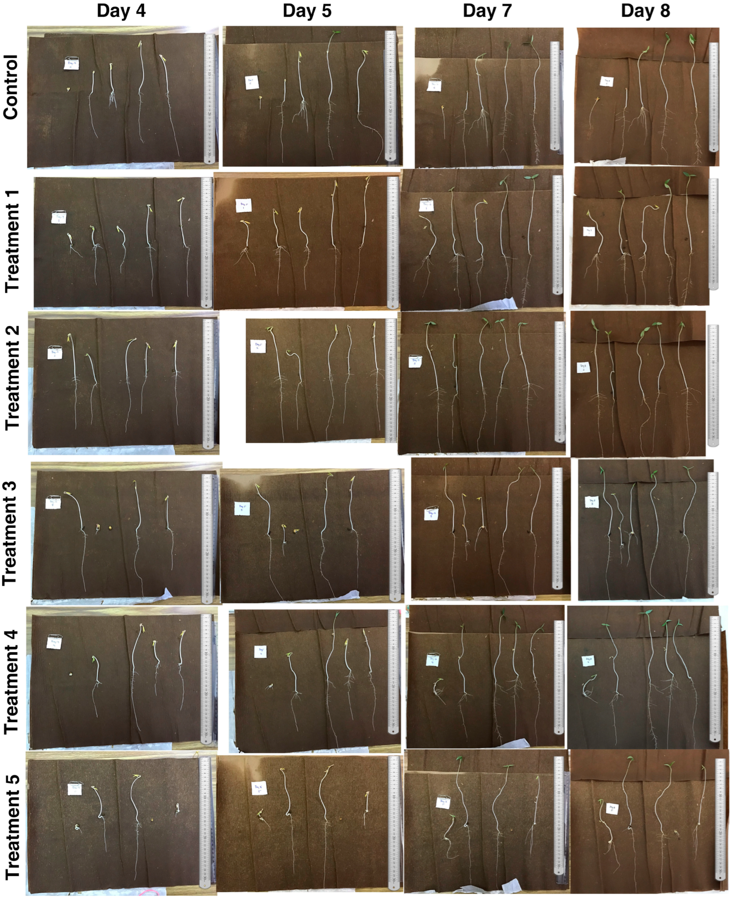
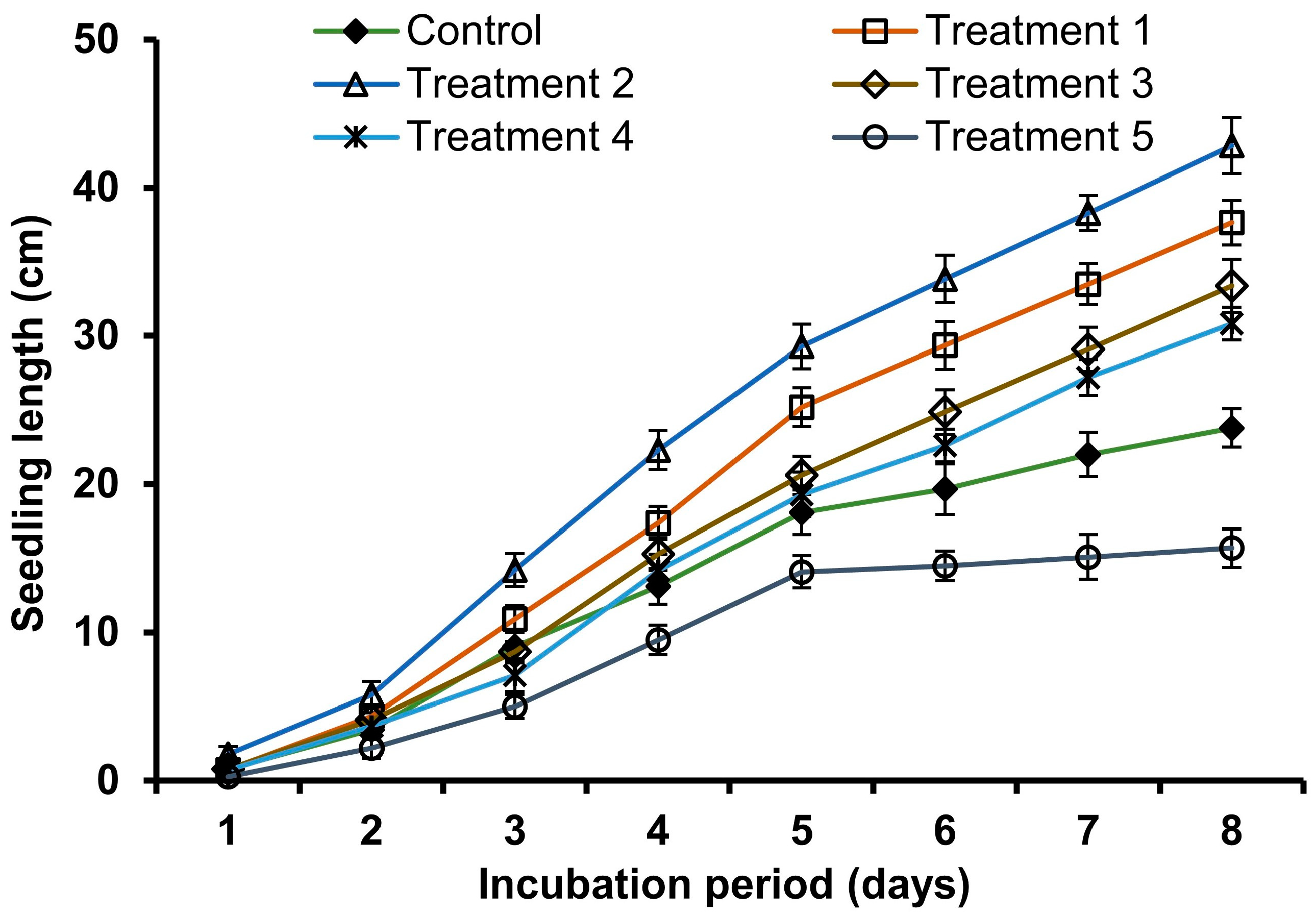
3.2. Growth Rate of V. radiata Seedlings

The growth rate of V. radiata seedlings was investigated during treatment to varying concentrations of Ni/Ni(OH)2 NPs (Treatment 1 to 5) and control conditions. The effect of Ni/Ni(OH)2 NPs on seedling lengths of V. radiata is shown in Figure 3. It is obvious that increasing the concentrations of Ni/Ni(OH)2 NPs in the growth medium affect the seedling lengths significantly. The maximum seedling length was obtained through Treatment 2 (42.84 cm), whereas in the control it was 23.8 cm after 8 days of incubation. This investigation shows that at lower concentrations of Ni/Ni(OH)2 NPs (Treatments 1 to 4) there was a positive effect on the length of seedlings but at higher concentrations of Ni/Ni(OH)2 NPs (Treatment 5), there was a marked decrease in the length of the seedling relative to the controls. This also confirmed Ni toxicity to the V. radiata seedlings at higher concentrations of exposure. This result agrees with the results of Parlak (2016), where increasing the concentration of nickel in the nutrient solution affected the height of the used plants significantly [28]. Nickle interferes with the intake of mineral nutrients necessary for the elongation of the plant, as shown by the considerable retardation in the growth of seedling lengths of Vigna sp. with increasing concentrations of Ni treatment [26]. Growth suppression may be accounted for by a significant decrease in cell wall flexibility brought about by the elevation of peroxidase activity for the lignification process [26,29]. This may be prompted by the cofactor function that Ni plays during germination. It was also clearly shown that excessive amounts of Ni may impede the intake of mineral elements such as K, Mg, Zn, and Fe, which are required for the germination of plants, resulting in stunted or diminished plant development. This is because the germination of plants cannot occur without these minerals [26,30].

3.3. Biomass of V. radiata Seedlings

Table 2 presents the effects of Ni/Ni(OH)2 NPs on the fresh and dry weights, respectively, of the V. radiata seedlings after 8 days of germination. It was found that the dry and fresh weights of V. radiata seedlings grown in Treatments 1 and 3 were not significantly different from those of seedlings produced in the control conditions (Table 2). Therefore, it was concluded that the fresh and dry weights of plants exposed to these concentrations of Ni/Ni(OH)2 NPs and in this experimental condition may not be significantly affected. This was contrary to the report presented by Khan and Khan (2010), where the treatment of chickpea with 100–400 ppm Ni did not significantly reduce plant biomass [31]. Inferentially, the finding that the metal had no appreciable impact on plant biomass suggests that neither the photosynthetic rate nor nutrient use were significantly altered. However, when V. radiata seedlings were exposed to Treatment 3, both their fresh and dry weights increased dramatically compared to the control. Moreover, both the fresh and dry weights of V. radiata seedlings are lower in comparison to the control during Treatment 4 and Treatment 5. As a result, the current study suggests that the concentration of Ni/Ni(OH)2 NPs under Treatments 4 and 5 may be hazardous to the plant, as it drastically decreases its biomass during the time of germination. Concentrations of Ni higher than 150 µg g−1 of the sample were found to be phytotoxic, resulting in a substantial change in plant biomass (fresh and dry weights) [32]. The significant reduction in the growth rate of V. radiata seedlings found in the current investigation may be attributed to the inhibition of biomolecule synthesis, enzyme activities such as protease and α-amylase activities, and utilization of food reserves [26].

3.4. Chlorophyll Content in Ni/Ni(OH)2 NPs Treated V. radiata Plants

It was observed that the V. radiata seedlings under Treatment 2 shows significantly higher chlorophyll a (45.04 µg g−1 fresh weight), chlorophyll b (1.21 µg g−1 fresh weight), and total chlorophyll contents (122.1 µg g−1 fresh weight) over control, where these contents were found to be 0.61, 0.32, and 32.4 µg g−1 fresh weight, respectively (Table 3), wherein Treatment 2 was most effective. Approximately similar results were obtained with Treatment 1, whereas Treatment 3 shows higher chlorophyll a content compared to control. The ability of plants to perform photosynthesis and generate primary biomass is related to the concentration of chlorophyll in their tissues. Furthermore, chlorophyll provides an indirect estimate of the nutrient status because nitrogen is directed to chlorophyll, and nitrogen is one of the utmost crucial constituents required for the growth of plants [33]. In addition, the chlorophyll amount present in the plant leaves is strongly correlated with its level of stress. Comparing control plants to those given a Ni/Ni(OH)2 NPs concentration (Treatments 1, 2, 3, and 4), we discovered that the latter had higher chlorophyll a, chlorophyll b, and total chlorophyll contents. However, the concentration of chlorophyll content does not show any significant variations under Treatment 5 in comparison to control vehicles. The reduction in chlorophyll accumulation in leaves of seedlings after 8 days of germination under Treatment 5 could be explained by the inhibition of chlorophyll biosynthesis in comparison to Treatments 1, 2, 3, and 4. Based on scientific evidence, it is likely that the reduction in chlorophyll contents can be attributed to the influence of Ni, which possibly displaced Mg2+ molecules from the central position of chlorophyll. Consequently, enzymes responsible for chlorophyll synthesis became less efficient, and as a result, the plants exhibited lower iron content [34].
The CSI shows an increase in the first three treatments (Treatment 1, 2, and 3) when compared to the control. Subsequently, a decrease is observed as the nanoparticle concentration in Treatment 5 increases. The maximum CSI value recorded was 23.73% for Treatment 2 after 8 days of incubation or germination (Table 4). This finding unequivocally demonstrates that the nanoparticles utilized in this study had a significant impact on the chlorophyll structure. Similar results were reported by Hong et al. [35] who observed a reduction in chloroplast photosensitivity in spinach upon treatment with TiO2 nanoparticles. According to other studies, plants treated with Ni (nickel) have exhibited lower chlorophyll concentration in their leaves [36]. Chlorosis, a condition characterized by yellowing of leaves, can be caused by deficiencies in both iron (Fe) and magnesium (Mg), as well as by inhibition of chlorophyll synthesis [37]. To assess a plant’s ability to withstand stress, researchers use the CSI. A high CSI indicates that the stress has little or no impact on the plants. When plants respond to stress with increased chlorophyll availability, their CSI also increases. This leads to higher dry matter yield, photosynthetic rate, and overall productivity. The findings demonstrate the adaptability of chlorophyll under adverse conditions [38].

3.5. Membrane Stability Index and Root Ion Leakage of Ni/Ni(OH)2 NP-Treated V. radiata Seedlings

One of the crucial components of typical plant physiology was the membrane stability index (MSI). Remarkably, Ni/Ni(OH)2 NPs also displayed distinct impacts on V. radiata plants (Table 4). The findings from the present study demonstrate that the utilization of Ni/Ni(OH)2 NPs led to a remarkable improvement in MSI when subjected to Treatment 2. The MSI was enhanced by an impressive 67.89% over the control value of 58.12% (Table 4). Additionally, Treatment 1 also showed a notable increase of 63.0% compared to the control, indicating a significant difference. However, no significant differences were observed in the values of Treatments 3, 4, and 5, as presented in Table 4, in comparison to the control. The observed effect could be attributed to the presence of Ni-based NPs. At lower concentrations, these NPs seem to enhance the antioxidant defense system, leading to a reduction in the levels of reactive oxygen species (ROS) in plants. Consequently, this results in improved relative water content and strengthened cell membrane integrity. Nevertheless, it is worth noting that as the concentration of A. racemosus leaf extract-fabricated Ni/Ni(OH)2 NPs increases (Treatment 3, 4, and 5), the MSI contents show a tendency to decrease. However, it was revealed that the treatment with Ni/Ni(OH)2 NPs resulted in a decrease in root ion conductivity. As the concentration of nanoparticles increased as shown in Table 4, the conductivity of root ions decreased. Treatments 1, 2, and 3 exhibited lower conductivity levels of 29.58%, 24.75%, and 27.84%, respectively, compared to the control’s conductivity of 35.53% (as presented in Table 4). Further, RIL value does not show a significant difference as compared to the control. The root system of plants is especially vulnerable to operational and environmental stresses. In particular, operational strains may diminish root viability [39,40]. Damage to the roots is required for the evaluation of fine root vitality, which is crucial for the survival of the seedlings. But even then, the harm is sometimes intangible or difficult to evaluate in a manner that is universally accepted. Root ion leakage is an acceptable test. It elucidates alterations in membrane function and the integrity of root cell membranes. Consequently, it would be expected to be a reliable predictor of root damage and, accordingly, of the seedling persistence potential [39,41]. Nanoparticles at higher concentrations also cause oxidative stress and electrolytic leakage, which affect membrane stability [42]. Even though there are a few reports of Ni-based NP-induced toxicity in plants underlining its dose-dependent toxicity, the whole scope of physicochemical perturbations is yet not elucidated [43]. ROS are important participants in normal cellular signal transduction that, at low concentrations, help in preserving cellular homeostasis [44]. Their integrative role in plants, from development to stress response, has been a topic of active research [45]. In the present investigation, it became obvious that Ni-based NPs-induced toxicity increased in a dose-dependent manner, possibly leading to an overproduction of ROS. Moreover, ROS is the crucial component of the stress management cascade, initially functioning in tandem, particularly at lower doses; however, at higher doses of Ni-based NP, it behaves antagonistically, leading to acute cellular damage [43]. An increase in Ni-based NP dosage brought about a steady deterioration of cellular membrane integrity, as was reported previously [46]. Earlier reports have indicated that capricious numbers of aggregated NiO-NP of various sizes and conformations clustered over the epidermal cell membranes, abrading them, and caused substantial damage to their surfaces before entering the cells [47]. Such damage to the membrane surfaces led to an immediate increase in cellular ROS [48]. Uncontrolled ROS upsurge induced the malfunction of the antioxidant profiles [43]. ROS acts as a double-edged sword that has immense importance in signal transduction and stress management. The exposure of plants to metals induces oxidative challenges through pathways specific to a particular metal [49], often culminating in a mismatch between production and neutralization of ROS, most notably H2O2, hydroxyl ions, and superoxide [50]. H2O2 is a selectively reactive, uncharged non-radical, having both oxidizing and reducing properties, making it crucial for energy-efficient stress mitigation [51]. The mitochondria, despite being the primary location of H2O2 generation, suffer maximum damage from its excess in the cell. It is noteworthy that nickel, a non-redox active metal, can indirectly lead to an increase in intracellular ROS levels in exposed tissues, either by inhibiting specific enzymes by blocking their binding sites, diminishing the cellular glutathione pool, or affecting NADPH oxidase, thus upending the cellular antioxidant profiles and creating a ROS furor in the experimental system [52].

4. Conclusions

The utilization of A. racemosus leaf extract-synthesized Ni/Ni(OH)2 NPs in this study has demonstrated their potential as a remarkable nano-fertilizer, particularly when utilized in lower concentrations. These bio-fabricated NPs showed promising results in enhancing the growth of V. radiata seedlings, as evidenced by significant improvements in various morphophysiological attributes after an 8-day germination period. The results from Treatment 2, which involved lower concentrations of Ni/Ni(OH)2 NPs, showcased significant positive effects on the seed germination; seedling length; biomass yield; chlorophyll a, chlorophyll b, and total chlorophyll content; CSI; and MSI of V. radiata seedlings. These results indicate the potential of bio-fabricated Ni/Ni(OH)2 NPs to serve as effective agents for promoting plant growth and development. Nevertheless, it is crucial to acknowledge and address the challenges associated with higher concentrations of Ni/Ni(OH)2 NPs, as their elevated levels may have adverse effects on the plants and the environment. Further research is required to investigate the long-term effects and potential accumulation of these nanoparticles in plant tissues to ensure their safety and sustainable use as a nano-fertilizer. Future lines of investigation should focus on addressing these challenges to develop sustainable and effective strategies for enhancing crop growth and yield using bio-fabricated nanoparticles. By understanding the safety profile and optimizing the application of Ni/Ni(OH)2 NPs, we can harness their full potential as a valuable tool in modern agriculture and contribute to the advancement of environmentally friendly and efficient farming practices.

Author Contributions

Conceptualization: A.K.S.; Investigation: A.P.; Writing—original draft: A.P. and S.S.; Writing—review & editing: A.K.S., R.G., P.P., F.I., L.Ș. and R.P.; Visualization: S.S., P.K.S., A.K.S., U.K.S., R.G., P.P., F.I., L.Ș. and R.P.; Supervision: A.K.S. and R.G.; Funding acquisition: F.I., L.Ș. and R.P. All authors have read and agreed to the published version of the manuscript.

Funding

The APC of this paper was funded by a project grant (Code 6PFE) under the scheme “Increasing the impact of excellence research on the capacity for Innovation and Technology Transfer within USV Timiosoara” Romania.

Data Availability Statement

Not applicable.

Conflicts of Interest

Authors declare no conflict of interest.

References

  1. Parisi, C.; Vigani, M.; Rodríguez-Cerezo, E. Agricultural nanotechnologies: What are the current possibilities? Nano Today 2014, 10, 124–127. [Google Scholar] [CrossRef]
  2. Shinde, S.; Paralikar, P.; Ingle, A.P.; Rai, M. Promotion of seed germination and seedling growth of Zea mays by magnesium hydroxide nanoparticles synthesized by the filtrate from Aspergillus niger. Arab. J. Chem. 2020, 13, 3172–3182. [Google Scholar] [CrossRef]
  3. Shahzad, B.; Tanveer, M.; Rehman, A.; Cheema, S.A.; Fahad, S.; Rehman, S.; Sharma, A. Nickel; whether toxic or essential for plants and environment—A review. Plant Physiol. Biochem. 2018, 132, 641–651. [Google Scholar] [CrossRef] [PubMed]
  4. Kaul, R.K.; Kumar, P.; Burman, U.; Joshi, P.; Agrawal, A.; Raliya, R.; Tarafdar, J.C. Magnesium and iron nanoparticles production using microorganisms and various salts. Mater. Sci. Pol. 2020, 30, 254–258. [Google Scholar] [CrossRef]
  5. Sekhon, B.S. Nanotechnology in agri-food production: An overview. Nanotechnol. Sci. Appl. 2014, 20, 31–53. [Google Scholar] [CrossRef] [Green Version]
  6. Siddiqui, M.H.; Al-Whaibi, M.H.; Firoz, M.; Al-Khaishany, M.Y. Role of nanoparticles in plants. In Nanotechnology and Plant Sciences: Nanoparticles and Their Impact on Plants; Springer: Berlin/Heidelberg, Germany, 2015; pp. 19–35. [Google Scholar] [CrossRef]
  7. Chhipa, H. Nanofertilizers and nanopesticides for agriculture. Environ. Chem. Lett. 2017, 15, 15–22. [Google Scholar] [CrossRef]
  8. Kolbert, Z.; Szőllősi, R.; Rónavári, A.; Molnár, Á. Nanoforms of essential metals: From hormetic phytoeffects to agricultural potential. J. Exp. Bot. 2022, 73, 1825–1840. [Google Scholar] [CrossRef]
  9. Dikshit, P.K.; Kumar, J.; Das, A.K.; Sadhu, S.; Sharma, S.; Singh, S.; Gupta, P.K.; Kim, B.S. Green Synthesis of Metallic Nanoparticles: Applications and Limitations. Catalysts 2021, 11, 902. [Google Scholar] [CrossRef]
  10. Bhandari, G.; Dhasmana, A.; Chaudhary, P.; Gupta, S.; Gangola, S.; Gupta, A.; Rustagi, S.; Shende, S.S.; Rajput, V.D.; Minkina, T.; et al. A Perspective Review on Green Nanotechnology in Agro-Ecosystems: Opportunities for Sustainable Agricultural Practices & Environmental Remediation. Agriculture 2023, 13, 668. [Google Scholar] [CrossRef]
  11. Jaji, N.D.; Lee, H.L.; Hussin, M.H.; Akil, H.M.; Zakaria, M.R.; Othman, M.B.H. Advanced nickel nanoparticles technology: From synthesis to applications. Nanotech. Rev. 2020, 9, 1456–1480. [Google Scholar] [CrossRef]
  12. Hill, D.; Barron, A.R.; Alexander, S. Comparison of hydrophobicity and durability of functionalized aluminium oxide nanoparticle coatings with magnetite nanoparticles–links between morphology and wettability. J. Colloid Interface Sci. 2019, 555, 323–330. [Google Scholar] [CrossRef] [PubMed]
  13. Bian, Z.; Das, S.; Wai, M.H.; Hongmanorom, P.; Kawi, S. A review on bimetallic nickel-based catalysts for CO2 reforming of methane. Chem. Phys. Chem. 2017, 18, 3117–3134. [Google Scholar] [CrossRef] [PubMed] [Green Version]
  14. Wang, D.; Jia, Y.; He, Y.; Wang, L.; Fan, J.; Xie, H.; Yu, W. Enhanced photothermal conversion properties of magnetic nanofluids through rotating magnetic field for direct absorption solar collector. J. Colloid Interface Sci. 2019, 557, 266–275. [Google Scholar] [CrossRef]
  15. Chaudhary, S.; Kaur, Y.; Jayee, B.; Chaudhary, G.R.; Umar, A. NiO nanodisks: Highly efficient visible-light driven photocatalyst, potential scaffold for seed germination of Vigna Radiata and antibacterial properties. J. Clean. Prod. 2018, 190, 563–576. [Google Scholar] [CrossRef]
  16. Singh, Y.; Sodhi, R.S.; Singh, P.P.; Kaushal, S. Biosynthesis of NiO nanoparticles using Spirogyra sp. cell-free extract and their potential biological applications. Mater. Adv. 2022, 3, 4991–5000. [Google Scholar] [CrossRef]
  17. Parveen, A.; Sonkar, S.; Yadav, T.P.; Sarangi, P.K.; Singh, A.K.; Singh, S.P.; Gupta, R. Asparagus racemosus leaf extract mediated bioconversion of nickel sulfate into nickel/nickel hydroxide nanoparticles: In vitro catalytic, antibacterial, and antioxidant activities. Biomass Convert. Biorefin. 2022, 1–17. [Google Scholar] [CrossRef]
  18. Hajra, A.; Mondal, N.K. Effects of ZnO and TiO2 nanoparticles on germination, biochemical and morphoanatomical attributes of Cicer arietinum L. Energ. Ecol. Environ. 2017, 2, 277–288. [Google Scholar] [CrossRef] [Green Version]
  19. Bayat, M.; Zargar, M.; Murtazova, K.M.-S.; Nakhaev, M.R.; Shkurkin, S.I. Ameliorating Seed Germination and Seedling Growth of Nano-Primed Wheat and Flax Seeds Using Seven Biogenic Metal-Based Nanoparticles. Agronomy 2022, 12, 811. [Google Scholar] [CrossRef]
  20. Rawat, P.S.; Kumar, R.; Ram, P.; Pandey, P. Effect of nanoparticles on wheat seed germination and seedling growth. Int. J. Agric. Biosyst. Eng. 2018, 12, 13–16. [Google Scholar]
  21. Bates, L.S.; Waldren, R.A.; Teare, I.D. Rapid determination of free proline for water-stress studies. Plant Soil 1973, 39, 205–207. [Google Scholar] [CrossRef]
  22. Kaloyereas, S.A. A New Method of Determining Drought Resistance. Plant Physiol. 1958, 33, 232. [Google Scholar] [CrossRef] [PubMed]
  23. Premachandra, G.S.; Saneoka, H.; Ogata, S. Cell membrane stability, an indicator of drought tolerance, as affected by applied nitrogen in soyabean. J. Agric. Sci. 1990, 115, 63–66. [Google Scholar] [CrossRef]
  24. Sairam, R.K. Effect of moisture-stress on physiological activities of two contrasting wheat genotypes. Indian J. Exp. Biol. 1994, 32, 594. [Google Scholar]
  25. Lutts, S.; Kinet, J.M.; Bouharmont, J. Changes in plant response to NaCl during development of rice (Oryza sativa L.) varieties differing in salinity resistance. J. Exp. Bot. 1995, 46, 1843–1852. [Google Scholar] [CrossRef]
  26. Asagba, S.O.; Apiamu, A.; Enokpe, F.E. Effects of nickel toxicity on the indices of germination and Ca2+ ATPase activity in cowpea plant (Vigna unguiculata). J. Appl. Sci. Environ. Manag. 2019, 23, 1147–1152. [Google Scholar] [CrossRef]
  27. Ashraf, M.Y.; Sadiq, R.; Hussain, M.; Ashraf, M.; Ahmad, M.S.A. Toxic effect of nickel (Ni) on growth and metabolism in germinating seeds of sunflower (Helianthus annuus L.). Biol. Trace Elem. Res. 2011, 143, 1695–1703. [Google Scholar] [CrossRef]
  28. Parlak, U.K. Effect of nickel on growth and biochemical characteristics of wheat (Triticum aestivum L.) seedlings. NJAS Wagening. J. Life Sci. 2016, 76, 1–5. [Google Scholar] [CrossRef]
  29. Bhalerao, S.A.; Sharma, A.S.; Poojari, A.C. Toxicity of Nickel in plants. Int. J. Pure Appl. Biosci. 2015, 3, 345–355. [Google Scholar]
  30. Ahmad, M.S.A.; Hussain, M.; Saddiq, R.; Alvi, A.K. Mungbean: A nickel indicator, accumulator or excluder? Bull. Environ. Contam. Toxicol. 2007, 78, 319–324. [Google Scholar] [CrossRef]
  31. Khan, M.R.; Khan, M.M. Effect of varying concentration of nickel and cobalt on the plant growth and yield of chickpea. Aust. J. Basic Appl. Sci. 2010, 4, 1036–1046. [Google Scholar]
  32. Pandey, V.K.; Gopal, R. Nickel toxicity effects on growth and metabolism of eggplant. Int. J. Veg. Sci. 2010, 16, 351–360. [Google Scholar] [CrossRef]
  33. Argenta, G.; Silva, P.R.F.D.; Sangoi, L. Leaf relative chlorophyll content as an indicator parameter to predict nitrogen fertilization in maize. Ciência Rural 2004, 34, 1379–1387. [Google Scholar] [CrossRef] [Green Version]
  34. Zvezdanović, J.; Petrović, S.; Lazarević, A. Nickel (II) interactions with chlorophylls in solution: Impact to degradation induced by UV-irradiation. Chem. Naissensis 2023, 5, 1–17. [Google Scholar] [CrossRef]
  35. Hong, F.; Zhou, J.; Liu, C.; Yang, F.; Wu, C.; Zheng, L.; Yang, P. Effect of nano-TiO2 on photochemical reaction of chloroplasts of spinach. Biol Trace Elem. Res. 2005, 105, 269–279. [Google Scholar] [CrossRef] [PubMed]
  36. Kaur, L.; Sharma, S.; Gadgil, K. Response of Indian mustard (Brassica juncea arawali) plants under nickel stress with special reference to nickel phytoextraction potential. EQA-Int. J. Environ. Qual. 2019, 34, 17–33. [Google Scholar] [CrossRef]
  37. Kaya, C.; Ashraf, M. The mechanism of hydrogen sulfide mitigation of iron deficiency-induced chlorosis in strawberry (Fragaria × ananassa) plants. Protoplasma 2019, 256, 371–382. [Google Scholar] [CrossRef]
  38. Mohan, M.M.; Narayanan, S.L.; Ibrahim, S.M. Chlorophyll stability index (CSI): Its impact on salt tolerance in rice. Int. Rice Res. Notes 2000, 25, 38–39. [Google Scholar]
  39. Radoglou, K.; Cabral, R.; Repo, T.; Hasanagas, N.; Sutinen, M.L.; Waisel, Y. Appraisal of root leakage as a method for estimation of root viability. Plant Biosyst. 2007, 141, 443–459. [Google Scholar] [CrossRef] [Green Version]
  40. McKay, H.M. A review of the effect of stresses between lifting and planting on nursery stock quality and performance. New Forests 1997, 13, 369–399. [Google Scholar] [CrossRef]
  41. Ritchie, G.A.; Landis, T.D. Seedling quality tests: Root electrolyte leakage. In Forest Nursery Notes; PNW Region. Winter; USDA Forest Service: Washington, DC, USA, 2006; pp. 6–10. [Google Scholar]
  42. Hatami, M.; Ghorbanpour, M. Defense enzyme activities and biochemical variations of Pelargonium zonale in response to nanosilver application and dark storage. Turk. J. Biol. 2014, 38, 130–139. [Google Scholar] [CrossRef]
  43. Manna, I.; Sahoo, S.; Bandyopadhyay, M. Effect of Engineered Nickel Oxide Nanoparticle on Reactive Oxygen Species-Nitric Oxide Interplay in the Roots of Allium cepa L. Front. Plant Sci. 2021, 12, 586509. [Google Scholar] [CrossRef] [PubMed]
  44. Romero-Puertas, M.C.; Sandalio, L.M. Nitric oxide level is self-regulating and also regulates its ROS partners. Front. Plant Sci. 2016, 7, 316. [Google Scholar] [CrossRef] [Green Version]
  45. Asgher, M.; Per, T.S.; Masood, A.; Fatma, M.; Freschi, L.; Corpas, F.J.; Khan, N.A. Nitric oxide signaling and its crosstalk with other plant growth regulators in plant responses to abiotic stress. Environ. Sci. Pollut. Res. 2017, 24, 2273–2285. [Google Scholar] [CrossRef]
  46. Manna, I.; Bandyopadhyay, M. Engineered nickel oxide nanoparticle causes substantial physicochemical perturbation in plants. Front. Chem. 2017, 5, 92. [Google Scholar] [CrossRef] [Green Version]
  47. Ates, M.; Demir, V.; Arslan, Z.; Camas, M.; Celik, F. Toxicity of engineered nickel oxide and cobalt oxide nanoparticles to Artemia salina in seawater. Water Air Soil Pollut. 2016, 227, 70. [Google Scholar] [CrossRef] [Green Version]
  48. Shang, L.; Nienhaus, K.; Nienhaus, G.U. Engineered nanoparticles interacting with cells: Size matters. J. Nanobiotechnol. 2014, 12, 5. [Google Scholar] [CrossRef] [Green Version]
  49. Rizwan, M.; Ali, S.; Qayyum, M.F.; Ok, Y.S.; Adrees, M.; Ibrahim, M.; Zia-ur-Rehman, M.; Farid, M.; Abbas, F. Effect of metal and metal oxide nanoparticles on growth and physiology of globally important food crops: A critical review. J. Hazard. Mater. 2017, 322, 2–16. [Google Scholar] [CrossRef]
  50. Cuypers, A.; Hendrix, S.; Amaral dos Reis, R.; De Smet, S.; Deckers, J.; Gielen, H.; Jozefczak, M.; Loix, C.; Vercampt, H.; Vangronsveld, J.; et al. Hydrogen peroxide, signaling in disguise during metal phytotoxicity. Front. Plant Sci. 2016, 7, 470. [Google Scholar] [CrossRef] [PubMed] [Green Version]
  51. Das, K.; Roychoudhury, A. Reactive oxygen species (ROS) and response of antioxidants as ROS-scavengers during environmental stress in plants. Front. Environ. Sci. 2014, 2, 53. [Google Scholar] [CrossRef] [Green Version]
  52. Valko, M.; Jomova, K.; Rhodes, C.J.; Kuča, K.; Musílek, K. Redox-and non-redox-metal-induced formation of free radicals and their role in human disease. Arch. Toxicol. 2016, 90, 1–37. [Google Scholar] [CrossRef] [PubMed]
Figure 1. Germination study of V. radiata seeds treated with various concentrations of Ni/Ni(OH)2 NPs (Treatment 1 at 2.74 mg mL−1; Treatment 2 at 5.48 mg mL−1; Treatment 3 at 8.22 mg mL−1; Treatment 4 at 10.96 mg mL−1; and Treatment 5 at 13.70 mg mL−1) and the control (received only distilled water) after day 1 (24 h), day 2 (48 h), and day 3 (72 h) of incubation.
Agronomy 13 02073 g001
Figure 2. Germination study of V. radiata seeds treated with various concentrations of Ni/Ni(OH)2 NPs (Treatment 1 at 2.74 mg mL−1; Treatment 2 at 5.48 mg mL−1; Treatment 3 at 8.22 mg mL−1; Treatment 4 at 10.96 mg mL−1; and Treatment 5 at 13.70 mg mL−1) and the control (received only distilled water) after day 4 to day 8 of incubation.
Agronomy 13 02073 g002
Figure 3. Effect of increasing concentration of Ni/Ni(OH)2 NPs (Treatment 1 at 2.74 mg mL−1; Treatment 2 at 5.48 mg mL−1; Treatment 3 at 8.22 mg mL−1; Treatment 4 at 10.96 mg mL−1; and Treatment 5 at 13.70 mg mL−1) and the control (received only distilled water) on V. radiata seedling lengths. Bars represent the standard deviation of the mean.
Agronomy 13 02073 g003
Table 1. Study of the germination % of V. radiata seeds at different concentrations of Ni/Ni(OH)2 NPs.
ConditionsGermination % of Seeds
Day 1 (24 h)Day 2 (48 h)Day 3 (72 h)
Control80 ± 5 a80 ± 4 b80 ± 2 b
Treatment 160 ± 2 b100 ± 0 a100 ± 0 a
Treatment 280 ± 3 a100 ± 0 a100 ± 0 a
Treatment 360 ± 2 b60 ± 4 c80 ± 2 b
Treatment 460 ± 3 b80 ± 3 b80 ± 4 b
Treatment 520 ± 2 c40 ± 3 d60 ± 2 c
Values are means ± standard deviations of six measurements (n = 6). Values bearing different superscripts column-wise (small letters; a–d) are different from each other (p < 0.05, Duncan’s new multiple range test). A distinct analysis was carried out for each column.
Table 2. Comparative assessment of maximum fresh and dry weights of V. radiata seedlings under different concentrations of Ni/Ni(OH)2 NPs after 8 days of incubation.
ConditionsFresh Weight (mg)Dry Weight (mg)
Control345.0 ± 8.1 c20.1 ± 0.5 b
Treatment 1347.4 ± 10.3 c21.0 ± 0.7 bc
Treatment 2386.5 ± 12.3 d24.9 ± 1.0 d
Treatment 3326.2 ± 13.6 c22.3 ± 0.6 c
Treatment 4306.4 ± 11.1 b19.8 ± 0.7 b
Treatment 5214.2 ± 8.5 a13.3 ± 0.6 a
Values are means ± standard deviations of six measurements (n = 6). Values bearing different superscripts column-wise (small letters; a–d) are different from each other (p < 0.05, Duncan’s new multiple range test). A distinct analysis was performed for each column.
Table 3. Chlorophyll content of leaves from V. radiata seedlings grown on medium containing varying concentrations of Ni/Ni(OH)2 NPs (Treatment 1 at 2.74 mg mL−1; Treatment 2 at 5.48 mg mL−1; Treatment 3 at 8.22 mg mL−1; Treatment 4 at 10.96 mg mL−1; Treatment 5 at 13.70 mg mL−1) and Control (received only distilled water).
Conditions Chlorophyll a
(µg g−1)
Chlorophyll b
(µg g−1)
Total Chlorophyll
(µg g−1)
Control0.61 ± 0.04 a0.32 ± 0.04 a32.4 ± 4.1 a
Treatment 142.12 ± 1.05 c1.14 ± 0.05 c116.4 ± 3.8 cd
Treatment 245.04 ± 1.18 d1.21 ± 0.07 cd122.1 ± 5.5 d
Treatment 341.37 ± 1.34 bc1.11 ± 0.05 bc115.5 ± 3.9 c
Treatment 439.21 ± 1.26 b1.03 ± 0.04 b105.3 ± 3.5 b
Treatment 50.61 ± 0.09 a0.33 ± 0.06 a33.0 ± 4.9 a
Values are means ± standard deviations of six measurements (n = 6). Values bearing different superscripts column-wise (small letters; a–d) are different from each other (p < 0.05, Duncan’s new multiple range test). A distinct analysis was performed for each column.
Table 4. Chlorophyll stability index (CSI), membrane stability index (MSI), and root ion leakage (RIL) in Ni/Ni(OH)2 NPs treated V. radiata seedlings after 8 days of germination.
TreatmentCSI (%)MSI (%)RIL (%)
Control19.61 ± 0.86 a58.12 ± 2.45 a35.53 ± 3.23 d
Treatment 121.36 ± 1.11 b63.0 ± 3.66 bc29.58 ± 3.11 bc
Treatment 223.73 ± 1.98 c67.89 ± 4.65 d24.75 ± 2.56 a
Treatment 321.43 ± 0.53 bc62.33 ± 2.98 ab27.84 ± 2.71 ab
Treatment 420.06 ± 0.85 a59.37 ± 3.13 ab32.9 ± 3.98 d
Treatment 518.83 ± 0.71 a57.89 ± 2.91 a33.51 ± 3.72 d
Values are means ± standard deviations of six measurements (n = 6). Values bearing different superscripts column-wise (small letters; a–d) are different from each other (p < 0.05, Duncan’s new multiple range test). A distinct analysis was performed for each column.
Disclaimer/Publisher’s Note: The statements, opinions and data contained in all publications are solely those of the individual author(s) and contributor(s) and not of MDPI and/or the editor(s). MDPI and/or the editor(s) disclaim responsibility for any injury to people or property resulting from any ideas, methods, instructions or products referred to in the content.

