Article

Growth and Photosynthetic Responses to Increased LED Light Intensity in Korean Ginseng (Panax ginseng C.A. Meyer) Sprouts

1
Shandong Facility Horticulture Bioengineering Research Center, Weifang University of Science and Technology, Shouguang 262700, China
2
Department of Horticulture, Division of Applied Life Science (BK21 Four Program), Graduate School of Gyeongsang National University, Jinju 52828, Republic of Korea
3
Institute of Agriculture and Life Science, Gyeongsang National University, Jinju 52828, Republic of Korea
4
Research Institute of Life Science, Gyeongsang National University, Jinju 52828, Republic of Korea
*
Author to whom correspondence should be addressed.
Agronomy 2023, 13(9), 2375; https://doi.org/10.3390/agronomy13092375
Submission received: 16 August 2023 / Revised: 7 September 2023 / Accepted: 11 September 2023 / Published: 13 September 2023
(This article belongs to the Topic Plant Responses to Environmental Stress)

Abstract

:
Compared to the traditional production of ginseng roots, Panax ginseng sprouts (PGSs) are currently regarded as a substitute due to the relatively short-term culture but still high nutrition. However, the optimal light intensity for the growth ability of PGSs and the characterizations of the responses of PGSs to the light intensity have been largely neglected. This study aimed to determine the influences of the light intensity on the growth, morphogenesis, and photosynthetic responses in PGSs. To this end, two-year-old ginseng rootlets were subjected to one of six light intensities (from 30 to 280 PPFD with 50 PPFD intervals) in a plant factory with artificial lighting (PFAL) via LED light for 10 weeks. On the whole, the recorded parameters of the PGSs showed gradually decreasing trends in response to the increasing light intensities. However, the 80 PPFD-treated PGSs possessed similar or greater root dry weights, leaf areas, carotenoids levels, and photosynthesis (the maximal PSII quantum yield) compared to those in the 30 PPFD regime. Additionally, photoinhibition symptoms as evidenced by chlorosis, necrosis, and stunted growth were observed as the light intensity attained 180 PPFD. Thus, 130 PPFD could be considered a safe point for the appearance of photoinhibition in PGSs. Taken together, we show that the light intensity range of 30–80 PPFD is suitable for maximizing the production of PGSs in PFALs.

1. Introduction

Ginseng (Panax ginseng C.A. Meyer), a perennial plant belonging to the Araliaceae family, has been extensively utilized as an important medicinal crop or tonic in East Asia, and particularly in China, Korea, and Japan [1]. Over the past 2000 years, people in these areas have used ginseng roots and extracts for medicinal purposes, such as boosting hematopoiesis [2], preventing circulatory shock impacts [3], regulating the immune function [4], and alleviating stress and fatigue [5]. Recently, the Korean ginseng market was estimated to be worth USD 1140 million, and it was the second largest producer and exporter after China in 2012 [6]. Moreover, fresh ginseng accounted for 50% of the production [6,7]. Because people’s perception of the pursuit of health food is growing, the demand for ginseng and related products has enormously increased and is believed to be greatly expanding.
However, commercial ginseng roots are harvested usually between the fourth year and sixth year of growth for the marketable size due to the fact that ginseng is a shade-loving sciophyte featuring a very low growth rate [8,9]. Also, the long cultivation period, transplant disease, a lack of fertile land, and intensive cultivation techniques have disrupted the farmers’ ability to cope with the increasing ginseng market demand [10]. In addition, harsh growing environments due to global warming and the excessive supplementation of pesticides have further incited scholars and academicians to find alternative cultural ways to improve the ginseng quality and yield.
Panax ginseng sprouts (PGSs) have recently been cultivated as a medicinal vegetable and are regarded as a crop with higher economic and potential values (twice the profit than ginseng seedlings) because they deliver a shorter cultivation period (approximately 6 weeks) and higher concentrations of nutritional compounds, such as ginseng saponins [9,11,12,13]. Compared to conventional ginseng cultivation places, such as greenhouses and farms, plant factories with artificial lighting (PFALs) can precisely control the growing conditions without being influenced by the altered external environment, imparting uniform, clean, and pesticide-free plants, which makes it feasible to grow high-quality PGSs quickly [14].
Light is a primary source of energy for photosynthetic organisms that affects plant growth and development [15]. The light intensity is a crucial trait determining the productive growth, morphogenesis, and physiological responses of ginseng plants. Also, plants have developed sophisticated mechanisms to react to various light regimes, such as light acclimation, which have rendered plant biochemical and morphological traits adapted for the prevailing light environments [16]. Because ginseng is a shade-loving herb that is readily vulnerable to photoinhibition, photobleaching, and chlorosis, shading installation is required during the cultivation period [8,17,18]. The suitable light intensity, therefore, is of great importance for the growth and production of the health-promoting phytochemicals in ginseng. Furthermore, the optimal light intensity for ginseng growth distinctly varies among ginseng ages, culture environments, ginseng cultivars, ginseng harvest times, etc. [11,14,18,19]. For instance, approximately 500 PPFD (75% shade from full sunlight) is considered as a safe point for growing ginseng according to Miskell’s report [20]. Jang showed that the range of 75–100 PPFD is effective for the growth and development of ginseng seedlings, while no more than 30 PPFD is widely adopted for the large-scale commercial culture of PGSs in Korea [17,21]. Nonetheless, how different light intensities supplied by PFALs affect the growth, morphology, and photosynthetic ability of PGSs needs to be determined.
Therefore, the experiment undertaken aimed to (1) investigate the morphological and photosynthetic responses of PGSs to different light intensities in a PFAL; (2) elucidate the relations among the investigated parameters; and (3) establish the proper light intensities beneficial for the practical cultivation of PGSs.

2. Materials and Methods

2.1. Plant Materials and Growth Conditions

Healthy two-year-old Korean ginseng roots (Panax ginseng C.A. Meyer) of similar sizes with main tap roots and tiny emerging shoots were selected as the plant material and ordered from a ginseng grower (Geumsan, Chungnam, Republic of Korea). The average fresh weight of the ginseng roots was 2.36 g. The roots were grown in ginseng culture baskets (L × W × H, 52.9 cm × 26.9 cm × 6.2 cm) containing commercial BVB medium (Bas Van Buuren Substrate, EN-12580, De Lier, the Netherlands) and watered with 1 L of tap water. Subsequently, all the planted ginseng roots were cultured in a controlled environment with an alternating diurnal regime of 12 h light (natural white LED light) and 12 h dark at a constant 15 °C and relative 70% humidity until the emerging of the sprouts following Kim’s method [9]. The germinated ginseng sprouts with similar morphologies and without any mechanical flaws were detected, selected, and transplanted to smaller-sized ginseng culture baskets for further experiments.

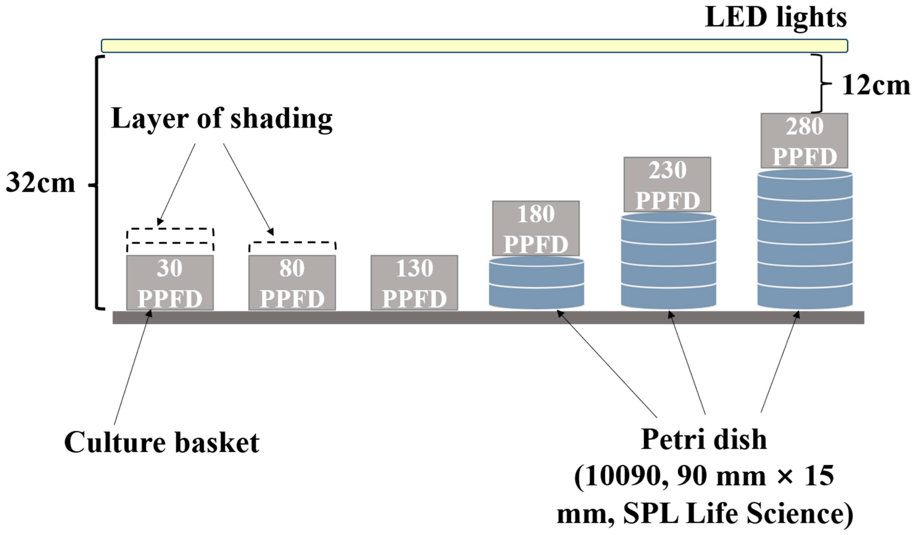
2.2. Experimental Design and Treatments

The transplanted ginseng plants were equally divided into 6 groups and subjected to 6 different LED-based (MEF50120, More Electronics Co., Ltd., Changwon, Republic of Korea) light intensities in a retrofitted plant factory. The spectrum of the LED light (ranging from 400 to 750 nm with a distinct peak at 450 nm in blue) was determined by using a hand-held spectroradiometer (MK550T, UPRtek, Miaoli, Taiwan). The light intensities gradually increased from 30 to 280 PPFD with an interval of 50 PPFD by adopting different layers of shading or adjusting the heights between the ginseng plants and the light source (Figure 1). The light uniformity per light setting was from 0.87 to 0.91. The culture environment was consistent with that described in Section 2.1. above. Subsequently, a multipurpose nutrient solution (MNS) formulated following our pioneer publication [22] was watered to the ginseng plants once a week at 500 mL for 10 weeks until distinct plant responses appeared. Each light intensity treatment was completely randomized by undertaking three independent biological replications, consisting of 12 plants each.

2.3. Destructive Sampling and Growth Parameter Measurements

The growth parameters of juvenile ginseng plants in different treatments, including the whole weights and root weights, shoot-related traits (shoot length, stem length, and stem diameter), and leaf-related parameters (leaf length and width, leaf area, petiole length), were individually investigated during the harvest. Specifically, the shoot length was measured on the basis of the above-ground part, while the stem length refers to the distance between the top of the root and the bottom of the leaf (or the first petiole). The stem diameter was determined at 2 cm above the top of the root. The three topmost leaves were separated from the petioles and subjected to a leaf area scanning apparatus (LI-3000, LI-COR Technology Company, Lincoln, NE, USA) for the determination of the leaf area.

2.4. Analysis of the Photosynthetic Parameters

The photosynthetic activity was estimated in terms of certain critical parameters, such as the photosynthesis pigments, the maximal PSII quantum yield (Fv/Fm).
The chlorophyll contents of chlorophyll a and b, together with carotenoids, were spectrophotometrically determined following the protocol found in Sims’s study [23], with minor modifications. Specifically, an amount of 0.10 g of fresh leaf samples was mixed with a 2 mL reaction medium consisting of 45% v/v ethanol, 45% v/v acetone, and 10% v/v ddH2O, and then this mixture was subjected to incubation at 4 °C overnight. During the incubation, mild shaking at 10 g was carried out via a rotator (AG, FINEPCR, Seoul, Republic of Korea). Afterward, the supernatant was transferred to the cuvette and spectrophotometrically measured at 645 nm, 663 nm, and 440 nm with a spectrophotometer (Libra S22, Biochrom, Corston, UK). Finally, the chlorophyll a and b, together with the carotenoid concentrations, were individually calculated by using the formulae below:
Chlorophyll   a = 12.72   ×   OD 663   2.59   ×   OD 645   ×   V Sample   fresh   weight
Chlorophyll   b = 22.88   ×   OD 645   4.67   ×   OD 663   ×   V Sample   fresh   weight
Carotenoids = 4.7   ×   OD 440   0.27   ×   ( Chl   a + Chl   b Sample   fresh   weight
where ‘V’ is the volume of the reaction solution, and the chlorophyll contents and carotenoids are expressed as mg·g−1 of fresh weight.
The maximal PSII quantum yield (Fv/Fm) was determined by using a hand-held FluorPen FP 100 (Photon Systems Instruments, Drásov, Czech Republic) system. In specific, the measurements were performed on the mid-lamina portion of the topmost leaf after a 40 min darkness adapting and a 0.6 s saturating light pulse. Thereafter, the maximum fluorescence (Fm) and minimum fluorescence (F0) were obtained, and the Fv/Fm was finally calculated following the equation as described by Kramer [24].

2.5. Statistical Analysis and Data Processing

The statistical analysis was carried out with SAS 8.2 statistical software (SAS Inst., Cary, NC, USA). The significant differences among different treatments were assessed via Duncan’s multiple range test at p = 0.05 and are shown by different letters (one-way ANOVA). The significant interactions between the light intensity and the investigated parameters were determined via the two-way ANOVA ‘Linear models’ approach of Fisher’s least-significant-difference (LSD) test (F-test), for which the light intensity was considered as the independent variable, while the acquired parameter data were regarded as the dependent variable. For all the variables, linear regression analysis was conducted according to the light intensities by using EXCEL 2019 software. All the figures were generated with GraphPad Prism 8.0.2 software. The heat map regarding the correlations among the investigated parameters and the PCA (principal component analysis) figure were plotted via the Origin 2022 program. All of the measurements were performed via at least three independent biological replications.

3. Results

3.1. Plant Growth and Morphylogy as Affected by Light Intensity

The ginseng growth attributes were markedly affected by the different light intensities after 8 weeks of cultivation. As is apparent in Figure 2, notable different morphological responses, including the shoot length, root size, stem diameter and length, and leaf size, were observed under the increased light intensities. On the whole, the growth parameters showed a decreasing trend in response to the increased light intensity.
Furthermore, it is notable that the ginseng plants developed phototoxicity symptoms when the light intensity improved from 180 to 280 PPFD, as characterized by chlorosis, leaf necrosis, and early defoliation (Figure 2).

3.2. Fresh Weight and Dry Weight as Affected by Light Intensity

The ginseng plant weight-related parameters, consisting of the whole fresh weight and whole dry weight, as well as the root fresh weight and root dry weight, were investigated after destructive harvest. It is noteworthy that the weight-related parameters exhibited a progressive decline according to the increased light intensities, regardless of the whole weight or root weight considered (Figure 3). Apparently, with the exception of the root dry weight, the whole fresh weight and whole dry weight together with the root fresh weight significantly decreased when the light intensity increased from 30 to 130 PPFD (Figure 3). In particular, the whole fresh weight and whole dry weight significantly decreased 18.2% and 23.1%, respectively, when the light intensity increased from 30 to 80 PPFD (Figure 3A).
However, significant (34.5%) decreases concerning the root dry weight were detected when the light intensities increased from 80 to 180 PPFD (Figure 3B). In addition, the light intensity-conferred impacts on the investigated fresh weight and dry weight were significant according to the F-test results (***, p ≤ 0.001).

3.3. Shoot Length, Stem Length, and Stem Diameter as Affected by Light Intensity

Consistent with the weight-related parameters above, the ginseng plant shoot length and stem length together with the stem diameter showed gradual decreasing trends as the light intensity increased (Figure 4). Also, the shoot length, stem length, and stem diameter significantly (23.1%, 20.2%, and 15.2%, respectively) declined when the light intensity increased from 30 to 80 PPFD. Significant interactions between the light intensity and shoot length and stem length together with the stem diameter were observed.
However, the ginseng plants displayed similar stem diameters when the light intensity increased from 180 to 230 PPFD and from 230 to 280 PPFD, while no significant statistical differences were imparted at 180 PPFD, 230 PPFD, or 280 PPFD (Figure 4B).

3.4. Leaf-Related Parameters as Affected by Light Intensity

The responses of the leaf-related parameters to the light intensity, such as the leaf length, leaf width, petiole length, and leaf area, were investigated. On the whole, the leaf parameters displayed progressive declines regarding the leaf status and leaf-related characteristics, as exhibited in Figure 5.
Specifically, the ginseng plants cultivated at 30 PPFD and 80 PPFD possessed similar leaf colors, which were more prone to green, whereas pale leaves and chlorosis were monitored when the light intensity reached 130 PPFD. Still, the ginseng plants grown under the 230 PPFD and 280 PPFD regimes developed leaf necrosis symptoms (Figure 5A). Moreover, these findings were further evidenced by the investigated leaf length and width, petiole length, and leaf area (Figure 5B–D). These four parameters showed markedly decreasing trends according to the increasing light intensity. As expected, significant interactions between the light intensity and leaf length, leaf width, and petiole length, together with the leaf area, were conferred. Interestingly, the ginseng plants treated with 30 PPFD possessed similar leaf areas compared with those under the 80 PPFD regime (Figure 5D).

3.5. Photosynthetic Activity as Affected by Light Intensity

The photosynthetic activity was estimated by means of traits, including the photosynthesis pigments (chlorophyll a and b, carotenoids) and the maximal PSII quantum yield (Fv/Fm). Consistent with the above findings, these four traits also displayed reduced tendencies in response to the increased light intensity. Also, the light intensity significantly influenced the photosynthesis pigments and Fv/Fm. Outstandingly, the total chlorophyll concentration of the ginseng leaves at 130 PPFD remarkably (22.4%) declined relative to that of the ginseng leaves treated with 30 PPFD (Figure 6A). However, it is worthy to note that slight fluctuations (no statistically significant differences, or even higher values in the 80 PPFD groups, were detected) in the carotenoids and Fv/Fm between the 30 PPFD and 80 PPFD treatments were recorded (Figure 6B,C).

3.6. Multivariate Data Analysis (PCA and Pearson Correlation Analysis) among the Investigated Parameters

To visualize the impacts of the light intensity on the ginseng growth and photosynthetic activity responses, together with the relations among the treatments and study parameters, PCA (principal component analysis) on the basis of the collected data set, including the growth traits and photosynthetic characteristics, was performed. The correlation degree was further determined via Pearson’s correlation matrix analysis (p ≤ 0.05).
The first two principal components of the PCA results explained 86.7% of the total variance, where PC1 captured 81.1% and PC2 comprised 5.6% (Figure 7A). As exhibited in Figure 7A, the ginseng samples treated lower than 130 PPFD were mainly distributed on the right quadrants of the PC1 scatter plot, whereas the other three higher-light-intensity-treated samples were located on the left quadrants of the PC1 scatter plot. Moreover, the samples separated on the right side of the PC1 scatter plot possessed higher values of all the studied parameters.
In addition, the correlation analysis data also showed the positive relations among the (1) chlorophylls and carotenoids; (2) the maximal PSII quantum yield and leaf-related traits (length, width, and area); (3) weight and stem diameter. The degrees of the correlations among these investigated parameters are intuitively denoted by the correlation coefficients (Figure 7B). In particular, conspicuously, the shoot length was significantly and positively correlated with the leaf width, petiole length, and root fresh weight (r = 0.93, 0.92, and 0.9, respectively). Similarly, the root fresh weight was positively correlated with the petiole length, leaf length, and leaf width (r = 0.91, 0.85, and 0.87, respectively), and the leaf area was positively correlated with the shoot length, petiole length, and whole fresh weight (r = 0.84, 0.86, and 0.81, respectively). Regarding the chlorophyll a content, it displayed positive correlations with the leaf length and leaf width (r = 0.89 and 0.86, respectively), while the maximal PSII quantum yield was positively correlated with the leaf area, petiole length, and chlorophyll a level (r = 0.91, 0.92, and 0.77, respectively) (Figure 7B).

4. Discussion

Light intensity is one of the most important light components that significantly affects plant photosynthesis and dictates the ginseng production capacity [8,17,25]. It is widely recognized that the ginseng responses to various light regimes (radiations and intensities) depend on the ginseng ages and cultivars, as well as on environmental factors [14,26,27,28]. The responses of PGSs regarding the physiology, morphology, and photosynthesis aspects to the light intensity are still speculative. Also, the light intensity at approximately 30 PPFD has been extensively adopted in practical ginseng productions, while Jang showed that the range of 75–100 PPFD was the most effective method for the growth and photosynthetic ability of ginseng seedlings [17,21]. Therefore, this study investigated the influences of the light intensity (30–280 PPFD) on the growth and photosynthesis of PGSs, concomitantly determining the correlations among the studied parameters, thereby delivering basic guidelines and knowledge for the culture of PGSs under different light intensity environments.

4.1. Growth and Morphological Characteristics

In our trials, the recorded ginseng physiological and morphological parameters were remarkably affected according to the increased light intensity. The light intensity as high as 180 PPFD significantly restricted the growth of the ginseng plants, as displayed by the decreased whole weight capacity and root development, together with the disturbed leaf performances (Figure 2, Figure 3 and Figure 5). These findings were in agreement with Lee’s report, indicating that the photoinhibition may have limited the electron transport even though the light saturation point was not reached for ginseng growth [29,30,31]. In particular, the ginseng plants cultivated with the light intensity exceeding 230 PPFD developed photoinhibition symptoms, as characterized by leaf chlorosis, necrosis, and stunted leaves (Figure 2) [20,32,33,34].
More importantly, the whole weight, root weight, shoot length, and stem length and diameter progressively decreased as the light intensity increased, revealing that 30 PPFD for the treatment of PGSs may be the most effective method for promoting the shoot and root growth. This is consistent with Kuronuma’s findings suggesting that ginseng plants could be dormant when high light intensity is applied after early growth cultivation [8]. Jang showed that the 25 PPFD-spiked ginseng seedlings possessed the longest shoots, which is parallel with the findings in the present study [17]. Interestingly, regarding the dry root weight between 30 PPFD and 80 PPFD, no statistical differences were conferred (Figure 3B), which may probably be ascribed to the finding that high light intensity is required during the ginseng early growth stage in order to break the dormancy [8,35].
Leaf formation and crop canopy maintenance play essential roles in the root growth and development of ginseng seedlings [36]. During the ginseng growth period, the shoot elongation first increased and then diminished, contributing to a decline in the biomass, along with all the consecutive biomass improvements migrating into the roots. Subsequently, the root weight increased via photosynthesis via the three completely expanded trifoliate leaves after the canopy was formed; therefore, the growth of the PGSs was established solely on the mentioned leaves [29,35,37,38]. In this study, the leaf areas of the ginseng plants treated with 30 PPFD were similar to those of the ginseng plants under the 80 PPFD regime (Figure 5A,D), indicating that the leaf development of the PGSs was not significantly affected when the light intensity was below 80 PPFD. However, the leaf-related parameters monitored in terms of the leaf length and width, together with the leaf area, were considerably curtailed as the light intensity supply increased to 130 PPFD (Figure 5B–D). Possibly, the ginseng leaf structure was vulnerable to damage by the excessive absorption of light energy [17,39]. Previous research showed that the ginseng leaf structure was prone to shrink to accommodate the stem extension below 50 PPFD, and to photomorphogenesis to prevent the excessive light over 125 PPFD [17,40], while our findings enlighten that the threshold is below 30 PPFD and less than 130 PPFD, probably because of the ginseng ages and culture conditions considered.

4.2. Photosynthetic Characteristics

Because light is an essential factor for photosynthesis, plants are able to acclimate to the changes in the light environment in which they are grown for maintained fitness and performance [41]. The acclimation is associated with the changes in multiple processes regarding the biochemical and metabolic aspects, such as the adjustments of pigments and the electron transfer rate, all of which influence photosynthesis [17,42,43,44]. In our experiment, the ginseng plants showed distinct photosynthetic responses to the fluctuating light intensities, and the recorded photosynthetic characteristics supported the ginseng growth (Figure 6).
The photosynthetic pigments in terms of chlorophyll a and b and carotenoids are the most abundant photoreceptors of plants, initiating the light signal transduction process [45,46]. In general, higher chlorophyll contents could further upgrade the light absorption rate and, therefore, the photosynthetic activity [45,46,47]. In line with the growth performance, in terms of the chlorophyll contents, regardless of the types, gradually declining trends were obtained as the light intensity increased (Figure 6A). Carotenoids play a pivotal role in the regulation and protection of photosynthesis, light collection, and the production of phytohormones [48,49]. Surprisingly, we noticed that the carotenoid concentrations of the 80 PPFD-cultured ginseng plants were similar to those of the ginseng plants grown at 30 PPFD (no statistical difference) (Figure 6B), probably because ginseng is a shade-preferring plant that requires the stimulation of more carotenoids for photoprotection in higher-light-intensity environments [8,49,50,51].
Additionally, most plants have evolved feedback regulatory acclimation mechanisms to cope with high-irradiance environments, as reflected by the fluctuations in the maximal PSII quantum yield (Fv/Fm) [52,53,54]. A gradually diminished trend of the Fv/Fm value as the light intensity exceeded 80 PPFD (Figure 6C) showed the inhibitions of the light intensity on the energy transformations and photosynthesis. These decreases in the photosynthetic characteristics were also due to the fact that the high-light-intensity supply could instigate the degradation of chlorophylls [54,55].

4.3. Correlations among the Studied Paremeters and Treatments

Consistent with the growth performances and recorded photosynthetic parameters, the PCA results also showed the distinct responses of the ginseng to the light intensity. We noticed that the 30 and 80 PPFD-cultured plants exhibited relatively greater values of the physiological parameters and photosynthetic capacities, whereas the samples treated no less than 180 PPFD had totally opposite responses to these (Figure 7A), suggesting that the safe point for avoiding the appearance of detrimental symptoms during the cultivation of PGSs may be lower than 180 PPFD.
Moreover, outstandingly, the root-related parameters, such as the root fresh weight, were associated with the shoot and leaf development, while the chlorophyll a and the maximal PSII quantum yield (Fv/Fm) were related to the leaf growth (Figure 7B). Therefore, the growth and photosynthesis activity of the studied PGSs were associated with the root production and leaf establishment [37,38,56].

5. Conclusions

Accordingly, this study investigated the growth and photosynthetic responses of Panax ginseng sprouts (PGSs) to the light intensity and concomitantly set the optimal LED-based photosynthetic photon flux density for the high-precision and practical production of PGSs in PFALs. Consequently, this study showed that the studied parameters had generally progressively declining responses to the increased light intensity (from 30 to 280 PPFD). Therefore, 30 PPFD could be considered as the most effective. However, regarding the root dry weight, leaf area, carotenoid level, and photosynthesis (the maximal PSII quantum yield), we found that there were no differences between the 30 PPFD groups and their 80 PPFD-cultured counterparts. In addition, the PGSs developed detrimental photoinhibition symptoms as the light increased to 180, 230, and 280 PPFD. Hence, these results demonstrated that 30–80 PPFD is a suitable range for the maximum yield capacity and photosynthetic characteristics of PGSs in PFALs. These findings could be implemented to improve the economic efficiency of commercial PGS productions.

Author Contributions

Conceptualization, B.R.J. and J.S.; methodology, B.R.J. and J.S.; software, J.S. and J.Y.; validation, B.R.J. and J.S.; formal analysis, B.R.J. and J.S.; investigation, J.S. and J.Y.; resources, B.R.J.; data curation, J.S.; writing—original draft preparation, J.S.; writing—review and editing, B.R.J. and J.S.; supervision, B.R.J.; project administration, B.R.J.; funding acquisition, B.R.J. All authors have read and agreed to the published version of the manuscript.

Funding

This research was supported by the ‘Leaders in Industry-University Cooperation 3.0’ Project, funded by the Ministry of Education and the National Research Foundation of Korea, project no. 202207800001. Jinnan Song and Jingli Yang were supported by the BK21 Four Program, the Ministry of Education, the Republic of Korea.

Data Availability Statement

Data sharing is not applicable to this article.

Acknowledgments

The authors acknowledge Yeji Moon (Department of Horticulture, College of Agriculture and Life Science, Gyeongsang National University) for her assistance in the preparation and cultivation of the ginseng root materials.

Conflicts of Interest

The authors declare no conflict of interest.

References

  1. Jung, M.Y.; Jeon, B.S.; Bock, J.Y. Free, esterified, and insoluble-bound phenolic acids in white and red Korean ginsengs (Panax ginseng C. A. Meyer). Food Chem. 2002, 79, 105–111. [Google Scholar] [CrossRef]
  2. Park, E.; Hwang, I.; Song, J.-Y.; Jee, Y. Acidic polysaccharide of Panax ginseng as a defense against small intestinal damage by whole-body gamma irradiation of mice. Acta Histochem. 2011, 113, 19–23. [Google Scholar] [CrossRef] [PubMed]
  3. Buettner, C.; Yeh, G.Y.; Phillips, R.S.; Mittleman, M.A.; Kaptchuk, T.J. Systematic review of the effects of ginseng on cardiovascular risk factors. Ann. Pharmacother. 2006, 40, 83–95. [Google Scholar] [CrossRef] [PubMed]
  4. Shin, M.-S.; Song, J.H.; Choi, P.; Lee, J.H.; Kim, S.-Y.; Shin, K.-S.; Ham, J.; Kang, K.S. Stimulation of innate immune function by Panax ginseng after heat processing. J. Agric. Food Chem. 2018, 66, 4652–4659. [Google Scholar] [CrossRef] [PubMed]
  5. Kim, Y.-J.; Zhang, D.; Yang, D.-C. Biosynthesis and biotechnological production of ginsenosides. Biotechnol. Adv. 2015, 33, 717–735. [Google Scholar] [CrossRef] [PubMed]
  6. Baeg, I.-H.; So, S.-H. The world ginseng market and the ginseng (Korea). J. Ginseng Res. 2013, 37, 1. [Google Scholar] [CrossRef]
  7. Chung, I.-M.; Lim, J.-J.; Ahn, M.-S.; Jeong, H.-N.; An, T.-J.; Kim, S.-H. Comparative phenolic compound profiles and antioxidative activity of the fruit, leaves, and roots of Korean ginseng (Panax ginseng Meyer) according to cultivation years. J. Ginseng Res. 2016, 40, 68–75. [Google Scholar] [CrossRef]
  8. Kuronuma, T.; Wang, Q.; Ando, M.; Watanabe, H. Effects of Different Light Intensities on the Growth and Accumulation of Photosynthetic Products in Panax ginseng C. A. Meyers. Environ. Control Biol. 2020, 58, 131–135. [Google Scholar] [CrossRef]
  9. Kim, Y.-J.; Nguyen, T.K.L.; Oh, M.-M. Growth and ginsenosides content of ginseng sprouts according to LED-based light quality changes. Agronomy 2020, 10, 1979. [Google Scholar] [CrossRef]
  10. Li, X.; Liu, J.; Zuo, T.-T.; Hu, Y.; Li, Z.; Xu, X.-Y.; Yang, W.-Z.; Guo, D.-A. Advances and challenges in ginseng research from 2011 to 2020: The phytochemistry, quality control, metabolism, and biosynthesis. Nat. Prod. Rep. 2022, 39, 875–909. [Google Scholar] [CrossRef]
  11. Islam, M.J.; Ryu, B.R.; Azad, M.O.K.; Rahman, M.H.; Rana, M.S.; Lim, J.-D.; Lim, Y.-S. Exogenous putrescine enhances salt tolerance and ginsenosides content in Korean ginseng (Panax ginseng Meyer) sprouts. Plants 2021, 10, 1313. [Google Scholar] [CrossRef]
  12. Cho, K.M.; Lee, H.Y.; Cho, D.Y.; Jung, J.G.; Kim, M.J.; Jeong, J.B.; Jang, S.-N.; Lee, G.O.; Sim, H.-S.; Kang, M.J. Comprehensive Comparison of Chemical Composition and Antioxidant Activity of Panax ginseng Sprouts by Different Cultivation Systems in a Plant Factory. Plants 2022, 11, 1818. [Google Scholar] [CrossRef]
  13. Jang, I.B.; Yu, J.; Suh, S.J.; Jang, I.B.; Kwon, K.B. Growth and ginsenoside content in different parts of ginseng sprouts depending on harvest time. Korean J. Med. Crop Sci. 2018, 26, 205–213. [Google Scholar] [CrossRef]
  14. Hwang, S.H.; Kim, S.C.; Seong, J.A.; Lee, H.Y.; Cho, D.Y.; Kim, M.J.; Jung, J.G.; Jeong, E.H.; Son, K.-H.; Cho, K.M. Comparison of ginsenoside contents and antioxidant activity according to the size of ginseng sprout has produced in a plant factory. J. Appl. Biol. Chem. 2021, 64, 253–261. [Google Scholar] [CrossRef]
  15. Yeh, N.; Chung, J.-P. High-brightness LEDs—Energy efficient lighting sources and their potential in indoor plant cultivation. Renew. Sustain. Energy Rev. 2009, 13, 2175–2180. [Google Scholar] [CrossRef]
  16. Son, I.-C.; Moon, K.H.; Song, E.Y.; Wi, S.H.; Seo, H.-H.; Moon, Y.E.; Reddy, V.; Yang, J.; Sicher, R.; Oh, S. Growth and physiological responses of Chinese cabbage to different light intensity until leafy head formation. Hortic. Sci. Technol. 2018, 36, 151–160. [Google Scholar]
  17. Jang, I.; Do, G.; Hwang, H.; Suh, S.; Yu, J.; Jang, I.; Moon, J.; Chun, C. Morphological development and photosynthetic acclimation of Panax ginseng seedlings to irradiation by light-emitting diodes (LEDs). Hortic. Environ. Biotechnol. 2021, 62, 571–579. [Google Scholar] [CrossRef]
  18. Nadeau, I.; Olivier, A. The biology and forest cultivation of American ginseng (Panax quinquefolius L.) in Canada. Can. J. Plant Sci. 2003, 83, 877–891. [Google Scholar]
  19. Zhang, Y.-C.; Li, G.; Jiang, C.; Yang, B.; Yang, H.-J.; Xu, H.-Y.; Huang, L.-Q. Tissue-specific distribution of ginsenosides in different aged ginseng and antioxidant activity of ginseng leaf. Molecules 2014, 19, 17381–17399. [Google Scholar] [CrossRef] [PubMed]
  20. Miskell, J.-A.; Parmenter, G.; Eaton-Rye, J.J. Decreased Hill reaction rates and slow turnover of transitory starch in the obligate shade plant Panax quinquefolius L. (American ginseng). Planta 2002, 215, 969–979. [Google Scholar] [CrossRef]
  21. Rural Development Administration (RDA). Good Agricultural Practice of Ginseng; Revised Ed.; Rural Development Administration: Suwon, Republic of Korea, 2012; pp. 77–79. [Google Scholar]
  22. Song, J.; Yang, J.; Jeong, B.R. Growth, quality, and nitrogen assimilation in response to high ammonium or nitrate supply in cabbage (Brassica campestris L.) and lettuce (Lactuca sativa L.). Agronomy 2021, 11, 2556. [Google Scholar] [CrossRef]
  23. Sims, D.A.; Gamon, J.A. Relationships between leaf pigment content and spectral reflectance across a wide range of species, leaf structures and developmental stages. Remote Sens. Environ. 2002, 81, 337–354. [Google Scholar] [CrossRef]
  24. Kramer, D.M.; Johnson, G.; Kiirats, O.; Edwards, G.E. New Fluorescence Parameters for the Determination of QA Redox State and Excitation Energy Fluxes. Photosynth. Res. 2004, 79, 209. [Google Scholar] [CrossRef] [PubMed]
  25. Sadiq, N.B.; Kwon, H.; Park, N.I.; Hamayun, M.; Jung, J.-H.; Yang, S.-H.; Jang, S.-W.; Kabadayı, S.N.; Kim, H.-Y.; Kim, Y.-J. The Impact of Light Wavelength and Darkness on Metabolite Profiling of Korean Ginseng: Evaluating Its Anti-Cancer Potential against MCF-7 and BV-2 Cell Lines. Int. J. Mol. Sci. 2023, 24, 7768. [Google Scholar] [CrossRef] [PubMed]
  26. Jayakodi, M.; Lee, S.-C.; Yang, T.-J. Comparative transcriptome analysis of heat stress responsiveness between two contrasting ginseng cultivars. J. Ginseng Res. 2019, 43, 572–579. [Google Scholar] [CrossRef] [PubMed]
  27. Li, M.-R.; Shi, F.-X.; Zhou, Y.-X.; Li, Y.-L.; Wang, X.-F.; Zhang, C.; Wang, X.-T.; Liu, B.; Xiao, H.-X.; Li, L.-F. Genetic and epigenetic diversities shed light on domestication of cultivated ginseng (Panax ginseng). Mol. Plant 2015, 8, 1612–1622. [Google Scholar] [CrossRef]
  28. Di, P.; Sun, Z.; Cheng, L.; Han, M.; Yang, L.; Yang, L. LED Light Irradiations Differentially Affect the Physiological Characteristics, Ginsenoside Content, and Expressions of Ginsenoside Biosynthetic Pathway Genes in Panax ginseng. Agriculture 2023, 13, 807. [Google Scholar] [CrossRef]
  29. Lee, B.; Pham, M.D.; Cui, M.; Lee, H.; Hwang, H.; Jang, I.; Chun, C. Growth and physiological responses of Panax ginseng seedlings as affected by light intensity and photoperiod. Hortic. Environ. Biotechnol. 2022, 63, 835–846. [Google Scholar] [CrossRef]
  30. Yang, D.-C.; Chae, Q.; Lee, S.-J.; Kim, Y.-H.; Kang, Y.-H. Effects of Light and Photosynthetic Electron Transport System on the Generation of Singlet Oxygen (1O2) in Ginseng Thylakoid Membrane. J. Ginseng Res. 1990, 14, 57–62. [Google Scholar]
  31. Lee, S.S. Characteristics of photosynthesis among new cultivars of ginseng (Panax ginseng C. A. Meyer). J. Ginseng Res. 2002, 26, 85–88. [Google Scholar]
  32. Nam, M.H.; Kim, S.I.; Liu, J.R.; Yang, D.C.; Lim, Y.P.; Kwon, K.-H.; Yoo, J.S.; Park, Y.M. Proteomic analysis of Korean ginseng (Panax ginseng C. A. Meyer). J. Chromatogr. B 2005, 815, 147–155. [Google Scholar]
  33. Nam, M.H.; Heo, E.J.; Kim, J.Y.; Kim, S.I.; Kwon, K.H.; Seo, J.B.; Kwon, O.; Yoo, J.S.; Park, Y.M. Proteome analysis of the responses of Panax ginseng C. A. Meyer leaves to high light: Use of electrospray ionization quadrupole-time of flight mass spectrometry and expressed sequence tag data. Proteomics 2003, 3, 2351–2367. [Google Scholar] [CrossRef]
  34. Jung, J.H.; Kim, H.-Y.; Kim, H.S.; Jung, S.H. Transcriptome analysis of Panax ginseng response to high light stress. J. Ginseng Res. 2020, 44, 312–320. [Google Scholar]
  35. Parmenter, G.; Littlejohn, R. Effect of shade on growth and photosynthesis of Panax ginseng. N. Z. J. Crop Hortic. Sci. 2000, 28, 255–269. [Google Scholar]
  36. Proctor, J.; Dorais, M.; Bleiholder, H.; Willis, A.; Hack, H.; Meier, V. Phenological growth stages of North American ginseng (Panax quinquefolius). Ann. Appl. Biol. 2003, 143, 311–317. [Google Scholar] [CrossRef]
  37. Kim, Y.-J.; Joo, S.C.; Shi, J.; Hu, C.; Quan, S.; Hu, J.; Sukweenadhi, J.; Mohanan, P.; Yang, D.-C.; Zhang, D. Metabolic dynamics and physiological adaptation of Panax ginseng during development. Plant Cell Rep. 2018, 37, 393–410. [Google Scholar] [PubMed]
  38. Proctor, J.T.; Louttit, D.; Jiao, J. Seasonal growth and root respiration of North American ginseng. J. Ginseng Res. 1998, 22, 161–167. [Google Scholar]
  39. Fan, X.-X.; Xu, Z.-G.; Liu, X.-Y.; Tang, C.-M.; Wang, L.-W.; Han, X.-L. Effects of light intensity on the growth and leaf development of young tomato plants grown under a combination of red and blue light. Sci. Hortic. 2013, 153, 50–55. [Google Scholar]
  40. Proctor, J.T.; Palmer, J.W.; Follett, J.M. Growth, dry matter partitioning and photosynthesis in North American ginseng seedlings. J. Ginseng Res. 2010, 34, 175–182. [Google Scholar] [CrossRef]
  41. Vialet-Chabrand, S.; Matthews, J.S.; Simkin, A.J.; Raines, C.A.; Lawson, T. Importance of fluctuations in light on plant photosynthetic acclimation. Plant Physiol. 2017, 173, 2163–2179. [Google Scholar]
  42. Terashima, I.; Hanba, Y.T.; Tazoe, Y.; Vyas, P.; Yano, S. Irradiance and phenotype: Comparative eco-development of sun and shade leaves in relation to photosynthetic CO2 diffusion. J. Exp. Bot. 2006, 57, 343–354. [Google Scholar]
  43. Athanasiou, K.; Dyson, B.C.; Webster, R.E.; Johnson, G.N. Dynamic acclimation of photosynthesis increases plant fitness in changing environments. Plant Physiol. 2010, 152, 366–373. [Google Scholar] [PubMed]
  44. Kono, M.; Terashima, I. Long-term and short-term responses of the photosynthetic electron transport to fluctuating light. J. Photochem. Photobiol. B 2014, 137, 89–99. [Google Scholar] [PubMed]
  45. Fleischer, W.E. The relation between chlorophyll content and rate of photosynthesis. J. Gen. Physiol. 1935, 18, 573. [Google Scholar] [CrossRef] [PubMed]
  46. Buttery, B.; Buzzell, R. The relationship between chlorophyll content and rate of photosynthesis in soybeans. Can. J. Plant Sci. 1977, 57, 1–5. [Google Scholar]
  47. Song, J.; Yang, J.; Jeong, B.R. Root GS and NADH-GDH play important roles in enhancing the ammonium tolerance in three bedding plants. Int. J. Mol. Sci. 2022, 23, 1061. [Google Scholar]
  48. Sathasivam, R.; Radhakrishnan, R.; Kim, J.K.; Park, S.U. An update on biosynthesis and regulation of carotenoids in plants. S. Afr. J. Bot. 2021, 140, 290–302. [Google Scholar]
  49. Demmig-Adams, B.; Gilmore, A.M.; Iii, W.W.A. In vivo functions of carotenoids in higher plants. FASEB J. 1996, 10, 403–412. [Google Scholar]
  50. Maltsev, Y.; Maltseva, K.; Kulikovskiy, M.; Maltseva, S. Influence of light conditions on microalgae growth and content of lipids, carotenoids, and fatty acid composition. Biology 2021, 10, 1060. [Google Scholar]
  51. Demmig-Adams, B.; ADAMS III, W. Carotenoid composition in sun and shade leaves of plants with different life forms. Plant Cell Environ. 1992, 15, 411–419. [Google Scholar]
  52. Öquist, G.; Chow, W. On the relationship between the quantum yield of Photosystem II electron transport, as determined by chlorophyll fluorescence and the quantum yield of CO2-dependent O2 evolution. Photosynth. Res. 1992, 33, 51–62. [Google Scholar] [CrossRef] [PubMed]
  53. Janka, E.; Körner, O.; Rosenqvist, E.; Ottosen, C.-O. Using the quantum yields of photosystem II and the rate of net photosynthesis to monitor high irradiance and temperature stress in chrysanthemum (Dendranthema grandiflora). Plant Physiol. Biochem. 2015, 90, 14–22. [Google Scholar] [PubMed]
  54. ElSayed, A.I.; Rafudeen, M.S.; Gomaa, A.M.; Hasanuzzaman, M. Exogenous melatonin enhances the reactive oxygen species metabolism, antioxidant defense-related gene expression, and photosynthetic capacity of Phaseolus vulgaris L. to confer salt stress tolerance. Physiol. Plant. 2021, 173, 1369–1381. [Google Scholar] [CrossRef]
  55. Song, J.; Yang, J.; Jeong, B.R. Difference between Day and Night Temperature (DIF) and Light Intensity Affect Growth and Photosynthetic Characteristics of Panax ginseng Meyer Sprouts. Plants 2023, 12, 2820. [Google Scholar] [CrossRef]
  56. Wang, Y.; Pan, J.Y.; Xiao, X.Y.; Lin, R.C.; Cheng, Y.Y. Simultaneous determination of ginsenosides in Panax ginseng with different growth ages using high-performance liquid chromatography–mass spectrometry. Phytochem. Anal. PCA 2006, 17, 424–430. [Google Scholar] [CrossRef] [PubMed]
Figure 1. The PGSs were cultivated under 6 different light intensity regimes.
Agronomy 13 02375 g001
Figure 2. The influence of light intensity on ginseng growth and morphology.
Agronomy 13 02375 g002
Figure 3. Effects of light intensity on ginseng (A) whole fresh weight and whole dry weight and (B) root fresh weight and root dry weight. Values are means ± SDs of n = 5 biological replicates. Significant differences for identical parameters among the light intensities are denoted by different letters, following Duncan’s multiple range test at p = 0.05 (one-way ANOVA). The significant interactions between the light intensity and the investigated parameters were determined via F-test and are indicated by different numbers of asterisks.
Agronomy 13 02375 g003
Figure 4. Effects of light intensity on ginseng (A) shoot length and stem length and (B) stem diameter. Values are means ± SDs of n = 5 biological replicates. Significant differences for identical parameters among different light intensities are denoted by different letters, following Duncan’s multiple range test at p = 0.05 (one-way ANOVA). The significant interactions between the light intensity and the investigated parameters were determined via F-test and are indicated by different numbers of asterisks (**, p < 0.01; and ***, p < 0.001).
Agronomy 13 02375 g004
Figure 5. The effects of light intensity on ginseng (A) leaf morphology and status, (B) leaf length and width, (C) petiole length, and (D) leaf area. The presented data are expressed as means ± SDs (n = 5 replicates). Different letters indicate significant differences according to Duncan’s multiple range test at p = 0.05 (one-way ANOVA). The significant interactions between the light intensity and the investigated leaf parameters were determined via F-test and are denoted by different numbers of asterisks (**, p < 0.01; and ***, p < 0.001).
Agronomy 13 02375 g005
Figure 6. The effects of light intensity on ginseng (A) chlorophyll contents, (B) carotenoids contents, and (C) the maximal quantum yield of photosystem II (Fv/Fm). The displayed values are expressed as means ± SDs (n = 5 independent replicates). Different lowercase letters indicate significant differences according to Duncan’s multiple range test at p = 0.05 (one-way ANOVA). The significant interactions between the light intensity and the investigated photosynthetic characteristics were determined via F-test and are denoted by different numbers of asterisks (**, p < 0.01; and ***, p < 0.001).
Agronomy 13 02375 g006
Figure 7. Multivariate data analysis using (A) PCA and (B) a heatmap of Pearson’s correlation matrix among the investigated growth and photosynthetic characteristics. Cyan color and brown color indicate low R values and high R values, respectively. Abbreviations: SHL: shoot length; STL: stem length; SD: shoot diameter; FW: whole fresh weight; DW: whole dry weight; RFW: root fresh weight; RDW: root dry weight; PL: petiole length; LL: leaf length; LW: leaf width; LA: leaf area; Cha: chlorophyll a; Chb: chlorophyll b; Ca: carotenoids; QY: the maximal PSII quantum yield (Fv/Fm).
Agronomy 13 02375 g007
Disclaimer/Publisher’s Note: The statements, opinions and data contained in all publications are solely those of the individual author(s) and contributor(s) and not of MDPI and/or the editor(s). MDPI and/or the editor(s) disclaim responsibility for any injury to people or property resulting from any ideas, methods, instructions or products referred to in the content.

