Article

First Report of Leaf Spot Disease (“Negrilla”) on Agave salmiana Otto Ex Salm-Dyck (ssp. salmiana) Plants Caused by Bipolaris zeae Zivan in Mexico

by
Teresa Romero-Cortes
1,
Victor H. Pérez España
1,
José A. Pescador-Rojas
1,
Eduardo Rangel-Cortés
2,
María M. Armendaríz-Ontiveros
1 and
Jaime A. Cuervo-Parra
1,*
1
Escuela Superior de Apan, Universidad Autónoma del Estado de Hidalgo, Carretera Apan-Calpulalpan, Km 8, Chimalpa Tlalayote, Apan 43900, Mexico
2
Facultad de Ciencias, Universidad Nacional Autónoma de México, Investigación Científica, C. U. Coyoacán, Ciudad de México 04510, Mexico
*
Author to whom correspondence should be addressed.
Agronomy 2024, 14(3), 623; https://doi.org/10.3390/agronomy14030623
Submission received: 20 February 2024 / Revised: 8 March 2024 / Accepted: 18 March 2024 / Published: 20 March 2024
(This article belongs to the Section Pest and Disease Management)

Abstract

:
Agave genera include slow-growing plants with cultural and economic roots dating back to pre-Columbian times in Mexico. Several species have a widespread presence in the country and are cultivated and/or used directly from the field to obtain various derived products. Agave salmiana is widely used in the region of the High Valleys of Apan, Hidalgo, Mexico. However, fungal diseases are causing considerable losses to Agave crops. For this reason, fungi strains from maguey plants from Apan, Hidalgo, with “Negrilla” disease symptoms were isolated and identified morphologically and molecularly. The results provide information on a new disease disseminated in A. salmiana plants, which causes symptoms such as black spots on the leaves due to pathogenic fungi of the genera Bipolaris. The morphological and molecular characterization located the phytopathogenic fungus as new isolates of Bipolaris zeae. Finally, the re-isolation of the causal agent of the disease was achieved in all pathogenicity tests, verifying that the symptoms observed in the maguey plants were caused by B. zeae, thus corroborating Koch’s postulates, and constituting the first report of this fungus as a pathogen of A. salmiana in Mexico.

1. Introduction

The Helminthosporium complex [1] includes the phytopathogen graminicolous fungi species and the most important genera are Drechslera, Bipolaris, and Exserohilum [2]. The Bipolaris Shoemaker genera [3] and their Cochliobolus Drechsler [4] sexual morph [5] are opportunistic and pathogenic microorganisms with a worldwide distribution, which are present in air, soil, and plants [6]. The Bipolaris species generally have pale, multiseptate, straight or curved conidia, without the presence of bulges in their cells. The Cochliobolus presents structures with an oscillate ascoma, pseudo paraphyses and bitunicate asci; and within its ascus, one typically finds the multiseptate and filiform ascospores with helical shape arrangement [1,7]. These pathogenic fungi are commonly associated with several disease symptoms, such as leaf spots and blights, melting out, and root and foot rot, reproducing mainly in crop plants of high economic value [1,8,9,10,11,12]. Crops such as wheat (Triticum spp.), corn (Zea mays), and rice (Oryza sativa) exhibit serious losses in grain yields due to diseases caused by some Bipolaris species [13,14,15]. Additionally, the Bipolaris and Cochliobolus species occur ubiquitously and are saprophytic and pathogenic to more than 60 plant host genera [16]. Similarly, these fungi cause human diseases such as dermatomycosis, keratitis, allergic sinusitis, central nervous system infections, disseminated infections, etc. [17,18,19,20]. However, the pathologies in humans caused by representatives of these genera are very infrequent, occurring only in patients with serious problems in their immune system [21].
The Bipolaris classification based on morphological characters was initially complicated because some species shared characteristics of morphology (sexual and asexual) with the genera Curvularia [8]. In addition, the conidia and the conidiophores are highly variable within species [22]. In an attempt to solve the conflict in the nomenclature for these two genera, much research has focused on molecular phylogenetics to relocate many species on the genera Bipolaris in Curvularia [5,6,23,24], which is helpful for establishing the correct location of new fungal isolates within the Bipolaris and Curvularia group [25,26]. In Mexico, different Bipolaris species have been registered as foliar pathogens of crops such as oat (Avena sativa L.), wheat, rice, barley (Hordeum vulgare), and maize [10,13,27,28,29]. Given that the genera Bipolaris has more than 60 species, each one with different physiological, phytopathological, biochemical, and biological characteristics [1,30], it is important its proper identification to control and manage diseases produced by these fungi complex group, as well as to identify disease symptomatology in plants that have not been previously infected. In this sense, Perez-España et al. [12] reported that the Agave salmiana plants present a fungal disease that is characterized by the existence of circular gray spots on the leaves, which become necrotic over time. These results are in agreement with Hernández et al. [31] study, where they reported the same symptomatology for the same Agave species caused by the etiological agent of Curvularia lunata, this study was carried out in different municipalities of Hidalgo, Mexico. Therefore, the identification of species involved in causing the symptomatology observed in Agave plants is an attractive alternative for mitigating, controlling, and preventing diseases produced by pathogenic fungi. As managing diseases in Agave plants is desirable, this research aims to morphologically and molecularly identify the causal agent of the “leaf spot” disease in A. salmiana plants and determine the pathogen(s) aggressiveness isolated in plants of the genera Agave cultured under field conditions.

2. Materials and Methods

2.1. Fungal Strains Isolation

Fungi strains were isolated from the cuticle layer of Agave salmiana with disease symptoms in the form of dark black spots (colloquially called “Negrilla” in Mexico) during the years 2019, 2020, and 2021 from the municipality of Apan, Hidalgo, Mexico, (19°39′20″ LN and 98°31′05″ LW, 2488 masl), under rainfed conditions. Samples were sterilized by immersion in a 3% sodium hypochlorite solution for 60 s, followed by two washes in sterile distilled water; dried on filter paper; and then placed on Petri dishes containing potato dextrose agar (PDA, Bioxon®, Becton Dickinson, Edomex, Mexico) in a moist chamber. A total of eight fungal strains were isolated and selected based on the characteristics developed during their culture. Fungi were incubated under dark conditions at 28 °C for one week [32] or until the colony covered the entire culture medium [10]. Subsequently, the selected growing colonies were subcultured on PDA under the same conditions until its analysis.

2.2. Morphological Characterization

Macroscopic morphological identification of the eight strains was performed on PDA and Sabouraud Dextrose Agar (SDA, DifcoTM, Sarks, MD, USA) culture media. Subsequently, the strains were morphologically identified at the macroscopic level, and the PDA medium was used to create the descriptions of microscopic structures. The strain identification was based on the comparisons with other Bipolaris strains [1,5,11,33] and complemented with new observations. Fifty measurements for each trait observed in each isolated species were made to calculate the length and width of the following structures: ascomata, conidiophores, conidia, and the number of dictyosepts per conidia. The morphological measurements were made with a compound microscope (Leica DM 3000, Leica Microsystems GmbH, Wetzlar, Germany) using bright field and a scanning electron microscope (SEM) (JEOL, Model IT300, Boston, MA, USA) both located at the Escuela Superior de Apan (ESAp-UAEH), Hidalgo, Mexico. The photographs were digitally generated using the CorelDraw 2017 software (CorelDRAW Graphics Suite, version 2017, Mexico City, Mexico).

2.3. Molecular Characterization

The genomic DNA from each strain was extracted using the Sanger sequencing method (Eurofins Genomics GmbH, Ebersberg, Germany). The strains DNA extraction was carried out following the methodology described by Cuervo-Parra et al. [34]. The PCR reactions and amplifications were performed in an automatic thermal cycler (Techne PRIME3, Series No. 31309, Cole Parmer, Staffordshire, UK), as described by Romero-Cortes et al. [35]. The amplicons were recovered and washed twice with TE buffer (Tris HCl 10 mM, EDTA 1 mM). The gene internal transcribed spacers, which contained the ITS I/5.8s/ITS II region of the ribosomal DNA (rDNA), were amplified using the primers ITS1 and ITS4 [36]. The purified PCR products were sequenced by Cinvestav-IPN (Irapuato, Mexico). The DNA sequences were assembled using the ChromasPro 2.1.10.1 software (Technelysium Pty Ltd, South Brisbane, Australia) and aligned with the BLAST® 2.15.0 software from the NCBI GenBank database [37]. The sequences were deposited in the GenBank database with the accession numbers ON630338 to ON630345. The phylogenetic analyses were performed by generating a tree with the Neighbor-joining statistic method (NJ), using the Tajima-Nei model, supported with the bootstrap test of the phylogeny method, using 1000 random replicates with the MEGA X 10.2.2 software [38].

2.4. Pathogenicity Tests

To comply with Koch’s postulates [39], healthy one-year-old Agave seedlings from healthy adult maguey plants were planted in an experimental plot, under a completely randomized experimental design, and experimental evaluation began one month after plant establishment [40]. Each experimental treatment consisted of six A. salmiana seedlings with 7 to 8 leaves sowed and inoculated with Bipolaris zeae (strain JCP-N07) in leaves and rhizosphere under field conditions. The pathogenicity experimental field was prepared with a substrate composed of sand (80 kg), limestone (120 kg), and black soil (200 kg) from the previous crop cycle. The experiment was performed in triplicate using 21 Agave seedlings as non-inoculated controls (T1) and 21 Agave seedlings inoculated with Bipolaris zeae (T2) in the year 2022.
The Agave plant inoculation with B. zeae was based on the technique modification described by Mata-Santoyo et al. [13]. To infect the A. salmiana plants of the spot leaf disease, a solution containing 15 mL in equal volumes of a 5 × 10⁶ conidia/mL and Tween 80 (1%) of B. zeae strain JCP-N07 in sterile distilled water was sprayed directly onto the leaves and rhizosphere of each Agave plant of T2. The T1 plants were sprayed with sterile distilled water. The evaluation began five days after the inoculation, according to the severity scale proposed by Perelló et al. [41], where 1 = free of infection; 2 = 1 to 5%; 3 = 5 to 12%; 4 = 12 to 20%; 5 = 20 to 35%; 6 = 35 to 45%; 7 = 45 to 60%; 8 = 60 to 80% and 9 = more than 80% of the leaf area was affected with necrosis and generalized chlorosis. The data obtained were converted to percentages and an ANOVA analysis of variance was performed using the Statistix 10.0 software [42] and the means were compared by Tukey’s HSD All-Pairwise test at a significant difference (p < 0.05).

3. Results and Discussion

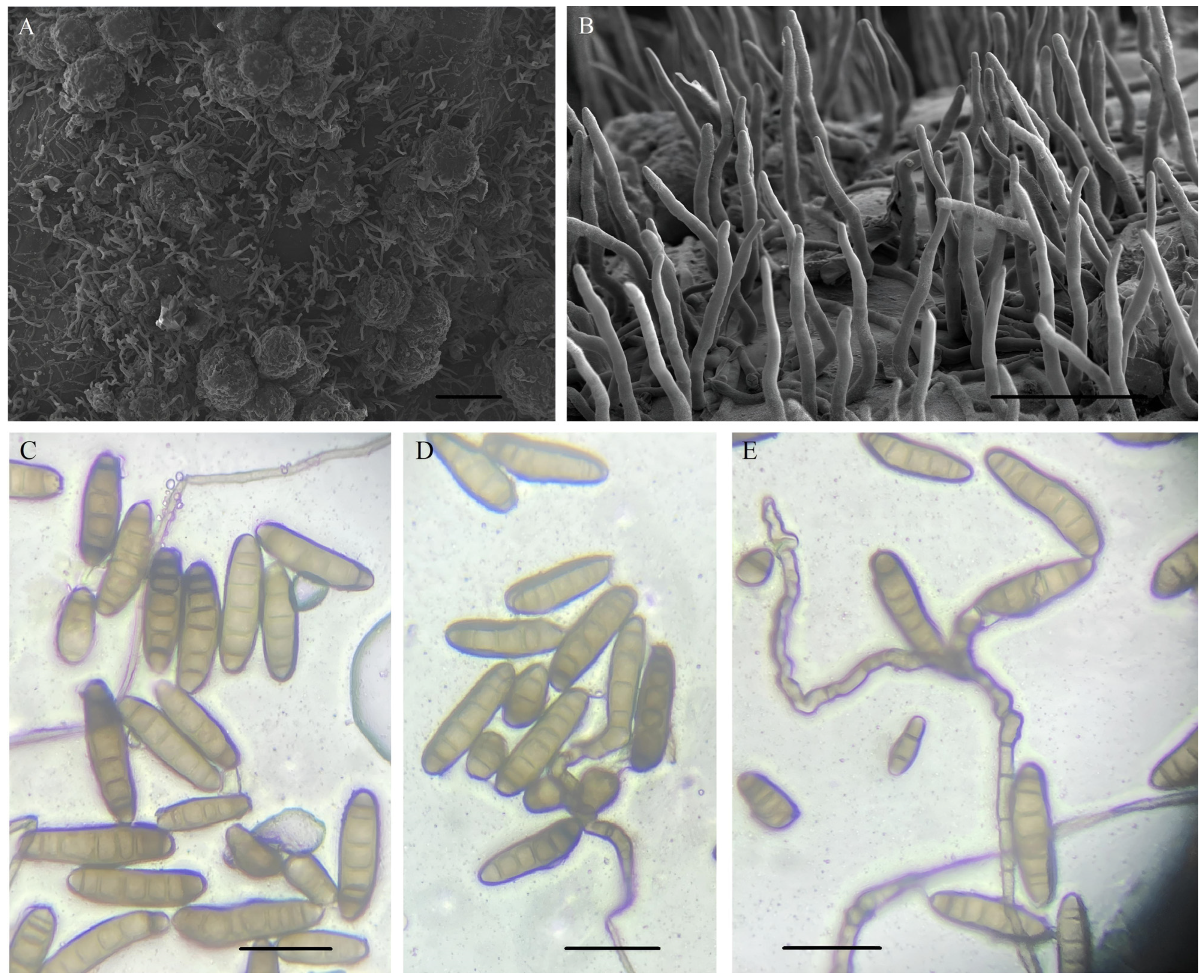
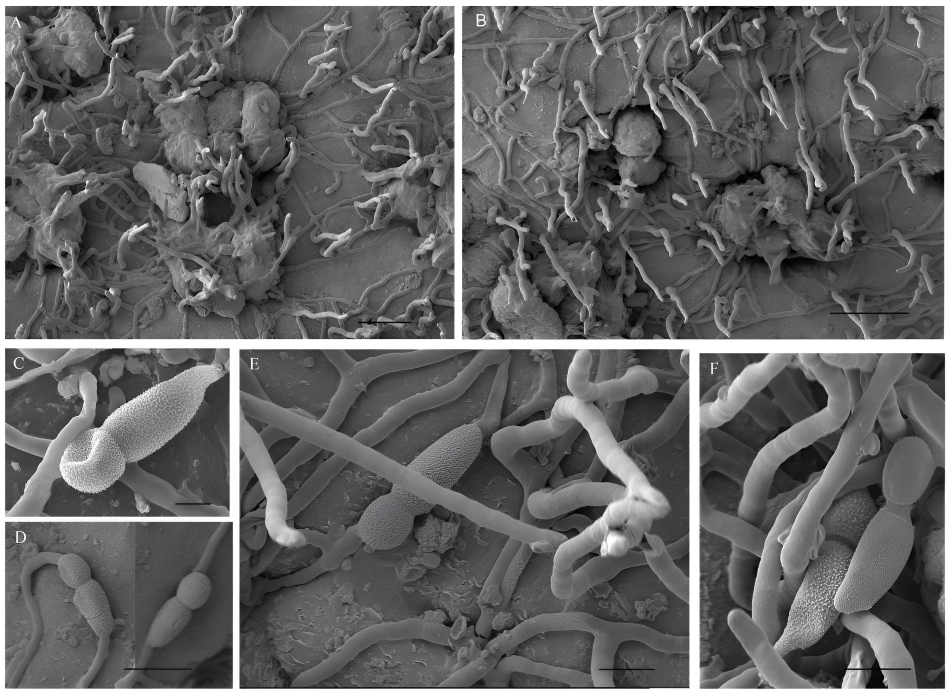
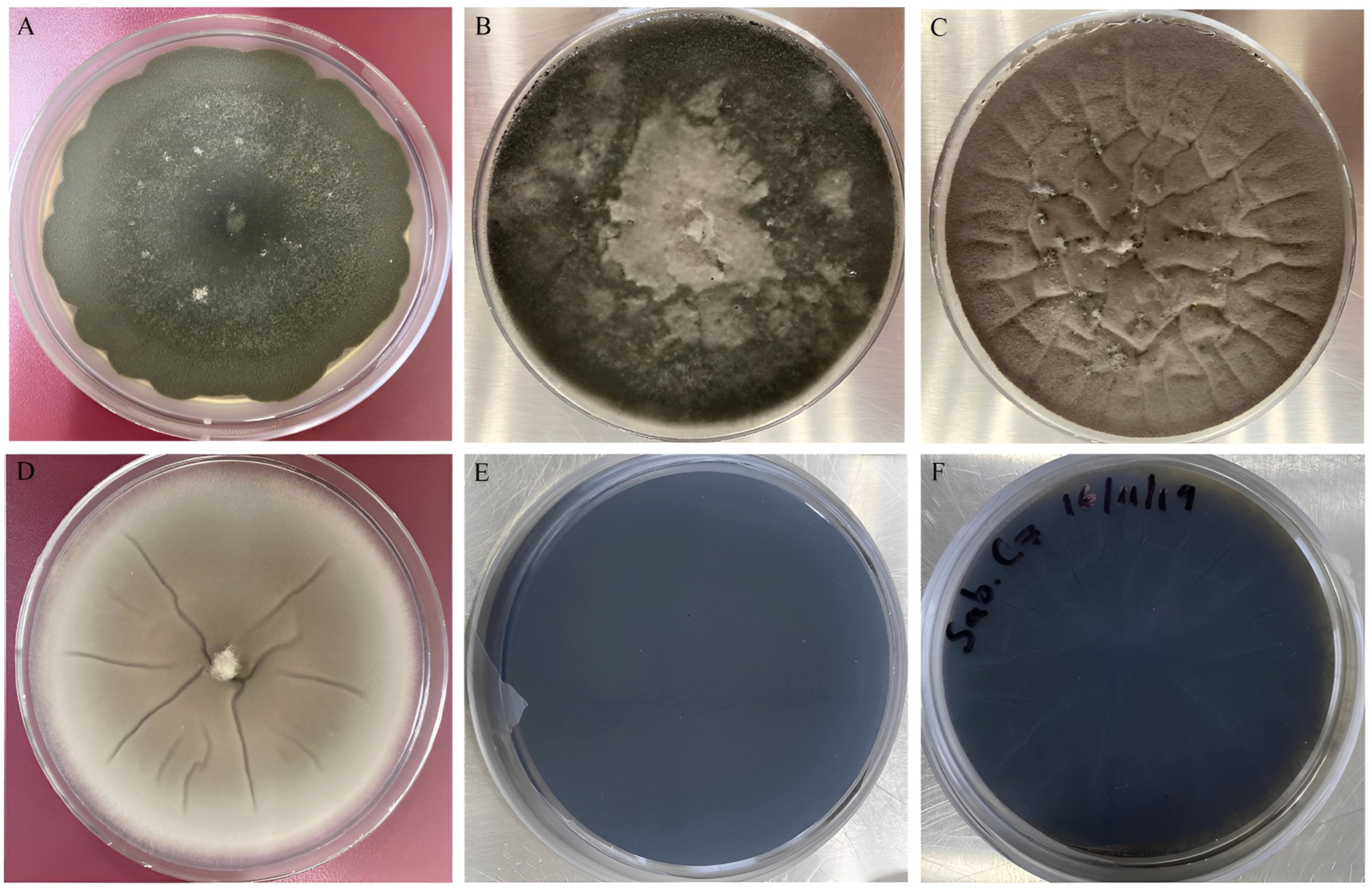
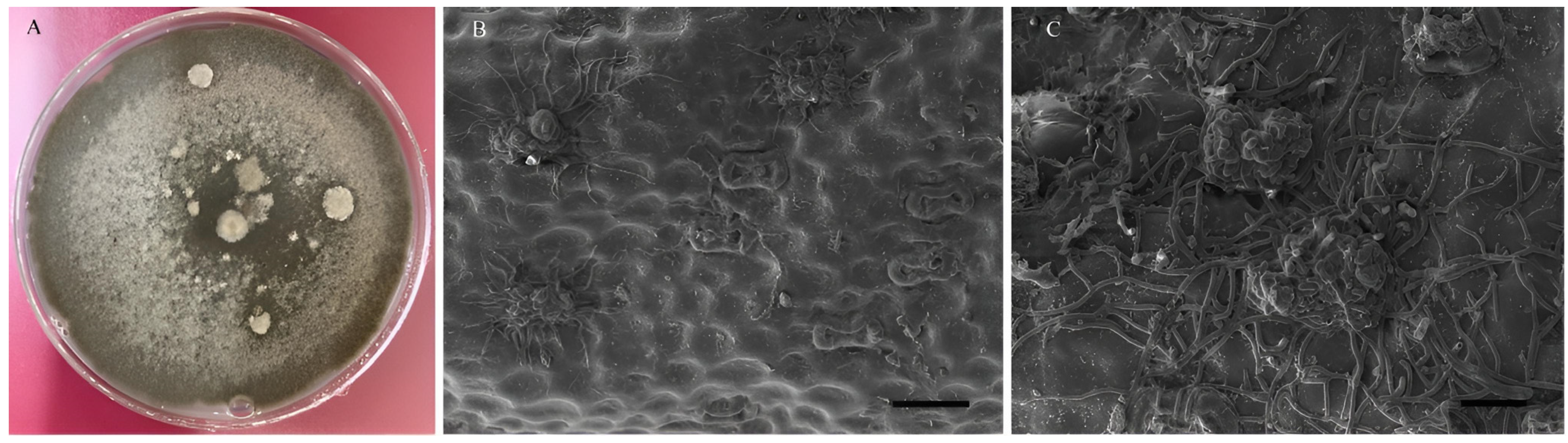
3.1. Morphological Characterization

The fungal colonies isolated and selected from A. salmiana plants, cultivated in PDA medium, showed a diameter of 44.8 to 45 mm (44.9 ± 0.1 mm) in 7 days at 28 °C, with an average of 6.41 mm per day. On the other hand, the colonies in the SDA medium reached a diameter between 44.9 and 45 mm (44.96 ± 0.05 mm) in 6 days at 28 °C, with an average of 7.49 mm per day. The fungal colonies color was initially white in the two different culture media (PDA and SDA), turning to olive green over time, and then changing to dark black in PDA medium (Figure 1A,B). In SDA medium, the colonies color changed from light brown to dark brown with the presence of some depressions (Figure 1C,D). The velvety texture in both media PDA and SDA was due to the presence of conidia. After 7 days of growth, the presence of pustules was observed, and the colonies showed a cottony texture, changing from olive green to dark gray (Figure 1A,B) and light gray (Figure 1C) over time in PDA and SDA culture media, respectively. The colonial growth observed was rosette-shaped (Figure 1A) to radial (Figure 1B) in PDA, and radial (Figure 1C,D) in SDA. The back colonies were black in PDA and SDA (Figure 1E,F). However, in SDA, the presence of striae is due to the folds observed on the front of the colonies (Figure 1F).
The fungi of the Cochliobolus genera are characterized by presenting brown conidiophores and conidia, straight or curved fusoid, and their germination occurs from a germ tube at each end [6]. In this sense, the colonies in the Petri dishes had abundant sporulation after 7 days. The hyphae observed were septate, branched, and smooth, with a thickness up to 7 µm, and could be warty sometimes [1]. The conidiophores were abundant, cylindrical, straight, or flexuous, geniculated at the upper part, smooth, dark brown, arising singly or sometimes in small groups, branched, septate (Figure 2A,B), with size of 80.67–372 µm long x 6.02–8.1 µm wide (av. = 222.15 µm, SE = 110.02, n = 50, long; av. = 7.5 µm, SE = 0.64, n = 50, wide). The conidiogenous nodes were dark below, with a smooth to slightly roughened surface. The conidia observed was 43–225 μm long × 12.02–22 µm wide (av. = 124.34 µm, SE = 57.31, n = 50; av. = 16.19 µm, SE = 2.24, n = 50), straight to slightly curved, smooth, obclavate to fusiform, hyaline to olivaceous brown when immature and brown to reddish brown when mature (Figure 2C–E), with end cells usually paler than middle cells, sometimes presenting Y-shaped conidia in PDA medium [6], with 6 to 14 distoseptate (av. = 9.3, SE = 2.52, n = 50), with the terminal cells cut by a thick dark septum, straight to slightly curved. The hilum was truncated, slightly protruding, 3–5 µm. In the Bipolaris fungi, the conidia septum ontogeny was observed as follows: the first-formed septum is median to submedian, the second septum delimits the basal cell of the mature conidium, and the third septum delimits the distal cell [1].
Additionally, on the A. salmiana leaves with the previously described disease symptoms of leaf spots, an additional unusual type of conidia was observed (Figure 3A,B), which was lecythiform, verrucose, septate, with a size of 31.02–24.56 µm long × 13.72–17.85 µm wide (av. = 27.31 µm, SE = 2, n = 50, long; av. = 16.20 µm, SE = 1.32, n = 50, wide) arising from single conidiophores (Figure 3C–E) or in small groups (Figure 3F), with a morphology similar to that observed in PDA culture medium. The differences observed were in their size, between 72.38–79.75 µm long × 3.58–5.57 µm wide (av. = 76.78 µm, SE = 2.38, n = 50, long; av. = 4.85 µm, SE = 0.74, n = 50, wide) and the bilateral branching at its base from the main hypha, with one or more than three conidiophores in the same hypha, distributed over the ascomata (Figure 3A) and to the surface of the cuticular layer with disease symptoms (Figure 3B).
The following sexual morphological characteristics were observed in PDA: the ascomata were globose, black with a well-defined ostiolar neck, the size was 318–510 µm long × 335.3–440 µm wide (av. = 427.86 µm, SE = 50.94, n = 50, long; av. = 398.73 µm, SE = 38.66, n = 50, wide; Figure 3A,B). The asci were thin-walled, cylindrical to broadly clavate, sessile or with a pedicel, with 4–8 ascospores packed helically in an ascus, 150–210 μm long × 18–20 μm wide, with filiform, hyaline, 6–8 septate ascospores, 260 μm long × 6–8 μm wide [6]. Similarly, the pseudoparaphyses were hyaline, filiform, and septate. The morphological characteristics of the conidiophores observed in the maguey plants were like those previously observed and described in PDA medium. The morphological characteristics of the different structures observed and the measurements obtained in this research permitted the identification of the studied strains as new B. zeae records from Mexico [6,11,33]. In addition, these results constitute the first report of this species as a pathogen of A. salmiana plants in the municipality of Apan, Hidalgo, in Mexico.
On the other hand, Figure 4A shows the formation of the Cochliobolus zeae ascomata. It was possible to observe the different stages of development of this structure in PDA medium. At the beginning of the colonial development (24 to 48 h), groups of hyphae with septa were observed growing on the culture medium (Figure 4B). Seven days after development, the hyphae began to intertwine with each other, giving rise to the formation of prosenchyma (Figure 4C). Subsequently, when the hyphae lost their individuality, they gave rise to the formation of a pseudoparenchyma (Figure 4D). Finally, the process of formation of the ascomata started when the pseudoparenchyma closed on itself (Figure 4E). Regarding the A. salmiana leaves, when the conidia germinate, the hypha penetrates the interior of the cuticle via the stomata ostioles (Figure 4F). After some time, the ascomata begins to emerge from the cuticle layer through the stomata (Figure 4G), an action that directly interferes with the exchange of gases and water. When the ascomata has reached its maximum size, it begins to produce conidiophores and hyphae. The hyphae first develop on the cuticle adjacent to the affected stomata (Figure 4H) and begin the conidiophores production, leading to conidia formation (Figure 4I).
It was possible to observe ascomata with abundant conidiophores production on the abaxial side of the cuticle layer of the longitudinal section of A. salmiana leaves with C. zeae leaf spot disease symptoms, which come out through the stomata, presenting abundant hyphae (Figure 5A). Also, abundant mycelium grew through the stomata on the adaxial side of the cuticle layer in different sections of the same affected maguey leaf (Figure 5B). In addition, when the cuticle layer was mechanically removed, it was possible to observe the mycelium growth in the inner tissue cells of the A. salmiana leaves (Figure 5C).
In other plant species, such as Pennisetum clandestinum, Setaria italica, Agropyron scabrifolium, Sorghum spp., Cenchrus ciliaris, Z. mays, Brachiaria brizantha, Setaria sphacelata, Triticum spp., Triticum aestivum, and A. sativa [43], B. zeae causes necrotic spots on leaves like those observed on the leaves of A. salmiana in this study. Also, additional damage caused by B. zeae, such as root system lesions, has been reported in Panicum maximum plants under greenhouse trials [44]. For its part, B. zeae causes brown spots in Fagopyrum esculentum, most commonly on the lower leaves, where the spots initially were light brown with an irregular border and pale brown center, and older spots become dark brown and often coalesced [11]. Small, irregular, pale-brown lesions on middle and lower leaves were found on Hemarthria altissima, which became dark brown to yellow over time, spreading to all parts of the plant [45]. Other Bipolaris species, such as Bipolaris microstegii, also cause ellipsoidal to irregular leaf spots in Microstegium vimineum plants, which are brown with a darker near-black border [46]. On the other hand, leaf spots caused by several Bipolaris species in maize are distinct from those observed in other plant hosts, with elongated strip lesions, or fusiform, elliptical lesions caused by Bipolaris maydis, or narrow linear lesions caused by Bipolaris zeicola, and subrotund lesions caused by Bipolaris saccharicola [47].
On the other hand, other fungi have been reported as possible pathological agents in several species of the Agave genera. For example, Epicoccum sorghinum has been reported as a possible pathogen in Agave tequilana Weber var. blue plants [48,49], A. angustifolia [50], and A. lechuguilla [51]. Also, the fungus Myrmaecium rubricosum has been isolated from A. tequilana plants [52] and A. lechuguilla [53]. Another genus of fungi responsible for causing rot in maguey plants buds is the fungus Fusarium lactis isolated from A. salmiana plants [54,55]. Finally, the fungus Curvularia lunata has been isolated from plants of Agave fourcroydes [56] and A. salmiana [31,57] plants, causing typical leaf spots of the disease known as “Negrilla” in other municipalities of the state of Hidalgo in Mexico.

3.2. Molecular Characterization

The nucleotide sequences of the rDNA and the internal transcribed spacer are useful tools for the identification and classification of the different species of fungi that may be present in a crop [58]. In this research, a total of eight new nucleotide sequences of phytopathogenic fungi belonging to the genera Bipolaris were characterized, together with additional sequences downloaded from the NCBI GenBank. For the amplification of the ITS I/5.8s/ITS II region of the rDNA of the phytopathogenic fungi isolated and identified morphologically in this study, the ITS1 (TCCGTAGGTGAACCTGCGG) and ITS4 (TCCTCCGCTTATTGATATGC) primers were used [36,59]. The length of the amplified sequences of the fungal strains isolated from A. salmiana plants ranged between 525 and 553 bp and were deposited in the NCBI GenBank database. The sequences pairwise analysis generated an identity percentage between 99.44 and 99.6%; this analysis was performed with the BLAST program [60] for the fungal sequences isolated from A. salmiana plants and other related Bipolaris sequences from GenBank. The BLAST analysis confirmed the previous morphological identification of the eight strains isolates from A. salmiana leaf tissue with black spots, thus placing all the isolates within the Bipolaris genera, corresponding to new isolates of B. zeae for Mexico (Table 1).
The sequences selected for this study had a length between 467 and 863 pb and the length of the consensus sequence for the multiple sequence alignment was 876 bp. The phylogenetic analysis was performed for the ITS I/5.8s/ITS II region of the rDNA, including the partial sequences of the 18S and 28S ribosomal genes, for both Bipolaris and Cochliobolus selected strains. The analysis with the NJ statistical method of the Mexican B. zeae sequences and 27 other related sequences from GenBank generated a tree (Figure 6). The phylogenetic analysis revealed three main clades (I, II, and III) and three individual sequences located at the phylogenetic tree base, represented by sequences from the genera Bipolaris and Cochliobolus, supported by the bootstrap method phylogeny test, using 1000 random replicates. The tree was rooted with the Curvularia spicifera GenBank sequence MH864011 as an outgroup to infer phylogenetic relationships [62]. The best tree, which has a total branch length = 0.15517150, is shown.
At the top of clade I of the phylogenetic tree, the Mexican B. zeae sequences isolated from the A. salmiana plants were located in two branches with bootstrap values of 99 and 97, obtained by the NJ method during the phylogenetic analysis, together with the phylogenetically related sequences of B. zeae from the NCBI GenBank database that were isolated from different species of cultivated plants of the families Asteraceae, Polygonaceae, and Poacea from South Africa [61], Canada [63], China [11], and the United States of America [6,64].
In the middle part of clade I, the two sequences of Bipolaris mediocris and the individual sequence of Bipolaris variabilis were located. The first two were isolated from the Netherlands [62] and the third one was isolated from the leaf spots on Pennisetum clandestinum in Argentina [33]. All these sequences were supported by a bootstrap statistic value of 99. At the bottom of clade I, three Bipolaris sorokiniana sequences were located, supported by a bootstrap value of 90. The first one was isolated from grass plants of the genera Lolium in the Solomon Island [5] and the other two sequences were isolated from barley seeds (Hordeum vulgare) with leaf spot symptoms [10]. It is worth mentioning that these two B. sorokiniana sequences and the other 14 sequences of fungi belonging to the leaf spot complex of barley were previously isolated in the same location in which the eight B. zeae strains were isolated in this study.
Finally, along with the B. sorokiniana sequences, one B. zeicola sequence isolated from Arachis hypogaea (groundnut) plants from South Africa, was in an individual branch [65]. Within clade II, the sequences of Cochliobolus microlaenae and Bipolaris peregianensis were in the same branch, with a bootstrap value of 73. The first one was isolated from China and the second one from Australia, and both were found in Cynodon dactylon plants [16]. In a sister branch with a bootstrap value of 67, the Cochliobolus chloridis and the Bipolaris cynodontis sequences were located, both isolated in Australia; the first one was from Chloris gayana plants [16] and the second one was from C. dactylon plants [5]. In another nearby branch, five Bipolaris oryzae sequences were located, all of them isolated in Thailand from Oryza sativa plants [5], with a bootstrap support value of 91. These three branches in turn had a bootstrap statistical support of 56 (Figure 6).
On the other hand, the sequences of Cochliobolus victoriae (anamorph: Bipolaris victoriae) were in clade III, isolated from A. sativa plants and the sequence of Cochliobolus carbonum (anamorph: B. zeicola); the first one was isolated in the USA [16] and the second one was isolated in Canada [8], supported by a bootstrap value of 73. Finally, at the bottom of the phylogenetic tree, the sequences of Cochliobolus melinidis, Cochliobolus sativus (anamorph: B. sorokiniana), and Cochliobolus heterostrophus (anamorph: B. maydis) were located as individual isolates, the first one isolated from China [16] and the other two from Canada [66,67]. It is important to comment that all these sequences correspond to different species of fungi of the genera Bipolaris/Cochliobolus that cause pathologies in one or several plant species with economic importance to humans [1]. The isolated Agave strains selected the A. salmiana plants as a new host, and they presented similar symptoms to those observed in other plant species attacked by different strains of B. zeae [6,11,45,68]. This result is probably attributed to the nucleotide changes observed in the sequences of B. zeae isolated from A. salmiana plants with respect to the other sequences of B. zeae isolated from different plant species obtained from the NCBI GenBank database. Finally, the sequence of C. spicifera was located at the phylogenetic tree base as an external group [62].

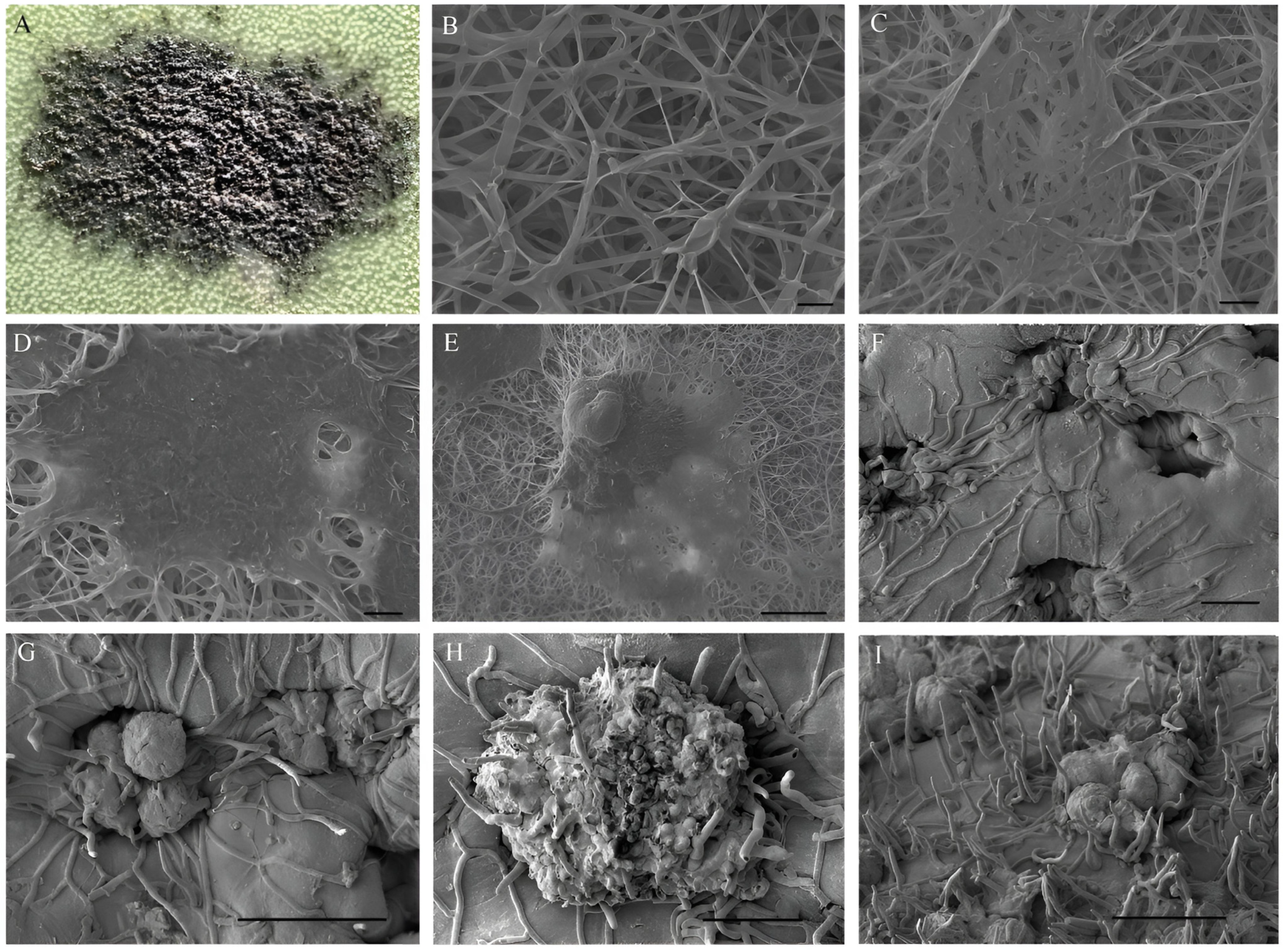
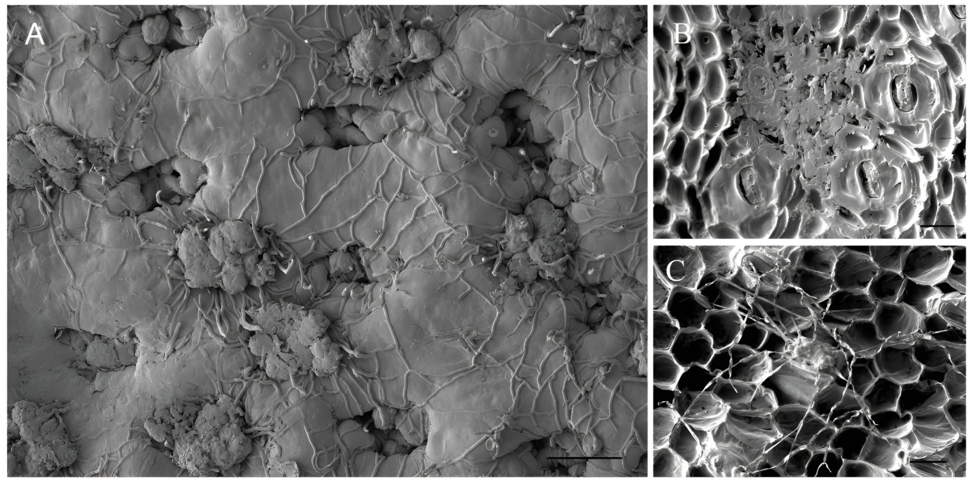
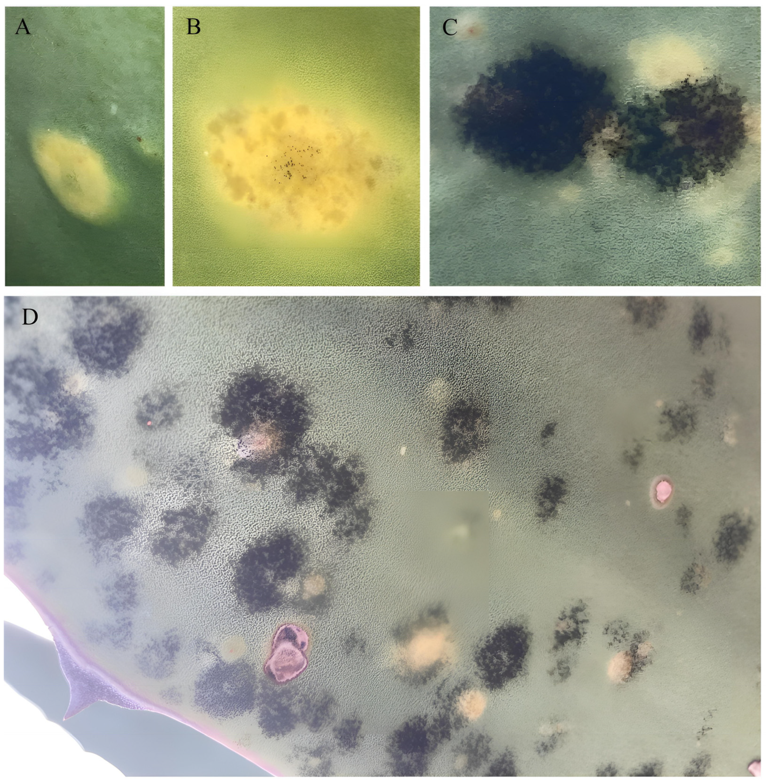
3.3. Bipolaris zeae Pathogenicity Tests

Once the B. zeae strain was isolated and characterized, an aqueous solution containing a high concentration of this strain (5 × 10⁶ conidia/mL) was prepared and sprayed onto Agave plants. Ninety days after the application of conidia suspension, all inoculated plants (T2) presented disease symptoms, showing foliar intensity damage of 10.92 ± 0.12a, considered as severe levels. The T1 plants did not present any disease symptoms in this period. The low severity levels observed in T2 plants may be explained by the chemical and structural composition of the maguey leaves, in relation to the quantity of epidermal cells, cutin, polysaccharides, waxes, and stomata present [12,69,70]. Figure 7D shows that all tissues of maguey leaves are invaded after a certain time, even though it takes a longer time for the B. zeae hyphae to colonize and invade the cuticle layer of maguey plants.
To confirm that the leaf spot symptoms observed in the A. salmiana seedlings were caused by the B. zeae conidia, samples of the infected tissues were taken and incubated in PDA medium, obtaining B. zeae colonies with the same morphological characteristics described above (Figure 8A). On the other hand, 20 days after B. zeae conidia inoculation, hyphae in different stages of development were observed emerging from the stomata on the maguey leaves of T2 plants (Figure 8B). Finally, it was possible to observe a completely developed ascomata with the presence of several formed conidiophores, around 40 to 50 days after inoculation (Figure 8C).
The species B. zeae were morphologically identified and molecularly characterized to corroborate, at a molecular level, the correct identity of the fungus isolated from the infected maguey plants in the pathogenicity test. The results of the pairwise alignment of sequences, with the Blast program, gave a percentage of identity of 100% with the accession ON630344 corresponding to the JCP-N07 strain of B. zeae (Table 1), which was used in this research to carry out the pathogenicity experiments. Additionally, this sequence was published in the GenBank database with OR553122 as the accession number. This pathogeny test and the molecular characterization confirm that B. zeae is the etiological agent of leaf disease symptoms observed in the evaluated maguey plants. In this regard, other species of the Bipolaris genera such as B. maydis and B. zeicola cause similar symptoms in corn crops [71] and Curvularia verruculosa and C. lunata in A. lechuguilla plants [72,73].

4. Conclusions

Agave salmiana has high economic and cultural importance in Mexico. Pathogens are an area of interest for Agave producers and researchers due to the losses they cause in Agave plantations and production. Agave pathogens tend to spread to other crops, since maguey plantations are usually among forage plants, grain, and cereal crops, and for this reason, pathogens are studied extensively. The genera Bipolaris have a cosmopolitan distribution, frequently found in the leaves, stems, and roots of different grasses. The results presented in this paper confirm that the fungus Bipolaris zeae participates in leaf spot disease (colloquially called “Negrilla”) observed on the leaves of Agave salmiana plants. It is expected that these findings could provide more insight for creating practice strategies (cultural practice, chemical control, or biological agents) to control pathogens propagation among crops in the region. Also, further study into other pathogens related to this disease, such as Curvularia lunata and C. verruculosa, is recommended.

Author Contributions

Conceptualization, J.A.C.-P. and T.R.-C.; methodology, J.A.C.-P.; software, J.A.C.-P.; validation, J.A.C.-P., V.H.P.E., J.A.P.-R., E.R.-C., M.M.A.-O. and T.R.-C.; formal analysis, J.A.C.-P.; investigation, J.A.C.-P., V.H.P.E. and T.R.-C.; resources, J.A.C.-P., V.H.P.E., J.A.P.-R., E.R.-C. and T.R.-C.; data curation, J.A.C.-P. and T.R.-C.; writing—original draft preparation, J.A.C.-P.; writing—review and editing, J.A.C.-P., V.H.P.E., J.A.P.-R., E.R.-C., M.M.A.-O. and T.R.-C.; visualization and supervision, J.A.C.-P. and T.R.-C.; project administration, J.A.C.-P. and T.R.-C. All authors have read and agreed to the published version of the manuscript.

Funding

This research received no external funding.

Data Availability Statement

Data are contained within the article.

Acknowledgments

All the authors thank the staff of the Escuela Superior de Apan-UAEH for having provided all the facilities required to carry out the field and laboratory experiments. We also appreciate the support of J.J. Pelayo-Cárdenas for his help checking and improving the English language of the final manuscript.

Conflicts of Interest

The authors declare no conflicts of interest and the funders had no role in the design of the study; in the collection, analysis, or interpretation of data; in the writing of the manuscript; or in the decision to publish the results.

References

  1. Sivanesan, A. Graminicolous Species of Bipolaris, Curvularia, Drechslera, Exserohilum and Their Teleomorphs, 10th ed.; No. 158; CAB International Mycological Institute: Wallingford, UK, 1987; pp. 10–267. [Google Scholar]
  2. Cardona, R.; González, M.S. Caracterización y patogenicidad de hongos del complejo Helminthosporium asociados al cultivo del arroz en Venezuela. Bioagro 2008, 20, 141–145. [Google Scholar]
  3. Shoemaker, R.A. Nomenclature of Drechslera and Bipolaris, grass parasites segregated from ‘Helminthosporium’. Canad. J. Bot. 1959, 37, 879–887. [Google Scholar] [CrossRef]
  4. Drechsler, C. Phytopathological and taxonomical aspects of Ophilobolus, Pyrenophora, Helminthosporium and a new genus Cochliobolus. Phytopathological 1934, 24, 973. [Google Scholar]
  5. Manamgoda, D.S.; Cai, L.; McKenzie, E.H.C.; Crous, P.W.; Madrid, H.; Chukeatirote, E.; Shivas, R.G.; Tan, Y.P.; Hyde, K.D. A phylogenetic and taxonomic re-evaluation of the Bipolaris–Cochliobolus—Curvularia Complex. Fungal Divers. 2012, 56, 131–144. [Google Scholar] [CrossRef]
  6. Manamgoda, D.S.; Rossman, A.Y.; Castlebury, L.A.; Crous, P.W.; Madrid, H.; Chukeatirote, E.; Hyde, K.D. The genus Bipolaris. Stud. Micol. 2014, 79, 221–288. [Google Scholar] [CrossRef] [PubMed]
  7. Zhang, Y.; Crous, P.W.; Schoch, C.L.; Hyde, K.D. Pleosporales. Fungal Divers. 2012, 53, 1–221. [Google Scholar] [CrossRef] [PubMed]
  8. Berbee, M.L.; Pirseyedi, M.; Hubbard, S. Cochliobolus phylogenetics and the origin of known, highly virulent pathogens, inferred from ITS and glyceraldehyde-3-phosphate dehydrogenase gene sequences. Mycologia 1999, 91, 964–977. [Google Scholar] [CrossRef]
  9. Castellanos-González, L.; De Mello-Prado, R.; Silva-Campos, C.N.; Barbosada-Silva, J.G. Desarrollo de la mancha foliar por Bipolaris maydis (teleomorfo: Cochliobolus heterostrophus) en maíz dulce en función de nitrógeno, potasio y silicio en invernadero. Cienc. Tecnol. Agropecuaria 2020, 21, e1508. [Google Scholar] [CrossRef]
  10. Romero-Cortes, T.; Zavala-González, E.A.; Pérez, E.V.H.; Aparicio-Burgos, J.E.; Cuervo-Parra, J.A. Characterization of Cochliobolus sativus and Pyrenophora teres fungi belonging to the leaf spot complex of barley (Hordeum vulgare) isolated from barley seeds in Mexico. Chilean J. Agric. Sci. 2021, 37, 277–289. [Google Scholar] [CrossRef]
  11. Chen, T.X.; Qi, Y.J.; Wang, L.H.; Li, C.J. First report of leaf spot disease on Fagopyrum esculentum caused by Bipolaris zeae in China. Plant Dis. 2021, 10, 3301. [Google Scholar] [CrossRef]
  12. Pérez-España, V.H.; Cuervo-Parra, J.A.; Aparicio, B.J.E.; Morales-Ovando, M.A.; Peralta, G.M.; Romero-Cortes, T. Importancia de la capa cuticular durante la colonización del hongo causante de la negrilla en Agave salmiana Otto ex Salm-Dyck ssp. salmiana. Rev. Mex. Cienc. For. 2022, 13, 158–168. [Google Scholar] [CrossRef]
  13. Mata-Santoyo, C.I.; Leyva-Mir, S.G.; Camacho-Tapia, M.; Tovar-Pedraza, J.M.; Huerta-Espino, J.; Villaseñor-Mir, H.E.; García-León, E. Aggressiveness of Bipolaris sorokiniana and Alternaria alternata isolates on wheat cultivars in Mexico. Rev. Mex. Fitopatol. 2018, 36, 432–443. [Google Scholar] [CrossRef]
  14. Romero-Cortes, T.; Tamayo-Rivera, L.; Morales-Ovando, M.A.; Aparicio-Burgos, J.E.; Pérez-España, V.H.; Peralta-Gil, M.; Cuervo-Parra, J.A. Growth and yield of purple Kculli corn plants under different fertilization schemes. J. Fungi 2022, 8, 433. [Google Scholar] [CrossRef] [PubMed]
  15. Bockus, W.W.; Bowden, R.L.; Hunger, R.M.; Morrill, W.L.; Murray, T.D.; Smiley, R.W. Diseases caused by fungi and fungus-like organisms. In Compendium of Wheat Diseases and Pests, 3rd ed.; The American Phytopatological Society: Saint Paul, MN, USA, 2010; pp. 15–86. [Google Scholar] [CrossRef]
  16. Manamgoda, D.S.; Cai, L.; Bahkali, A.H.; Chukeatirote, E.; Hyde, K.D. Cochliobolus: An overview and current status of species. Fungal Divers. 2011, 51, 3–42. [Google Scholar] [CrossRef]
  17. Kobayashi, H.; Sano, A.; Aragane, N.; Fukuoka, M.; Tanaka, M.; Kawaura, F.; Fukuno, Y.; Matsuishi, E.; Hayashi, S. Disseminated infection by Bipolaris spicifera in an immunocompetent subject. Med. Mycol. 2008, 46, 361–365. [Google Scholar] [CrossRef] [PubMed]
  18. El Khizzi, N.; Bakheshwain, S.; Parvez, S. Bipolaris: A plant pathogen causing human infections: An emerging problem in Saudi Arabia. Res. J. Microbiol. 2010, 5, 212–217. [Google Scholar] [CrossRef]
  19. Piontelli, L.E. Opportunistic species of clinical relevance in the Bipolaris Shoemaker & Curvularia Boedijn genera: Its characterization under newest taxonomic criteria. Taxonomía 2015, 30, 40–63. [Google Scholar]
  20. Carballo, C.M.; Matthews, S.; Spesso, M.F.; Consigli, C. Primer aislamiento en la Argentina de Bipolaris cynodontis en la uña. Dermatol. Argen. 2018, 24, 133–135. [Google Scholar]
  21. Guarner, J.; Brandt, E.M. Histopathologic diagnosis of fungal infections in the 21st century. Clin. Microbiol. Rev. 2011, 24, 247–280. [Google Scholar] [CrossRef]
  22. Tan, Y.P.; Crous, P.W.; Shivas, R.G. Eight novel Bipolaris species identified from John L. Alcorn’s collections at the Queensland Plant Pathology Herbarium (BRIP). Mycol. Prog. 2016, 15, 1203–1214. [Google Scholar] [CrossRef]
  23. da Cunha, K.C.; Sutton, D.A.; Fothergill, A.W.; Cano, J.; Gené, J.; Madrid, H.; De Hoog, S.; Crous, P.W.; Guarro, J. Diversity of Bipolaris species in clinical samples in the United States and their antifungal susceptibility profiles. J. Clin. Microbiol. 2012, 50, 4061–4066. [Google Scholar] [CrossRef] [PubMed]
  24. Madrid, H.; da Cunha, K.C.; Gené, J.; Dijksterhuis, J.; Cano, J.; Sutton, D.A.; Guarro, J.; Crous, P.W. Novel Curvularia species from clinical specimens. Persoonia 2014, 33, 48–60. [Google Scholar] [CrossRef] [PubMed]
  25. Kashyap, A.S.; Manzar, N.; Maurya, A.; Mishra, D.D.; Singh, R.P.; Sharma, P.K. Development of Diagnostic Markers and Applied for Genetic Diversity Study and Population Structure of Bipolaris sorokiniana Associated with Leaf Blight Complex of Wheat. J. Fungi 2023, 9, 153. [Google Scholar] [CrossRef] [PubMed]
  26. Manzar, N.; Kashyap, A.S.; Maurya, A.; Rajawat, M.V.S.; Sharma, P.K.; Srivastava, A.K.; Roy, M.; Saxena, A.K.; Singh, H.V. Multi-Gene Phylogenetic Approach for Identification and Diversity Analysis of Bipolaris maydis and Curvularia lunata Isolates Causing Foliar Blight of Zea mays. J. Fungi 2022, 8, 802. [Google Scholar] [CrossRef] [PubMed]
  27. Mereles, G.L. Calidad Fisiológica, Identificación de Hongos y su Incidencia en la Pudrición de la Mazorca de Maíz Zea mays L. en el Municipio de Tepalcing., Morelos. Bachelor’s Thesis, Universidad Autónoma Agraria Antonio Narro, Coahuila, México, December 2018. [Google Scholar]
  28. Leyva-Mir, S.G.; Villaseñor-Mir, H.E.; Tovar-Pedraza, J.M.; García-León, E. Respuesta de genotipos de avena a la infección por Bipolaris victoriae y Bipolaris sorokiniana. Rev. Mex. Cienc. Agric. 2019, 10, 1023–1034. [Google Scholar] [CrossRef]
  29. Rodríguez-Pedroso, A.T.; Plascencia-Jatomea, M.; Bautista-Baños, S.; Ventura-Zapata, E.; Cortez-Rocha, M.O.; Ramírez-Arrebato, M.A. Efecto in vitro de un quitosano de masa molecular media sobre dos cepas de Bipolaris oryzae aisladas en México y Cuba. Biotecnol. Apl. 2021, 38, 2201–2205. [Google Scholar]
  30. Sivanesan, A. New Bipolaris, Curvularia, and Exserohilum species. Mycol. Res. 1992, 6, 485–489. [Google Scholar] [CrossRef]
  31. Hernández, J.J.; Hernández, D.E.M.; López, V.E.; Álvarez, C.J. Aislamiento e identificación del fitopatógeno causal de viruela o “negrilla” en Agave salmiana de municipios del estado de Hidalgo, México. Sci. Fungorum 2022, 53, e1425. [Google Scholar] [CrossRef]
  32. Ramírez-Cariño, H.F.; Guadarrama-Mendoza, P.C.; Sánchez-López, V.; Cuervo-Parra, J.A.; Ramírez-Reyes, T.I.; Valadez-Blanco, R. Biocontrol of Alternaria alternata and Fusarium oxysporum by Trichoderma asperelloides and Bacillus licheniformis in tomato plants. Anton. Leeuw. Int. J G. 2020, 113, 1247–1261. [Google Scholar] [CrossRef]
  33. Marin-Felix, Y.; Groenewald, J.Z.; Cai, L.; Chen, Q.; Marincowitz, S.; Barnes, I.; Bensch, K.; Braun, U.; Camporesi, E.; Damm, U.; et al. Genera of phytopathogenic fungi: GOPHY 1. Stud. Mycol. 2017, 86, 99–216. [Google Scholar] [CrossRef]
  34. Cuervo-Parra, J.A.; Sánchez-López, V.; Romero-Cortes, T.; Ramírez-Lepe, M. Hypocrea/Trichoderma viridescens ITV43 with potential for biocontrol of Moniliophthora roreri Cif & Par, Phytophthora megasperma and Phytophthora capsici. Afr. J. Microbiol. Res. 2014, 8, 1704–1712. [Google Scholar] [CrossRef]
  35. Romero-Cortes, T.; López-Pérez, P.A.; Pérez, E.V.H.; Medina-Toledo, A.K.; Aparicio-Burgos, J.E.; Cuervo-Parra, J.A. Confrontation of Trichoderma asperellum VSL80 against Aspergillus niger via the effect of enzymatic production. Chil. J. Agric. Sci. 2019, 35, 68–80. [Google Scholar] [CrossRef]
  36. Kendall, J.M.; Rygiewicz, P.T. Fungal-specific PCR primers developed for analysis of the ITS region of environmental DNA extracts. BMC Microbiol. 2005, 5, 28. [Google Scholar] [CrossRef]
  37. Zhang, Z.; Schwartz, S.; Wagner, L.; Miller, W. A greedy algorithm for aligning DNA sequences. J. Comput. Biol. 2000, 7, 203–214. [Google Scholar] [CrossRef] [PubMed]
  38. Kumar, S.; Stecher, G.; Li, M.; Knyaz, C.; Tamura, K. MEGA X: Molecular evolutionary genetics analysis across computing platforms. Mol. Biol. Evol. 2018, 35, 1547–1549. [Google Scholar] [CrossRef] [PubMed]
  39. Reveles, H.R.G.; Maldonado, T.C.; Chávez, R.I.; Muñoz, E.J.J.; Moreno, G.A. Vigencia de los Postulados de Koch en la reproducción de Trichinella spiralis en modelo experimental. Rev. Electrón. Vet. 2014, 15, 1–20. [Google Scholar]
  40. Vázquez, D.E.; García, N.J.R.; Peña, V.C.B.; Ramírez, T.H.M.; Morales, R.V. Tamaño de la semilla, emergencia y Desarrollo de la plántula de maguey (Agave salmiana Otto ex Salm-Dyck). Rev. Fitotec. Mex. 2011, 34, 167–173. [Google Scholar] [CrossRef]
  41. Perelló, A.; Sisterna, M.N.; Cortese, P. Scale for appraising the leaf blight of wheat caused by Alternaria triticimaculans. Cereal Res. Commun. 1998, 26, 189–194. [Google Scholar] [CrossRef]
  42. Analytical Software, Statistix 10, 1st ed.; Analytical Software: Tallahassee, FL, USA, 2017; pp. 7–445.
  43. Sisterna, M.; Wolcan, S. Bipolaris zeae on Pennisetum clandestinum in Argentina. Summa Phytopathol. 1990, 16, 184–188. [Google Scholar]
  44. Nazareno, d.A.J.R.; d’Avila, C.M.J.; Michalski, M.V.; Rodrigues, R.A.; Santos, S.M.; Saraiva, N.A.S. Transmissao e Patogenicidade de duas Espécies de Bipolaris Associadas as Sementes de Panicum Maximum Jacq, 1st ed.; Embrapa Cerrados: Brasilia, Brazil, 2009; p. 16. [Google Scholar]
  45. Xue, L.H.; Liu, Y.; Wu, W.X.; Liang, J.; Zhang, L.; Huang, X.Q.; Zhou, X.Q.; Johnson, R.D.; Li, C.J.; Wang, J.J.; et al. First Report of Leaf Spot of Hemarthria altissima Caused by Bipolaris zeae in China. Plant Dis. 2016, 101, 243. [Google Scholar] [CrossRef]
  46. Minnis, M.A.; Rossman, Y.A.; Kleczewski, M.N.; Flory, L.S. Bipolaris microstegii Minnis, Rossman, Kleczewski & S.L. Flory, sp. nov. Persoonia 2012, 29, 150–151. [Google Scholar]
  47. Sun, X.; Qi, X.; Wang, W.; Liu, X.; Zhao, H.; Wu, C.; Chang, X.; Zhang, M.; Chen, H.; Guoshu, G. Etiology and Symptoms of Maize Leaf Spot Caused by Bipolaris spp. in Sichuan, China. Pathogens 2020, 9, 229. [Google Scholar] [CrossRef] [PubMed]
  48. Campos-Rivero, G.; Sánchez-Teyes, L.F.; De la Cruz-Arguijo, E.; Ramírez-González, M.S.; Larralde-Corona, C.P.; Narvaez-Zapata, J.A. Bioprospecting for Fungi with Potential Pathogenic Activity on Leaves of Agave tequilana Weber var. Azul. GenBank Direct Submission, 11 October 2018. Available online: https://www.ncbi.nlm.nih.gov/nucleotide/MK041914.1 (accessed on 1 March 2024).
  49. Valencia, Y.T.; Martin, M.R.; Cruz, L.J.R.; Pérez, B.D.; Magaña, A.A.; Cortés, V.A.; Nexticapan, G.A. Identification and Molecular Characterization Phytopathogenic Fungi in Agave tequilana in Mexico. GenBank Direct Submission, 2 August 2023. Available online: https://www.ncbi.nlm.nih.gov/nucleotide/OR388091.1 (accessed on 1 March 2024).
  50. Mora-Aguilera, J.A.; Cabrera-Huerta, E.; Nava-Díaz, C.; Hernández-Castro, E. First report of Epicoccum sorghinum on Agave Mezcal (Agave angustifolia). GenBank Direct Submission, 19 June 2018. Available online: https://www.ncbi.nlm.nih.gov/nucleotide/MH497563.1 (accessed on 1 March 2024).
  51. Cuervo-Parra, J.A.; Romero-Cortes, T.; Aparicio-Burgos, J.E. Isolation and Molecular Identification of Phytopathogenic Fungi, Associated with Agave salmiana and Agave lechuguilla Plants with Disease Symptoms “Negrilla”, in the State of Hidalgo, Mexico. GenBank Direct Submission, 25 May 2023. Available online: https://www.ncbi.nlm.nih.gov/nucleotide/OR047544.1 (accessed on 1 March 2024).
  52. Campos-Rivero, G.; Sánchez-Teyes, L.F.; De la Cruz-Arguijo, E.; Ramírez-González, M.S.; Larralde-Corona, C.P.; Narvaez-Zapata, J.A. Bioprospecting for Fungi with Potential Pathogenic Activity on Leaves of Agave tequilana Weber var. Azul. GenBank Direct Submission, 11 October 2018. Available online: https://www.ncbi.nlm.nih.gov/nucleotide/MK041894.1 (accessed on 1 March 2024).
  53. Cuervo-Parra, J.A.; Romero-Cortes, T.; Aparicio-Burgos, J.E. Isolation and Molecular Identification of Phytopathogenic Fungi, Associated with Agave salmiana and Agave lechuguilla Plants with Disease Symptoms “Negrilla”, in the State of Hidalgo, Mexico. GenBank Direct Submission, 25 May 2023. Available online: https://www.ncbi.nlm.nih.gov/nucleotide/OR047545.1 (accessed on 1 March 2024).
  54. Flores-Nuñez, N.M.; Camarena-Pozos, D.A.; Partida-Martínez, L.P. Volatiles Emitted by Fungi Associated with the Phyllosphere of Agaves and Cacti are Diverse and Able to Promote Plant Growth. GenBank Direct Submission, 3 April 2020. Available online: https://www.ncbi.nlm.nih.gov/nucleotide/MT279265.1 (accessed on 1 March 2024).
  55. Cuervo-Parra, J.A.; Romero-Cortes, T.; Aparicio-Burgos, J.E. Isolation and Molecular Identification of Phytopathogenic Fungi, Associated with Agave salmiana and Agave lechuguilla Plants with Disease Symptoms “Negrilla”, in the State of Hidalgo, Mexico. GenBank Direct Submission, 25 May 2023. Available online: https://www.ncbi.nlm.nih.gov/nucleotide/OR047542.1 (accessed on 1 March 2024).
  56. Tapia-Tussell, R.; Solis, S.; Pérez-Brito, D. Identification and Molecular Characterization of Ligninolytic Fungi from Yucatan, Mexico. GenBank Direct Submission, 17 June 2009. Available online: https://www.ncbi.nlm.nih.gov/nucleotide/GQ280375.1 (accessed on 1 March 2024).
  57. Cuervo-Parra, J.A.; Romero-Cortes, T.; Aparicio-Burgos, J.E. Isolation and Molecular Identification of Phytopathogenic Fungi, Associated with Agave salmiana and Agave lechuguilla Plants with Disease Symptoms “Negrilla”, in the State of Hidalgo, Mexico. GenBank Direct Submission, 25 May 2023. Available online: https://www.ncbi.nlm.nih.gov/nucleotide/OR047532.1 (accessed on 1 March 2024).
  58. Mohammadi, A.; Amini, Y. Molecular characterization and identification of Acrostalagmus luteoalbus from Saffron in Iran. Agric. Sci. Dev. 2015, 4, 16–18. [Google Scholar]
  59. Toju, H.; Tanabe, A.S.; Yamamoto, S.; Sato, H. High-coverage ITS primers for the DNA-based identification of ascomycetes and basidiomycetes in environmental samples. PLoS ONE 2012, 7, e40863. [Google Scholar] [CrossRef] [PubMed]
  60. Morgulis, A.; Coulouris, G.; Raytselis, Y.; Madden, T.L.; Agarwala, R.; Schäffer, A.A. Database indexing for production MegaBLAST searches. Bioinformatics 2008, 24, 1757–1764. [Google Scholar] [CrossRef] [PubMed]
  61. Visagie, C.M.; Bhiya, T.; Kgatle, G. Alternaria Diversity from South Africa. GenBank Direct Submission. 22 May 2020. Available online: https://www.ncbi.nlm.nih.gov/nucleotide/MT505870.1 (accessed on 10 June 2022).
  62. Vu, D.; Groenewald, M.; de Vries, M.; Gehrmann, T.; Stielow, B.; Eberhardt, U.; Al-Hatmi, A.; Groenewald, J.Z.; Cardinali, G.; Houbraken, J.; et al. Large-scale generation and analysis of filamentous fungal DNA barcodes boosts coverage for kingdom fungi and reveals thresholds for fungal species and higher taxon delimitation. Stud. Mycol. 2019, 92, 135–154. [Google Scholar] [CrossRef] [PubMed]
  63. Berbee, M.L. Molecular Phylogenetics of Cochliobolus. GenBank Direct Submission, 30 July 1998. Available online: https://www.ncbi.nlm.nih.gov/nuccore/af081452 (accessed on 13 June 2022).
  64. Bohl, S.K.; Harmon, P.F.; Clay, K.; Goss, E.M.; Flory, S.L. Widespread Independent Emergence and Accumulation of Fungal pathogens Reduce Fecundity of an Invasive Grass. GenBank Direct Submission, Submitted 10 August 2014. Available online: https://www.ncbi.nlm.nih.gov/nuccore/km507725 (accessed on 13 June 2022).
  65. Adetunji, M.C.; Mwanza, M. Fungal Profile of Groundnut sold in Mafikeng, North West, South Africa. GenBank Direct Submission, 26 April 2019. Available online: https://www.ncbi.nlm.nih.gov/nuccore/mk841439 (accessed on 13 June 2022).
  66. Berbee, M.; Pirseyedi, M.; Hubbard, S. GenBank Direct Submission. 10 June 1998. Available online: https://www.ncbi.nlm.nih.gov/nuccore/af071325 (accessed on 13 June 2022).
  67. Berbee, M.; Pirseyedi, M.; Hubbard, S. GenBank Direct Submission. 10 June 1998. Available online: https://www.ncbi.nlm.nih.gov/nuccore/af071329 (accessed on 13 June 2022).
  68. Ahmadpour, A.; Donyadoost-Chelan, M.; Heidarian, Z.; Javan-Nikkhah, M. New species of Bipolaris and Curvularia on grass species in Iran. Rostaniha 2011, 12, 39–49. [Google Scholar]
  69. Staiger, S.; Seufert, P.; Arand, K.; Burghardt, M.; Popp, C.; Riederer, M. The permeation barrier of plant cuticles: Uptake of active ingredients is limited by very long-chain aliphatic rather than cyclic wax compounds. Pest Manag. Sci. 2019, 75, 3405–3412. [Google Scholar] [CrossRef]
  70. Pérez-España, V.H.; Cuervo-Parra, J.A.; Paz-Camacho, C.; Morales-Ovando, M.A.; Gómez-Aldapa, C.A.; Rodríguez-Jiménez, G.C.; Robles-Olvera, V.J.; López-Pérez, P.A.; Romero-Cortes, T. General characterization of cuticular membrane isolated from Agave salmiana. Int. J. Bio-resour. Stress Manag. 2019, 10, 046–052. [Google Scholar] [CrossRef]
  71. Centro Internacional de Mejoramiento de Maíz y Trigo (CIMMYT). Enfermedades del Maíz: Una Guía Para su Identificación en el Campo, 4th ed.; CIMMYT: Ciudad de México, Mexico, 2004; pp. 18–33. [Google Scholar]
  72. Cuervo-Parra, J.A.; Romero-Cortes, T.; Aparicio-Burgos, J.E. Isolation and Molecular Identification of phytopathogenic Fungi, Associated with Agave salmiana and Agave lechuguilla Plants with Disease Symptoms “Negrilla”, in the State of Hidalgo, Mexico. GenBank Direct Submission, 25 May 2023. Available online: https://www.ncbi.nlm.nih.gov/nucleotide/OR047538.1 (accessed on 1 March 2024).
  73. Cuervo-Parra, J.A.; Romero-Cortes, T.; Aparicio-Burgos, J.E. Isolation and Molecular Identification of Phytopathogenic Fungi, Associated with Agave salmiana and Agave lechuguilla Plants with Disease Symptoms “Negrilla”, in the State of Hidalgo, Mexico. GenBank Direct Submission, 25 May 2023. Available online: https://www.ncbi.nlm.nih.gov/nucleotide/OR047536.1 (accessed on 1 March 2024).
Figure 1. Bipolaris zeae morphology in PDA and Sabouraud medium. (A,B) Pustules showing morphological and color variations in PDA after 7 days, as well as growth ranging from rosette to radial. (C,D) Few pustules grew on the mycelium with a cracked appearance due to the great abundance of folds observed in SDA medium. Reverse colony morphology (E) in PDA and (F) in SDA culture media.
Agronomy 14 00623 g001
Figure 2. Bipolaris zeae morphology in plant and PDA medium. (A,B) SEM ascomata and several conidiophores growing on the maguey leaf. (CE) Conidia with 3-7-distosetate, and conidiophore shape in PDA. Scale bars: (A) = 100 μm, (B) = 50 μm, (CE) = 20 μm.
Agronomy 14 00623 g002
Figure 3. Bipolaris zeae colonies on maguey plants. (A,B) SEM ascomata with abundant conidiophores emerging from them, as well as hyphae with conidiophores on the maguey leaf. (CF) SEM conidia and conidiophore morphological structures produced on the maguey leaf. Scale bars: (A,B) = 50 μm, (C) = 5 μm, (D) = 20 μm, (E,F) = 10 μm.
Agronomy 14 00623 g003
Figure 4. Cochliobolus zeae ascomata morphology in Agave salmiana (A,FI) and in PDA medium (BE). Macroscopic morphology of the leaf spot disease observed in maguey leaves (A). Set of hyphae with the presence of septa (B). Intertwined hyphae begin to form prosenchyma (C). Hyphae that have lost their individuality give rise to pseudoparenchyma (D). Pseudoparenchyma in the ascomata process forming (E). Hyphae enter the interior of the cuticle via stomata ostioles (F). Ascomata emerging from the stoma (G). Fully formed ascomata with hyphae growing on the left (H). Groups of ascomata with abundant hyphae and conidiophores growing on the maguey leaf (I). Scale bars: (B) = 10 µm; (C,D) = 20 µm; (F,H) = 50 µm; (E,G,I) = 100 µm.
Agronomy 14 00623 g004
Figure 5. Cochliobolus colonies grown on Agave salmiana plant observed in SEM. (A) Ascomata with abundant hyphae and conidiophores emerge from the stomas on the adaxial side of the maguey leaf. (B) Mycelium growing through the stomas by the adaxial side of the cuticular layer from the maguey leaf. (C) Mycelium growing inside the internal parts of the stroma of the maguey leaf without the cuticular layer. Scale bars: (A) = 100 μm, (B) = 50 μm, (C) = 50 μm.
Agronomy 14 00623 g005
Figure 6. Phylogenetic analysis generated for Bipolaris from a Neighbor-joining analysis based on the ITS alignment. The numbers in the nodes are the bootstrap values.
Agronomy 14 00623 g006
Figure 7. Leaf spots observed on the leaves of maguey plants. Early leaf spot symptoms on maguey leaves (A). Leaf spot observable symptoms in maguey leaves from 30 to 35 days of inoculation (B). Leaf spot developed on the Agave leave two months after starting the inoculation (C). Adult maguey leaf with B. zeae colonies, 5 months after the onset of the first symptoms (D).
Agronomy 14 00623 g007
Figure 8. Leaf spot caused by Bipolaris zeae in plant and PDA medium. (A) B. zeae morphological characteristics observed in PDA after 7 days of growth. (B,C) Micrographs of ascomata in different development stages and conidiophores growing on the maguey leaf cuticle. Scale bars: (B) = 100 μm, (C) = 50 μm.
Agronomy 14 00623 g008
Table 1. Fungal identity based on sequencing of the ITS I/5.8s/ITS II region of rDNA.
StrainStrain AccessionSpeciesTotal ScoreQuery Cover (%)Identity (%)Accesion Closest HitAccession Length (pb)References
JCP-N01ON630338B. zeae100310099.64MT505870863Visagie et al. [61]
JCP-N02ON630339B. zeae98510099.63MT505870863Visagie et al. [61]
JCP-N03ON630340B. zeae97610099.63MT505870863Visagie et al. [61]
JCP-N04ON630341B. zeae97610099.44MT505870863Visagie et al. [61]
JCP-N05ON630342B. zeae95910099.62MT505870863Visagie et al. [61]
JCP-N06ON630343B. zeae98510099.63MT505870863Visagie et al. [61]
JCP-N07ON630344B. zeae101110099.64MT505870863Visagie et al. [61]
JCP-N08ON630345B. zeae96810099.62KJ909787568Manamgoda et al. [6]
Disclaimer/Publisher’s Note: The statements, opinions and data contained in all publications are solely those of the individual author(s) and contributor(s) and not of MDPI and/or the editor(s). MDPI and/or the editor(s) disclaim responsibility for any injury to people or property resulting from any ideas, methods, instructions or products referred to in the content.

