Article

Glomus mongioiense, a New Species of Arbuscular Mycorrhizal Fungi from Italian Alps and the Phylogeny-Spoiling Issue of Ribosomal Variants in the Glomus Genus

1
Institute of Biology, Biotechnology and Environmental Protection, Faculty of Natural Sciences, University of Silesia in Katowice, 40-032 Katowice, Poland
2
Technical Institute of Bakrajo, Sulaimani Polytechnic University, Sulaymaniyah 46001, Iraq
3
Institute of Agronomy, Hungarian University of Agriculture and Life Sciences, 2100 Gödöllő, Hungary
4
Departamento de Botânica e Zoologia, Universidade Federal do Rio Grande do Norte, Campus Universitário, Natal 59072-970, RN, Brazil
5
Mycorrhizal Applications LLC at Bio-Research & Development Growth Park, St. Louis, MO 36132, USA
*
Author to whom correspondence should be addressed.
These authors contributed equally to this work.
Agronomy 2024, 14(7), 1350; https://doi.org/10.3390/agronomy14071350
Submission received: 4 April 2024 / Revised: 12 June 2024 / Accepted: 18 June 2024 / Published: 21 June 2024

Abstract

:
Glomus mongioiense, a new species of arbuscular mycorrhizal fungi (AMF) in the family Glomeraceae, was isolated from rhizosphere soil collected from a meadow in the Italian Alps. The novelty of the species and its relationship with other species of the same genus were obtained by morphological and phylogenetic (45S nrDNA + RPB1 gene) analyses. Two glomoid spore-producing AMF isolates from a saltmarsh of the Scottish Highlands and maritime sand dunes of the Baltic Sea in Poland, were also included in this study and later found to be conspecific with G. rugosae. Phylogenetic placement analysis using environmental sequences indicated that G. mongioiense sp. nov. seems to be a rare species. Furthermore, the molecular and phylogenetic analysis provided important insights into the presence of highly divergent ribosomal variants in several Glomus species, with potential negative implication in phylogeny and species recognition.

1. Introduction

Arbuscular mycorrhizal fungi (AMF) are key components of the soil microbiome. They live in close relationship with the vast majority of plant species in forest or agricultural systems, benefiting them by increasing water and nutrient absorption and mitigating biotic and abiotic stress. They also enhance soil structure and stability by stimulating root expansion and developing a hyphal network that contributes to the formation of stable macroaggregates, thereby decreasing soil erosion [1,2]. Additionally, AMF hyphae are able to recruit specific microbial populations with multiple plant growth promoting traits such as phosphate solubilization, nitrogen fixation, plant growth regulation and biocontrol activity, increasing the functional diversity and richness of rhizosphere microorganisms [3,4].
Because of the benefits they provide, AMF have key ecological and agronomic roles, in fact several species within the Glomeraceae family are frequently used in commercial inoculants [5]. However, the richness of species that make up this group is still poorly understood, indeed there still exists a great disparity between the number of potential species detected by environmental sequences and the number of species formally described. Currently, around 360 AMF species have been described, distributed in 49 genera and 17 families [6,7,8].
The description in 1885 of Glomus macrocarpum Tul. & C. Tul. and G. microcarpum Tul. & C. Tul. by [9] marked the recognition of the first fungal genus within the Glomeromycota, despite both species were being later transferred to the genus Endogone [10]. Glomus was reinstated by [11] with the revision of the family Endogonaceae, entailing the reestablishment of the original species (G. macrocarpum and G. microcarpum), the description of new Glomus species, and the transfer of other species from Endogone to Glomus. Subsequently, for at least four decades, all species with glomoid-type spores (spores produced terminally, sub-terminally or intercalary from subtending hyphae) were included in Glomus. Until 2010, Glomus encompassed nearly 50% of the described richness within Glomeromycota. However, phylogenetic analysis has revealed that the genus was polyphyletic, and several Glomus species were reorganized into other taxonomic categories from genus to class level [7,12,13,14,15,16].
The phylogeny of Glomus has been limited for many years by the absence of the holotype and molecular data for the type species. In fact, G. macrocarpum was lectotypified by [17] only in 1983, while in 2010 [12] established a molecular epitype of G. macrocarpum and defined it as the only species in Glomus sensu stricto. The other species originally recognized as Glomus were therefore categorized as Glomus sensu lato due to their uncertain or unknown phylogenetic data. Ref. [15] described G. tetrastratosum Błaszk., Chwat & Góralska based on its phylogenetic proximity to G. macrocarpum, and five other new species (G. bareae Błaszk., Niezgoda, B.T. Goto & Kozłowska, G. ibericum A. Guillén, F.J. Serrano-Tamay, J.B. Peris & I. Arrillaga, G. atlanticum Błaszk., Niezgoda, B.T. Goto, Moreira & Magurno, G. chinense F.X. Yu, B.T. Goto, H.Y. Feng & Yong Jun Liu, and G. rugosae Błaszk., B.T. Goto & Niezgoda) were recently described [18,19,20,21,22], expanding the Glomus sensu stricto clade. Morphologically, this clade is represented by glomoid species occurring in loose clusters or, rarely, in hypogeous or epigeous unorganized glomerocarps, forming a single spore wall with two to four layers.
During investigations of AMF assemblages associated with plants growing on the European Alps, in a Scottish saltmarsh area, and on Baltic sandy dunes, we obtained single-spore cultures of three glomoid fungi, thereafter recognized as members of the genus Glomus. One species is new to science, here described under the name G. mongioiense sp. nov., based on the morphology of the spores and the phylogenetic analysis of 45S nrDNA and RPB1 genes. The other two isolates (namely G. sp. Highlander and G. sp. Hel) were found to be conspecific with the recently described G. rugosae.
Moreover, the analyses performed showed the presence of two highly divergent ribosomal variants in the newly described G. mongioiense, as well as in G. rugosae, G. macrocarpum, G. chinense, G. ibericum and G. tetrastratosum, with potential negative implications in phylogeny and species recognition.

2. Materials and Methods

2.1. Sampling and Single Species Pot Cultures

The field inoculum containing Glomus mongioie was collected on the foothills of Monte Mongioie in the Ligurian Alps, Italy (44°09′34.7″ N 7°46′29.2″ E), characterized by rich mesophilous grasslands and calcitic-dolomitic soil. The plant species composition included Aquilegia atrata W.D.J.Koch, Gentiana ligustica R.Vilm. & Chopinet, Leontopodium nivale subsp. alpinum (Cass.) Greuter, Nigritella sp., Potentilla sp., Helictotrichon sedenense (DC.) Holub, Aster alpinus L., Pulsatilla alpina (L.) Delarbre, Linum alpinum L., Carex firma Host, Saxifraga caesia L., S. cernua L., Trifolium thalii Vill., Trollius europaeus L., Polygala alpestris Rchb, and Festucetum sp. The site is characterized by a low temperature climate with mean annual values of 3.0 to 5.9 °C and mean annual precipitation of 1001–1500 mm [23]. The samples were collected by F. Magurno on 17 August 2019.
The field inoculum containing Glomus sp. Highlander was collected in the saltmarsh habitat of Beauly Firth (57°30′13.9″ N 4°19′06.6″ W), Scotland, UK, which is a part of the Moray Firth and, together with the Inverness Firth, form the estuarine component of the Moray Basin system. The soils are mixosaline to saline, mainly mineral, and pH is circumneutral [24]. The climate of the area is temperate oceanic, with a mean annual temperature of 9.3 °C. The mean annual rainfall is relatively low (624.4 mm) due to the “rain shadow” effect caused by surrounding mountains [24,25]. The plant communities consist of Zostera angustifolia Rchb., Z. noltii Hornem., Carex recta Boott as well as Juncus gerardi Loisel., Plantago maritima L., Armeria maritima (Mill.) Willd. and Festuca rubra L. [24,26]. The field samples were collected by F. Magurno on 29 October 2018.
Glomus sp. Hel was found to be associated with the roots of Leymus arenarius Hochst. growing on the sand dunes of the Baltic Sea near Jastarnia, Poland (54°42′23.2452″ N 18°40′7.7988″ E) on the Hel Peninsula. The coastal climate of the peninsula is shaped by oceanic and continental influences, with an average annual temperature of about 9 °C, and mean annual rainfall of 750 mm. The soils developed on the dunes consist of unconsolidated or partly consolidated material [27]. The material was collected on 9 July 2021 by K. Bierza from yellow dune communities, representing earlier successional stages of tall-grass perennial swards with the domination of L. arenarius and Ammophila arenaria (L.) Link.
Field-collected mixtures of rhizosphere soils and root fragments collected from the three locations were used for inoculation of pot trap cultures with Plantago lanceolata L. as host plant. After approximately five months, spores and loose clusters were isolated from the substrate and used to establish pure species pot cultures in a mixture of sterile river sand and bentonite clay (9:1, v/v). The cultures were checked at regular intervals of three months to detect the presence of spores.

2.2. Morphological Analysis

Spore clusters and spores were mounted in water, polyvinyl alcohol–lactic acid–glycerol (PVLG), and a mixture of PVLG and Melzer’s reagent (1:1, v/v). Morphological characteristics of spores and their sub-cellular structure were characterized based on at least 50–100 spores, examined and photographed using dissecting and compound microscopes. Color names were derived from [28]. The terminology of spore characters follows [21,29,30,31]. Types of spore wall layers are those defined by [32,33]. The nomenclature of fungi and the authors of fungal names are from the Index Fungorum database at www.indexfungorum.org (accessed on 1 April 2024). The terms “glomerospores” and “glomerocarps” were used for spores and fruit bodies (sporocarps) produced by AM fungi, respectively, as [34,35] proposed. Voucher specimens (holotype and isotype) of the new species were deposited in the UFRN–Fungos herbarium, Brazil.

2.3. Molecular Analysis

Genomic DNA was extracted from single clusters of spores with the DNeasy PowerSoil Pro kit (Qiagen, Hilden, Germany), following the producer’s instructions with the modifications as follows: the first step of vortexing was replaced by crushing the spores with a micropestle inside a vial containing 100 μL of CD1 solution; the kit’s solutions volume was arranged proportionally until the first washing step; and final elution was performed in a 30 μL volume. Amplicons of 45S nrDNA partial genes (thereafter 45S) were obtained by nested PCR with the primer pairs SSUmAf–LSUmAr and SSUmCf–LSUmBr [36] using the Phusion Plus DNA Polymerase (Thermo Fisher Scientific, Waltham, MA, USA) with universal annealing temperature of 60 °C, according to the producer’s instructions. RPB1 amplicons were obtained from G. mongioiense by PCR with the primers RPB1-4F1 in combination with RPB1-5R as in [31] targeting the region from exon 4 to 5, and from Glomus sp. Highlander and Hel with the primers RPB1–3F alfa and RPB1–5R beta [37] targeting the region from exon 3 to 5. PCR amplicons were purified with GeneJET PCR Purification Kit (Thermo Fisher Scientific, Waltham, MA, USA), cloned with CloneJET PCR Cloning Kit (Thermo Fisher Scientific, Waltham, MA, USA) and sequenced at Genomed S.A. (Warsaw, Poland). Both 45S and RPB1 sequences were deposited in GenBank (45S: PP639264–PP639280, PP753776; RPB1: PP661647–PP661653).

2.4. Phylogenetic Analyses

For the phylogenetic accommodation of the three AMF isolates, two datasets of 45S sequences and one dataset of RPB1 partial gene sequences were prepared, representing all the sequenced members of the genus Glomus, upon verification by blastn analysis of the affiliation of the isolates to the genus. Sequences of the two described Complexispora species [37] were included to represent the outgroup in the phylogenetic analysis.
The 45S datasets contained 57 (45Sds1) and 46 (45Sds2) sequences, overlapping the partial 18S–ITS–partial 28S regions. The 45Sds1 dataset differed by the presence of alternative gene variant sequences from Glomus species (G. mongioiense, G. sp. Highlander, G. sp. Hel, G. macrocarpum, G. chinense, G. ibericum and G. tetrastratosum), to test their influence on the stability of species clades in the phylogenetic inference.
The RPB1 dataset contained 30 sequences, the longest spanning from intron 1 to exon 5, the shortest from exon 4 to 5. Exon 1 was not considered due to its relatively short length (ca 30 bp).
The datasets were aligned separately with the online version of MAFFT 7 using the E-INS-i iterative refinement methods (http://mafft.cbrc.jp/alignment/server/). Two concatenated alignments were prepared combining the 45S (ds1 and ds2) and the RPB1 alignments using a custom Python script.
The four alignments (45Sds1, 45Sds2, 45Sds1 + RPB1, 45Sds2 + RPB1) were used as input for the Bayesian inference (BI) and maximum likelihood (ML) phylogenetic inference, performed via CIPRES Science Gateway 3.1 [38]. The 45S alignments were divided into five partitions: 18S, ITS1, 5.8S, ITS2, and 28S. Eight additional RPB1 partitions, representing four exons and four introns, were employed in the concatenated alignment. GTR + I + G nucleotide substitution model was used for each partition in both BI and ML analyses [39]. Four Markov chains were run over one million generations in MrBayes 3.2 [40], sampling every 1000 generations, with a burn-in at 30% sampled trees. The ML phylogenetic tree inference was performed with RAxML-NG 1.0.1 [41], using a maximum likelihood/1000 bootstrapping run, and an ML–estimated proportion of invariable sites and base frequencies. For each alignment, the resulting phylogenetic trees were visualized, merged and edited in TreeGraph 2 [42]. Any discrepancy in the topology between BI and ML trees was recorded. Clades were considered supported with Bayesian posterior probabilities ≥ 0.95 and ML bootstrap values ≥ 70%.
The percentages of identity intraspecies and between the new species and the closest relatives were calculated using the blastn suite-2sequences tool of NCBI.
To detect other possible occurrences of the new species, blastn was used to retrieve uncultured Glomeromycota sequences from GenBank with percentage of identity > 96% with at least one of the 45S sequences of the species of interest. The environmental sequences were aligned with the db1 and their phylogenetic placement was established by RAxML–EPA [43], followed by the placement mass accumulation in GAPPA v0.8.0 [44], with threshold > 0.9, similarly to [45].

3. Results

3.1. Taxonomy

Glomus mongioiense Magurno, Uszok, M.B. Queiroz, sp. nov.
Figure 1A–F.
MycoBank No. MB 854237
Etymology: mongioiense, referring to the Monte Mongioie, where this species was originally found.
Diagnosis: Differs from G. rugosae and G. macrocarpum, the closest phylogenetic relatives (see Section 3.2), in the spore wall structure with three layers (vs. four layers and two layers, respectively) and the nucleotide composition of 45S nrDNA and RPB1 genes.
Description: Glomerospores produced in small aggregates (5–20 spores), globose to subglobose, (45–)86(–110) µm diam., rarely ovoid, 30–95 × 45–100 µm, with one subtending hypha (Figure 1A–F), adherents in live roots. Glomerospores formed terminally or rarely intercalary on hyphae, in maturity yellowish white (4A2) to greyish yellow (4B5) and may slightly darken to yellow brown when ageing in soil. Spore wall is 4–17 µm thick in total, consisting of three layers (Figure 1A,C–F). The first layer (swl1) is hyaline, thin (0.8–)1.0(–1.1) µm, evanescent, mucilaginous and expanding (10 µm) in spores mounted in PVLG (Figure 1C,E,F). The second layer (swl2) is uniform, permanent, semi-flexible, smooth, hyaline, (0.5–)1.0(–1.5) µm thick, adherent of spore wall layer 3 (swl3). The swl3 is structural (rigid), laminate, smooth, yellowish to greyish yellow, (1.0–)1.5(–2.0) µm thick (Figure 1C,E,F). The pigmentation of swl3 is continuous with subtending hypha wall (Figure 1A,C,F). Melzer’s reaction is reddish white (11A2) to brownish violet (11D) in spore wall layers 1 (Figure 1B,D). Mature spores lose Melzer’s reaction after a few weeks. Spore wall layers are continuous with subtending hypha layers. Subtending hypha (sh) is generally present, single, straight, or cylindrical (Figure 1A–C,E,F), yellowish white (4A2) to greyish yellow (4B5), acquiring a light yellow color to hyaline 50–150 µm afar from the spore base. Subtending wall (3.0–10.0) µm at the point of attachment, occluded by septum formed by spore wall (swl3) thickening (Figure 1A,B). Occlusion of the spore contents is by ingrowths of the laminated spore wall component at 10–15 µm distant from the spore base (Figure 1A). Germination unknown. Spore development involves the differentiation of the hyaline hyphal wall layer into hyaline, semi-persistent spore wall layers (swl1–2) and then a laminate, structural (rigid) layer (swl3) that becomes pigmented with increasing numbers of developing sublaminae. After the spores mature, their pore is closed by the introverted thickening of swl3 and an additional bridging septum arising from the laminate wall layer. Sporocarps not found in field samples or pot cultures.
Specimen examined: Spores obtained in a single-species culture established from a trap culture using soil from Monte Mongioie, a mountain of the Ligurian Alps located in the southern Piedmont, Italy (44°09′34.7″ N 7°46′29.2 E″), August 2019, F. Magurno. (Holotype: UFRN-Fungos 3678; Isotype: UFRN–Fungos 3679).
Distribution: So far, the new fungus has been only detected in Italy, living in the rhizosphere of herbaceous mountain vegetation in the Ligurian Alps. The RAxML-EPA and GAPPA affiliation analysis using environmental sequences showed no sequences potentially representing G. mongioiense, suggesting that this is probably a rare species in the world. In single-species culture, G. mongioiense formed mycorrhiza with Plantago lanceolata, producing spores, arbuscules, vesicles, and intra- and extraradical hyphae.
Glomus sp. Highlander and G. sp. Hel
Figure 2A–G.
The isolates Glomus sp. Highlander and G. sp. Hel exhibited no notable morphological differences, and only minor differences when compared with G. rugosae. As described in G. rugosae, both isolates produced glomerospores in small aggregates (2–20 spores), with one subtending hypha (Figure 2A–F) adherents in live roots. The glomerospores are globose to subglobose, pale yellow (4A3) to greyish yellow (4B3), and consist of a single spore wall composed of four layers (Figure 2A,C–G). The first layer (swl1) is hyaline, thin (1.0–)1.5(–1.8) µm, evanescent, mucilaginous and expanding (14 µm) in spores mounted in PVLG (Figure 2A,C), sloughed off in mature spores.
The second layer (swl2) is hyaline, uniform, permanent, semi-flexible, smooth, (0.5–)1.0(–1.5) µm thick, and adherent of spore wall layer 3 (swl3). The swl3 is structural (rigid), laminate, smooth, pale yellow to greyish yellow, and (1.0–)1.5(–3.0) µm thick (Figure 2C–G). The fourth layer (swl4) is hyaline, permanent, flexible, smooth, 0.5–1.0 µm thick, adherent to the inner surface of the swl3 and difficult to see in young spores. In Melzer’s reagent, a reddish white (7A2) to deep red (8A3) reaction is observed in swl1 and swl3 (Figure 2B,D,F,G), differing from G. rugosae, which shows a Melzer’s reaction only in swl1. Mature spores lose Melzer’s reaction after a couple of weeks. The subtending hypha (sh) is generally present, single, straight or cylindrical, with four layers in the wall (shwl1–4) continuous and concolorous with the spore wall layers (swl1–4) (Figure 2A,B,D,F).
Specimen examined: Spores obtained in a single–species culture established from a trap culture using soil from a saltmarsh of the Beauly Firth, Scotland, UK (57°30′13.9″ N 4°19′06.6″ W), October 2018, F. Magurno, UFRN-Fungos 3680. Spores from a single–species culture established from a trap culture inoculated with soil and roots of Leymus arenarius from sand dunes of the Baltic Sea, Poland (54°42′23.2452″ N 18°40′7.7988″ E), July 2021, K. Bierza.

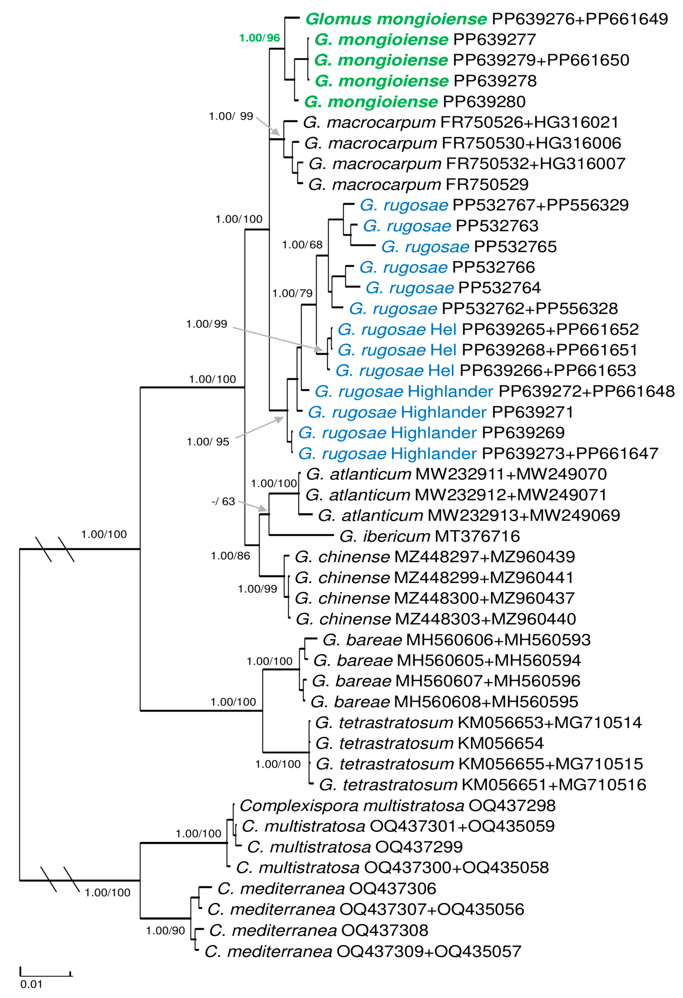
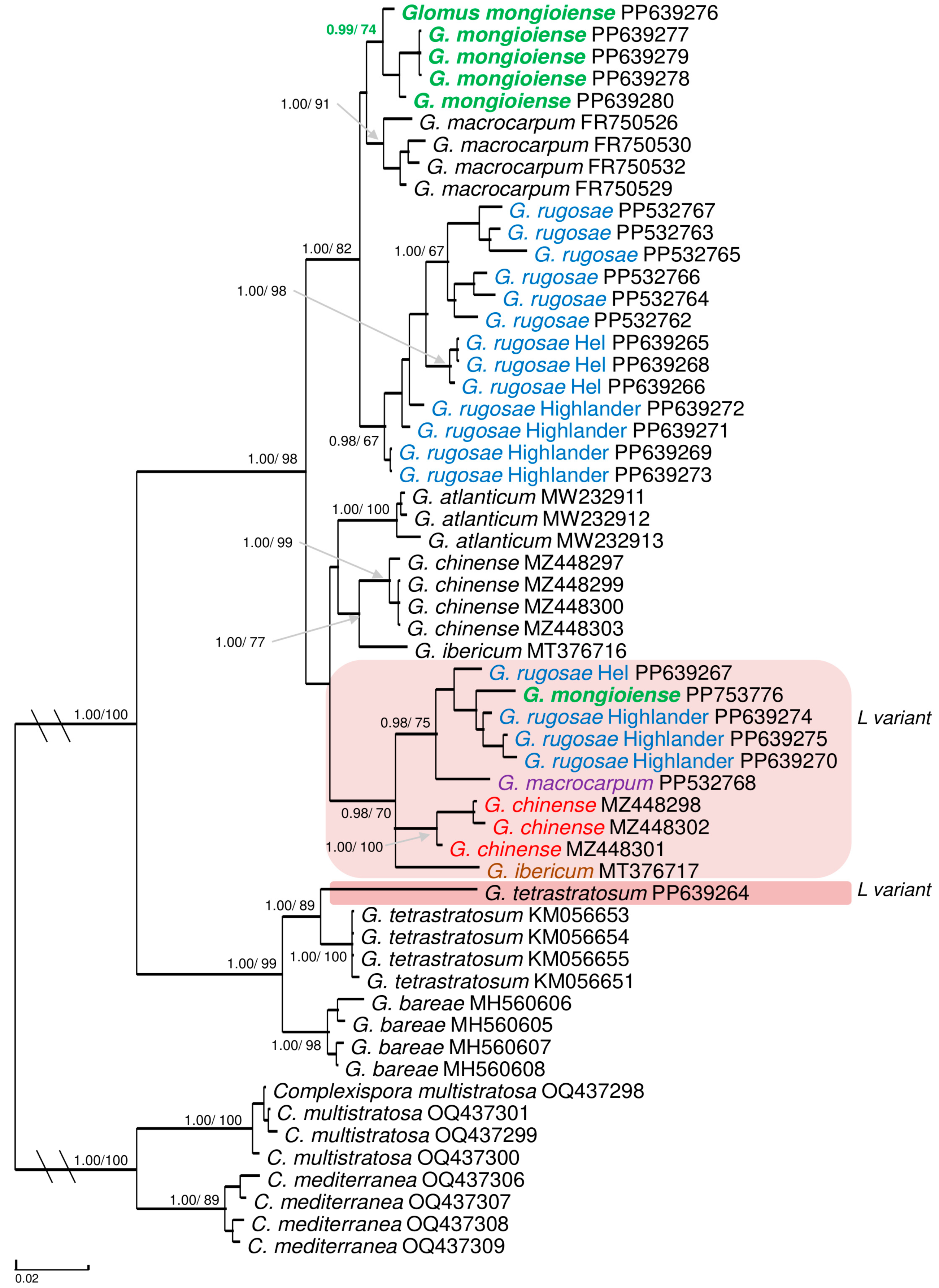
3.2. Molecular and Phylogenetic Analyses

Overall, 18 sequences for the 45S locus and 7 sequences for the RPB1 locus were obtained in the study. Among the 45S sequences, 6 were obtained for Glomus mongioiense, 7 for the isolate Highlander, 4 for the isolate Hel, and 1 for G. tetrastratosum. Of the RPB1 locus, 2 sequences were obtained for G. mongioiense, 2 for the isolate Highlander, and 3 for the isolate Hel.
The visual inspection of the 45Sds1 alignment allowed us to recognize the presence of two variants, S (=short) and L (=long) among the sequences of G. mongioiense and the 2 isolates Highlander and Hel, differing mostly by a ca. 70–85 bp motif at the beginning of ITS1 (Figure S1). A similar ITS1 pattern was observed in sequences of G. macrocarpum, G. chinense and G. ibericum retrieved from GenBank, and in a sequence from G. tetrastratosum obtained from an isolate at the AMF collection of the University of Silesia in Katowice.
Both the phylogenies generated from the 45Sds2 alignment (without L variant sequences) alone (Figure S2) or concatenated (Figure 3) showed identical topology.
The sequences of G. mongioiense populated an independent, supported clade, sister to G. macrocarpum and to G. rugosae, the latter including the sequences from the Highlander and Hel isolates. Glomus mongioiense’s clade received in both analyses full BI support as well as the clades of the neighbor species G. rugosae and G. macrocarpum. The ML support of G. mongioiense and G. rugosae, moderate (78% and 82%, respectively) in the 45S phylogeny, increased considerably in the concatenated analysis (96% and 95%, respectively). Inside the G. rugosae clade, the isolate originally used to describe the species received low (66% and 68%) ML support in both analyses, the Hel isolate received full/high ML support, while sequences of the Highlander isolate scattered basally to the clade formed by the previous two.
When the L variant sequences were included in the analysis (45Sds1), the 45S phylogeny was drastically affected (Figure 4). The L variant sequences of the neighboring species G. mongioiense, G. rugosae and G. macrocarpum clustered together with the L variant sequences of G. chinense and G. ibericum in a “long branch” clade (BI = 0.98, ML = 70%). Furthermore, the support of the G. rugosae clade strongly decreased in both the ML and BI analyses (BI = 0.98, ML = 67%), while the ML support of G. mongioiense was moderately affected (74%). On the other hand, the L variant sequence of G. tetrastratosum did not escape its species clade but penalized the ML support (89%). The concatenated analysis did not restore the expected topology of the trees (Figure S3), leaving the L variant sequences in an independent but very weakly supported clade (BI = 0.96, ML = 55%), but it did it mitigate the negative effect on the ML support of G mongioiense, G. rugosae and G. tetrastratosum (84%, 78% and 98%, respectively).
The 45S sequences inside the G. mongioiense clade shared a percentage of identity >98.5%. Considering the neighboring species, the percentage of identity within G. rugosae ranged between 94.9% and 98.2%, with G. macrocarpum between 95.6% and 96.5%. In G. rugosae, considering the three isolates, the intraspecies variability did not exceed 4%.
Considering the L variants clade, the included species shared a percentage of identity between ca 95.2% and 97.8%. G. ibericum was not considered in this comparison because of an additional dubious duplication of a long fragment at the beginning of the long subunit. Comparing the L variants with the S sequences inside each species, the level of dissimilarity was ca 8% for G. mongioiense, 4.5–7% for G. rugosae, 7% for G. macrocarpum and 5.5% for G. chinense. The L variant of G. tetrastratosum differed by 9–10% from the other L variant sequences, and by 6% from the sequences of its own species clade.
The percentage of identity of RPB1 sequences was higher than 99.3% in G. mongioiense. The species shared 99–99.2% of identity with G. rugosae and 98.4–98.6% with G. macrocarpum.

4. Discussion

The morphological analysis, supported by the phylogenetic inference using single and concatenated loci, confirmed that the AMF isolate obtained from the Ligurian Alps (Italy) represents a new species of the genus Glomus, here described as G. mongioiense. Furthermore, the analysis accommodated the isolates from saltmarshes (Highlands, Scotland) and coastal sand dunes (Hel Peninsula, Poland), both from hypersaline environments, in the recently described G. rugosae.
The Hel isolate and the original isolate used to describe G. rugosae (from the same peninsula on the Baltic Sea) shared the highest degree of molecular identity and formed two distinct sister clades. Sequences from the isolate from Scotland, on the other hand, did not form a distinct clade but scattered basally to the clade hosting the sequences from the other two isolates. The use of three isolates with a certain degree of genetic divergence penalized the support of the species clade, particularly in the 45S ML phylogeny. The separateness of the Highlander isolate from the two Baltic isolates probably reflects the geographic distance and influence of pedoclimatic factors acting on the mycorrhizal fungi.
Finally, the molecular and phylogenetic analysis showed the presence of ribosomal sequences (called L variants) in Glomus species, carrying an insertion in the ITS1, being highly divergent and negatively affecting both the topologies and species supports in the trees. These sequences should not be classified as chimeras as, when blasted in NCBI, the insertion in the ITS1 did not give any match other than the sequences they belong to. Clearly, the genome sequencing of a Glomus species would be necessary to finally confirm this assumption.

4.1. Comparison with Glomus Species

The only species non-sequenced with similar morphology to that of G. mongioiense is G. kerguelense. Both species produce yellow–colored glomoid–like spores with a three–layered spore wall and a subtending hypha [7,46]. The main differences between these species reside in the phenotypic and histochemical characters of the spore wall layers. Instead of the uniform, hyaline, thin (0.8–1.4 µm) spore wall layer 2 of G. mongioiense, G. kerguelense has a laminate, hyaline to pale yellow, thicker (4.3–6.8 µm) layer. Spore wall layer 3 in both species is laminate and yellow, but in G. kerguelense it is clearly thicker (1.6–2.2 µm), and, most importantly, its inner surface in maturing spores is covered with granular processes, forming a granular/spongiform appearance that disappears with spore maturing (vs. the always smooth appearance in G. mongioiense). None of the spore wall layers of G. kerguelense stains in Melzer’s reagent like spore wall layer 1 of G. mongioiense.
Glomus atlanticum, G. bareae and G. chinense, all Glomus species sensu stricto, also present three layers in the spore wall, as in G. mongioiense [18,20,21]. Similarly to G. mongioiense, in G. atlanticum and G. bareae the Melzer’s reaction is observed in the swl1, but these species differ significantly. The swl3 in G. mongioiense is laminated and greyish yellow (vs. laminated and orange–brown in G. atlanticum and flexible and hyaline in G. bareae) [20]. In G. chinense, the second uniform and hyaline slw2 is missing as in G. mongioiense. In addition, the Melzer’s reaction in G. chinense is observed in swl1–2 (vs. only in swl1 in G. mongioiense). The main difference of G. mongioiense from its phylogenetically closest species (G. macrocarpum and G. rugosae) is in the spore wall. While G. mongioiense presents three layers, Glomus macrocarpum presents two layers and G. rugosae four layers.

4.2. Ribosomal Variants: A “Forgotten” Issue

The existence of ribosomal variants and their possible implication in the phylogenetic placement of AMF species have been known for a long time. As early as 20 years ago, [47] warned about the possibility of misleading phylogenetic information using ribosomal markers in environmental studies. Based on the analysis of the 28S D2 region, [47] detected several variants in isolates of Glomus claroideum and G. etunicatum (currently Entrophospora claroidea and E. etunicata; [7]) with a level of divergence up to 19% bp differences among the sequences. Ref. [48] investigated the rRNA gene diversity amplifying the 5.8S-partial 28S region from single spores in nine populations of Claroideoglomus (=Entrophospora) species (E. claroidea, E etunicata, E. lutea). According to the study, two groups of sequences, called S and L variants, were detected, clustering in two distinct supported clades as a result of ancestral polymorphism. Each clade was populated by sequences from the three species investigated. The S (short) variant differed from the L (long) variant primarily by a 100 bp deletion in the ITS2 and by multiple nucleotide polymorphisms along the 28S region. Both variants were considered functional after verification of gene expression and estimation of the structural stability. Finally, recent comparative analysis of genomes from several isolates of Rhizoglomus irregulare has confirmed the presence in this species of up to 11 divergent rRNA copies on each haplotype investigated, located on different chromosomes [49,50].
In the present study we enlarged the knowledge of this phenomenon inside the Glomus genus. Highly divergent variants were obtained from the newly described G. mongioiense, the two isolates of G. rugosae and G. tetrastratosum. Additional sequences from G. macrocarpum, G. chinense and G. ibericum were retrieved from GenBank. Overall, six over eight Glomus species with 45S sequences were represented by at least one L variant sequence. In contrast from that observed by [48], the differences between the variants and the sequences typically used to represent the species lied mostly in the ITS1 locus, due to a motif ca 60–80 bp long.
In a pilot analysis, when only L variant sequences of G. rugosae were employed in the phylogenetic inference, their presence did not alter the topology of the trees but negatively affected the support of G. rugosae clade. When L variant sequences from six Glomus species were included in the analysis, they drastically impacted the tree topology, attracting each other in a single clade and therefore trespassing the species boundaries in a similar manner to that observed by [48]. Only the L variant of G. tetrastratosum remained in its own species clade.
Ancestral polymorphism cannot be employed as the only mechanism to explain the origin of these variants. If this were to be the case, phylogeny of the L variants, single or as clade, would have been expected to be placed in a sister position to the clade of “conventional” S sequences, as in [48]. Supposedly, as has also been suggested by the visual inspection of the alignments, events of recombination between the ribosomal copies could have occurred, preventing a separation of distinct clades as for paralogous genes.
In conclusion, independently of their origin, the presence of highly divergent ribosomal variants represents a potential issue in the Glomeromycota phylogeny, especially when based on 45S sequences only. In the current study we focused only on variants easy to spot because of their degree of dissimilarity. Likely, as in R. irregulare, more ribosomal copies might be found in a single genome, raising the question of what amount of intraspecies diversity could actually be explained by intragenomic diversity. Besides affecting the support of species clades, L variants could lead to the overestimation of the species present in environmental studies, or, when obtained from an isolate without their “conventional” S counterpart, to the recognition of “artificial” species. Further investigations using genome sequencing might be needed to understand the magnitude of this phenomenon in other taxa in the Glomeromycota.

Supplementary Materials

The following supporting information can be downloaded at https://www.mdpi.com/article/10.3390/agronomy14071350/s1, Figure S1: Bird’s-eye view of ITS1 alignment; Figure S2: 50% majority-rule consensus tree from the Bayesian analysis of dataset ds2 (S variant sequences); Figure S3: 50% majority-rule consensus tree from the Bayesian analysis of dataset ds1 concatenated with RPB1 dataset.

Author Contributions

All authors contributed to the study concept and design. F.M.: conceptualization, resources, methodology, formal analysis, writing—original draft, writing—review and editing, supervision; S.U.: conceptualization, methodology, investigation, formal analysis, writing—review and editing; K.B.: resources, data curation, writing—review and editing; J.B.: writing—review and editing; Z.K.: writing—review and editing; M.B.d.Q.: methodology, investigation, formal analysis, writing—original draft, writing—review and editing; L.C.: writing—original draft, writing—review and editing, Supervision. All authors have read and agreed to the published version of the manuscript.

Funding

This research received no external funding.

Data Availability Statement

Datasets generated and/or analyzed during the current study are available from the corresponding author upon request. Sequences obtained in the study are available in Genbank with the following accession numbers: 45S: PP639264-PP639280, PP753776; and RPB1: PP661647-PP661653.

Acknowledgments

The authors wish to thank Paola Galluzzo for her precious contribution to the description of the floristic characteristics of Monte Mongioie.

Conflicts of Interest

Author Leonardo Casieri is employed by the company Mycorrhizal Applications LLC. The remaining authors declare that the research was conducted in the absence of any commercial or financial relationships that could be construed as a potential conflict of interest.

References

  1. Wilkes, T.I.; Warner, D.J.; Edmonds-Brown, V.; Davies, K.G.; Denholm, I. Zero Tillage Systems Conserve Arbuscular Mycorrhizal Fungi, Enhancing Soil Glomalin and Water Stable Aggregates with Implications for Soil Stability. Soil Syst. 2021, 5, 4. [Google Scholar] [CrossRef]
  2. Fall, A.F.; Nakabonge, G.; Ssekandi, J.; Founoune-Mboup, H.; Apori, S.O.; Ndiaye, A.; Badji, A.; Ngom, K. Roles of Arbuscular Mycorrhizal Fungi on Soil Fertility: Contribution in the Improvement of Physical, Chemical, and Biological Properties of the Soil. Front. Fungal Biol. 2022, 3, 723892. [Google Scholar] [CrossRef] [PubMed]
  3. Wang, B.; Xiao, Q.; Geng, X.; Lin, K.; Li, Z.; Li, Y.; Chen, J.; Li, X. Arbuscular mycorrhizal fungi alter rhizosphere bacterial diversity, network stability and function of lettuce in barren soil. Sci. Hortic. 2024, 323, 112533. [Google Scholar] [CrossRef]
  4. Pandit, A.; Kochar, M.; Srivastava, S.; Johny, L.; Adholeya, A. Diversity and Functionalities of Unknown Mycorrhizal Fungal Microbiota. Microbiol. Res. 2022, 256, 126940. [Google Scholar] [CrossRef] [PubMed]
  5. Salomon, M.J.; Demarmels, R.; Watts-Williams, S.J.; McLaughlin, M.J.; Kafle, A.; Ketelsen, C.; Soupir, A.; Bücking, H.; Cavagnaro, T.R.; van der Heijden, M.G.A. Global evaluation of commercial arbuscular mycorrhizal inoculants under greenhouse and field conditions. Appl. Soil Ecol. 2022, 169, 104225. [Google Scholar] [CrossRef]
  6. Wijayawardene, N.N.; Hyde, K.; Dai, D.; Sánchez-García, M.; Goto, B.T.; Saxena, R.K.; Erdoğdu, M.; Selçuk, F.; Rajeshkumar, K.C.; Aptroot, A.; et al. Outline of Fungi and fungus-like taxa. Mycosphere 2022, 13, 53–453. [Google Scholar] [CrossRef]
  7. Błaszkowski, J.; Sánchez-García, M.; Niezgoda, P.; Zubek, S.; Fernández, F.; Vila, A.; Al-Yahya’ei, M.N.; Symanczik, S.; Milczarski, P.; Malinowski, R.; et al. A new order, Entrophosporales, and three new Entrophospora species in Glomeromycota. Front. Microbiol. 2022, 13, 962856. [Google Scholar] [CrossRef]
  8. Silva, G.A.; Corazon-Guivin, M.A.; de Assis, D.M.A.; Oehl, F. Blaszkowskia, a new genus in Glomeraceae. Mycol. Prog. 2023, 22, 74. [Google Scholar] [CrossRef]
  9. Tulasne, L.R.; Tulasne, C. Fungi nonnulli hipogaei, novi v. minus cognito act. Giorn. Bot. Ital. 1845, 2, 55–63. [Google Scholar]
  10. Tulasne, L.R.; Tulasne, C. Fungi Hypogaei. Histoire et Monographie des Champignons Hypogds; Legare Street Press: Paris, France, 1851. [Google Scholar]
  11. Gerdemann, J.W.; Trappe, J.M. The Endogonaceae of the Pacific Northwest. Micol. Mem. 1974, 5, 1–76. [Google Scholar]
  12. Schüßler, A.; Walker, C. The Glomeromycota: A Species List with New Families and New Genera; Royal Botanic Garden Edinburgh: Edinburgh, UK, 2010. [Google Scholar]
  13. Oehl, F.; Silva, G.A.; Goto, B.T.; Maia, L.C.; Sieverding, E. Glomeromycota: Two new classes and a new order. Mycotaxon 2011, 116, 365–379. [Google Scholar] [CrossRef]
  14. Oehl, F.; Silva, G.A.; Goto, B.T.; Sieverding, E. Glomeromycota: Three new genera and glomoid species reorganized. Mycotaxon 2011, 116, 75–120. [Google Scholar] [CrossRef]
  15. Błaszkowski, J.; Chwat, G.; Góralska, A.; Ryszka, P.; Kovács, G.M. Two new genera, Dominikia and Kamienskia, and D. disticha sp. nov. in Glomeromycota. Nova Hedwig. 2015, 100, 225–238. [Google Scholar] [CrossRef]
  16. Sieverding, E.; Silva, G.A.; Reinhard, B.; Oehl, F. Rhizoglomus, a new genus of the Glomeraceae. Mycotaxon 2015, 129, 373–386. [Google Scholar] [CrossRef]
  17. Berch, S.M.; Fortin, J.A. Lectotypification of Glomus macrocarpum and proposal of new combinations: Glomus australe, Glomus versiforme, and Glomus tenebrosum (Endogonaceae). Can. J. Bot. 1983, 61, 2608–2617. [Google Scholar] [CrossRef]
  18. Błaszkowski, J.; Niezgoda, P.; Goto, B.T.; Kozłowska, A. Halonatospora gen. nov. with H. pansihalos comb. nov. and Glomus bareae sp. nov. (Glomeromycota; Glomeraceae). Botany 2018, 96, 737–748. [Google Scholar] [CrossRef]
  19. Guillén, A.; Serrano-Tamay, F.J.; Peris, J.B.; Arrillaga, I. Glomus ibericum, Septoglomus mediterraneum, and Funneliformis pilosus, three new species of arbuscular mycorrhizal fungi. Mycologia 2020, 112, 819–828. [Google Scholar] [CrossRef] [PubMed]
  20. Błaszkowski, J.; Niezgoda, P.; Zubek, S.; Meller, E.; Milczarski, P.; Malicka, M.; Goto, B.T.; Woźniak, G.; Moreira, H.; Magurno, F. Dominikia bonfanteae and Glomus atlanticum, two new species in the Glomeraceae (phylum Glomeromycota) with molecular phylogenies reconstructed from two unlinked loci. Mycol. Prog. 2021, 20, 131–148. [Google Scholar] [CrossRef]
  21. Yu, F.; Goto, B.T.; Magurno, F.; Błaszkowski, J.; Wang, J.; Ma, W.; Feng, H.; Liu, Y. Glomus chinense and Dominikia gansuensis, two new Glomeraceae species of arbuscular mycorrhizal fungi from high altitude in the Tibetan Plateau. Mycol. Prog. 2022, 21, 32. [Google Scholar] [CrossRef]
  22. Błaszkowski, J.; Zubek, S.; Milczarski, P.; Malinowski, R.; Goto, B.T.; Niezgoda, P. Glomus rugosae, a new arbuscular mycorrhizal species in Glomeraceae (phylum Glomeromycota) from maritime sand dunes of Poland and an ash pond of Czech Republic. Phytotaxa 2024, 644, 271–280. [Google Scholar] [CrossRef]
  23. Fratianni, S.; Acquaotta, F. The climate of Italy. In Landscapes and Landforms of Italy; Soldati, M., Marchetti, M., Eds.; Springer: Berlin/Heidelberg, Germany, 2017; pp. 29–38. [Google Scholar]
  24. Firth, C.; Fleet, L. Information Sheet on Ramsar Wetlands (RIS). 2006. Available online: http://www.ramsar.org/ris/key_ris_index.htm (accessed on 1 April 2024).
  25. Gunn, G.F. Measuring nocturnal near-surface urban heat island intensity in the small, mid-latitude city of Inverness, Scotland. Scott. Geogr. J. 2023, 140, 192–209. [Google Scholar] [CrossRef]
  26. Polderman, P.J.; Polderman-Hall, R.A. Algal communities in Scottish saltmarshes. Br. J. Psychol. 1980, 15, 59–71. [Google Scholar] [CrossRef]
  27. Woch, M.W.; Kapusta, P.; Stanek, M.; Możdżeń, K.; Grześ, I.M.; Rożej-Pabijan, E.; Stefanowicz, A.M. Effects of invasive Rosa rugosa on Baltic coastal dune communities depend on dune age. NeoBiota 2023, 82, 163–187. [Google Scholar] [CrossRef]
  28. Kornerup, A.; Wanscher, J.H. Methuen Handbook of Colour, 3rd ed.; Methuen Publishing: London, UK, 1983. [Google Scholar]
  29. Furrazola, E.; Torres-Arias, Y.; Ferrer, R.L.; Herrera, R.A.; Berbara, R.; Goto, B.T. Glomus crenatum (Glomeromycetes), a new ornamented species from Cuba. Mycotaxon 2011, 116, 132–143. [Google Scholar] [CrossRef]
  30. Goto, B.T.; Jardim, J.G.; da Silva, G.A.; Furrazola, E.; Torres-Arias, Y.; Oehl, F. Glomus trufemii (Glomeromycetes), a new sporocarpic species from Brazilian sand dunes. Mycotaxon 2012, 120, 1–9. [Google Scholar] [CrossRef]
  31. Błaszkowski, J.; Jobim, K.; Niezgoda, P.; Meller, E.; Malinowski, R.; Milczarski, P.; Zubek, S.; Magurno, F.; Casieri, L.; Bierza, W.; et al. New Glomeromycotan Taxa, Dominikia glomerocarpica sp. nov. and Epigeocarpum crypticum gen. nov. et sp. nov. From Brazil, and Silvaspora gen. nov. From New Caledonia. Front. Microbiol. 2021, 12, 655910. [Google Scholar] [CrossRef] [PubMed]
  32. Walker, C. Taxonomic concepts in the Endogonaceae: Spore wall characteristics in species descriptions. Mycotaxon 1983, 18, 443–455. [Google Scholar]
  33. Błaszkowski, J. Glomeromycota; W. Szafer Institute of Botany, Polish Academy of Sciences: Kraków, Poland, 2012. [Google Scholar]
  34. Goto, B.T.; Maia, L.C. Glomerospores: A new denomination for the spore of Glomeromycota, a group molecularly distinct from the zygomycota. Mycotaxon 2006, 96, 129–132. [Google Scholar]
  35. Jobim, K.; Błaszkowski, J.; Niezgoda, P.; Kozłowska, A.; Zubek, S.; Mleczko, P.; Chachuła, P.; Ishikawa, N.K.; Goto, B.T. New sporocarpic taxa in the phylum Glomeromycota: Sclerocarpum amazonicum gen. et sp. nov. in the family Glomeraceae (glomerales) and Diversispora sporocarpia sp. nov. in the Diversisporaceae (Diversisporales). Mycol. Prog. 2019, 18, 369–384. [Google Scholar] [CrossRef]
  36. Krüger, M.; Stockinger, H.; Krüger, C.; Schüßler, A. DNA-based species level detection of Glomeromycota: One PCR primer set for all arbuscular mycorrhizal fungi. New Phytol. 2009, 183, 212–223. [Google Scholar] [CrossRef]
  37. Błaszkowski, J.; Yamato, M.; Niezgoda, P.; Zubek, S.; Milczarski, P.; Malinowski, R.; Meller, E.; Malicka, M.; Goto, B.T.; Uszok, S.; et al. A new genus, Complexispora, with two new species, C. multistratosa and C. mediterranea, and Epigeocarpum japonicum sp. nov. Mycol. Prog. 2023, 22, 34. [Google Scholar] [CrossRef]
  38. Miller, M.A.; Pfeiffer, W.; Schwartz, T. Creating the CIPRES science gateway for inference of large phylogenetic trees. In Proceedings of the Gateway Computing Environments Workshop, New Orleans, LA, USA, 14 November 2010; pp. 1–8. [Google Scholar]
  39. Abadi, S.; Azouri, D.; Pupko, T.; Mayrose, I. Model selection may not be a mandatory step for phylogeny reconstruction. Nat. Commun. 2019, 10, 934. [Google Scholar] [CrossRef] [PubMed]
  40. Ronquist, F.; Teslenko, M.; Mark, P.V.D.; Ayres, D.L.; Darling, A.; Hohna, S.; Larget, B.; Liu, L.; Suchard, M.A.; Huelsenbeck, J.P. MrBayes 3.2: Efficient Bayesian phylogenetic inference and model choice across a large model space. Syst. Biol. 2012, 61, 539–542. [Google Scholar] [CrossRef] [PubMed]
  41. Kozlov, A.M.; Darriba, D.; Flouri, T.; Morel, B.; Stamatakis, A. RAxML-NG: A fast, scalable, and user-friendly tool for maximum likelihood phylogenetic inference. Bioinformatics 2019, 35, 4453–4455. [Google Scholar] [CrossRef] [PubMed]
  42. Stöver, B.C.; Müller, K.F. TreeGraph 2: Combining and visualizing evidence from different phylogenetic analyses. BMC Bioinform. 2010, 11, 7. [Google Scholar] [CrossRef] [PubMed]
  43. Berger, S.A.; Krompass, D.; Stamatakis, A. Performance, accuracy, and web server for evolutionary placement of short sequence reads under maximum likelihood. Syst. Biol. 2011, 60, 291–302. [Google Scholar] [CrossRef] [PubMed]
  44. Czech, L.; Barbera, P.; Stamatakis, A. Genesis and Gappa: Processing, analyzing and visualizing phylogenetic (placement) data. Bioinformatics 2020, 36, 3263–3265. [Google Scholar] [CrossRef] [PubMed]
  45. Malicka, M.; Magurno, F.; Piotrowska-Seget, Z. Phenol and Polyaromatic Hydrocarbons are Stronger Drivers Than Host Plant Species in Shaping the Arbuscular Mycorrhizal Fungal Component of the Mycorrhizosphere. Int. J. Mol. Sci. 2022, 23, 12585. [Google Scholar] [CrossRef] [PubMed]
  46. Dalpé, Y.; Plenchette, C.; Frenot, Y.; Gloaguen, J.C.; Strullu, D.G. Glomus kerguelense, a new Glomales species from sub-Antarctic. Mycotaxon 2002, 84, 51–60. [Google Scholar]
  47. Rodriguez, A.; Clapp, J.P.; Robinson, L.; Dodd, J.C. Studies on the diversity of the distinct phylogenetic lineage encompassing Glomus claroideum and Glomus etunicatum. Mycorrhiza 2005, 15, 33–46. [Google Scholar] [CrossRef]
  48. VanKuren, N.W.; den Bakker, H.C.; Morton, J.B.; Pawlowska, T.E. Ribosomal RNA gene diversity, effective population size, and evolutionary longevity in asexual Glomeromycota. Evolution 2013, 67, 207–224. [Google Scholar] [CrossRef] [PubMed]
  49. Sperschneider, J.; Yildirir, G.; Rizzi, Y.S.; Malar, M.C.; Nicol, A.M.; Sorwar, E.; Villeneuve-Laroche, M.; Chen, E.C.H.; Iwasaki, W.; Brauer, E.K.; et al. Arbuscular mycorrhizal fungi heterokaryons have two nuclear populations with distinct roles in host–plant interactions. Nat. Microbiol. 2023, 8, 2142–2153. [Google Scholar] [CrossRef] [PubMed]
  50. Yildirir, G.; Sperschneider, J.; Malar, M.C.; Chen, E.C.H.; Iwasaki, W.; Cornell, C.; Corradi, N. Long reads and Hi-C sequencing illuminate the two-compartment genome of the model arbuscular mycorrhizal symbiont Rhizophagus irregularis. New Phytol. 2022, 233, 1097–1107. [Google Scholar] [CrossRef] [PubMed]
Figure 1. (AF) Glomus mongioiense. (A,B) Spores in loose clusters with three wall layers (swl) and one subtending hypha (sh). (A,CF) Spore wall layers (swl1–3). (F) Subtending hyphal wall layers (shwl2–3) continuous with spore wall layers. (A,C,E,F) Spores in PVLG. (B,D) Spores in PVLG + Melzer’s reagent.
Agronomy 14 01350 g001
Figure 2. (A,B,DF) Glomus sp. Highlander isolate. (C,G) Glomus sp. Hel isolate. (A,B) Spores in loose clusters. (A,CG) Spore wall layers (swl1–4). (A) Subtending hyphal wall layers (shwl1–4) continuous with spore wall layers. (A,C,E) Spores in PVLG. (B,D,F,G) Spores in PVLG + Melzer’s reagent.
Agronomy 14 01350 g002
Figure 3. 50% majority-rule consensus tree from the Bayesian analysis of 45S sequences concatenated with RPB1 sequences of G. mongioiense, seven species of Glomus sensu stricto in possession of molecular data, and two Complexispora species (the closest taxa to Glomus in the family Glomeraceae) serving as outgroup. The new species is in bold-green-colored font. Sequences of G. rugosae, including the Highlander and Hel isolates are in blue-colored font. Bayesian posterior probabilities ≥ 0.95 and ML bootstrap values ≥ 60% are shown near the branches. All parameters of the convergence diagnostic (potential scale reduction factor and standard deviation of split frequencies) indicated that the convergence was obtained in the BI analysis. Bar indicates 0.01 expected change per site per branch. Outgroup and ingroup branches are shortened to 50% in length to improve visibility (indicated by \\).
Agronomy 14 01350 g003
Figure 4. 50% majority-rule consensus tree from the Bayesian analysis of dataset ds1 (S and L variant sequences). The dataset consisted of 45S sequences of G. mongioiense, seven species of Glomus sensu stricto in possession of molecular data, and two Complexispora species (the closest taxa to Glomus in the family Glomeraceae) serving as outgroup. The new species is in bold–green–colored font. Sequences of G. rugosae, including the Highlander and Hel isolates are in blue–colored font. The clades hosting L variant sequences are highlighted with colored boxes, and colored fonts and are used to distinguish the several species included. Bayesian posterior probabilities ≥ 0.95 and ML bootstrap values ≥ 60% are shown near the branches. All parameters of the convergence diagnostic (potential scale reduction factor and standard deviation of split frequencies) indicated that the convergence was obtained in the BI analysis. Bar indicates 0.02 expected change per site per branch. Outgroup and ingroup branches are shortened to 50% in length to improve visibility (indicated by \\).
Agronomy 14 01350 g004
Disclaimer/Publisher’s Note: The statements, opinions and data contained in all publications are solely those of the individual author(s) and contributor(s) and not of MDPI and/or the editor(s). MDPI and/or the editor(s) disclaim responsibility for any injury to people or property resulting from any ideas, methods, instructions or products referred to in the content.

