Article

Selection and Characterization of Somaclonal Variants of Prata Banana (AAB) Resistant to Fusarium Wilt

by
Mileide dos Santos Ferreira
1,
Tamyres Amorim Rebouças
2,
Anelita de Jesus Rocha
2,
Wanderley Diaciso dos Santos Oliveira
1,
Ana Carolina Lima Santos dos Santos
3,
João Pedro Falcón Lago de Jesus
3,
Andresa Priscila de Souza Ramos
2,
Claudia Fortes Ferreira
2,
Janay Almeida dos Santos-Serejo
2,
Fernando Haddad
2 and
Edson Perito Amorim
4,*
1
Department of Biology, State University of Feira de Santana, Feira de Santana 44036-900, BA, Brazil
2
Department of Biology, Embrapa Mandioca e Fruticultura, Cruz das Almas 44380-000, BA, Brazil
3
Center for Environmental and Biological Agricultural Sciences, Federal University do Recôncavo da Bahia, Cruz das Almas 44380-000, BA, Brazil
4
Department of Plant Genetic Improvement, Embrapa Mandioca e Fruticultura, Cruz das Almas 44380-000, BA, Brazil
*
Author to whom correspondence should be addressed.
Agronomy 2024, 14(8), 1740; https://doi.org/10.3390/agronomy14081740
Submission received: 15 July 2024 / Revised: 30 July 2024 / Accepted: 7 August 2024 / Published: 8 August 2024
(This article belongs to the Section Crop Breeding and Genetics)

Abstract

:
Fusarium wilt, caused by the fungus Fusarium oxysporum f. sp. cubense (Foc), is one of the most devastating diseases affecting banana cultivation worldwide. Although Foc tropical race 4 (TR4) has not yet been identified in Brazilian production areas, the damage caused by races 1 and subtropical 4 is the main cause of production losses, especially affecting cultivars of the Prata subgroup. Thus, the induction of somaclonal variation is a promising strategy in biotechnology to generate genetic variability and develop resistant varieties. This study aimed to induce somaclonal variation in the Prata Catarina cultivar (AAB genome) using successive subcultures in Murashige and Skoog (MS) medium enriched with the plant regulator Thiadizuron (TDZ) at two concentrations: 1 and 2 mg/L. After evaluating the symptoms, we selected 13 resistant somaclones that were not infected by the fungus. Histochemical and histological analyses of the somaclones indicated possible defense mechanisms that prevented colonization and/or infection by Foc, such as intense production of phenolic compounds and the presence of cellulose and callose in the roots. Some somaclones showed no pathogen structures in the xylem-conducting vessels, indicating possible pre-penetration resistance. Furthermore, molecular studies indicated that the genetic alterations in the somaclones may have induced resistance to Foc without compromising the agronomic characteristics of the commercial genotype.

1. Introduction

Bananas (Musa spp.) are the most consumed fresh fruit worldwide, with an estimated annual production of 114 million tons [1]. Currently, banana production is spread across several tropical and subtropical regions, especially Asia, Latin America and the Caribbean, and Africa. The largest producers are India (34.5 million tons), China (11.8 million tons), Indonesia (9.2 million tons), and Nigeria (8.0 million tons) [2]. Bananas of the Cavendish subgroup are grown on a large scale for export, dominating the American and European markets; however, there are hundreds of other cultivars used worldwide, mainly for domestic consumption and local or regional markets, playing a crucial role in the diet of the populations of the Indo-Malaysian, Asian, East African, and Latin American and Caribbean regions [3].
In Latin America and the Caribbean, Brazil is the largest producer of bananas, with an annual production of 7 million tons in an area of approximately 460,000 hectares [3]. This results in a productivity of 15 tons per hectare, annually generating more than USD 2.5 billion [2]. This production is mostly conducted by small producers throughout the country, and cultivars of the Prata subgroup (AAB genome) are widely grown and preferred by consumers owing to their unique texture and flavor. Notably, 70% of the area of banana cultivation is occupied by cultivars from this subgroup, especially the Prata Catarina cultivar, namely, a natural mutant derived from the Prata-Anã cultivar, with higher productivity and fruit quality [4]. Thus, the Prata banana plays a vital role, both in the agricultural economy and Brazilian food security [4,5].
Banana production is limited by several abiotic and biotic factors, including soil or water salinity [6], dry environments [7], weevil borer (Cosmopolites sordidus), nematodes (Meloidogyne spp., Pratylenchus coffeae, and Radopholus similis) [8], viruses (BBTV, BSV, etc.) [9], Xanthomonas vasicola pv. musacearum [10], Pseudocercospora fijiensis, responsible for Black Sigatoka [11], and Fusarium oxysporum f. sp. cubense (Foc), which causes Fusarium wilt [12].
Brazilian banana production, as in other countries, is threatened by the spread of Fusarium oxysporum f. sp. cubense tropical race 4 (Foc TR4), considered the most aggressive strain of Foc, which causes Fusarium wilt and has spread to different production areas [13,14,15]. Foc TR4 can infect a wide range of banana cultivars, including those of the Prata subgroup, causing devastation to plantations in Asia, Africa, and Latin America [16,17].
Despite the potential destructive impact of Foc TR4 in Brazilian cultivation areas, considering its absence in the country, race 1 and subtropical race 4 (STR4) are currently the biggest limitations to fruit production in the country, especially in the irrigated perimeters in the northern parts of Minas Gerais and Bahia, Ribeira Valley (state of São Paulo), and northern part of Santa Catarina; thus, extensive areas cultivated with Prata bananas are unviable for cultivation owing to the high infestation by race 1 [18]. Therefore, producers have replaced Prata bananas with Cavendish bananas, but this option is risky, considering the preference of Brazilian consumers for Prata banana types. Additionally, STR4 has caused damage to banana production in the southeastern and southern regions of Brazil, which experience harsh winters, facilitating infection by Foc [19].
The disease is identified by symptoms such as yellowing and wilting of the leaves, as well as cracks in the pseudostem. After the pathogen infects the roots, the fungus multiplies in the vascular tissue, causing a collapse that eventually leads to the plant’s death. The economic losses are severe, resulting in drastic reductions in fruit production and quality, which severely impacts banana farms. Fusarium wilt is particularly severe because it can spread rapidly through the soil via crop residues, and its spores can persist for over 30 years, making disease management and control a significant challenge for producers [13,15,18,20].
In this context, the best strategy to contain the damage caused by Fusarium oxysporum f. sp. cubense is the use of resistant cultivars in addition to other tools, such as biological control and proper soil management, which together can mitigate the effects of the disease [20,21,22]. The use of tissue culture is another effective technique to prevent the spread of Foc, as this method allows for the propagation of genetically identical plants, ensuring uniformity and quality in production [23,24].
Accordingly, banana breeding programs at research institutions in different regions of the world have focused their efforts on exploiting the plant’s genetic resistance to the pathogen to obtain a means of long-term control of the disease. These programs use different breeding strategies, especially hybridization, transgenesis or gene editing, mutagenesis, and in vitro induction of somaclonal variation [23].
Among the aforementioned breeding methods, the induction of somaclonal variation has been widely used as an efficient option for the genetic improvement of various crops. This technique involves cultivating plant cells in a culture medium supplemented with cytokinins, which are plant regulators that promote cell division and high multiplication rates, such as Thidiazuron (TDZ). This is followed by successive in vitro subcultures, which can induce genetic changes, providing variability and generating somaclonal variants of interest. These factors are decisive in generating spontaneous and selectable genetic variations, enabling traits of interest to be obtained for breeding [24]. The variations observed in plants can arise from a number of factors, including somatic mutations, epigenetics, and stresses during in vitro cultivation. After inducing somaclonal variation, an accurate phenotypic evaluation of the somaclones must be conducted to identify and select those with desirable characteristics for commercial cultivation, especially disease resistance [25,26,27].
In field tests, some somaclonal variants of “Cavendish” have been obtained and shown to have some level of tolerance to Foc TR4 [28]. In another study, somaclonal variants of the “Grande Naine” banana plant were identified in a greenhouse [29]. The most famous banana somaclone that is widespread in areas contaminated by Foc TR4 is Formosana (GCTCV 218), developed by the Taiwan Biodiversity Research Institute (TBRI) in Taiwan [30]. In sugarcane, a somaclone with resistance to brown rust (Puccinia melanocephala) was identified [31]. In another study, a somaclone with promising agronomic characteristics related to grain yield in wheat was identified [32]. In studies on rice (Oryza sativa L. cv. Nipponbare), three somaclones were selected with resistance to the fungus Magnaporthe oryzae, which causes rice brusone [33]. These results validate the application potential of the induction of somaclonal variation for genetic improvement in various crops.
In this study, we generated the first somaclonal variants of the Prata subgroup banana cv. Prata Catarina through in vitro cultivation supplemented with the plant regulator TDZ. In the greenhouse, we selected somaclones resistant to Fusarium wilt based on a bioassay in beds infested with a strain of Foc STR4. To assess the extent of genetic diversity present in the selected somaclones, we used the molecular markers inter-retrotransposon amplified polymorphism (IRAP), retrotransposon–microsatellite amplified polymorphism (REMAP), and inter-simple sequence repeat (ISSR); to observe plant–pathogen interactions, we evaluated compounds related to plant defense responses by means of histological and histochemical analyses.

2. Materials and Methods

2.1. Plant Material

Seedlings of the Prata Catarina (AAB) cultivar were used for multiplication and the induction of somaclonal variation. This cultivar is a natural mutation selected in plantation areas in Brazil, derived from the Prata-Anã cultivar.
The seedlings were subcultured in Murashige and Skoog (MS) medium [34], supplemented with indoleacetic acid (IAA) (1.6 mL/L) and adenine hemisulfate (80 mg/L), to which different concentrations of TDZ were added. The treatments comprised two doses of TDZ: treatment 1 (T1), where the MS medium was supplemented with 1 mg of TDZ per liter; treatment 2 (T2), using 2 mg of TDZ per liter.
Five subcultures were employed for each treatment, with an interval ranging between 40 and 60 days, depending on the development of the plants. At the end of the subcultivation of the two treatments, 2400 plants were subjected to resistance assessment in the greenhouse, with 1200 plants for each dose of TDZ. Additionally, 240 commercial Prata Catarina seedlings that did not receive supplementation with TDZ were selected to serve as the positive control. In the greenhouse, the plants were maintained at temperatures between 25 and 27 °C, with a relative humidity of 50 to 60%. During soil preparation, we ensured the appropriate conditions by providing sufficient amounts of macro and micronutrients, as well as organic matter. The plants were irrigated using drip irrigation, following the specific recommendations for the crop.

2.2. Preparation of the Foc Inoculum

In this study, we used isolate CNPMF (National Center for Cassava and Fruit Research) 229, selected from the biological collection of the Phytopathology Laboratory at Embrapa Mandioca e Fruticultura. The isolate was chosen owing to its virulence and ability to aggressively infect banana varieties, including the Cavendish subgroup; when inoculated under controlled conditions, the same characteristics were observed in isolate CNPMF 218, classified as STR4, both of which were collected in the same region, namely, in the state of Santa Catarina [23,35]. The isolate was cultivated on potato dextrose agar medium, in BOD at 25 °C, under a 12-h photoperiod. After colony growth, a suspension of conidia was prepared at a concentration of 106 colony forming units (CFUs) and approximately 20 mL was deposited on 1 kg of sterilized rice. The medium was then incubated at 25 °C with a 12-h photoperiod. After 20 days, CFUs were quantified through a series of dilutions to assess spore concentration and viability. The CFUs were counted in a Neubauer chamber, and the concentration used for soil infestation in the greenhouse beds was 106 conidia/g of rice or inoculum [36].

2.3. Evaluation of Resistance in the Greenhouse

After 60 days of acclimatization, the somaclone seedlings were transferred to a greenhouse and planted in beds measuring 10 x 1 m, with soil infested with isolate CNPMF 229. After 90 days of planting, the somaclones were assessed for resistance to Foc, exposing the roots to contact with the pathogen spores. To achieve this, cross-sections were made in the rhizomes of the seedlings and the internal symptoms of rhizome discoloration were assessed using the scale proposed by Dita et al. [37], namely, 1: no symptoms; 2: rhizome with initial discoloration; 3: discoloration of the rhizome throughout the vascular system; 4: rhizome with necrosis in most internal tissues; and 5: completely necrotic rhizome.
Based on the scores, an analysis of variance was conducted on the disease index (DI) estimates at a 5% significance level. The data were depicted in a boxplot graph with the DI results of the treatments. To calculate the DI, the scores obtained in the evaluations of the internal symptoms of the disease were transformed (0 to 4). The number of replications was based on the number of evaluated plants, namely, 1200 for each treatment. The analyses were conducted using the R software v. 4.3.3 [38].

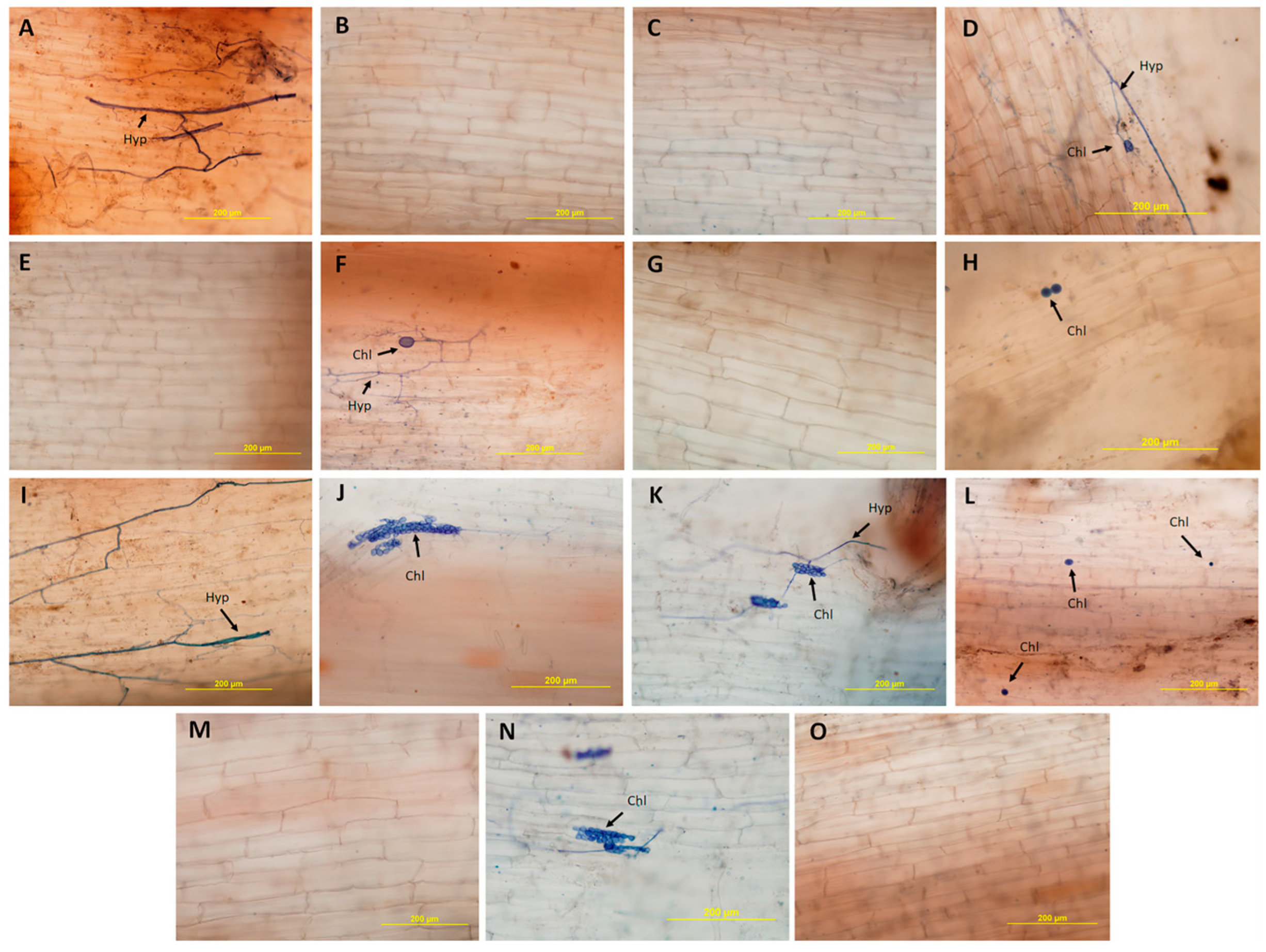
2.4. Histological and Histochemical Analysis

Root fragments of the somaclones classified as resistant to isolate CNPMF 229 were collected and immersed in Karnovsky’s solution [39] for a period of 48 h. The fragments were then dehydrated using an increasing series of ethanol at 3 h intervals, ranging from 30 to 100%. Infiltration and embedding were conducted using the historesin embedding kit (hydroxyethyl methacrylate, Leica, Heldelberg, Germany). After polymerization of the historesin, histological sections measuring 8 µm were obtained using a Leitz 1516 microtome (Thermo Fisher Scientific, Waltham, MA, USA). These sections were mounted on histological slides and stained with ferric chloride for 3 h to detect phenolic compounds [40], and calcofluor white (0.01%) to detect cellulose. To detect callose, the slides were stained with aniline blue (0.05%) for 5 to 10 min [41]. The histological sections were subsequently analyzed and photographed using a B x S1 fluorescence microscope (Olympus Latin America, Miami, FL, USA).
The analysis of root clarification and staining of fungal structures was conducted according to the method described by Phillips and Hayman [42]. The roots were immersed in a 10% potassium hydroxide (KOH) solution at room temperature for 48 h, followed by immersion in a 1% HCl solution for 30 min. Trypan blue dye in a 0.05% solution (lactic acid/glycerol/water = 2:1:1) was applied for 1 h. After staining, the slides were prepared and fragments were microphotographed using an optical microscope (Olympus Latin America, Miami, FL, USA).

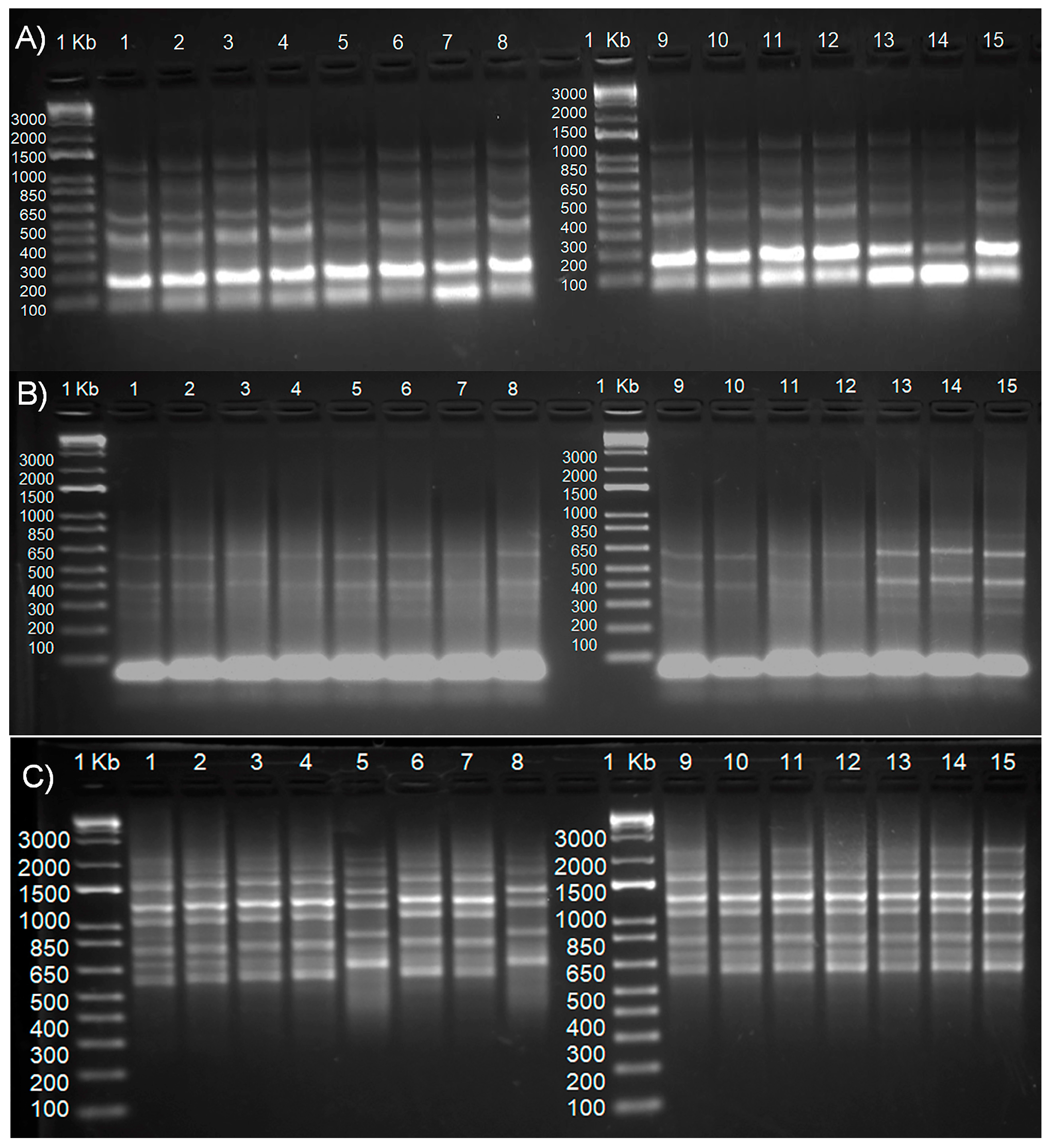
2.5. Molecular Analysis

Material Collection and DNA Extraction

Samples of young leaves from the resistant somaclones in T1 and T2, as well as the control, were collected and taken to Embrapa’s Molecular Biology laboratory for DNA extraction using the methodology proposed by Doyle and Doyle [43], adapted by a previous study [44]. The DNA was quantified and its quality was assessed on a 1% agarose gel stained with GelRed®® (Biotium, Inc., 46117 Landing Parkway Fremont, CA, USA), and subjected to an electrophoretic run at 80 V for 1 h; subsequently, it was visualized using a UV transluminator.
PCR amplification and analysis using IRAP, REMAP, and ISSR markers.
Inter-retrotransposon amplified polymorphism (IRAP) is used to detect genetic polymorphisms between retrotransposons. This marker is based on the amplification of DNA regions located between two retrotransposons. Retrotransposon–microsatellite amplified polymorphism (REMAP) combines retrotransposon elements with microsatellites (short repetitive sequences) to amplify genetic polymorphisms. Inter-simple sequence repeat (ISSR) uses primers established in microsatellite regions to amplify the regions between these simple repetitive sequences. These markers are highly effective in genetic studies as they allow for efficient and precise detection of genetic variations [45,46].
The IRAP marker analysis was based on the method described by Kalendar et al. [45]. The 20 μL reaction mixture comprised 25 ng of DNA, 0.3 μm primer, 2.5 mm MgCl, 0.2 mm deoxynucleotide triphosphate (dNTPs), 10× Taq buffer, and 0.3 U Taq DNA polymerase. The amplifications were conducted in a Veriti 96-Well Thermal Cycler (0.2 mL), Life Technologies, Waltham, MA, USA, with the following settings: 1 cycle at 94 °C for 3 min; 35 cycles at 94 °C for 30 s, 42 °C for 1 min, and 72 °C for 45 s; and 1 cycle at 72 °C for 5 min and 4 °C.
Amplification between simple sequence repeats (ISSR) was conducted using the method described by Sankar [46]. The 25 μL reaction mixture comprised 50 ng of template DNA, 0.2 μm primer, 2.5 mm MgCl, 20 mm dNTPs, 10× Taq buffer, and 0.2 U Taq DNA polymerase. The amplifications were conducted in a Veriti 96-Well Thermal Cycler (0.2 mL), Life Technologies, with the following setting: 1 cycle at 94 °C for 3 min; 39 cycles at 94 °C for 40 s, 48 °C for 40 s, and 72 °C for 1 min; and 1 cycle at 72 °C for 5 min and 4 °C.
The long terminal repeat (LTR) reverse primer 7286 REMAP was combined with seven LTR-SSR primers (Table 1), according to Kalendar et al. [45]. REMAP amplifications were conducted with a final volume of 25 μL, containing 50 ng of DNA, 0.2 μm LTR primer, 0.3 μm ISSR primer, 2.5 mm MgCl 2, 2 mm dNTPs, 10× Taq buffer, and 0.2 U Taq DNA polymerase. The amplifications were conducted in a Veriti 96-Well Thermal Cycler (0.2 mL), Life Technologies, with the following settings: 1 cycle at 94 °C for 3 min; 30 cycles at 94 °C for 30 s, 58 °C for 1 min, and 72 °C for 45 s; 1 cycle at 72 °C for 5 min and 4 °C.
The amplification products were separated on a 2.0% agarose gel and subjected to an electrophoretic run at 80 V for 3 to 4 h. They were stained with GelRed®® and visualized using a UV transluminator (Loccus, Cotia, Brazil).

3. Results

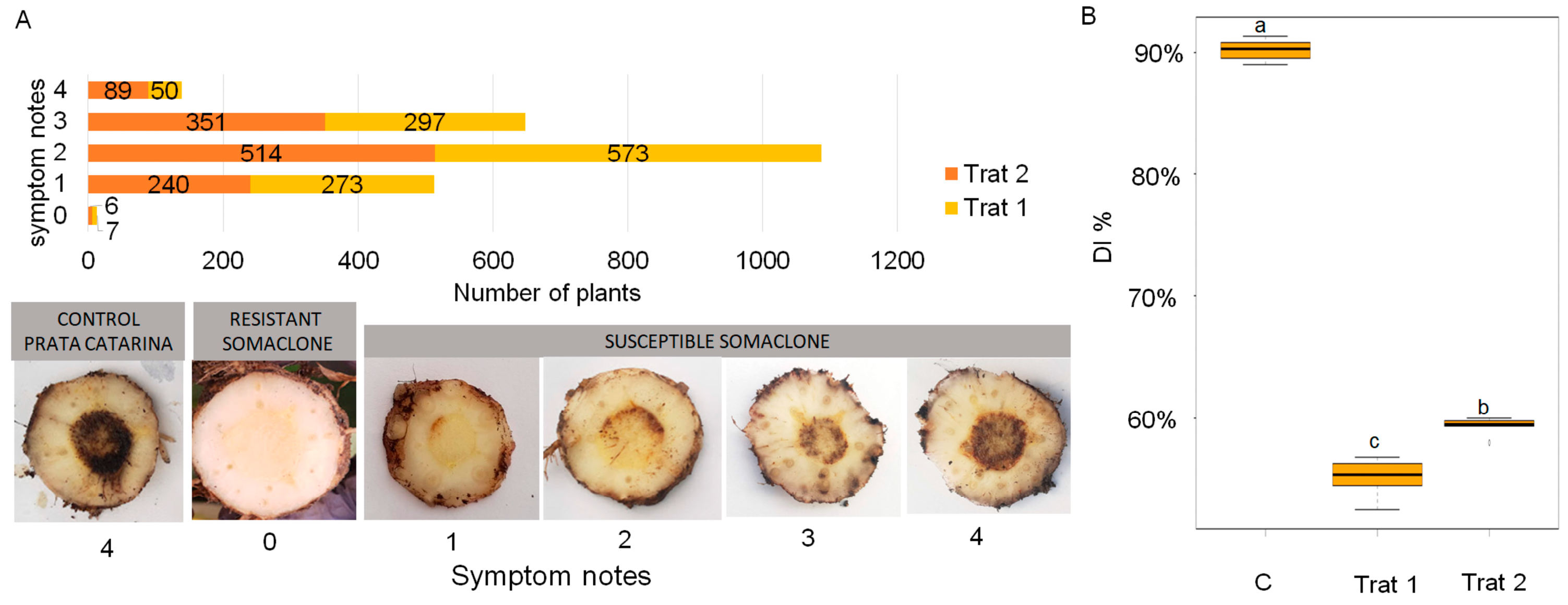
3.1. Resistance Assessment in the Greenhouse

Thirteen somaclones were selected with no symptoms (score 0) when inoculated with isolate CNPMF 229; namely, seven from T1 (TDZ dose 1 mg/L) named S1 to S7, and six from T2 (TDZ dose 2 mg/L), named S8 to S13. The susceptible plants between the treatments differed in terms of the aggressiveness of the isolate. In T1, 273 plants showed rhizomes with initial discoloration (score 1); 573 plants showed discoloration of rhizomes throughout the vascular system (score 2); 297 plants showed rhizomes with necrosis in most internal tissues (score 3); and 50 plants showed completely necrotic rhizomes (score 4). In T2, 240, 514, and 351, plants were classified with symptoms associated with grades 1, 2, and 3, and 89 plants were totally necrotic (grade 4). These results indicate that TDZ induced less resistance in T2 as 29% of the plants received scores of 3 or 4, which indicate greater aggressiveness of the pathogen. In T1, most plants received scores of 1 or 2, enabling the classification of genotypes as highly resistant or resistant (Figure 1). Based on these results, and for the Prata Catarina cultivar, a TDZ dose of 1 mg/L is ideal for inducing resistance to Foc in future studies.
The DI percentage of the controls for the Prata Catarina cultivar reached over 90%. In T2, the average DI percentage of the somaclone population was 60%, whereas T1 showed the lowest DI percentage, at approximately 50% (Figure 1).

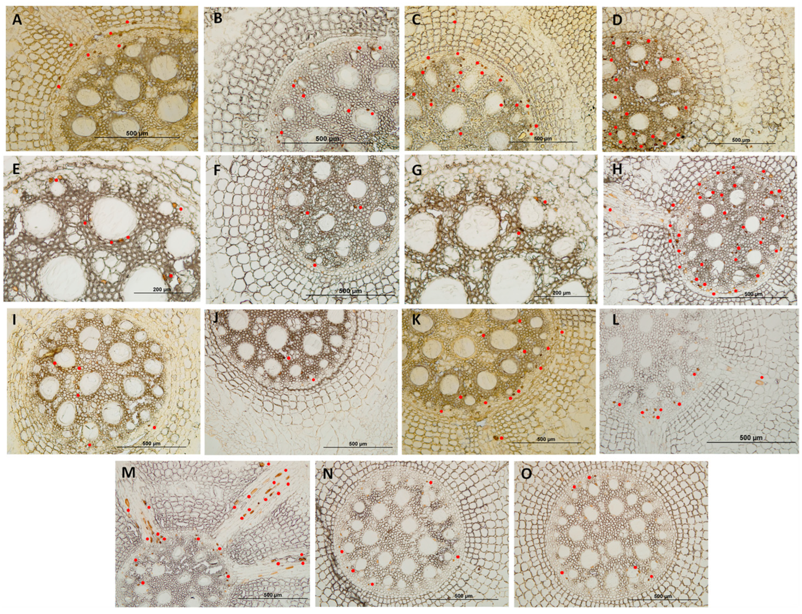
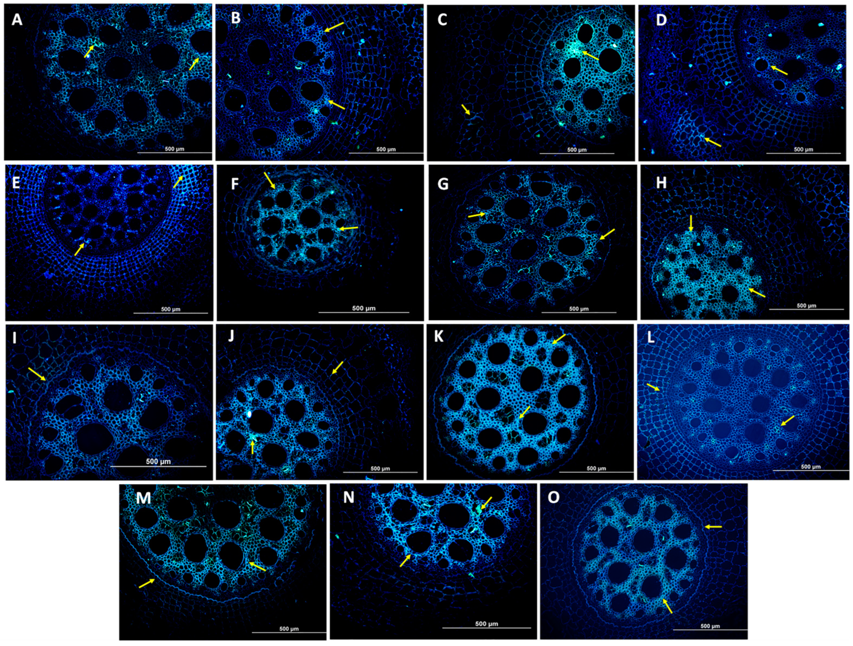
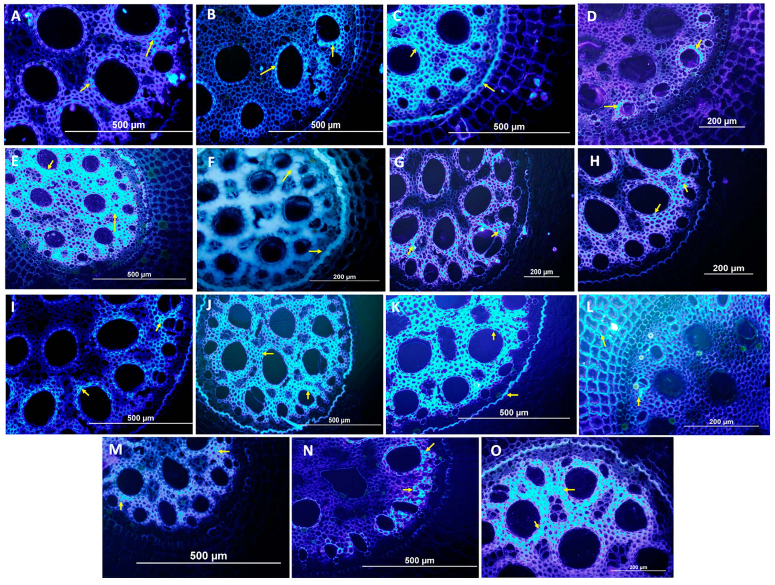
3.2. Histological and Histochemical Evaluation

To ascertain phenolic compounds, we detected small dots with a dark brown color in the rhizome tissue of all the somaclones and controls. The resistant somaclones S2, S3, and S7, associated with T1, showed a higher concentration of phenolic compounds compared with the control and other somaclones from the same treatment, as shown in Figure 2C,D,H.
In T2, the resistant somaclones S9 and S11 (K and M) showed higher concentrations of phenolic compounds compared with the control and other resistant somaclones from the same treatment. This difference is shown in Figure 2.
We observed that among the two treatments, somaclones treated with a 1 mg/L dose of TDZ (T1) showed the most intense production of phenolic compounds.
When assessing the presence of callose in the roots, somaclones S2 (C), S5 (F), and S7 (H), associated with T1, showed higher concentrations of this compound within the same treatment, indicated by the higher intensity of fluorescent light in the vascular tissue compared with the other somaclones and control. Considering T2, only somaclone S13 (O) showed a lower concentration of callose compared with the control. Notably, the somaclones derived from T2 showed higher concentrations of callose than those from T1 (Figure 3).
The analysis of cellulose showed that somaclones S2 (C) and S4 (E), linked to T1, showed the highest amount of this compound, indicated by the bluish-white color in the root tissues, followed by somaclones S5 (F), S6 (G), and S7 (H) compared with the controls. Conversely, somaclones S1 (B) and S3 (D) indicated the lowest concentration of cellulose in this treatment, as shown in Figure 4.
In T2, somaclones S8 (J), S9 (K), and S13 (O) showed the highest concentration of cellulose. The other somaclones showed a lower or equal concentration of cellulose compared with the controls. T2 showed a greater number of somaclones with the presence of cellulose, as shown in Figure 4.
In the evaluation of root whitening and staining, only the presence of hyphae was observed in the controls of the two treatments without the presence of chlamydospores, which were detected in the tissue of the vascular system of somaclones S3 (D), S5 (F), and S6 (H) in T1. In somaclones S1 (B), S2 (C), S4 (E), and S6 (G) of the same treatment, no pathogen structures were observed.
In T2, no pathogen structures were observed in the root tissue of somaclones S11 (M) and S13 (O). In somaclones S8 (J), S9 (K), S10 (L), and S11 (N) of the same treatment, the presence of chlamydospores was observed, as shown in Figure 5.

3.3. PCR Amplification and Marker Analysis

The IRAP, REMAP, and ISSR markers could not identify significant genetic differences between the resistant somaclones and control (cultivar Prata Catarina), as indicated by the band patterns identified in the somaclones, which are identical to those in the control, as shown in Figure 6A–C. Notably, the genetic alterations induced by the doses of TDZ in the somaclones only affected the level of resistance of the plants. Thus, we inferred that the selected resistant somaclones may have the same agronomic and sensory profile as the commercial Prata Catarina cultivar; this is an important fact as it increases the chances of adoption by producers and consumers. Notably, a complete agronomic and sensory characterization of the resistant somaclones will be the subject of the subsequent studies.

4. Discussion

4.1. Assessment of Resistance in the Greenhouse

In this study, we induced somaclonal variants derived from the Prata Catarina cultivar using the plant growth regulator TDZ at two concentrations, 1 and 2 mg/L. We evaluated 2400 somaclones for their resistance to Fusarium wilt. Among these, 13 were resistant after phenotyping in greenhouse conditions using soil infested with the pathogen, seven were resistant with the 1 mg/L dose, and six were resistant with the 2 mg/L dose, thereby corresponding to a selection pressure of 0.5%.
In our study, we observed a greater number of highly resistant and resistant somaclones at a TDZ dose of 1 mg/L (280 somaclones or 23% of the total, corresponding to scores of 0 and 1) compared with those at a TDZ dose of 2 mg/L (246 somaclones or 20%, corresponding to scores of 0 and 1). In banana cultivation, TDZ is used as a plant regulator to induce somaclonal variation and generate Grand Naine (Cavendish) banana somaclones resistant to Foc STR4, as indicated by Rebouças et al. [29]. The authors obtained two resistant somaclones, corresponding to an average of 1% resistant somaclones. The TDZ dose of 1 mg/L used in the study by Rebouças et al. [29] reinforces the results obtained in this study.
The potential of the technique of inducing somaclonal variation combined with supplementation with plant regulators has also been effective in crops such as wheat (Triticum aestivum L.) [32] and ornamental plants [47]. In these contexts, the authors obtained positive effects on agronomic characteristics such as grain yield, reduced plant height, number, size, and weight of flowers, leaves, and stems, as well as reduced flowering induction time and variations in flower types and colors.
In our study, no morphological changes were observed in the somaclones, such as changes in leaf color or plant size, at least until three months of plant development and evaluation. A complete agronomic characterization of the somaclones will be conducted in subsequent stages of study.
The efficacy of TDZ reported in our study and in the current literature is related to the fact that it acts as a hormone, triggering various functions in plant tissues, including an increase in the formation of lateral buds and the development of plants with a more desirable architecture for agricultural or ornamental production. [48]. TDZ has various effects on fruit crops; for example, it can improve fruit size in kiwi (Actinidia deliciosa “Hayward”), pear (Pyrus communis L. cv “Spadona” and “Coscia”), and grapes (Vitis vinifera cv “Simone”) [49,50,51] and increase yield in pears (P. calleryana cv “Hosui” and “Packham’s Triumph”) and cucumbers (Cucumis sativa L.) [52,53]. It has high cytokinin activity in in vitro cultures, promoting high rates of multiplication and shoot formation; it is effective in inducing callus and regenerating plants from plant tissues.
TDZ is also commonly used in plant tissue cultures to promote shoot formation [54,55,56]; it has an influence on morphogenesis and rooting efficiency when used in concentrations above threshold levels and/or for prolonged periods [57,58]. Ferreira et al. [24] analyzed the role of somaclonal variation in plant breeding, observing that various plant regulators are used to induce somaclonal variation. Among these regulators, TDZ was remarkable, especially at doses of 1 and 2 mg/L. Moreover, according to the authors, thousands of plants subjected to TDZ treatment are required to facilitate the selection of somaclones with desirable agronomic characteristics, especially genetic resistance to pathogens.
Thus, the concentrations of the plant regulator, together with the subcultures, cause genetic variations that can result in different phenotypes compared with the original matrices. In grapevines (Vitis vinifera) subjected to somaclonal variation induction, starting from 12 in vitro subcultures in the presence of 1-naphthaleneacetic acid (NAA) and TDZ [59], phenotypes divergent from the commercial cultivar were observed. Similarly, Bi-dabadi et al. [60] observed variation in the phenotypes of the banana cultivars “Berangan Intan”, “Berangan”, and “Rastali” when subcultured in vitro six times in the presence of benzylaminopurine (BAP) and TDZ. In our study, TDZ doses of 1 and 2 mg/L, combined with five subcultures, generated efficient genetic alterations in terms of inducing resistance to Fusarium wilt.
Regardless of the studied culture, one determining factor for the induction of somaclonal variation in vitro is the number of subcultures to which the explant is submitted. Notably, from the fifth subcultivation onward, the explants can already undergo somaclonal variation [24]. In our study, we conducted five subcultivations and identified resistant somaclones. Similarly, Miyao et al. [33] employed five subcultures on lines regenerated from cell cultures of rice (Oryza sativa L. cv. Nipponbare) and obtained three lines with resistance to the fungus Magnaporthe oryzae, which causes rice brusone. These results confirm those of in vitro variations from the fifth subcultivation cycle onward.

4.2. Analysis of Resistance Mechanisms by Histological and Histochemical Evaluations

Some resistance mechanisms may have been activated in the somaclones selected as resistant in this study. Somaclones S2, S3, and S7 (C, D, and H) in T1, and S9 and S11 in T2, showed the highest concentrations of phenolic compounds (Figure 2). Phenols are substances produced when a plant is infected by a pathogen and are accumulated in the vascular system to prevent the spread of infection. This strategy was identified by Rocha et al. [23], who studied the interaction between Musa sp vs. Fusarium oxysporum f. sp. cubense, with the aim of quantifying the virulence levels of different isolates when inoculated into resistant and susceptible banana cultivars. They observed a higher concentration of phenolic compounds in the roots of resistant plants. Similarly, Soares et al. [61] reported greater production and accumulation of phenolic compounds in cultivars resistant to Pseudocercospora fijiensis, based on histochemical analyses, which also enabled the identification of the presence of callose in the leaves of resistant genotypes in a greater quantity compared with that in susceptible cultivars. Phenolic compounds are associated with defense mechanisms and responses to adverse environmental conditions and are slightly involved in cell growth and development [62,63].
During fluorescence analysis, the presence of callose was observed in both treatments of the resistant somaclones. This observation suggests that the somaclones activated signaling pathways capable of detecting the presence of Foc in the roots through pathogen-associated molecular patterns (PAMPs), which are specific molecules found in various pathogenic microorganisms, such as bacteria, viruses, fungi, and parasites. Conversely, pattern recognition receptors (PRRs) are receptors present in cells that recognize these patterns, initiating an immune response to fight the infection. These processes enable the production and deposition of callose at the pathogen’s infection sites, strengthening the cell wall and hindering penetration by the fungus. This phenomenon, known as PAMP-triggered immunity (PTI), includes certain responses, such as the accumulation of callose, which may have occurred in the resistant somaclones in this study, wherein the presence of callose was observed in greater concentration in somaclones S2 (C), S5 (F), S7 (H), and S9 (K). This indicated that through the action of PAMPs, the presence of Foc in the roots of the somaclones and activation of this post-formed resistance mechanism could be detected.
In resistant plants, callose formation is rapidly induced, deposited mainly at infection points to reinforce the cell walls and prevent penetration by the pathogen [64,65,66,67]. In different bean genotypes inoculated with Fusarium oxysporum f. sp. phaseoli (Fop), an increase in the production of phenolic compounds and carbohydrates was observed, as well as the deposition of callose inside the xylem vessels, specifically in the resistant genotypes [68].
The third component evaluated in our study, related to post-formed resistance mechanisms, was the presence of cellulose. Thus, we conducted fluorescence analysis to observe the presence of cellulose in the xylem-conducting vessels in the roots of the resistant somaclones. The somaclones from T2, especially S8 (J), S9 (K), and S13 (O), showed greater cellulose deposition compared with the control (Figure 4). Rocha et al. [23] also detected these compounds in banana cultivars after inoculation with Foc. During the interaction with Foc 229A isolate, which was also used in this study, cellulose was observed in the Prata-Anã and Grande Naine cultivars.
When a plant is attacked by a pathogen, it activates defense responses to protect its tissues, and one of these responses involves the production of cellulose, a crucial element of the cell wall. Cellulose strengthens cell walls, hindering the penetration and spread of the pathogen. Additionally, plants deposit additional layers of cellulose around the site of infection, creating physical barriers that isolate the pathogen and prevent its spread. The presence of pathogens also activates signaling pathways that increase cellulose production, regulating genes related to its biosynthesis. These mechanisms help plants to resist infection and defend themselves against pathogens [65,66,67].
Phytopathogenic fungi produce enzymes that degrade the cell wall, such as cellulases, facilitating their invasion of host tissues. These enzymes break down cell wall components, such as wax and the cuticle, enabling the penetration and spread of the pathogen [69,70]. In this context, the resistant somaclones developed defense mechanisms with cellulose accumulation in the cell wall, which proved to be efficient against Foc. Thus, the pathogen was unable to produce enough enzymes to degrade the cellulose in the cell wall of the hosts owing to the high concentration of this compound in the vascular tissue of the resistant somaclones (Figure 4).
In the root clarification and staining technique, the absence of pathogen structures was observed in the resistant somaclones in both treatments with TDZ, indicating that these genotypes developed resistance mechanisms preventing the penetration of Foc, such as the action of phenolic compounds, callose, and cellulose. The presence of chlamydospores was detected in the tissue of the vascular system of somaclones S3, S5, and S6 in T1, and S8, S9, S10, and S12 in T2. The analyzed somaclones showed pathogen structures in the vascular system; however, successful infection by the pathogen was prevented. This resistance can be attributed to the aforementioned mechanisms or possible genetic alterations that have not yet been investigated.
These findings indicate that the pathogen was unable to successfully establish infection in somaclones of T1 and T2 owing to the effectiveness of the existing defense mechanisms, differently from what occurred in the control treatment. In somaclone S2, the three defense mechanisms analyzed in this study were identified: phenolic compounds, callose, and cellulose. In somaclone S7, phenolic compounds and callose were observed, whereas in somaclone S9, phenolic compounds and cellulose were detected. Somaclone S13 showed the presence of callose and cellulose. The other somaclones showed only one of the studied defense mechanisms. Notably, by presenting three different resistance mechanisms, somaclone S2 may be superior to the others selected in this study as the pathogen will need to overcome three different obstacles for successful infection.
Post-formed mechanisms of genetic resistance, such as phenolic compounds, callose, and cellulose, may have been activated by the resistant plants that contained structures of the pathogen in the root tissue, which did not enable the spread of infection (Figure 5). These findings can be explained by the fact that chlamydospores and microconidia germinate around the root tip and between the root hairs of banana genotypes, before penetrating the epidermal cells and moving through the intercellular elongation zone to start the infection process [29,71,72]. Thus, the somaclones that did not contain Foc spores may have developed resistance mechanisms that did not enable the entry of the pathogen into the root tissues, which must be verified in future studies.

4.3. Analysis of the Extent of Genetic Diversity in Somaclones Using Molecular Markers IRAP, REMAP, and ISSR

The molecular markers used in our study (IRAP, REMAP, and ISSR) were selected to cover different parts of the somaclone genome, enabling the detection of genetic variations between somaclones and between somaclones and their parents.
Our results indicated that the resistant somaclones have high genetic similarity with the Prata Catarina cultivar, from which they were derived. The array of markers used indicated that the bands/allele patterns were mostly similar to the commercial cultivar. Thus, we inferred that the somaclones did not differ agronomically from Prata Catarina in terms of the agronomic characters, such as bunch weight, number of fruits, or the sensory profile. This information will be verified in future studies on the commercial potential of somaclones through field experiments.
Ferreira et al. [24] indicated that the IRAP and REMAP markers are considered efficient molecular markers for investigating the genetic variability in various crops. These markers are based on retrotransposons that are dispersed throughout the plant genome and can contain thousands of copies, thus contributing to the size, structure, diversity, and variation of the genome, which is a factor that can affect gene function [73].
Muhammad et al. [74] used random amplified polymorphic DNA (RAPD) markers, which revealed greater polymorphism compared with IRAP markers when analyzing somaclones derived from the silk subgroup (AAB) banana cultivar called “Rasthali.” They concluded that somaclonal variation appears to be derived from multiple indels scattered throughout the genome, as a response to stress induced by micropropagation. Therefore, for the comprehensive characterization of somaclonal variants, more than one DNA marker system must be employed to detect variations in various regions of the genome, as was used in our study.
Another molecular marker used to identify genetic variations, in addition to the aforementioned markers, is amplified fragment length polymorphism (AFLP). This molecular technique is widely used to detect genetic variations between different DNA samples, combining the digestion of DNA with restriction enzymes and selective amplification of DNA fragments using polymerase chain reaction (PCR), enabling the detailed analysis of somaclonal variation [24]. Munsamy et al. [31] investigated the increased frequency of somaclonal variants of sugarcane plants (Saccharum spp.) produced in vitro using the AFLP marker. They identified genetic variations in at least one sugarcane somaclone that was resistant to brown rust (Puccinia melanocephala).
Today, next-generation sequencing technology is increasingly being used to study somaclonal variation and genetic variability in plants. Approaches, including whole-genome sequencing, offer a more detailed view of genetic changes in somaclones [75]. Therefore, although the traditional methods discussed in this study are still widely used, whole-genome sequencing technologies have become more accessible and offer a more comprehensive and accurate perspective on somaclonal variation. These new technologies will be employed in our future studies on the selected resistant somaclones.

5. Conclusions

In this study, the application of the plant regulator TDZ proved to be effective in producing 13 somaclones derived from the cultivar Prata Catarina (subgroup Prata, AAB), with resistance to Fusarium wilt. The 1 mg/L dose was more efficient in obtaining somaclones in terms of the number of resistant plants. Molecular analyses did not reveal genetic changes in the somaclones; it is possible that their agronomic characteristics remained unchanged compared to those of the commercial genotype. However, more studies are essential to support this hypothesis. Results from the histological and histochemical evaluations corroborate the observed resistance, indicating the presence of phenolic compounds, callose, and cellulose as possible contributing mechanisms to this resistance. These findings indicate the presence of post-formed resistance mechanisms in the resistant somaclones. Our results were promising, indicating that somaclonal variation induction is an effective and efficient approach for developing banana cultivars resistant to this disease. However, additional studies will be conducted with the resistant somaclones to obtain information on their commercial value, thereby benefiting regions where the disease limits the production of Prata bananas in various producing areas of Brazil.

Author Contributions

M.d.S.F. and E.P.A. conceived the idea that gave rise to the theme of this article. M.d.S.F., F.H., C.F.F., J.A.d.S.-S. and E.P.A. developed the research project. M.d.S.F., A.C.L.S.d.S., J.P.F.L.d.J., W.D.d.S.O., A.P.d.S.R., A.C.L.S.d.S., J.P.F.L.d.J. and T.A.R. helped in the development of research work activities. M.d.S.F. wrote the article. E.P.A., C.F.F., F.H., J.A.d.S.-S., T.A.R. and A.d.J.R. provided technical guidance and research supervision and s critical review of the study. All authors collaborated in the editing part of the writing process. All authors have read and agreed to the published version of the manuscript.

Funding

This research was funded by IITA/The Bill and Melinda Gates Foundation—Accelerated. Breeding of Better Bananas, ID OPP1093845.

Data Availability Statement

The datasets presented in this article are not readily available because the data are part of an ongoing study. Requests to access the datasets should be directed to the corresponding author.

Acknowledgments

The authors thank the Graduate Program in Biotechnology (PPGBiotec) of the State University of Feira de Santana, as well as FAPESB (Bahia Research Foundation) for granting DSc. scholarships to M.d.S.F., and CNPq (National Council for Scientific and Technological Development) for the research productivity grants for E.P.A. and C.F.F. The authors thank CAPES (Coordination for the Improvement of Higher Education Personnel) for granting DSc. scholarships to W.D.d.S.O.

Conflicts of Interest

The authors declare no conflicts of interest.

References

  1. FAO. Available online: https://www.fao.org/world-banana-forum/zh/ (accessed on 8 June 2024).
  2. FAOSTAT. 2024. Available online: https://www.fao.org/faostat/en/#data (accessed on 10 May 2024).
  3. FAO. Banana Statistical Compendium 2021; Food and Agriculture Organization of the United Nations: Rome, Italy, 2022. [Google Scholar]
  4. Lichtemberg, L.A.; Hinz, R.H.; Malburg, J.L.; Peruch, L.A.M.; Sonego, M. Agriculture of Santa Catarina SCS451—Catarina—New Cultivar of Banana Plant of the Prata Subgroup; Epagri—Experimental Station of Itajaí: Santa Catarina, Brazil, 2011. [Google Scholar]
  5. Soni, K.B.; Jadhav, P.R.; Alex, S. Genetic Improvement of Banana. In Genetic Engineering of Crop Plants for Food and Health Security: Volume 1; Springer Nature: Singapore, 2024; pp. 305–329. [Google Scholar]
  6. Willadino, L.; Camara, T.R.; Ribeiro, M.B.; Amaral, D.O.J.; Suassuna, F.; Silva, M.V.D. Mechanisms of tolerance to salinity in banana: Physiological, biochemical, and molecular aspects. Rev. Bras. Frutic. 2017, 39, 1–8. [Google Scholar] [CrossRef]
  7. Nansamba, M.; Sibiya, J.; Tumuhimbise, R.; Karamura, D.; Kubiriba, J.; Karamura, E. Breeding banana (Musa spp.) for drought tolerance: A review. Plant Breed. 2020, 139, 685–696. [Google Scholar] [CrossRef]
  8. Monteiro, J.D.; Santos, M.; Santos, J.R.P.; Cares, J.E.; Marchão, R.L.; Amorim, E.P.; Costa, D.D.C. Identification of plant parasitic nematodes in triploid and tetraploid bananas in brazil. Rev. Caatinga 2020, 33, 865–877. [Google Scholar] [CrossRef]
  9. Sairam, S.; Selvarajan, R.; Handanahalli, S.S.; Venkataraman, S.; Sairam, S.; Selvarajan, R.; Handanahalli, S.S.; Venkataraman, S. Towards understanding the structure of the capsid of Banana Bunchy Top Virus. BioRxiv 2020. [Google Scholar] [CrossRef]
  10. Studholme, D.J.; Wicker, E.; Abrare, S.M.; Aspin, A.; Bogdanove, A.; Broders, K.; Dubrow, Z.; Grant, M.; Jones, J.B.; Karamura, G.; et al. Transfer of Xanthomonas campestris pv. arecae and X. campestris pv. musacearum to X. vasicola (Vauterin) as X. vasicola pv. arecae comb. nov. and X. vasicola pv. musacearum comb. nov. and description of X. vasicola pv. vasculorum pv. nov. Phytopathology 2020, 110, 1153–1160. [Google Scholar] [CrossRef] [PubMed]
  11. Nascimento, F.D.S.; Sousa, Y.M.; Rocha, A.D.J.; Ferreira, C.F.; Haddad, F.; Amorim, E.P. Sources of black Sigatoka resistance in wild banana diploids. Rev. Bras. Frutic. 2020, 42, e-038. [Google Scholar] [CrossRef]
  12. Gonçalves, Z.S.; Haddad, F.; Amorim, V.B.O.; Ferreira, C.F.; Oliveira, S.A.S.; Amorim, E.P. Agronomic characterization and identification of banana genotypes resistant to Fusarium wilt race 1. Eur. J. Plant Pathol. 2019, 155, 1093–1103. [Google Scholar] [CrossRef]
  13. Ploetz, R.C. Fusarium wilt management in banana: A review with special reference to tropical race 4. Crop Prot. 2015, 73, 7–15. [Google Scholar] [CrossRef]
  14. Ploetz, R.C. Gone Bananas? Current and Future Impact of Fusarium Wilt on Production. In Plant Diseases and Food Security in the 21st Century; Scott, P., Strange, R., Korsten, L., Gullino, M.L., Eds.; Springer Nature: Cham, Switzerland, 2021; Volume 10, pp. 21–32. [Google Scholar] [CrossRef]
  15. García-Bastidas, F.A.; Quintero-Vargas, J.C.; Ayala-Vasquez, M.; Schermer, T.; Seidl, M.F.; Santos-Paiva, M.; Noguera, A.M.; Aguilera-Galvez, C.; Wittenberg, A.; Hofstede, R.; et al. First report of Fusarium wilt Tropical race 4 in Cavendish bananas caused by Fusarium odoratissimum in Colombia. Plant Dis. 2020, 104, 994. [Google Scholar] [CrossRef]
  16. Maryani, N.; Lombard, L.; Poerba, Y.S.; Subandiyah, S.; Crous, P.W.; Kema, G.H.J. Phylogeny and genetic diversity of the banana Fusarium wilt pathogen Fusarium oxysporum f. sp. cubense in the Indonesian centre of origin. Stud. Mycol. 2019, 92, 155–194. [Google Scholar] [CrossRef]
  17. Martínez, G.; Olivares, B.O.; Rey, J.C.; Rojas, J.; Cardenas, J.; Muentes, C.; Dawson, C. The Advance of FusariumWilt Tropical Race 4 in Musaceae of Latin America and the Caribbean: Current Situation. Pathogens 2023, 12, 277. [Google Scholar] [CrossRef] [PubMed]
  18. Costa, S.N.; Bragança, C.A.D.; Ribeiro, L.R.; Amorim, E.P.; Silva, S.O.; Dita, M.; Laranjeira, F.F.; Haddad, F. Genetic structure of Fusarium oxysporum f. sp. cubense in diferent regions from Brazil. Plant Pathol. 2015, 64, 37–146. [Google Scholar] [CrossRef]
  19. Batista, I.C.A.; Heck, D.W.; Santos, A.; Alves, G.; Ferro, C.G.; Dita, M.; Haddad, F.; Michereff, S.J.; Correia, K.C.; da Silva, C.F.B.; et al. The Population of Fusarium oxysporum f. sp. cubense in Brazil Is Not Structured by Vegetative Compatibility Group or by Geographic Origin. Phytopathology 2022, 112, 2416–2425. [Google Scholar] [CrossRef] [PubMed]
  20. Dita, M.; Barquero, M.; Heck, D.; Mizubute, E.S.G.; Staver, C.P. Fusarium wilt of banana: Current knowledge on epidemiology and research needs toward sustainable disease management. Front. Plant Sci. 2018, 9, 1468–1488. [Google Scholar] [CrossRef]
  21. Pegg, K.G.; Coates, L.M.; O’Neill, W.T.; Turner, D.W. The epidemiology of fusarium wilt of Banana. Front. Plant Sci. 2019, 10, 1395. [Google Scholar] [CrossRef] [PubMed]
  22. Jamil, F.N.; Tang, C.N.; Saidi, N.B.; Lai, K.S.; Baharum, N.A. Fusarium wilt in banana: Epidemics and management strategies. In Horticultural Crops; Kossi Baimey, H., Hamamouch, N., Adjiguita Kolombia, Y., Eds.; IntechOpen: London, UK, 2019; pp. 229–331. [Google Scholar]
  23. Rocha, A.D.; Soares, J.M.; Nascimento, F.D.; Rocha, A.D.; Amorim, V.B.; Ramos, A.P.; Ferreira, C.F.; Haddad, F.; Amorim, E.P. Molecular, histological and histochemical responses of banana cultivars challenged with Fusarium oxysporum f. sp. cubense with different levels of virulence. Plants 2022, 11, 2339. [Google Scholar] [CrossRef] [PubMed]
  24. Ferreira, M.D.; Rocha, A.D.; Nascimento, F.D.; Oliveira, W.D.; Soares, J.M.; Rebouças, T.A.; Morais Lino, L.S.; Haddad, F.; Ferreira, C.F.; Santos-Serejo, J.A.; et al. The role of somaclonal variation in plant genetic improvement: A systematic review. Agronomy 2023, 13, 730. [Google Scholar] [CrossRef]
  25. Davey, M.R.; Anthony, P. Plant Cell Culture: Essential Methods; John Wiley & Sons: Hoboken, NJ, USA, 2010. [Google Scholar]
  26. Bairu, M.W.; Aremu, A.O.; Van Staden, J. Somaclonal variation in plants: Causes and detection methods. Plant Growth Regul. 2011, 63, 147–173. [Google Scholar] [CrossRef]
  27. Jain, S.M.; Brar, D.S.; Ahloowalia, B.S. (Eds.) Somaclonal Variation and Induced Mutations in Crop Improvement; Springer Science & Business Media: Berlin/Heidelberg, Germany, 2013. [Google Scholar]
  28. Hou, B.H.; Tsai, Y.H.; Chiang, M.H.; Tsao, S.M.; Huang, S.H.; Chao, C.P.; Chen, H.M. Cultivar-specific markers, mutations, and chimerisim of Cavendish banana somaclonal variants resistant to Fusarium oxysporum f. sp. cubense tropical race 4. BMC Genom. 2022, 23, 470. [Google Scholar] [CrossRef]
  29. Rebouças, T.A.; Rocha, A.d.J.; Cerqueira, T.S.; Adorno, P.R.; Barreto, R.Q.; Ferreira, M.; Lino, L.S.M.; Amorim, V.B.D.; Santos-Serejo, J.A.D.; Haddad, F.; et al. Pre-selection of banana somaclones resistant to Fusarium oxysporum f. sp. cubense, subtropical race 4. Crop Prot. 2021, 147, 105692. [Google Scholar] [CrossRef]
  30. Hwang, S.C.; Ko, W.H. Cavendish banana cultivars resistant to Fusarium wilt acquired through somaclonal variation in Taiwan. Plant Dis. 2004, 88, 580–587. [Google Scholar] [CrossRef] [PubMed]
  31. Munsamy, A.; Rutherford, R.S.; Snyman, S.J.; Watt, M.P. 5-Azacytidine as a tool to induce somaclonal variants with useful traits in sugarcane (Saccharum spp.). Plant Biotechnol. Rep. 2013, 7, 489–502. [Google Scholar] [CrossRef]
  32. Wang, K.; Wang, S.; Zhou, X.; Lin, Z.; Li, J.; Du, L.; Tang, Y.; Xu, H.; Yan, Y.; Ye, X. Development, identification, and genetic analysis of a quantitative dwarfing somatic variation line in wheat. Crop Sci. 2013, 53, 1032–1041. [Google Scholar] [CrossRef]
  33. Miyao, A.; Nakagome, M.; Ohnuma, T.; Yamagata, H.; Kanamori, H.; Katayose, Y.; Takahashi, A.; Matsumoto, T.; Hirochika, H. Molecular spectrum of somaclonal variation in regenerated rice revealed by whole-genome sequencing. Plant Cell Physiol. 2012, 53, 256–264. [Google Scholar] [CrossRef] [PubMed]
  34. Murashige, T.; Skoog, T. A revised medium for rapid growth and bioassays with tobacco tissue cultures. Physiol. Plantarum. 1962, 15, 473–497. [Google Scholar] [CrossRef]
  35. Velame, K.V.; Rocha, A.D.; Ferreira, M.D.; Haddad, F.; Amorim, V.B.; Pestana, K.N.; Ferreira, C.F.; Alves Santos de Oliveira, S.; Amorim, E.P. Evidence of Correlation between Pathogenicity, Avirulence Genes, and Aggressiveness of Fusarium oxysporum f. sp. cubense in Banana “Cavendish” and “Prata” Subgroups. Horticulturae 2024, 10, 228. [Google Scholar] [CrossRef]
  36. Madigan, M.T.; Martinko, J.M.; Bender, K.S.; Buckley, D.H.; Stahl, D.A. Microbiologia de Brock, 14th ed.; Artmed Editora: Porto Alegre, Brazil, 2016. [Google Scholar]
  37. Dita, M.A.; Pérez Vicente, L.; Martínez, E. Inoculation of Fusarium oxysporum f. sp. cubense causal agent of fusarium wilt in banana. In Technical Manual: Prevention and Diagnostic of Fusarium Wilt of Banana Caused by Fusarium oxysporum f. sp. cubense Tropical Race 4 (TR4); Pérez Vicente, L., Dita, M.A., de la Parte, E.M., Eds.; Food and Agriculture Organization of the United Nations: Rome, Italy, 2014; pp. 55–58. [Google Scholar]
  38. R Core Team. R: A Language and Environment for Statistical Computing; The R Foundation for Statistical Computing: Vienna, Austria, 2020; Volume 1. [Google Scholar]
  39. Karnovsky, M.J. A formaldehydeglutaraldehyde fixative of high osmolality for use in electron microscopy. J. Cell Biol. 1965, 27, 137. [Google Scholar]
  40. Johansen, D.A. Plant Microtechnique; Mc Graw Hill: New York, NY, USA, 1940; 523p. [Google Scholar]
  41. Hughes, J.; McCully, M.E. The use of an optical brightener in the study of plant structures. Stain Technol. 1975, 50, 319–329. [Google Scholar] [CrossRef] [PubMed]
  42. Phillips, J.M.; Hayman, D.S. Improved procedures for clearing roots and staining parasitic and vesicular-arbuscular mycorrhizal fungi for rapid assessment of infection. Trans. Br. Mycol. Soc. 1970, 55, 158–161. [Google Scholar] [CrossRef]
  43. Doyle, J.J.; Doyle, J.L. Isolation of plant DNA from fresh tissue. Focus Rockv. 1990, 12, 13–15. [Google Scholar]
  44. Ferreira, C.F.; Gutierrez, D.L.; Kreuze, J.F.; Iskra-Caruana, M.L.; Chabannes, M.; Barbosa, A.C.O.; Santos, T.A.; Silva, A.G.S.; Santos, R.M.F.; Amorim, E.P.; et al. Rapid plant dna and rna extraction protocol using a bench drill brief note. Genet. Mol. Res. 2019, 18, gmr18394. [Google Scholar] [CrossRef]
  45. Kalendar, R.; Grob, T.; Regina, M.; Suoniemi, A.; Schulman, A. IRAP and REMAP: Two new retrotransposon-based DNA fingerprinting techniques. Theor. Appl. Genet. 1999, 98, 704–711. [Google Scholar] [CrossRef]
  46. Sankar, A.A.; Moore, G.A. Evaluation of inter-simple sequence repeat analysis for mapping in Citrus and extension of the genetic linkage map. Theor. Appl. Genet. 2001, 102, 206–214. [Google Scholar] [CrossRef]
  47. Eeckhaut, T.; Van Houtven, W.; Bruznican, S.; Leus, L.; Van Huylenbroeck, J. Somaclonal variation in Chrysanthemum× morifolium protoplast regenerants. Front. Plant Sci. 2020, 11, 607171. [Google Scholar] [CrossRef] [PubMed]
  48. Erland, L.A.; Giebelhaus, R.T.; Victor, J.M.; Murch, S.J.; Saxena, P.K. The morphoregulatory role of thidiazuron: Metabolomics-guided hypothesis generation for mechanisms of activity. Biomolecules 2020, 10, 1253. [Google Scholar] [CrossRef] [PubMed]
  49. Famiani, F.; Proietti, P.; Pilli, M.; Battistelli, A.; Moscatello, S. Effects of application of thidiazuron (TDZ), gibberellic acid (GA 3), and 2,4-dichlorophenoxyacetic acid (2,4-D) on fruit size and quality of Actinidia deliciosa ‘Hayward’. N. Z. J. Crop Hortic. Sci. 2007, 35, 341–347. [Google Scholar] [CrossRef]
  50. Stern, R.; Shargal, A.; Flaishman, M. Thidiazuron increases fruit size of ‘Spadona’ and ‘Coscia’ pear (Pyrus communis L.). J. Hortic. Sci. Biotechnol. 2003, 78, 51–55. [Google Scholar] [CrossRef]
  51. Reynolds, A.; Wardle, D.; Zurowski, C.; Looney, N. Phenylureas CPPU and Thidiazuron Affect YieldComponents, Fruit Composition, and Storage Potential of Four Seedless Grape Selections. J. Am. Soc. Hortic. Sci. 1992, 1, 85–89. [Google Scholar] [CrossRef]
  52. Pasa, M.S.; Silva, C.P.D.; Carra, B.; Brighenti, A.F.; Souza, A.L.K.D.; Petri, J.L. Thidiazuron (TDZ) increases fruit set and yield of ‘Hosui’ and ‘Packham’s Triumph’ pear trees. An. Acad. Bras. Ciênc. 2017, 89, 3103–3110. [Google Scholar] [CrossRef]
  53. Yang, Y.-Z.; Lin, D.-C.; Guo, Z.-Y. Promotion of fruit development in cucumber (Cucumis sativus) by thidiazuron. Sci. Hortic. 1992, 50, 47–51. [Google Scholar] [CrossRef]
  54. Shen, X.; Kane, M.E.; Chen, J. Effects of genotype, explant source, and plant growth regulators on indirect shoot organogenesis in Dieffenbachia cultivars. In Vitro Cell Dev Biol Plant 2008, 44, 282–288. [Google Scholar] [CrossRef]
  55. Ali, H.M.; Khan, T.; Khan, M.A.; Ullah, N. The multipotent thidiazuron: A mechanistic overview of its roles in callogenesis and other plant cultures in vitro. Biotechnol. Appl. Biochem. 2022, 69, 2624–2640. [Google Scholar] [CrossRef] [PubMed]
  56. Tymoszuk, A.; Kulus, D. In Vitro Technology and Micropropagated Plants; MDPI: Basel, Switzerland, 2024. [Google Scholar] [CrossRef]
  57. Dewir, Y.H.; Murthy, H.N.; Ammar, M.H.; Alghamdi, S.S.; Al-Suhaibani, N.A.; Alsadon, A.A.; Paek, K.Y. In vitro rooting of leguminous plants: Difficulties, alternatives, and strategies for improvement. Hortic. Environ. Biotechnol. 2016, 57, 311–322. [Google Scholar] [CrossRef]
  58. Dewir, Y.H.; Nurmansyah Naidoo, Y.; Teixeira da Silva, J.A. Thidiazuron-induced abnormalities in plant tissue cultures. Plant Cell Rep. 2018, 37, 1451–1470. [Google Scholar] [CrossRef] [PubMed]
  59. Pop, R.; Pamfil, D.; Hârța, M.; Clapa, D.; Cordea, M.I. Assessment of somaclonal variation in micropropagated grapevine cultivars using molecular markers. Sci. Pap. Ser. B Hortic. 2023, LXVII, 1. [Google Scholar]
  60. Bidabadi, S.S.; Meon, S.; Wahab, Z.; Mahmood, M. Study of genetic and phenotypic variability among somaclones induced by BAP and TDZ in micropropagated shoot tips of banana (Musa spp.) using RAPD markers. J. Agric. Sci. 2010, 2, 49–60. [Google Scholar]
  61. Soares, J.M.; Rocha, A.D.; Nascimento, F.D.; Amorim, V.B.; Ramos, A.P.; Ferreira, C.F.; Haddad, F.; Amorim, E.P. Gene Expression, Histology and Histochemistry in the Interaction between Musa sp. and Pseudocercospora fijiensis. Plants 2022, 11, 1953. [Google Scholar] [CrossRef] [PubMed]
  62. Alonso-Herrada, J.; Rico-Reséndiz, F.; Campos-Guillén, J.; Guevara-González, R.G.; Torres-Pacheco, I.; Cruz-Hernández, A. Establishment of in vitro regeneration system for Acaciella angustissima (Timbe) a shrubby plant endemic of México for the production of phenolic compounds. Ind. Crops Prod. 2016, 86, 49–57. [Google Scholar] [CrossRef]
  63. Dias, M.I.; Sousa, M.J.; Alves, R.C.; Ferreira, I.C. Exploring plant tissue culture to improve the production of phenolic compounds: A review. Ind. Crops Prod. 2016, 82, 9–22. [Google Scholar] [CrossRef]
  64. Wu, S.; Shan, L.; He, P. Microbial signature-triggered plant defense responses and early signaling mechanisms. Plant Sci. 2014, 228, 118–126. [Google Scholar] [CrossRef]
  65. Wang, B.; Andargie, M.; Fang, R. The function and biosynthesis of callose in high plants. Heliyon 2022, 8, e09248. [Google Scholar] [CrossRef] [PubMed]
  66. Ellinger, D.; Voigt, C.A. Callose biosynthesis in Arabidopsis with a focus on pathogen response: What we have learned within the last decade. Ann. Bot. 2014, 114, 1349–1358. [Google Scholar] [CrossRef] [PubMed]
  67. Li, N.; Lin, Z.; Yu, P.; Zeng, Y.; Du, S.; Huang, L.J. The multifarious role of callose and callose synthase in plant development and environment interactions. Front. Plant Sci. 2023, 14, 1183402. [Google Scholar] [CrossRef] [PubMed]
  68. De Quadros, F.M.; de Freitas, M.B.; Simioni, C.; Ferreira, C.; Stadnik, M.J. Redox status regulation and action of extra-and intravascular defense mechanisms are associated with bean resistance against Fusarium oxysporum f. sp. phaseoli. Protoplasma 2020, 257, 1457–1472. [Google Scholar] [CrossRef] [PubMed]
  69. Kikot, G.E.; Hours, R.A.; Alconada, T.M. Contribution of cell wall degrading enzymes to pathogenesis of Fusarium graminearum: A review. J. Basic Microbiol. 2009, 49, 231–241. [Google Scholar] [CrossRef] [PubMed]
  70. Najjarzadeh, N.; Matsakas, L.; Rova, U.; Christakopoulos, P. How carbon source and degree of oligosaccharide polymerization affect production of cellulase-degrading enzymes by Fusarium oxysporum f. sp. lycopersici. Front. Microbiol. 2021, 12, 652655. [Google Scholar] [CrossRef] [PubMed]
  71. Xiao, R.F.; Zhu, Y.-J.; Li, Y.-D.; Liu, B. Studies on vascular infection of Fusarium oxysporum f. sp. cubense race 4 in banana by field survey and green fluorescent protein reporter. Int. J. Phytopathol. 2013, 2, 44–51. [Google Scholar] [CrossRef]
  72. Warman, N.M.; Aitken, E.A. The movement of Fusarium oxysporum f. sp. cubense (sub-tropical race 4) in susceptible cultivars of banana. Front. Plant Sci. 2018, 9, 420641. [Google Scholar] [CrossRef]
  73. Sheeja, T.E.; Kumar, I.P.V.; Giridhari, A.; Minoo, D.; Rajesh, M.K.; Babu, K.N. Amplified Fragment Length Polymorphism: Applications and recent developments. Mol. Plant Taxon. 2021, 2222, 187–218. [Google Scholar]
  74. Muhammad, A.J.; Othman, F.Y. Characterization of Fusarium wilt-resistant and Fusarium wilt-susceptible somaclones of banana cultivar Rastali (Musa AAB) by random amplified polymorphic DNA and retrotransposon markers. Plant Mol. Biol. Report. 2005, 23, 241–249. [Google Scholar] [CrossRef]
  75. Skarzyńska, A.; Pawełkowicz, M.; Pląder, W. Genome-wide discovery of DNA variants in cucumber somaclonal lines. Gene 2020, 736, 144412. [Google Scholar] [CrossRef] [PubMed]
Figure 1. Internal symptoms of Fusarium wilt in the somaclones of Prata Catarina (AAB) banana plants evaluated in the greenhouse. (A) Bar graph with the number of plants with each grade of symptoms according to the grading scale, which varied from 1 to 4, and cross-section of the rhizome with the respective degrees of symptoms. (B) Boxplot of the internal disease symptom indices (DI%). Trat 1: treatment 1, with a TDZ dose of 1 mg/L; Trat 2: treatment 2, with a TDZ dose of 2 mg/L. The treatments differ statistically, as indicated by the letters a, b, and c.
Agronomy 14 01740 g001
Figure 2. Cross-sectional micrographs of the roots of somaclones of the cultivar Prata Catarina, considered resistant to infection by Foc isolate CNPMF 229. The red dots indicate the presence of phenolic compounds. Controls of (A) T1 and (I) T2: (B) S1, (C) S2, (D) S3, (E) S4, (F) S5, (G) S6, and (H) S7 are resistant somaclones in T1; (J) S8, (K) S9, (L) S10, (M) S11, (N) S12, and (O) S13, are resistant somaclones in T2.
Agronomy 14 01740 g002
Figure 3. Fluorescence micrographs of cross-sections of the roots of somaclones of the cultivar Prata Catarina, considered resistant to infection by Foc isolate CNPMF 229. The yellow arrows indicate fluorescent regions with the presence of callose. Controls of (A) T1 and (I) T2: (B) S1, (C) S2, (D) S3, (E) S4, (F) S5, (G) S6, and (H) S7 represent the resistant somaclones in T1; (J) S8, (K) S9, (L) S10, (M) S11, (N) S12, and (O) S13 represent the resistant somaclones in T2.
Agronomy 14 01740 g003
Figure 4. Fluorescence micrographs of cross-sections of the roots of somaclones of the cultivar Prata Catarina, considered resistant to infection by Foc isolate CNPMF 229. The yellow arrows indicate the presence of cellulose. Controls of (A) T1 and (I) T2: (B) S1, (C) S2, (D) S3, (E) S4, (F) S5, (G) S6, and (H) S7represent the resistant somaclones in T1; (J) S8, (K) S9, (L) S10, (M) S11, (N) S12, and (O) S13 represent the resistant somaclones in T2.
Agronomy 14 01740 g004
Figure 5. Micrographs of root fragments from somaclones of the cultivar Prata Catarina, considered resistant to infection by Foc isolate CNPMF 229. The arrows indicate chlamydospores (Chl) and fungal hyphae (Hyp). Controls of (A) T1 and (I) T2: (B) S1, (C) S2, (D) S3, (E) S4, (F) S5, (G) S6, and (H) S7 represent the resistant somaclones in T1; (J) S8, (K) S9, (L) S10, (M) S11, (N) S12, and (O) S13 represent the resistant somaclones in T2.
Agronomy 14 01740 g005
Figure 6. Molecular analysis to identify genetic changes in somaclones resistant to Foc isolate CNPMF 229. (A) Inter-retrotransposon amplified polymorphism (IRAP) markers Sukula + LTR6149 combination; (B) retrotransposon–microsatellite amplified polymorphism (REMAP) markers REMAP: LTR reverse 7286 + 8387; (C) inter-simple sequence repeat (ISSR) markers ISSR-7. 1kb Invitrogem®® (Waltham, MA, USA) marker; Prata Catarina cultivar controls (1 and 9), 2: S1, 3: S2, 4: S3, 5: S4, 6: S5, 7: S6, and 8: S7; these correspond to the resistant somaclones in T1. 10: S8, 11: S9, 12: S10, 13: S11, 14: S12, and 15: S13; these correspond to the resistant somaclones in T2.
Agronomy 14 01740 g006
Table 1. List of markers used to discriminate somaclones of Prata banana (AAB).
Initiator IdentificationNucleotide Sequence (5–3)Annealing Temperature (°C)
REMAP *
LTR reverse 7286 GGAA11CATAGCATGGATAA
TAAACGATTATC
54 °C; 58 °C
8081(GA)9C54 °C
8082(CT)9G54 °C
8385(CAC)7G58 °C
8386(GTG)7C58 °C
8387(CA)10G54 °C
8564(CAC)7T58 °C
8565GT(CAC)758 °C
IRAP **
LTR6149 + TR6150 CTCGCTCGCCCACTACATCAACCGCGTTTATT
CTGGTTCGCCCCATCTCTATCTATCCACACATGTA
42 °C
LTR6150 + 5′LTR2 CTGGTTCGCCCCATCTCTATCTATCCACACATGTA
ATCATTGCCTCTAGGGCATAATTC
42 °C
3′LTR + LTR6150 TGTTTCCCATGCGACGTTCCCCAACA
CTGGTTCGCCCCATCTCTATCTATCCACACATGTA
42 °C
5′LTR2 + Nikita ATCATTGCCTCTAGGGCATAATTC
CGCATTTGTTCAAGCCTAAACC
42 °C
3′LTR + Nikita TGTTTCCCATGCGACGTTCCCCAACA
CGCATTTGTTCAAGCCTAAACC
46 °C
Nikita + LTR6149 CGCATTTGTTCAAGCCTAAACC
CTCGCTCGCCCACTACATCAACCGCGTTTATT
46 °C
5′LTR2 + LTR6150 ATCATTGCCTCTAGGGCATAATTC
CTGGTTCGCCCCATCTCTATCTATCCACACATGTA
46 °C
Sukula + LTR6150 GATAGGGTCGCATCTTGGGCGTGAC
CTGGTTCGCCCCATCTCTATCTATCCACACATGTA
46 °C
ISSR ***
ISSR-7(AG)9 48 °C
ISSR-23(AG)8AT 45 °C
* REMAP (retrotransposon–microsatellite amplified polymorphism); ** IRAP (inter-retrotransposon amplified polymorphism); *** ISSR (inter-simple sequence repeat).
Disclaimer/Publisher’s Note: The statements, opinions and data contained in all publications are solely those of the individual author(s) and contributor(s) and not of MDPI and/or the editor(s). MDPI and/or the editor(s) disclaim responsibility for any injury to people or property resulting from any ideas, methods, instructions or products referred to in the content.

