Article

Alterations in Physiological Parameters and Secondary Metabolites of Astragalus adsurgens Infected by the Pathogen Alternaria gansuensis

1
Grassland Research Center of National Forestry and Grassland Administration, Institute of Ecological Protection and Restoration, Chinese Academy of Forestry, Beijing 100091, China
2
State Key Laboratory of Herbage Improvement and Grassland Agro-Ecosystems, Lanzhou University, Lanzhou 730020, China
3
Engineering Research Center of Grassland Industry, Ministry of Education, Lanzhou University, Lanzhou 730020, China
4
Key Laboratory of Grassland Livestock Industry Innovation, Ministry of Agriculture and Rural Affairs, Lanzhou University, Lanzhou 730020, China
5
Gansu Tech Innovation Center of Western China Grassland Industry, Lanzhou University, Lanzhou 730020, China
6
College of Pastoral Agriculture Science and Technology, Lanzhou University, Lanzhou 730020, China
7
Gansu Grassland Technical Extension Station, Lanzhou 730046, China
8
Department of Plant Biology, Rutgers University, New Brunswick, NJ 08901, USA
9
Department of Entomology, Plant Pathology and Weed Science, New Mexico State University, Las Cruces, NM 88003, USA
*
Authors to whom correspondence should be addressed.
Agronomy 2024, 14(9), 1892; https://doi.org/10.3390/agronomy14091892 (registering DOI)
Submission received: 30 July 2024 / Revised: 17 August 2024 / Accepted: 23 August 2024 / Published: 24 August 2024
(This article belongs to the Special Issue Grass and Forage Diseases: Etiology, Epidemic and Management)

Abstract

:
Alternaria gansuensis, a seed-borne fungus of standing milkvetch (Astragalus adsurgens), is the most common pathogen of this plant species and causes yellow stunt and root rot. Although plant resistance to this disease has been identified, a better understanding of the nature of this resistance will help improve and optimize its implementation in standing milkvetch. The effects of A. gansuensis on the physiology of standing milkvetch were assessed in a 4-week study comparing a resistant plant variety, Shanxi, and a susceptible variety, Ningxia. In the first week, there was an obvious decrease in photosynthesis (P) in inoculated plants, especially in the susceptible variety, but there were no changes in stomatal conductance (Sc). From the second week on, P and Sc decreased progressively, and significant stem lesions were observed concomitantly. Water use efficiency (WUE) increased slightly in the second week but then decreased significantly from the third week. Physiological changes observed for the resistant variety of standing milkvetch were less dramatic than those of the susceptible variety. Hyphae were observed around inoculation lesions of the plants. Culture filtrate (CF) of A. gansuensis induced changes in extracellular pH and conductivity, especially in the susceptible variety samples. Tissue integrity changes in the plants correlated with the decrease in P. Secondary metabolite compounds were extracted from the plants and 21 types of compounds were identified. The composition and proportion of secondary metabolites were markedly altered by the pathogen, and these differences may indicate potential mechanisms of disease resistance to A. gansuensis in standing milkvetch.

1. Introduction

Standing milkvetch (Astragalus adsurgens) is a salt-tolerant plant that is widely planted in China, Mongolia, Canada, and some parts of the USA [1] and plays a crucial role in local ecosystems. Since the 1970s in China, it has been widely applied in arid, semi-arid, sandy, and desert areas, especially in the Loess Plateau. This plant species has been sown on over 11.50 million ha and is widely distributed in China for its abilities of windbreak, sand stabilization, and conservation of soil and water [2,3]. Standing milkvetch has strong adaptability and good drought tolerance, can be used as green manure and a nectar-producing plant, and is an important source of forage with medium quality and high yield [4].
Disease is a major factor limiting the pasture productivity and persistence of standing milkvetch [5]. Of the pathogens affecting standing milkvetch pastures, Alternaria gansuensis is the most common seed-borne fungus in China and causes plant yellow stunt and root rot. Frequencies of A. gansuensis isolated from seeds of standing milkvetch in Gansu, Shaanxi, Ningxia, and Inner Mongolia were 0.2% to 44.6%, and the average mortality increased by 26% and 68%, respectively, in the third and fourth year of a multi-year study [6,7]. Research has been conducted to determine the biological characteristics and pathogenicity of A. gansuensis [8] and evaluate disease resistance of standing milkvetch varieties in the laboratory, greenhouse, and field [9,10,11]. However, the mechanistic basis for standing milkvetch yellow stunt and root rot caused by A. gansuensis and the physiological basis for disease resistance of different varieties of milkvetch have yet to be elucidated.
In many plants, alterations in physiological indicators such as stomatal conductance, photosynthesis, and hydraulic capacity were observed after infection with a pathogen [12,13,14,15]. The pathogens induced root rot or necrosis of tissues and this resulted in reductions in both the photosynthetic rate and accompanying water supply [16,17]. In inoculated plants, physiological alterations developed swiftly and homogeneously in leaves, even with a lack of visible symptoms in the inoculated plants. Tissue necrosis occurred after inoculation, also without any visible damage to plants initially. Thus, a decline in photosynthesis is the first measurable decrease caused by tissue necrosis, before any perceptible changes in hydraulic conductivity and other parameters [18]. Leaves and roots distant from inoculation sites also showed symptoms suggesting that migrating toxins may act as inducers of plant host defenses. Many types of toxins and other substances are produced and translocated by pathogens into host cells [19]. These substances can participate in colonization, induce disease, and affect host tissues, including interfering in the active defense response of the host [15].
The main objectives of the current study were to determine the effects of A. gansuensis on host physiology by observing changes in net photosynthesis, hydraulic conductivity, and other parameters of resistant and susceptible varieties of standing milkvetch plants inoculated with the pathogen. The influence of culture filtrate (CF) on plant tissue integrity and physiology will also be examined, along with the secondary metabolites of the plants. A possible mechanism underlying disease resistance of A. gansuensis in standing milkvetch is proposed based on findings from the study.

2. Materials and Methods

2.1. Greenhouse Trials

Commercial potting soil used in the study had the following properties: pH 7.8, phosphorus 29.6 mg kg–1, potassium 108.1 mg kg–1, and available nitrogen 93.5 mg kg–1. Potting soil was sterilized in an autoclave at 121 °C for 4 h [6], and then one hundred surface-sterilized plastic pots (15 cm by 15 cm) were filled with 1 kg sterile soil.
Seeds of Astragalus adsurgens Shanxi, the resistant variety, and Astragalus adsurgens Ningxia, the susceptible variety, were surface-disinfected in 75% ethanol for 3 min, rinsed three times with sterilized distilled water, and then treated with sodium hypochlorite (1% available chlorine) for 10 min, after which they were rinsed three times with sterilized distilled water. After surface sterilization, all seeds were planted in the sterilized potting soil. All pots were arranged randomly and placed in a greenhouse under 16 h light/8 h dark at 18–25 °C with watering every 3 days. One hundred pots of 15-month-old plants were used in the study.
Alternaria gansuensis, isolate MHLZU-HX0401, was obtained from infected standing milkvetch roots. Colonies were maintained on wheat hay decoction agar (WHDA) in 90 mm diameter Petri dishes incubated at 22 °C.

2.2. Stomatal Conductance (Sc), Photosynthesis (P), and Water Use Efficiency (WUE)

To analyze the physiological responses of standing milkvetch plants to A. gansuensis, 50 pots of 15-month-old plants were used as inoculated treatments. Mycelial plugs (5 mm diameter) were cut from 8-week-old isolates of A. gansuensis grown in WHDA [18]. An incision, 0.5 mm in length, was made in the stem of each plant and one plug was placed on the incision for A. gansuensis colonization. Each inoculation point was covered with moist and sterilized muslin cloth and sealed with adhesive tape. The remaining 50 pots of plants were inoculated with sterile WHDA as controls.
Stomatal conductance (Sc, mol m−2 s−1), net photosynthetic rate (P, CO2 assimilation, µmol m−2 s−1), and photosynthetic photon flux density (PPFD, µmol m−2 s−1) were measured weekly after inoculation using a LICOR 6400 system. Fifty standing milkvetch plants were monitored per time and treatment as duplicate measurements. When PPFD > 300 µmol m−2 s−1, values of net photosynthesis and stomatal conductance were recorded [20,21]. When light saturation occurred (PPFD > 500 µmol m−2 s−1), water use efficiency (WUE) was calculated as the P-to-Sc ratio.
The lesion lengths in millimeters of all pots of inoculated and control plants were measured.
Comparisons between inoculated and control plants were analyzed and mean differences were determined using independent sample t-tests (SPSS Statistics 19.0).

2.3. Toxicity Determination of A. gansuensis Culture Filtrate (CF)

CF was prepared by culturing isolates of A. gansuensis in WHDA for approximately 8 weeks in an incubator at 22 °C. One hundred 15 mL sterile centrifuge tubes were each filled with 10 mL wheat hay decoction (WHD) and a 5 mm diameter mycelial plug from A. gansuensis culture was added to every tube. Tubes were then incubated at a constant temperature shaker at 120 rpm in the dark at 22 °C. After incubation for 5 days, each culture was filtered through a sterile 0.2 µm membrane. All CFs were collected and combined from each centrifuge tube.
Control plants were used to analyze the effect of A. gansuensis CF on membrane permeability. Fifty uninfected plants were used and samples (stems and leaves) were obtained from each plant. All samples were surface-disinfected in 75% ethanol for 1 min, treated with sodium hypochlorite (0.5% available chlorine) for 3 min, then rinsed with sterilized deionized water three times. Next, 0.50 g stems and 0.50 g leaves of each plant were placed in sterile centrifuge tubes and 10 mL CF was added. Control treatments comprised sterile centrifuge tubes containing the same quantity of stems and leaves with 10 mL sterile WHD. Immediately after addition of CF or sterile WHD to the stems and leaves, the pH and conductivity (initial levels) in each tube were measured with a sterile pH meter and conductivity meter, respectively [20,21]. The tubes were then incubated at 22 °C with a 16 h light/8 h dark cycle for 72 h before being placed in a shaker set at 120 rpm for 60 min [22]. After standing for six hours, pH and conductivity values were finally measured. To identify the changes in pH and electrolyte leakage induced by the CF, the initial pH and conductance (in µS) levels were subtracted from the final measured values. The samples comprised one resistant variety and one susceptible variety, 50 pots each, with 10 pots each per group for a total of 5 replicates. A healthy plant was taken from each pot for this study.
Data were subjected to ANOVA (the data have been tested for normal distribution), comparisons between CF treatments and control treatments were analyzed, and mean differences were determined using independent sample t-tests (SPSS Statistics 19.0).

2.4. Phytochemical Investigation

After detection of physiological parameters, 2 kg of the two plant varieties, both inoculated and control treatments, were collected, respectively, to test for secondary metabolites. Stems and leaves were cold-soaked and extracted three times with petroleum ether, ethyl acetate, and 95% ethanol, successively. The chemical constituents extracted from inoculated and control plants of the two varieties of standing milkvetch were separated systematically by conventional column chromatography, preparative thin-layer chromatography, reversed silica gel column chromatography, and combined recrystallization. Various spectroscopy techniques (infrared spectra analysis, electron ionization mass spectrometry, high-resolution electrospray ionization mass spectroscopy, carbon-13 nuclear magnetic resonance, and two-dimensional nuclear magnetic resonance) were used to analyze and identify all the extracted compounds. The phytochemical investigation procedures followed were as described by Liu [23] and Rao and Ravishankar [24], with minor modifications. The melting point was determined by Kofler micromelting point analyzer and Nicolet 170SXFT-IR infrared spectrometer. EIMS was determined by HP5988AGCMS mass spectrometer. Specific rotatory measurements were automatically recorded with a Perkin Elmer 341. The spectra of CHCl3, 1H NMR (300, 400 MHz) and 13C NMR (75, 100 MHz) were determined by Varian Mecury-300BB and Bruker AM-400 superconducting nuclear magnetic resonance spectrometers. HRESIMS was determined by Bruker Daltonics APEXII 47e mass spectrometer. Silica gel for column chromatography (200–300 mesh) and GF254 (10–40 m) for thin-layer chromatography are produced by Qingdao Ocean Chemical Plant. The reversed silica gel used is (R-C18), with a 254 nm UV lamp for TLC test, supplemented with 5% H2SO4-ethanol solution or 10% FeCl3- ethanol solution, heated to develop color. After all the diseased plants were crushed, they were extracted by cold soaking with 90% ethanol at room temperature three times, combined with the extraction solvent for vacuum distillation, and the total extract was 200 g after drying. It was extracted with petroleum ether, ethyl acetate, and n-butanol, respectively. The petroleum ether part was heavy (20 g), the ethyl acetate part was heavy (35 g), and the n-butanol part and the water phase part The petroleum ether part and the ethyl acetate part were mixed with 20 g and 35 g silica gel (200–300 mesh), and then wet-loaded with 200 g and 400 g silica gel (200–300 mesh), respectively, for column chromatography. Petroleum ether: ethyl acetate gradient elution, each 200 mL, was the first portion. The 30 g silica gel column chromatography and chloroform/acetone (25:1, 15:1, 8:1, 4:1, 2:1, 1:1) to carry out the gradient elution by TLC test in chloroform/acetone (15:1) have a good point through the reverse phase silica gel (R-C18) with methanol/water (4:1) elution and then by PTLC. The compound was obtained by expanding with chloroform/acetone (8:1).

3. Results

3.1. Infection Rate and Lesion Length

At the end of the experiment, the infection rate of A. gansuensis, determined by reisolation, was 92.5%, and the average lesion length was significant different (p < 0.05) with 15.1 ± 3.1 mm and 26.7 ± 4.6 mm for the resistant variety and susceptible variety, respectively. The average lesion length was 7.6 ± 1.2 mm for the control treatments.

3.2. Sc, P, and WUE in Standing Milkvetch Inoculated with A. gansuensis

The first parameter influenced by inoculation of varieties of standing milkvetch with A. gansuensis was net P, which decreased by 18.9% and 8.9% in the first week of varieties Ningxia and Shanxi, respectively. The reductions continued in the following three weeks, reaching 65.9% and 31.4% reductions in the fourth week for varieties Ningxia and Shanxi, respectively (Figure 1A,B; p < 0.05 vs. control).
Compared with control values, Sc decreased by 22.2% and 10.3% in the second week for varieties Ningxia and Shanxi, respectively. These reductions continued in the following two weeks, reaching reductions of 33.9% and 19.7% in the fourth week for Ningxia and Shanxi, respectively (Figure 1C,D; p < 0.05 vs. control).
During the first week post-inoculation, there was no significant change in WUE between inoculated and control plants. However, in the second week, the WUE for the inoculated plants of varieties Ningxia and Shanxi increased by 5.4% and 1.4%, respectively, compared with control plants, but these differences were not significant. By the third week, the WUE had declined and this decrease continued, reaching 48.3% and 14.6% of the control level in the fourth week for varieties Ningxia and Shanxi, respectively (Figure 1E,F; p < 0.05 vs. control).
Diseased spots were observed on the leaves of inoculated plants in the fourth week, indicating that physiological alterations occurred prior to the development of visible symptoms.

3.3. Toxic Effects of CF of A. gansuensis

The probability that A. gansuensis could secrete substances influencing plant tissue integrity and function was assessed by placing stems and leaves into the CF from the pathogen. Loss of membrane integrity in the stems and leaves caused by A. gansuensis CF was shown by significant decreases in pH (p < 0.05; Figure 2A) and significant increases in conductivity (p < 0.05; Figure 2B) of the bathing solution. This indicated that A. gansuensis CF induced tissue liberation of electrolytes. Necrosis was observed on the stems and leaves of CF treatments but not on control treatments.

3.4. Relationship between the Relative Decline in Net P and Extracellular Increase in Conductivity, Extracellular Decrease in pH, and Percentage of Necrosis

The reduction in net P induced by A. gansuensis in the two varieties of standing milkvetch was positively and significantly (p < 0.001) correlated with the increase in extracellular electrical conductivity (Figure 3A; R = 0.895 and 0.897 for Ningxia and Shanxi, respectively), the decrease in extracellular pH (Figure 3B; R = 0.920 and 0.911 for Ningxia and Shanxi, respectively), and the increase in percentage of necrosis (Figure 3C; R = 0. 922 and 0.923 for Ningxia and Shanxi, respectively).

3.5. Phytochemical Investigation

A total of 25 compounds were obtained. The structures of 21 compounds were determined (Figure 4). Aap 1 was a novel compound in standing milkvetch.
Secondary metabolites obtained from control plants had more compound structure types, such as derivatives of benzene, aliphatic groups, sesquiterpenes, steroids, and triterpenes, than those from inoculated plants. The main components were triterpenes and aliphatic compounds. In inoculated plants, the structure types of the extracted compounds were mainly benzene derivatives, aliphatic compounds, and triterpenes, with aliphatic compounds being the predominant ones. Five secondary metabolite compounds were obtained from all samples, but eight compounds were only obtained from control samples or inoculated samples, respectively. In control samples, compounds with the highest relative content were Aap 13 and Aap 18, while Aap 16 and Aap 17 had the highest relative content in the inoculated samples. Of the 13 compounds obtained from samples inoculated with A. gansuensis, the relative content of 9 compounds (Aap 1 to 3, 7 to 10, 15, and 17) was lower in Ningxia (susceptible variety) than in Shanxi (resistant variety) plants (Table 1).

4. Discussion

Inoculation of standing milkvetch with A. gansuensis was followed by progressive and significant reduction in net P, Sc, and WUE in both plant varieties (p < 0.05). Invasion by A. gansuensis could destroy stem stomata, leading to extensive death of cortex, phloem, and xylem cells. The influence on plant physiology can largely be explained by the disruption in vascular bundle transport, as well as by blockage or obstruction of the transport system by chlamydospores, hyphae, and tyloses [12,25]. Changes in plant structures and functions can lead to losses in water and gas supply capacity. Obstructions in the water transport system in plants and accompanying water deficits contribute to the decline in photosynthetic rate and increases in stomatal closure and result in decreases in plant vigor and rapid death of leaves and stems of the whole plant, especially in early periods of growth [15]. In the final week of this study (week four), necrosis was observed in leaves which induced a rapid decline in P and Sc values. WUE values increased in the second week and could be caused by a significant reduction in Sc and a slight reduction in P (p < 0.05). In this way, a better use of water resources during water stress is implemented by plants [15]. However, in this study, the blockage or obstruction of the transport system meant the water supply was not adequate to maintain the WUE and this led to a significant decrease in Sc and P values in both varieties (p < 0.05).
Many fungi are known to secrete a wide range of toxins [19,26,27]. The action of such toxins on plant morphological and structural features and the impact in inducing plant defense mechanisms have been evaluated in numerous studies. Apoptosis, necrosis, and alterations in extracellular pH and membrane permeability in different hosts have been observed in these studies [18,28,29,30,31]. However, previously, there has been no evidence that A. gansuensis produces toxins.
Electrolyte leakage, alterations in extracellular pH in stems and leaves, as well as leaf necroses in vitro and distant from the inoculated lesions were observed in this study in both resistant and susceptible varieties of standing milkvetch inoculated with A. gansuensis. This implied that there may be alternative reasons besides obstructions in the water transport system that led to physiological changes and apoptosis or necrosis in the plants. It was hypothesized that the standing milkvetch plants suffered a detrimental impact caused by unknown toxins secreted by the pathogen A. gansuensis. Alterations in membrane integrity and function caused by the CF of A. gansuensis were clearly demonstrated in this study. Similar results were observed in studies on Austrocedrus chilensis, Onobrychis viciaefolia, and other plants [15,32]. However, the mechanisms responsible for these detrimental impacts have not yet been explored.
Significant positive correlations between the decrease in net P and the changing rates in pH and the increase in conductivity and necroses were observed in both susceptible and resistant varieties of standing milkvetch (p < 0.05). The association of alterations in physiological parameters with necroses observed in leaves was distant from the site inoculated lesions. Furthermore, necroses occurred in stems and leaves treated with A. gansuensis CF in vitro. From this combination of observations, it was speculated that toxins could be involved, and such toxins might have detrimental impacts on net P [18,30,33,34,35]. Net photosynthesis can be detected easily; this parameter is positively and significantly correlated with physiological dysfunction and necrosis, and it could be a rapid way to monitor disease occurrence or resistance evaluation.
In this study, changes in P, Sc, WUE, conductivity, and pH were much greater in infected plants of the susceptible variety than in the resistant variety, although the disease severity index showed the same trend between the two varieties in resistance evaluation [10,11]. The observed changes in physiological measurements from this study support that Shanxi is the resistant variety of A. gansuensis.
The main secondary metabolites of plant disease defenses are phenolic compounds, terpenoids, and alkaloids. Phenolic compounds play an important role in resisting infection by pathogens and insects, and many of these compounds affect the molecular targets of animals or microorganisms in non-specific ways [36]. Monoterpenes and sesquiterpenes are volatile compounds that can have various effects, including attracting, avoiding, or transmitting information to insects, and also have the function of sterilization [37]. In addition, many alkaloids have antibacterial properties [38]. Therefore, the study of secondary metabolites produced by plants of different resistance varieties following inoculation with a pathogen may facilitate understanding of the disease resistance mechanism. In the current study, 21 different secondary metabolite compounds were obtained from standing milkvetch plants. Infection with the pathogen A. gansuensis caused the disappearance of eight kinds of compounds and the appearance of another eight compound types, including a new compound, Aap 1. The composition and proportion of secondary metabolites were markedly altered by the pathogen, and the relative contents of nine compounds were higher in the inoculated resistant variety than in the susceptible variety. These compounds may be related to disease resistance. However, the mechanism by which these compounds are produced and their effects on disease resistance remain unknown. Future studies should aim to identify the toxins in the CF of A. gansuensis and evaluate which toxins trigger alterations in physiological parameters in host tissues. The mechanisms of these physiological alterations and the differences between susceptible and resistant plant varieties will also be explored.

5. Conclusions

By studying the physiological reasons for the differences between the resistant plant variety and susceptible variety, it was found that although the resistant plant variety and susceptible variety had significant changes in light and rate, stomatal conductance and water use efficiency after inoculation of pathogens, the physiological changes in susceptible varieties were greater, which directly affected the physiological metabolism and health of plants. In the samples of susceptible variety, the changes in extracellular pH and conductivity induced by the culture filtrate (CF) of A. gansuensis were particularly obvious. Tissue integrity changes in the plants correlated with the decrease in P. Secondary metabolite compounds were extracted from the plants and 21 types of compound were identified. The composition and proportion of secondary metabolites were markedly altered by the pathogen, and these differences may indicate potential mechanisms of disease resistance to A. gansuensis in standing milkvetch.

Author Contributions

X.H. performed the experiment and data analyses. X.H. and B.Y. wrote and edited the manuscript. J.F.W. and R.C.; writing—review and editing. X.L.; resources. C.L.; supervision. All authors have read and agreed to the published version of the manuscript.

Funding

This research was funded by the National Key R & D Program of China (2022YFD140110X), National Key R & D Program of China (2022YFD1300802), the Grassland plant disease investigation in Gansu Province (2023HTBA03608), the Gansu Forestry and Grassland Administration—Grassland monitoring and evaluation technology support project of Gansu Province (2021-794), and the Fundamental Research Funds for the Central Universities (lzujbky-2022-kb02).

Data Availability Statement

All applicable data are published and referenced in the paper.

Conflicts of Interest

The authors declare no conflicts of interest.

References

  1. Li, X.; Wang, Y.; Guo, P.; Zhang, Z.; Cui, X.; Hao, B.; Guo, W. Arbuscular mycorrhizal fungi facilitate Astragalus adsurgens growth and stress tolerance in cadmium and lead contaminated saline soil by regulating rhizosphere bacterial community. Appl. Soil Ecol. 2023, 187, 104842. [Google Scholar] [CrossRef]
  2. Su, S.F. Astragalus Adsurgens; Agriculture Press: Beijing, China, 1985. [Google Scholar]
  3. Yin, Y.L.; Nan, Z.B.; Li, C.J.; Hou, F.J. Root-invading fungi of milk vetch on the Loess Plateau, China. Agric. Ecosyst. Environ. 2008, 124, 51–59. [Google Scholar] [CrossRef]
  4. Liu, Y.; Feng, X.; Gao, P.; Li, Y.; Christensen, M.J.; Duan, T. Arbuscular mycorrhiza fungi increased the susceptibility of Astragalus adsurgens to powdery mildew caused by Erysiphe pisi. Mycology 2018, 28, 223–232. [Google Scholar] [CrossRef] [PubMed]
  5. Nan, Z.B. Seedborne fungi of Astragelus adsurgen-environment, pathogenicity and disease control. Acta Prataculturae Sin. 1998, 1, 12–18. [Google Scholar]
  6. Wang, Z.; Wang, Q. Cultivating Erect Milkvetch (Astragalus adsurgens Pall.) (Leguminosae) Improved Soil Properties in Loess Hilly and Gullies in China. J. Integr. Agric. 2013, 12, 1652–1658. [Google Scholar] [CrossRef]
  7. Li, Y.Z.; Nan, Z.B. First report of yellow stunt and root rot of standing milkvetch caused by Embellisia sp. from China. Plant. Pathol. 2008, 57, 780. [Google Scholar] [CrossRef]
  8. Li, Y.Z.; Nan, Z.B. Nutritional study on Embellisia astragali, a fungal pathogen of milk vetch (Astragalus adsurgens). Anton. Leeuw. Int. J. G. 2009, 95, 275–284. [Google Scholar] [CrossRef]
  9. Liu, J.L.; Li, Y.Z.; Nan, Z.B. Design of species-specific PCR method for the detection of pathogen Embellisia astragali in standing milk vetch seeds. Lett. Appl. Microbiol. 2015, 60, 372–378. [Google Scholar] [CrossRef]
  10. Yu, B.H.; Nan, Z.B.; Li, Y.Z.; Lin, H.L. Resistance of standing milkvetch (Astragalus adsurgens) varieties to Embellisia astragali. Crop Pasture Sci. 2012, 63, 351–359. [Google Scholar] [CrossRef]
  11. Zeng, C.Y.; Zhu, X.; Cui, Z.; Li, Y.Z. Antifungal activity of plant extracts against Embellisia astragali, the fungal causal agent of yellow dwarf and root-rot disease of standing milkvetch. Crop Pasture Sci. 2017, 66, 735–739. [Google Scholar] [CrossRef]
  12. Collins, B.; Parke, J.; Lachenbruch, B.; Hansen, E. The effects of Phytophthora ramorum infection on hydraulic conductivity and tylosis formation in tanoak sapwood. Can. J. For. Res. 2009, 39, 1766–1776. [Google Scholar] [CrossRef]
  13. Huang, C.; Cun, Y.; Yu, H.; Tong, Z.; Xiao, B.; Song, Z.; Wang, B.; Li, Y.; Liu, Y. Transcriptomic profile of tobacco in response to Tomato zonate spot orthotospovirus infection. Virol. J. 2017, 14, 153. [Google Scholar] [CrossRef]
  14. Prokopová, J.; Mieslerová, B.; Hlaváčková, V.; Hlavinka, J.; Lebeda, A.; Nauš, J.; Špundová, M. Changes in photosynthesis of Lycopersicon spp. plants induced by tomato powdery mildew infection in combination with heat shock pre-treatment. Physiol. Mol. Plant Pathol. 2010, 74, 205–213. [Google Scholar] [CrossRef]
  15. Vélez, M.L.; Silva, P.V.; Troncoso, O.A.; Greslebin, A.G. Alteration of physiological parameters of Austrocedrus chilensis by the pathogen Phytophthora austrocedrae. Plant Pathol. 2012, 61, 877–888. [Google Scholar] [CrossRef]
  16. Chang, Q.; Liu, J.; Wang, Q.; Han, L.N.; Liu, J.; Li, M.; Huang, L.L.; Yang, J.R.; Kang, Z.S. The effect of Puccinia striiformis f. sp. tritici on the levels of water-soluble carbohydrates and the photosynthetic rate in wheat leaves. Physiol. Mol. Plant Pathol. 2013, 84, 131–137. [Google Scholar]
  17. Dawson, P.; Weste, G. Changes in water relations associated with infection by Phytophthora cinnamomi. Aust. J. Bot. 1982, 30, 393–400. [Google Scholar] [CrossRef]
  18. Manter, D.K.; Kelsey, R.G.; Karchesy, J.J. Photosynthetic declines in Phytophthora ramorum-infected plants develop prior to water stress and in response to exogenous application of elicitins. Phytopathology 2007, 97, 850–856. [Google Scholar] [CrossRef]
  19. Hardham, A.R.; Cahill, D.M. The role of oomycete effectors in plant–pathogen interactions. Funct. Plant Biol. 2010, 37, 919–925. [Google Scholar] [CrossRef]
  20. Gyenge, J.E.; Fernández, M.E.; Schlichter, T. Influence of radiation and drought on gas exchange of Austrocedrus chilensis seedlings. Bosque 2007, 28, 220–225. [Google Scholar] [CrossRef]
  21. Gyenge, J.E.; Fernández, M.E.; Schlichter, T.M. Are differences in productivity between native and exotic trees in N.W. Patagonia related to differences in hydraulic conductance. Trees 2008, 22, 483–490. [Google Scholar] [CrossRef]
  22. Ezeibekwe, I.O.; Ofong, A.U.; Mbagwu, F.N.; Unamba, C.I.N. Toxin production by fungi isolated from rotten pawpaw fruits in parts of Imo and Abia states of Nigeria. Rep. Opin. 2009, 1, 41–44. [Google Scholar]
  23. Liu, Q. Studies on the Structures and Bioactivities of Secondary Metabolites from Three Southern Plants. Ph.D. Thesis, Lanzhou University, Lanzhou, China, 2010. [Google Scholar]
  24. Rao, S.R.; Ravishankarb, G.A. Plant cell cultures: Chemical factories of secondary metabolites. Biotechnol. Adv. 2002, 20, 101–153. [Google Scholar] [PubMed]
  25. Parke, J.L.; Oh, E.; Voelker, S.; Hansen, E.M.; Buckles, G.; Lachenbruch, B. Phytophthora ramorum colonizes tanoak xylem and is associated with reduced stem water transport. Phytopathology 2007, 97, 1559–1567. [Google Scholar] [CrossRef]
  26. Costet, L.; Cordelier, S.; Dorey, S.; Baillieul, F.; Fritig, B.; Kauffmann, S. Relationship between localized acquired resistance (LAR) and the hypersensitive response (HR): HR is necessary for LAR to occur and salicylic acid is not sufficient to trigger LAR. Mol. Plant Microbe Interact. 1999, 12, 655–662. [Google Scholar] [CrossRef]
  27. Kamoun, S. A catalogue of the effector secretome of plant pathogenic oomycetes. Annu. Rev. Phytopathol. 2006, 44, 41–60. [Google Scholar] [CrossRef]
  28. Brummer, M.; Arend, M.; Fromm, J.; Schlenzig, A.; Oßwald, W.F. Ultrastructural changes in immunocytochemical localization of the elicitin quercinin in Quercus robur L. roots infected with Phytophthora quercina. Physiol. Mol. Plant Pathol. 2002, 61, 109–120. [Google Scholar] [CrossRef]
  29. Desender, S.; Andrivon, D.; Val, F. Activation of defence reactions in Solanaceae: Where is the specificity? Cell Microbiol. 2007, 9, 21–30. [Google Scholar] [CrossRef]
  30. Fleischmann, F.; Koehl, J.; Portz, R.; Beltrame, A.B.; Oßwald, W. Physiological changes of Fagus sylvatica seedlings infected with Phytophthora citricola and the contribution of its elicitin ‘‘citricolin’’ to pathogenesis. Plant Biol. 2005, 7, 650–658. [Google Scholar] [CrossRef]
  31. Vleeshouwers, V.G.A.A.; Van, D.W.; Govers, F.; Kamoun, S.; Colon, L. The hypersensitive response is associated with host and nonhost resistance to Phytophthora infestans. Planta 2000, 210, 853–864. [Google Scholar] [CrossRef]
  32. Nie, H.X.; Gao, F.; Duan, T.Y.; Li, Y.Z. Progress in research on the diseases of Onobrychis viciaefolia. Acta Prataculturae Sin. 2014, 23, 302–312. [Google Scholar]
  33. Clemenz, C.; Fleischmann, F.; Ha¨berle, K.H.; Matyssek, R.; Oßwald, W. Photosynthetic and leaf water potential responses of Alnus glutinosa saplings to stem-base inoculation with Phytophthora alni subsp. alni. Tree Physiol. 2008, 11, 1703–1711. [Google Scholar] [CrossRef] [PubMed]
  34. Robin, C.; Capron, G.; Desprez-loustau, M.L. Root infection by Phytophthora cinnamomi in seedlings of three oak species. Plant Pathol. 2001, 50, 708–716. [Google Scholar] [CrossRef]
  35. Kostic, I.; Nikolic, N.; Milanovic, S.; Milenkovic, I.; Pavlovic, J.; Paravinja, A.; Nikolic, M. Silicon modifies leaf nutriome and improves growth of oak seedlings exposed to phosphorus deficiency and Phytophthora plurivora infection. Front. Plant Sci. 2023, 29, 1265782. [Google Scholar] [CrossRef] [PubMed]
  36. Wink, M.; Mohamed, G.I.A. Evolution of chemical defense traits in the Leguminosae: Mapping of distribution patterns of secondary metabolites on a molecular phylogeny inferred from nucleotide sequences of the rbcL gene. Biochem. Syst. Ecol. 2003, 31, 897–917. [Google Scholar] [CrossRef]
  37. Song, Q.Y.; Nan, Z.B.; Gao, K.; Song, H.; Tian, P.; Zhang, X.X.; Li, C.J.; Xu, W.B.; Li, X.Z. Antifungal, phytotoxic, and cytotoxic activities of metabolites from Epichloë bromicola, a fungus obtained from Elymus tangutorum grass. J. Agric. Food Chem. 2015, 63, 8787–8792. [Google Scholar] [CrossRef]
  38. Xia, C.; Li, N.N.; Zhang, Y.W.; Li, C.J.; Zhang, X.X.; Nan, Z.B. Role of Epichloë Endophytes in defense responses of cool-season grasses to pathogens: A review. Plant Dis. 2018, 102, 2061–2073. [Google Scholar] [CrossRef]
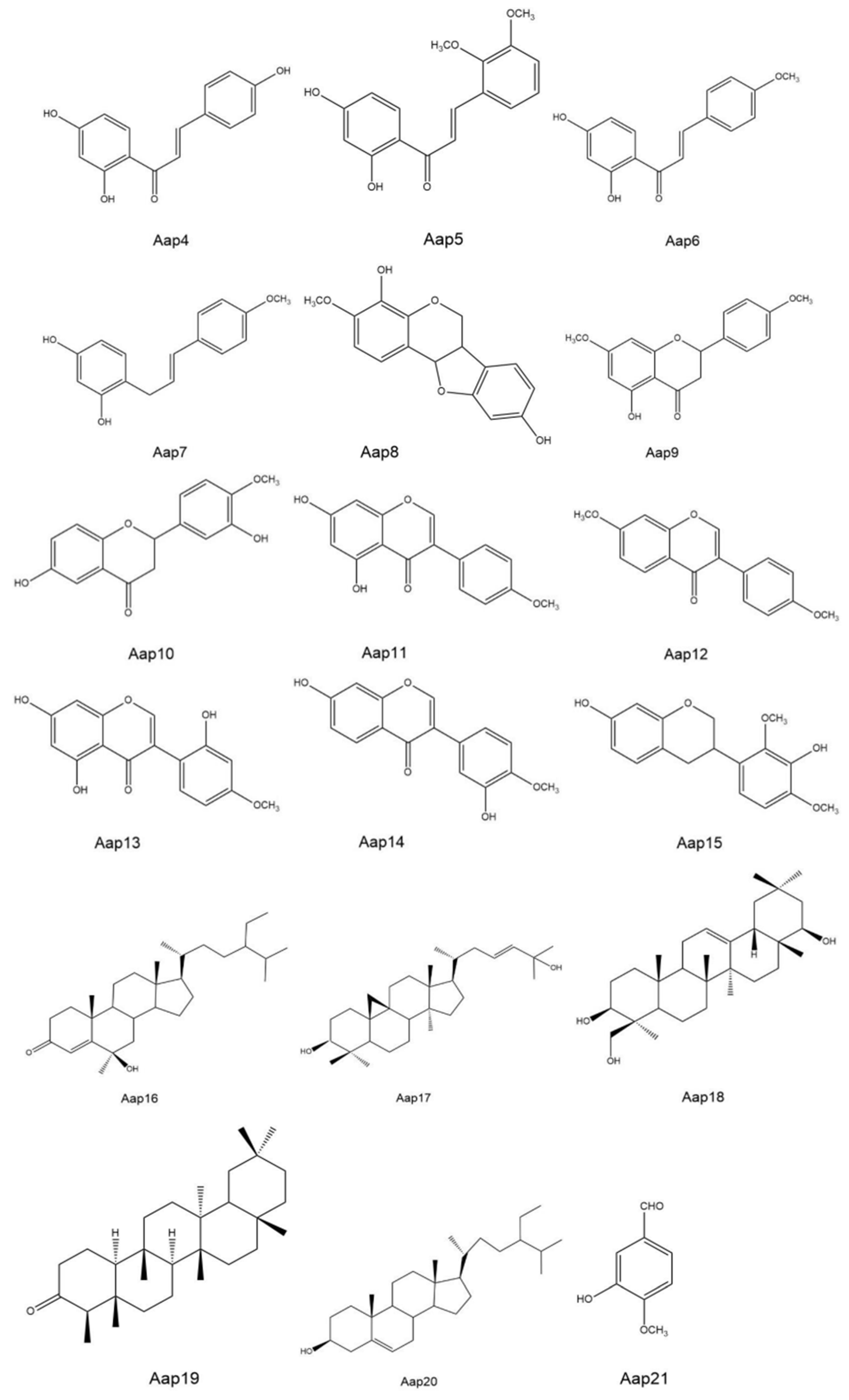
Figure 1. Weekly measurements of net photosynthetic rate (P) (A,B), stomatal conductance (Sc) (C,D), and water use efficiency (WUE) (E,F) for two varieties of standing milkvetch. Each data point represents the mean ± standard error. *, p < 0.05 vs. control.
Agronomy 14 01892 g001
Figure 2. Extracellular alterations in electrical conductivity (A) and pH (B) in bathing solution of leaves and stems of standing milkvetch (varieties Shanxi and Ningxia) exposed to sterile WHD (control) or culture filtrate from A. gansuensis. Each bar represents the mean ± standard error. *, p < 0.05 vs. control.
Agronomy 14 01892 g002
Figure 3. Relationship between relative decline in net photosynthetic rate and extracellular increase in conductivity (A), extracellular decrease in pH (B), and percentage of necrosis (C) induced by A. gansuensis in two varieties of standing milkvetch.
Agronomy 14 01892 g003
Figure 4. Compound structure of secondary metabolites determined in this study.
Agronomy 14 01892 g004aAgronomy 14 01892 g004b
Table 1. Relative content comparison of 21 secondary metabolites between two varieties and treatments of standing milkvetch.
Secondary MetaboliteRelative Content (%) in:
Control TreatmentInoculated with A. gansuensis
ShanxiNingxiaShanxiNingxia
Aap 1: Methyl 3- (2, 4-dihydroxyphenyl) -5- (4-methoxyphenyl) -5-oxopentanoate0.000.005.362.85
Aap 2: 2,4,4’trihydroxy-3-isopentenyl chalcone4.924.5111.218.41
Aap 3: 2, 4-dihydroxy-3’,4’ -dimethoxy-chalcone0.970.262.511.36
Aap 4: 2,4,4 ’-trihydroxy-chalcone0.951.530.953.25
Aap 5: 2, 4-dihydroxy-2’,3’ -dimethoxy-chalcone0.210.330.000.00
Aap 6: 2, 4-dihydroxy-4’ methoxychalcone0.480.250.000.00
Aap 7: 2, 4-dihydroxy-4’-methoxy- chalalkyl0.811.642.371.27
Aap 8: 8-4’ dihydroxy-7-methoxy- hydroxypterocarpan0.000.001.310.95
Aap 9: 5-hydroxy-7,4’-dimethoxy- dihydroflavone0.000.008.335.68
Aap 10: 6,3’-dihydroxy-4’-methoxy- dihydroflavone0.000.0012.019.33
Aap 11: 5, 7-dihydroxy-4’-methoxy-isoflavone0.000.002.173.58
Aap 12: 7,4’-dimethoxy-isoflavone6.688.120.000.00
Aap 13: 5,7,2’-trihydroxy-4’ methoxy-isoflavone13.5611.930.000.00
Aap 14: 7,3’-dihydroxy-4’ -methoxy-isoflavone10.235.560.000.00
Aap 15: 7, 3-dihydroxy-2’,4’-dimethoxy-isoflavane0.000.000.620.45
Aap 16: steroid-4-ene-6 -alcohol-3-ketone0.000.0011.9714.68
Aap 17: cycloaltin-26-ene 3β, 28-diol0.000.0028.2126.56
Aap 18: Oleanolic-12-ene 3β,22β, 23-triol24.6530.981.252.48
Aap 19: friedelin8.226.560.000.00
Aap 20: β-sitosterol8.056.830.000.00
Aap 21: vanillin1.723.430.000.00
Disclaimer/Publisher’s Note: The statements, opinions and data contained in all publications are solely those of the individual author(s) and contributor(s) and not of MDPI and/or the editor(s). MDPI and/or the editor(s) disclaim responsibility for any injury to people or property resulting from any ideas, methods, instructions or products referred to in the content.

