Article

Establishment of a Serology- and Molecular-Combined Detection System for Youcai Mosaic Virus and Its Application in Various Host Plants

1
Department of Plant Pathology, College of Plant Protection, Yangzhou University, Yangzhou 225009, China
2
Joint International Research Laboratory of Agriculture and Agri-Product Safety, Ministry of Education of China, Yangzhou University, Yangzhou 225009, China
*
Author to whom correspondence should be addressed.
Agronomy 2024, 14(9), 1900; https://doi.org/10.3390/agronomy14091900 (registering DOI)
Submission received: 5 August 2024 / Revised: 20 August 2024 / Accepted: 23 August 2024 / Published: 25 August 2024
(This article belongs to the Section Crop Breeding and Genetics)

Abstract

:
The youcai mosaic virus (YoMV) can infect a diverse array of crop species, such as Raphanus sativus, Brassica napus, Solanum nigrum, and Rehmannia glutinosa, causing substantial economic damage. This study aimed to develop a rapid, sensitive, and economical diagnostic method for YoMV. We successfully expressed and purified the recombinant His-CPYoMV-YZ protein in E. coli BL21, which was used to immunize New Zealand White rabbits, generating high-titer polyclonal antibodies (PAb-CPYoMV-YZ). Additionally, a serological-based reverse transcription loop-mediated isothermal amplification (S-RT-LAMP) assay was refined, combining serological and molecular detection techniques to enhance practicality. Utilizing PAb-CPYoMV-YZ, we developed four techniques for detecting YoMV: Western blot, dot immunoblotting assay, enzyme-linked immunosorbent assay (ELISA), and S-RT-LAMP. YoMV isolates from various regions and hosts were analyzed. The results indicated that PAb-CPYoMV-YZ was highly effective in detecting YoMV across a range of hosts and isolates from diverse regions. This study fills an important gap in the serological detection of YoMV and provides a practical tool for on-site diagnosis and control of YoMV infection.

1. Introduction

R. sativus, commonly known as radish, is a member of the Brassicaceae family and represents one of the world’s oldest cultivated crops. In 2020, the global radish planting area was approximately 1.133 million hectares, producing about 47.82 million tons of radishes. In China, radish is extensively cultivated due to its high productivity and excellent storage resistance [1]. Viral diseases pose significant threats to radish cultivation, primarily affecting the foliage. The major symptoms include foliage deformity, leaf crumpling, dwarfism, and stunted growth [2]. These viral diseases are among the most severe affecting radishes [3]. Numerous viruses have been identified that can infect radishes, including turnip mosaic virus (TuMV, Potyvirus), cucumber mosaic virus (CMV, Cucumovirus), youcai mosaic virus (YoMV, Tobamovirus), potato virus Y (PVY, Potyvirus), potato virus X (PVX, Potexvirus), tomato mosaic virus (ToMV, Tobamovirus), tobacco mosaic virus (TMV, Tobamovirus), cucumber green mottle mosaic virus (CGMMV, Tobamovirus), broad bean wilt virus (BBWV, Fabavirus), broad bean wilt virus 2 (BBWV2, Fabavirus), Brassica yellows virus (BrYV, Polerovirus), radish mosaic virus (RaMV, Comovirus), Raphanus sativus cryptic virus 3 (RsCV3, Partitiviridae), Raphanus sativus chrysovirus 1 (RasCV1, Alphachrysovirus), grapevine leafroll-associated virus (GLRaV, Ampelovirus), grapevine virus A (GVA, Vitivirus), and Brassica napus RNA virus 1 (BnRV1, Waikavirus) [2,4,5,6,7,8,9].
Youcai mosaic virus (YoMV) belongs to the genus Tobamovirus, subgroup 3 [10], and is a positive-sense, single-stranded RNA virus with rod-shaped viral particles. It is also known as Chinese rape mosaic virus (CRMV) and oilseed rape mosaic virus (ORMV) [11,12]. YoMV can infect a variety of plants, including members of the Brassicaceae, Solanaceae, and Cucurbitaceae families [13]. The virus was initially identified in the plant species B. campestris in eastern China by Wei et al. in 1958 [14], and the first nucleotide sequencing of the YoMV genome was completed by Aguilar et al. in 1996 [15]. YoMV has been detected in China, Japan, Korea, Germany, Spain, Ukraine, Switzerland, and many other countries [16,17,18]. In 2022, Li et al. first reported YoMV on radish in Huizhou, Guangdong [19]. Our laboratory identified YoMV in wild S. nigrum in 2023 [20]. YoMV contains four open reading frames (ORFs), which encode a 125 kDa protein, a 182 kDa protein, a 30 kDa movement protein (MP), and a 17.7 kDa coat protein (CP). It primarily spreads through sap, and diseased soil can also cause infection [21,22].
Several techniques have been developed for the identification and detection of YoMV. Cai et al. employed reverse transcription polymerase chain reaction (RT-PCR) to identify YoMV in oilseed rape [3]. Wang et al. identified the presence of YoMV in R. glutinosa using double-stranded RNA technology and sequence-independent amplification (SIA) [21]. Pang et al. used small RNA deep sequencing to detect YoMV and ReMV (rehmannia mosaic virus) in R. glutinosa [23]. Li et al. confirmed the presence of YoMV in radish using small RNA sequencing [19]. Qin et al. developed a rapid reverse transcription loop-mediated isothermal amplification (RT-LAMP) method for detecting YoMV in Dioscorea oppositifolia and R. glutinosa [24]. However, these testing methods require professional technicians, a well-equipped experimental environment, and are relatively costly. Furthermore, there are limited data on the identification of YoMV in plants using polyclonal antibody-based serological assays. Therefore, there is an urgent need for a highly specific, sensitive, high-throughput, and user-friendly method for detecting and controlling YoMV in field conditions.
In this study, we successfully cloned the CPYoMV-YZ gene sequence from the pCB301-YoMV-YZ-2 vector using PCR, constructed a prokaryotic expression vector for the CPYoMV-YZ protein, and purified the recombinant His-CPYoMV-YZ protein with an N-terminal His tag. A polyclonal antibody against the His-CPYoMV-YZ protein (PAb-CPYoMV-YZ) was generated in New Zealand White rabbits by immunizing them with prokaryotically expressed His-CPYoMV-YZ as the antigen. Using PAb-CPYoMV-YZ, we developed serological assays, including Western blotting, dot blotting, and enzyme-linked immunosorbent assay (ELISA), to effectively detect YoMV in various plants. The S-RT-LAMP assay was developed by combining a serological method based on PAb-CPYoMV-YZ with a molecular detection technique, aiming to improve the specificity and sensitivity of the detection system [25]. The PAb-CPYoMV-YZ prepared in this study exhibited a good titer and high specificity, providing a new method for the rapid and efficient detection of YoMV in various plants.

2. Materials and Methods

2.1. Construction of Recombinant CPYoMV-YZ Protein Expression Vector

The prokaryotic expression vectors were constructed according to the methodology previously outlined by [25]. In our experiments, we successfully cloned the CPYoMV-YZ gene sequence from the pCB301-YoMV-YZ-2 (2023, Yangzhou Solanum nigrum isolate) recombinant cloning vector by PCR using Phanta Max Super-Fidelity DNA Polymerase (Vazyme, Nanjing, China). The restriction sites for EcoR I and Xho I were introduced into the fragment by PCR using the primers CPYoMV-F-EcoR I and CPYoMV-R-Xho I (Supplementary Table S1). The purified PCR product and pET-28(a)+ vector were double-digested with EcoR I and Xho I. The pET-28(a)-CPYoMV-YZ recombinant vector was constructed by purifying the digested products using the EasyPure PCR purification kit (Trans, Beijing, China) and ligating the target fragment with vector using T4 DNA ligase (Takara, Beijing, China). The recombinant plasmids were validated by Sanger sequencing at Sangon Biotech (Shanghai, China) using an Applied Biosystems 3730XL (Thermo Fisher, Shanghai, China) sequencer, and the results were analyzed using SnapGene software. Protein size prediction was performed using the ExPASy-ProtParam tool (SIB Swiss Institute of Bioinformatics, Lausanne, Switzerland). Finally, the recombinant vector was transformed into E. coli BL21, and four positive monoclonal colonies were selected for further use.

2.2. Prokaryotic Expression and Purification of Recombinant CPYoMV-YZ Protein

The correct pET-28(a)-CPYoMV-YZ recombinant vector was transformed into E. coli BL21, and four positive monoclonal colonies were selected to test the small-scale expression of the putative His-CPYoMV-YZ protein with induction by IPTG. The recombinant proteins expressed in the four positive clones were analyzed by sodium dodecyl sulfate-polyacrylamide gel electrophoresis (SDS-PAGE) and stained with Coomassie Brilliant Blue (CBB). The clone exhibiting the highest expression level of the His-CPYoMV-YZ protein was selected for large-scale expression and purification following a previously described method [26] (refer to chapter 12).

2.3. Production of Polyclonal Antibody against His-CPYoMV-YZ Protein

The purified and concentrated target protein was injected into New Zealand White rabbits. The rabbits were administered a total antigen dose of 3 mg via subcutaneous injection at multiple sites on four occasions, with each injection given at 7-day intervals. After 35 days of immunization, 1 mL of rabbit blood was drawn from the ear vein, and the antiserum was incubated at 4 °C overnight to determine the titer following the method of Zhang et al. [27]. Immunoglobulin G (IgG) was precipitated by the addition of a 10% sodium thimerosal solution, and a series of purification steps [26] yielded a high-purity anti-CPYoMV-YZ polyclonal antibody (PAb-CPYoMV-YZ).

2.4. Western Blotting, Immuno-Dot Blotting, and ELISA Detection of YoMV

A serological assay for YoMV in radish was developed using PAb-CPYoMV-YZ. Western blotting, immuno-dot blotting, and ELISA were performed according to standard protocols [28].
For Western blotting, 0.2 g of the samples (rabbit leaves, N. benthamiana leaves, S. nigrum leaves, R. glutinosa leaves) were frozen in liquid nitrogen and subsequently ground using a TH-Mini handheld homogenizer (LABGIC, Beijing, China). Protein samples were prepared using SDS-PAGE protein sample loading buffer (5X) (Beyotime, Shanghai, China), as previously described [25]. Alkaline phosphatase (AP)-coupled anti-rabbit immunoglobulin (AP-A) (Sangon Biotech, Shanghai, China) was used as the secondary antibody.
For immuno-dot blotting, the leaves were ground, and the total proteins were extracted and processed as previously described, with minor modifications [26]. Purified recombinant protein His-CPYoMV-YZ was used as a positive control, while total protein extracted from healthy radish was used as a negative control. During the initial antibody incubation phase, the antiserum was diluted 101, 102, 103, 104, 105, and 106-fold.
To perform the ELISA, leaves from 62 radish samples suspected to be infected with YoMV (Huizhou, Guangdong) were frozen and ground into powder in liquid nitrogen. Thereafter, 500 µL of Protein Extraction Buffer (0.02 M PBS buffer with 1% cocktail) was added to the mixture, which was then placed on ice for 20 min. The mixture was subsequently centrifuged at 12,500 rpm for 15 min. The supernatant was collected, and approximately 100 µL was transferred into a 96-well ELISA plate (100 µL/well), covered with aluminum foil, and then incubated at 37 °C for 3 h. Next, 100 µL per well of the PAb-CPYoMV-YZ, diluted to 1.0 × 10⁴-fold, was added as previously described with some modifications [26]. After the reaction was completed, measure the OD405 and OD450 using a microplate reader (Model 650, Bio-Rad, Shanghai, China). The incidence rate was calculated as the percentage of positive samples out of the total number of samples. The negative control was radish samples not infected with YoMV, while positive control was radish samples infected with YoMV.

2.5. RT-PCR for YoMV

Plant leaves were subjected to liquid nitrogen, snap-frozen, and subsequently ground into powder using a 4 cm diameter punch (LABGIC, Beijing, China). The resulting plant tissue powder was then placed into pre-cooled 1.5 mL RNase-free Eppendorf tubes. Total RNA was extracted from plant samples using the TakaRa MiniBEST Universal RNA Extraction Kit (Takara, Beijing, China). The extracted RNA was subsequently stored in an ultra-low temperature freezer. RNA concentrations were quantified using a NanoPhotometer N60 (IMPLEN, Munich, Germany), and RNA integrity was assessed by electrophoresis on a 2% agarose gel. The cDNA was synthesized using the PrimeScript RT-PCR Kit (TaKaRa, Beijing, China), and the CPYoMV-YZ coding sequence was amplified by PCR with primers CPYoMV-F and CPYoMV-R (Supplementary Table S1). The PCR products were analyzed by 1% agarose gel electrophoresis and stained with ethidium bromide (EB).

2.6. S-RT-LAMP Assay for YoMV Using Prepared PAb-CPYoMV-YZ

Referring to the S-RT-LAMP experimental method proposed by Hua et al. [25], the effects of false positives were minimized based on the original method by extending the UV lamp elimination time, using sodium hypochlorite to wipe the ultra-clean table, and adding glycerol to the reaction system. S-RT-LAMP experiments were performed using total RNA extracted from the immunoprecipitation products of radish (Brassicaceae, Raphanus, Guangdong, 2022), R. glutinosa (Scrophulariaceae, Rehmannia, Henan, 2022), S. nigrum (Solanaceae, Solanum, Yangzhou, 2023), and N. benthamiana (Solanaceae, Nicotiana, Yangzhou, 2024) samples identified as infected with YoMV, and subjected to gel electrophoresis on a 3% agarose. We designed four sets of primers for the RT-LAMP assay using PrimerExplorer V5 (http://primerexplorer.jp/e, Lanpu Bio-tech, Beijing, China), targeting highly conserved regions of the CPYoMV-YZ genome sequence (Supplementary Table S2). A reaction time of 60 min and a temperature of 60 °C were chosen.

2.7. Multiple Sequence Alignment and Phylogenetic Tree Construction of the CPYoMV Gene.

All CP gene sequences of YoMV isolates were downloaded from the NCBI database and aligned using DNAman for multiple sequence alignment. The CPYoMV-YZ sequences were compared with the CP sequences of other viruses in the same genus and with isolates from other regions of YoMV using MEGA 11 software (version 11.0.13). A CP gene-based phylogenetic tree of YoMV was constructed using the neighbor-joining method (1000 cycles).

3. Results

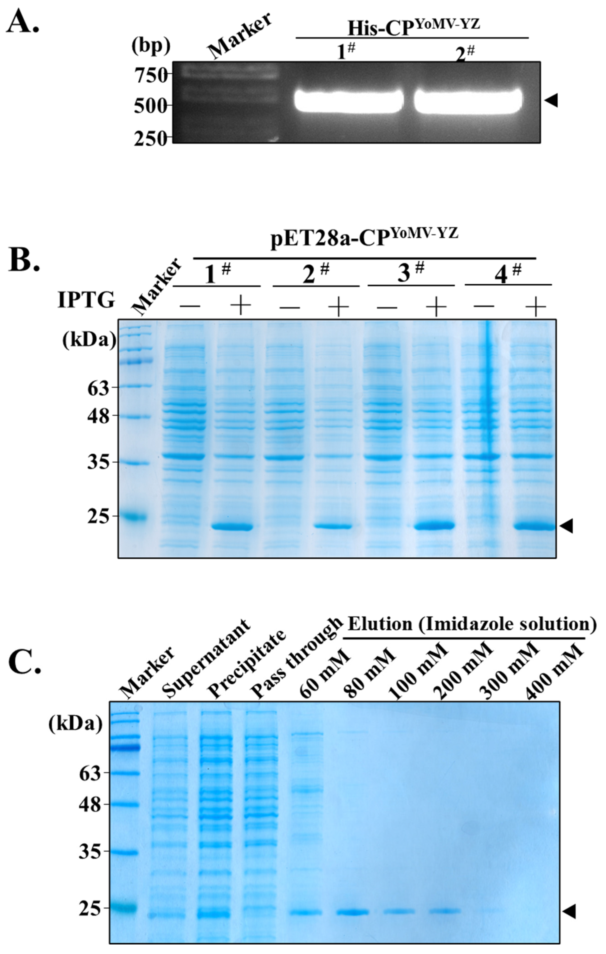
3.1. Production of Recombinant CPYoMV-YZ Protein

By analyzing the CP gene-based phylogenetic tree of YoMV isolates from different regions (Supplementary Figure S1), the CPYoMV-YZ sequence was found to be highly conserved, making the CPYoMV-YZ protein an optimal candidate for the detection of YoMV from diverse sources. We successfully amplified the CPYoMV-YZ sequence containing EcoR I and Xho I cleavage sites using PCR (Figure 1A). The pET-28-CPYoMV-YZ recombinant vector was successfully constructed and transformed into E. coli BL21. Positive monoclonal colonies were selected for subsequent experiments.
Small-scale expression experiments demonstrated that all four mono-clones expressed a target protein with 22 kDa under IPTG induction (Figure 1B), which were consistent with the predicted size. Clones 1, 3, and 4 exhibited relatively high expression levels of the target proteins, while clone 2 showed lower expression. Thus, clone 3 was further selected for subsequent large-scale expression and purification of the target protein.
Following large-scale induction experiments, SDS-PAGE analysis of the gradient imidazole eluent revealed that concentrations of 60 mM, 80 mM, 100 mM, and 200 mM imidazole solution were effective in eluting the recombinant proteins bound to the resin (Figure 1C).

3.2. Antiserum Production Using Recombinant His-CPYoMV-YZ Protein

The His-CPYoMV-YZ protein, purified by affinity chromatography, was analyzed using SDS-PAGE. The results demonstrated that the purity and concentration of the target protein were high, making it suitable for antiserum preparation (Figure 2A). The titer of the prepared anti-His-CPYoMV-YZ antiserum was determined by enzyme-linked immunosorbent assay (ELISA), which showed that the optical density (OD) at 450 nm of the antiserum remained above 0.6 even after a 64,000-fold dilution (Figure 2B, red line). Immuno-dot blotting experiments were conducted using antiserum dilutions of 1:101, 1:102, 1:103, 1:104, 1:105, and 1:106. The results demonstrated that even when the antiserum was diluted up to 104 times, a significant chromogenic reaction was still observed (Figure 2C). The specificity and sensitivity of the prepared antisera were further investigated by Western blotting using diluted antisera with concentration gradient ratios of 1:1000, 1:2000, 1:5000, 1:10,000, and 1:20,000. The immunoblotting results showed that specific bands remained visible at a dilution of 1:20,000 (Figure 2D). The results showed that the prepared anti-His-CPYoMV-YZ antiserum exhibited high sensitivity and good quality.

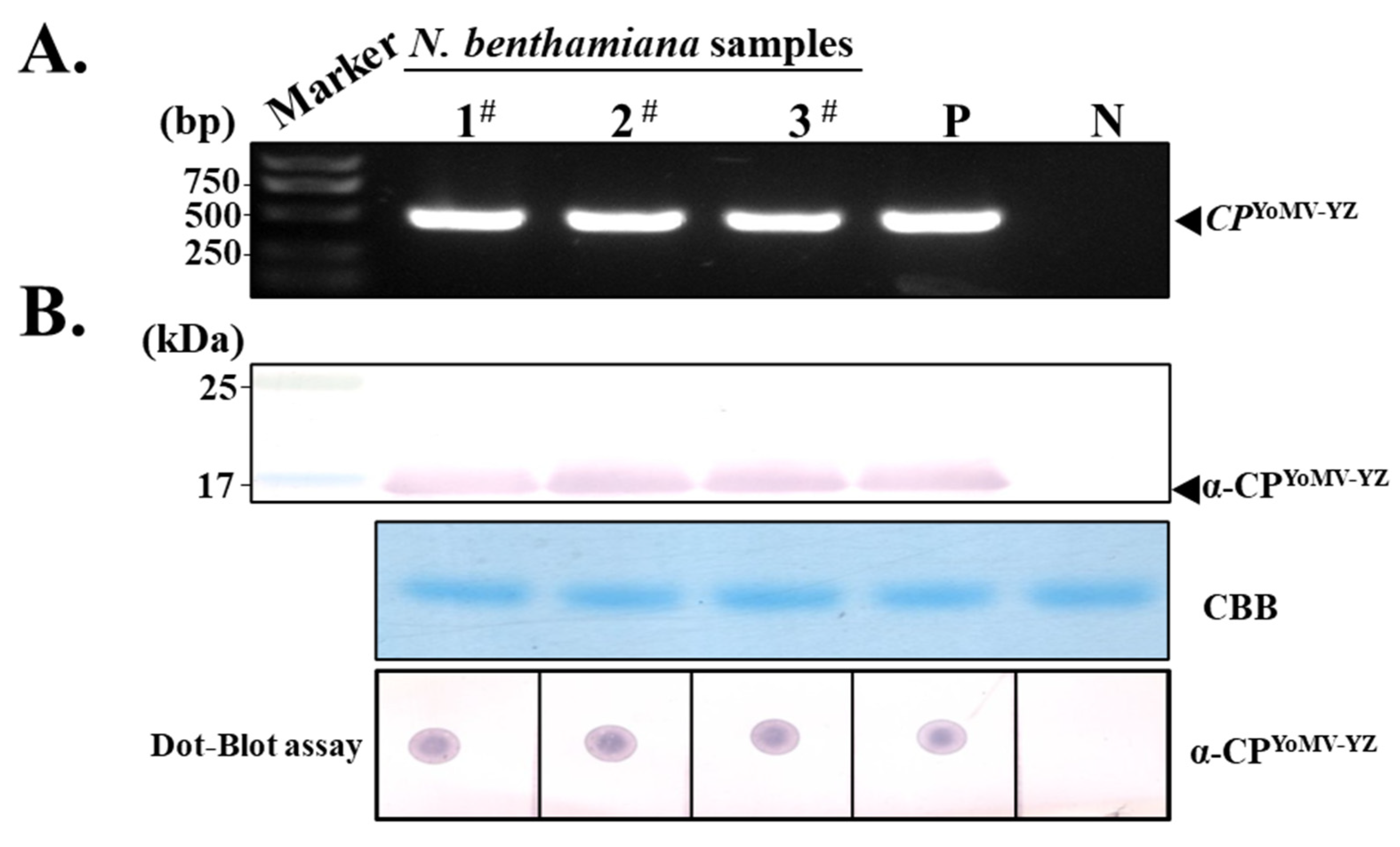
3.3. Evaluation of Purified PAb-CPYoMV-YZ by Western Blotting and Immuno-Dot Blotting

To enhance the precision of YoMV detection using the obtained antisera, we investigated the potential of purified PAb-CPYoMV-YZ for identifying YoMV in Nicotiana benthamiana. RT-PCR results indicated that samples 1, 2, and 3 were infected with YoMV (Figure 3A). Subsequently, the samples were subjected to Western blot analysis using PAb-CPYoMV-YZ diluted 10−4-fold. The results showed distinct, specific bands on the blotting membrane, indicating both the titers and purity of the purified PAb-CPYoMV-YZ (Figure 3B, upper panel). Additionally, samples were assayed by dot blotting with purified recombinant His-CPYoMV-YZ protein as the positive control and total N. benthamiana protein infiltrated with the Agrobacterium-mediated pCB301 empty plasmid as the negative control. The results showed that even at a 10−4 dilution of PAb-CPYoMV-YZ, a distinct color development reaction could still be observed on the blot strips, indicating that PAb-CPYoMV-YZ exhibited high titer and sensitivity (Figure 3B, bottom panel).

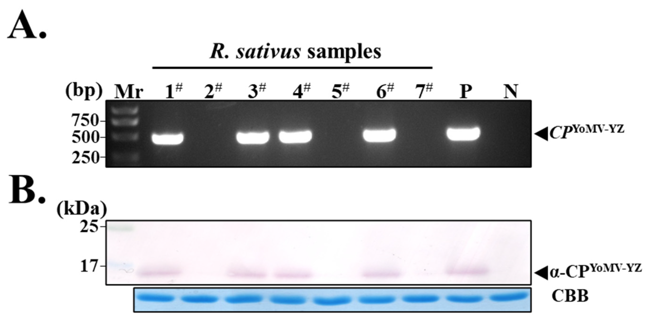
3.4. Screening of YoMV-Positive and Negative Radish Plants by Molecular and Serological Methods

We sought to detect the presence of YoMV in seven suspected infected radish samples collected from Huizhou, Guangdong Province. The results showed that the expected size fragments (approximately 474 bp) were amplified in samples 1, 3, 4, and 6, indicating that they were infected by YoMV (Figure 4A). In contrast, samples 2, 5, and 7 did not exhibit this amplification (Figure 4A). Furthermore, the CPYoMV-YZ antiserum was used for Western blot detection in the seven radish samples. The results showed that only samples 1, 3, 4, and 6 exhibited corresponding bands on the membrane, indicating that these samples were infected by YoMV. Conversely, samples 2, 5, and 7 were not infected (Figure 4B), which was consistent with the RT-PCR results. These results demonstrated that the prepared antibodies specifically recognized YoMV-infected plants in the field.

3.5. Establishment of ELISA and Immuno-Dot Blotting Assays for YoMV in Radish

The results demonstrated that when PAb-CPYoMV-YZ was diluted to 10−4-fold, a pronounced color reaction was observed on the membrane (Figure 5A). Statistical analysis identified six samples as incidence cases, with an incidence rate of 9.68%. Concurrently, we conducted ELISA experiments (Figure 5B,C). The results were consistent with those obtained from the immuno-dot blotting assays and OD405 readings, indicating that the method can be employed for the detection of YoMV.

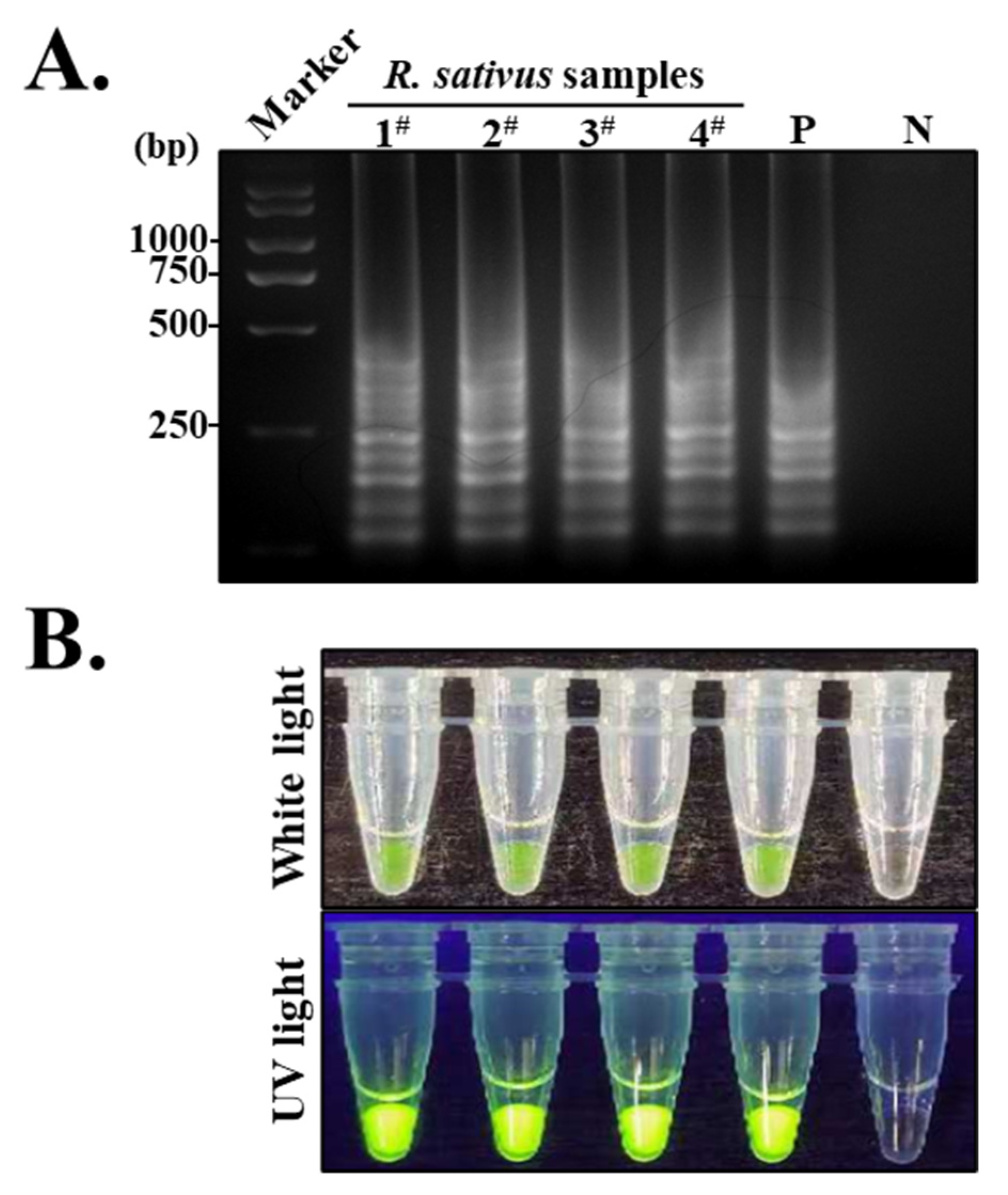
3.6. Establishment of an S-RT-LAMP Assay Based on the CPYoMV-YZ Gene

The results showed that samples 1, 2, 3, and 4 exhibited identical trapezoidal bands to those observed in the positive controls. In contrast, the negative control did not produce any discernible bands (Figure 6A). Meanwhile, SYBR Green I nucleic acid dye was added to S-RT-LAMP amplification products. The positive control exhibited a green hue under white light, whereas the negative control displayed an orange coloration. The positive amplification products exhibited a pronounced fluorescence under UV light, whereas the negative control did not show a discernible fluorescence response. The samples infiltrated with YoMV exhibited a reaction pattern similar to that of the positive controls (Figure 6B). The results of the fluorescence experiments were consistent with those of the gel electrophoresis. These results demonstrate that the established S-RT-LAMP assay system is an effective and cost-efficient method for the rapid detection of YoMV in various samples.

3.7. Purified PAb-CPYoMV-YZ for YoMV Detection in a Wide Range of Plants

Recently, we detected the presence of YoMV in a sample of R. glutinosa from Henan Province, designated YoMV-HN (unpublished). Detection of YoMV in radish, R. glutinosa, S. nigrum, and N. benthamiana was conducted using the S-RT-LAMP technique.
Following electrophoresis on a 3% agarose gel, the results showed that the samples of R. glutinosa from Henan and S. nigrum from Yangzhou exhibited trapezoidal bands identical to those observed in the samples of radish from Guangdong and the positive sample of N. benthamiana. In contrast, the negative control did not produce any discernible bands (Figure 7A). The results of the fluorescence reaction, conducted after adding SYBR Green I nucleic acid dye to the S-RT-LAMP amplification product, were consistent with those of the gel electrophoresis (Figure 7B). Western blot experiments demonstrated that the radish, R. glutinosa, S. nigrum, and N. benthamiana samples exhibited identical specific bands on the membranes, whereas the negative controls did not (Figure 7C). The results obtained from immuno-dot blotting experiments were identical (Figure 7C). The outcomes of the Western blot and immune-dot blotting experiments align with those of the S-RT-LAMP experiments. These results illustrate that the PAb-CPYoMV-YZ prepared in this experiment is capable of rapid and sensitive detection of YoMV in multiple genera and families of plants. The aforementioned results demonstrate that the PAb-CPYoMV-YZ prepared in this experiment is capable of specifically and sensitively detecting YoMV in plants belonging to multiple genera and families, including Brassicaceae, Scrophulariaceae, and Solanaceae.

4. Discussion

The control of plant viral diseases has historically been a major challenge, as many vegetables and crops are susceptible to viral infections. Recent studies have shown that YoMV can infect a diverse range of herbs, including R. glutinosa, S. nigrum, Stellaria media, Plantago asiatica, and D. oppositifolia [20,21,29]. Additionally, YoMV has been observed to affect various crops, such as B. campestris, radish, and Nicotiana tabacum [3,19,21]. To date, 27 different YoMV isolates have been reported. In this study, we focused on the YoMV Yangzhou isolate and the YoMV Guangdong isolate, which were obtained in our laboratory.
However, the infectivity and pathogenicity of different YoMV isolates can vary considerably across plant species. The YoMV-GD (NCBI accession number: OK655842.1) isolate was observed to infect both N. tabacum and N. benthamiana. Five days after infection, N. benthamiana exhibited symptoms such as wilting and necrosis, while seven days after infection, N. tabacum exhibited severe necrosis and leaf curling [19]. This evidence indicates that YoMV-GD isolates are highly pathogenic to N. benthamiana, similar to the YoMV-HK1(NCBI accession number: MG001349.1) isolate. Conversely, the YoMV-Wh (NCBI accession number: EU571218.1) isolate can infect Brassicaceae plants but is unable to infect most Solanaceae plants, including N. tabacum [3]. The YoMV-YZ-2 (NCBI accession number: OR261028.1), YoMV-YZ-3 (NCBI accession number: OR261029.1), and YoMV-YZ-8 (NCBI accession number: OR261030.1) isolates discovered earlier in our laboratory exhibited similar toxicity and showed mild necrosis symptoms on the fifth day. However, they were less virulent than YoMV-GD [20]. YoMV-HN (unpublished) isolates, on the other hand, were less toxic than YoMV-YZ.
However, multiple sequence comparisons revealed that the sequence similarity between YoMV-GD and YoMV-YZ-2 is remarkably high, reaching 98.57% (Supplementary Figure S2). Comparison of the CP sequences revealed a similarity of 99.79%, with only one base difference (Supplementary Figure S3). An evolutionary tree based on the CPYoMV gene showed that YoMV-GD and YoMV-YZ are part of the same extensive branch and are closely related (Supplementary Figure S1). Our findings indicate that YoMV-GD and YoMV-YZ are part of the same extensive branch of the evolutionary tree and are closely related. However, our analysis also revealed significant differences in their pathogenicity and infection capacity. These findings suggest that there is no significant correlation between viral pathogenicity and geographic or sequence similarity. Therefore, it is evident that the diagnosis and control of virus diseases in the field cannot rely solely on symptoms. Furthermore, reliance on high-throughput sequencing technology is hindered by the issues of delayed results and high costs. Consequently, it is imperative to prepare antisera against YoMV CP and develop serological-based assays to facilitate the rapid diagnosis of this disease in the field.
Polymerase chain reaction (PCR) was employed to amplify the CPYoMV-YZ gene from the pCB301-YoMV-YZ-2 plasmid. The recombinant His-CPYoMV-YZ protein was expressed using the E. coli BL21 strain (Figure 1), and the purified protein was used as an antigen to immunize New Zealand White rabbits to obtain high-quality antiserum. Several accurate and sensitive serological assays for YoMV have been developed using the specific His-CPYoMV-YZ polyclonal antiserum. (Figure 2). The antiserum obtained was also shown to have good titer. The optimal concentration of PAb-CPYoMV-YZ for YoMV detection was determined using YoMV-positive N. benthamiana samples (Figure 3). IgG antibodies were purified from the polyclonal antisera to obtain PAb-CPYoMV-YZ. A high-throughput, accurate, and sensitive field YoMV immuno-dot blot and ELISA assay was established using PAb-CPYoMV-YZ (Figure 4 and Figure 5). Additionally, we enhanced the approach by Hua et al. [25] by integrating PAb-CPYoMV-YZ with conventional RT-LAMP methodology, developing an S-RT-LAMP assay for the CPYoMV-YZ gene. By using sodium hypochlorite for disinfection, adding a glycerol liquid seal, and adding templates in different rooms to avoid false positives, the reliability of S-RT-LAMP was further improved (Figure 6). The optimal temperature for detecting YoMV using S-RT-LAMP technology was determined to be 60 °C for 60 min (Supplementary Figure S4). Based on the established YoMV rapid test system, various plants from different sources infected with different YoMV isolates were tested in the laboratory to verify the practicality and extensiveness of the obtained PAb-CPYoMV-YZ (Figure 7). The results demonstrated that the prepared PAb-CPYoMV-YZ effectively detected YoMV from different sources, with good specificity and high efficiency. It should be noted that this experiment tested samples from a limited number of regions, presenting certain inherent limitations. Consequently, it would be beneficial to obtain samples from more regions in order to facilitate further analysis of YoMV isolates across a wider range of hosts and geographical locations.
Furthermore, our previous study showed that the ReMV polyclonal antiserum or PAb-CP ReMV may exhibit cross-reactivity with TMV and YoMV in serological testing [30]. However, our experiments demonstrated that different viral CP proteins could be distinguished by their size differences and validated by sequencing.
Taken together, these results indicate that the CPYoMV-YZ antiserum and PAb-CPYoMV-YZ obtained in this experiment are very effective for the detection of YoMV, with good titer, sensitivity, and specificity. They can also be used in rapid detection of YoMV in different crops, making this method practical and economical. This study addresses a significant gap in the field of serological detection of YoMV, paving the way for further research on the pathogenesis of YoMV.

Supplementary Materials

The following supporting information can be downloaded at: https://www.mdpi.com/article/10.3390/agronomy14091900/s1, Supplementary Table S1: Primers used for YoMV CP gene cloning and virus detection by RT-PCR; Supplementary Table S2: The four oligonucleotide primers used for the S-RT-LAMP assay for the detection of YoMV CP; Supplementary Figure S1: Phylogenetic evolutionary tree of different isolates based on YoMV CP was constructed using MEGA; Supplementary Figure S2: Results of complete genome sequence comparison between YoMV YZ-2 and YoMV GD; Supplementary Figure S3: Results of CP gene sequence comparison between YoMV YZ-2 and YoMV GD; Supplementary Figure S4: S-RT-LAMP amplification temperature probe experiment for YoMV.

Author Contributions

K.Z., D.L. and C.F. carried out the experiments. Y.H., H.C. and C.F. wrote the manuscript with support from K.Z. K.Z. supervised the project. C.F. and K.Z. conceived the initial idea and designed the experiments and analysed the data. M.W. and J.H. collected experimental data and provided software support and planted the plants. All authors met all four of these criteria. All authors have read and agreed to the published version of the manuscript.

Funding

This work was funded by the National Natural Science Foundation of China (32372486, 31801699), the Excellent Youth Fund of Jiangsu Natural Science Foundation (BK20220116), Jiangsu Province Agricultural Science and technology independent innovation fund project (SCX(24)3116), the Postgraduate Research and Practice Innovation Program of the Jiangsu Province (SJCX24_2279), and the Chinese Government Scholarship (China Scholarship Council, CSC) (File No. 202108320223), and the Young and Middle-aged Academic Leaders of the “Qinglan Project” of Yangzhou University.

Institutional Review Board Statement

Not applicable.

Data Availability Statement

All data are available within the article and its Supplementary Materials.

Acknowledgments

We thank all the other members of the plant virology laboratory at the college of plant protection at Yangzhou University for their helpful suggestions and constructive criticism.

Conflicts of Interest

The authors declare no conflicts of interest.

References

  1. Bao, C.; Wang, J.; Hu, T.; Hu, H.; Wang, W.; Wei, Q. Discussion on the Current Situation of Development and Breeding Direction of Radish Industry in China. J. Zhejiang Agric. Sci. 2019, 60, 707–710. [Google Scholar]
  2. Yuan, W.; Yuan, S.; Gan, C.; Cui, L.; Deng, X. Molecular Identification and Control of Radish Virus Disease in Hubei Province. J. CJ. Veg. 2015, 8, 61–62. [Google Scholar]
  3. Cai, L.; Xu, Z.; Chen, K.; Yan, L.; Hou, M. Identification and Characterization of Oilseed Rape Mosaic Virus (ORMV) Wh Strain. J. Plant Prot. 2005, 32, 4. [Google Scholar]
  4. Wang, L.; Chen, X.; Chen, W.; Tan, Q.; Yang, X. Detection of Main Viral Species of Vegetable Virus Diseases in Guizhou. GD. Agric. Sci. 2015, 20, 63–67. [Google Scholar]
  5. Chung, J.; Han, J.-Y.; Kim, J.; Ju, H.; Gong, J.; Seo, E.-Y.; Hammond, J.; Lim, H.-S. Survey of Viruses Present in Radish Fields in 2014. Res. Plant Dis. 2015, 21, 235–242. [Google Scholar] [CrossRef]
  6. Choi, S.-K.; Park, S.-H.; Jeon, B.-W.; Jang, S.-W.; Yoon, J.-Y. First Report of Youcai Mosaic Virus in Raphanus Raphanistrum Subsp. Sativus in Korea. Plant Dis. 2017, 101, 1959. [Google Scholar] [CrossRef]
  7. Chen, X.-R.; Wang, Y.; Zhao, H.-H.; Zhang, X.-Y.; Wang, X.-B.; Li, D.-W.; Yu, J.-L.; Han, C.-G. Brassica Yellows Virus’ Movement Protein Upregulates Anthocyanin Accumulation, Leading to the Development of Purple Leaf Symptoms on Arabidopsis Thaliana. Sci. Rep. 2018, 8, 16273. [Google Scholar] [CrossRef] [PubMed]
  8. Qiu, X.; Tan, Q.; Xiao, Q.; Mei, S.; Zhang, J. Pathogen Identification of Virus Disease and Evaluation for Germplasm Disease Resistance in Radish, Acta Hortic. Sin. 2020, 47, 1947–1955. [Google Scholar]
  9. Liu, Y.; Li, F.; Li, Y.; Zhang, S.; Gao, X.; Xie, Y.; Yan, F.; Zhang, A.; Dai, L.; Cheng, Z.; et al. Identification, Distribution and Occurrence of Viruses in the Main Vegetables of China. Sci. Agric. Sin. 2019, 52, 239–261. [Google Scholar]
  10. Lartey, R.T.; Voss, T.C.; Melcher, U. Tobamovirus Evolution: Gene Overlaps, Recombination, and Taxonomic Implications. Mol. Biol. Evol. 1996, 13, 1327–1338. [Google Scholar] [CrossRef]
  11. Gibbs, A.; Tien, P.; Kang, L.; Tian, Y.; Randles, J. Classification of several tobamoviruses isolated in China on the basis of the amino acid composition of their virion proteins. Intervirology 1982, 18, 160–163. [Google Scholar] [CrossRef] [PubMed]
  12. Palukaitis, P.; Randles, J.W.; Tian, Y.T.; Kang, L.Y.; Tien, P. Taxonomy of several tobamoviruses from China as determined by molecular hybridization analysis with complementary DNA. Intervirology. 1981, 16, 136–141. [Google Scholar] [CrossRef]
  13. Ju, H.-K.; Kim, I.-H.; Hu, W.-X.; Kim, B.; Choi, G.-W.; Kim, J.; Lim, Y.P.; Domier, L.L.; Hammond, J.; Lim, H.-S. A Single Nucleotide Change in the Overlapping MP and CP Reading Frames Results in Differences in Symptoms Caused by Two Isolates of Youcai Mosaic Virus. Arch. Virol. 2019, 164, 1553–1565. [Google Scholar] [CrossRef]
  14. Wei, J.; Shen, S.; Wang, J.; Zhang, C.; Zhu, Y. A Preliminary Study of Brassica Campestris and Turnip Mosaic Virus in East China. Acta Phytophy. Acta Pharm. Sin. 1958, 4, 2. [Google Scholar]
  15. Aguilar, I.; Sanchez, F.; Martin, A.M.; Martinez-Herrera, D.; Ponz, F. Nucleotide Sequence of Chinese Rape Mosaic Virus (Oilseed Rape Mosaic Virus), a Crucifer Tobamovirus Infectious on Arabidopsis thaliana. Plant Mol. Biol. 1996, 30, 191–197. [Google Scholar] [CrossRef] [PubMed]
  16. Fuji, S.; Mochizuki, N.; Fujinaga, M.; Ikeda, M.; Shinoda, K.; Uematsu, S.; Furuya, H.; Naito, H.; Fukumoto, F. Incidence of Viruses in Alstroemeria Plants Cultivated in Japan and Characterization of Broad Bean Wilt Virus-2, Cucumber Mosaic Virus and Youcai Mosaic Virus. J. Gen. Plant Pathol. 2007, 73, 216–221. [Google Scholar] [CrossRef]
  17. Zhu, H.; Hong, J.; Ye, R.; Chen, J.; Yu, S.; Adams, M.J. Sequence Analysis Shows That Ribgrass Mosaic Virus Shanghai Isolate (RMV-Sh) Is Closely Related to Youcai Mosaic Virus. Arch. Virol. 2001, 146, 1231–1238. [Google Scholar] [CrossRef]
  18. Kwon, S.-J.; Kim, Y.-B.; Back, C.-K.; Chung, B.-N. First Report of a Mixed Infection of Youcai Mosaic Virus and Rehmannia Mosaic Virus in Rehmannia Glutinosa in Korea. Plant Dis. 2017, 07, 1128. [Google Scholar] [CrossRef]
  19. Li, Z.; Tang, Y.; She, X.; Yu, L.; Lan, G.; He, Z. Molecular Characteristics and Pathogenicity Analysis of Youcai Mosaic Virus Guangdong Isolate Infecting Radish. Sci. Agric. Sin. 2022, 55, 2752–2761. [Google Scholar]
  20. Gu, T.; Feng, C.; Hua, Y.; Liu, D.; Chen, H.; He, Z.; Xu, K.; Zhang, K. Molecular Characterization and Pathogenicity of an Infectious cDNA Clone of Youcai Mosaic Virus on Solanum nigrum. Int. J. Mol. Sci. 2024, 25, 1620. [Google Scholar] [CrossRef]
  21. Wang, D.; Zhang, X.; Yang, F.; Niu, Y. Identification and Analysis of Complete Genomic Sequence of Youcai Mosaic Virus Rehmannia Glutinosa Isolate in Shanxi. Acta Phytophy. Sin. 2016, 46, 598–604. [Google Scholar]
  22. Mansilla, C.; Aguilar, I.; Martínez-Herrera, D.; Sánchez, F.; Ponz, F. Physiological Effects of Constitutive Expression of Oilseed Rape Mosaic Tobamovirus (ORMV) Movement Protein in Arabidopsis Thaliana. Transgenic Res. 2006, 15, 761–770. [Google Scholar] [CrossRef]
  23. Pang, X. Identification of Virus Infecting Atractylodes macrocephala Koidz, Pinellia ternata Breit and Rehmannia glutinosa Libosch by Small RNA Deep Sequencing. SXAU. 2018, 6, 45–50. [Google Scholar]
  24. Qin, Y.; Wang, F.; Wen, Y.; Zhao, Z.; Gao, S.; Zhang, D.; Li, S.; Liu, Y.; Liu, Y.; Lu, S.; et al. Establishment of RT-LAMP Method for Rapid Detection of Youcai Mosaic Virus. Acta Phytophy. Sin. 2023, 1024, 001. [Google Scholar]
  25. Hua, Y.; Feng, C.; Gu, T.; Chen, H.; Liu, D.; Xu, K.; Zhang, K. Development of Polyclonal Antibodies and a Serological-Based Reverse-Transcription Loop-Mediated Isothermal Amplification (S-RT-LAMP) Assay for Rice Black-Streaked Dwarf Virus Detection in Both Rice and Small Brown Planthopper. Viruses 2023, 15, 2127. [Google Scholar] [CrossRef] [PubMed]
  26. Zhang, K.; Zhuang, X.; Xu, H.; Gan, H.; He, Z.; Chen, J. Development of Polyclonal Antibodies-Based Serological Methods and a Dig-Labelled DNA Probe-Based Molecular Method for Detection of the Vicia Cryptic Virus-M in Field Plants. J. Virol. Methods. 2022, 299, 114331. [Google Scholar] [CrossRef]
  27. Zhang, K.; Zhuang, X.; Xu, H.; Guo, X.; He, Z.; Xu, K.; Liu, F. Sensitive and High-Throughput Polyclonal Antibody-Based Serological Methods for Rice Stripe Virus Detection in Both Rice and Small Brown Planthopper. Crop Prot. 2021, 144, 105599. [Google Scholar] [CrossRef]
  28. Barker, K. At the Bench: A Laboratory Navigator; Cold Spring Harbor Laboratory Press: New York, NY, USA, 2004. [Google Scholar]
  29. Qin, Y.; Wang, F.; Cai, L.; Gao, S.; Wen, Y.; Liu, Y.; Lu, C.; Yang, J.; Li, X.; Qi, W.; et al. First Report of Youcai Mosaic Virus Infecting Yam in China. Plant Dis. 2023, 107, 1247. [Google Scholar] [CrossRef]
  30. Chen, J.; Feng, C.; Guo, X.; Zhou, Y.; Gu, T.; Zhuang, X.; Cheng, L.; Zhang, K. Development of Polyclonal Antibodies-Based Serological Methods for Detection of the Rehmannia Mosaic Virus in Field Plants. Front. Sustain. Food Syst. 2022, 6, 1013470. [Google Scholar] [CrossRef]
Figure 1. Construction of the prokaryotic expression vector pET-28(a)-CPYoMV-YZ and purification of the recombinant His-CPYoMV-YZ protein. (A) Agarose gel analysis of PCR-amplified products using the pCB301-YoMV-YZ-2 plasmid as a template and primers specific for CPYoMV-YZ. Numbers 1# and 2# represent the 474 bp CP coding region sequence, and black arrows mark their positions. (B) SDS-PAGE (12.5%) analysis of His-CPYoMV-YZ protein expression in four independent E. coli BL21 transformants (1#, 2#, 3#, 4#), showing the expression levels of the target proteins in the presence (+) or absence (−) of IPTG induction. Black triangles indicate the bands of the target protein. (C) SDS-PAGE (12.5%) analysis of affinity chromatography-purified eluates of prokaryotically expressed target proteins. Supernatants, precipitates, and column flow-throughs collected after centrifugation of bacterial cell extracts and column binding. Protein profiles of eluates collected after elution from Ni-NTA resin columns with the indicated concentrations of imidazole (60–400 mM) are shown. Black triangles indicate the bands of the target protein.
Agronomy 14 01900 g001
Figure 2. Three serological methods were utilized to evaluate the quality of the prepared anti-His-CPYoMV-YZ protein antisera. (A) SDS-PAGE analysis of affinity column-purified His-CPYoMV-YZ protein. The numbers on the left indicate the molecular weight markers. (B) The titer of crude antiserum was determined by enzyme-linked immunosorbent assay (ELISA). The ELISA was performed using a commercial secondary antibody labeled with horseradish peroxidase (HRP). The x-axis represents the dilution factor of the antiserum in phosphate-buffered saline (PBS). The y-axis represents the absorbance value measured at 450 nm. The red line indicates an OD450 value of 0.6. (C) The crude antiserum was diluted to different concentrations in PBS, and immuno-dot blot experiments were performed with purified His-CPYoMV-YZ as the antigen. The numbers on the left indicate various dilution factors. (D) The crude antiserum with step-wise increased dilutions was evaluated by Western blotting. The crude antiserum was diluted to different concentrations in PBS, and purified His-CPYoMV-YZ was used as the antigen. The numbers on the left indicate protein markers.
Agronomy 14 01900 g002
Figure 3. Purified PAb-CPYoMV-YZ specifically detects YoMV in N. benthamiana using Western blot and dot-blot assays. (A) Agarose gel analysis of RT-PCR products from three N. benthamiana plants infiltrated with Agrobacterium tumefaciens harboring pCB301-YoMV-YZ-2 plasmid. RT-PCR amplification was performed using specific primers for CPYoMV-YZ. “P” represents the pCB301-YoMV-YZ-2 plasmid (positive control); “N” represents total RNA from a N. benthamiana plant after infiltration with Agrobacterium harboring the pCB301 empty plasmid (negative control). (B) Western blot and immuno-dot blot detection of YoMV in N. benthamiana using PAb-CPYoMV-YZ diluted 10,000-fold. The samples loaded in each lane are the same as in (A). “P” represents purified His-CPYoMV-YZ protein (positive control); “N” represents total protein from a N. benthamiana plant after infiltration with Agrobacterium harboring the pCB301 empty plasmid (negative control). The large subunit of Rubisco complex was stained with Coomassie Brilliant Blue (CBB), serving as loading control.
Agronomy 14 01900 g003
Figure 4. Serological Assay of the R. sativus samples collected from the field using purified anti-CPYoMV-YZ polyclonal antibody (PAb-CPYoMV-YZ) (A) Agarose gel analysis of RT-PCR products from seven field-collected radish plants. Specific primers for CPYoMV-YZ were used for amplification. “P” represents the radish samples previously verified to be infected with YoMV (positive control). “N” represents the radish samples confirmed to be uninfected with YoMV (negative control). (B) Seven radish samples were analyzed by Western blot using PAb-CPYoMV-YZ at a 10,000-fold dilution. The samples loaded in each lane are the same as in (A). “P” represents total protein from radish infected with YoMV (positive control). “N” represents total protein from healthy radish (negative control). Black triangular arrows indicate the range of target sizes. The large subunit of Rubisco complex was stained with Coomassie Brilliant Blue (CBB), serving as loading control.
Agronomy 14 01900 g004
Figure 5. High-throughput immuno-dot blotting test and ELISA for YoMV detection in radish samples from wild field. (A) Immuno-dot blotting test results for 62 field samples collected from Guangdong Province, China. Wells A1 and A2 represent the negative and positive controls, respectively. The samples from wells A3–A8, B1–B8, C1–C8, D1–D8, E1–E8, F1–F8, G1–G8, and H1–H8 are the 62 radish samples from Guangdong. (B) Blotting results of the ELISA experiments corresponding to the samples in panel A. (C) ELISA absorbance (OD405) readings measured at 405 nm for the samples shown in panel B, presented as histograms. Negative control was radish samples not infected with YoMV; positive control was radish samples infected with YoMV.
Agronomy 14 01900 g005
Figure 6. Application of the optimized S-RT-LAMP assay for the detection of YoMV-infected radish. (A) RT-LAMP results: lanes 2–6 represent the 1#, 2#, 3#, and 4# YoMV-infected samples. Lanes 6 and 7 indicate the positive and negative controls, respectively. Lane 1 is the DNA marker. (B) SYBR Green I was added to the reaction system, and the color change was directly observed under white and UV light. The samples loaded in each lane are the same as in (A). “P” represents the radish samples previously verified to be infected with YoMV (positive control). “N” represents the radish samples confirmed to be uninfected with YoMV (negative control).
Agronomy 14 01900 g006
Figure 7. Detection of YoMV in various plants using purified PAb-CPYoMV-YZ (A) S-RT-LAMP results: lanes 2–5 represent samples from radish, R. glutinosa, S. nigrum, and N. benthamiana, respectively. Lane 6 is N. benthamiana not infected with YoMV as a negative control. (B) SYBR Green I was added to the reaction system, and the color change was directly observed under white and UV light. (C) Detection of YoMV in a wide range of plant samples using a PAb-CPYoMV-YZ-based serum assay. Except for the negative control, the lanes showed consistent specific bands and spot imprints similar to those of the positive control (N. benthamiana). ‘N’ represents N. benthamiana samples not infected with YoMV (negative control). N. benthamiana samples subjected to Agrobacterium infiltration with pCB301-YoMV-YZ-2 were treated as positive controls.
Agronomy 14 01900 g007
Disclaimer/Publisher’s Note: The statements, opinions and data contained in all publications are solely those of the individual author(s) and contributor(s) and not of MDPI and/or the editor(s). MDPI and/or the editor(s) disclaim responsibility for any injury to people or property resulting from any ideas, methods, instructions or products referred to in the content.

