Article

Community Diversity of Endophytic Bacteria in the Leaves and Roots of Pea Seedlings

1
Qingdao Academy of Agricultural Sciences, Qingdao 266121, China
2
College of Biological Engineering, Qingdao University of Science & Technology, Qingdao 266101, China
3
Tropical Research and Education Center, Department of Plant Pathology, Institute of Food and Agricultural Sciences, University of Florida, Homestead, FL 33031, USA
4
Tropical Research and Education Center, Environmental Horticulture Department, Institute of Food and Agricultural Sciences, University of Florida, Homestead, FL 33031, USA
*
Authors to whom correspondence should be addressed.
Agronomy 2024, 14(9), 2030; https://doi.org/10.3390/agronomy14092030
Submission received: 25 July 2024 / Revised: 28 August 2024 / Accepted: 30 August 2024 / Published: 5 September 2024

Abstract

:
Endophytic bacteria from pea (Pisum sativum L.) plants play important roles in regulating plant growth, health, and nutrition. To enhance the understanding of endophytic bacteria in peas, twenty pea cultivars, two chickpeas, and two broad bean cultivars were planted into artificial soils for 4 weeks. Leaves and roots were collected from plants and sterilized. Endophytic bacterial DNAs were isolated from sterilized materials (leaves, roots, and seeds) and used as templates to detect the bacterial diversity by amplifying the 16S V3–V4 region. The Remel Tryptose Soya Agar (TSA) medium, the aluminum sec-butoxide (ASb) medium, and the yeast extract mannitol agar (YMA) medium were used to isolate bacteria from sterilized leaves and roots, respectively. The plant growth-promoting (PGP) properties of these isolated bacteria, such as the solubilization of phosphorus and potassium and the production of Indole-3-acetic acid (IAA), 1-aminocyclopropane-1-carboxylic acid (ACC) deaminase, nitrogenase, pectinase, and cellulose, were studied in vitro. Bacterial isolates were processed for 16S rDNA gene sequencing and performed molecular identification by reconstruction of the phylogenetic tree using the neighborhood association approach in the software MEGA X. Results indicated that the majority of the bacterial communities were shared among leaves, roots, and seeds of pea plants. In both the leaves and roots of pea plants, the prominent phyla identified were Pseudomonadota, Bacteroidota, and Bacillota, with dominant genera such as Rhizobium, Bacteroides, Blautia, and Prevotella prevailing at the genus level. The samples from leaves and roots had unique dominant bacterial genera. In total, 48 endophytic bacteria strains were isolated from leaves and roots, of which 16 strains were from roots and 32 strains were from leaves. The majority of the isolates from leaves (78.13%) and roots (75%) had the ability to produce indole-3-acetic acid (IAA). Moreover, isolates from roots also had greater ability to produce 1-amino-cyclopropane-1-carboxylic acid (ACC) deaminase (81.25%) than those from leaves (62.5%). This study demonstrated the unique distribution of endophytes in leaves and roots of pea, which can have great potential in pea production.

1. Introduction

In nature, plants and animals are rich in microbiomes. Biological studies have shown the co-evolution of plants and their endogenous microorganisms. In some cases, the connection is mutually beneficial: endophytes live in the host by sharing and exchanging the products of metabolic and physiological processes [1,2]. Host plants often provide nutrients and a stable habitat, while endophytes provide nitrogen and phosphorus by cycling the elements from the environment and protect plants from the influence of other microorganisms and abiotic stresses [3,4,5]. The interaction between plants and microorganisms influences the agroecology and plant evolution, which leads to great interest in studying plant-related microbiomes. Considering the microbial community as a complex plant trait, genetic tools such as DNA fingerprinting and high-throughput sequencing can be applied to reveal the role of plant-related endophytes [6,7,8,9].
Studies have shown that the endophytic bacteria community can determine the seedling establishment and growth of the host plant by influencing nutrition and hormone levels [10,11,12] or driving the evolution of multi-disease resistances [10]. In addition, these isolated endophytic bacteria had the plant growth-promoting (PGP) characteristics, such as phosphorus and potassium cycling by the solubilization of phosphorus and potassium from the environment, nitrogen cycling by biological nitrogen fixation with the bacterial enzyme nitrogenase, and the production of indole-3-acetic acid (IAA) and 1-amino-cyclopropane-1-carboxylic acid (ACC) deaminase [13,14,15]. The primary cell wall plays a restraint force on plant cell growth [16]. In an oversimplified sketch, the primary cell wall was described as a network of cellulose microfibrils, connected by hemicellulose linkages, embedded in a pectin matrix [17]. Recent observations suggest that pectinase and cellulase activities in plant cell walls of growing cells not only play a role in wall patterning, hydration, and stress relaxation during growth but also provide a driving force for cell wall expansion [18]. As Cosgrove (2023) [19] suggested, “growing walls of plant cells combine two seemingly incompatible properties, namely, mechanical strength and dynamic extensibility, that is, irreversible yielding (also known as creep), sometimes regulated within seconds”. Thus, cellulase and pectinase activities of endophytic bacteria were also regarded as potential PGP traits in this study. Therefore, understanding the community structure and PGP traits of plant-associated microbiota is an important topic to provide important knowledge required for sustainable agriculture.
Pea (Pisum sativum L.) is a cool-season food legume rich in vitamins, minerals, proteins, and carbohydrates, and provides balanced nutrition. Pea has been widely used in different food products and considered a cheap protein source [20,21]. The diversity and PGP characteristics of endophytic bacteria at pea leaves and roots were supposed to play an important role on pea growth and development. Therefore, understanding of endophytic bacteria involved in pea development at the seedling stage is important for pea development. This paper investigates community diversity and the PGP traits of endophytic bacteria from leaves and roots at the pea seedling stage. The research holds promise in screening Plant Growth-Promoting (PGP) endophytes, offering deeper insights into the growth and development of various pea cultivars. Results from this study could directly benefit the application of plant endophytes in pea production. Our study sought to test the following hypotheses: the bacterial microbiota assemblage of seed habitat, root endosphere, and rhizosphere is dominated by deterministic processes of assembly; both vertical and horizontal transmission contribute to the observed differences in the root-associated bacterial communities; and the plant phenotypes could be predicted from the core bacterial microbiota, which is represented by a small community but that is highly connected to other microbes. This work was developed to go beyond descriptive understanding of the plant-associated microbiota by developing predictive interpretations.

2. Materials and Methods

2.1. Sample Collection

Plant samples were listed in Supplement Table S1 and divided into eight groups, including dry pea (abbr. DGP, cultivar QK No. 01 to 04), fresh pea (FGP, QK No. 05 to 08), lotus pea (SP, QK No. 09 to 12), sweet crisp pea (SCP, QK No. 13 to 16), pea tip (SEP, QK No. 17 to 19), and pea sprout (SHP, QK No. 20). These twenty pea cultivars represent different ecotypes and market types in China. Pea, chickpea, and broad bean are the major cool-season grain legume crops planted worldwide, and they are also primarily for human consumption. Two chickpeas (CHP, QK No. 21 to 22) and broad beans (BRB, QK No. 23 to 24) were used as control groups. In addition, artificial soils without seeds were placed in a −80 °C refrigerator and used as the control to produce background soil microbiome. The seeds, i.e., 180 pea seeds, 18 chickpea seeds, and 18 broad bean seeds, were planted into pots filled with artificial soils consisting of Pindstrup sphagnum moss peat (JingDong shopping center, Beijing, China). The seeds were grown in hoop houses at the Qingdao Academy of Agricultural Sciences, Qingdao, Shandong, China (36°09′8.05″ N, 120°25′15.77″ E, 15 m above sea level). Three repeats were made for each cultivar. After one week, each cultivar only selected 3 plants for the present experiment. Then, a total of 72 plants were used in this study. Bacteria of seeds and soils were endophytic bacterial banks of the host plants [12]. Thus, soils with no seeds planted were also placed in these plastic houses. Plants were sampled from the pots four weeks after seedling, following the procedure described below. Soil was removed from the surface of the roots by gently shaking and subsequent vigorously vertexing in sterile tap water. The leaves and root samples were aseptically removed from plants and sterilized by immersing in 70% ethanol for 3 min and rinsing 5–7 times in double-distilled water to remove surface bacterial effects. The Tryptose Soya Agar (TSA) medium was used to ensure the complete removal of bacteria by incubating water samples from the latest rinse at 28 °C for 3 days. Finally, the samples were packed with sterile Whatman1 filter paper and stored at −20 °C for DNA extraction. For simplicity, abbreviations (LQK: leaves of seedings; RQK: roots of seedings; LZM: isolates from leaves of seedlings; and RZM: isolates from roots of seedlings) were used across the experiment.

2.2. Bacterial Genomic DNA Extraction and Illumina MiSeq Sequencing

Total genomic DNA was extracted from leaves, roots, soil, and seeds of plants using the QIAamp 96 PowerFecal QIAcube HT DNA Extraction Kit (Qiagen, Dusseldorf, Germany), following the manufacturer’s instructions. Quality and quantity of DNA were determined with NanoDrop and agarose gel. Genomic DNA was diluted to the final concentration of 1 ng/μL and stored at −20 °C prior to library preparation. Using the diluted genomic DNA as the template, the 16S V3-V4 region was amplified by PCR with the specific barcoded primer pairs 343 F and 798 R described in Nossa et al. (2010) [22] and Takara Ex Taq high-fidelity enzyme (Takara Suzo Co., Ltd., Tokyo, Japan). The PCR steps are described as follows: DNA was initially denaturized at 94 °C for 5 min, followed by 30 cycles of 94 °C for 45 s, 55 °C for 40 s, and 72 °C for 60 s, and a final elongation step at 72 °C for 10 min. PCR products were detected by electrophoresis, purified with magnetic beads and a two-round PCR template, and amplified for another round of PCR. After the final round of purification using AMPure XP beads (Beckman Coulter Inc., Brea, CA, USA), the resulting amplicon was quantified using the Qubit dsDNA assay kit (Thermo Fisher Scientific, Cleveland, OH, USA). Equal amounts of purified amplicons were then pooled for sequencing on the Illumina Miseq platform at Shanghai OE Biotech. Co., Ltd. (Shanghai, China).

2.3. Bioinformatic and Statistical Analyses

Raw sequencing reads were pre-processed to detect and cut off ambiguous bases (N) using Trimmomatic software V0.32 [23]. Sequencing reads with a quality score below 20 were removed by the sliding window trimming approach. The remaining high-quality paired-end reads were assembled with FLASH software v1.2.11 [24]. Parameters of assembly were used as follows: 10 bp of minimal overlapping, 200 bp of maximum overlapping, and 20% of maximum mismatch rate. QIIME software (Version 1.8.0) [25] was used to remove ambiguous reads, homologous or chimeric sequences, and assemblies below 200 base pairs. Assemblies with at least 75% of their bases scoring above Q20 were preserved for further analyses. Primer sequences were removed and clustered to generate operational taxonomic units (OTUs) using VSEARCH software V1.9.0 with a 97% similarity cutoff [26]. To improve taxonomic classification, we only used exact sequence matching. The representative read of each OTU was selected using the QIIME package. All representative reads were annotated and blasted against Greengens (16s rDNA) using RDP classifier (confidence threshold 70%) [27], and classified using the UNITE database (ITSs rDNA) [28]. Based on Bray–Curtis distances and permutational multivariate analysis of variance (PERMANOVA), the principal component analysis (PCA) was generated to measure the significance differences of beta-diversity as described in Lv et al. (2021) [29] using the MicrobiomeAnalyst R package [30]. The Superheat v 1.0.0 package was used to create a heatmap of genome coverage paired with a dendrogram based on Pearson distance calculated using the cor function in R [31].

2.4. Isolation of Endophytes from the Leaves and Roots of Pea Plants

The Remel Tryptose Soya Agar (TSA) medium was used for isolating common bacteria from the leaves and roots of the pea seedlings. The diazotrophic bacteria were subsequently screened by aluminum sec-butoxide (ASb) medium and cultured on the yeast extract mannitol agar (YMA) medium. TSA medium was as follows (per liter). Tryptone 15.0 g, soya peptone 5.0 g, NaCl 30.0 g, and bacteriological agar 15.0 g. ASb medium was as follows (per liter). MgSO4•7H2O 0.20 g, NaCl 0.2 g, CaSO4•2H2O 0.10 g, K2HPO4 0.2 g, CaCO3 5.0 g, and bacteriological agar 15 g. And YMA medium was as follows (per liter). Mannitol 14 g, yeast powder 4.5 g, MgSO4•7H2O 0.1 g, K2HPO4 0.4 g, NaCl 0.3 g, CaCl2 0.01 g, and bacteriological agar 15 g. Each of these three media was dissolved in 1 L of distilled water and autoclaved at 121 °C for 15 min. After cooling in a water bath at 55 °C, 15–20 mL of the medium was poured into each Petri dish. These compositions were of analytical grade and purchased from Sinopharm Chemical Reagent Co., Ltd. (Shanghai, China).
Prior to grinding the surface-sterilized leaf or root samples, 300 sample tissues were first rolled onto sterile TSA plates to eliminate the possibility of culturing bacteria from the surface. Samples with visible bacterial colonies growing from the plant portions were discarded. The tissue was grinded and homogenized in a 1.5 mL tube, and serial dilutions were made for plating on TSA and Asb plates. After growing in the microbiological incubator at 30 °C for 3–7 days, bacterial colonies of different morphologies were picked from the plates and sub-cultured on the same media. All colonies (nearly 6000 colonies) were stored at −80 °C for further analysis. The fastest-growing strains (50 strains) among strains with similar morphology were selected to perform subsequent experiments.

2.5. Characterization of Endophytic Bacteria for Plant Growth-Promoting (PGP) Traits

The endophytic bacterial isolates were studied in vitro for PGP properties, including the solubilization of phosphorus and potassium and the production of IAA, ACC deaminase, nitrogenase, pectinase, and cellulase. The isolates were first activated on YMA medium, then incubated in an incubator at 30 °C or shaken in YM broth at 120 rpm.

2.5.1. Solubilization of Phosphorus and Potassium

The isolates were inoculated to the yolk agar plate with appropriate medium to determine the solubilization capacity of the isolate on phosphorous and potassium using previously described methods [32,33,34]. The inorganic phosphorus (Ca3(PO4)2) medium was used for phosphorus solubilizing capability, while the potassium silicate medium was used for potassium solubilizing capacity. The isolates were inoculated at 30 °C for 3 days to identify the colonies with turbid phenotype, an indication of hydrolysis led by the endophyte bacterial. The diameter of the hydrolysis cycle was measured to determine the solubilizing capacity of each isolate.

2.5.2. Production of IAA and ACC Deaminase

The production of the phytohormone IAA was assayed based on a colorimetric method [35,36]. Precultures of the endophytic isolates were grown in beef extract peptone medium containing 0.5 g/L tryptophan for 24 h. Then, cell-free supernatants were mixed with Salkowski reagent (0.01 M FeCl3 in 35% perchloric acid) at the ratio of 2:1 and incubated in the dark for 30 min at room temperature. The production of IAA was indicated by a reddish color with an absorption peak at 530 nm. Given the ACC deaminase may enhance plant growth by lowering plant ethylene levels, we also investigated the presence of ACC deaminase in isolated strains. ACC deaminase activity was assayed using ACC as the sole nitrogen source [33,37]. The isolates were successively inoculated onto Asb, Dworkin, and Foster’s (DF) medium [38] and a modified DF (MDF) medium and incubated for 24 h. The bacteria that grew on the MDF medium were ACC-positive strains and were considered to produce ACC deaminase.

2.5.3. Determination of Enzyme Activities

The nitrogenase activity of the strains was determined as previously described [39,40,41]. Bacterial cultures (1 mL) were added into a 13 mL cylinder bottle with a rubber plug. The next steps are described as follows: A total of 1 mL air was replaced with 1 mL 99.99% acetylene with a syringe for 24 h, 2 mL saturated sodium chloride solution was injected, then 200 µL gas was extracted and injected into the prepared absorption solution. The absorption value was determined by colorimetry at 548 nm after 5 min of shock.
In addition, the isolates were inoculated into the holes of the pectinase medium and hydroxy cellulose sodium medium and incubated for 3 days [33,42]. The pectinase activity was emulated by the pectinase medium-grown isolated strains as follows: The pectinase medium grown isolated strains were stained with 1% CTAB solution for 10 min and then decolorized with 1 mol/L sodium chloride solution. The hydroxy cellulose sodium medium grown isolated strains were dyed with 1 g/L Congo red solution for 30 min and then decolorized with 1 mol/L sodium chloride solution. By measuring the diameter of the colony and hydrolysis circle, the capacity of the strains to produce pectinase and cellulase was evaluated, respectively.

2.5.4. PCR Identification of Endophytic Bacterial Isolates

In order to group the isolates for selection of culturable strains for further experiments, PCR identification was carried out. The DNA template was generated from colonies by using the Tsingke genome extraction kit (Qiagen, Hilden, Germany). Extracted DNA was amplified by the PCR technology using the universal 16S rDNA primers fD1 (CCGAATTCGTCGACAACAGAGTTTGATCCTGGCTCAG) and rp2 (CCCGGGATCCAAGCTTACGGCTACCTTGTTACGACTT) (Tsingke Company, Beijing, China). The PCR reactions (DNA was initially denaturized at 94 °C for 5 min, followed by 30 cycles of 94 °C for 45 s, 55 °C for 40 s, and 72 °C for 60 s, and a final elongation step at 72 °C for 10 min) and their analyses on agarose gels were carried out as described by Grönemeyer et al. (2014) [43]. Finally, the extracted DNA from PCR products was sent to the Tsingke company (Beijing, China) for sequencing. 16S rDNA sequences were compared with those available in the GenBank databases and identified to the strain level, species level, or genus level by the 16S rDNA sequence similarity [44].

2.5.5. Alignment of Sequences and Construction of Phylogenetic Trees

The quality of the sequences from the isolate was assessed using BioEdit 7.2.5 [45]. The best-matching 16S rRNA gene sequences of species strains were downloaded from the National Centre of Biotechnology Information website (NCBI) (http://blast.ncbi.nlm.nih.gov, accessed on 28 August 2024) and used as the reference sequences. Visual calibration was performed in BioEdit 7.2, and the high-variable region was calibrated using an integrated crystal calibrator. To a considerable extent, shorter sequences were deleted and pruned from the calibrated drape. The isolate sequences from the present study and the best-matching 16S rRNA sequences of each isolate from NCBI were used for the phylogenetic analyses. Reconstruction of the phylogenetic tree was carried out in MEGA X (https://www.megasoftware.net/ accessed on 28 August 2024) [46]. The phylogenetic tree based on the neighborhood association approach [47] was inferred from the evolutionary distance calculated by the maximum synthetic likelihood method with 1000 bootstraps [48]. The position containing gaps was neglected in the pairwise distance computations.

3. Results

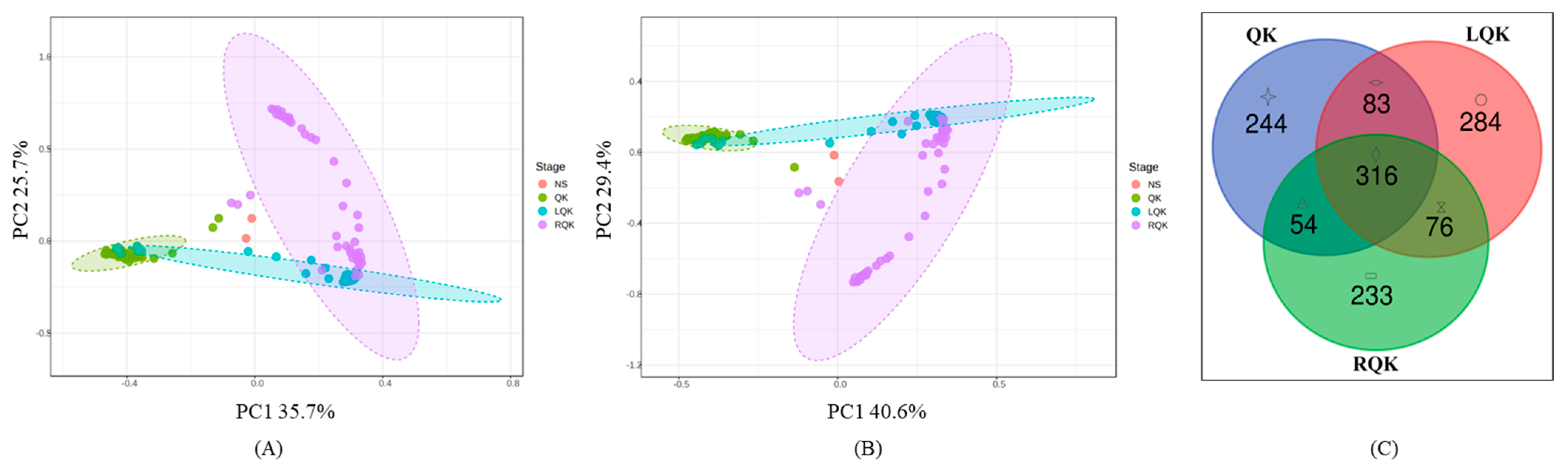
3.1. Microbial Community Structure and Distribution

A total of 10,761 operational taxonomic units (OTUs) were identified using 97% sequence similarity, and the abundance of OTUs of all samples was assessed according to Kwak et al. (2018) [49]. A total of 28 phyla were detected in 24 samples (Figure 1A). Of them, a higher percentage of the phyla were Pseudomonadota, Bacteroidota, Bacillota, Actinomycetota, Fusobacteriota, and Acidobacteriota. In the leaves, the dominant phyla were Bacteroidota (25–40%), Bacillota (25–30%), and Pseudomonadota (16–20%). In the roots, the dominant phyla were Pseudomonadota (18–90%), Bacteroidota (10–40%) and Bacillota (10–30%). At the genus level, a total of 497,775 sequences from the bacterial isolates were identified as unknown sequences that have not been classified into genera. The unknown sequences accounted for 15.83% of the total sequences, indicating that there were many potential new isolates to be discovered (Figure 1B). The dominant genera were Rhizobium, Bacteroides, Blautia, and Prevotella. In the leaves, the dominant genera were Bacteroides (30–50%), Prevotella (5–10%), and Blautia (5–10%). In the roots, the dominant genera were Rhizobium (20–95%), Bacteroides (2–45%), and Prevotella (5%). Roots were mainly correlated with Rhizobium, and leaves were mainly correlated with Bacteroides. Interestingly, both roots and leaves were correlated with Bacteroides.

3.2. OTU Cluster Analysis of Different Kinds of Samples

Two principal component analyses (PCA) were performed on the endogenous OTUs derived from leaves of seedlings (LQK), roots of seedlings (RQK), unplanted soil (NS), and seeds (QK). The first and second axes explained 35.7% and 25.7% of the total variation, respectively, and contributed 61.4% variation, which may reflect some difference existed between endophytic bacterial diversity of seeds and roots (Figure 2A). Furthermore, the Veen diagram showed that the endogenous OTUs from leaves, roots, and seeds shared more than half of the communities (Figure 2B).

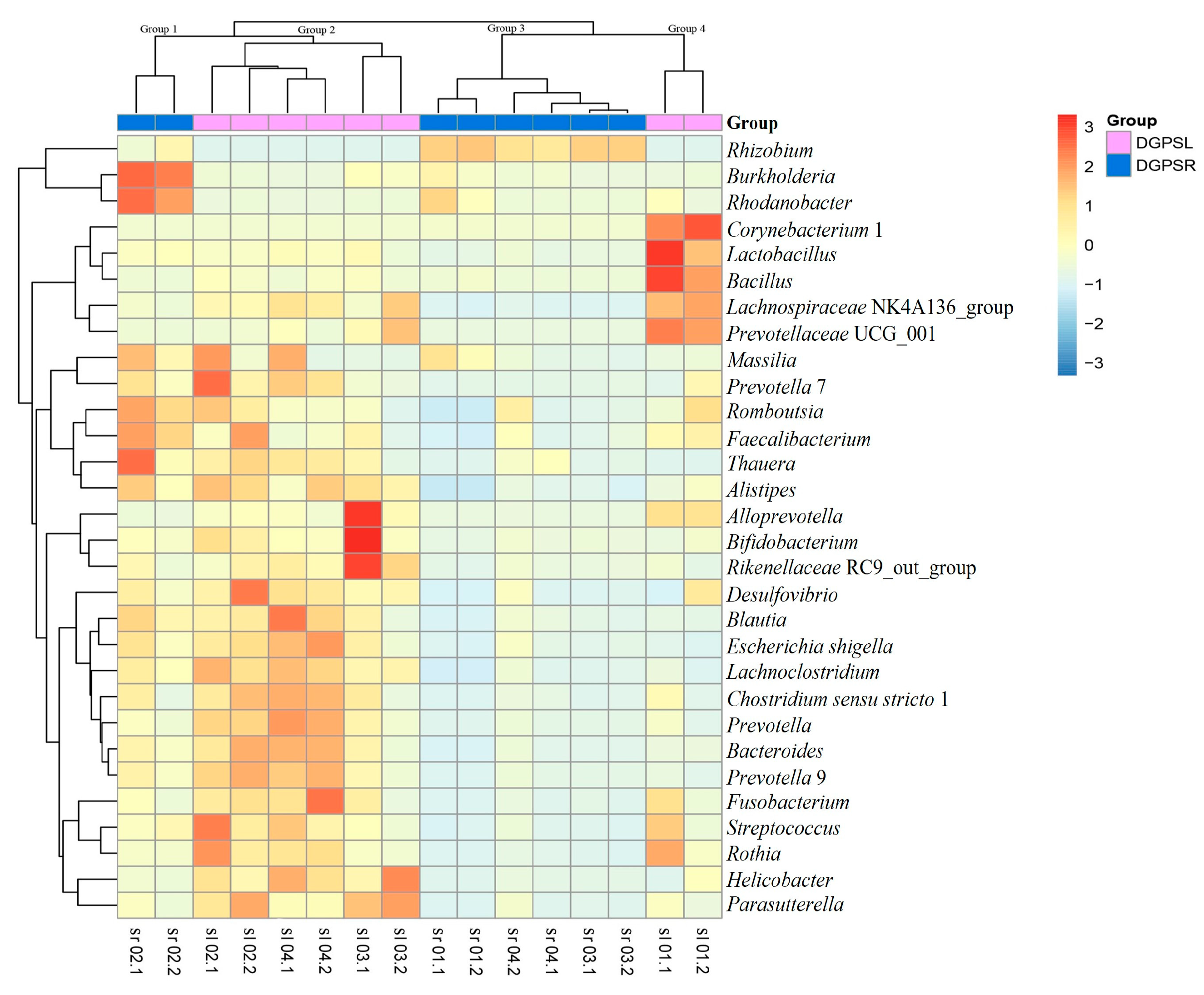
3.3. Phylogenetic Tree and Heatmap

The top 30 OTUs were selected to develop the heatmap and the evolutionary tree. The OTU abundance also varied in different pea cultivars. For example, the heatmap of dry green pea (DGP) cultivars showed that the 16 samples were divided into four clusters (Figure 3). They were Group 1 (RQK 02.1, RQK 02.2) composed by the endogenous OTUs from roots of two plants of one dry pea cultivar QK 02, Group 2 (LQK 02.1, LQK 02.2, LQK 03.1, LQK 03.2, LQK 04.1, and LQK 04.2) composed by the endogenous OTUs from leaves of six plants in three dry pea cultivars (QK 02, QK 03, and QK 04), Group 3 (RQK 01.1, RQK 01.2, RQK 03.1, RQK 03.2, RQK 04.1, and RQK 04.2) composed by the endogenous OTUs from roots of six plants in three dry pea cultivars (QK 01, QK 03, and QK 04), and Group 4 (LQK 01.1, LQK 01.2) composed by endogenous OTUs from leaves of two plants of one dry pea cultivar QK 01. The most representative bacterial genera in Group 1, Group 2, Group 3, and Group 4 were Burkholderia, Rhizobium, Corynebacterium, and Alistipes, respectively.

3.4. Isolation and Identification of Endophytes from the Leaves and Roots of Pea Plants

After surface sterilization, no bacteria were cultivated from the surface of the leaves and roots. However, culturable bacteria were retrieved from the inside of these leaves and roots. This indicated that the bacteria isolated under these conditions were indeed endophytes. Among them, 48 endophytic azotobacter isolates, 16 from roots and 32 from leaves, were selected for analyses of PGP properties and construction of phylogenetic trees.
Most isolates were identified as Bacillota and Pseudomonadota (Table 1, Figure 4 and Figure 5). Among the tested isolates, LZM 26 showed the highest solubilization of organophosphorus, which was identified as Pantoea dispersa. LZM 23 showed the highest solubilization of inorganic phosphorus, and LZM 21 demonstrated the highest solubility of potassium. LZM 26 and LZM 21 were both identified as Rahnella aquatilis.

3.5. Solubilization of Phosphorus and Potassium of Endophytic Isolates

Among the tested endophytic isolates from leaves and roots, 12 isolates were positive for the generation of the turbidity solubilization circle on the organophosphorus medium. Twenty-three isolates were positive for the generation of transparent solubilization circles on the inorganic phosphorus plate. Six isolates were positive for the generation of the transparent solubilization circle on the potassium plate (Table 1). We found that 34.83% of the endophytes isolated from leaves had the ability to solubilize organophosphorus, while only 6.25% of the isolates from roots had this ability. Furthermore, 15.63% of the endophytes isolated from leaves had the ability to solubilize potassium, while 12.50% of the isolates from roots had this ability. More isolates from root capacity show the capacity to solubilize inorganic phosphate (56.25%) than those from leaves (43.75%).

3.6. Production of IAA and ACC Deaminase of Isolates

Among the tested isolates, 33 were positive for IAA production and 15 were negative (Table 1). The spectrophotometric assay revealed that these isolates produced varying levels of IAA in vitro. LZM 21, belonging to Rahnella aquatilis, produced the highest IAA (Figure 4A and Figure 5). Thirty-six isolates were positive for producing ACC deaminase, and most of these isolates were identified as Bacillota and Pseudomonadota (Figure 5). The majority of the endophytic bacteria isolated from this study produced IAA and ACC deaminase, indicating the growth-promoting characteristics of those microbiomes at the seedling stage. Approximately 78.13% of the isolates from leaves and 75% of the isolates from roots had the ability to produce IAA. Furthermore, more isolates from roots had the ability to produce ACC deaminase (81.25%) than those from leaves (62.5%).

3.7. Determination of Nitrogenase, Cellulase, and Pectinase Activities of Endophytic Isolates

Among the tested isolates, 40 were able to grow in N-free semisolid medium and reduce acetylene. Most isolates were identified as Bacillota and Pseudomonadota. LZM 7 showed the highest nitrogenase activity that was related to Acinetobacter schindleri (Table 1). Twenty-four isolates were positive for the production of the solubilization circle on the sodium hydroxycellulose plate. Sixteen isolates were positive for pectinase. RZM 9 that showed the highest cellulase capacity and pectinase capacity was related to Bacillus siamensis (Figure 5). Cellulase and pectinase were related to cell wall synthesis and leaf growth at the seedling stage. We found that 96.88% of endophytes isolated from leaves had the ability to fix nitrogen, while only 50% of isolates from roots had this ability. Furthermore, more isolates from roots had the ability to solubilize hydroxycellulose (56.25%) than those from leaves (37.5%). Moreover, more isolates from roots had the ability to solubilize pectin (37.5%) than those from leaves (25%). The endophytic bacteria in leaves with these properties were more prominent than in roots.

3.8. Phylogenetic Analysis

The 16S rRNA gene amplification sequences of the 48 isolates were used for phylogenetic analysis. The redundant sequences were eliminated among the isolates, and the tree was constructed by aligning the unique sequences of the isolates. Phylogenetic analysis set many isolates to the species level (Figure 5). Among these endophytes, 23 isolates were assigned to Pseudomonadota, 23 were assigned to Bacillota, and 2 were assigned to Actinomycetota. Two isolates were grouped into Actinomycetes, showing the lowest diversity. The greatest species diversity was observed among the Bacillota group, and most isolates belonged to Bacillus. Three isolates of Bacillus sp., Burkholderia sp., and Rahnella sp. were also identified in this study. These isolates may co-evolve with the plants to adapt to specific habitats, and their taxonomic status still needs to be further determined by multi-phase taxonomy. All 16S rRNA gene sequences reported in this study had been deposited on NCBI (http://blast.ncbi.nlm.nih.gov/Blast.cgi, accessed on 28 August 2024).
When considering the abundance of genera, Bacillus had the highest abundance of 44% (21/48) irrespective of the tissue, followed by Acinetobacter making up 13% (6/48) of the cultured bacteria isolated in this study. Regarding the isolates from tissue, Bacillus was predominant in leaves and roots. Most isolates from the leaves and roots belong to the same genera. But four specific isolates in other genera were only separated from leaves rather than roots, i.e., Peanibacillus, Microbacterium, Acinetobacter, and Curtobacterium. Interestingly, three isolates only from roots were specific, and they belong to Leifsonia, Burkholderia, and Pseudomonas, respectively.

4. Discussion

4.1. Diversity of Endophytic Communities in Leaves and Roots

Previous studies have shown that the species of endophytic bacteria varied under different vegetation and host conditions [50]. In our study, the endophytic community structures in the leaves of the pea plants were significantly different from those in the roots, supporting the conclusions of many studies previously reported [7,37,51,52].
The species of endophytic communities in the leaves and roots of the pea seedlings were found to be quite different. For example, Bacteroidota accounted for the highest proportion in seedling leaves, while Pseudomonadota accounted for the highest proportion in seedling roots. Given different functions of leaves and roots in plants, the composition of endophytic communities can fluctuate accordingly [53,54,55]. Bacteroidota has been reported to be involved in C metabolism, such as the degradation of organic matter and nitrite oxidation in legume crops [56,57]. The Pseudomonadota was reported to play a key role in energy metabolism and aboveground biomass and total biomass [58,59,60,61]. Leaf serves as the primary tissue for converting and storing carbon for energy, while roots mainly direct the absorption and transformation of water, nutrients, and minerals. The root or leaf endosphere-dwelling bacterial microbiota must specialize and coevolve with hosts [62], and their colonization was related to bacterial colonization traits (e.g., chemotaxis towards specific resources and production of enzymes) and interplay with host–plant immune responses [8]. Thus, the relative abundance of an OTU belonging to the genus Pseudomonadota was possibly correlated with pea plants belowground N content, and the relative abundance of one OTU of the genus Bacteroidota was possibly correlated with aboveground C content (Figure 1).

4.2. Niche Clustering Analysis at Seedling Stage

Vertical transmission of endophytes with beneficial characteristics can be utilized to promote plant growth and health and benefit the next generation through seeds [63]. In this study, an extensive taxonomic overlap between the leaf, root, and seed microbiota suggested that the microbial communities were similar between different tissue types in the seedling stage of pea with frequent microbial translocations. The PCA analyses showed no clear isolation based on ecological niches of LQK, RQK, NS, and QK, suggesting the presence of shared core bacterial communities (Figure 2). Moreover, there were nearly 400 mutual bacterial communities in leaf, root, and seed tissues, indicating a potential vertical transmission of seed endophytes. Furthermore, the bacterial taxa colonized in seeds exhibited higher vertical transmission capacities in leaves than roots at the seedling stage. He et al. (2023) discovered that 41.38% of the leaf endophytic bacteria of Vallisneria natans were from seeds; only 0.42% of the root endophytic bacteria were derived from seeds [64]. However, these results indicated bacterial taxa of seeds had an important effect on the endophytic bacterial community of leaves and roots.

4.3. Phylogenetic Analysis and Potential Plant Growth Promotion of Endophytic Isolates

The growth-promoting characteristics of the isolated endophytes were identified in this study. Many endophytic isolates, such as RZM 9, RZM 12, and LZM 7, showed PGP traits. DNA sequencing and phylogenetic analysis revealed that RZM 9 belonged to Bacillus. Meanwhile, RZM 9 demonstrated strong cellulase and pectinase activities. Cellulase provides the ductility required for growth of cell walls, while pectinase can link cellulose, both of which are associated with vigorous leaf growth in plants at the seedling stage [65]. Phylogenetic analysis revealed that RZM 12 belonged to Burkholderia, which was in low abundance but important for functions in nitrogen fixation with degradation of aromatic compounds [66,67]. Results of phylogenetic analysis indicated that LZM 7 belonged to Acinetobacter. Acinetobacter was proved to promote the growth of beetroot [68,69]. The endophytes obtained in this study possessed PGP characteristics, and they could be applied to promote the growth of pea plants at the seedling stage. It is important to screen PGP endophytes for improving agronomic traits of pea cultivars. In addition, further research on diversity and transmission of endophytes during the whole growth period of pea and their role in plant growth and development is of great interest to biologists [33,68]. This study enables a deeper understanding of endophytic bacteria composition during pea growth.

5. Conclusions

A total of 48 strains of endophytic bacteria with plant growth-promoting (PGP) characteristics were isolated, of which 16 strains were from roots and 32 strains were from leaves. Among the 48 isolates, 12 isolates had the ability to solubilize organic phosphorus, 23 isolates solubilized inorganic phosphorus, 33 produced IAA, 36 isolates were positive for the production of ACC deaminase, 40 produced nitrogenase, 24 isolates were able to produce cellulase, and 16 isolates produced pectinase. In addition, isolates from leaves and roots had different plant growth-promoting traits. For example, more isolates from roots had the ability to solubilize hydroxycellulose (56.25%) and pectin (37.5%) than those from leaves (37.5% and 25%, respectively). Results from this present study suggest the existence of an active and diverse endophytic bacteria at the seedling stage of pea. The composition of the endophyte community warrants future functional study with the ultimate goal of identifying PGP endophytes that can be used as alternatives to improve the growth of pea plants.

Supplementary Materials

The following supporting information can be downloaded at: https://www.mdpi.com/article/10.3390/agronomy14092030/s1, Table S1. The origin and type of samples.

Author Contributions

J.H.: Conceptualization, investigation, methodology, formal analysis, validation, visualization, and writing—original draft, review and editing; Q.L.: conceptualization, investigation, methodology, and writing—original draft and review and editing; F.S.: investigation, methodology, formal analysis, and writing—review and editing; X.C.: investigation, formal analysis, and writing—review and editing; L.L.: investigation, methodology, formal analysis, validation, and visualization; L.F.: investigation, methodology, and writing—review and editing; S.Z.: conceptualization, and writing—original draft, review, and editing; X.W.: formal analysis, writing—original draft, review, and editing; X.Z.: conceptualization, investigation, methodology, writing—original draft, review, and editing, supervision, and funding acquisition. All authors have read and agreed to the published version of the manuscript.

Funding

This research was supported by “The China Agriculture Research System of MOF and MARA–Food legumes (CARS-08)”, the Presidential Foundation of the QINGDAO Academy of Agriculture Sciences, and the Qingdao Municipal Project for Science and Technology in Public Benefit (No. 14-2-3-35-nsh).

Data Availability Statement

Conflicts of Interest

The authors declare no conflict of interest.

References

  1. Kraiser, T.; Gras, D.E.; Gutiérrez, A.G.; González, B.; Gutiérrez, R.A. A holistic view of nitrogen acquisition in plants. J. Exp. Bot. 2011, 62, 1455–1466. [Google Scholar] [CrossRef]
  2. Reinhold-Hurek, B.; Hurek, T. Living inside plants: Bacterial endophytes. Curr. Opin. Plant Biol. 2011, 14, 435–443. [Google Scholar] [CrossRef]
  3. Ali, S.; Charles, T.C.; Glick, B.R. Delay of flower senescence by bacterial endophytes expressing 1-aminocyclopropane-1-carboxylate deaminase. J. Appl. Microbiol. 2012, 113, 1139–1144. [Google Scholar] [CrossRef]
  4. Ali, S.; Charles, T.C.; Glick, B.R. Amelioration of high salinity stress damage by plant growth-promoting bacterial endophytes that contain ACC deaminase. Plant Physiol. Biochem. 2014, 80, 160–167. [Google Scholar] [CrossRef] [PubMed]
  5. Berendsen, R.L.; Pieterse, C.M.; Bakker, P.A. The rhizosphere microbiome and plant health. Trends Plant Sci. 2012, 17, 478–486. [Google Scholar] [CrossRef] [PubMed]
  6. Lau, J.A.; Lennon, J.T. Evolutionary ecology of plant-microbe interactions: Soil microbial structure alters selection on plant traits. New Phytol. 2011, 192, 215–224. [Google Scholar] [CrossRef] [PubMed]
  7. Wagner, M.R.; Lundberg, D.S.; Rio, T.G.D.; Tringe, S.G.; Dangl, J.L.; Mitchell-Olds, T. Host genotype and age shape the leaf and root microbiomes of a wild perennial plant. Nat. Commun. 2016, 7, 12151. [Google Scholar] [CrossRef]
  8. Guo, J.J.; Ling, N.; Li, Y.; Li, K.; Ning, H.L.; Shen, Q.R.; Guo, S.W.; Vandenkoornhuyse, P. Seed-borne, endospheric and rhizospheric core microbiota as predictors of plant functional traits across rice cultivars are dominated by deterministic processes. New Phyt. 2021, 230, 2047–2060. [Google Scholar] [CrossRef]
  9. Ku, Y.L.; Han, X.T.; Lei, Y.T.; Zhang, M.; Zhao, Z. Different sensitivities and assembly mechanisms of the root-associated microbial communities of Robinia pseudoacacia to spatial variation at the regional scale. Plant Soil 2023, 486, 621–637. [Google Scholar] [CrossRef]
  10. Agler, M.T.; Ruhe, J.; Kroll, S.; Morhenn, C.; Kim, S.T.; Weigel, D.; Kemen, E.M. Microbial Hub Taxa Link Host and Abiotic Factors to Plant Microbiome Variation. PLoS Biol. 2016, 14, e1002352. [Google Scholar] [CrossRef]
  11. Nelson, E.B. The seed microbiome: Origins, interactions, and impacts. Plant Soil 2018, 422, 7–34. [Google Scholar] [CrossRef]
  12. Aziz, U.; Rehmania, M.S.; Wang, L.; Luo, X.F.; Xian, B.S.; Wei, S.W.; Wang, G.D.; Shu, K. Toward a Molecular Un-derstanding of Rhizosphere, Phyllosphere, and Spermosphere Interactions in Plant Growth and Stress Response. Crit. Rev. Plant Sci. 2021, 40, 479–500. [Google Scholar] [CrossRef]
  13. Burbano, J.L.; Hurek, C.S.; Reinhold-Hurek, B. Isolation and characterization of root-associated bacteria from agricultural crops in the Kavango region of Namibia. Plant Soil. 2012, 356, 67–82. [Google Scholar]
  14. Jorquera, M.A.; Shaharoona, B.; Nadeem, S.M.; Mora, M.D.L.L.; Crowley, D.E. Plant Growth-Promoting Rhizobacteria Associated with Ancient Clones of Creosote Bush (Larrea tridentata). Microb. Ecol. 2012, 64, 1008–1017. [Google Scholar] [CrossRef]
  15. Bulgarelli, D.; Schlaeppi, K.; Spaepen, S.; Themaat, E.V.L.V.; Schulze-Lefert, P. Structure and functions of the bacterial microbiota of plants. Annu. Rev. Plant Biol. 2013, 64, 807–838. [Google Scholar] [CrossRef]
  16. Cosgrove, D.J. Growth of the plant cell wall. Nat. Rev. Mol. Cell Biol. 2005, 6, 850–861. [Google Scholar] [CrossRef]
  17. Mirabet, V.; Das, P.; Boudaoud, A.; Hamant, O. The role of mechanical forces in plant morphogenesis. Annu. Rev. Plant Biol. 2011, 62, 365–385. [Google Scholar] [CrossRef] [PubMed]
  18. Haas, K.T.; Wightman, R.; Peaucelle, A.; Hofte, H. The role of pectin phase separation in plant cell wall assembly and growth. Cell Surf. 2021, 7, 100054. [Google Scholar] [CrossRef] [PubMed]
  19. Cosgrove, D.J. Structure and growth of plant cell walls. Nat. Rev. Mol. Cell Biol. 2023, 25, 340–358. [Google Scholar] [CrossRef]
  20. Ofek, M.; Hadar, Y.; Minz, D. Colonization of cucumber seeds by bacteria during germination. Environ. Microbiol. 2011, 13, 2794–2807. [Google Scholar] [CrossRef]
  21. Wu, X.; Li, N.; Hao, J.; Hu, J.; Zhang, X.; Blair, M.W. Genetic diversity of Chinese and global pea (Pisum sativum L.) collections. Crop Sci. 2016, 57, 1574–1584. [Google Scholar] [CrossRef]
  22. Nossa, C.W.; Oberdorf, W.E.; Aas, J.A.; Paster, B.J.; DeSantis, T.Z.; Brodie, E.L.; Malamud, D.; Poles, M.A.; Pei, Z. Design of 16s rRNA gene primers for 454 pyrosequencing of the human foregut microbiome. World J. Gastroenterol. 2010, 16, 4135. [Google Scholar] [CrossRef] [PubMed]
  23. Bolger, A.M.; Lohse, M.; Usadel, B. Trimmomatic: A flexible trimmer for Illumina sequence data. Bioinformatics 2014, 30, 2114–2120. [Google Scholar] [CrossRef] [PubMed]
  24. Reyon, D.; Tsai, S.Q.; Khayter, C.; Foden, J.A.; Sander, J.D.; Joung, J.K. FLASH assembly of TALENs for high-throughput genome editing. Nat. Biotechnol. 2012, 30, 460–465. [Google Scholar] [CrossRef]
  25. Caporaso, J.G.; Kuczynski, J.; Stombaugh, J.; Bittinger, K.; Bushman, F.D.; Costello, E.K. QIIME allows analysis of high-throughput community sequencing data. Nat. Methods 2010, 7, 335–336. [Google Scholar] [CrossRef]
  26. Edgar, R.C.; Haas, B.J.; Clemente, J.C.; Quince, C.; Knight, R. UCHIME improves sensitivity and speed of chimera detection. Bioinformatics 2011, 27, 2194. [Google Scholar] [CrossRef]
  27. Wang, Q.; Garrity, G.M.; Tiedje, J.M.; Cole, J.R. Naive Bayesian classifier for rapid assignment of r RNA sequences into the new bacterial taxonomy. Appl. Environ. Microbiol. 2017, 73, 5261–5267. [Google Scholar] [CrossRef]
  28. Kõljalg, U.; Larsson, K.H.; Abarenkov, K.; Nilsson, R.H.; Alexander, I.J.; Eberhardt, U.; Erland, S.; Høiland, K.; Kjøller, R.; Larsson, E.; et al. UNITE: A database providing web-based methods for the molecular identification of ectomycorrhizal fungi. New Phyt. 2005, 166, 1063–1068. [Google Scholar] [CrossRef]
  29. Lv, X.; Wang, Q.; Zhang, X.; Hao, J.; Li, L.; Chen, W.; Li, H.; Wang, Y.; Ma, C.; Wang, J.; et al. Community structure and associated networks of endophytic bacteria in pea roots throughout plant life cycle. Plant Soil 2021, 468, 225–238. [Google Scholar] [CrossRef]
  30. Dhariwal, A.; Chong, J.; Habib, S.; King, I.L.; Agellon, L.B.; Xia, J. MicrobiomeAnalyst: A web-based tool for compre-hensive statistical, visual and meta-analysis of microbiome data. Nucleic Acids Res. 2017, 45, W180–W188. [Google Scholar] [CrossRef]
  31. Barter, R.L.; Yu, B. Superheat: An R package for creating beautiful and extendable heatmaps for visualizing complex data. J. Comput. Graph. Stat. 2018, 27, 910–922. [Google Scholar] [CrossRef]
  32. Bai, W.; Hu, R.; Zhang, J.; Feng, L.; Xu, H. A study on the isolation, screening and phosphate solubilizing capacity of phosphate solubilizing bacteria in the rhizosphere of corn. J. South China Agric. Univ. 2013, 34, 167–176. [Google Scholar]
  33. Borah, A.R.; Das, R.; Mazumdar Thakur, D. Culturable endophytic bacteria of Camellia species endowed with plant growth promoting characteristics. J. Appl. Microbiol. 2019, 127, 825–844. [Google Scholar] [CrossRef]
  34. Etesami, H.; Emami, S.; Alikhani, H.A. Potassium solubilizing bacteria (KSB): Mechanisms, promotion of plant growth, and future prospects A review. J. Soil Sci. Plant Nut. 2017, 17, 897–911. [Google Scholar] [CrossRef]
  35. Gordon, S.A.; Weber, R.P. Colorimetric estimation of indoleacetic acid. Plant Physiol. 1951, 26, 192–195. [Google Scholar] [CrossRef] [PubMed]
  36. Penrose, D.M.; Glick, B.R. Methods for isolating and characterizing ACC deaminase-containing plant growth-promoting rhizobacteria. Physiol. Plant. 2003, 118, 10–15. [Google Scholar] [CrossRef]
  37. Chimwamurombe, P.M.; Grönemeyer, J.L.; Reinhold-Hurek, B. Isolation and characterization of culturable seed-associated bacterial endophytes from gnotobiotically grown Marama bean seedlings. FEMS Microbiol. Ecol. 2016, 92, 1574–6941. [Google Scholar] [CrossRef]
  38. Jacobson, C.B.; Pasternak, J.J.; Glick, B.R. Partial purification and characterization of 1-aminocyclopropane-1-carboxylate de-aminase from the plant growth promoting rhizobacterium Pseudomonas putida GR12-2. Can. J. Microbiol. 1994, 40, 1019–1025. [Google Scholar] [CrossRef]
  39. Dai, Z.G. Colorimetric determination of nitrogenase activity in rhizobia. Ningxia Agric. Sci. Technol. 1981, 3, 43–44. [Google Scholar]
  40. Tariq, M.; Hameed, S.; Yasmeen, T.; Zahid, M.; Zafar, M. Molecular characterization and identification of plant growth promoting endophytic bacteria isolated from the root nodules of pea (Pisum sativum L.). World J. Microb. Biot. 2014, 30, 719–725. [Google Scholar] [CrossRef]
  41. Wang, X.N.; Luo, J.H.; Hao, J.J.; Zhang, X.Y.; Liu, L.; Fu, L.P.; Wang, J.L.; Han, Y.H.; Liu, Q.L. Plant growth-promoting activities of endophytic nitrogen -fixing bacteria isolated from seedings of faba bean (Vicia faba L.). J. Agric. Sci. 2020, 22, 33–39. [Google Scholar]
  42. Da Silva, A.C.; Rachid, C.T.C.D.C.; De Jesus, H.E.; Rosado, A.S.; Peixoto, R.S. Predicting the biotechnological potential of bacteria isolated from Antarctic soils, including the rhizosphere of vascular plants. Polar Biol. 2017, 40, 1393–1407. [Google Scholar] [CrossRef]
  43. Grönemeyer, J.L.; Kulkarni, A.; Berkelmann, D.; Hurek, T.; Reinhold-Hurek, B. Identification and characterization of rhizobia indigenous to the Okavango region in Sub-Saharan Africa. Appl. Environ. Microb. 2014, 80, AEM-02417. [Google Scholar] [CrossRef]
  44. Drancourt, M.; Bollet, C.; Carlioz, A.; Martelin, R.; Gayra, J.P.; Raoult, D. 16S Ribosomal DNA sequence analysis of a large collection of environmental and clinical unidentifiable bacterial isolates. J. Clin. Microbiol. 2000, 38, 3623–3630. [Google Scholar] [CrossRef]
  45. Hall, T.A. BioEdit: A user-friendly biological sequence alignment editor and analysis program for Windows 95/98/NT. Nucleic Acids Symp. 1999, 41, 95–98. [Google Scholar]
  46. Langenwalder, D.B.; Schmidt, S.; Gilli, U.; Pantchev, N.; Ganter, M.; Silaghi, C.; von Loewenich, F.D. Genetic characterization of Anaplasma phagocytophilum strains from goats (Capra aegagrus hircus) and water buffalo (Bubalus bubalis) by 16S rRNA gene, ankA gene and multilocus sequence typing. Ticks Tick-Borne Dis 2009, 10, 101267. [Google Scholar] [CrossRef] [PubMed]
  47. Saitou, N.; Nei, M. The neighbor-joining method: A new method for reconstructing phylogenetic trees. Mol. Biol. Evol. 1987, 4, 406–425. [Google Scholar]
  48. Tamura, K.; Nei, M.; Kumar, S. Prospects for inferring very large phylogenies by using the neighbor-joining method. Proc. Natl. Acad. Sci. USA 2004, 101, 11030–11035. [Google Scholar] [CrossRef]
  49. Kwak, M.J.; Kong, H.G.; Choi, K.; Kwon, S.K.; Song, J.Y.; Lee, J.; Lee, P.A.; Choi, S.Y.; Seo, M.; Lee, H.J.; et al. Rhizosphere microbiome structure alters to enable wilt resistance in tomato. Nat. Biotechnol. 2018, 36, 1117. [Google Scholar] [CrossRef]
  50. Zuo, X.A.; Zhao, H.L.; Zhao, X.Y.; Guo, Y.R.; Yun, J.Y.; Wang, S.K.; Miyasaka, T.J.E.G. Vegetation pattern variation, soil degradation and their relationship along a grassland desertification gradient in Horqin Sandy Land, northern China. Environ. Geol. 2009, 58, 1227–1237. [Google Scholar] [CrossRef]
  51. Luo, J.; Tao, Q.; Jupa, R.; Liu, Y.; Li, T. The role of vertical transmission of shoot endophytes in root-associated microbiome assembly and heavy metal hyperaccumulation in sedum alfredii. Environ. Sci. Technol. 2019, 53, 6954–6963. [Google Scholar] [CrossRef]
  52. Bai, Y.; Müller, D.B.; Srinivas, G.; Garrido-Oter, R.; Potthoff, E.; Rott, M.; Dombrowski, N.; Münch, P.C.; Spaepen, S.; Remus-Emsermann, M.; et al. Functional overlap of the Arabidopsis leaf and root microbiota. Nature 2015, 528, 364–369. [Google Scholar] [CrossRef] [PubMed]
  53. Ettinger, C.L.; Voerman, S.E.; Lang, J.M.; Stachowicz, J.J.; Eisen, J.A. Microbial communities in sediment from Zostera marina patches, but not the Z. marina leaf or root microbiomes, vary in relation to distance from patch edge. PeerJ 2017, 5, e3246. [Google Scholar] [CrossRef] [PubMed]
  54. Wei, N.; Ashman, T.L. The effects of host species and sexual dimorphism differ among root, leaf and flower microbiomes of wild strawberries in situ. Sci. Rep. 2018, 8, 5195. [Google Scholar] [CrossRef] [PubMed]
  55. Yang, R.; Liu, P.; Ye, W.X. Illumina-based analysis of endophytic bacterial diversity of tree peony (Paeonia Sect. Moutan) roots and leaves. Braz. J. Microbiol. 2017, 48, 695–705. [Google Scholar] [CrossRef]
  56. Cai, S.; Zhao, X. Responses of bacterial communities and nitrogen-cycling microbiomes to the conversion from cereals to legumes in the rice-based system. Appl. Soil Ecol. 2024, 195, 105241. [Google Scholar] [CrossRef]
  57. Adnan, M.; Wahid, F.; Fahad, S.; Arif, M.; Shi, S.M.; He, X.H. Mycorrhizae and soil health. In Improving Soil Health; Burleigh Dodds Science Publishing Limited: Cambridge, UK, 2023; pp. 5–83. [Google Scholar]
  58. Sasse, J.; Martinoia, E.; Northen, T. Feed your friends: Do plant exudates shape the root microbiome? Trends Plant Sci. 2017, 23, 25–41. [Google Scholar] [CrossRef]
  59. Degli Esposti, M.; Martinez Romero, E. A survey of the energy metabolism of nodulating symbionts reveals a new form of respiratory complex I. FEMS Microbiol. Ecol. 2016, 92, fiw084. [Google Scholar] [CrossRef]
  60. Mukhopadhya, I.; Hansen, R.; El-Omar, E.M.; Hold, G.L. IBD—What role do Proteobacteria play? Nat. Rev. Gastroenterol. Hepatol. 2012, 9, 219–230. [Google Scholar] [CrossRef]
  61. Liu, A.; Contador, C.A.; Fan, K.; Lam, H.M. Interaction and regulation of carbon, nitrogen, and phosphorus metabolisms in root nodules of legumes. Front. Plant Sci. 2018, 9, 426283. [Google Scholar] [CrossRef]
  62. Kemen, A.C.; Agler, M.T.; Kemen, E. Host–microbe and microbe–microbe interactions in the evolution of obligate plant parasitism. New Phyt. 2015, 206, 1207–1228. [Google Scholar] [CrossRef] [PubMed]
  63. Truyens, S.; Weyens, N.; Cuypers, A.; Vangronsveld, J. Bacterial seed endophytes: Genera, vertical transmission and interaction with plants. Environ. Microbiol. Rep. 2015, 7, 40–50. [Google Scholar] [CrossRef]
  64. He, R.; Hu, S.; Li, Q.; Zhao, D.; Wu, Q.L.; Zeng, J. Greater transmission capacities and small-world characteristics of bacterial communities in the above-than those in the below-ground niches of a typical submerged macrophyte, Vallisneria natans. Sci. Total Environ. 2023, 903, 166229. [Google Scholar] [CrossRef] [PubMed]
  65. Braybrook, S.A.; JöNsson, H. Shifting foundations: The mechanical cell wall and development. Curr. Opin. Plant Biol. 2016, 29, 115–120. [Google Scholar] [CrossRef] [PubMed]
  66. Suárez-Moreno, Z.R.; Caballero-Mellado, J.; Coutinho, B.G.; Mendonça-Previato, L.; James, E.K.; Venturi, V. Common features of environmental and potentially beneficial plant-associated Burkholderia. Microb. Ecol. 2012, 63, 249–266. [Google Scholar] [CrossRef]
  67. İnceoğlu, Ö.; Al-Soud, W.A.; Salles, J.F.; Semenov, A.V.; Elsas, J.D.V. Comparative analysis of bacterial communities in a potato field as determined by pyrosequencing. PLoS ONE 2011, 6, 1932–6203. [Google Scholar] [CrossRef] [PubMed]
  68. Shi, Y.; Lou, K.; Li, C. Growth promotion effects of the endophyte Acinetobacter johnsonii strain 3-1 on sugar beet. Symbiosis 2011, 54, 159–166. [Google Scholar] [CrossRef]
  69. Cherif-Silini, H.; Silini, A.; Yahiaoui, B.; Ouzari, I.; Boudabous, A. Phylogenetic and plant-growth-promoting characteristics of Bacillus isolated from the wheat rhizosphere. Ann. Microbiol. 2016, 66, 1087–1097. [Google Scholar] [CrossRef]
Figure 1. The histograms of endogenous communities. (A) Taxonomic comparison of the 28 most abundant phyla were selected to draw the histogram of phylum level. (B) Taxonomic comparison of the 27 most abundant genera were selected to draw the histogram of genus level.
Agronomy 14 02030 g001
Figure 2. Distribution of endophytes in different tissues of pea seedling developmental stage. (A) The principal component analyses (PCA) diagram of total OTUs from leaves of seedling plants (LQK). (B) The principal component analyses (PCA) diagram of total OTUs from roots of seedling plants (RQK). (C) The Veen diagram shows common and unique OTUs among LQK, RQK, and unplanted seeds (QK).
Agronomy 14 02030 g002
Figure 3. The heatmaps of endogenous communities based on the 30 top OTUs.
Agronomy 14 02030 g003
Figure 4. The Indole-3-acetic acid (IAA) production and nitrogenase activity. (A) The nitrogenase activity capacity. (B) The IAA production capacity. LZM: the isolate from seedling leaf; RZM: the isolate from seedling root.
Agronomy 14 02030 g004
Figure 5. Neighbor-joining phylogenetic tree of endophyte isolates and best-matching 16S rRNA sequences derived from National Center for Biotechnology Information (NCBI). LZM: the isolate from seedling leaf; RZM: the isolate from seedling root. The isolates identified in the present study were shown in bold.
Agronomy 14 02030 g005
Table 1. Plant growth-promoting characteristics of endophytes.
No.PhylotypeCandidate ID.Accession NumberInorganic PhosphorusOrganic PhosphorusPotassiumCellulasePectinaseIAAACCNitrogenase Activity
1LZM1Acinetobacter johnsoniiMN709198-+---+-+
2LZM2Acinetobacter johnsoniiMN709199++---+++
3LZM3Bacillus australimarisMN709173---+++++
4LZM4Bacillus aeriusMN709174-+-+++++
5LZM5Acinetobacter johnsoniiMN709200---+++-+
6LZM6Acinetobacter johnsoniiMN709201-+---+++
7LZM7Acinetobacter johnsoniiMN709202++-+-+-+
8LZM8Rahnella sp.MN709205-+-+--++
9LZM9Bacillus australimarisMN709182---++-++
10LZM10Enterobacter roggenkampiiMN709209-----+++
11LZM11Paenibacillus tundraeMN709193---+--+-
12LZM12Acinetobacter oryzaeMN709203+--+++++
13LZM13Paenibacillus tundraeMN709194---+++++
14LZM14Bacillus australimarisMN709183++---+++
15LZM15Pantoea agglomeransMN709214++-+++++
16LZM16Bacillus safensisMN709187------++
17LZM17Bacillus australimarisMN709181+-++++++
18LZM18Acinetobacter lwoffiiMN709204-----+++
19LZM19Enterobacter ludwigiiMN709211+------+
20LZM20Bacillus xiamenensisMN709188---+-+++
21LZM21Rahnella aquatilisMN709206+-+--+++
22LZM22Bacillus aeriusMN709179-+---+++
23LZM23Rahnella aquatilisMN709207+-++++++
24LZM24Bacillus australimarisMN709184-----+++
25LZM25Bacillus pumilusMN709185++++++-+
26LZM26Pantoea brenneriMN709213-+-+--++
27LZM27Microbacterium hydrocarMN709195----+--+
28LZM28Paenibacillus taichungensisMN712330+-++-+++
29LZM29Pantoea agglomeransMN712331+--+-+++
30LZM30Pantoea agglomeransMN709215+----+++
31LZM31Pantoea agglomeransMN709216---+++++
32LZM32Bacillus pumilusMN709186+----+++
33RZM1Bacillus megateriumMN709191-----+++
34RZM2Enterobacter ludwigiiMN709212---++---
35RZM3Bacillus thuringiensisMN709190+----++-
36RZM4Pseudomonas baeticaMN709197-----+-+
37RZM5Pantoea vagansMN709217+-+--+++
38RZM6Bacillus subtilisMN709178---+++++
39RZM7Bacillus halotoleransMN709176---+-+--
40RZM8Enterobacter roggenkampiiMN709210------+-
41RZM9Bacillus sp.MN709177+--+++++
42RZM10Leifsonia shinshuensisMN709196--------
43RZM11Bacillus mojavensisMN709175+--+++--
44RZM12Burkholderia sp.MN712332+----++-
45RZM13Bacillus cereusMN709189++-+-+-+
46RZM14Bacillus aeriusMN709180+----+++
47RZM15Rahnella aquatilisMN709208+----++-
48RZM16Bacillus megateriumMN709192+-+---++
Note: LZM means the isolate from seedling leaf; RZM means the isolate from seedling root; “+”, mean positive of treatment; “-”, mean negative of treatment.
Disclaimer/Publisher’s Note: The statements, opinions and data contained in all publications are solely those of the individual author(s) and contributor(s) and not of MDPI and/or the editor(s). MDPI and/or the editor(s) disclaim responsibility for any injury to people or property resulting from any ideas, methods, instructions or products referred to in the content.

