Article

The Microbiological Activity of Soil in Response to Gliotoxin, the “Lethal Principle” of Trichoderma

by
Anastasia V. Teslya
,
Elena V. Gurina
,
Artyom A. Stepanov
,
Aleksandr V. Iashnikov
and
Alexey S. Vasilchenko
*
Laboratory of Antimicrobial Resistance, Institute of Environmental and Agricultural Biology (X-BIO), Tyumen State University, Tyumen 625003, Russia
*
Author to whom correspondence should be addressed.
Agronomy 2024, 14(9), 2084; https://doi.org/10.3390/agronomy14092084
Submission received: 15 August 2024 / Revised: 10 September 2024 / Accepted: 11 September 2024 / Published: 12 September 2024

Abstract

:
Trichoderma is a soil-dwelling microorganism that has many benefits for plants and is therefore widely used in agriculture. Among the secondary metabolites produced by Trichoderma, gliotoxin (GT) is one of the most studied. The antagonistic effect of GT on other fungi was first discovered by R. Weindling in 1934. He referred to it as the “lethal principle” of Trichoderma. Despite the long history of studying GT, its impact on the soil microbial community has remained largely unexplored. In our work, we investigated the response of the soil microbial community to different doses of GT (10–500 µM per kg) and different durations (7–56 days) of exposure. We measured microbiological parameters (CO2 emission, microbial biomass (MB)), calculated the eco-physiological indices and determined the activity of soil enzymes involved in the C, N, P and S cycles. We identified three types of microbial responses to GT: inhibition, stress and stimulation. The inhibitory effect developed only by day 56 and in the samples treated with 500 μM GT. The stress effect (increased CO2 emission and decreased MB) of GT on microbial communities was predominant. Soil extracellular enzymes also responded to GT to varying degrees. A stimulating effect of GT on enzyme activity was noted for β-D-1,4-cellobiosidase and β-1,4-glucosidase. The activity of arylsulfatase and leucine aminopeptidase decreased under the influence of GT up to day 28, but by the end of the experiment, there was a restoration of activity. We did not observe any significant changes in the activity of β-1,4-xylosidase, β-1,4-N-acetyl-glucosaminidase or acid phosphatase. The results obtained showed that GT at high, “man-made” doses can inhibit the microbiological activity of soil, but at naturally occurring concentrations, it can have a stimulating effect on soil microbiome functionality.

1. Introduction

The increasing demand for environmentally friendly agricultural chemicals is driving advances in research to find alternative, natural methods to stimulate plant growth and increase productivity, as well as to protect crops against phytopathogens. Fungal and bacterial microbial inoculants are an important alternative to agrochemicals [1,2].
The extracellular enzymes of the soil microbial community play a crucial role in the cycling of essential nutrients and the overall biogeochemical processes in ecosystems. The use of microbial inoculants is intended to help maintain the health of soil and, in turn, its fertility. However, there is a lack of full understanding of how the use of microbiological products affects the interactions between microorganisms in soils and, consequently, the essential functions of the soil ecosystem. The benefits that plants can experience are realized both indirectly, through increased systemic resistance, and directly, for example, through the bioconversion of essential nutrients into available forms and direct pathogen antagonism. The ability to produce antibiotics is one of the main characteristics of microbial agents taken into account when screening and selecting useful strains [3]. It is worth noting that the action of antibiotics produced by microbial agents affects not only the pathogenic components of the microbiome but also other bacterial and fungal taxa. The fact that secondary metabolites produced by microorganisms in biopesticides may have undesirable effects on non-target organisms is even a reason for criticism and discussion of their use in agriculture [4,5]. To date, there have been no systematic studies on the impact of antibiotics produced by PGP (plant growth-promoting) microorganisms on the structural and functional properties of soils. However, interested readers can refer to a review on the effect of pharmaceutical antibiotics on soil functions [6].
Recently, the ecological role of antibiotics has become a topic of discussion, as it defines the properties of these substances as signaling molecules that influence transcription and translation processes, modulating biochemical activities in natural populations on both intraspecific and interspecific levels [7]. From the perspective of agricultural practices, there is a significant lack of understanding about the fundamental processes that take place in soil when antibiotic-producing microorganisms are present.
Trichoderma is one of the most sought-after PGP microorganisms for use in cropping systems [8]. Trichoderma’s secondary metabolites are not only good fungicides but also plant growth stimulants [9]. Gliotoxin (GT) is one of these well-known secondary metabolites produced by Trichoderma virens as well as other fungi: Aspergillus, Eurotium, Neosartorya, Penicillium and Acremonium [10]. Since the discovery of GT as an antibiotic in 1934, it has been recognized as one of the key factors in determining the effectiveness of antimicrobial preparations based on Trichoderma virens; R. Weindling referred GT as the “lethal principle” of Trichoderma against Rhizoctonia solani [11,12,13,14,15,16,17,18]. In soils, Trichoderma virens has the ability to produce approximately 1 microgram of gliotoxin per gram of soil [19]. In a vermiculite medium, gliotoxin was detected in an amount of 0.42 μg/cm3, in composted mineral soil—0.36 μg/cm3, in clay soil—0.20 μg/cm3 and in sandy soil—0.02 μg/cm3 [20]. GT was found to inhibit the growth of Fusarium solani [21], Phytophthora capsici [22], Macrophomina phaseolina [23], Rhizoctonia solani [24] and Pythium ultimum [25]. Recently, it has been found that GT is active against various types of bacteria and is more effective against those with a Gram-positive cell wall [26]. Trichoderma is one of the most extensively studied fungi with beneficial properties for cultivated plants. More detailed information on the plant-beneficial characteristics of Trichoderma species can be found in review articles [27,28,29]. Since its discovery in the 20th century, the functional and ecological roles of GT have only begun to become clearer. For example, recent research has found that, in addition to its fungicidal and bactericidal properties, GT also induces transcriptomic reprogramming in plants, priming their immune responses and defenses [30]. Today, there is a lack of knowledge regarding the effects of GT on soil microorganisms, the related transformation of nutrients in soil and the associated biochemical processes. This information is essential for understanding how microbial products function.
In this work, we focused on studying the effect of GT on soil microorganisms by assessing the key functional indicators of soil health, such as the respiratory activity of soil microbiota, microbial biomass and enzymatic activity. This study presents an evaluation of the ecological and physiological state of the microbial community in response to GT. We hypothesize that (1) gliotoxin will act as an external inducer of physiological activity (including as an energy source), promoting an increase in the content of microbial biomass and its respiratory activity; (2) the activity of soil enzymes will increase under the influence of GT; (3) over time, the stimulating effect will weaken due to the degradation of GT in soils and the adaptation of the microbial community to new conditions. In soils with a low dose of GT, the return to initial indicators or near-control conditions will occur more rapidly. Additionally, it is possible that high doses of GT may have a stress-inducing effect and lead to a reduction in microbial biomass and enzyme activity in the soil.

2. Materials and Methods

2.1. Soil Sample Preparation and Experimental Design

GT was obtained by cultivating Aspergillus fumigatus UTMN in liquid Weindling broth, as described in the work [31]. The soil for the experiment was collected using the “checkerboard” method (four points at the corners and one in the center) from the surface (0–15 cm) of the agrocenosis (coordinates: 57.0912, 65.3738). The collected soil was delivered to the laboratory in polyethylene bags (“zip” bags).
In the laboratory, soil samples were cleared of visible plant debris and stones and mixed into one sample. The mixed sample was then thoroughly mixed and sifted through a sieve with a pore diameter of 2 mm. For each GT concentration (10, 50 and 500 μM kg−1 soil), five replicate microcosms were prepared by transferring carefully prepared soil into plastic containers with multiple holes of 1–2 mm in diameter (Figure S1). Each soil sample was mixed with different concentrations of GT diluted in distilled water. The control soils were mixed with water. All soil samples were then homogenized by mixing thoroughly for 5 min to ensure the uniform distribution of GT and water. The water-holding capacity of the soils was adjusted to 60%. Containers with GT-treated soils were placed at a depth of 0–5 cm in the agrocenosis (where the soil was initially collected). Soil samples were collected from each microcosm after 7, 14, 28 and 56 days of incubation. The experimental period was June–August 2023 (Table S1).

2.2. Chemical Analysis

For basic soil parameters, the soil pHKCl and pHH2O were measured potentiometrically using an Orion Star A 111 pH meter (Thermo Scientific, Waltham, MA, USA) according to the international standard ISO 10390 [32]. The total carbon (TC) and total nitrogen (TN) contents were determined in triplicate using a Vario EL III elemental analyzer (Elementar, Langenselbold, Germany). The soil was gray (Luvic Phaeozems, according to the WRB classification) with a loamy texture (clay 28.5%, silt 27.7%, sand 43.8%), pHH2O 6.2 and pHKCl 5.6. The content of TC determined in this way was considered equal to the SOC content, since carbonates are absent from the studied Luvic Phaeozems.

2.3. Extraction and Detection of GT in Soil

GT residues were quantified using chloroform extraction. To perform this, 500 mg of soil was taken from each microcosm and placed in a test tube (2 mL). The soil was then mixed with 1 mL of chloroform solvent. The tubes were shaken for 1 h at 150 rpm in a shaker–incubator. After that, the samples were centrifuged for 10 min at 13,000 rpm. This extraction procedure was repeated three times in order to maximize the amount of GT extracted from the soil. The extracts were combined, and then the solvent was removed by evaporation on a vacuum rotary evaporator (Heidolph Hei-VAP Expert Control, Schwabach, Germany). The dry residue was redissolved in DMSO (1 mL) and applied to a 6 mL C18 column for solid-phase extraction (Phenomenex, Torrance, CA, USA). The resulting extract was lyophilized, redissolved again in DMSO (1.5 mL) and analyzed by high-performance liquid chromatography (HPLC) (Agilent 1260 Infinity II, Agilent Technologies, Santa Clara, CA, USA) using the analytical standard of gliotoxin.

2.4. Microbiological Analysis

Basal respiration (BR) was determined by the emission of CO2 from the soil of the microcosms with and without GT using a LI-830 gas analyzer (LI-COR Biosciences, Lincoln, NE, USA). The procedure for determining BR is described in [26,33]. The substrate-induced respiration (SIR) method was used to determine the microbial biomass carbon content (MBSIR). The procedure for determining SIR and MBSIR is described in [26,33]. CO2 emission measured by the SIR method was recorded using an LI-830 gas analyzer. BR and SIR values were expressed in CO2 g−1 soil h−1 [34]. MBSIR was expressed in μg C g−1 soil. Measurements of BR, SIR and, respectively, MBSIR were performed in 3-fold replicates on the 7th, 14th, 28th and 56th days after the introduction of GT into the microcosms.
The ecological and physiological functioning of the microbial community in the presence of GT and without it was assessed using the following indicators: QR, qCO2, MBSIR/SOC and qCO2/SOC. The description of eco-physiological indicators is presented in detail in the work [26].

2.5. Enzymatic Analysis

The activities of β-D-1,4-cellobiosidase (CB), β-1,4-glucosidase (βG), β-1,4-xylosidase (βX), β-1,4-N-acetyl-glucosaminidase (NAG), L-leucine aminopeptidase (LAP), acid phosphatase (AP) and arylsulfatase (ARS) were determined fluorimetrically using fluorogenically labeled substrates [35]. Measurements were performed in 96-well black plates. Standard (4-MUB and AMC) and substrate solutions were prepared in defined concentrations. A one-gram soil sample was mixed with 50 mL of sterile water. The soil suspension was homogenized using ultrasound and shaken on a horizontal shaker. The enzyme assay protocol is detailed in [36]. The fluorescence of solutions in microplates was measured at the moment of substrate addition after 30, 60, 120 and 180 min. The excitation wavelength was 360 nm, and the emission wavelength was 450 nm. Measurements were carried out on days 7, 14, 28 and 56 of incubation after treatment with GT. For each microcosm, we calculated 6 analytical replicates (wells of the microplate). The activity was calculated using the formulas presented in the works [37,38].

2.6. Statistical Processing

The obtained results were statistically manipulated using Origin 2021 (OriginLab Corporation, Northampton, MA, USA) software. The Shapiro–Wilk test was used to assess the normal distribution of values. In the presence of a normal distribution, the two-sample t-test was used, whereas if normality was rejected, the Mann–Whitney test was used. Two-way ANOVA analysis was used to assess the impacts of co-incubation time (sampling time), applied GT concentrations and their interaction on soil enzyme activities. Tukey’s comparison test was used to assess the significance within and between groups. Differences were considered significant at p-values < 0.05. The correlation between gliotoxin dose and metabolic/respiratory quotients was calculated by correlation–regression analysis using MS Excel (2016).

3. Results

3.1. Soil Organic Carbon

In the soil, the organic carbon (SOC) content was 17.4 ± 0.07 g kg−1, and the TN content was 1.99 ± 0.11 g kg−1. The SOC was also measured in the samples with different doses of GT and at different times of co-incubation (Table 1). On the 7th day of incubation, there was a 41.3% increase in SOC content in microcosms treated with 500 μM GT (p < 0.005). The decrease in SOC in microcosms with 50 μM GT was not statistically significant (p > 0.05). In samples treated with a low concentration of GT (10 μM), there was no change in SOC content (p > 0.05). A significant increase in the SOC was observed in samples with 50 μM GT after 14 days of co-incubation (by 19.4%), and the SOC content in samples with 500 μM GT remained quite high compared to that of the control (32.3%). The changes recorded on day 28 were not statistically significant. However, at the end of the experiment, in samples with 50 μM of GT, there was a significant increase in SOC of 22.2% compared to that in the control group (p < 0.05).

3.2. GT Residues in Soils

Using HPLC, we assessed the residual amounts of GT in the microcosms during incubation. It was found that, on the 7th day of co-incubation, approximately 10% of the introduced concentrations of GT could be identified in all microcosms. On the 14th day, GT in the microcosms with 10 and 50 μM was not detected, and in the microcosms with 500 μM GT, about 7% of the initial dose was detected. The following day, GT was detected in microcosms at a level of 0.5% of the initial 500 μM (Figure 1).

3.3. Microbial Activity of Soil under the Influence of GT

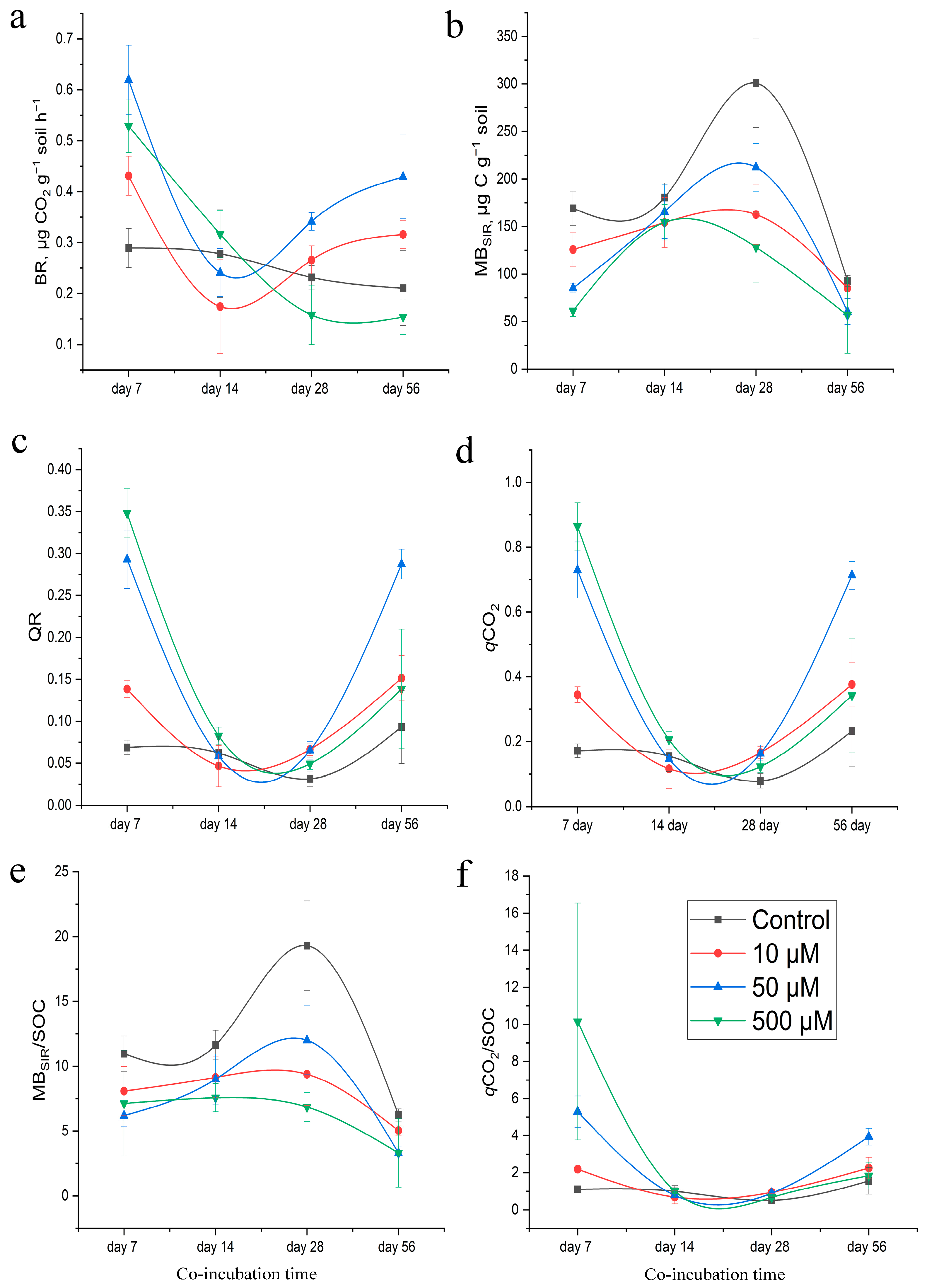
During the experiment, there were changes in microbial activity (Figure 2). On the 7th day, all samples treated with GT showed an increase in respiratory activity compared to the control samples, which suggests that GT had an effect on the microorganisms (p < 0.001) (Figure 2a).
After 14 days, in samples with 10 μM GT, basal respiration decreased by 37.3% (p < 0.005). In samples with 50 and 500 μM GT, a decrease in the respiratory activity of microbial communities was also observed, but when compared with the control samples, these changes were not significant (p > 0.05). On the 28th day of incubation, a further increase in microbial respiration was observed in the 10 and 50 µM GT samples. By the 56th day, in the 10 µM GT group, BR was 49.9% higher than that in the control (p < 0.005), while in the 50 μM group, it was 103.5% higher (p < 0.001). In the soil samples treated with 500 μM GT, after the stimulation period on day 7, the basal respiration decreased and reached a steady state on day 28. On day 56, the BR value corresponded to that on day 28.
The MBSIR index in soils treated with 10, 50 and 500 μM of GT decreased by 25.7%, 49.6% and 63.7% on the 7th day of incubation, respectively (p < 0.001) (Figure 2b). On the 14th and 28th days, there was a slight increase in microbial biomass compared to that at the initial point. However, these values continued to remain below those of the control group (p < 0.001). At the end of the incubation period, the difference in MBSIR between the treated samples (10, 50 and 500 μM) and the control was 8.1% (p < 0.01), 34.7% (p < 0.001) and 38.9% (p < 0.05), respectively.

3.4. Eco-Physiological Changes in Soils under the Influence of GT

Soils treated with GT showed higher QR and qCO2 values than controls (Figure 2c,d). The QR and qCO2 values were highest on the 7th day and then decreased. Also, on the 7th and 14th days of the experiment, it was found that as the concentration of GT increased, as did the values of QR and qCO2 (R2 = 0.6, R2 = 0.8). In the following days, we did not observe strong relationships between the concentration of GT and the eco-physiological parameters (day 28—R2 = 0.01, day 56—R2 = 0.02).
The microbial biomass carbon-to-soil organic carbon ratio (MBSIR/SOC) was significantly lower in GT-treated soil compared to that in control soil (p < 0.005). On the contrary, qCO2/SOC values in the soils with GT were higher than those in the control samples (p < 0.01) (Figure 2e,f). The increase in qCO2/SOC in samples with 500 μM GT on the 14th and 56th days was not significant (p > 0.05).

3.5. Enzymatic Activity of Soil under the Influence of Gliotoxin

The activities of extracellular soil enzymes responded differently to the introduction of varying doses of GT during the experiment.
Activity of C-cycle enzymes. In soil samples with 10 μM GT, a significant increase (by 1.2 times) in the activity of β-D-1,4-cellobiosidase (CB) was observed on the 56th day of incubation compared to that in the control (p < 0.05) (Figure 3a). In the samples with 50 μM of GT, on day 7, CB activity decreased by a factor of 1.1 (p < 0.005). On days 14 and 28, CB activity increased by factors of 1.1 and 1.3, respectively (p < 0.005). By the end of the incubation period, the CB activity levels were similar to those of the control samples. We did not observe a significant effect of the 500 μM GT treatment on the cells on the 7th day of the experiment (p > 0.05). On the 14th day, enzyme activity increased 1.2-fold compared to that in the control group (p < 0.001) and remained at this level until the end of the incubation period (p < 0.05).
GT significantly reduced the activity of β-1,4-glucosidase (βG) on the 7th day of incubation in samples with 50 μM GT (p < 0.005) and 500 μM GT (p < 0.05) (Figure 3b). A dose of 10 µM GT did not have a significant effect on βG activity over the course of the 7-day experiment (p > 0.05). However, on the 14th day of the experiment, the activity of βG significantly increased in all studied samples compared to the control group (p < 0.001). At the end of the incubation period (day 56), high βG activity was only observed in the 10 μM GT and 500 μM GT samples (p < 0.05). In samples with 50 μM GT, activity was 1.2 times lower than that in the control group (p < 0.005).
No significant changes in β-1,4-xylosidase (βX) activity were observed depending on the dose of GT over the 28-day period of the experiment (Figure 3c). However, on the 56th day, there was a 1.2-fold increase in βX activity compared to that in the control, which was statistically significant (p < 0.001).
Activity of P-cycle enzymes. During 4 weeks of incubation, the differences in acid phosphatase (AP) activity observed between the soils treated with GT and the control group were not statistically significant (p > 0.1) (Figure 3d). Toward the end of the incubation period, AP activity increased slightly in samples with 10 and 500 μM of GT (p < 0.001).
Activity of N-cycle enzymes. On the 7th day of incubation, the activity of L-leucine aminopeptidase (LAP) was lower than the control value in all experimental samples (p < 0.05) (Figure 3e). Differences in LAP activity on days 14 and 28 between the control and GT-treated samples were not significant (p > 0.2). The end of the incubation period was characterized by an increase in LAP activity in the 50 μM GT and 500 μM GT samples (p < 0.005).
No significant changes in β-1,4-N-acetyl-glucosaminidase (NAG) activity were observed (Figure 3f). The maximum NAG activity was only observed in 500 μM GT samples on the 7th and 28th days of incubation (3.5% and 9.8% higher than the control, respectively, p < 0.001 and p < 0.05).
Activity of S-cycle enzymes. On the 7th day of the experiment, arylsulfatase (ARS) activity in samples containing 10 μM of GT increased (p < 0.001) (Figure 3g). In samples with 50 μM, it decreased (p < 0.05). And in samples with 500 μM, the activity was similar to that in the control group. The opposite trend was noted on the 14th day of the experiment. The greatest inhibition of ARS activity was observed on the 28th day of the experiment. In the 10 μM sample, GT was 1.9 times lower than in the control. In the 50 and 500 μM samples, GT was also 1.5 times lower compared to the control (p < 0.001). By the end of the incubation period, enzyme activity was restored and was significantly higher than that in the controls in samples containing 10 µM (p < 0.01) and 500 µM (p < 0.001).
The interaction between sampling time and used GT concentration had a significant impact on βG, βX, AP, NAG and LAP activities but did not significantly affect CB and ARS activities (Table 2).

4. Discussion

The ability of the soil to mineralize organic compounds and cycle nutrients is a crucial function of its microbial community. The stability of this microbiome therefore determines the productivity of plants and hence the yield. When investigating natural biological compounds (or microbial agents) for possible use in agronomy, it is important to understand their impact on the indigenous soil microbiota. The effects of the biopreparation (positive, negative or neutral) will depend on the physical and chemical properties of the soil (soil texture, aggregates, pH, organic matter, humidity, seasonality, temperature, etc.) [38,39]. Previously, we conducted a laboratory experiment in which we assessed the effect of GT on the structural and functional properties of the soil microbiome [36]. We continued our study by changing the controlled conditions of the laboratory to the uncontrolled conditions of a real agricultural system.
When evaluating the obtained results, we interpreted the positive responses for both respiration and biomass as a stimulating effect of GT on the microbial community. A positive respiration response and a negative biomass response indicated that the microbial community was under stress. Negative responses to both respiration and biomass suggested an inhibitory effect. A negative respiration response and a positive biomass response could indicate a dormant or maintenance state of the microbial community [40].

4.1. Effect of GT on Soil Microbial Activity

The first reaction of the microbial community to the treatment of soils with different concentrations of GT (7th day of the experiment) was a surge in respiratory activity. We believe that GT, consisting mainly of carbon, oxygen, nitrogen, hydrogen and sulfur, acted as an easily accessible substrate for the physiological and metabolic needs of microorganisms [41], contributing to the increased mineralization of carbon sources to CO2 in soils. Various studies have convincingly demonstrated the possibility of microorganisms using antibiotics, pesticides and other chemicals as sources of carbon and nitrogen [42,43,44,45,46,47]. There is also a possibility that the release of available carbon occurred as a result of the death of microbial cells [48,49]. Our previous laboratory study showed that introducing GT into soil leads to changes in the structure of both bacterial and fungal communities within the soil microbiome. GT-resistant bacteria and fungi begin to dominate [36]. We believe that the restructuring of the microbial community will lead to a decrease in microbial biomass against the background of increased CO2 production. Under stressful conditions, microbial communities use energy from the consumed substrate to maintain cellular functions and their functional state within the system. They also use this energy to adapt to new conditions and not to convert carbon into biomass (i.e., not for growth) [50,51,52,53,54]. This is confirmed by the MBSIR/SOC and qCO2/SOC indicators, which are used to assess the availability of soil organic carbon, its “quality” and the efficiency of its utilization by microorganisms [55,56,57,58].
In our study, the decrease in MBSIR/SOC and increase in qCO2 indicate that most of the carbon from organic substrates was directed to catabolic processes. Therefore, it can be argued that under stress, microorganisms use organic carbon “poorly” and “inefficiently”. The increase in the qCO2/SOC index values in samples treated with GT, especially at concentrations of 500 μM and 50 μM, indicates a decreased efficiency in carbon utilization. This is not beneficial for soil microbial communities, as it means that the carbon needed for their growth is being lost. On the contrary, the higher the MBSIR/SOC ratio and the lower the qCO2 (in the control samples and samples with 10 μM GT), the greater the proportion of carbon from organic substrates that is directed toward anabolic processes, leading to the accumulation of microbial biomass carbon. Lower qCO2/SOC index values indicate higher carbon use efficiency due to the presence of stable biomass in the control soils and in the samples treated with 10 μM of GT.
The values of the metabolic (qCO2) and respiratory (QR) coefficients [51,59] confirm the high energy costs of microbial communities to maintain their vital activity in tests with GT. By the end of the incubation period (day 56), the content of microbial biomass in soil samples at 10 μM increased relative to the control value. Lower GT concentrations probably affect the physiology of microorganisms, but to a lesser extent; microbial communities under these conditions return to their original functional state more quickly. The effect of concentrations of 50 μM GT and 500 μM GT was more significant. In samples with 50 μM GT, the microbial communities remained under stress (with low microbial biomass and high values for BR and qCO2). In samples with 500 μM GT, at the end of the experiment, not only had the microbial biomass not recovered (it corresponded to the level on day 7), but the respiratory activity of the microbial communities was also lower than that in the control group. It turns out that the stress effect on soils with 500 μM GT not only did not weaken over time, but signs of the inhibition of the microbial community also appeared. It can be assumed that the observed effects (weakening effect, inhibitory effect) of the influence of GT by the end of the experiment are associated with its decomposition in the soil environment.
In our previous study it was shown that in soils with pHH2O 7.3 and pHKCl 6.26, by the 14th day, even in samples with a high concentration (500 μM), GT is not detected [36]. This result is supported by another study showing that in alkaline soils with a pH of 7.4, GT was completely degraded within 5 days [23]. In acidic soils, it took 10 days for complete degradation [23]. The relationship between pH and GT stability was also shown in earlier studies examining the effect of pH on the fungistatic activity of aqueous solutions of GT [60,61]. In our study, in soil samples (pHH2O 6.2, pHKCl 5.6) with GT concentrations of 10 and 50 μM, GT was already not detected on the 14th day of the experiment. In samples with a concentration of 500 μM, GT was detected even on the 56th day.
It turns out that the direct effect of GT on the soil microbial community lasts for approximately seven days. After that, we see the consequences of stress exposure, which is known as the post-antibiotic effect [62]. We associate the low rate of GT decomposition in the 500 μM GT samples with the predominance of clay and silt fractions in these soils (clay 28.5%, silt 27.7%, respectively). Due to the strong bond with clay minerals, GT becomes less accessible to microorganisms. Consequently, microbial communities are under the inhibitory influence of GT for a long time, and their activity begins to be suppressed. The influence of clay minerals on the rate of decomposition of various substances (antibiotics, secondary metabolites, biopesticides, etc.) is shown in the works [20,63,64]. Microbial communities in the control soils were the most energetically efficient (“slow metabolism”); their functioning was more stable throughout the experiment (low qCO2 and QR values). In the control samples, we added only water at a level of 60% of the total moisture capacity. This level of water is considered optimal for the vital activity of microorganisms and therefore for determining the overall potential of these soils. In such a “stable” system, the energy expenditure of microorganisms is not only used for maintaining their vital activities but also for growth processes. A certain balance between microbial biomass and mineralization processes is evident in the samples. It is likely that, in control samples compared to those with GT, microorganisms with a K strategy predominate. These microorganisms are represented by a greater variety of species that are more effective at assimilating the substrate through anabolic processes. Therefore, the “slow metabolism” of microbial communities in control soils indicates the physical and chemical stability of soil properties and functions, as well as the overall ecological balance.

4.2. Changes in Enzymatic Activity

Another key soil parameter is the activity of soil enzymes, which are very sensitive to external factors [65,66]. In our study, we found that, depending on the dose, GT can act as a stressor for soil microbiota to a greater or lesser extent. The restructuring of the soil microbiome and its metabolic activity was an immediate response to exposure to GT [36,67]. The higher the metabolic activity of soil microbial communities, the more enzymes are enriched in the soil, which is necessary for acquiring carbon [68,69]. The fact that enzyme activity is linked to the metabolic activity of soil microorganisms is indicated by higher CO2 emissions in soils treated with GT compared to control soils [69]. On the other hand, the activity of C-cycle enzymes, especially βG, may increase as labile carbon reserves are depleted [70,71]. βG intensively catalyzes the hydrolysis of disaccharides in soil, converting them into glucose. This process provides energy sources for soil microbial communities and stimulates their activity [72]. This is indicated by an increase in mineralization activity, which occurs against the background of a decrease in microbial biomass.
It should also be noted that, in soils with a high proportion of clay and silt, extracellular enzymes remain active and stable for a longer period. Enzymes that are bound to clay particles are effectively incorporated and participate in catalytic processes, providing energy for the soil microbiome when it is under stress or when there is a lack of biomass availability [73,74,75].
We also expected that soil treatment with GT, which affects microbial communities and the activity of C-cycle enzymes, would lead to changes in phosphatase activity. However, the hypothesis proposed was not fully supported. The increase in phosphatase activity was only observed on the 56th day of the experiment, and it was by a factor of 1.06. It turns out that GT did not act as a driver of phosphatase activity. Perhaps the enzymes were stabilized by the clay part of the soil (clay 28.5%, silt 27.7%) against the background of sufficiently available phosphorus for the soil microbiome [76,77]. Alternatively, the loss of easily accessible carbon in the soil, which began after the addition of GT, led to a decrease in enzyme abundance, such that the activity corresponded with that of control soils at the early stages of incubation [78]. The increase in phosphatase activity in the soil toward the end of the experiment may have been due to the stimulation of fungi and bacteria, which could have altered their quantity and composition under the influence of GT.
Among Thermoleophilia, there are species in which functional genes are associated with not only the C cycle but also the P cycle, and they determine phosphatase activity [79,80]. In the assimilation and cycling of phosphorus, a significant role is played by the bacteria Actinobacteria, Acidobacteria, Chloroflexi, Methylomirabilota and Bdellovibrionota, as well as the fungi Ascomycota and Basidiomycota [80]. There is evidence that some species of the Mortierella family are involved in soil carbon cycling, phosphorus solubilization and mobilization, lipid metabolism, chitin degradation and even soil bioremediation [81,82,83,84]. There is evidence that these bacteria and fungi listed are predominant in soils supplemented with GT [36]. The absence of an effect of antibiotics on phosphatase activity was demonstrated in the works [85,86,87].
Soil N-acquisition enzymes, including NAG and LAP, can serve as indicators of energy N demand [64,86]. The resource allocation model suggests that N enrichment should decrease the activities of N-acquisition enzymes due to the decreased N demand under high N availability [88,89,90,91]. This model explains the activity of LAP, which we described in samples treated with GT. The activity did not decrease significantly by the 7th day of the experiment. After that, the enzyme levels did not differ significantly from those in the control group. NAG activity, on the contrary, increased, especially in samples with a high dose of GT. NAG activity can be determined by the bacterial community: Acidobacteria, Proteobacteria, Chloroflexi, Planctomycetota and Armatimonadota [80], as well as Solicoccozyma fungi [92]. In a study by Liu et al., the relationship between Bacteroidetes, Chloroflexi, Bdellovibrionota, the number of symbiotic fungi and LAP activity was also investigated [80]. The lower the number of fungi, the lower the LAP activity [80]. The presence of bacteria and fungi responsible for nitrogen cycling was also demonstrated in our previous work [34].
Arylsulfatase (ARS) accelerates sulfur mineralization and catalyzes the conversion of organic sulfide to inorganic sulfur, which is of great importance for the geochemical cycle of sulfur [93]. Microbial ARS production is stimulated by low S conditions and is often directly linked to the microbial biomass [94]. This enzyme may indicate the presence of fungi in the soil, as only fungi contain ether sulfates, which are the substrate for ARS [95]. In our recent study [36], we found that GT favors bacteria that oxidize thiosulfate, such as Thiobacillus spp. These bacteria are obligate autotrophs that use elemental sulfur, thiosulfate or polythionates as energy sources [96,97]. We can speculate that if bacteria of this genus are present in our samples, they could use GT as a source of energy.

5. Conclusions

GT, when introduced into the soil, has different effects on the assessed parameters during the incubation period. In the presence of GT, microbial communities become more metabolically active. The greatest changes in microbial biomass, respiration and enzyme activity were observed when the soil was treated with GT at a concentration of 500 μM. Over time, the effect of GT weakens, and an indirect post-antibiotic effect develops. The development of microbial community response scenarios, including enzymatic activity in response to “stress”, is determined by the antibiotic dose and the properties of the soil itself. Therefore, it is essential to understand the interplay between various soil properties, such as soil type, texture, moisture content, acidity, organic matter content and others, as well as the dosage of the preparation and its spatial and temporal variability in relation to microbiological parameters and the enzymatic activity of soil. This knowledge will be crucial in determining how GT or GT-producing fungi can be utilized in agricultural practice. This work serves as a starting point for further studies on (1) the impact of gliotoxin on microbial properties in different soil types; (2) the influence of gliotoxin in the rhizosphere of soil; and (3) how soil texture affects the behavior of gliotoxin and its capacity to determine microbial properties.

Supplementary Materials

The following supporting information can be downloaded at: https://www.mdpi.com/article/10.3390/agronomy14092084/s1, Figure S1: The experimental microcosms; Table S1: Meteorological conditions for the summer period of 2023.

Author Contributions

Conceptualization, A.V.T. and A.S.V.; methodology, A.V.T.; formal analysis, A.V.T.; investigation, E.V.G., A.V.I. and A.A.S.; resources, A.V.T.; data curation, A.V.T.; writing—original draft preparation, A.V.T. and A.S.V.; writing—review and editing, A.V.T. and A.S.V.; visualization, A.V.T. and A.S.V.; supervision, A.V.T.; project administration, A.V.T.; funding acquisition, A.V.T. All authors have read and agreed to the published version of the manuscript.

Funding

The work was supported by the Russian Science Foundation 23-24-00648 (https://www.rscf.ru/project/23-24-00648/) (assessed on 12 September 2024).

Data Availability Statement

The original contributions presented in the study are included in the article/Supplementary Materials, further inquiries can be directed to the corresponding author.

Acknowledgments

This work was partially performed using resources from the Research Resource Center & Natural Resource Management and Physico-Chemical Research (University of Tyumen).

Conflicts of Interest

The authors declare no conflicts of interest.

References

  1. Keswani, C.; Singh, H.B.; Hermosa, R.; García-Estrada, C.; Caradus, J.; He, Y.W.; Mezaache-Aichour, S.; Glare, T.R.; Borriss, R.; Vinale, F.; et al. Antimicrobial secondary metabolites from agriculturally important fungi as next biocontrol agents. Appl. Microbiol. Biotechnol. 2019, 103, 9287–9303. [Google Scholar] [CrossRef] [PubMed]
  2. Rodrigo, S.; García-Latorre, C.; Santamaria, O. Metabolites produced by fungi against fungal phytopathogens: Review, implementation and perspectives. Plants 2021, 11, 81. [Google Scholar] [CrossRef] [PubMed]
  3. Glare, T.; Caradus, J.; Gelernter, W.; Jackson, T.; Keyhani, N.; Köhl, J.; Marrone, P.; Morin, L.; Stewart, A. Have biopesticides come of age? Trends Biotechnol. 2012, 30, 250–258. [Google Scholar] [CrossRef]
  4. Deising, H.B.; Gase, I.; Kubo, Y. The unpredictable risk imposed by microbial secondary metabolites: How safe is biological control of plant diseases? J. Plant Dis. Prot. 2017, 124, 413–419. [Google Scholar] [CrossRef]
  5. Koch, E.; Becker, J.O.; Berg, G.; Hauschild, R.; Jehle, J.; Köhl, J.; Smalla, K. Biocontrol of plant diseases is not an unsafe technology! J. Plant Dis. Prot. 2018, 125, 121–125. [Google Scholar] [CrossRef]
  6. Cycoń, M.; Mrozik, A.; Piotrowska-Seget, Z. Antibiotics in the soil environment—Degradation and their impact on microbial activity and diversity. Front. Microbiol. 2019, 10, 338. [Google Scholar] [CrossRef]
  7. Davies, J. What are antibiotics? Archaic functions for modern activities. Mol. Microbiol. 1990, 4, 1227–1232. [Google Scholar] [CrossRef]
  8. Shahriar, S.A.; Islam, M.N.; Chun, C.N.W.; Kaur, P.; Rahim, M.A.; Islam, M.M.; Uddain, J.; Siddiquee, S. Microbial Metabolomics Interaction and Ecological Challenges of Trichoderma Species as Biocontrol Inoculant in Crop Rhizosphere. Agronomy 2022, 12, 900. [Google Scholar] [CrossRef]
  9. Tyśkiewicz, R.; Nowak, A.; Ozimek, E.; Jaroszuk-Ściseł, J. Trichoderma: The Current Status of Its Application in Agriculture for the Biocontrol of Fungal Phytopathogens and Stimulation of Plant Growth. Int. J. Mol. Sci. 2022, 23, 2329. [Google Scholar] [CrossRef]
  10. Smith, E.B.; Dolan, S.K.; Fitzpatrick, D.A.; Doyle, S.; Jones, G.W. Towards understanding the gliotoxin detoxification mechanism: In vivo thiomethylation protects yeast from gliotoxin cytotoxicity. Microb. Cell 2016, 3, 120–125. [Google Scholar] [CrossRef]
  11. Weindling, R. Studies on a lethal principle effective in the parasitic action of Trichoderma lignorum on Rhizoctonia solani and other soil fungi. Phytopathology 1934, 24, 1153–1179. [Google Scholar]
  12. Stanley, N.F.; Mills, J.A. The biological activity of a substance resembling gliotoxin produced by a strain of Aspergillus fumigatus. Aust. J. Exp. Biol. Med. Sci. 1946, 24, 133–138. [Google Scholar] [CrossRef] [PubMed]
  13. Miller, P.A.; Milstrey, K.P.; Trown, P.W. Specific inhibition of viral ribonucleic acid replication by Gliotoxin. Science 1968, 159, 431–432. [Google Scholar] [CrossRef] [PubMed]
  14. Waring, P.; Eichner, R.D.; Müllbacher, A. The chemistry and biology of the immunomodulating agent gliotoxin and related epipolythiodioxopiperazines. Med. Res. Rev. 1988, 4, 499–524. [Google Scholar] [CrossRef] [PubMed]
  15. Pahl, H.L.; Krauss, B.; Schulze-Osthoff, K.; Decker, T.; Traenckner, E.B.M.; Vogt, M.; Myers, C.; Parks, T.; Warring, P.; Mühlbacher, A.; et al. The immunosuppressive fungal metabolite gliotoxin specifically inhibits transcription factor NF-κB. J. Exp. Med. 1996, 183, 1829–1840. [Google Scholar] [CrossRef]
  16. Vigushin, D.M.; Mirsaidi, N.; Brooke, G.; Sun, C.; Pace, P.; Inman, L.; Moody, C.J.; Coombes, R.C. Gliotoxin is a dual inhibitor of farnesyltransferase and geranylgeranyltransferase I with antitumor activity against breast cancer in vivo. Med. Oncol. 2004, 21, 21–30. [Google Scholar] [CrossRef]
  17. Scharf, D.H.; Remme, N.; Heinekamp, T.; Hortschansky, P.; Brakhage, A.A.; Hertweck, C. Transannular disulfide formation in gliotoxin biosynthesis and its role in self-resistance of the human pathogen Aspergillus fumigatus. J. Am. Chem. Soc. 2010, 132, 10136–10141. [Google Scholar] [CrossRef]
  18. Kim, Y.S.; Park, S.J. Gliotoxin from the marine fungus Aspergillus fumigatus induces apoptosis in HT1080 fibrosarcoma cells by downregulating NF-κB. Fish. Aquat. Sci. 2016, 19, 35. [Google Scholar] [CrossRef]
  19. Wright, J.M. Production of Gliotoxin in Soils. Nature 1956, 177, 896. [Google Scholar] [CrossRef]
  20. Lumsden, R.D.; Locke, J.C.; Adkins, S.T.; Walter, J.F.; Ridout, C.J. Isolation and localization of the antibiotic gliotoxin produced by Gliocladium virens from alginate prill in soil and soilless media. Phytopathology 1992, 82, 230–235. [Google Scholar] [CrossRef]
  21. Aliaa, R.E.S. Control of root-rot diseases of Phaseolus vulgaris using gliotoxin. Malays J. 2008, 4, 40–43. [Google Scholar] [CrossRef]
  22. Tomah, A.A.; Abd Alamer, I.S.; Li, B.; Zhang, J.Z. A new species of Trichoderma and gliotoxin role: A new observation in enhancing biocontrol potential of T. virens against Phytophthora capsici on chili pepper. Biol. Control 2020, 145, 104261. [Google Scholar] [CrossRef]
  23. Jayalakshmi, R.; Oviya, R.; Premalatha, K.; Mehetre, S.T.; Paramasivam, M.; Kannan, R.; Theradimani, M.; Pallavi, M.S.; Mukherjee, P.K.; Ramamoorthy, V. Production, stability and degradation of Trichoderma gliotoxin in growth medium, irrigation water and agricultural soil. Sci. Rep. 2021, 11, 16536. [Google Scholar] [CrossRef] [PubMed]
  24. Lewis, J.A.; Roberts, D.P.; Hollenbeck, M.D. Induction of cytoplasmic leakage from Rhizoctonia solani hyphae by Gliocladium virens and partial characterization of a leakage factor. Biocontrol Sci. Technol. 1991, 1, 21–29. [Google Scholar] [CrossRef]
  25. Roberts, D.P.; Lumsden, R.D. Effect of extracellular metabolites from Gliocladium virens on germination of sporangia and mycelial growth of Pythium ultimum. Phytopathology 1990, 80, 461–465. [Google Scholar] [CrossRef]
  26. Vasilchenko, A.V.; Poshvina, D.V.; Semenov, M.V.; Timofeev, V.N.; Iashnikov, A.V.; Stepanov, A.A.; Pervushina, A.N.; Vasilchenko, A.S. Triazoles and Strobilurin Mixture Affects Soil Microbial Community and Incidences of Wheat Diseases. Plants 2023, 12, 660. [Google Scholar] [CrossRef] [PubMed]
  27. Guzmán-Guzmán, P.; Kumar, A.; de Los Santos-Villalobos, S.; Parra-Cota, F.I.; Orozco-Mosqueda, M.D.C.; Fadiji, A.E.; Hyder, S.; Babalola, O.O.; Santoyo, G. Trichoderma species: Our best fungal allies in the biocontrol of plant diseases—A review. Plants 2023, 12, 432. [Google Scholar] [CrossRef]
  28. Poveda Arias, J. Trichoderma as biocontrol agent against pests: New uses for a mycoparasite. Biol. Control 2021, 159, 104634. [Google Scholar] [CrossRef]
  29. Ferreira, F.V.; Musumeci, M.A. Trichoderma as biological control agent: Scope and prospects to improve efficacy. World J. Microbiol. Biotechnol. 2021, 37, 90. [Google Scholar] [CrossRef]
  30. Zaid, R.; Koren, R.; Kligun, E.; Gupta, R.; Leibman-Markus, M.; Mukherjee, P.K.; Kenerley, C.M.; Bar, M.; Horwitz, B.A. Gliotoxin, an immunosuppressive fungal metabolite, primes plant immunity: Evidence from Trichoderma virens-tomato interaction. mBio 2022, 13, e00389-22. [Google Scholar] [CrossRef]
  31. Vasilchenko, A.S.; Gurina, E.V.; Drozdov, K.A.; Vershinin, N.A.; Kravchenko, S.V.; Vasilchenko, A.V. Exploring the antibacterial action of gliotoxin: Does it induce oxidative stress or protein damage? Biochimie 2023, 214, 86–95. [Google Scholar] [CrossRef] [PubMed]
  32. Pansu, M.; Gautheyrou, J. Handbook of Soil Analysis: Mineralogical, Organic and Inorganic Methods; Springer Science & Business Media: Berlin/Heidelberg, Germany, 2007. [Google Scholar]
  33. Vasilchenko, A.V.; Galaktionova, L.V.; Tretyakov, N.Y.; Dyachkov, S.M.; Vasilchenko, A.S. Impact of agricultural land use on distribution of microbial biomass and activity within soil aggregates. Soil Use Manag. 2023, 39, 618–633. [Google Scholar] [CrossRef]
  34. Anderson, T.H.; Domsch, K.H. Application of eco-physiological quotients (qCO2 and qD) on microbial biomasses from soils of different cropping histories. Soil Biol. Biochem. 1990, 22, 251–255. [Google Scholar] [CrossRef]
  35. Razavi, B.S.; Blagodatskaya, E.; Kuzyakov, Y. Nonlinear temperature sensitivity of enzyme kinetics explains canceling effect—A case study on loamy haplic Luvisol. Front. Microbiol. 2015, 6, 1126. [Google Scholar] [CrossRef]
  36. Teslya, A.V.; Gurina, E.V.; Poshvina, D.V.; Stepanov, A.A.; Iashnikov, A.A.; Vasilchenko, A.S. Fungal Secondary Metabolite Gliotoxin Enhances Enzymatic Activity in Soils by Reshaping Their Microbiome. 2024. Available online: https://ssrn.com/abstract=4824592 (accessed on 10 September 2024).
  37. German, D.P.; Weintraub, M.N.; Grandy, A.S.; Lauber, C.L.; Rinkes, Z.L.; Allison, S.D. Optimization of hydrolytic and oxidative enzyme methods for ecosystem studies. Soil Biol. Biochem. 2011, 43, 1387–1397. [Google Scholar] [CrossRef]
  38. Nieder, R.; Benbi, D.K.; Reichl, F.X.; Nieder, R.; Benbi, D.K.; Reichl, F.X. Health risks associated with pesticides in soils. Soil Compon. Hum. Health 2018, 503–573. [Google Scholar] [CrossRef]
  39. Koba, O.; Golovko, O.; Kodešová, R.; Fér, M.; Grabic, R. Antibiotics degradation in soil: A case of clindamycin, trimethoprim, sulfamethoxazole and their transformation products. Environ. Pollut. 2017, 220, 1251–1263. [Google Scholar] [CrossRef]
  40. Frkova, Z.; Vystavna, Y.; Koubová, A.; Kotas, P.; Grabicová, K.; Grabic, R.; Kodešová, R.; Chroňáková, A. Microbial responses to selected pharmaceuticals in agricultural soils: Microcosm study on the roles of soil, treatment and time. Soil Biol. Biochem. 2020, 149, 107924. [Google Scholar] [CrossRef]
  41. Moreno, J.L.; Aliaga, A.; Navarro, S.; Hernandez, T.; García, C. Effects of atrazine on microbial activity in semiarid soil. Appl. Soil Ecol. 2007, 35, 120–127. [Google Scholar] [CrossRef]
  42. Fenton, J.J.; Harsch, H.H.; Klein, D. Production of volatile nitrogenous compounds from the degradation of streptomycin by Pseudomonas maltophilia. J. Bacteriol. 1973, 116, 1267–1272. [Google Scholar] [CrossRef]
  43. Johnsen, J. Utilization of benzylpenicillin as carbon, nitrogen and energy source by a Pseudomonas fluorescens strain. Arch. Microbiol. 1977, 115, 271–275. [Google Scholar] [CrossRef] [PubMed]
  44. Dantas, G.; Sommer, M.O.; Oluwasegun, R.D.; Church, G.M. Bacteria subsisting on antibiotics. Science 2008, 320, 100–103. [Google Scholar] [CrossRef] [PubMed]
  45. Dantas, G.; Sommer, M.O. Ecological and clinical consequences of antibiotic subsistence by environmental microbes. In Antimicrobial Resistance in the Environment; Wiley-Blackwell: Hoboken, NJ, USA, 2012; pp. 29–41. [Google Scholar] [CrossRef]
  46. Perri, R.; Kolvenbach, B.A.; Corvini, P.F. Subsistence and complexity of antimicrobial resistance on a community-wide level. Environ. Microbiol. 2020, 22, 2463–2468. [Google Scholar] [CrossRef]
  47. Kumar, M.; Yadav, A.N.; Saxena, R.; Paul, D.; Tomar, R.S. Biodiversity of pesticides degrading microbial communities and their environmental impact. Biocatal. Agric. Biotechnol. 2021, 31, 101883. [Google Scholar] [CrossRef]
  48. Muñoz-Leoz, B.; Ruiz-Romera, E.; Antigüedad, I.; Garbisu, C. Tebuconazole application decreases soil microbial biomass and activity. Soil Biol. Biochem. 2011, 43, 2176–2183. [Google Scholar] [CrossRef]
  49. Bonfleur, E.J.; Tornisielo, V.L.; Regitano, J.B.; Lavorenti, A. The effects of glyphosate and atrazine mixture on soil microbial population and subsequent impacts on their fate in a tropical soil. Water Air Soil Pollut. 2015, 226, 21. [Google Scholar] [CrossRef]
  50. Chander, K.; Dyckmans, J.; Joergensen, R.; Meyer, B.; Raubuch, M. Different sources of heavy metals and their long-term effects on soil microbial properties. Biol. Fertil. Soils 2001, 34, 241–247. [Google Scholar] [CrossRef]
  51. Anderson, T.H. Microbial eco-physiological indicators to asses soil quality. Agric. Ecosyst. Environ. 2003, 98, 285–293. [Google Scholar] [CrossRef]
  52. Frische, T.; Höper, H. Soil microbial parameters and luminescent bacteria assays as indicators for in situ bioremediation of TNT-contaminated soils. Chemosphere 2003, 50, 415–427. [Google Scholar] [CrossRef]
  53. Rui, Y.; Murphy, D.V.; Wang, X.; Hoyle, F.C. Microbial respiration, but not biomass, responded linearly to increasing light fraction organic matter input: Consequences for carbon sequestration. Sci. Rep. 2016, 6, 35496. [Google Scholar] [CrossRef]
  54. Eisenhauer, N.; Klier, M.; Partsch, S.; Sabais, A.C.; Scherber, C.; Weisser, W.W.; Scheu, S. No interactive effects of pesticides and plant diversity on soil microbial biomass and respiration. Appl. Soil Ecol. 2009, 42, 31–36. [Google Scholar] [CrossRef]
  55. Anderson, T.H.; Domsch, K.H. Ratios of microbial biomass carbon to total organic carbon in arable soils. Soil Biol. Biochem. 1989, 21, 471–479. [Google Scholar] [CrossRef]
  56. Joergensen, R.G.; Castillo, X. Interrelationships between microbial and soil properties in young volcanic ash soils of Nicaragua. Soil Biol. Biochem. 2001, 33, 1581–1589. [Google Scholar] [CrossRef]
  57. Klose, S.; Wernecke, K.D.; Makeschin, F. Microbial activities in forest soils exposed to chronic depositions from a lignite power plant. Soil Biol. Biochem. 2004, 36, 1913–1923. [Google Scholar] [CrossRef]
  58. Joergensen, R.G.; Emmerling, C. Methods for evaluating human impact on soil microorganisms based on their activity, biomass, and diversity in agricultural soils. J. Plant Nutr. Soil Sci. 2006, 169, 295–309. [Google Scholar] [CrossRef]
  59. Bedolla-Rivera, H.; Negrete-Rodríguez, M.X.; Medina-Herrera, M.; Gámez-Vázquez, F.; Álvarez-Bernal, D.; Samaniego-Hernández, M.; Gámez-Vázquez, A.; Conde-Barajas, E. Development of a soil quality index for soils under different agricultural management conditions in the Central Lowlands of Mexico: Physicochemical, biological and ecophysiological indicators. Sustainability 2020, 12, 9754. [Google Scholar] [CrossRef]
  60. Brian, P.W.; Hemming, H.G. Gliotoxin, a fungistatic metabolic product of Trichoderma viride. Ann. Appl. Biol. 1945, 32, 214–220. [Google Scholar] [CrossRef]
  61. Jefferys, E.G. The stability of antibiotics in soils. Microbiology 1952, 7, 295–312. [Google Scholar] [CrossRef]
  62. Craig, W.A. The postantibiotic effect. Clin. Microbiol. Newsl. 1991, 13, 121–124. [Google Scholar] [CrossRef]
  63. Albert, J.; Muñoz, K. Kinetics of microbial and photochemical degradation of aflatoxin B1 in a sandy loam and clay soil. Sci. Rep. 2022, 12, 16849. [Google Scholar] [CrossRef]
  64. Albert, J.; More, C.; Korz, S.; Muñoz, K. Soil Microbial Responses to Aflatoxin Exposure: Consequences for Biomass, Activity and Catabolic Functionality. Soil Syst. 2023, 7, 23. [Google Scholar] [CrossRef]
  65. Dick, R.P. Soil enzyme activities as indicators of soil quality. Defin. Soil Qual. A Sustain. Environ. 1994, 35, 107–124. [Google Scholar] [CrossRef]
  66. Sinsabaugh, R.L.; Lauber, C.L.; Weintraub, M.N.; Ahmed, B.; Allison, S.D.; Crenshaw, C.; Contosta, A.R.; Cusack, D.; Frey, S.; Gallo, M.E.; et al. Stoichiometry of soil enzyme activity at global scale. Ecol. Lett. 2008, 11, 1252–1264. [Google Scholar] [CrossRef] [PubMed]
  67. Singh, B.K.; Walker, A. Microbial degradation of organophosphorus compounds. FEMS Microbiol. Rev. 2006, 30, 428–471. [Google Scholar] [CrossRef]
  68. Doran, J.W. Soil microbial and biochemical changes associated with reduced tillage. Soil Sci. Soc. Am. J. 1980, 44, 765–771. [Google Scholar] [CrossRef]
  69. Trasar-Cepeda, C.; Leirós, M.C.; Gil-Sotres, F. Hydrolytic enzyme activities in agricultural and forest soils. Some implications for their use as indicators of soil quality. Soil Biol. Biochem. 2008, 40, 2146–2155. [Google Scholar] [CrossRef]
  70. Cenini, V.L.; Fornara, D.A.; McMullan, G.; Ternan, N.; Lajtha, K.; Crawley, M.J. Chronic nitrogen fertilization and carbon sequestration in grassland soils: Evidence of a microbial enzyme link. Biogeochemistry 2015, 126, 301–313. [Google Scholar] [CrossRef]
  71. Zeglin, L.H.; Stursova, M.; Sinsabaugh, R.L.; Collins, S.L. Microbial responses to nitrogen addition in three contrasting grassland ecosystems. Oecologia 2007, 154, 349–359. [Google Scholar] [CrossRef]
  72. Tabatabai, M.A. Soil enzymes. Methods Soil Anal. Part 2 Microbiol. Biochem. Prop. 1994, 5, 775–833. [Google Scholar] [CrossRef]
  73. Babaniyi, B.R.; Thompson, S.O.; Ogundele, O.D.; Oluwole, O.F. Effects of Agrochemicals on Soil Microbial Enzymes. In Ecological Interplays in Microbial Enzymology; Springer Nature: Singapore, 2022; pp. 353–377. [Google Scholar] [CrossRef]
  74. Feketeová, Z.; Hrabovský, A.; Šimkovic, I. Microbial features indicating the recovery of soil ecosystem strongly affected by mining and ore processing. Int. J. Environ. Res. Public Health 2021, 18, 3240. [Google Scholar] [CrossRef]
  75. Quiquampoix, H.; Burns, R.G. Interactions between proteins and soil mineral surfaces: Environmental and health consequences. Elements 2007, 3, 401–406. [Google Scholar] [CrossRef]
  76. Naidja, A.; Huang, P.M.; Bollag, J.M. Enzyme-clay interactions and their impact on transformations of natural and anthropogenic organic compounds in soil. J. Environ. Qual. 2000, 29, 677–691. [Google Scholar] [CrossRef]
  77. Zimmerman, A.R.; Ahn, M.-Y. Organo-mineral–enzyme interaction and soil enzyme activity. Soil Enzymol. 2011, 22, 271–292. [Google Scholar] [CrossRef]
  78. Silva-Olaya, A.M.; Mora-Motta, D.A.; Cherubin, M.R.; Grados, D.; Somenahally, A.; Ortiz-Morea, F.A. Soil enzyme responses to land use change in the tropical rainforest of the Colombian Amazon region. PLoS ONE 2021, 16, e0255669. [Google Scholar] [CrossRef]
  79. Li, C.; He, Y.Q.; Cui, L.Q.; Albuquerque, L.; Chen, R.W.; Long, L.J.; Tian, X.P. Miltoncostaea marina gen. nov. sp. nov., and Miltoncostaea oceani sp. nov., a novel deep branching phylogenetic lineage within the class Thermoleophilia isolated from marine environments, and proposal of Miltoncostaeaceae fam. nov. and Miltoncostaeales ord. nov. Syst. Appl. Microbiol. 2021, 44, 126216. [Google Scholar] [CrossRef]
  80. Liu, Y.; Dong, L.; Zhang, H.; Deng, Y.; Hu, B.; Wang, W. Distinct roles of bacteria and fungi in mediating soil extracellular enzymes under long-term nitrogen deposition in temperate plantations. For. Ecol. Manag. 2023, 529, 120658. [Google Scholar] [CrossRef]
  81. Osorio, N.W.; Habte, M. Synergistic effect of a phosphate-solubilizing fungus and an arbuscular mycorrhizal fungus on leucaena seedlings in an Oxisol fertilized with rock phosphate. Botany 2013, 91, 274–281. [Google Scholar] [CrossRef]
  82. Phillips, L.A.; Ward, V.; Jones, M.D. Ectomycorrhizal fungi contribute to soil organic matter cycling in sub-boreal forests. ISME J. 2014, 8, 699–713. [Google Scholar] [CrossRef]
  83. Zhang, K.; Bonito, G.; Hsu, C.M.; Hameed, K.; Vilgalys, R.; Liao, H.L. Mortierella elongata increases plant biomass among non-leguminous crop species. Agronomy 2020, 10, 754. [Google Scholar] [CrossRef]
  84. Costa, O.Y.; Pijl, A.; Houbraken, J.; van Lith, W.; Kuramae, E.E. Soil substrate source drives the microbes involved in the degradation of gelatin used as a biostimulant. Appl. Soil Ecol. 2023, 189, 104906. [Google Scholar] [CrossRef]
  85. Liu, B.; Li, Y.; Zhang, X.; Wang, J.; Gao, M. Effects of chlortetracycline on soil microbial communities: Comparisons of enzyme activities to the functional diversity via Biolog EcoPlates™. Eur. J. Soil Biol. 2015, 68, 69–76. [Google Scholar] [CrossRef]
  86. Yang, Q.; Zhang, J.; Zhu, K.; Zhang, H. Influence of oxytetracycline on the structure and activity of microbial community in wheat rhizosphere soil. J. Environ. Sci. 2009, 21, 954–959. [Google Scholar] [CrossRef] [PubMed]
  87. Wei, X.; Wu, S.C.; Nie, X.P.; Yediler, A.; Wong, M.H. The effects of residual tetracycline on soil enzymatic activities and plant growth. J. Environ. Sci. Health 2009, 44, 461–471. [Google Scholar] [CrossRef] [PubMed]
  88. Schimel, J.; Becerra, C.A.; Blankinship, J. Estimating decay dynamics for enzyme activities in soils from different ecosystems. Soil Biol. Biochem. 2017, 114, 5–11. [Google Scholar] [CrossRef]
  89. Chen, H.; Li, D.; Zhao, J.; Xiao, K.; Wang, K. Effects of nitrogen addition on activities of soil nitrogen acquisition enzymes: A meta-analysis. Agric. Ecosyst. Environ. 2018, 252, 126–131. [Google Scholar] [CrossRef]
  90. Sinsabaugh, R.L.; Moorhead, D.L. Resource allocation to extracellular enzyme production: A model for nitrogen and phosphorus control of litter decomposition. Soil Biol. Biochem. 1994, 26, 1305–1311. [Google Scholar] [CrossRef]
  91. Allison, S.D. Cheaters, diffusion and nutrients constrain decomposition by microbial enzymes in spatially structured environments. Ecol. Lett. 2005, 8, 626–635. [Google Scholar] [CrossRef]
  92. Zhang, S.; Chen, Y.; Tuo, Y.; Li, H.; Wang, Y. Effects of lime nitrogen on the fungi community of tobacco soil and its correlation analysis. In IOP Conference Series: Earth and Environmental Science; IOP Publishing: Bristol, UK, 2022; Volume 1087, p. 012059. [Google Scholar] [CrossRef]
  93. Ma, C.; Hu, B.; Wei, M.B.; Zhao, J.H.; Zhang, H.Z. Influence of matured compost inoculation on sewage sludge composting: Enzyme activity, bacterial and fungal community succession. Bioresour. Technol. 2019, 294, 122165. [Google Scholar] [CrossRef]
  94. Dotaniya, M.L.; Aparna, K.; Dotaniya, C.K.; Singh, M.; Regar, K.L. Role of soil enzymes in sustainable crop production. In Enzymes in Food Biotechnology; Academic Press: Cambridge, MA, USA, 2019; pp. 569–589. [Google Scholar] [CrossRef]
  95. Kertesz, M.A.; Mirleau, P. The role of soil microbes in plant sulphur nutrition. J. Exp. Bot. 2004, 55, 1939–1945. [Google Scholar] [CrossRef] [PubMed]
  96. Katayama, Y.; Kuraishi, H. Characteristics of Thiobacillus thioparus and its thiocyanate assimilation. Can. J. Microbiol. 1978, 24, 804–810. [Google Scholar] [CrossRef]
  97. Kumar, M.; Zeyad, M.T.; Choudhary, P.; Paul, S.; Chakdar, H.; Rajawat, M.V. Thiobacillus. In Beneficial Microbes in Agro-Ecology; Academic Press: Cambridge, MA, USA, 2020; pp. 545–557. [Google Scholar] [CrossRef]
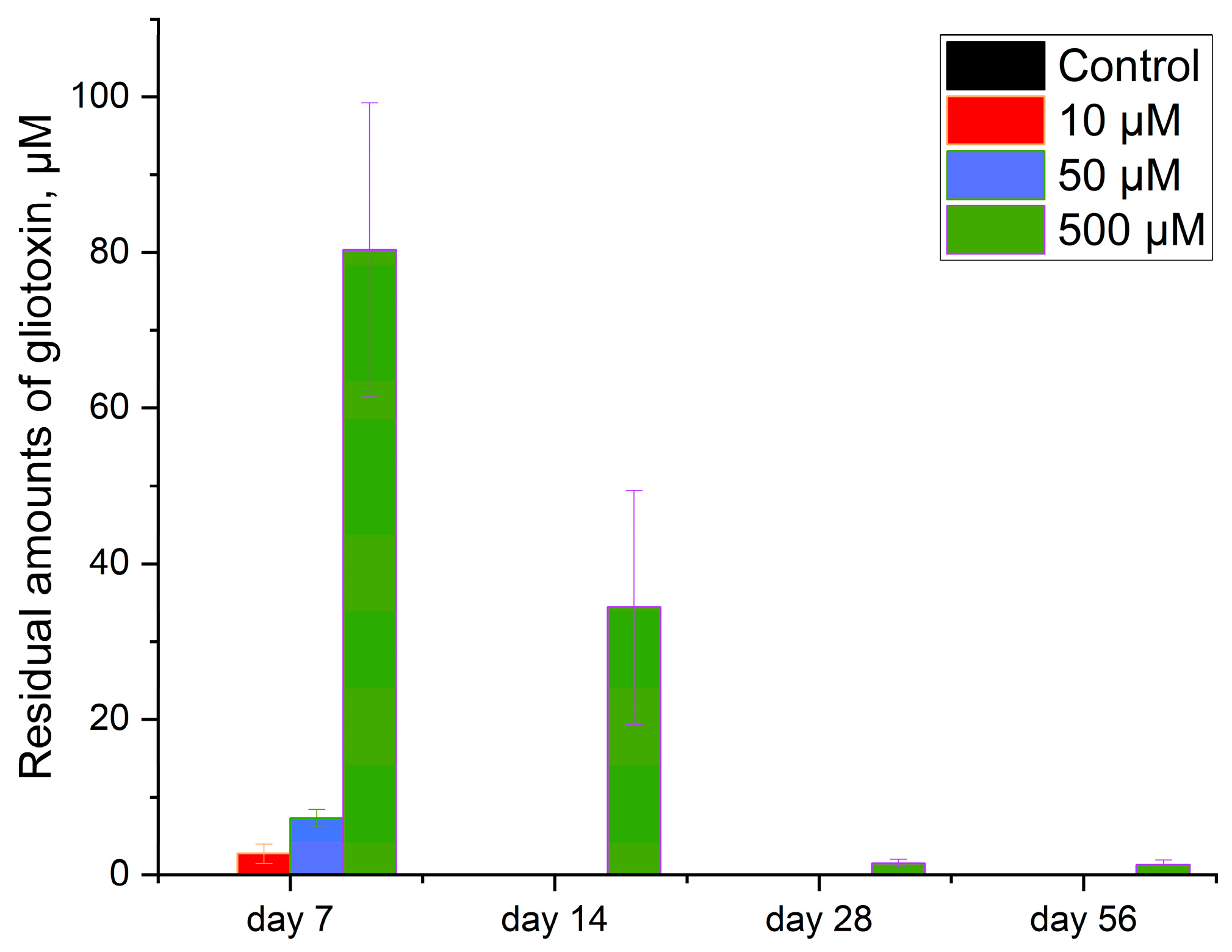
Figure 1. Available gliotoxin in soil as a function of time was determined by HPLC detection of residual quantities. Data are presented as mean ± standard deviation.
Agronomy 14 02084 g001
Figure 2. Response of the soil microbial community to the application of various doses of gliotoxin. (a) Basal (microbial) respiration; (b) microbial biomass; (c) coefficient of microbial respiration, QR; (d) metabolic coefficient or specific respiration of microbial biomass, qCO2, μg CO2 mg−1 MBSIR h−1; (e) share of microbial biomass carbon in organic carbon, MBSIR/SOC, %; (f) qCO2/SOC, μg CO2 mg−1 and MBSIR h−1 (gSOCg−1 soil)−1. Data are presented as mean ± standard deviation.
Agronomy 14 02084 g002
Figure 3. Changes in soil enzymatic activity under the influence of different doses of gliotoxin. Data are presented as mean ± standard deviation.
Agronomy 14 02084 g003
Table 1. Soil organic carbon content in samples with different doses of GT and different incubation periods.
GT Dose, µM kg−1 SoilSoil Organic Carbon, g kg−1
Incubation Time, Days
7142856
Control15.5 ± 0.5815.5 ± 0.4315.7 ± 1.3614.9 ± 0.74
1015.8 ± 1.3816.9 ± 0.0217.5 ± 1.2316.9 ± 1.21
5013.9 ± 1.1118.5 ± 0.92 *17.9 ± 2.1318.2 ± 1.19 *
50021.9 ± 1.63 **20.5 ± 0.62 ***18.4 ± 2.4918.1 ± 2.47
* p < 0.05, ** p < 0.005, *** p < 0.001 (t-test). Data are presented as mean ± standard deviation.
Table 2. Two-way ANOVA analysis of the effect of experimental conditions on soil enzymatic activity.
EnzymesStatisticsFactors
Sampling Time (A)GT Concentration (B)A × B
βGF52.6313.6033.11
p-value3.21 × 10−183.86 × 10−75.80 × 10−19
CBF1.231.411.32
p-value0.310.250.26
βXF9.597.828.70
p-value2.04 × 10−51.35 × 10−43.75 × 10−7
APF22.346.1214.23
p-value2.30 × 10−109.01 × 10−41.16 × 10−10
NAGF246.107.11126.60
p-value4.41 × 10−382.97 × 10−41.57 × 10−36
LAPF103.242.8653.05
p-value3.38 × 10−260.042841.17 × 10−24
ARSF5.901.113.51
p-value0.001160.351010.00421
Disclaimer/Publisher’s Note: The statements, opinions and data contained in all publications are solely those of the individual author(s) and contributor(s) and not of MDPI and/or the editor(s). MDPI and/or the editor(s) disclaim responsibility for any injury to people or property resulting from any ideas, methods, instructions or products referred to in the content.