Article

Identification and Antagonistic Potential of Bacillus atrophaeus against Wheat Crown Rot Caused by Fusarium pseudograminearum

by
Shengzhi Guo
1,†,
Arneeb Tariq
1,†,
Jun Liao
1,
Aowei Yang
1,
Xinyan Jiang
1,
Yanling Yin
1,2,
Yuan Shi
1,
Changfu Li
1,
Junfeng Pan
1,
Dejun Han
3,* and
Xihui Shen
1,*
1
State Key Laboratory for Crop Stress Resistance and High-Efficiency Production, Shaanxi Key Laboratory of Agricultural and Environmental Microbiology, College of Life Sciences, Northwest A&F University, Yangling District, Xianyang 712100, China
2
Xinjiang Production and Construction Crops Key Laboratory of Protection and Utilization of Biological Resources in Tarim Basin, College of Life Sciences, Tarim University, Alar 843300, China
3
State Key Laboratory for Crop Stress Resistance and High-Efficiency Production, College of Agronomy, Northwest A&F University, Yangling District, Xianyang 712100, China
*
Authors to whom correspondence should be addressed.
These authors contributed equally to this work.
Agronomy 2024, 14(9), 2135; https://doi.org/10.3390/agronomy14092135
Submission received: 20 August 2024 / Revised: 16 September 2024 / Accepted: 18 September 2024 / Published: 19 September 2024

Abstract

:
Fusarium pseudograminearum (Fpg) is a significant pathogen responsible for fusarium crown rot (FCR) in wheat (Triticum aestivum L.), a disease with devastating impacts on crop yield. The utilization of biocontrol bacteria to combat fungal diseases in plants is a cost-effective, eco-friendly, and sustainable strategy. In this trial, an endophytic bacterial species, designated as SW, was isolated from the roots of wheat. The strain exhibited potent antagonistic effects against Fpg and reduced the FCR disease severity index by 76.07 ± 0.33% in a greenhouse pot trial. Here, 106 colony-forming units (CFUs)/mL of the SW strain was determined to be the minimum dose required to exhibit the antagonism against Fpg. The strain was identified as Bacillus atrophaeus using genome sequencing and comparison with type strains in the NCBI database. Whole-genome sequencing analysis revealed that SW harbors genes for siderophores, antifungal metabolites, and antibiotics, which are key contributors to its antagonistic activity. Additionally, the strain’s ability to utilize various carbon and nitrogen sources, successfully colonize wheat root tissues as an endophyte, and form biofilms are critical attributes for promoting plant growth. In summary, these findings demonstrate the ability of Bacillus atrophaeus to control FCR disease in wheat in a sustainable agricultural setting.

1. Introduction

Fusarium pseudograminearum (Fpg) is the primary contributor of Fusarium crown rot (FCR) in wheat, which causes the browning and blackening of seeds, seedlings, roots, crowns, and particularly lower stems, severely impeding water and sap flow [1]. This disease has become one of the most significant threats to wheat yield, predominantly affecting regions with low precipitation such as parts of North China, Australia, and the Northwestern United States [2,3,4]. In China, FCR caused by Fpg was first documented in Henan Province in 2012 [5]. Typically, FCR causes a 10% yield loss, but under favorable conditions for disease spread, i.e., high humidity, warm temperatures, and organic matter in the soil, the loss can exceed 30% [2,6]. Although Fpg primarily invades the root system and lower nodes, its mycotoxins, including zearalenone (ZEN), deoxynivalenol (DON), and DON derivatives, can also be found in the crown, posing significant health risks to both humans and animals [7,8]. In humans, mycotoxins can adversely affect various organs and systems, such as the liver, kidneys, and immune, reproductive and developmental systems, and can also cause cancer by triggering oxidative stress, inhibiting protein synthesis, and altering DNA [9]. In animals, symptoms such as gastrointestinal dysfunction, reduced reproduction, impaired feed conversion efficiency, lowered milk and egg production, and anemia have been reported [10]. In symptomatic wheat tissues, disease intensity is positively correlated with the likelihood of pathogen isolation, Fpg quantity, and mycotoxin concentration. Even asymptomatic tissues may accumulate mycotoxins, with the Fpg biomass and mycotoxin concentration depending on the proximity to diseased tissue and its severity [11]. Various fungicides, such as fludioxonil, difenoconazole, tebuconazole, and azoxystrobin, have been tested and found to be effective for controlling Fpg [12,13,14]. Seed treatments like Cruiser Plus and Celest have shown good control in greenhouse and field trials [13]. However, resistance to tebuconazole and fludioxnil has been detected in some Fpg isolates in China [12]. Moreover, prolonged use of fungicides may degrade soil quality and harm the environment [15].
To reduce reliance on chemical fungicides in agriculture settings, there is an increasing focus on utilizing biological control methods for disease suppression [9]. The recruitment of natural hostile microorganisms as a biological control tool has arisen as an alternative choice [16]. Biocontrol agents (BCAs) show great promise as substitutes for chemical fungicides in controlling phytopathogens [17,18]. Hydrolytic enzymes and antagonistic secondary metabolites are common mechanisms of plant growth-promoting (PGP) bacteria; these compounds exhibit potent antifungal effects against plant pathogenic fungi and can enhance induced systemic resistance (ISR) in susceptible hosts [19]. When plants are exposed to biocontrol bacteria, it can trigger a cascade of defense reactions, including protection against oxidative stress [20], formation of secondary metabolites [21], increased activity of defense-related enzymes, and overexpression of defense-associated genes [22]. The diverse antibacterial compounds produced by Bacillus species make them highly desirable BCAs for agricultural use. Lipopeptides, siderophores, non-ribosomally synthesized peptides (NRSP), polyketides, and bacteriocins are the primary antimicrobial metabolites produced by these microbes, accounting for 5–8% of their genomes [23].
Several Bacillus species possess enzyme complexes known as non-ribosomal peptide synthetase (NRPS), which include fengycin, surfactin, and iturins, all known for their ability to hinder fungal mycelia. Recent research highlighted that fengycin synthesized by Bacillus spp. can effectively reduce F. graminearum [22] by damaging its reproductive and vegetative structures in wheat [24,25]. Additional, fengycin has been shown to trigger ISR in Solanum lycopersicum against Sclerotinia sclerotiorum [26]. Furthermore, bacillomycin D derived from Bacillus spp. has been documented to effectively inhibit the growth of F. graminearum in wheat [27]. The lipopeptides synthesized by B. atrophaeus can mitigate oxidative stress in cucumber powdery mildew caused by Sphaerotheca fuliginea [28], as well as combat cotton rhizoctoniosis [29] and apple ring rot disease [30].
This research aims to identify endophytic strains from healthy wheat roots and evaluate their biocontrol potential against Fpg both in vivo and in vitro. The SW strain, exhibiting strong antifungal activity, was identified as Bacillus atrophaeus using morphological analysis and whole genome sequencing. In addition to its antagonistic behavior against Fpg, the strain demonstrated several plant growth-promoting traits. In greenhouse pot experiments, the strain significantly suppressed disease incidence and promoted seedling growth. These findings indicate that B. atrophaeus SW holds great promise as a BCA. This research presents a biological strategy to combat wheat crown rot, laying the foundation for innovative and sustainable agricultural practices.

2. Materials and Methods

2.1. Sample Collection and Isolation of Strains

Healthy and thriving wheat plants from Henan Province (35.77° N, 115.39° E, China) were carefully sampled for strain isolation. Briefly, 3 g of root tissue was surface sterilized [31] using 75% (v/v) ethanol for 1 min, followed by soaking in 3% (v/v) NaClO for 5 min. Finally, the samples were rinsed with sterilized distilled water five times. The sterilized root tissue was placed on sterile filter paper to absorb excess water and then ground in a pre-sterilized pestle and mortar on a sterile clean bench until homogenized. Serial dilutions of the homogenous solution were prepared using sterilized saline phosphate buffer (PBS: 8 g/L NaCl, 0.2 g/L KCl, 1.42 g/L Na2HPO4, 0.27 g/L KH2PO4, pH 7.4) and spread onto Luria–Bertani (LB) agar [32]. After 48 h of incubation at 30 °C, the isolates were further purified by streaking until single colonies were obtained. Purified strains were preserved at −80 °C in LB broth supplemented with autoclaved 20% (v/v) glycerol.

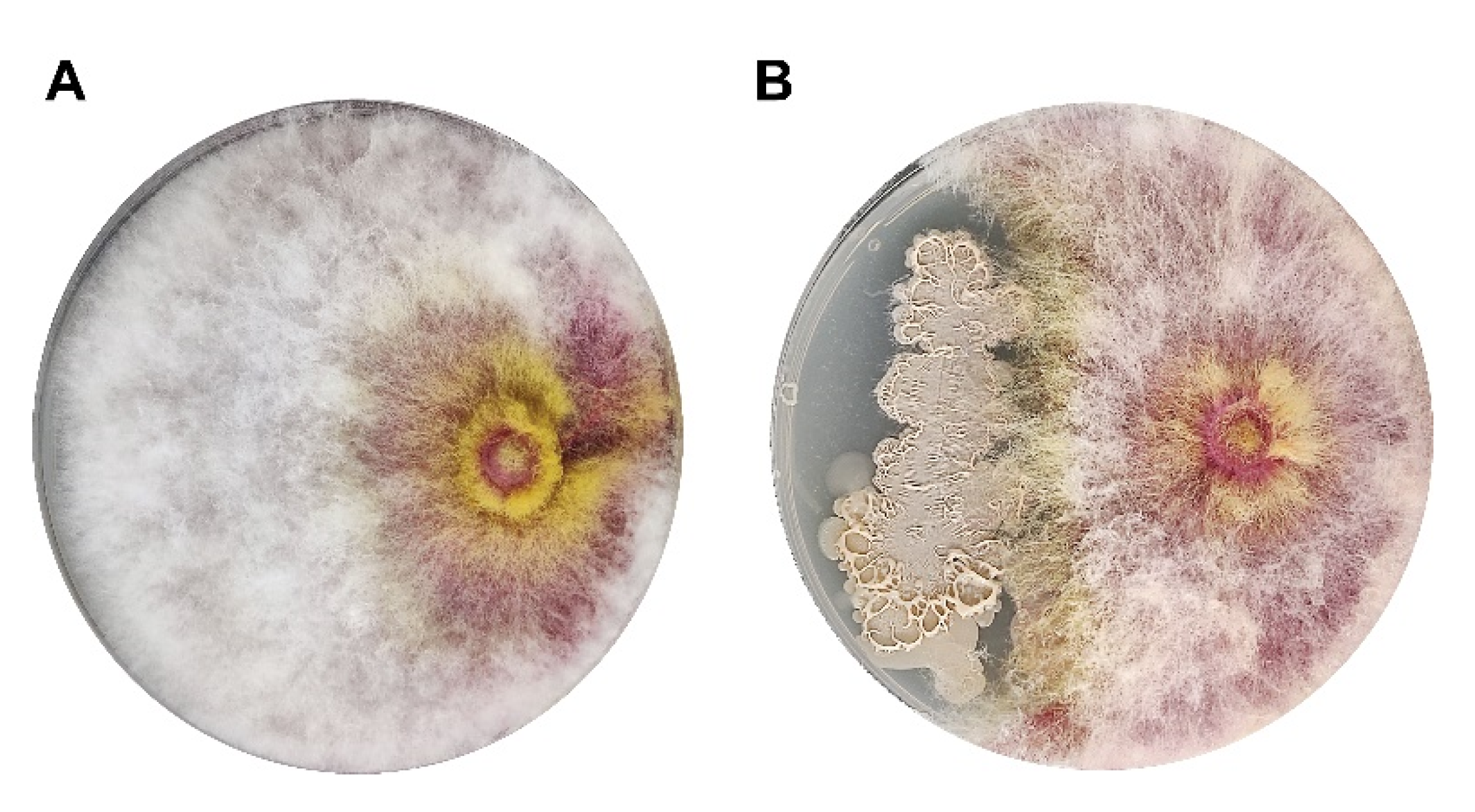
2.2. Co-Inoculation Assay for the Screening of Biocontrol Strains

Mycelia of Fpg were inoculated in a sodium carboxymethylcellulose (Na-CMC) broth containing 2.5 g/L dipotassium hydrogen phosphate, 2.5 g/L disodium hydrogen phosphate, 20 g/L sodium carboxymethyl cellulose, 2.0 g/L peptone, and 0.5 g/L yeast extract and incubated for 5 days at 26 °C. The Na-CMC suspension was filtered through eight strata of autoclaved gauze, and the filtrate was centrifuged at 3500 rpm for 10 min to collect conidia. The conidial pellet was put in autoclaved PBS to attain a final concentration of 1 × 10⁵ conidia/mL, referred to as the conidia solution of Fpg. Afterward, 200 µL conidia solution was evenly placed onto a potato dextrose agar (PDA) [33] plates, serving as the screening plates for isolating biocontrol strains.
The previously isolated root endophytic strains were cultivated in 5 mL LB broth at 30 °C in a shaker at 180 rpm for 24 h, followed by centrifugation for 5 min at 4500 rpm. The upper layer was disposed, and the precipitated pellet was resuspended in autoclaved PBS to achieve an OD600 of 1.0, measured using an EPOCH plate reader (Bio-Tek® Instruments, Highland Park, IL, USA).
For screening biocontrol strains, each plate was marked at regular intervals and coated with 10 µL of each bacterial PBS suspension. The plates were incubated upright at 26 °C, and the emergence of inhibition zones around the tested strains was noted after 7 days. This protocol was repeated three times.
Strains that exhibited inhibition zone were further assessed for antifungal activity using a dual culture assay on PDA plates. The procedure was executed as previously outlined, with few modifications [34]. A 5 mm diameter Fpg mycelium was placed 2 cm from the edge of the plate, and 10 µL of the bacteria suspension was placed 2 cm from the opposite edge on an 8 cm diameter plate. Control plates were treated with sterile PBS instead of bacterial suspension. The Petri plates were kept upright at 26 °C for 7 days until the Fpg mycelium extended to the edge of the control plate. This experiment was repeated thrice for reproducible results.

2.3. Minimum Inhibitory Dose

The conidia solution of the Fpg was prepared as described in Section 3.2. The SW strain was cultured in 5 mL of LB broth at 30 °C in a shaker at 180 rpm for 24 h. After centrifugation, both the Fpg conidia solution and the SW bacterial suspension were resuspended in sterile PBS. The 2 × 10⁵ conidia/mL solution was mixed with different concentrations of the SW strain: 5 × 101, 5 × 10², 5 × 10³, 5 × 10⁴, 5 × 10⁵, 5 × 10⁶, 5 × 10⁷, or 5 × 10⁸ CFU/mL. PDA plates were marked into equal-sized grid intervals, and 6 μL of each solution was applied to the corresponding grid. Each set of experiments was performed in triplicate. The plates were incubated upright at 26 °C for 5 days. The minimum concentration of the SW bacterial solution at which Fpg did not grow was recorded as the minimum inhibitory concentration.

2.4. Strain Identification

Morphologic and phenotypic identification of the test strain was performed using scanning electron microscopy (SEM) with a Nano SEM-450 instrument (FEI Co., Hillsboro, OH, USA) and 16S ribosomal RNA analysis. Universal primers 27F (5′-AGAGTTTGATCMTGGCTCAG-3′) and 1492R (5′-TACGGYTACCTTGTTACGACTT-3′) were used for PCR amplification of the 16S rRNA gene of SW. The resulting 16S rRNA gene sequence was compared against sequences available in the NCBI database.

2.5. Assays for Plant Growth-Promoting Traits

Strain SW was regularly cultivated in 5 mL LB broth at 30 °C for 24 h with constant shaking at 180 rpm. After cultivation, the cells were harvested and suspended in sterilized PBS to attain a 1 × 10⁸ CFU/mL concentration for subsequent experimental protocols. The strain’s ability to use of different carbon and nitrogen compounds as energy source was examined by cultivating it in minimal salt medium (1.5 g/L K2HPO4·3H2O, 0.5 g/L KH2PO4, 1 g/L NaCl, 0.2 g/L MgSO4·7H2O, pH 7.2) [35] fortified with several compounds as the sole energy source (the carbon sources included D-fructose, D-ribose, D-raffinose, glucose, sucrose, trehalose, maltose, soluble starch, xylose, and xylan, and the nitrogen sources include tryptone, ammonium nitrate, yeast, urea, glycine, casein, glutamate, and ammonium sulfate). During the examination of carbon compounds utilization, nitrogen-containing compounds (ammonium sulfate and sodium nitrate) were incorporated as energy sources, while glucose was used in the assessment of nitrogen sources.
The synthesis of siderophore was tested using the Chrome Azurol S (CAS) assay (Haibo Co., HB9132, Qingdao, China). Briefly, strain SW was cultured in LB medium at 30 °C with shaking at 180 rpm for 24 h. The bacterial concentration was then adjusted to 1 × 10⁸ CFU/mL using sterile PBS. Subsequently, 10 µL of the SW PBS suspension was spotted onto CAS medium. The culture plate was incubated at 30 °C in the dark for 3 days. The appearance of an orange-yellow halo around the strain indicated its ability to produce siderophores. Pseudomonas khavaziana SR9 was used as a positive control [36].
Antibiotic resistance was assessed by fortifying LB broth with varying levels of antibiotics. Strain SW was initially cultured in LB medium at 30 °C with shaking at 180 rpm for 24 h. Subsequently, the strain was transferred to fresh LB broth at a 1% (v/v) inoculum. Each tube was supplemented with different concentrations of common laboratory antibiotics (ampicillin, kanamycin, chloramphenicol, nalidixic acid, ceftazidime, rifampicin, ciprofloxacin, tetracycline, gentamicin, and streptomycin) and kept in a 180-rpm shaker at 30 °C for another 24 h. The growth of the strain was then recorded.

2.6. Whole Genome Sequencing, Annotation, and Analysis

The commercially available TIANamp Bacteria DNA Kit (Tiangen Biotech Co., Beijing, China) was used to extract the strain’s DNA followed by sequencing with the Magigene (Shenzhen, China) Pacbio Sequel IIe system. The resulting third-generation sequencing reads were processed for assembly using MECAT2 [37]. Gene predication was performed using Glimmer 3 [38], while non-coding rRNA and tRNA were identified using rRNAmmer 1.2 [39] and tRNAscan-SE 2.0 [40], respectively. Initial alignment for sRNA prediction was conducted using the Rfam database [41] and further refined using the cmsearch tool [42]. Prophage occurrence was evaluated using phage_finder (v2.1) [43]. The genome sequence was annotated using RAST (http://rast.nmpdr.org/, accessed on 17 September 2024) [44,45]. Antibiotic resistance genes and carbohydrate-active enzymes were predicted using the Comprehensive Antibiotic Resistance [46] and the Carbohydrate-Active Enzymes Databases [47], respectively. Secondary metabolite biosynthetic gene clusters were annotated and analyzed using antiSMASH 7.0 [48]. The CVTree v4 [49] method was employed to construct a whole genome-based phylogenomic tree. Average nucleotide identity among selected Bacillus species was calculated using FastANI [50], and digital DNA–DNA hybridization (dDDH) values were measured using the Genome-to-Genome Distance Calculator 3.0 [51].

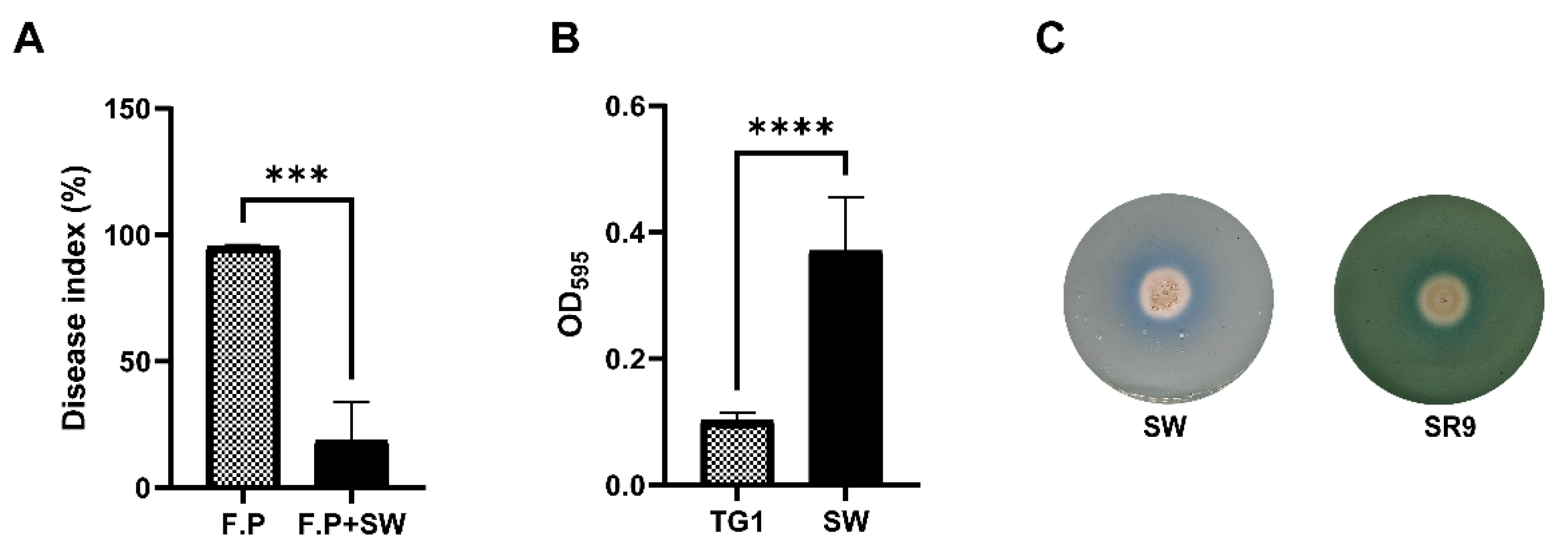
2.7. Biofilm Adhesion Assay

The biofilm formation ability of the SW strain was quantified using the crystal violet staining method [52]. The SW strain was cultivated in MSgg medium (Coolaber Co., SL0130, Beijing, China) at 30 °C with constant shaking at 180 rpm for 48 h. After diluting the strain suspension to an OD600 of 0.05, 170 µL was added to each well of a 96-well microtiter plate. The plate was incubated at 30 °C for 48 h in a static position, followed by the removal of the supernatant. The wells were then gently washed three times with PBS to eliminate any non-adherent cells. The remaining adherent cells were stained with 0.1% (w/v) crystal violet and subsequently washed with PBS. To dissolve the remaining stain, 200 µL of ethanol was added, and the absorbance of the solution was measured at 590 nm using a microplate reader.

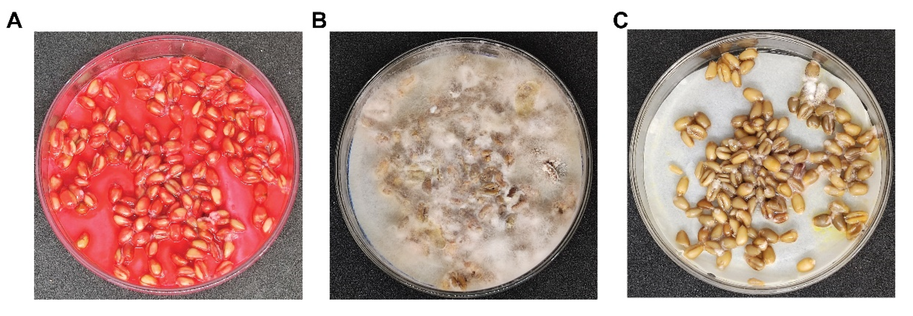
2.8. In Vitro Infection of Wheat Kernels

To evaluate the biocontrol activity of the SW strain on wheat seeds, 24-h water- soaked and sterilized seeds were placed on sterilized and moistened filter paper in the Petri plate. The wheat grains (cv. Jimai22) were evenly sprayed with three different solutions.
5 mL Fpg conidia solution (2 × 10⁵ conidia/mL) + 5 mL PBS.
5 mL Fpg conidia solution (2 × 10⁵ conidia/mL) + 5 mL chemical fungicide fludioxonil (5 g/L).
5 mL Fpg conidia solution (2 × 10⁵ conidia/mL) + 5 mL SW (108 CFU/mL).
Each spray treatment was performed in triplicate. The plates were incubated at 26 °C for 5 days, with sterilized water added as needed to maintain proper moisture content.

2.9. Biocontrol Efficiency of SW in Pot Experiment

The biocontrol potential of the SW against Fpg in wheat was evaluated under controlled greenhouse conditions, with minor adjustments to a previously defined method [53].
The Fpg mycelium was inoculated into millet seeds sterilized at 121 °C for 30 min. The flasks were kept at 26 °C for 7 days in a static position and manually shaken twice daily to ensure complete colonization of the millet seeds. Peat soil was sterilized in an autoclave at 121 °C for 40 min, and 100 g was placed into each plastic pot measuring 6.5 cm in diameter and 8.5 cm in height. Plump winter wheat seeds (cv. Jimai22) were washed with water, surface sterilized with ethanol, and sown in soil-filled pots with 12 seeds per pot. The pots were then placed in a growth chamber with a 12-h light/dark cycle. After three days of germination, each pot was watered with 40 mL SW PBS suspension (1 × 10⁸ CFU/mL), while the control group received an equal volume of PBS. At the one-tiller and one-leaf stage, 10 grains of infected millet were placed at the base of each seedling and covered with a layer of the same soil as used for pot filling.
After 4 weeks of sowing, the disease severity index and biocontrol efficacy of the SW strain were recorded. Six pots were used per treatment, and the entire experiment was repeated three times.
Disease severity was assessed using the following grading scale [54]: 0, no disease symptoms; 1, yellow or brown discoloration from root to beneath the first leaf sheath; 3, browning but not blackening of first leaf sheath; 7, browning up to the third leaf sheath; 9, root rot leading to death or wilting of leaves.
Disease   index =   E a c h   d i s e a s e   g r a d e × n u m b e r   o f   i n f e c t e d   p l a n t s   o f   t h a t   d i s e a s e   g r a d e T h e   t o t a l   n u m b e r   o f   p l a n t s   s u r v e y e d × 9 × 100
Control   efficiency = D i s e a s e   i n d e x   o f   c o n t r o l d i s e a s e   i n d e x   o f   t r e a t m e n t D i s e a s e   i n d e x   o f   c o n t r o l × 100 %

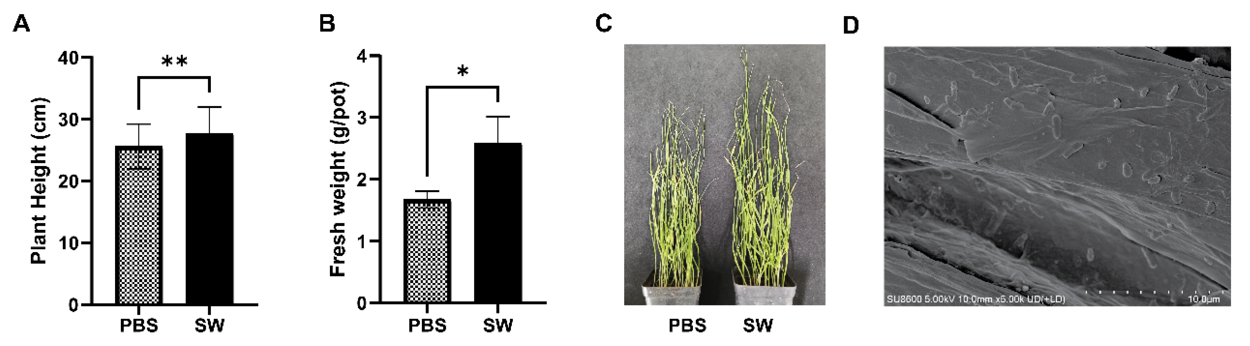
2.10. Colonization of SW and Plant Growth Promotion Pot Experiment

A pot experiment was conducted in the greenhouse to evaluate the plant growth-promoting effects of strain SW in winter wheat (Triticum aestivum L. cv. Jimai22). Peat soil and wheat seeds were sterilized, and pots were filled as described in the previous section. The seedlings were germinated in the dark at 26 °C for 2 days and then transplanted into pots filled with sterile peat soil, with 12 seedlings per pot, maintained under a 12-h light/dark cycle. One week after transplanting, each pot in the SW treatment group was irrigated with 40 mL of the strain suspension (1 × 10⁸ CFU/mL), while an equal volume of sterile water was added to the control group pots. One-week post-inoculation, the roots were collected, washed with sterile PBS to remove soil, and fixed with 4% glutaraldehyde at 4 °C for 12 h. The samples were then rinsed four times with 0.1 M PBS (pH 6.8) and dehydrated in a graded ethanol series for 10 min each. After drying and gold sputter-coating, the colonization of SW was examined using a Nano SEM-450 instrument (FEI Co., Hillsboro, OH, USA).
Plant biomass and height were recorded 30 days after inoculation. The experiment was conducted three times to ensure precise results.

2.11. Statistical Analysis

The data were statistically analyzed in Graph Pad Prism software (GraphPad Prism 9) using an unpaired two-tailed Student’s t test. The significance level was set at p < 0.05.

3. Results

3.1. Characterization and Identification of SW

The SW strain exhibited a milky white, beaded appearance and was non-pigmented on LB agar plates after 24 h of incubation (Figure 1A). Microscopically, the cells were observed to be organized in pairs or chains of straight rods with rounded ends (Figure 1B). Amplification of the 16S rRNA gene using universal primers 27F and 1492R, followed by a Basic Local Alignment Search Tool (BLAST) search on the NCBI database, identified the strain as belonging to the Bacillus genus (Table S1). The 16S rRNA gene sequence of strain SW has been deposited in the GenBank databases under accession number PQ186995. Phylogenetic analysis further clarified the position of strain SW within the B. atrophaeus cluster. It was found to cluster on the same branch as B. atrophaeus NRRL NRS 213T (Figure 1C) with an average nucleotide identity of 98.26% (Figure 2) and a digital DNA–DNA hybridization value of 89.4% (Table S2). These results confirmed the identification of the SW strain as B. atrophaeus. The sequence data have been submitted to the NCBI database under accession number GCA_039519175.1.

3.2. Biocontrol Efficacy of Wheat Endophytic Strain SW

Among a series of strains isolated from wheat roots using the serial dilution technique, strain SW exhibited strong antagonistic potential against Fpg and remarkably hindered mycelial extension in a co-inoculation assay (Figure 3).
In a greenhouse pot experiment, treatments without inoculation of the SW biocontrol agent showed more pronounced Fpg crown rot symptoms, including dark brown stems near the first node. The disease severity index exhibited a statistically significant decrease of 76.07 ± 0.33% in strain-inoculated plants compared to the control group. These data suggest that strain SW is an effective biocontrol agent against Fpg (Figure 4A).

3.3. Secondary Metabolite Analysis and Plant Beneficial Characters

Given the biocontrol potential demonstrated by strain SW, an analysis was conducted to assess its capacity for generating secondary metabolites. The results indicated the contribution of 11 diverse gene clusters in the biosynthesis of different valuable products (Table 1). The analysis revealed the presence of seven non-ribosomal peptide synthetase (NRPS) clusters, which include genes related to antibiotics such as bacillibactin, surfactin, zwittermicin A, bacillaene, and fengycin, and other antibacterial substances. In addition to NRPS, the analysis indicated the presence of other clusters involved in the synthesis of antibiotics like carbapenem, specifically 1-carbapen-2-em-3-carboxylic acid. Furthermore, one thiopeptide and two terpene biosynthetic clusters were predicted. Although no ‘Most similar known cluster’ was identified for these gene clusters, their corresponding genomic locations were successfully mapped, indicating the potential presence of novel biosynthetic pathways that have not yet been characterized. The predicted results of this analysis suggest that SW can exhibit significant biocontrol effects.
Strain SW exhibited survival capability in the presence of low concentrations of ampicillin, gentamicin, nalidixic acid, chloramphenicol, ciprofloxacin, kanamycin, and ceftazidime, with the exception of rifampicin and tetracycline. It also demonstrated the ability to withstand high concentrations of ampicillin and streptomycin (Table S3). The strain was capable of utilizing various carbon and nitrogen sources, except xylose and glutamate (Table S4). In a biofilm synthesis test on a microtiter plate, strain SW showed significant potential to produce biofilm (OD590 = 0.37) compared to the control (OD590 = 0.10) (Figure 4B). Additionally, strain SW turned the blue color of CAS medium to orange-yellow, indicating its ability to produce siderophores (Figure 4C).

3.4. Minimum Inhibitory Dose Estimation of SW Strain to Antagonize Fpg

To determine the minimum effective concentration of the SW spore suspension required to inhibit the mycelial growth of Fpg on the PDA Petri plates, various concentrations ranging from 5 × 101 to 5 × 10⁸ CFU/mL were tested against 105 conidia/mL Fpg suspension. At specific bacterial concentrations, fungal growth was unable to spread beyond the surrounding area. Notably, spore suspensions at concentrations of 5 × 10⁶, 5 × 10⁷, and 5 × 10⁸ CFU/mL effectively inhibited fungal mycelium growth within their respective zones. Therefore, 5 × 10⁶ CFU/mL was identified as the minimum effective biocontrol dose against a 105/mL Fpg conidial suspension (Figure 5).

3.5. SW Strain Mimics Fludioxonil Function in Wheat Kernel Experiments

In a Petri plate experiment on wheat kernels, the SW strain mimicked the action of fludioxonil fungicide, which is known to retard mycelial growth by strongly inhibiting germ tube elongation and delaying conidia formation. There was no significant difference in the inhibitory effect between 5 g/L fludioxonil and a concentration of 108 CFU/mL of the SW strain against a 2 × 10⁵ conidia/mL Fpg suspension. Both the SW strain and fludioxonil strongly inhibited the mycelial growth on seeds in Petri plates compared to the control (Figure 6).

3.6. Growth Promotion and Colonization Effects of the SW Strain in Wheat Plants

To analyze the growth enhancement effects of the SW strain in wheat seedlings, vegetative parameters such as plant height and fresh weight were recorded and statistically evaluated in both groups. In the greenhouse, a wheat pot experiment was conducted in the absence of Fpg. A significant increase in plant fresh weight and height was observed in both SW-inoculated and non-inoculated strains. This suggests that the SW strain, in addition to triggering ISR against disease, also has a notable effect on plant growth (Figure 7). One week after the application of the SW suspension, scanning electron microscopy images revealed the colonization of SW in the wheat roots (Figure 7D).

4. Discussion

Over the last few decades, a vast number of plant growth-promoting (PGP) bacteria have been screened, and their biocontrol potentials have been explored [55,56]. The implementation of biocontrol strains to antagonize fungal pathogens is a crucial strategy for reducing pesticide use and promoting plant health, thereby contributing to sustainable agriculture [57,58,59]. A biocontrol agent exhibiting multiple plant growth-promoting traits in addition to biological control activity is more promising under field conditions compared to those with only antagonistic activity against pathogens [60,61]. The antagonistic ability was assessed in vitro using dual culture assays of wheat endophytes and the common mycopathogen, i.e., Fpg. A significant proportion of the tested strains exhibited antagonistic effects, with strain SW displaying the highest antifungal potential. Based on morphological attributes, 16S rRNA sequencing, and complete genome sequences, strain SW was identified as B. atrophaeus.
Bacillus is a widely recognized genus of biocontrol agents with significant potential in controlling plant diseases [62]. Successful implementation of biocontrol potential requires an extensive understanding of the underlying mechanisms [63,64]. Bacillus spp. employ diverse strategies to exert their biocontrol potential, such as antibiosis, competition for nutrients, parasitism, or plant growth promotion. It is widely acknowledged that direct interaction and establishment of ecological niches are crucial for PGP bacteria against numerous pathogens [18,65]. Biochemical tests revealed that strain SW can utilize a diverse array of both carbon and nitrogen sources as substrates, greatly enhancing its potential for colonization. The SW strain was able to produce biofilm on the artificial surface, enabling intimate associations within the host plant. Previous research has demonstrated that biofilm formation is a major contributor to plant pathogenesis [66]. Biofilms provide a shielded microenvironment where bacteria reside and maintain their colonies under favorable conditions despite external environmental stresses. They acquire nutrients from root exudates and impact the plant by ensuring a firm root system, maintaining root zone moisture, and moderating the soil pH, leading to improved biogeochemical cycling [67].
Biocontrol agents employ various strategies to mitigate the harmful effects of phytopathogens, including antibiosis and siderophore synthesis [68]. The antibiosis functions of the SW strain are particularly significant for controlling fungal diseases. In this study, the SW strain demonstrated resistance to multiple concentrations of different antibiotics, which is consistent with previous reports in the literature [35]. Genome analysis revealed that up to 5% of the Bacillus genome comprises genes involved in the antibiotic biosynthetic pathway [69]. Lipopeptide antibiotics are the most predominantly produced antibiotics in B. atrophaeus [29]. Previous studies have reported that B. atrophaeus produces extracellular hydrolytic enzymes and lipopeptide antibiotics, which cause oxidative cell disruption, ultrastructural damage, and genomic disturbances leading to pathogen necrosis [29,70]. Antismash database analysis identified numerous antibiotic-producing gene clusters in the SW strain genome, most predominantly including zwittermicin A and bacillaene. Another important class of secondary metabolites, biosurfactins, was also identified. There are five dominant types of biosurfactants: glycolipids, lipopeptides and lipoproteins, phospholipids and fatty acids, and particulate and polymeric biosurfactants. Multiple studies have documented the synthesis of surfactins from diverse biocontrol strains of Bacillus [71,72], which are significant due to their antibacterial and antifungal potential [73]. Fengycin, in particular, exhibits potent antifungal activity against filamentous fungi by interacting with sterols and phospholipids in fungal cell membranes, thereby disrupting their structure and permeability [74]. Fengycin produced by Bacillus strains has been shown to control Fusarium moniliforme and Seclerotinia scleritiorium infections in rapeseed [75]. Genome mapping of the SW strain revealed some gene clusters with no correspondence to previously known gene clusters, suggesting the presence of novel metabolites yet to be discovered. Siderophore chemicals, such as bacillibactin synthesized by the SW strain, have the potential to mitigate disease and boost plant growth. Bacillibactin consists of small peptide molecules and functional groups with a side chain that binds ferric ions through a set of ligands. It hinders the infection capacity of phytopathogens by restricting Fe in the soil–root interface and promotes root and shoot growth. Siderophore production is a common attribute of diverse groups of antagonistic plant growth-promoting rhizobacteria (PGPR), such as Pseudomonas, Bacillus, Rhizobium, Azotobactor, Enterobacter, and Serratia [38,76,77,78,79].
Bacterial colonization in plant tissues is essential for bacteria to exert beneficial effects on plants, enhancing plant health and productivity [80]. Long-term root colonization by biocontrol bacteria is crucial for effective crop protection against soil-borne phytopathogens [81]. Bacteria that effectively colonize plant roots access the root surface through two mechanisms: passive migration via water fluxes or active swimming propelled by flagella [82]. Active flagellar propulsion is controlled by a unique genetic motif that has been previously identified in most Bacillus spp. [83]. Additionally, colonizing bacterial strains release enzymes to develop an extracellular matrix, utilizing carbon sources derived from hydrolyzing the cellulose, hemicellulose, and pectin of the plant host cell wall [84]. Previous research has reported that Bacillus strains secrete these enzymes [85,86]. Fpg conidia may overwinter in the wheat seeds to complete their life cycle, with infected seed germination leading to high disease spread and yield loss [87]. The SW strain effectively combats the disease incidence on wheat kernels, comparable to fludioxonil fungicide. The SW strain suspension also effectively controlled the disease index in the greenhouse pot experiment. These results align with the previous reports indicating that biopriming of seeds with a B. atrophaeus suspension reduces the disease index by more than 50% [88]. Due to the presence and potential production of useful secondary metabolites and antibiotic gene clusters present in the SW strain genome, along with confirmed siderophore and biofilm-producing characteristics, the growth promotion effect was significant in terms of plant height and fresh weight in the greenhouse pot experiment. In summary, B. atrophaeus SW is an endophytic bacterium with strong antagonistic capabilities against Fpg and promotes wheat plant growth.

5. Conclusions

The biocontrol potential of Bacillus atrophaeus SW, which was isolated from wheat root tissues and identified using 16S rRNA, whole genome sequence, ANI, and dDNA–DNA hybridization, was demonstrated by its strong in vitro antifungal activity against Fpg in wheat plants. Additionally, the SW strain demonstrated biofilm formation, siderophore production, the presence of antibiotic-related genes, and the ability to utilize various carbon and nitrogen sources, all of which contribute to restricting the mycelial growth of Fpg. The B. atrophaeus SW strain also showed excellent biocontrol potential against Fpg in wheat kernel experiments and wheat plants grown under greenhouse conditions. This strain holds great promise as a cost-effective and eco-friendly approach to managing FCR. However, further investigations are necessary to uncover any additional beneficial compounds. While numerous Bacillus species exhibit broad-spectrum antifungal activity, only a few are commercially viable as antagonistic organisms. Therefore, further research is warranted to identify and purify the key compounds in the secondary metabolites of SW and to develop fungicide formulations.

Supplementary Materials

The following supporting information can be downloaded at: https://www.mdpi.com/article/10.3390/agronomy14092135/s1, Table S1: The similarity index of SW strain with other strains of Bacillus genus calculated by 16s rRNA gene sequence. Table S2: Digital DNA-DNA-Hybridization values of related strains to SW. Table S3: Antimicrobial susceptibility test of SW strain. Table S4: Carbon and nitrogen source utilization of strain SW.

Author Contributions

Conceptualization, J.P. and X.S.; Funding acquisition, X.S.; Investigation, C.L.; Methodology, S.G., A.T., J.L., A.Y., X.J., Y.Y. and Y.S.; Project administration, X.S.; Resources, D.H.; Software, S.G.; Supervision, C.L., J.P., D.H. and X.S.; Writing—original draft, A.T.; Writing—review & editing, S.G., A.T., J.L., A.Y., X.J., Y.Y., Y.S., C.L., J.P. and D.H. and X.S. All authors have read and agreed to the published version of the manuscript.

Funding

This work was supported by the grant of the National Natural Science Foundation of China (32330004 to X.S.).

Data Availability Statement

The 16S rRNA gene sequence of strain SW has been deposited in the GenBank/EMBL/DDBJ databases under accession number PQ186995. The whole-genome sequence of strain SW has been deposited in the GenBank/EMBL/DDBJ databases under accession number GCA_039519175.1. Strain SW (CGMCC 30329) was deposited at the China General Microbiological Culture Collection Center (CGMCC).

Acknowledgments

Technical help was rendered by Life Science Research Core Services at Northwest A&F University (Kerang Huang). Supplementary assistance was provided by the Teaching and Research Core Facility at the College of Life Science (Ningjuan Fan, Xiyan Chen, and Hui Duan).

Conflicts of Interest

The authors declare that the research was conducted in the absence of any commercial or financial relationships that could be construed as a potential conflict of interest.

References

  1. Dong, Q.; Liu, Q.; Goodwin, P.H.; Deng, X.; Xu, W.; Xia, M.; Zhang, J.; Sun, R.; Wu, C.; Wang, Q. Isolation and Genome-Based Characterization of Biocontrol Potential of Bacillus Siamensis YB-1631 against Wheat Crown Rot Caused by Fusarium pseudograminearum. J. Fungi 2023, 9, 547. [Google Scholar] [CrossRef] [PubMed]
  2. Smiley, R.W.; Gourlie, J.A.; Easley, S.A.; Patterson, L.-M.; Whittaker, R.G. Crop Damage Estimates for Crown Rot of Wheat and Barley in the Pacific Northwest. Plant Dis. 2005, 89, 595–604. [Google Scholar] [CrossRef]
  3. Xu, F.; Yang, G.; Wang, J.; Song, Y.; Liu, L.; Zhao, K.; Li, Y.; Han, Z. Spatial Distribution of Root and Crown Rot Fungi Associated with Winter Wheat in the North China Plain and Its Relationship with Climate Variables. Front. Microbiol. 2018, 9, 1054. [Google Scholar] [CrossRef] [PubMed]
  4. Backhouse, D.; Abubakar, A.A.; Burgess, L.W.; Dennisc, J.I.; Hollaway, G.J.; Wildermuth, G.B.; Wallwork, H.; Henry, F.J. Survey of Fusarium Species Associated with Crown Rot of Wheat and Barley in Eastern Australia. Australas. Plant Pathol. 2004, 33, 255–261. [Google Scholar] [CrossRef]
  5. Li, H.L.; Yuan, H.X.; Fu, B.; Xing, X.P.; Sun, B.J.; Tang, W.H. First Report of Fusarium Pseudograminearum Causing Crown Rot of Wheat in Henan, China. Plant Dis. 2012, 96, 1065. [Google Scholar] [CrossRef]
  6. Xu, F.; Song, Y.L.; Zhou, Y.L.; Zhang, H.; Wang, J.M.; Li, Y.H.; Han, Z.H. Occurrence Dynamics and Characteristics of Fusarium Root and Crown Rot of Wheat in Henan Province during 2013–2016. Plant Prot 2016, 42, 126–132. [Google Scholar]
  7. Beccari, G.; Prodi, A.; Pisi, A.; Nipoti, P.; Onofri, A.; Nicholson, P.; Pfohl, K.; Karlovsky, P.; Gardiner, D.M.; Covarelli, L. Development of Three Fusarium Crown Rot Causal Agents and Systemic Translocation of Deoxynivalenol Following Stem Base Infection of Soft Wheat. Plant Pathol. 2018, 67, 1055–1065. [Google Scholar] [CrossRef]
  8. Moretti, A.; Panzarini, G.; Somma, S.; Campagna, C.; Ravaglia, S.; Logrieco, A.F.; Solfrizzo, M. Systemic Growth of F. Graminearum in Wheat Plants and Related Accumulation of Deoxynivalenol. Toxins 2014, 6, 1308–1324. [Google Scholar] [CrossRef] [PubMed]
  9. Ülger, T.G.; Uçar, A.; Çakıroğlu, F.P.; Yilmaz, S. Genotoxic Effects of Mycotoxins. Toxicon 2020, 185, 104–113. [Google Scholar] [CrossRef]
  10. Pier, A.C. Major Biological Consequences of Aflatoxicosis in Animal Production. J. Anim. Sci. 1992, 70, 3964–3967. [Google Scholar] [CrossRef]
  11. Xu, F.; Shi, R.; Liu, L.; Li, S.; Wang, J.; Han, Z.; Liu, W.; Wang, H.; Liu, J.; Fan, J. Fusarium Pseudograminearum Biomass and Toxin Accumulation in Wheat Tissues with and without Fusarium Crown Rot Symptoms. Front. Plant Sci. 2024, 15, 1356723. [Google Scholar] [CrossRef] [PubMed]
  12. Zhang, N.; Xu, Y.; Zhang, Q.; Zhao, L.; Zhu, Y.; Wu, Y.; Li, Z.; Yang, W. Detection of Fungicide Resistance to Fludioxonil and Tebuconazole in Fusarium Pseudograminearum, the Causal Agent of Fusarium Crown Rot in Wheat. PeerJ 2023, 11, e14705. [Google Scholar] [CrossRef] [PubMed]
  13. Zhang, N.; Yuan, S.; Zhang, Q.; Liu, W.; Zhou, Y.; Yang, W. Screening Fungicides for Controlling Wheat Crown Rot Caused by Fusarium Pseudograminearum across Hebei Province in China. Agriculture 2022, 12, 1643. [Google Scholar] [CrossRef]
  14. Hou, Y.; Guo, Y.; Wang, L.; He, S.; Zheng, W.; Liu, S.; Xu, J. Impact of Phenamacril on the Growth and Development of Fusarium Pseudograminearum and Control of Crown Rot of Wheat. Plant Dis. 2023, 107, 3843–3850. [Google Scholar] [CrossRef] [PubMed]
  15. Hao, X.; Gu, Y.; Zhang, H.; Wang, X.; Liu, X.; Chen, C.; Wang, C.; Zhang, X.; Liu, X.; Shen, X. Synthetic Microbial Community Promotes Bacterial Communities Leading to Soil Multifunctionality in Desertified Land. Microorganisms 2024, 12, 1117. [Google Scholar] [CrossRef]
  16. Mukhtar, T.; Vagelas, I.; Javaid, A. New Trends in Integrated Plant Disease Management. Front. Agron. 2023, 4, 1104122. [Google Scholar] [CrossRef]
  17. Chen, C.; Xin, K.; Liu, H.; Cheng, J.; Shen, X.; Wang, Y.; Zhang, L. Pantoea Alhagi, a Novel Endophytic Bacterium with Ability to Improve Growth and Drought Tolerance in Wheat. Sci. Rep. 2017, 7, 41564. [Google Scholar] [CrossRef]
  18. Farhat, F.; Tariq, A.; Waseem, M.; Masood, A.; Raja, S.; Ajmal, W.; Iftikhar, I.; Zulfiqar, U.; Maqsood, M.F. Plant Growth Promoting Rhizobacteria (PGPR) Induced Improvements in the Growth, Photosynthesis, Antioxidants, and Nutrient Uptake of Rapeseed (Brassica napus L.). Gesunde Pflanz. 2023, 75, 2075–2088. [Google Scholar] [CrossRef]
  19. Fira, D.; Dimkić, I.; Berić, T.; Lozo, J.; Stanković, S. Biological Control of Plant Pathogens by Bacillus Species. J. Biotechnol. 2018, 285, 44–55. [Google Scholar] [CrossRef]
  20. Ahn, I.-P.; Lee, S.-W.; Suh, S.-C. Rhizobacteria-Induced Priming in Arabidopsis Is Dependent on Ethylene, Jasmonic Acid, and NPR1. Mol. Plant-Microbe Interact. 2007, 20, 759–768. [Google Scholar] [CrossRef]
  21. Rahman, A.; Uddin, W.; Wenner, N.G. Induced Systemic Resistance Responses in Perennial Ryegrass against M Agnaporthe Oryzae Elicited by Semi-purified Surfactin Lipopeptides and Live Cells of Bacillus amyloliquefaciens. Mol. Plant Pathol. 2015, 16, 546–558. [Google Scholar] [CrossRef]
  22. Chowdhury, S.P.; Hartmann, A.; Gao, X.; Borriss, R. Biocontrol Mechanism by Root-Associated Bacillus Amyloliquefaciens FZB42–a Review. Front. Microbiol. 2015, 6, 780. [Google Scholar] [CrossRef] [PubMed]
  23. Compant, S.; Duffy, B.; Nowak, J.; Clément, C.; Barka, E.A. Use of Plant Growth-Promoting Bacteria for Biocontrol of Plant Diseases: Principles, Mechanisms of Action, and Future Prospects. Appl. Environ. Microbiol. 2005, 71, 4951–4959. [Google Scholar] [CrossRef] [PubMed]
  24. Zhang, L.; Sun, C. Fengycins, Cyclic Lipopeptides from Marine Bacillus Subtilis Strains, Kill the Plant-Pathogenic Fungus Magnaporthe Grisea by Inducing Reactive Oxygen Species Production and Chromatin Condensation. Appl. Environ. Microbiol. 2018, 84, e00445-18. [Google Scholar] [CrossRef] [PubMed]
  25. Fan, X.; He, F.; Ding, M.; Geng, C.; Chen, L.; Zou, S.; Liang, Y.; Yu, J.; Dong, H. Thioredoxin Reductase Is Involved in Development and Pathogenicity in Fusarium graminearum. Front. Microbiol. 2019, 10, 393. [Google Scholar] [CrossRef]
  26. Farzand, A.; Moosa, A.; Zubair, M.; Khan, A.R.; Massawe, V.C.; Tahir, H.A.S.; Sheikh, T.M.M.; Ayaz, M.; Gao, X. Suppression of Sclerotinia Sclerotiorum by the Induction of Systemic Resistance and Regulation of Antioxidant Pathways in Tomato Using Fengycin Produced by Bacillus Amyloliquefaciens FZB42. Biomolecules 2019, 9, 613. [Google Scholar] [CrossRef]
  27. Gu, Q.; Yang, Y.; Yuan, Q.; Shi, G.; Wu, L.; Lou, Z.; Huo, R.; Wu, H.; Borriss, R.; Gao, X. Bacillomycin D Produced by Bacillus amyloliquefaciens Is Involved in the Antagonistic Interaction with the Plant-Pathogenic Fungus Fusarium graminearum. Appl. Environ. Microbiol. 2017, 83, e01075-17. [Google Scholar] [CrossRef]
  28. Zhang, X.; Li, B.; Wang, Y.; Guo, Q.; Lu, X.; Li, S.; Ma, P. Lipopeptides, a Novel Protein, and Volatile Compounds Contribute to the Antifungal Activity of the Biocontrol Agent Bacillus Atrophaeus CAB-1. Appl. Microbiol. Biotechnol. 2013, 97, 9525–9534. [Google Scholar] [CrossRef]
  29. Chen, L.; Zhang, H.; Zhao, S.; Xiang, B.; Yao, Z. Lipopeptide Production by Bacillus Atrophaeus Strain B44 and Its Biocontrol Efficacy against Cotton Rhizoctoniosis. Biotechnol. Lett. 2021, 43, 1183–1193. [Google Scholar] [CrossRef]
  30. Mu, Y.; Yue, Y.; Gu, G.; Deng, Y.; Jin, H.; Tao, K. Identification and Characterization of the Bacillus Atrophaeus Strain J-1 as Biological Agent of Apple Ring Rot Disease. J. Plant Dis. Prot. 2020, 127, 367–378. [Google Scholar] [CrossRef]
  31. Pang, F.; Wang, T.; Zhao, C.; Tao, A.; Yu, Z.; Huang, S.; Yu, G. Novel Bacterial Endophytes Isolated from Winter Wheat Plants as Biocontrol Agent against Stripe Rust of Wheat. BioControl 2016, 61, 207–219. [Google Scholar] [CrossRef]
  32. Njoloma, J.P.B. Isolation of Endophytic Bacteria from Leaves, Stems, or Roots. In Endophytic Microbes: Isolation, Identification, and Bioactive Potentials; Humana: New York, NY, USA, 2022; pp. 3–6. [Google Scholar]
  33. Atlas, R.M. Handbook of Microbiological Media; CRC Press: Boca Raton, FL, USA, 2004; ISBN 0429129033. [Google Scholar]
  34. Chen, J.; Hu, L.; Chen, N.; Jia, R.; Ma, Q.; Wang, Y. The Biocontrol and Plant Growth-Promoting Properties of Streptomyces Alfalfae XN-04 Revealed by Functional and Genomic Analysis. Front. Microbiol. 2021, 12, 745766. [Google Scholar] [CrossRef] [PubMed]
  35. Wu, T.; Guo, S.-Z.; Zhu, H.-Z.; Yan, L.; Liu, Z.-P.; Li, D.-F.; Jiang, C.-Y.; Corvini, P.F.-X.; Shen, X.-H.; Liu, S.-J. The Sulfonamide-Resistance Dihydropteroate Synthase Gene Is Crucial for Efficient Biodegradation of Sulfamethoxazole by Paenarthrobacter Species. Appl. Microbiol. Biotechnol. 2023, 107, 5813–5827. [Google Scholar] [CrossRef]
  36. Guo, S.; Liu, Y.; Yin, Y.; Chen, Y.; Jia, S.; Wu, T.; Liao, J.; Jiang, X.; Kareem, H.A.; Li, X. Unveiling the Multifaceted Potential of Pseudomonas khavaziana Strain SR9: A Promising Biocontrol Agent for Wheat Crown Rot. Microbiol. Spectr. 2024, e00712-24. [Google Scholar] [CrossRef]
  37. Xiao, C.-L.; Chen, Y.; Xie, S.-Q.; Chen, K.-N.; Wang, Y.; Han, Y.; Luo, F.; Xie, Z. MECAT: Fast Mapping, Error Correction, and de Novo Assembly for Single-Molecule Sequencing Reads. Nat. Methods 2017, 14, 1072–1074. [Google Scholar] [CrossRef]
  38. Delcher, A.L.; Bratke, K.A.; Powers, E.C.; Salzberg, S.L. Identifying Bacterial Genes and Endosymbiont DNA with Glimmer. Bioinformatics 2007, 23, 673–679. [Google Scholar] [CrossRef] [PubMed]
  39. Lagesen, K.; Hallin, P.; Rødland, E.A.; Stærfeldt, H.-H.; Rognes, T.; Ussery, D.W. RNAmmer: Consistent and Rapid Annotation of Ribosomal RNA Genes. Nucleic Acids Res. 2007, 35, 3100–3108. [Google Scholar] [CrossRef]
  40. Chan, P.P.; Lin, B.Y.; Mak, A.J.; Lowe, T.M. TRNAscan-SE 2.0: Improved Detection and Functional Classification of Transfer RNA Genes. Nucleic Acids Res. 2021, 49, 9077–9096. [Google Scholar] [CrossRef]
  41. Griffiths-Jones, S.; Bateman, A.; Marshall, M.; Khanna, A.; Eddy, S.R. Rfam: An RNA Family Database. Nucleic Acids Res. 2003, 31, 439–441. [Google Scholar] [CrossRef]
  42. Nawrocki, E.P.; Eddy, S.R. Infernal 1.1: 100-Fold Faster RNA Homology Searches. Bioinformatics 2013, 29, 2933–2935. [Google Scholar] [CrossRef]
  43. Fouts, D.E. Phage_Finder: Automated Identification and Classification of Prophage Regions in Complete Bacterial Genome Sequences. Nucleic Acids Res. 2006, 34, 5839–5851. [Google Scholar] [CrossRef] [PubMed]
  44. Overbeek, R.; Olson, R.; Pusch, G.D.; Olsen, G.J.; Davis, J.J.; Disz, T.; Edwards, R.A.; Gerdes, S.; Parrello, B.; Shukla, M. The SEED and the Rapid Annotation of Microbial Genomes Using Subsystems Technology (RAST). Nucleic Acids Res. 2014, 42, D206–D214. [Google Scholar] [CrossRef] [PubMed]
  45. Aziz, R.K.; Bartels, D.; Best, A.A.; DeJongh, M.; Disz, T.; Edwards, R.A.; Formsma, K.; Gerdes, S.; Glass, E.M.; Kubal, M. The RAST Server: Rapid Annotations Using Subsystems Technology. BMC Genom. 2008, 9, 75. [Google Scholar] [CrossRef] [PubMed]
  46. Alcock, B.P.; Huynh, W.; Chalil, R.; Smith, K.W.; Raphenya, A.R.; Wlodarski, M.A.; Edalatmand, A.; Petkau, A.; Syed, S.A.; Tsang, K.K. CARD 2023: Expanded Curation, Support for Machine Learning, and Resistome Prediction at the Comprehensive Antibiotic Resistance Database. Nucleic Acids Res. 2023, 51, D690–D699. [Google Scholar] [CrossRef] [PubMed]
  47. Cantarel, B.L.; Coutinho, P.M.; Rancurel, C.; Bernard, T.; Lombard, V.; Henrissat, B. The Carbohydrate-Active EnZymes Database (CAZy): An Expert Resource for Glycogenomics. Nucleic Acids Res. 2009, 37, D233–D238. [Google Scholar] [CrossRef]
  48. Blin, K.; Shaw, S.; Augustijn, H.E.; Reitz, Z.L.; Biermann, F.; Alanjary, M.; Fetter, A.; Terlouw, B.R.; Metcalf, W.W.; Helfrich, E.J.N. AntiSMASH 7.0: New and Improved Predictions for Detection, Regulation, Chemical Structures and Visualisation. Nucleic Acids Res. 2023, 51, W46–W50. [Google Scholar] [CrossRef]
  49. Zuo, G. CVTree: A Parallel Alignment-Free Phylogeny and Taxonomy Tool Based on Composition Vectors of Genomes. Genom. Proteom. Bioinforma 2021, 19, 662–667. [Google Scholar] [CrossRef]
  50. Jain, C.; Rodriguez-R, L.M.; Phillippy, A.M.; Konstantinidis, K.T.; Aluru, S. High Throughput ANI Analysis of 90K Prokaryotic Genomes Reveals Clear Species Boundaries. Nat. Commun. 2018, 9, 5114. [Google Scholar] [CrossRef]
  51. Meier-Kolthoff, J.P.; Carbasse, J.S.; Peinado-Olarte, R.L.; Göker, M. TYGS and LPSN: A Database Tandem for Fast and Reliable Genome-Based Classification and Nomenclature of Prokaryotes. Nucleic Acids Res. 2022, 50, D801–D807. [Google Scholar] [CrossRef]
  52. Zhang, L.; Li, S.; Liu, X.; Wang, Z.; Jiang, M.; Wang, R.; Xie, L.; Liu, Q.; Xie, X.; Shang, D. Sensing of Autoinducer-2 by Functionally Distinct Receptors in Prokaryotes. Nat. Commun. 2020, 11, 5371. [Google Scholar] [CrossRef]
  53. Zhang, J.; Zhu, W.; Goodwin, P.H.; Lin, Q.; Xia, M.; Xu, W.; Sun, R.; Liang, J.; Wu, C.; Li, H. Response of Fusarium Pseudograminearum to Biocontrol Agent Bacillus Velezensis YB-185 by Phenotypic and Transcriptome Analysis. J. Fungi 2022, 8, 763. [Google Scholar] [CrossRef]
  54. Paulitz, T.C.; Steffenson, B.J. Biotic Stress in Barley: Disease Problems and Solutions. Barley 2010, 307–354. [Google Scholar] [CrossRef]
  55. Yadav, A.N. Beneficial Plant-Microbe Interactions for Agricultural Sustainability. J. Appl. Biol. Biotechnol. 2021, 9, 1–4. [Google Scholar] [CrossRef]
  56. Liu, H.; Li, J.; Carvalhais, L.C.; Percy, C.D.; Prakash Verma, J.; Schenk, P.M.; Singh, B.K. Evidence for the Plant Recruitment of Beneficial Microbes to Suppress Soil-borne Pathogens. New Phytol. 2021, 229, 2873–2885. [Google Scholar] [CrossRef]
  57. Ayaz, M.; Li, C.-H.; Ali, Q.; Zhao, W.; Chi, Y.-K.; Shafiq, M.; Ali, F.; Yu, X.-Y.; Yu, Q.; Zhao, J.-T. Bacterial and Fungal Biocontrol Agents for Plant Disease Protection: Journey from Lab to Field, Current Status, Challenges, and Global Perspectives. Molecules 2023, 28, 6735. [Google Scholar] [CrossRef]
  58. Bonaterra, A.; Badosa, E.; Daranas, N.; Francés, J.; Roselló, G.; Montesinos, E. Bacteria as Biological Control Agents of Plant Diseases. Microorganisms 2022, 10, 1759. [Google Scholar] [CrossRef]
  59. Ons, L.; Bylemans, D.; Thevissen, K.; Cammue, B.P.A. Combining Biocontrol Agents with Chemical Fungicides for Integrated Plant Fungal Disease Control. Microorganisms 2020, 8, 1930. [Google Scholar] [CrossRef]
  60. Sun, G.; Yao, T.; Feng, C.; Chen, L.; Li, J.; Wang, L. Identification and Biocontrol Potential of Antagonistic Bacteria Strains against Sclerotinia Sclerotiorum and Their Growth-Promoting Effects on Brassica napus. Biol. Control 2017, 104, 35–43. [Google Scholar] [CrossRef]
  61. Zhang, X.; Zhou, Y.; Li, Y.; Fu, X.; Wang, Q. Screening and Characterization of Endophytic Bacillus for Biocontrol of Grapevine Downy Mildew. Crop Prot. 2017, 96, 173–179. [Google Scholar] [CrossRef]
  62. Zalila-Kolsi, I.; Mahmoud, A.B.; Ali, H.; Sellami, S.; Nasfi, Z.; Tounsi, S.; Jamoussi, K. Antagonist Effects of Bacillus spp. Strains against Fusarium graminearum for Protection of Durum Wheat (Triticum turgidum L. subsp. durum). Microbiol. Res. 2016, 192, 148–158. [Google Scholar] [CrossRef]
  63. Kabeerdass, N.; Al Otaibi, A.; Rajendran, M.; Manikandan, A.; Kashmery, H.A.; Rahman, M.M.; Madhu, P.; Khan, A.; Asiri, A.M.; Mathanmohun, M. Bacillus-Mediated Silver Nanoparticle Synthesis and Its Antagonistic Activity against Bacterial and Fungal Pathogens. Antibiotics 2021, 10, 1334. [Google Scholar] [CrossRef] [PubMed]
  64. Balthazar, C.; Novinscak, A.; Cantin, G.; Joly, D.L.; Filion, M. Biocontrol Activity of Bacillus Spp. and Pseudomonas Spp. against Botrytis Cinerea and Other Cannabis Fungal Pathogens. Phytopathology 2022, 112, 549–560. [Google Scholar] [CrossRef] [PubMed]
  65. Villa-Rodriguez, E.; Parra-Cota, F.; Castro-Longoria, E.; López-Cervantes, J.; de los Santos-Villalobos, S. Bacillus Subtilis TE3: A Promising Biological Control Agent against Bipolaris Sorokiniana, the Causal Agent of Spot Blotch in Wheat (Triticum turgidum L. subsp. durum). Biol. Control 2019, 132, 135–143. [Google Scholar] [CrossRef]
  66. Lugtenberg, B.; Kamilova, F. Plant-Growth-Promoting Rhizobacteria. Annu. Rev. Microbiol. 2009, 63, 541–556. [Google Scholar] [CrossRef]
  67. Sauer, K.; Stoodley, P.; Goeres, D.M.; Hall-Stoodley, L.; Burmølle, M.; Stewart, P.S.; Bjarnsholt, T. The Biofilm Life Cycle: Expanding the Conceptual Model of Biofilm Formation. Nat. Rev. Microbiol. 2022, 20, 608–620. [Google Scholar] [CrossRef] [PubMed]
  68. Goswami, D.; Thakker, J.N.; Dhandhukia, P.C. Portraying Mechanics of Plant Growth Promoting Rhizobacteria (PGPR): A Review. Cogent Food Agric. 2016, 2, 1127500. [Google Scholar] [CrossRef]
  69. Khurana, H.; Sharma, M.; Verma, H.; Lopes, B.S.; Lal, R.; Negi, R.K. Genomic Insights into the Phylogeny of Bacillus Strains and Elucidation of Their Secondary Metabolic Potential. Genomics 2020, 112, 3191–3200. [Google Scholar] [CrossRef]
  70. Zubair, M.; Farzand, A.; Mumtaz, F.; Khan, A.R.; Sheikh, T.M.; Haider, M.S.; Yu, C.; Wang, Y.; Ayaz, M.; Gu, Q.; et al. Novel Genetic Dysregulations and Oxidative Damage in Fusarium graminearum Induced by Plant Defense Eliciting Psychrophilic Bacillus atrophaeus TS1. Int. J. Mol. Sci. 2021, 22, 12094. [Google Scholar] [CrossRef]
  71. Fan, H.; Ru, J.; Zhang, Y.; Wang, Q.; Li, Y. Fengycin Produced by Bacillus Subtilis 9407 Plays a Major Role in the Biocontrol of Apple Ring Rot Disease. Microbiol. Res. 2017, 199, 89–97. [Google Scholar] [CrossRef]
  72. Li, Y.; Han, L.-R.; Zhang, Y.; Fu, X.; Chen, X.; Zhang, L.; Mei, R.; Wang, Q. Biological Control of Apple Ring Rot on Fruit by Bacillus Amyloliquefaciens 9001. Plant Pathol. J. 2013, 29, 168. [Google Scholar] [CrossRef]
  73. Guillén-Navarro, K.; López-Gutiérrez, T.; García-Fajardo, V.; Gómez-Cornelio, S.; Zarza, E.; De la Rosa-García, S.; Chan-Bacab, M. Broad-Spectrum Antifungal, Biosurfactants and Bioemulsifier Activity of Bacillus Subtilis Subsp. Spizizenii—A Potential Biocontrol and Bioremediation Agent in Agriculture. Plants 2023, 12, 1374. [Google Scholar] [CrossRef] [PubMed]
  74. Tang, Q.; Bie, X.; Lu, Z.; Lv, F.; Tao, Y.; Qu, X. Effects of Fengycin from Bacillus Subtilis FmbJ on Apoptosis and Necrosis in Rhizopus Stolonifer. J. Microbiol. 2014, 52, 675–680. [Google Scholar] [CrossRef] [PubMed]
  75. Xue, J.; Sun, L.; Xu, H.; Gu, Y.; Lei, P. Bacillus Atrophaeus NX-12 Utilizes Exosmotic Glycerol from Fusarium Oxysporum f. Sp. Cucumerinum for Fengycin Production. J. Agric. Food Chem. 2023, 71, 10565–10574. [Google Scholar] [CrossRef]
  76. Wu, L.; Xie, Y.; Li, J.; Han, M.; Yang, X.; Chang, F. The Effect of Two Siderophore-Producing Bacillus Strains on the Growth Promotion of Perennial Ryegrass under Cadmium Stress. Microorganisms 2024, 12, 1083. [Google Scholar] [CrossRef]
  77. Nithyapriya, S.; Lalitha, S.; Sayyed, R.Z.; Reddy, M.S.; Dailin, D.J.; El Enshasy, H.A.; Luh Suriani, N.; Herlambang, S. Production, Purification, and Characterization of Bacillibactin Siderophore of Bacillus Subtilis and Its Application for Improvement in Plant Growth and Oil Content in Sesame. Sustainability 2021, 13, 5394. [Google Scholar] [CrossRef]
  78. Crosa, J.H.; Walsh, C.T. Genetics and Assembly Line Enzymology of Siderophore Biosynthesis in Bacteria. Microbiol. Mol. Biol. Rev. 2002, 66, 223–249. [Google Scholar] [CrossRef]
  79. Ni, M.; Wu, Q.; Wang, J.; Liu, W.C.; Ren, J.H.; Zhang, D.P.; Zhao, J.; Liu, D.E.W.; Rao, Y.H.; Lu, C.G. Identification and Comprehensive Evaluation of a Novel Biocontrol Agent Bacillus Atrophaeus JZB120050. J. Environ. Sci. Health Part B 2018, 53, 777–785. [Google Scholar] [CrossRef]
  80. Knights, H.E.; Jorrin, B.; Haskett, T.L.; Poole, P.S. Deciphering Bacterial Mechanisms of Root Colonization. Environ. Microbiol. Rep. 2021, 13, 428–444. [Google Scholar] [CrossRef]
  81. Lugtenberg, B.; Leveau, J.H.J. Biocontrol of Plant Pathogens: Principles, Promises and Pitfalls. In The Rhizosphere: Biochemistry and Organic Substances at the Soil-Plant Interface; CRC Press, Inc.: Boca Raton, FL, USA, 2007; pp. 267–296. [Google Scholar]
  82. Vacheron, J.; Desbrosses, G.; Bouffaud, M.-L.; Touraine, B.; Moënne-Loccoz, Y.; Muller, D.; Legendre, L.; Wisniewski-Dyé, F.; Prigent-Combaret, C. Plant Growth-Promoting Rhizobacteria and Root System Functioning. Front. Plant Sci. 2013, 4, 356. [Google Scholar] [CrossRef]
  83. Magno-Pérez-Bryan, M.C.; Martinez-Garcia, P.M.; Hierrezuelo, J.; Rodriguez-Palenzuela, P.; Arrebola, E.; Ramos, C.; De Vicente, A.; Pérez-García, A.; Romero, D. Comparative Genomics within the Bacillus Genus Reveal the Singularities of Two Robust Bacillus Amyloliquefaciens Biocontrol Strains. Mol. Plant-Microbe Interact. 2015, 28, 1102–1116. [Google Scholar] [CrossRef]
  84. Guo, S.; Li, X.; He, P.; Ho, H.; Wu, Y.; He, Y. Whole-Genome Sequencing of Bacillus Subtilis XF-1 Reveals Mechanisms for Biological Control and Multiple Beneficial Properties in Plants. J. Ind. Microbiol. Biotechnol. 2015, 42, 925–937. [Google Scholar] [CrossRef] [PubMed]
  85. Malik, M.S.; Rehman, A.; Khan, I.U.; Khan, T.A.; Jamil, M.; Rha, E.S.; Anees, M. Thermo-Neutrophilic Cellulases and Chitinases Characterized from a Novel Putative Antifungal Biocontrol Agent: Bacillus Subtilis TD11. PLoS ONE 2023, 18, e0281102. [Google Scholar] [CrossRef] [PubMed]
  86. Gharieb, M.M.; Abo-Zaid, G.A.; Bashir, S.I.; Hafez, E.E. Screening and Molecular Identification of Cellulase-Producing Bacillus Spp. from Agricultural Soil: Its Potential in Biological Control. Middle East J. Appl. Sci 2020, 10, 272–278. [Google Scholar]
  87. EFSA PLH Panel (EFSA Panel on Plangt Health); Bragard, C.; Baptista, P.; Chatzivassiliou, E.; Di Serio, F.; Gonthier, P.; Jaques Miret, J.A.; Justesen, A.F.; MacLeod, A.; Magnusson, C.S. Pest Categorisation of Fusarium Pseudograminearum. EFSA J. 2022, 20, e07399. [Google Scholar]
  88. Bencheikh, A.; Hicham, M.; Meriem, D.B.; Asma, G.; Khalida, B.; Rouag, N. Efficiency of Durum Wheat Seeds Biopriming by Rhizobacteria in the Biocontrol of Fusarium Culmorum and Fusarium Chlamydosporum Infecting Durum Wheat in Algeria. Arch. Phytopathol. Plant Prot. 2022, 55, 653–675. [Google Scholar] [CrossRef]
Figure 1. Identification of SW. (A) Colony morphology of the SW strain on an LB agar plate. (B) SEM image of the SW strain. Both images were captured after 48 h of incubation at 30 °C. (C) Circular phylogenetic tree constructed using the genome sequence of the SW strain, illustrating its phylogenetic position among type strains. The SW strain clustered with B. atrophaeus NRRL NRS 213T. Pseudomonas fluorescens ATCC 13525T was used as an outgroup. ‘T’ denotes type strain.
Agronomy 14 02135 g001
Figure 2. Average nucleotide identity (ANI) analysis of related strains to SW within the genus Bacillus. The color bar scale (80–95%) represents the ANI comparison of strain SW with various type strains within the genus Bacillus. The SW strain exhibited 98.26% ANI with B. atrophaeus NRRL NRS 213.
Agronomy 14 02135 g002
Figure 3. Dual culture plate assay against phytopathogenic fungus after 7 days of culturing. (A) Fpg and (B) Fpg streaked against strain SW. Inhibition of mycelium growth compared to control plates indicates the biocontrol capability of strain SW.
Agronomy 14 02135 g003
Figure 4. (A) Disease severity index in Fpg (F.P)-infected wheat plants and enhanced resistance with SW strain suspension (F.P + SW). The SW strain suspension was applied 3 days after germination, and Fusarium-infected millet was inoculated 1 week after sowing. The disease index was calculated after 30 days. (B) Biofilm formation of strain SW assessed using the crystal violet staining method and quantified with optical density (OD) measurements. The Escherichia coli TG1 strain was used as a negative control. ***, p < 0.001; ****, p < 0.0001. Data are presented as mean ± SD from three biological replicates. (C) Siderophore production by strain SW is indicated by the color change from blue to orange-yellow on a CAS agar plate. Pseudomonas khavaziana SR9 was used as positive control.
Agronomy 14 02135 g004
Figure 5. Calculation of the minimum inhibitory dose of the SW strain against Fpg. The SW strain was cultured in LB liquid medium at 30 °C for 24 h. After centrifugation, the bacterial cells were added to a 105 conidia/mL solution of Fpg to achieve final concentration of 5 × 101, 5 × 10², 5 × 10³, 5 × 10⁴, 5 × 10⁵, 5 × 10⁶, 5 × 10⁷, or 5 × 10⁸ CFU/mL. Here, 6 µL of each concentration was placed on marked grids of a PDA plate that was incubated in an upright position at 26 °C for 5 days. The experiment was performed in triplicate.
Agronomy 14 02135 g005
Figure 6. In vitro infection of 24-h pre-soaked wheat kernels and biocontrol activity of the SW strain compared with the fungicide fludioxonil. (A) Fpg conidial suspension (2 × 10⁵ conidia/mL) + chemical fungicide fludioxonil (5 g/L). (B) F. pseudograminearum conidial suspension (2 × 10⁵ conidia/mL). (C) Fpg conidia suspension (2 × 10⁵ conidia/mL) + SW (108 CFU/mL).
Agronomy 14 02135 g006
Figure 7. The effect of the SW strain on growth promotion in wheat plants. (A) Plant height significantly increased with SW strain inoculation compared to the PBS solution without the strain. (B) The increase in fresh weight was also significant with SW strain inoculation. (C) Pictorial representation showing the difference in height between SW-inoculated and non-inoculated plants. (D) Scanning electron microscopy image of wheat roots 7 days post-irrigation with SW. *, p < 0.05; **, p < 0.01.
Agronomy 14 02135 g007
Table 1. List of predicted secondary metabolite biosynthetic gene clusters of SW.
ClusterTypeMost Similar Known ClusterSimilarity (%)LocationLength (bp)
1NRP a-metallophore,
NRPS b
bacillibactin100305,340–357,20651,867
2thiopeptide, LAP c//370,220–400,32230,103
3NRPS//1,514,481–1,561,48847,008
4NRPSsurfactin861,688,924–1,754,33765,414
5NRPS, T1PKS dzwittermicin A182,034,896–2,116,24381,348
6terpene//2,517,449–2,538,29120,843
7TransAT-PKS, PKS-like, T3PKS, NRPS, NRPS-likebacillaene1003,120,507–3,245,201124,685
8NRPS, betalactone, transAT-PKSfengycin1003,349,068–3,491,645142,578
9terpene//3,537,638–3,559,52721,890
10NRP-metallophore, NRPSbacillibactin573,579,460–3,625,98646,527
11T3PKS1-carbapen-2-em-3-carboxylic acid163,657,809–3,698,90641,098
a NRP: non-ribosomal peptides; b NRPS: non-ribosomal peptide synthetase; c LAP: leucine aminopeptidase; d PKS: polyketide synthases.
Disclaimer/Publisher’s Note: The statements, opinions and data contained in all publications are solely those of the individual author(s) and contributor(s) and not of MDPI and/or the editor(s). MDPI and/or the editor(s) disclaim responsibility for any injury to people or property resulting from any ideas, methods, instructions or products referred to in the content.

