Article

Mechanistic Insights into Silicon-Enhanced Cadmium Detoxification in Rice: A Spatiotemporal Perspective

1
College of Geography and Oceanography, Minjiang University, Fuzhou 350108, China
2
Observation and Research Station of Land Consolidation in Hilly Region of Southeast China, MNR, Fuzhou 350108, China
3
Fuzhou Institute of Oceanography, Fuzhou 350108, China
*
Authors to whom correspondence should be addressed.
Agronomy 2025, 15(10), 2331; https://doi.org/10.3390/agronomy15102331
Submission received: 30 July 2025 / Revised: 28 September 2025 / Accepted: 30 September 2025 / Published: 2 October 2025
(This article belongs to the Special Issue Rice Cultivation and Physiology)

Abstract

The spatiotemporal regulatory mechanism underlying silicon (Si)-mediated cadmium (Cd) detoxification in rice (Oryza sativa L.) was investigated using non-invasive micro-test technology (NMT), combined with physiological and biochemical analyses. The results revealed the following: (1) Si significantly inhibited Cd2+ influx into rice roots, with the most pronounced reductions in ion flux observed under moderate Cd stress (Cd50, 50 μmol·L−1), reaching 35.57% at 7 days and 42.30% at 14 days. Cd accumulation in roots decreased by 34.03%, more substantially than the 28.27% reduction observed in leaves. (2) Si application enhanced photosynthetic performance, as evidenced by a 14.21% increase in net photosynthetic rate (Pn), a 32.14% increase in stomatal conductance (Gs), and a marked restoration of Rubisco activity. (3) Si mitigated oxidative damage, with malondialdehyde (MDA) and hydrogen peroxide (H2O2) levels reduced by 11.29–21.88%, through the upregulation of antioxidant enzyme activities (SOD, APX, CAT increased by 15.34–38.33%) and glutathione metabolism (GST activity and GSH content increased by 60.78% and 51.35%, respectively). (4) The mitigation effects of Si were found to be spatiotemporally specific, with stronger responses under Cd50 than Cd100 (100 μmol·L−1), at 7 days (d) compared to 14 d, and in roots relative to leaves. Our study reveals a coordinated mechanism by which Si modulates Cd uptake, enhances photosynthetic capacity, and strengthens antioxidant defenses to alleviate Cd toxicity in rice. These findings provide a scientific basis for the application of Si in mitigating heavy metal stress in agricultural systems.

1. Introduction

Since the identification of “Itai-itai disease” in 20th-century Japan, cadmium (Cd) has been globally recognized as a serious environmental pollutant that significantly threatens human health. Due to the exceptional mobility of Cd within the soil–plant continuum, this heavy metal can be easily absorbed and accumulated in crops, which causes substantial concerns about food safety and security. As a staple for nearly half of the global population, rice is particularly critical for the dietary intake of Asians and has become a major target for research [1]. Worldwide, considering the epidemiologically well-demonstrated bioaccumulation potential in plants, persistence in the environment, and multifaceted toxicity of Cd, to which chronic exposure may cause kidney dysfunction, osteoporosis, liver damage, and cancer [2], effective control of Cd pollution is urgently needed.
As the fundamental biochemical process underpinning the ecosystems on Earth, photosynthesis is pivotal in maintaining the metabolic homeostasis essential for plant growth and development. Extensive research has proven the profound inhibitory effects on photosynthesis exerted by Cd stress. Short-term exposure to Cd can disrupt the stomatal conductance and pigment synthesis in a plant, and in the long term, ultrastructural damage to chloroplasts and reductions in the activity of key carbon fixation enzymes, such as ribulose-1,5-bisphosphate carboxylase and oxygenase (Rubisco) [3,4], may result. In higher plants, Cd toxicity generally manifests in multiple physiological disorders, including an imbalance in the enzyme system, abnormal water metabolism, disrupted mineral nutrient uptake, inhibited protein synthesis, and altered membrane permeability. These effects will inevitably lead to significant declines in crop yield and quality [5,6,7].
Surpassed only by oxygen, silicon (Si) is the second most abundant element on Earth. Although it is not an essential mineral for higher plants, it has been extensively studied and found to show a significant effect in enhancing plants’ growth and development, as well as their resistance to biotic and abiotic stresses. Most interestingly, the role of Si in mitigating Cd toxicity in diverse plant species has been widely observed [8,9,10]. However, most evidence remains rooted in endpoint measurements and static observations, leaving a critical knowledge gap in our understanding of the real-time, dynamic processes governing this interaction. In higher plants, the enhancement of antioxidants is considered the primary factor associated with the function of Si in alleviating Cd stress [11]. For instance, an application of exogenous Si was found to significantly heighten the activities of superoxide dismutase (SOD), catalase (CAT), and ascorbate peroxidase (APX) while reducing the contents of malondialdehyde (MDA) and hydrogen peroxide (H2O2) [12]; promote reactive oxygen species (ROS) scavenging capacity; suppress lipid peroxidation; and maintain cell membrane integrity [13], benefiting the stress resistance of higher plants. Since the antioxidative defense system in a plant comprises both enzymes (e.g., SOD, CAT, and APX) and antioxidants (e.g., glutathione, GSH), Si-mediated regulation in the network can logically reduce Cd accumulation and improve the safety of crops. At present, because the spatiotemporal regulatory effects of Si in different plant tissues and organs are still unclear, substantial application for Cd detoxification on farmlands can yet be materialized.
Plants are more efficient in absorbing Cd than in absorbing lead (Pb) and mercury (Hg), and thus, they are more likely to accumulate Cd in their organs, causing safety concerns for crops. Thus, as indicated by studies [14], applying Si to relegate Cd uptake in the roots of a rice plant through net Cd influx reduction was a plausible approach for remedying the pollution problem. Our previous work confirmed that the presence of Si significantly decreased the Cd content in roots, stems, and leaves [15], enhancing the resistance of a rice plant to Cd stress [16]. Those attempts to decipher the mechanism were confined to in vitro experiments with short-term treatments, which failed to capture the real-time dynamics of Si-Cd interactions at the ion flux level in live rice plants. Hence, this study took advantage of the non-invasive micro-test technology (NMT) to explore uncharted territory. For electrophysiology analysis, the highly sensitive NMT can monitor the direction, flow rate, and dynamic changes of ion movements in live plant tissues in real time [17], which was instrumental for a study on heavy metal toxicity in organisms to successfully unveil the Cd transport in hyperaccumulator plants like Sedum alfredii [18].
Therefore, this study aimed to elucidate the spatiotemporal regulatory mechanisms by which Si modulates Cd uptake and detoxification in rice (Oryza sativa L. cv. Lemont) under Cd stress. We investigated the dynamic effects of Si on root Cd2+ flux, photosynthetic performance, antioxidant activity, and Cd translocation across plant tissues. We hypothesized the following: (1) Si application significantly reduces net Cd2+ influx in rice roots in a time-dependent manner, thereby limiting Cd translocation to aboveground tissues. (2) Si enhances photosynthetic capacity under Cd stress by improving stomatal conductance (Gs), chlorophyll content, and Rubisco activity. (3) Si activates the antioxidant defense system, characterized by elevated enzymatic activity (SOD, CAT, APX) and reduced ROS accumulation (e.g., H2O2, MDA), contributing to improved cellular integrity and Cd tolerance. To validate the above hypotheses, this study integrated NMT, photosynthetic measurement (LI-6400), and multi-level physiological indicator analysis to conduct a Cd-Si interaction experiment in the rice cultivar “Lemont”. By elucidating the Si-mediated systemic Cd tolerance mechanism through multiple pathways—“absorption–transport–photoprotection–oxidative balance”—this research aims to deepen our understanding of the Si-mediated Cd detoxification mechanisms in rice and provide physiological evidence to support the development of low-Cd rice cultivars through targeted agronomic or genetic interventions.

2. Materials and Methods

2.1. Rice Plant Cultivation

The rice seedlings were cultivated in May 2024 at the Teaching and Experimentation Base of Minjiang University in Fuzhou, Fujian. Seeds of healthy Lemont rice were first rinsed with tap water and soaked for 24 h prior to a surface sterilization with 10% sodium hypochlorite. The seeds were subsequently washed thoroughly with double-distilled water and germinated in darkness at 30 °C in an incubator for 48 h. Germinated seeds were evenly planted in pots filled with fine moist sand. At the three-leaves–one-bud stage, uniformly robust seedlings were transplanted 6 to a plastic pot and secured with perforated foam boards and sponges. Rice plants were maintained in a growth chamber under a 14 h/10 h light/dark cycle, 30/25 °C day/night temperatures, 400 μmol·m−2·s−1 light intensity, and 80% relative humidity. Each pot (17.5 cm in top diameter × 13.2 cm in height) contained 2 L of nutrient solution prepared according to Yoshida et al. (1976) [19] with pH adjusted to 5.6, and this solution was replaced every 5 days (d). A two-factor completely randomized treatment design applying Cd at 0 (CK), 5 μmol·L−1 (Cd5), 50 μmol·L−1 (Cd50), and 100 μmol·L−1 (Cd100) and Si at 0 (−Si) and 1.5 mmol·L−1 (+Si) [20,21] in the pot was conducted. Each treatment was conducted with three biological replicates, and all indicators were measured at 7 d and 14 d after the treatment. Analytical-grade CdCl2·2.5H2O and Na2SiO3 were used as the sources of Cd and Si, respectively, and were purchased from Sigma-Aldrich (St. Louis, MO, USA). Cd and Si stock solutions were prepared by dissolving CdCl2·2.5H2O and Na2SiO3, respectively, in ultrapure water (18.2 MΩ·cm), followed by filter sterilization using a 0.22 μm membrane filter prior to use.
The NMT employed to determine the Cd-ion flux on the epidermal cells of the root tip was NMT100-SIM-YG (Younger, Falmouth, MS, USA). IFLUXES/IMFLUXES 1.0 for data acquisition and MageFlux software (V.2.0.0) for conversion by Xu-Yue Science & Technology Co. Ltd. (Bejing, China) were used. Cd flow rates were taken by placing the electrode 300 μm from the root tip surface in 240 s at a sampling cycle of 6 s per measurement with the 5 μmol·L−1 and 50 μmol·L−1 calibration solutions for the 0, 5 μmol·L−1, 50 μmol·L−1, and 100 μmol·L−1 measurements.

2.2. Determination of Cd and Si Contents in Roots and Leaves

Root and leaf samples were oven-dried at 65 °C to a constant weight and ground to pass through a 100-mesh sieve. Cd content was determined using microwave digestion followed by inductively coupled plasma mass spectrometry (ICP-MS, NexION 300X, PerkinElmer, Waltham, MA, USA) according to Lin et al. (2017) [22]. Specifically, 0.15 g of sample was accurately weighed and digested with 5 mL of HNO3 and 1 mL of H2O2 (30%) using a four-step gradient program (power: 400–800 W, pressure: 0.5–2.0 MPa, time: 2–5 min) to thoroughly decompose organic matter. The digested solution was diluted to 30 mL with double-distilled water, and Cd concentration was quantified using an ICP-MS. For quality assurance, a certified reference material (GBW(E)083788) was used to establish the calibration curve, while a procedural blank prepared with 2% HNO3 in ultrapure water was included throughout the analysis to monitor and correct for potential contamination from reagents and the environment.
For Si, samples were digested at high pressure prior to the molybdenum blue colorimetric determination as per Lin et al. (2017) [22]. A 0.1 g sample was subjected to alkaline digestion with 3 mL of 50% NaOH in a high-pressure-resistant plastic tube using an autoclave at 121 °C for 30 min. Following digestion, the resulting mixtures were filtered and brought to a final volume of 50 mL using double-distilled water. Si content was quantified spectrophotometrically at 810 nm via the molybdenum blue colorimetric method.

2.3. Measurements of Photosynthetic Parameters and Rubisco Activity

Leaf photosynthetic parameters were measured using a LI-6400 Portable Photosynthesis System (LI-COR Biosciences, Lincoln, NE, USA). For each treatment, healthy leaves plucked from three rice plants of the same age were enclosed in the chamber. The instrument was warmed up in advance and set at the red–blue light source of 6400-02B with the intensity of 1200 μmol·m−2·s−1 on a built-in system prior to taking measurements. After the net photosynthetic rate (Pn) reading stabilized, data on Pn, Gs, and intercellular CO2 concentration (Ci) of the samples were collected.
Rubisco activity was determined using a microplate spectrophotometric method [23]. Tissue samples were rapidly homogenized in a buffer solution containing protease inhibitors to obtain an extract for a 10–15 m pre-incubation with CO2/Mg2+. Using D-ribulose-1,5-bisphosphate (RuBP) as the substrate, the reaction mixture contained auxiliary enzymes, phosphoglycerate kinase (PGK) and glyceraldehyde-3-phosphate dehydrogenase (GAPDH). Decline in the NADH absorbance at 340 nm was used to calculate Rubisco activity.

2.4. Determinations of Oxidative Stress Markers and Antioxidant Enzyme Activities in Tissues

For MDA and H2O2 assays, tissue was homogenized in ice-cold TCA to quench enzymatic activity and centrifuged to obtain supernatant. MDA content was determined following Heath and Packer (1968) [24] by reacting supernatant with TBA-TCA at 95 °C for 30 min, measuring absorbance at 532 nm and 600 nm, and calculating concentration using an extinction coefficient of 155 mM−1·cm−1. H2O2 content was determined using the phosphate buffer extraction method [25]. The supernatant was reacted with 0.1% titanium sulfate (in 20% H2SO4, v/v) to form a peroxide–titanium complex, and absorbance was measured at 410 nm. Concentration was quantified using a H2O2 standard curve.
Following homogenization of rice tissue (0.5 g) in an ice-cold PVPP-containing phosphate buffer (pH 7.8), the extract was centrifuged (12,000× g, 20 min, 4 °C) to obtain a supernatant for analyzing SOD, CAT, APX, and GST activities, as well as GSH content. The activity of SOD was determined by the Giannopolitis & Ries method (1977) [26] with one enzyme unit defined as the amount required to inhibit 50% of NBT photoreduction at 560 nm. CAT activity was assayed by measuring the initial rate of H2O2 decomposition at 240 nm for 60 s in potassium phosphate buffer (pH 7.0) with diluted enzyme extract, following Aebi (1984) [27]. APX activity was quantified by the method of Nakano & Asada (1981) [28] in a reaction mixture containing 50 mmol·L−1 potassium phosphate buffer (pH 7.0), 0.5 mmol·L−1 ascorbate, 0.1 mmol·L−1 H2O2, and 0.1 mmol·L−1 EDTA, by monitoring the decrease in absorbance at 290 nm for 1–3 min. GST activity was measured using the CDNB substrate method [29] in an assay system containing reduced GSH and CDNB in potassium phosphate buffer (pH 6.5) by continuously recording the increase in absorbance at 340 nm resulting from formation of the GSH-CDNB conjugate. GSH content was determined by the DTNB method [30], in which sulfhydryl groups react with DTNB to produce 2-nitro-5-thiobenzoic acid (TNB), measured at 412 nm after appropriate incubation in sodium phosphate buffer containing EDTA.

2.5. Statistical Analysis

All data are presented as mean ± standard deviation (SD) of three biological replicates. Two-way ANOVA was performed using the SPSS 16.0 software followed by Tukey’s honest significant difference (HSD) test for post hoc analysis (p < 0.05). Graphs were created by GraphPad Prism 10.

3. Results

3.1. Photosynthetic Response and Si Mitigating Effect of Rice Plants Under Cd Stress

In response to Cd stress, the rice plants significantly increased in Pn by 9.44% in 7 d and 14.21% in 14 d as well as Gs by 4.41% in 7 d and 20% in 14 d (Figure 1). Both parameters declined in a Cd-dose- and exposure-time-dependent manner, with the maximum reductions reaching 56.12% for Pn and 64.44% for Gs. In contrast, Ci rose significantly under Cd50 or Cd100, showing the stomatal and non-stomatal limitations of Si mitigation at high Cd concentrations. The efficacy of Si was most pronounced under Cd50, with an 11.19% increase in Pn. But, under Cd100, the increase was reduced from 9.4% to 6.6% over time. For Gs, Si improved from 4.32% to 16.61% in 7 d and from 14.29% to 32.14% in 14 d with a prolonged treatment of Cd5 or Cd50. It became non-significant in 14 d under Cd100. Notably, the observed increase in Pn with Si application occurred without a concurrent increase in Ci. This suggests that Si primarily enhanced photosynthetic efficiency through non-stomatal mechanisms rather than by facilitating CO2 diffusion via stomata. Specifically, Si appeared to improve the biochemical capacity of photosynthesis by protecting and enhancing Rubisco activity, which followed a trend similar to Pn (Figure 2). Rubisco activity was significantly suppressed by Cd stress but was markedly restored by Si addition (Table 1).
These results indicate that Si alleviates Cd-induced photosynthetic inhibition mainly by improving biochemical processes, particularly Rubisco activity, rather than through stomatal regulation. The efficacy of Si mitigation, however, depended on both the concentration and duration of Cd stress.

3.2. Si Spatiotemporally Regulated Cd Influx and Optimized Cd-Si Distribution in Rice Tissues

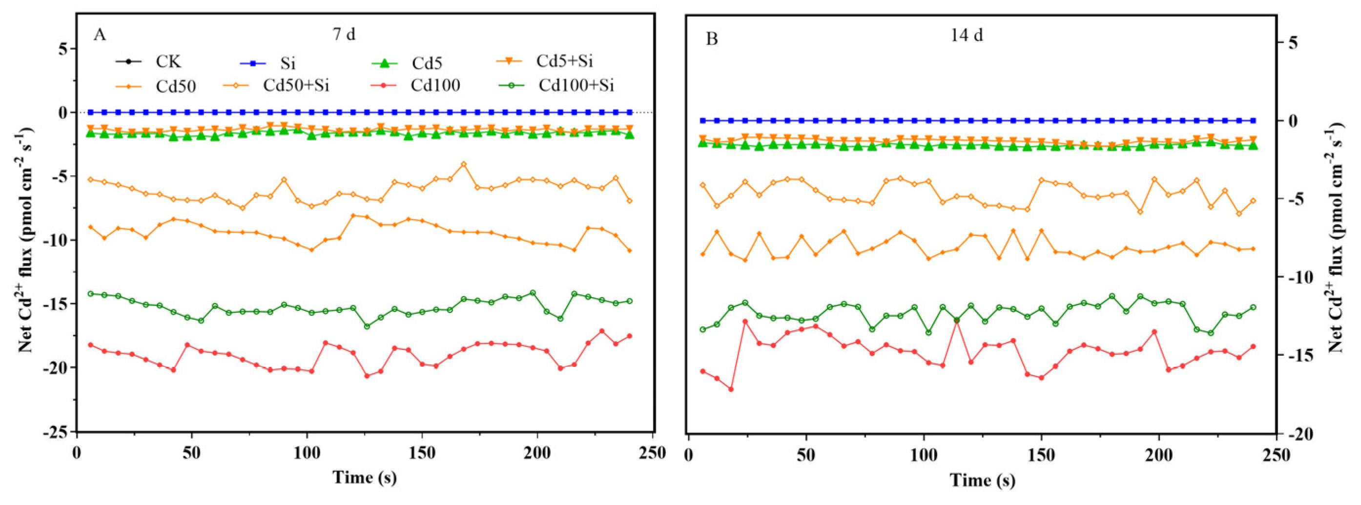
The Cd2+ influx into the cells on the surface of rice root tips measured at 7 d after treatment exhibited a concentration-dependent increase, with values ranging from 1.62 to 19.01 pmol·cm−2·s−1 across different Cd treatment levels (Figure 3 and Figure 4). By 14 d, although the influx continued to rise with increasing Cd concentrations, the rate of increase was markedly slower, particularly under high Cd exposures (e.g., 100 μmol·L−1), indicating a significant attenuation of influx capacity due to prolonged Cd stress. In the presence of Si, the flow rate was significantly reduced by 16.46–35.57% in 7 d and 17.33–42.30% in 14 d. Among all treatments, the greatest reduction occurred under Cd50 + Si. The effect of Si-mediated Cd flow rate interference was time- and Cd-concentration-dependent.
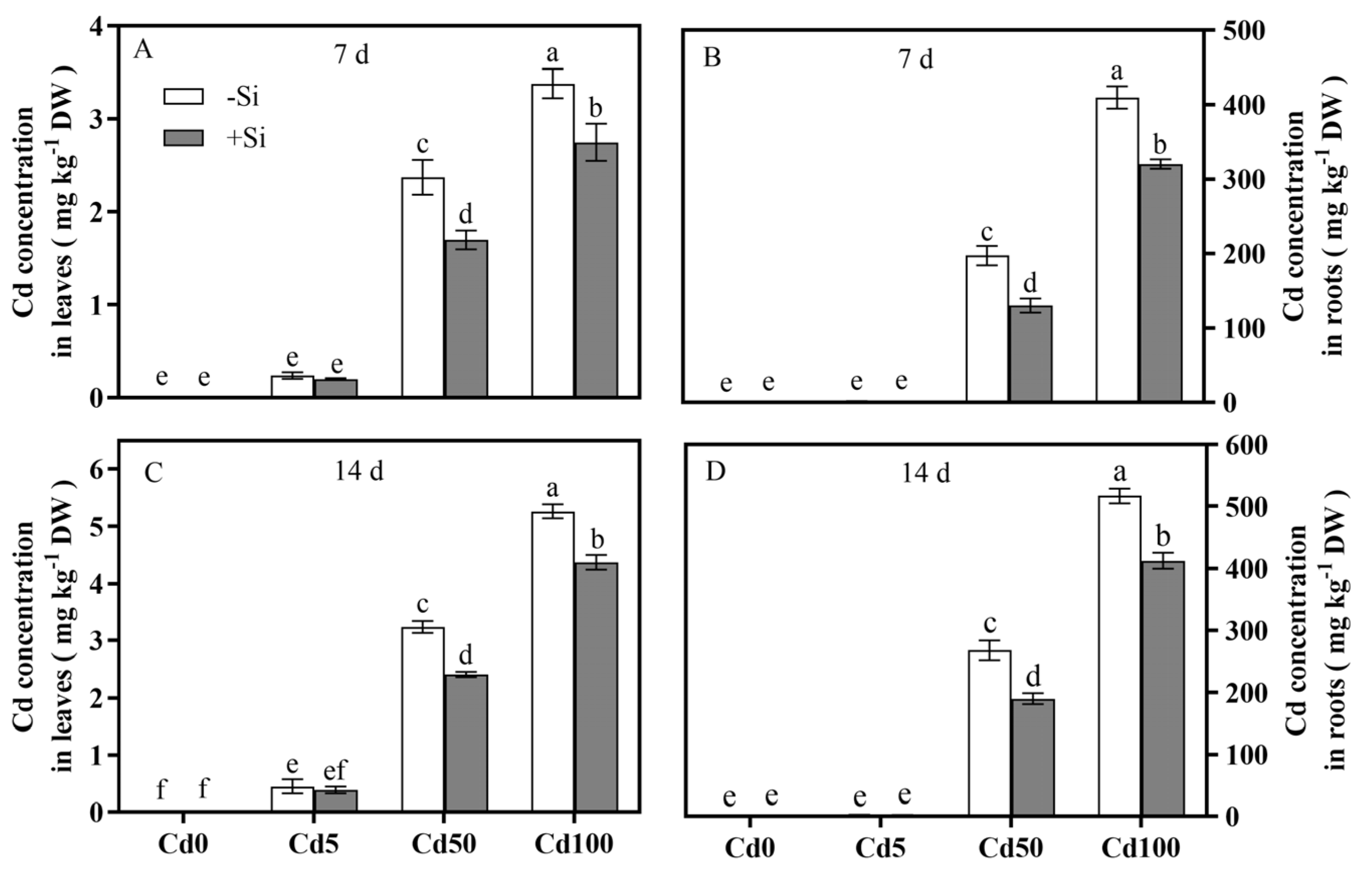
The results showed that the Cd content in plant tissues was dependent on the dosage of Cd treatment, and that high concentrations of Cd significantly promoted Cd accumulation (Figure 5). In 7–14 d of cultivation, the leaf Cd in the seedlings increased the most of all treatments by 55.62% under Cd100. The accumulation was substantially higher in the roots than in the leaves, with temporal increases of 35.9% under Cd50 and 26.23% under Cd100 treatments. Addition of Si significantly reduced the Cd uptake and translocation, as the Cd content decreased by 16.67–28.27% in 7 d and 13.33–25.62% in 14 d in the leaves, with the most significant inhibition of 28.27% under Cd50 + Si in 7 d, and in the roots, 34.03% in 7 d and 29.10% in 14 d, with the most significant inhibition of 21.81% under Cd100 + Si in 7 d and 20.20% in 14 d. A significant Cd-Si interaction (Table 1) by the addition of Si that effectively mitigated the Cd uptake in plant tissues through a spatiotemporally specific regulatory network was shown by ANOVA.
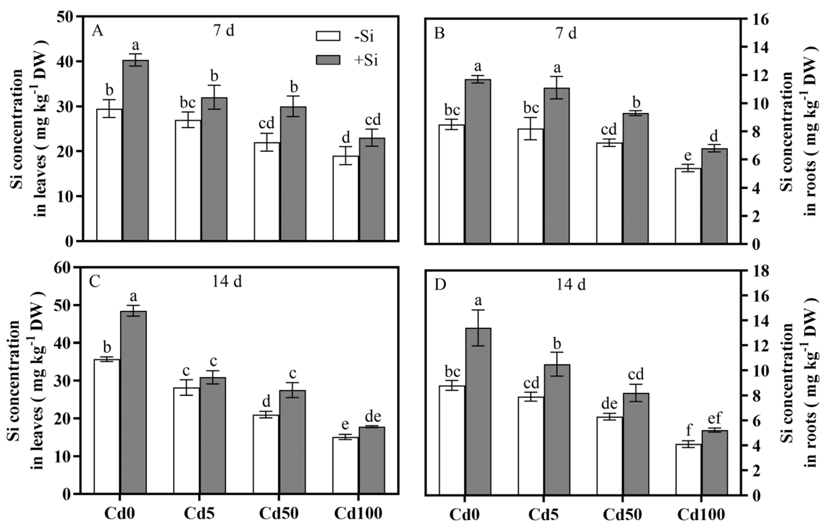
The exogenous Si significantly increased Si in the roots and leaves (Figure 6), but the presence of Cd under Cd × Si created an interaction between the two elements that hindered the Si uptake and content of the tissues (Figure 6 and Table 1). In the cases of higher dosage of Cd, e.g., Cd50 and Cd100, the hindrance intensified with exposure time. For example, under Cd100 + Si, the roots contained 36.47% less Si in 7 d and 53.41% less in 14 d, and the leaves 35.59% less in 7 d and 57.7% less in 14 d. Nonetheless, the existence of Si mitigated the Cd-induced suppression on the Si uptake by the roots, as 35.37% improvement was realized under Cd5 + Si in 7 d and 30.16% in 14 d. On the other hand, no significant effect was observed under Cd100 + Si in 14 d. Meanwhile, in the leaves, a maximal recovery of 36.36% occurred under Cd50 + Si in 7 d and 30.95% in 14 d. It seemed that the inhibition of Cd on Si accumulation in rice tissues was also concentration- and time-dependent. Unfortunately, prolonged Cd100 stress would make the suppressive effect irreversible.

3.3. Spatiotemporal Characteristics of Si-Mediated Cd Toxicity Alleviation via MDA and H2O2 Reductions in Plant Tissues

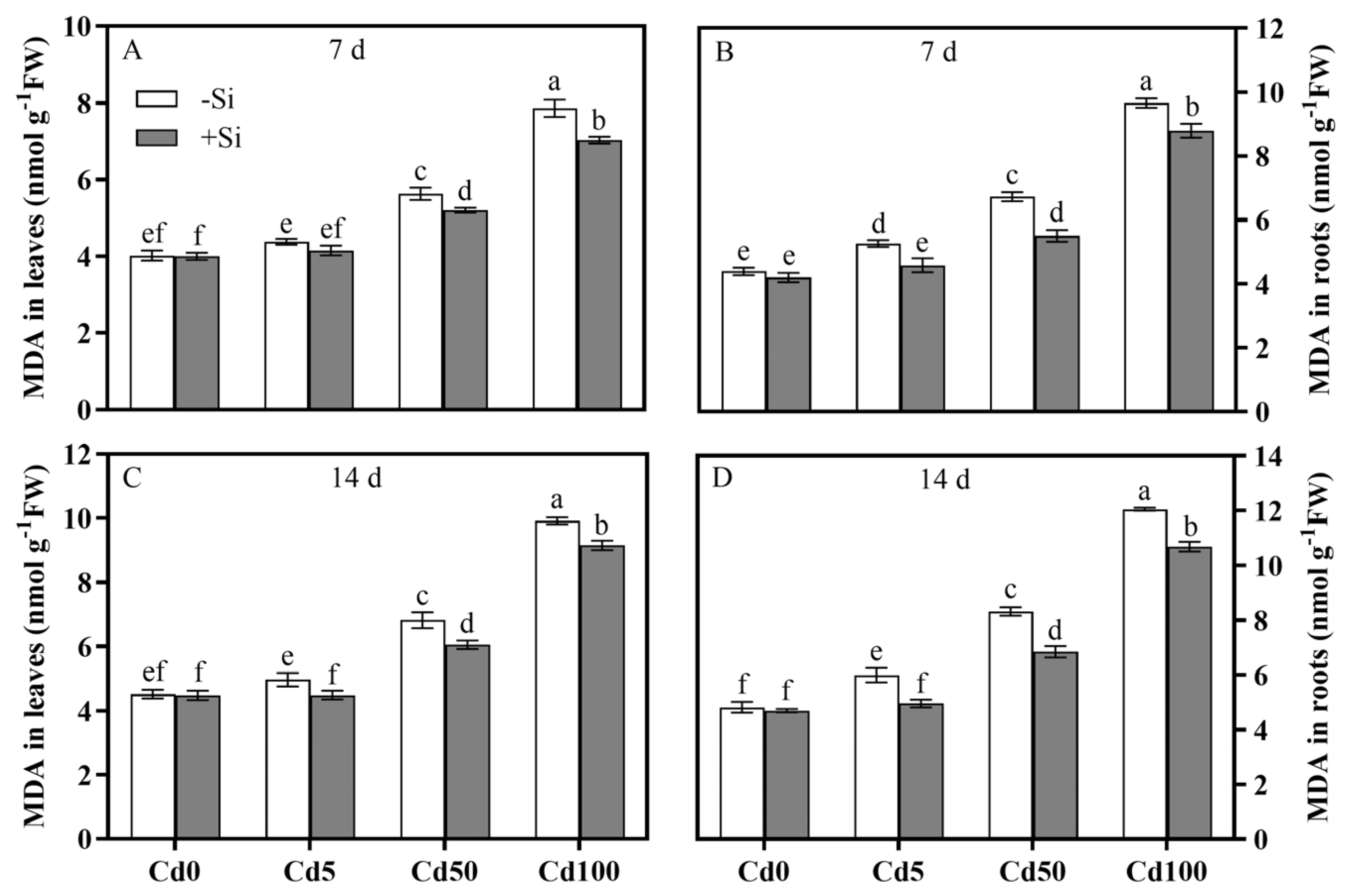
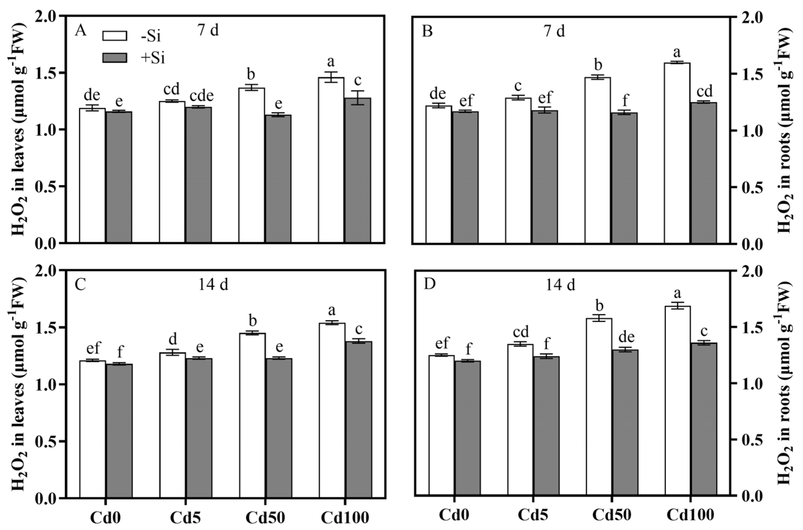
Cd exerted a significant oxidative stress on rice plants in a dose- and time-dependent manner, as shown in Figure 7; Figure 8. A 7 d exposure of the rice plants to Cd significantly elevated the markers of oxidative damage on the leaves, including MDA increases of 8.96%, 40.05%, and 95.52% and H2O2 increases of 5.04%, 15.13%, and 22.69% under Cd5, Cd50, and Cd100, respectively. The effects were more severe on the roots under Cd100, being 1.21–1.23× higher on MDA and 1.1× higher on H2O2. Significant Cd-Si interaction (Table 1) by the Si addition significantly minimized the Cd-induced oxidative damage in a concentration- and time-dependent manner. The MDA reductions in the leaves ranged from 5.25 to 10.56% with the peak decrease of 11.29% under Cd50 + Si in 14 d, and in the roots, peak decreases were 18.15% in 7 d and 17.79% in 14 d under Cd50 + Si; the H2O2 reductions in the roots increased with increasing Cd concentration and were maximized at −21.88% under Cd100 + Si in 7 d, and in the leaves, there was a unimodal response that peaked at −17.52% under Cd50 + Si. The prolonged Cd100 stress suppressed the Cd toxicity mitigation efficiency of Si, indicating the existence of a concentration- and time-dependent threshold.

3.4. Spatiotemporal Characteristics of Si-Mediated Cd Toxicity Alleviation via Antioxidant Defense System Regulation of Rice Plants

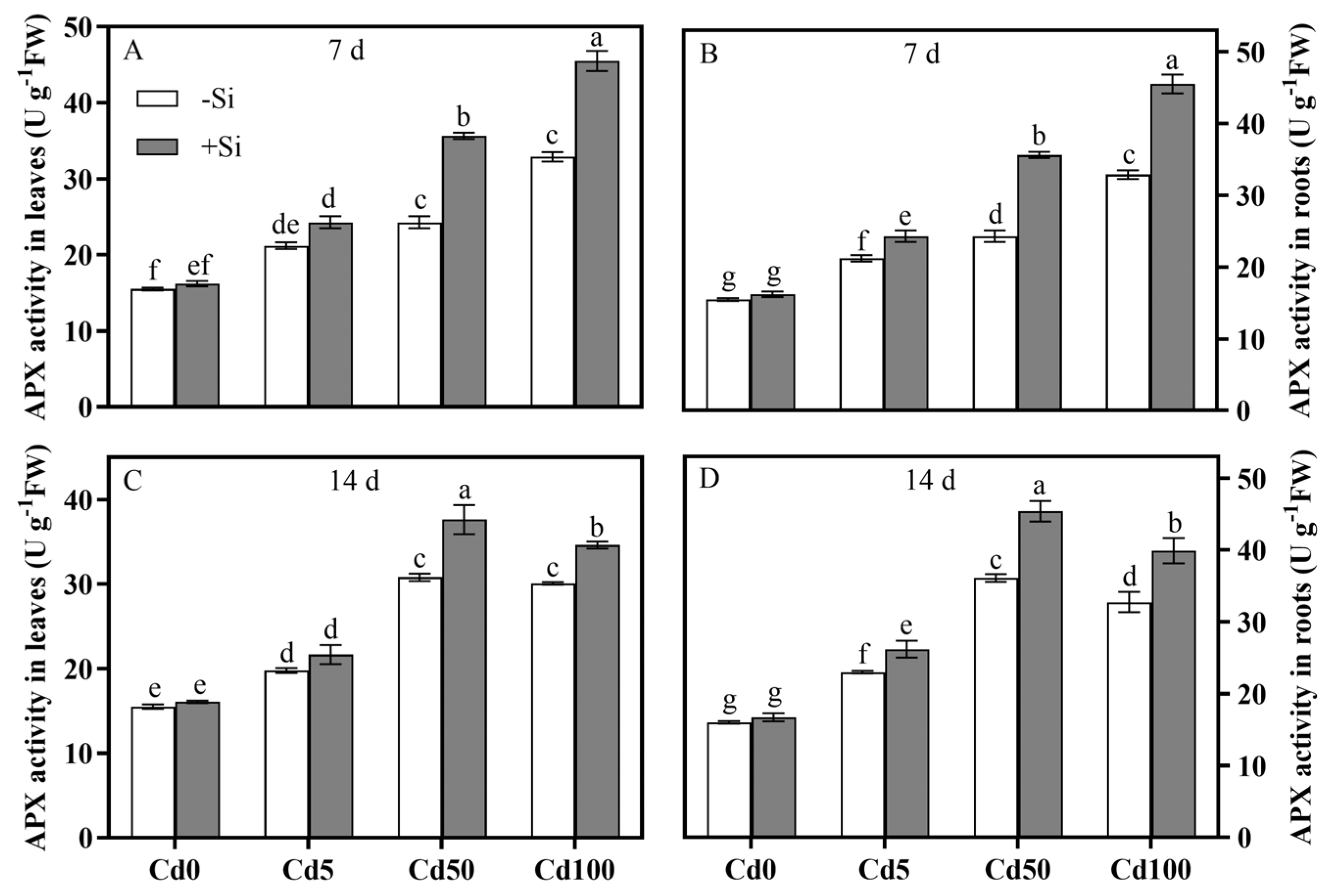
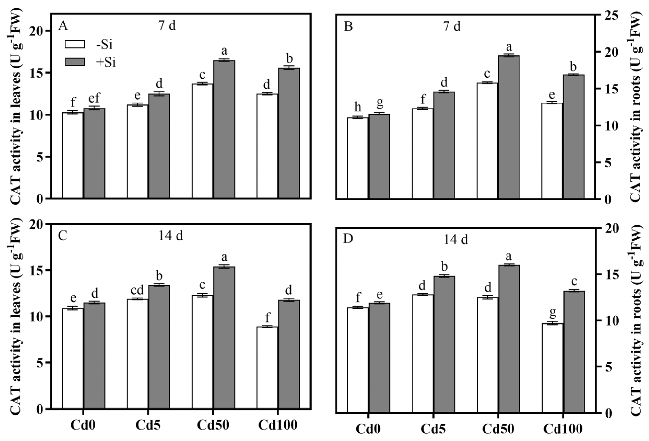
All Cd treatments significantly elevated the SOD and APX activities in the roots and leaves over the control. In 7 d of Cd exposure, the activities rose progressively with increasing Cd concentration. By the 14th day, they declined after an initial rise, but APX did not differ significantly under Cd50 and Cd100 (Figure 9 and Figure 10). This suggested that a threshold on the enzyme activity was reached when Cd stress was extended beyond 7 d. On the other hand, Si continued to positively affect SOD and APX during the same period. At 7 d, the SOD increased by 6.34–15.34% in the leaves and 12.6–20.99% in the roots, while the APX rose by 11.22–25.04% in the leaves and 14.62–38.33% in the roots. At 14 d, the maximal SOD increases of 10.08% in the leaves and 14.68% in the roots and the peak APX increases of 22.14% in the leaves and 25.76% in the roots were found under Cd50 + Si, followed by declines under higher Cd concentrations. The CAT activity increased with increasing Cd concentration but was lower under Cd100 than under Cd50 in 7 d. However, at 14 d, it dropped sharply by 18.35% in the leaves and 14.91% in the roots under Cd100. The potential risk of collapse in the plant antioxidant system loomed. Si applications would counteract and curtail the risk as shown by the CAT activity increases of 12.61–32.58% in the leaves and 15.63–36.08% in the roots at 14 d with the presence of Si (Figure 11). Furthermore, with the added Si, even though the protection was undermined by a prolonged Cd stress, significant Cd-Si interactions (Table 1) began to reduce the oxidative damage by activating relevant enzymes.
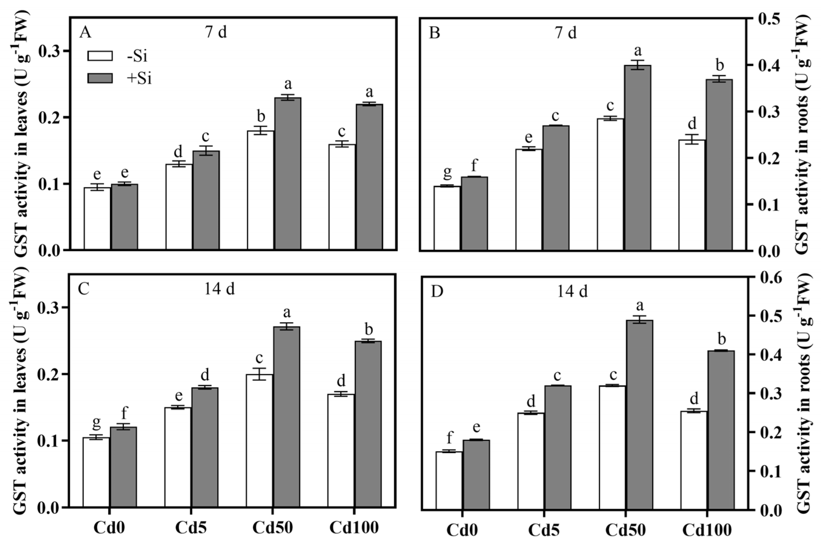
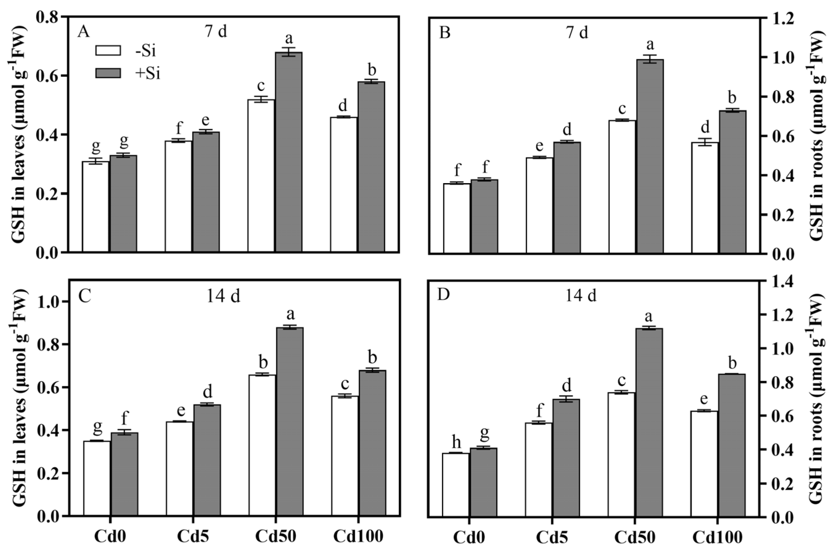
Under Cd50, the GST activity peaked with an increase of 89.47–113.33% over the control and was enhanced by Si addition continuously with increases of 20–47.06% in the leaves and 28.12–60.78% in the roots in 14 d (Figure 12). Displaying a unimodal response, the GSH content was the highest under Cd50 at 14 d, and under Cd50 + Si, the most significant 33.33% increase in the leaves and 51.35% increase in the roots appeared at 14 d (Figure 13). Apparently, Si could deliver optimal long-term protection for the rice plants when Cd concentration in the soil remained at a moderate level.

4. Discussion

4.1. Cd-Induced Photosynthetic Inhibition and Si-Mediated Alleviation

Cd stress significantly inhibits plant photosynthesis. The mechanism includes both stomatal and non-stomatal limitations [31]. Huang et al. (2019) [3] reported that the stress significantly decreases Gs and CO2 supply to the chloroplast stroma, limiting the availability of carbon assimilation substrates and ultimately lowering photosynthetic efficiency. The current study found that under the low Cd stress of 5 μmol·L−1 (Cd5), the rice plants responded by actively closing stomata to reduce Cd uptake, resulting in a decreased Gs and a limited Pn. Plants applied dominantly stomatal restriction to resist the stress, as shown by the decreased Gs without much change in Ci. When Cd stress was prolonged, non-stomatal factors gradually started to play an increasingly dominant role. The photosynthetic inhibition exerted by the moderate and severe Cd stress in this study manifested in reduced Pn, Gs, and Rubisco activity with a significantly elevated Ci. Inhibited non-stomatal Rubisco function could impair the carbon fixation in the Calvin cycle. Recent studies have reached a similar conclusion in barley [32] and cotton [33]. Therefore, it was the triad conditions of a lowered Pn, an accentuated Ci, and suppressed Rubisco activity that showcased a damaged enzyme system and disrupted electron transport chain, the culprit of compromised photosynthesis by the invasion of Cd in a rice plant.
Si can mitigate the ultrastructure damage caused by Cd stress on rice chloroplasts [34]. The present study also found that exogenous Si significantly, and most stably under moderate Cd50, alleviated the Cd-induced photosynthesis inhibition in the rice plants, which was attributed to either an improved stomatal function or a protective photosynthetic apparatus as the primary contributor. The alleviation effect of Si showed distinct concentration and time dependence, as well as tissue-specific mechanisms: in roots, Si primarily reduces Cd translocation to shoots by forming Si-Cd complexes; in leaves, it enhances antioxidant capacity and maintains photosystem integrity to mitigate oxidative damage. Specifically, Si exerts a dual mechanism: it reduces Cd uptake at the root level and enhances ROS scavenging in leaves, thereby mitigating direct Cd toxicity while protecting the photosynthetic apparatus from oxidative damage. This coordinated action enables the simultaneous enhancement of antioxidant enzyme activities and Pn, demonstrating Si’s role as an effective plant stress regulator with significant potential for application in Cd-contaminated agricultural systems. The study by Rizwan et al. (2019) [35] demonstrated a similar observation, showing that the foliar application of nSiO on rice leaves progressively increased Pn and Gs under Cd stress. Distinctly, the effect shown in this study was concentration- and time-dependent, since the changes in Gs and Pn intensified under prolonged low and moderate Cd stress but became statistically insignificant by 14 d under Cd100. This suggests a potential threshold effect of Si-mediated remediation. An irreversible disruption in the root cells might have occurred when Cd concentration exceeded the capacity of Si to perform a successful repair. Moreover, the Ci, which was significantly elevated under Cd50 or Cd100, was not affected at all by Si because Si regulates stomatal aperture, not CO2 diffusion. As a key enzyme involved in photosynthetic carbon assimilation, Rubisco directly affects Pn. The present study found that Rubisco activity was significantly inhibited by Cd stress, which could be partially restored by Si addition, particularly under a low to moderate Cd concentration. This paralleled the Pn changes and further confirmed that Si sustained photosynthetic efficiency by protecting the enzyme system and/or reducing Cd in the leaves. Nevertheless, an escalated Cd stress could lead to oxidative damage or protein denaturation with a permanent loss of the stimulatory effect of Si on Rubisco.
In summary, Si application could significantly alleviate the ill effects caused by low to moderate Cd stress on the photosynthesis of rice plants by means of improving stomatal conductance and Rubisco activity, but the efficacy diminished if the Cd concentration was too high or the exposure lasted too long.

4.2. Effects of Si on Cd Absorption and Translocation

Using NMT and spatiotemporal dynamic analysis on rice tissues, this study found that it was through a spatiotemporal-dependent regulatory mechanism that Si was capable of inhibiting Cd-ion influx and optimizing the Cd-Si distribution in the root cells of a rice plant. Specifically, the Cd influx kinetics was biphasic concentration- and time-dependent; the flow rate rose with an increased Cd concentration from 0 to 100 μmol·L−1, approaching a saturation point in the biomolecular binding sites in the roots of a plant, and then was impeded as the stress lasted from 7 d to 14 d. The inhibitory effect of Si on Cd influx was significantly spatiotemporally specific, as it was significantly greater on the 14th than the 7th day under Cd5 or Cd50, peaked under Cd50, and significantly declined under Cd100.
As reported by Yamaji et al. (2015) [36], variations in Si accumulation in rice are largely determined by the specific Si transporters, such as Lsi1. Building on previous experimental evidence that the overexpression or RNAi of Lsi1 significantly alters the Cd absorption in transgenic rice [16], Lsi1 per se can directly or indirectly affect Cd transport as well [37]. Closely associated with downregulated Cd absorption- and transport-related genes (e.g., Nramp1/5 and HMA3) [22,38], Cd alleviation by Si in rice might also be accomplished by means of Si mediating competitive inhibition through Lsi1.
The current study found that Cd accumulation in rice plants was organ-specific (roots > leaves) and time-dependent (14 d > 7 d). As the initial entry point and acceptor of Cd translocating from soil into a plant, roots naturally receive more of the pollutant than do the leaves. Acting as a barrier, this accumulation is simply a self-protection strategy of a plant [39]. As shown in this study, under prolonged stress, Cd increased significantly more in the leaves (i.e., 55.62%) than in the roots (i.e., 26.23–35.9%). Thus, a long-lasting exposure would allow ample time for Cd to fill the chelating capacity or activate the xylem transport mechanisms of a plant [40]. Elevated Cd accumulation in leaves heightens their susceptibility to metal-induced oxidative stress. Nevertheless, leaves exhibit a constitutively stronger antioxidant defense system than roots. Despite a comparatively smaller increase in antioxidant activity, their superior baseline capacity effectively counteracts Cd toxicity via enhanced production of glutathione and other antioxidative metabolites, thus sustaining redox homeostasis under prolonged stress [13]. In addition to competing for binding sites and absorption, Si also significantly redistributed the Cd in the spatiotemporally specific regulatory networks, providing another crucial venue for the toxicity alleviation in plants. Besides being a physical barrier, Si-induced cell wall silicification was theorized to preoccupy some binding sites in the root molecules and reduce the Cd influx by co-precipitation or competitive adsorption [41]. The significant Si accumulation in the roots observed in this study (Figure 6) seemed to support the hypothesis. The inhibitory effect of Si on Cd influx was slightly hampered by prolonged stress, as shown by a decline from 34.03% to 29.10% under Cd50 + Si. This was postulated to be due to the impaired Si metabolic pathways under high Cd concentration, e.g., 100 μmol·L−1. Figure 6 illustrates the absence of any significant recovery of Si intake under Cd100 + Si in 14 d. The high Cd level caused a 57.7% Si reduction in the leaves in 14 d, which was significantly greater than that induced by low Cd concentrations. The irreversible effect could be the result of damage to certain functions of the silicic acid transporter (e.g., Lsi1 and Lsi2) [42]. The Si application yielded optimal Cd mitigation under Cd50 with a 30.95% increase in leaf Si. But it provided only a limited remediation under Cd100. The asymmetric outcomes might stem from the disruption of the stoichiometric balance between Si and Cd in competing for plant absorption initiated by high Cd concentrations. Therefore, in applying Si for rice farming, the heavy metal pollution thresholds should be considered.

4.3. Effects of Cd and Si on Antioxidant System and Glutathione Metabolism of Rice Plants

In plant cells, ROS, such as O2−·, ·OH, and H2O2, are kept at low levels under homeostatic conditions to cause negligible physiological effects. Excessive Cd disrupts the equilibrium, leading to oxidative damage to DNA, RNA, proteins, and membrane lipids [43]. This study found that the Cd-induced oxidative stress on the rice plants was distinctly dose- and time-dependent, with significant increases in MDA and H2O2, and that the damage tended to be more severe in the roots than in the leaves. The observation was consistent with previous findings attributed to the tissue-specific Cd concentration and accumulation rate variations that resulted in differential responses in tissues [44]. Si application has been shown to effectively mitigate the adverse effects of Cd on plants, such as cotton and bitter gourd, by reducing MDA and H2O2 [45,46]. In Lemont rice, the present study demonstrated the intriguing roles of Si in alleviating Cd stress, reducing MDA, being significantly tissue-specific (i.e., root vs. leaf) as well as time- and concentration-dependent (i.e., significant leaf MDA reduction of 11.29% in 14 d under Cd50 vs. diminished protective effect under Cd100), and limiting ROS scavenging capacity. The efficacy of Si in mitigating Cd toxicity was directly determined by the Cd intensity, tissue type, and stress duration a rice plant encountered. The decomposition of H2O2 in the process was presumably mediated by Si with the activation of antioxidant enzymes [47].
Plants have evolved a defense system that comprises antioxidant enzymes (e.g., SOD, APX, and CAT) and non-enzymatic antioxidants (e.g., GSH, ascorbic acid, etc.) to maintain a normal cellular redox homeostasis to prevent excessive accumulation of ROS [48]. The present study found that the activities of SOD, APX, and CAT in rice were stimulated under 7 d of Cd exposure, likely as an adaptive response to counteract the initial burst of ROS. However, these activities were significantly suppressed under Cd100 at 14 d. This suppression may be attributed to the overproduction of ROS exceeding the detoxification capacity of the antioxidants, leading to irreversible oxidative damage to the enzymes and disruption of their structural integrity. For instance, the leaf CAT activity declined 18.35% under prolonged high Cd stress that induced oxidative damage in cells with a structurally compromised antioxidant system. Si might achieve the enhancement of antioxidant enzyme activities and reduction in oxidative stress effects by (1) preventing Cd from binding to the active sites on SOD/APX/CAT and (2) upregulating the expression levels of antioxidant enzyme genes. Similar observations were reported in wheat [49]. But the contrasting result by Liu et al. (2013) [50] in Solanum nigrum L. showed an activity reduction under Cd stress. The discrepancies between the two reports could be due to the differences in the test species, treatment durations, and/or experimental conditions. The stimulatory effects of Si depended on the stress intensity and duration a plant was encountering. In the current study on Lemont rice, it was most pronounced and stable under moderate Cd stress. The APX activity in the leaves rose by 22.14% after 14 d of the treatment, paralleling the changes in the photosynthetic and growth indicators, whereas prolonged exposure to Cd100 led to a continually declining CAT activity despite Si application. It was postulated that when oxidative damage to the tissues exceeded a threshold, the protective capacity of Si was jeopardized.
Serving as a precursor of phytochelatins (PCs) as well as a crucial antioxidant in plant cells, GSH plays a pivotal role in Cd detoxification [51]. This study found that, being the key enzyme catalyzing GSH-Cd conjugation, GST was most highly activated with a 103.57–113.33% increase in the roots under Cd50. In the presence of Si, it further reached 60.78% elevation in 14 d. Similar findings for wheat leaves were reported by Debona et al. (2014) [52]. Gao et al. (2016) [53] revealed that GSH facilitates Cd transport to plastids through GST-mediated chelation but restricts the influx to the shoots. In this study, Si concurrently raised the GSH content with a 51.35% increase in the Cd50-treated roots in 14 d. This suggested that Si participation promoted the compartmentalization of Cd into vacuoles or apoplasts via the GSH-PCs synthesis pathway. Under Cd100, however, the GSH/GST response was significantly attenuated, possibly due to (1) depletion of the glutathione pool, (2) saturation of GST activity, and/or (3) collapsed redox homeostasis.
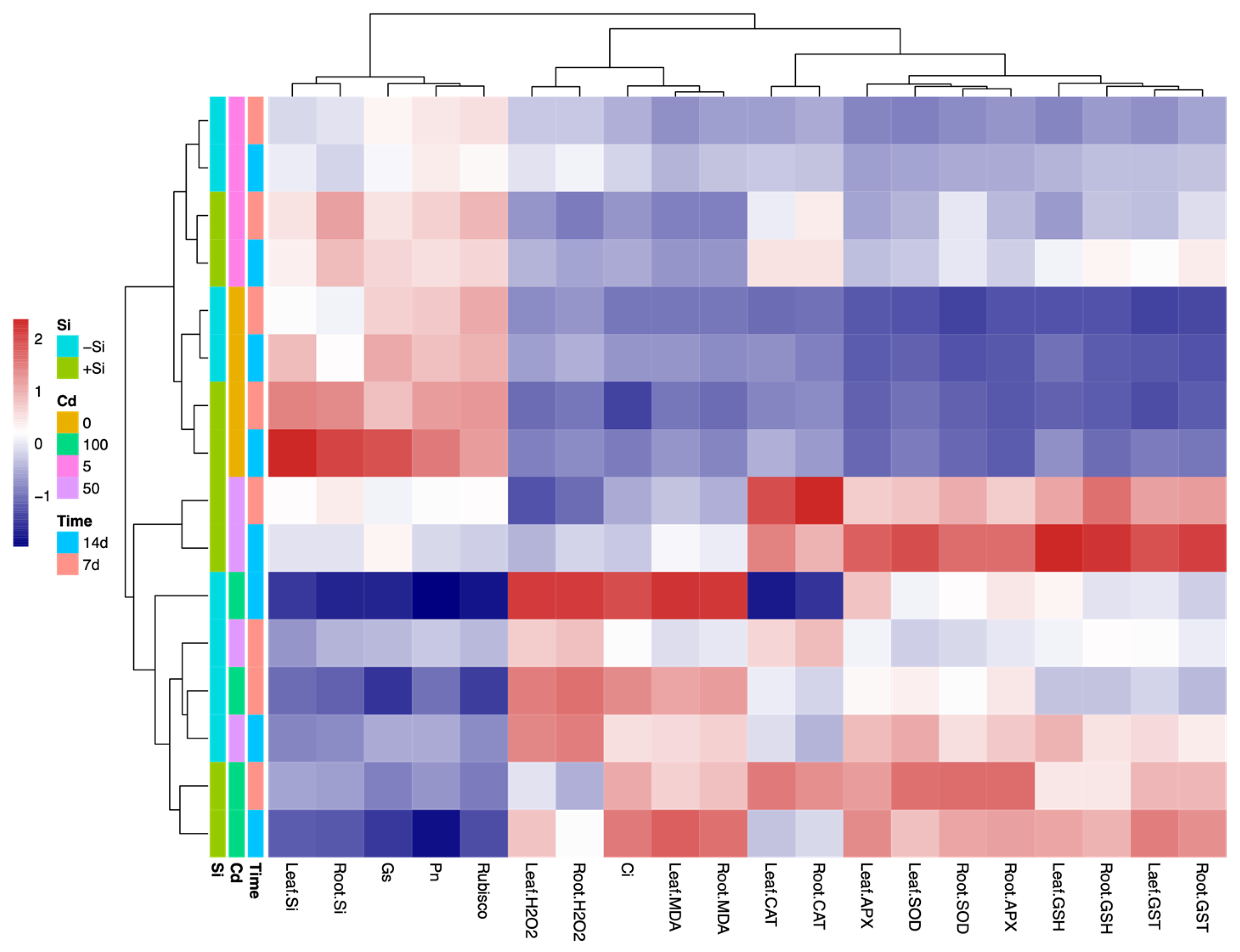
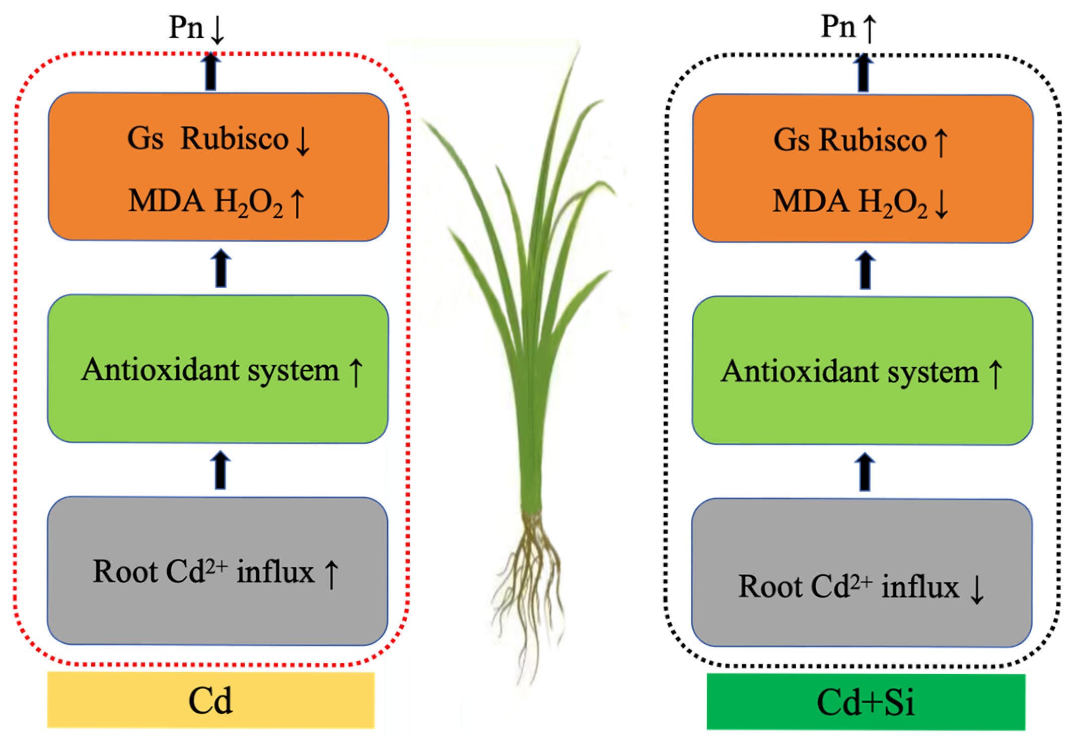
Finally, a summary and graphical model were developed to illustrate the mechanistic pathway by which Si alleviates cadmium stress in rice (Figure 14 and Figure 15). The mechanism highlights that Si markedly inhibits Cd2+ influx into rice roots, enhances photosynthetic performance, reduces oxidative damage (evidenced by lower levels of MDA and H2O2), and ultimately sustains photosynthetic efficiency under Cd stress.

5. Conclusions

This study systematically elucidates the physiological mechanisms by which Si alleviates Cd toxicity in rice. The main conclusions are as follows: Si employs a spatiotemporally specific regulatory mechanism to inhibit Cd2+ influx into root cells and optimizes Cd-Si tissue distribution, with the most pronounced effects observed under Cd50 and 14 d of treatment, while higher Cd concentrations (Cd100) or prolonged stress diminishes its efficacy. In terms of photosynthesis, Si significantly enhances Pn and Gs by improving stomatal function, protecting photosystem structure, and restoring Rubisco activity. It mitigates both direct Cd toxicity and oxidative damage through a root–leaf synergistic mechanism—reducing absorption in roots and scavenging ROS in leaves. Regarding antioxidation, Si effectively maintains redox homeostasis by boosting the activities of SOD, APX, and CAT, as well as promoting GSH synthesis and GST-mediated Cd compartmentalization. However, the alleviating effect of Si exhibits concentration and time dependence; under high Cd or prolonged stress, its efficacy significantly declines due to impaired Si metabolic pathways and saturated antioxidant systems. In summary, Si mitigates Cd toxicity through multi-pathway synergism—“reducing absorption, enhancing antioxidation, and repairing photosynthetic systems”—with its practical effectiveness depending on environmental Cd pollution levels and exposure duration. This study provides a theoretical foundation for the safe application of Si in Cd-contaminated farmland.

Author Contributions

Conceptualization, H.L., S.C. and S.J.; methodology, H.L., M.J., S.C. and S.J.; software, H.L. and S.J.; validation, H.L., S.C. and S.J.; formal analysis, H.L., S.C. and S.J.; investigation, H.L.; resources, H.L. and S.C.; data curation, H.L., S.C. and S.J.; writing—original draft preparation, H.L., S.C., M.J. and S.J.; writing—review and editing, H.L., S.C., M.J. and S.J.; visualization, H.L.; supervision, S.C. and S.J.; project administration, H.L., S.C., M.J. and S.J.; funding acquisition, S.C. and S.J. All authors have read and agreed to the published version of the manuscript.

Funding

This research was funded by the National Natural Science Foundation of China (No. 32001109), the Natural Science Foundation of Fujian Province, China (No. 2023J011577, 2025J011270), the Fujian Provincial Social Science Foundation (FJ2022B153), and the Horizontal Project (No.2025MHX168).

Data Availability Statement

The data presented in this study are available on request from the corresponding author.

Conflicts of Interest

The authors declare no conflicts of interest.

References

  1. Wang, P.; Chen, H.; Kopittke, P.M.; Zhao, F.J. Cadmium contamination in agricultural soils of China and the impact on food safety. Environ. Pollut. 2019, 249, 1038–1048. [Google Scholar] [CrossRef] [PubMed]
  2. Zhang, H.; Reynolds, M. Cadmium exposure in living organisms: A short review. Sci. Total Environ. 2019, 678, 761–767. [Google Scholar] [CrossRef] [PubMed]
  3. Huang, Y.; Xi, Y.; Gan, L.; Johnson, D.; Wu, Y.; Ren, D.; Liu, H. Effects of lead and cadmium on photosynthesis in Amaranthus spinosus and assessment of phytoremediation potential. Int. J. Phytoremediat. 2019, 21, 1041–1049. [Google Scholar] [CrossRef]
  4. Tran, T.A.; Popova, L.P. Functions and toxicity of cadmium in plants: Recent advances and future prospects. Turk. J. Bot. 2013, 37, 1–3. [Google Scholar] [CrossRef]
  5. Shaari, N.E.M.; Tajudin, M.T.F.M.; Khandaker, M.M.; Majrashi, A.; Alenazi, M.M.; Abdullahi, U.A.; Mohd, K.S. Cadmium toxicity symptoms and uptake mechanism in plants: A review. Braz. J. Biol. 2022, 84, e252143. [Google Scholar] [CrossRef]
  6. Hu, Y.; Wang, H.; Jia, H.; Peng, M.; Zhu, T.; Liu, Y.; Wei, J. Effects of Cd treatment on morphology, chlorophyll content and antioxidant enzyme activity of Elymus nutans Griseb., a native plant in Qinghai-Tibet Plateau. Plant Signal. Behav. 2023, 18, 2187561. [Google Scholar] [CrossRef]
  7. Wu, F.; Fan, J.; Ye, X.; Yang, L.; Hu, R.; Ma, J.; Ma, S.; Li, D.; Zhou, J.; Nie, G.; et al. Unraveling cadmium toxicity in Trifolium repens L. seedling: Insight into regulatory mechanisms using comparative transcriptomics combined with physiological analyses. Int. J. Mol. Sci. 2022, 23, 4612. [Google Scholar] [CrossRef]
  8. Ji, X.; Liu, S.; Juan, H.; Bocharnikova, E.A.; Matichenkov, V.V. Effect of silicon fertilizers on cadmium in rice (Oryza sativa) tissue at tillering stage. Environ. Sci. Pollut. Res. 2017, 24, 10740–10748. [Google Scholar] [CrossRef]
  9. Wu, J.; Mock, H.P.; Giehl, R.F.H.; Pitann, B.; Mühling, K.H. Silicon decreases cadmium concentrations by modulating root endodermal suberin development in wheat plants. J. Hazard. Mater. 2019, 364, 581–590. [Google Scholar] [CrossRef]
  10. Kollárová, K.; Kusá, Z.; Vatehová-Vivodová, Z.; Lišková, D. The response of maize protoplasts to cadmium stress mitigated by silicon. Ecotoxicol. Environ. Saf. 2019, 170, 488–494. [Google Scholar] [CrossRef]
  11. Hou, L.; Ji, S.; Zhang, Y.; Wu, X.; Zhang, L.; Liu, P. The mechanism of silicon on alleviating cadmium toxicity in plants: A review. Front. Plant Sci. 2023, 14, 1141138. [Google Scholar] [CrossRef]
  12. Song, A.; Li, Z.; Zhang, J.; Xue, G.; Fan, F.; Liang, Y. Silicon-enhanced resistance to cadmium toxicity in Brassica chinensis L. is attributed to Si-suppressed cadmium uptake and transport and Si-enhanced antioxidant defense capacity. J. Hazard. Mater. 2009, 172, 74–83. [Google Scholar] [CrossRef]
  13. Chen, H.; Huang, X.; Chen, H.; Zhang, S.; Fan, C.; Fu, T.; He, T.; Gao, Z. Effect of silicon spraying on rice photosynthesis and antioxidant defense system on cadmium accumulation. Sci. Rep. 2024, 14, 15265. [Google Scholar] [CrossRef]
  14. Liu, J.; Ma, J.; He, C.; Li, X.; Zhang, W.; Xu, F.; Lin, Y.; Wang, L. Inhibition of cadmium ion uptake in rice (Oryza sativa) cells by a wall-bound form of silicon. New Phytol. 2013, 200, 691–699. [Google Scholar] [CrossRef]
  15. Lin, H.M.; Fang, C.X.; Li, Y.Z.; Lin, W.W.; He, J.Y.; Lin, R.Y.; Lin, W.X. Effect of silicon on grain yield of rice under cadmium-stress. Acta Physiol. Plant. 2016, 38, 186. [Google Scholar] [CrossRef]
  16. Lin, H.M.; He, J.Y.; Lin, W.W.; Li, Y.Z.; Fang, C.X.; Lin, W.X. Lsi1-regulated Cd uptake and phytohormones accumulation in rice seedlings in presence of Si. Plant Growth Regul. 2018, 86, 149–157. [Google Scholar] [CrossRef]
  17. Han, M.; Yang, H.; Yu, G.; Jiang, P.; You, S.; Zhang, L.; Lin, H.; Liu, J.; Shu, Y. Application of Non-invasive Micro-test Technology (NMT) in environmental fields: A comprehensive review. Ecotoxicol. Environ. Saf. 2022, 240, 113706. [Google Scholar] [CrossRef] [PubMed]
  18. Sun, J.; Wang, R.; Liu, Z.; Ding, Y.; Li, T. Non-invasive microelectrode cadmium flux measurements reveal the spatial characteristics and real-time kinetics of cadmium transport in hyperaccumulator and nonhyperaccumulator ecotypes of Sedum alfredii. J. Plant Physiol. 2013, 170, 355–359. [Google Scholar] [CrossRef]
  19. Yoshida, S.; Forno, D.A.; Cock, J.H.; Gomez, K.A. Laboratory Manual for Physiological Studies of Rice; The International Rice Research Institute: Manila, Philippines, 1976. [Google Scholar]
  20. Adrees, M.; Ali, S.; Rizwan, M.; Zia-Ur-Rehman, M.; Ibrahim, M.; Abbas, F.; Farid, M.; Qayyum, M.F.; Irshad, M.K. Mechanisms of silicon-mediated alleviation of heavy metal toxicity in plants: A review. Ecotoxicol. Environ. Saf. 2015, 119, 186–197. [Google Scholar] [CrossRef] [PubMed]
  21. Shi, X.; Zhang, C.; Wang, H.; Zhang, F. Effect of Si on the distribution of Cd in rice seedlings. Plant Soil 2005, 272, 53–60. [Google Scholar] [CrossRef]
  22. Lin, H.M.; Fang, C.X.; Li, Y.Z.; Lin, W.W.; He, J.Y.; Lin, R.Y.; Lin, W.X. Cadmium-stress mitigation through gene expression of rice and silicon addition. Plant Growth Regul. 2017, 81, 91–101. [Google Scholar] [CrossRef]
  23. Sulpice, R.; Tschoep, H.; Von Korff, M.; Büssis, D.; Usadel, B.; HÖHne, M.; Witucka-Wall, H.; Altmann, T.; Stitt, M.; Gibon, Y. Description and applications of a rapid and sensitive non-radioactive microplate-based assay for maximum and initial activity of D-ribulose-1,5-bisphosphate carboxylase/oxygenase. Plant Cell Environ. 2007, 30, 1163–1175. [Google Scholar] [CrossRef]
  24. Heath, R.L.; Packer, L. Photoperoxidation in isolated chloroplasts: I. Kinetics and stoichiometry of fatty acid peroxidation. Arch. Biochem. Biophys. 1968, 125, 189–198. [Google Scholar] [CrossRef]
  25. Lin, C.C.; Kao, C.H. Effect of NaCl stress on H2O2 metabolism in rice leaves. Plant Growth Regul. 2000, 30, 151–155. [Google Scholar] [CrossRef]
  26. Giannopolitis, C.N.; Ries, S.K. Superoxide dismutases: I. Occurrence in higher plants. Plant Physiol. 1977, 59, 309–314. [Google Scholar] [CrossRef]
  27. Aebi, H. Catalase in vitro. In Method in Enzymology; Academic Press: Cambridge, MA, USA, 1984; Volume 105, pp. 121–126. [Google Scholar] [CrossRef]
  28. Nakano, Y.; Asada, K. Hydrogen peroxide is scavenged by ascorbate-specific peroxidase in spinach chloroplasts. Plant Cell Physiol. 1981, 22, 867–880. [Google Scholar] [CrossRef]
  29. Jakoby, W.B. The glutathione S-transferases a group of multifunctional detoxification proteins. Adv. Enzymol. Relat. Areas Mol. Biol. 1978, 46, 383–414. [Google Scholar] [CrossRef] [PubMed]
  30. Law, M.Y.; Charles, S.A.; Halliwell, B. Glutathione and ascorbic acid in spinach (Spinacia oleracea) chloroplasts. The effect of hydrogen peroxide and paraquat. Biochem. J. 1983, 210, 899–903. [Google Scholar] [CrossRef]
  31. Song, J.; Sun, Z.; Saud, S.; Fahad, S.; Nawaz, T. Exploring the deleterious effects of heavy metal cadmium on antioxidant defense and photosynthetic pathways in higher plants. Plant Stress 2025, 15, 100716. [Google Scholar] [CrossRef]
  32. He, S.; Lian, X.; Zhang, B.; Liu, X.; Yu, J.; Gao, Y.; Zhang, Q.; Sun, H. Nano silicon dioxide reduces cadmium uptake, regulates nutritional homeostasis and antioxidative enzyme system in barley seedlings (Hordeum vulgare L.) under cadmium stress. Environ. Sci. Pollut. Res. 2023, 30, 67552–67564. [Google Scholar] [CrossRef]
  33. An, M.; Wang, H.; Fan, H.; Ippolito, J.A.; Meng, Y.E.; Li, Y.; Wang, K.; Wei, C. Effects of modifiers on the growth, photosynthesis, and antioxidant enzymes of cotton under cadmium toxicity. J. Plant Growth Regul. 2019, 38, 1196–1205. [Google Scholar] [CrossRef]
  34. Tripathi, D.K.; Singh, V.P.; Kumar, D.; Chauhan, D.K. Rice seedlings under cadmium stress: Effect of silicon on growth, cadmium uptake, oxidative stress, antioxidant capacity and root and leaf structures. Chem. Ecol. 2012, 28, 281–291. [Google Scholar] [CrossRef]
  35. Rizwan, M.; Ali, S.; ur Rehman, M.Z.; Malik, S.; Adrees, M.; Qayyum, M.F.; Alamri, S.A.; Alyemeni, M.N.; Ahmad, P. Effect of foliar applications of silicon and titanium dioxide nanoparticles on growth, oxidative stress, and cadmium accumulation by rice (Oryza sativa). Acta Physiol. Plant. 2019, 41, 35. [Google Scholar] [CrossRef]
  36. Yamaji, N.; Sakurai, G.; Mitani-Ueno, N.; Ma, J.F. Orchestration of three transporters and distinct vascular structures in node for intervascular transfer of silicon in rice. Proc. Natl. Acad. Sci. USA 2015, 112, 11401–11406. [Google Scholar] [CrossRef] [PubMed]
  37. Ma, J.F.; Yamaji, N. Silicon uptake and accumulation in higher plants. Trends Plant Sci. 2006, 11, 392–397. [Google Scholar] [CrossRef]
  38. Kumar, K. Plant Metal and Metalloid Transporters; Srivastava, S., Ed.; Springer: Berlin/Heidelberg, Germany, 2022. [Google Scholar]
  39. Clemens, S.; Aarts, M.G.M.; Thomine, S.; Verbruggen, N. Plant science: The key to preventing slow cadmium poisoning. Trends Plant Sci. 2013, 18, 92–99. [Google Scholar] [CrossRef]
  40. Uraguchi, S.; Fujiwara, T. Cadmium transport and tolerance in rice perspectives for reducing grain cadmium accumulation. Rice 2012, 5, 5. [Google Scholar] [CrossRef]
  41. Wu, J.W.; Shi, Y.; Zhu, Y.X.; Wang, Y.C.; Gong, H.J. Mechanisms of enhanced heavy metal tolerance in plants by silicon: A review. Pedosphere 2013, 23, 815–825. [Google Scholar] [CrossRef]
  42. Ma, J.F.; Yamaji, N.; Tamai, K.; Mitani, N. Genotypic difference in silicon uptake and expression of silicon transporter genes in rice. Plant Physiol. 2007, 145, 919–924. [Google Scholar] [CrossRef]
  43. Luo, P.; Wu, J.; Li, T.T.; Shi, P.; Ma, Q.; Di, D.W. An overview of the mechanisms through which plants regulate ROS homeostasis under cadmium stress. Antioxidants 2024, 13, 1174. [Google Scholar] [CrossRef]
  44. He, J.; Qin, J.; Long, L.; Ma, Y.; Li, H.; Li, K.; Jiang, X.; Liu, T.; Polle, A.; Liang, Z.; et al. Net cadmium flux and accumulation reveal tissue-specific oxidative stress and detoxification in Populus × canescens. Physiol. Plantarum 2011, 143, 50–63. [Google Scholar] [CrossRef]
  45. Farooq, M.A.; Ali, S.; Hameed, A.; Ishaque, W.; Mahmood, K.; Iqbal, Z. Alleviation of cadmium toxicity by silicon is related to elevated photosynthesis, antioxidant enzymes; suppressed cadmium uptake and oxidative stress in cotton. Ecotoxicol. Environ. Saf. 2013, 96, 242–249. [Google Scholar] [CrossRef]
  46. Sun, H.; He, S.; Liu, T.; Zhang, Q.; Yu, J.; Gao, Y.; Wang, X. Alleviation of cadmium toxicity by nano-silicon dioxide in Momordica charantia L. Seedlings. J. Soil Sci. Plant Nutr. 2023, 23, 1060–1069. [Google Scholar] [CrossRef]
  47. Mišúthová, A.; Slováková, Ľ.; Kollárová, K.; Vaculík, M. Effect of silicon on root growth, ionomics and antioxidant performance of maize roots exposed to As toxicity. Plant Physiol. Biochem. 2021, 168, 155–166. [Google Scholar] [CrossRef] [PubMed]
  48. Marques, D.N.; Amaral Carvalho, M.E.; Piotto, F.A.; Batagin-Piotto, K.D.; Nogueira, M.L.; Gaziola, S.A.; Azevedo, R.A. Antioxidant defense response in plants to cadmium stress. In Cadmium Tolerance in Plants; Academic Press: Cambridge, MA, USA, 2019; pp. 423–461. [Google Scholar] [CrossRef]
  49. Heile, A.O.; Zaman, Q.u.; Aslam, Z.; Hussain, A.; Aslam, M.; Saleem, M.H.; Abualreesh, M.H.; Alatawi, A.; Ali, S. Alleviation of cadmium phytotoxicity using silicon fertilization in wheat by altering antioxidant metabolism and osmotic adjustment. Sustainability 2021, 13, 11317. [Google Scholar] [CrossRef]
  50. Liu, J.; Zhang, H.; Zhang, Y.; Chai, T. Silicon attenuates cadmium toxicity in Solanum nigrum L. by reducing cadmium uptake and oxidative stress. Plant Physiol. Biochem. 2013, 68, 1–7. [Google Scholar] [CrossRef]
  51. Fan, X.M.; Qin, L.; Wang, J.X.; Zhan, F.D.; He, Y.M.; Zu, Y.Q. A review of glutathione metabolism and cadmium tolerance in plants. J. West China For. Sci. 2019, 48, 50–56. (In Chinese) [Google Scholar] [CrossRef]
  52. Debona, D.; Rodrigues, F.A.; Rios, J.A.; Nascimento, K.J.T.; Silva, L.C. The effect of silicon on antioxidant metabolism of wheat leaves infected by Pyricularia oryzae. Plant Pathol. 2014, 63, 581–589. [Google Scholar] [CrossRef]
  53. Gao, L.; Chang, J.; Chen, R.; Li, H.; Lu, H.; Tao, L.; Xiong, J. Comparison on cellular mechanisms of iron and cadmium accumulation in rice: Prospects for cultivating Fe-rich but Cd-free rice. Rice 2016, 9, 39. [Google Scholar] [CrossRef]
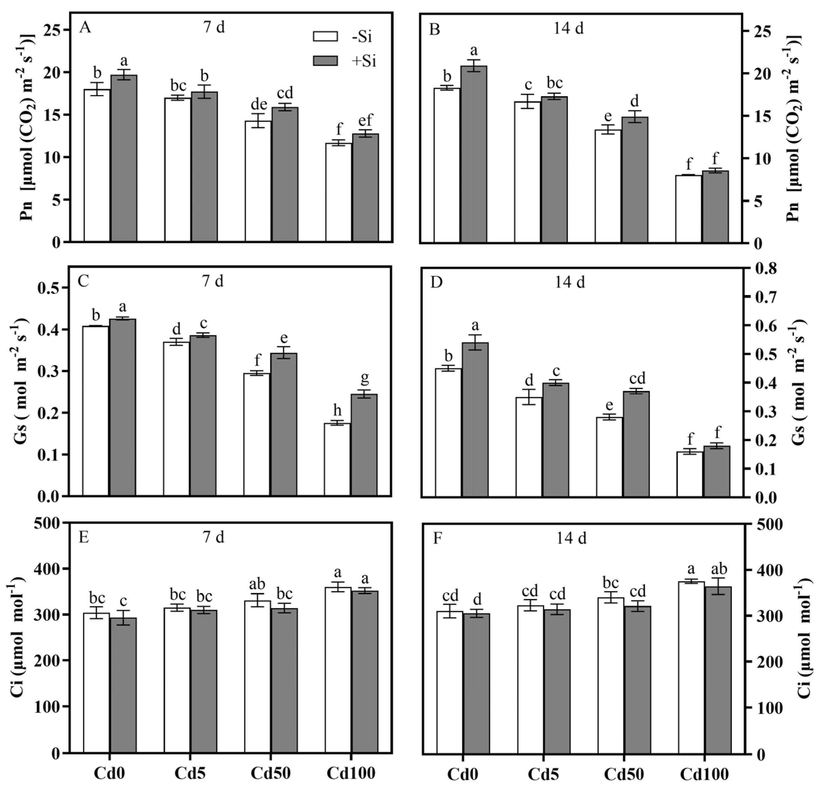
Figure 1. Photosynthetic parameters of rice leaves at 7 d and 14 d: (A) Pn at 7 d; (B) Pn at 14 d; (C) Gs at 7 d; (D) Gs at 14 d; (E) Ci at 7 d; (F) Ci at 14 d. Cd0, Cd5, Cd50, Cd100 denote 0 (control), 5, 50, 100 μmol·L−1 Cd; −Si and +Si denote 0 and 1.5 mmol·L−1 Si. Error bars show ±SD (n = 3); different letters denote significant differences (p < 0.05).
Agronomy 15 02331 g001
Figure 2. Rubisco activities in rice leaves at 7 d (A) and 14 d (B). Cd0, Cd5, Cd50, Cd100 denote 0 (control), 5, 50, 100 μmol·L−1 Cd; −Si and +Si denote 0 and 1.5 mmol·L−1 Si. Error bars show ±SD (n = 3); different letters denote significant differences (p < 0.05). FW: fresh weight.
Agronomy 15 02331 g002
Figure 3. Actual time-dependent Cd flow rates in rice roots at 7 d (A) and 14 d (B). Cd0, Cd5, Cd50, Cd100 denote 0 (control), 5, 50, 100 μmol·L−1 Cd; −Si and +Si denote 0 and 1.5 mmol·L−1 Si. Negative values represent influx.
Agronomy 15 02331 g003
Figure 4. Average Cd flow rates in rice roots at 7 d (A) and 14 d (B). Cd0, Cd5, Cd50, Cd100 denote 0 (control), 5, 50, 100 μmol·L−1 Cd; −Si and +Si denote 0 and 1.5 mmol·L−1 Si. Negative values represent influx. Error bars show ±SD (n = 3); different letters denote significant differences (p < 0.05).
Agronomy 15 02331 g004
Figure 5. Cd contents in rice leaves and roots at 7 d and 14 d: (A) leaves at 7 d; (B) roots at 7 d; (C) leaves at 14 d; (D) roots at 14 d. Cd0, Cd5, Cd50, Cd100 denote 0 (control), 5, 50, 100 μmol·L−1 Cd; −Si and +Si denote 0 and 1.5 mmol·L−1 Si. Error bars show ±SD (n = 3); different letters denote significant differences (p < 0.05). DW: dry weight.
Agronomy 15 02331 g005
Figure 6. Si contents in rice leaves and roots at 7 d and 14 d: (A) leaves at 7 d; (B) roots at 7 d; (C) leaves at 14 d; (D) roots at 14 d. Cd0, Cd5, Cd50, Cd100 denote 0 (control), 5, 50, 100 μmol·L−1 Cd; −Si and +Si denote 0 and 1.5 mmol·L−1 Si. Error bars show ±SD (n = 3); different letters denote significant differences (p < 0.05). DW: dry weight.
Agronomy 15 02331 g006
Figure 7. MDA contents in rice leaves and roots at 7 d and 14 d: (A) leaves at 7 d; (B) roots at 7 d; (C) leaves at 14 d; (D) roots at 14 d. Cd0, Cd5, Cd50, Cd100 denote 0 (control), 5, 50, 100 μmol·L−1 Cd; −Si and +Si denote 0 and 1.5 mmol·L−1 Si. Error bars show ±SD (n = 3); different letters denote significant differences (p < 0.05). FW: fresh weight.
Agronomy 15 02331 g007
Figure 8. H2O2 contents in rice leaves and roots at 7 d and 14 d: (A) leaves at 7 d; (B) roots at 7 d; (C) leaves at 14 d; (D) roots at 14 d. Cd0, Cd5, Cd50, Cd100 denote 0 (control), 5, 50, 100 μmol·L−1 Cd; −Si and +Si denote 0 and 1.5 mmol·L−1 Si. Error bars show ±SD (n = 3); different letters denote significant differences (p < 0.05). FW: fresh weight.
Agronomy 15 02331 g008
Figure 9. SOD activities in rice leaves and roots at 7 d and 14 d: (A) leaves at 7 d; (B) roots at 7 d; (C) leaves at 14 d; (D) roots at 14 d. Cd0, Cd5, Cd50, Cd100 denote 0 (control), 5, 50, 100 μmol·L−1 Cd; −Si and +Si denote 0 and 1.5 mmol·L−1 Si. Error bars show ±SD (n = 3); different letters denote significant differences (p < 0.05). FW: fresh weight.
Agronomy 15 02331 g009
Figure 10. APX activities in rice leaves and roots at 7 d and 14 d: (A) leaves at 7 d; (B) roots at 7 d; (C) leaves at 14 d; (D) roots at 14 d. Cd0, Cd5, Cd50, Cd100 denote 0 (control), 5, 50, 100 μmol·L−1 Cd; −Si and +Si denote 0 and 1.5 mmol·L−1 Si. Error bars show ±SD (n = 3); different letters denote significant differences (p < 0.05). FW: fresh weight.
Agronomy 15 02331 g010
Figure 11. CAT activities in rice leaves and roots at 7 d and 14 d: (A) leaves at 7 d; (B) roots at 7 d; (C) leaves at 14 d; (D) roots at 14 d. Cd0, Cd5, Cd50, Cd100 denote 0 (control), 5, 50, 100 μmol·L−1 Cd; −Si and +Si denote 0 and 1.5 mmol·L−1 Si. Error bars show ±SD (n = 3); different letters denote significant differences (p < 0.05). FW: fresh weight.
Agronomy 15 02331 g011
Figure 12. GST activities in rice leaves and roots at 7 d and 14 d: (A) leaves at 7 d; (B) roots at 7 d; (C) leaves at 14 d; (D) roots at 14 d. Cd0, Cd5, Cd50, Cd100 denote 0 (control), 5, 50, 100 μmol·L−1 Cd; −Si and +Si denote 0 and 1.5 mmol·L−1 Si. Error bars show ±SD (n = 3); different letters denote significant differences (p < 0.05). FW: fresh weight.
Agronomy 15 02331 g012
Figure 13. GSH contents in rice leaves and roots at 7 d and 14 d: (A) leaves at 7 d; (B) roots at 7 d; (C) leaves at 14 d; (D) roots at 14 d. Cd0, Cd5, Cd50, Cd100 denote 0 (control), 5, 50, 100 μmol·L−1 Cd; −Si and +Si denote 0 and 1.5 mmol·L−1 Si. Error bars show ±SD (n = 3); different letters denote significant differences (p < 0.05). FW: fresh weight.
Agronomy 15 02331 g013
Figure 14. Summary of the physiological and biochemical effect of Si-mediated Cd detoxification in rice at 7 d and 14 d.
Agronomy 15 02331 g014
Figure 15. Graphical mechanism of proposed model in this study.
Agronomy 15 02331 g015
Table 1. p-values for the effects of Cd, Si, and their interaction (Cd × Si) at 7 d and 14 d after treatment.
ParameterCdSiCd × Si
7 d14 d7 d14 d7 d14 d
Net Cd2+ flux 0.00010.00010.00010.00010.00010.0001
Cd concentration in leaf0.00010.00010.00010.00010.00010.0001
Cd concentration in root0.00010.00010.00010.00010.00010.0001
Si concentration in leaf0.00010.00010.00010.00010.04050.0001
Si concentration in root0.00010.00010.00010.00010.01590.0037
Pn0.00010.00010.00010.00010.45880.0115
Gs0.00010.00010.00010.00010.00010.0033
Ci0.00010.00010.03940.04330.81480.7932
Rubisco0.00010.00010.00010.00010.0040.0409
MDA in leaf0.00010.00010.00010.00010.00050.0043
MDA in root0.00010.00010.00010.00010.00050.0001
H2O2 in leaf0.00010.00010.00010.00010.00010.0001
H2O2 in root0.00010.00010.00010.00010.00010.0001
SOD in leaf0.00010.00010.00010.00010.00020.0392
SOD in root0.00010.00010.00010.00010.00010.0002
APX in leaf0.00010.00010.00010.00010.00010.0001
APX in root0.00010.00010.00010.00010.00010.0001
CAT in leaf0.00010.00010.00010.00010.00010.0001
CAT in root0.00010.00010.00010.00010.00010.0001
GST in leaf0.00010.00010.00010.00010.00010.0001
GST in root0.00010.00010.00010.00010.00010.0001
GSH in leaf0.00010.00010.00010.00010.00010.0001
GSH in root0.00010.00010.00010.00010.00010.0001
Disclaimer/Publisher’s Note: The statements, opinions and data contained in all publications are solely those of the individual author(s) and contributor(s) and not of MDPI and/or the editor(s). MDPI and/or the editor(s) disclaim responsibility for any injury to people or property resulting from any ideas, methods, instructions or products referred to in the content.