Article

Genome-Wide Identification and Expression Analysis of the Pepper β-1,3-gucanase Gene Family in Response to Phytophthora capsici Stresses

1
Institute of Vegetables, Anhui Academy of Agricultural Sciences, Hefei 230001, China
2
Key Laboratory of Horticultural Crop Germplasm Innovation and Utilization (Co-Construction by Ministry and Province), Institute of Horticulture, Anhui Academy of Agricultural Sciences, Hefei 230001, China
3
Anhui Provincial Key Laboratory for Germplasm Resources Creation and High-Efficiency Cultivation of Horticultural Crops, Institute of Vegetables, Anhui Academy of Agricultural Sciences, Hefei 230001, China
4
College of Life Sciences, Anhui Agricultural University, Hefei 230036, China
5
College of Agriculture, Anhui Science and Technology University, Chuzhou 233100, China
*
Authors to whom correspondence should be addressed.
Agronomy 2025, 15(4), 802; https://doi.org/10.3390/agronomy15040802
Submission received: 8 February 2025 / Revised: 17 March 2025 / Accepted: 21 March 2025 / Published: 24 March 2025
(This article belongs to the Section Plant-Crop Biology and Biochemistry)

Abstract

:
Peppers are widely cultivated around the world, yet they suffer from infections caused by Phytophthora capsici fungi throughout the year, which severely impacts their yield. The β-1,3-glucanase gene has been shown in previous studies to significantly enhance plants’ ability to cope with both biotic and abiotic stresses, including fungal infections. However, its function in peppers has not been reported. In this study, 80 CaBG genes were initially identified, before being filtered down to 54 CaBGs in peppers, and analyses conducted on the physicochemical properties, chromosomal localization, phylogenetic tree relationships, synteny, promoters, and gene expression levels of the BG gene family. The results indicated that these 54 CaBG genes are located on 12 chromosomes. Phylogenetic tree analysis classified the CaBGs into three subfamilies, α, β, and γ, each with its own specific functions, with the γ subfamily being associated with disease resistance in peppers. Synteny analysis showed that CaBG genes are relatively conserved and have not undergone extensive whole-genome duplication events. Promoter analysis indicated that CaBGs are induced by plant hormones and various external stressors. Transcriptomic and RT-qPCR analyses revealed that the CaBG7 and CaBG12 genes were significantly activated following infection by P. capsici, with the expression levels of these two genes being markedly higher in resistant plants compared to susceptible ones. Based on the phylogenetic tree and gene expression analysis, we propose that CaBG7 and CaBG12 may be key genes for disease resistance in chili peppers. This study provides a theoretical basis for breeding P. capsici-resistant chili peppers by molecular breeding methods.

1. Introduction

Phytophthora belongs to the family Pythiacea, there are more than 120 species, most of which are important plant pathogens [1]. Phytophthora root and collar rot, caused by Phytophthora capsici (P. capsici), is the most serious disease in pepper production [2,3], and it causes more than USD 100 million in losses worldwide annually [4]. P. capsici belongs to oomycetes, and there are more differences with fungi in terms of trophic structure, genome composition, metabolic pathways, and reproduction, and common fungal inhibitors are basically ineffective against them [5]. At present, control of oomycete diseases caused by P. capsici is still a worldwide problem, and the production of chemical-based agents, resulting in pathogen resistance, pharmaceutical residues and environmental pollution, and other issues is increasingly prominent [6]. Therefore, it is particularly important to find key genes that can suppress P. capsici and to develop molecularly improved varieties resistant to P. capsici for pepper. The cell wall is a critical biological structure that interfaces with host cells and plays a pivotal role in the pathogenicity and virulence of invasive fungi. Composed primarily of β-1,3-glucanase, chitin, and mannoproteins, the cell wall’s composition and the ratio of these components vary significantly across different fungal species, influencing their functional properties and interactions with host organisms [7]. β-1,3-glucanase is a polysaccharide formed through glycosidic linkages of glucose, and β-1,3-glucanase accounts for the majority of β-1,3-glucanases in fungal cell walls. β-1,3-glucanases are synthesized by plasma membrane-associated synthetases and are transported to the cell wall during synthesis [8].
β-1,3-glucanase belongs to the glycoside hydrolase family 17, which is the Pathogenesis related-2 (PR-2) protein [9]. The β-1,3-glucanase gene family has been extensively characterized across various species, with its functional roles partially elucidated through comprehensive research efforts [10,11,12]. It can hydrolyze the main component of the pathogen cell wall, β-1,3-glucanase, effectively inhibiting the growth of pathogens, and it serves as a crucial defense factor in the plant defense system [13]. β-1,3-glucanase can be expressed in plant–pathogen interactions to improve plant defenses [14,15]. In Arabidopsis, there are 50 AtBGs, all of which contain an N-terminal signal peptide and a glycosyl hydrolase family 17 (GH-17) domain, with some also containing a C-terminal domain X8 (also known as CBM43) [16]. In tomato (Solanum lycopersicum), a phylogenetic analysis identified 51 candidate β-1,3-glucanases that were distributed in three clusters (α, β, and γ), with evolutionary relations previously characterized in the model Arabidopsis thaliana [17]. Previous studies have shown that β-1,3-glucanase genes can be significantly expressed when plants are subjected to pathogenic bacteria or abiotic stresses, thereby increasing plant stress tolerance. For example, with a soybean gene SGN1 encoding β-1,3-glucanase, the expression of SGN1 was strongly induced by a variety of defense-related signals, such as treatment with H2O2, wounding, or treatment with fungal elicitor prepared from Phytophthora spp, as well as inoculation with Pseudomonas syringae [18]. The transient overexpression of the sugarcane (Saccharum officinarum) β-1,3-glucanase gene ScGluD2 in tobacco leaves induced the plant’s defense response and enhanced its resistance to Pseudomonas solanacearum and Botrytis cinerea [19]. In vitro antifungal assays revealed that a wheat (Triticum aestivum) recombinant β-1,3-glucanase significantly inhibited the spore formation and hyphal morphology of common wheat grain fungi [20]. A β-1,3-glucanase gene (PnGlu1) from Panax notoginseng exhibits a significantly induced expression in response to treatments with methyl jasmonate, salicylic acid, ethylene, hydrogen peroxide, and Fusarium solani inoculation. Recombinant PnGlu1 protein demonstrates high inhibitory activity against the growth of Fusarium solani and Fusarium oxysporum. Additionally, tobacco plants overexpressing PnGlu1 show strong resistance to Fusarium solani infection [21]. A novel β-1,3-glucanase, Gns6, was found to have significant antifungal activity against Mycobacterium bovis in rice [21].
Peppers, belonging to the Solanaceae family, are currently the most widely cultivated vegetable species in China [22]. With the expansion of pepper cultivation in China, the impact of P. capsici infestation on pepper crops has become increasingly severe. P. capsici seriously affects the growth and development of peppers by infecting its stems, leaves, and other parts, which can lead to widespread plant death and significant yield loss [2,4]. In previous studies, extensive research has been conducted on the molecular mechanisms of pepper resistance to P. capsici, primarily focusing on signal transduction pathways following pathogen infection and biological control strategies [23,24,25]. Additionally, significant progress has been made in screening pepper varieties resistant to P. capsici, with researchers successfully identifying multiple elite disease-resistant cultivars, contributing greatly to reducing the damage caused by P. capsici and improving pepper yields [26,27]. However, the precise identification of key genes directly involved in the disease resistance process and the enhancement of pepper resistance through molecular breeding techniques remain critical objectives in current research. Therefore, identifying key genes involved in pepper resistance to P. capsici blight is crucial. In this study, we conducted a comprehensive genome-wide identification of the β-1,3-glucanase gene family in peppers, identifying a total of 54 β-1,3-glucanase genes. We performed bioinformatic analyses to characterize the family of β-1,3-glucanase genes and expression patterns under biotic stress induced by P. capsica, providing a theoretical basis for the selection and breeding of pepper varieties resistant to P. capsici.

2. Materials and Methods

2.1. Plant Materials

In our pre-laboratory studies, we identified several resistant and susceptible pepper varieties by direct infestation of pepper plants by P. capsici and molecular marker experiments. In this experiment, the tested chili pepper varieties include a high-resistant variety R (V06C1825) and a susceptible variety S (H15-077), bred by the Institute of Vegetables, Anhui Academy of Agricultural Sciences. The P. capsici fungus HFGL-1-24 was cultured and provided by the Institute of Plant Protection and Agro-Product Safety, Anhui Academy of Agricultural Sciences. Pepper plants were cultivated in a constant-temperature laboratory incubator (20–25 °C in 16 h (h)/8 h light/dark cycles with 60% relative humidity; light intensity: 20,000 lux; regular watering and fertilization).

2.2. Genome-Wide Characterization of the β-1,3-glucanase Gene Family in Peppers

The chili pepper Zunla-1_v3.0 genome, selected for this study, was obtained from http://www.bioinformaticslab.cn/PepperBase/ (accessed on 8 February 2025) [28]. The Arabidopsis genome was obtained from https://www.arabidopsis.org/ (accessed on 8 February 2025), and the tomato genome annotation (ITAG2.4) was obtained from https://solgenomics.net/ (accessed on 8 February 2025). The hidden Markov model (HMM) profile of the glycoside hydrolase family 17 domain (PF00332) was obtained from the Pfam website (http://pfam.xfam.org/, accessed on 8 February 2025) and was employed as a query to identify all possible β-1,3-glucanases using HMMER (V3.0) software (e-value ≤ 1 × 10⁻¹⁰) [29]. The identification of conserved domains was performed using NCBI CDD tools (https://www.ncbi.nlm.nih.gov/cdd, accessed on 8 February 2025) and the InterPro database (https://www.ebi.ac.uk/interpro/, accessed on 8 February 2025) to identify the CaBG gene family members in pepper and name them based on their chromosomal positions.
Candidate proteins were further subjected to multiple sequence alignment to remove duplicates and incomplete protein sequences. The physicochemical properties of CaBG family members were analyzed using the online tool the ExPASy Proteomics Server (http://expasy.org/, accessed on 20 November 2024) [30]. Subcellular localization analysis of each gene family member was performed using the WoLF PSORT online tool [31]. The chromosome distribution map of the β-1,3-glucanase family genes in pepper was constructed using TBtools software v2.0 [32].

2.3. Phylogenetic Analysis of β-1,3-glucanase Family Genes from Pepper, Arabidpsis, and Tomato

To find out the evolutionary relationship of the β-1,3-glucanase gene family of chili peppers, we combined β-1,3-glucanase protein sequences of pepper, Arabidopsis, and tomato to construct a phylogenetic tree. A multiple sequence alignment of BG proteins was performed using ClustalX2.1 (Multiple Alignment Parameters, Gap Opening: 10.00, Gap Extension: 0.20). The phylogenetic tree was constructed with MEGA 7.0 using the neighbor-joining method and 1000 bootstrap replicates [33].

2.4. CaBGs Gene Motif and Structure Analysis

The genomic, CDS, and protein sequences of CaBGs were obtained from the pepper Zunla-1_v3.0 genome (http://www.bioinformaticslab.cn/PepperBase/, accessed on 21 November 2024) [28]. The exon/intron analysis of CaBGs was conducted using GSDS 2.0 (http://gsds.gao-lab.org/index.php, accessed on 22 November 2024). The motif analysis of CaBGs was performed in MEME (http://meme-suite.org/, accessed on 25 November 2024), with parameters set as follows: default for advanced options and a maximum of 10 motifs.

2.5. Cis-Element and Synteny Analysis of CaBGs

MCScanX software (using built-in parameters) was employed to investigate the collinearity relationship among the BG gene families in pepper, Arabidopsis and tomato (CPU for BlastP: 2, E-value: 1 × 10⁻¹⁰, Num of BlastHits: 5) [34]. The results were visualized using TBtools software v2.0 [32]. The cis-acting elements of the promoters of the pepper BG family genes were analyzed within the 2.0 Kb upstream sequences from the transcription start sites of the BG family genes using the online website PlantCare (https://bioinformatics.psb.ugent.be/webtools/plantcare/html/, accessed on 8 February 2025).

2.6. Analysis of Expression Patterns in Roots and Stems of Pepper Plants Under P. capsici Biostress

The laboratory conducted a preliminary screening and selected a susceptible variety (H15-077) and a resistant variety (V06C1825). The root irrigation method was employed to inoculate the seedlings of the susceptible variety H15-077 and the resistant variety V06C1825 with P. capsici (HFGL-1-24). Twelve hours prior to inoculation, the substrate was thoroughly watered to achieve a saturated moisture state. A glass rod was used to create holes approximately 1 cm away from the pepper plants in the substrate, into which 5 mL of spore suspension was injected. Each experimental material consisted of 15 plants. Following inoculation, the plants were incubated at 28 °C in dark conditions with high humidity for 24 h, after which they were maintained under normal management conditions. After inoculating these two pepper varieties with P. capsici, stem tissues were collected at 0 days (1 h), 1 day, 3 days, and 5 days post-inoculation. The tissues were quickly frozen in liquid nitrogen, with three biological replicates for each variety and stage to eliminate errors between samples. RNA extraction and cDNA library construction were carried out by Beijing Biomarker Technologies Co., Ltd. (Beijing, China). The cDNA libraries were then sequenced using the Illumina (San Diego, CA, USA) high-throughput sequencing platform to obtain the raw data (NCBI accession number PRJNA1234221). The percentage of Q30 bases of each sample of clean data obtained after sequencing quality control was not less than 93.86%, and the comparison efficiency of reads of each sample with the reference genome ranged from 93.15% to 95.27%. In order to analyze the effect of P. capsici on the gene expression of CaBGs in pepper roots, we also obtained and analyzed publicly available raw transcriptome data (NCBI accession number: PRJNA960669) from the roots of the A198 pepper variety under P. capsici biotic stress [4]. After raw data filtering, sequencing error rate checking, and GC content distribution checking, we obtain clean reads for subsequent analysis. We used FPKM (Fragments Per Kilobase of transcript per Million fragments mapped) as a measure of transcript or gene expression level [34]. A heatmap of their expression under P. capsici stress conditions in pepper was visualized using TBtools v2.0 [32].

2.7. RNA Extraction and cDNA Reverse Transcription

After inoculating the highly resistant variety R (V06C1825) and the susceptible variety S (H15-077) with P. capsici, stem tissues were collected from the uninfected pepper samples (pepper plants before infestation by P. capsici) as the control group (CK), as well as from the pepper plants at 0 day (1 h), 1 d, 3 d, and 5 d post-inoculation. Three biological replicates of samples from each stage were taken to eliminate errors between samples. All the collected samples were rapidly frozen in liquid nitrogen and then stored at −80 °C for subsequent RNA extraction. Samples were hand-ground in liquid nitrogen using mortar and pestle before mRNA extraction. Total RNA was extracted using the TIANGEN RNAprep pure kit (Tiangen, Beijing, China), following the manufacturer’s instructions. Reverse transcription was performed using an HiScript III 1st Strand cDNA Synthesis Kit (+gDNA wiper) (Vazyme Biotech, Najing, China). Three replications of samples were performed for each stage.

2.8. RT-qPCR Analysis

All RT–qPCR primers were designed using Beacon Designer 7 software (Table S3). The RT–qPCR was performed with a CFX96 TouchTM Real-Time PCR Detection System (BIO-RAD, Hercules, CA, USA), with three biological replicates for each sample. The relative gene expression levels were calculated using the 2−∆∆CT method [35]. CaACT (Genebank: AY572427.1) was used as reference gene for resistant variety R (V06C1825) and the susceptible variety S (H15-077) pepper qPCR [36].

2.9. Data Collection and Statistical Analysis

Data are presented as means ± SE of at least three independent experiments. The experimental data were analyzed by one-way ANOVA using SPSS 25.0 software; multiple comparisons were performed using Duncan’s method (p < 0.05). Statistical analyses were performed using GraphPad Prism 8.0 software.

3. Results

3.1. Identification and Physicochemical Characteristics of the β-1,3-glucanase (BG) Family Members in Pepper

This research identified 80 CaBG genes in the pepper genome through the use of the hidden Markov model PF00332. Candidate proteins were identified through conserved domain and subjected to multiple sequence alignment to remove duplicates and incomplete protein sequences, with 54 remaining after removing duplicates and short fragments. The CaBG proteins exhibited a variation in amino acid length, ranging from 195 amino acids (aas) in CaBG6 to 583 aas in CaBG37, with corresponding molecular weights ranging from 22.03 kDa (CaBG6) to 63.17 kDa (CaBG7). The isoelectric points ranged from 4.62 (CaBG54) to 9.48 (CaBG12), and there were 40 CaBG proteins with isoelectric points less than 7, which accounted for the majority of the CaBG family, indicating that most of the proteins in the CaBG family were acidic proteins. The most predicted sites for the subcellular localization of CaBG proteins were chloroplast (chlo) and plasma membrane (plas), with 13 genes, followed by extracellular (extr) with 9 genes and then vacular membrane with 7 genes (Table S1).

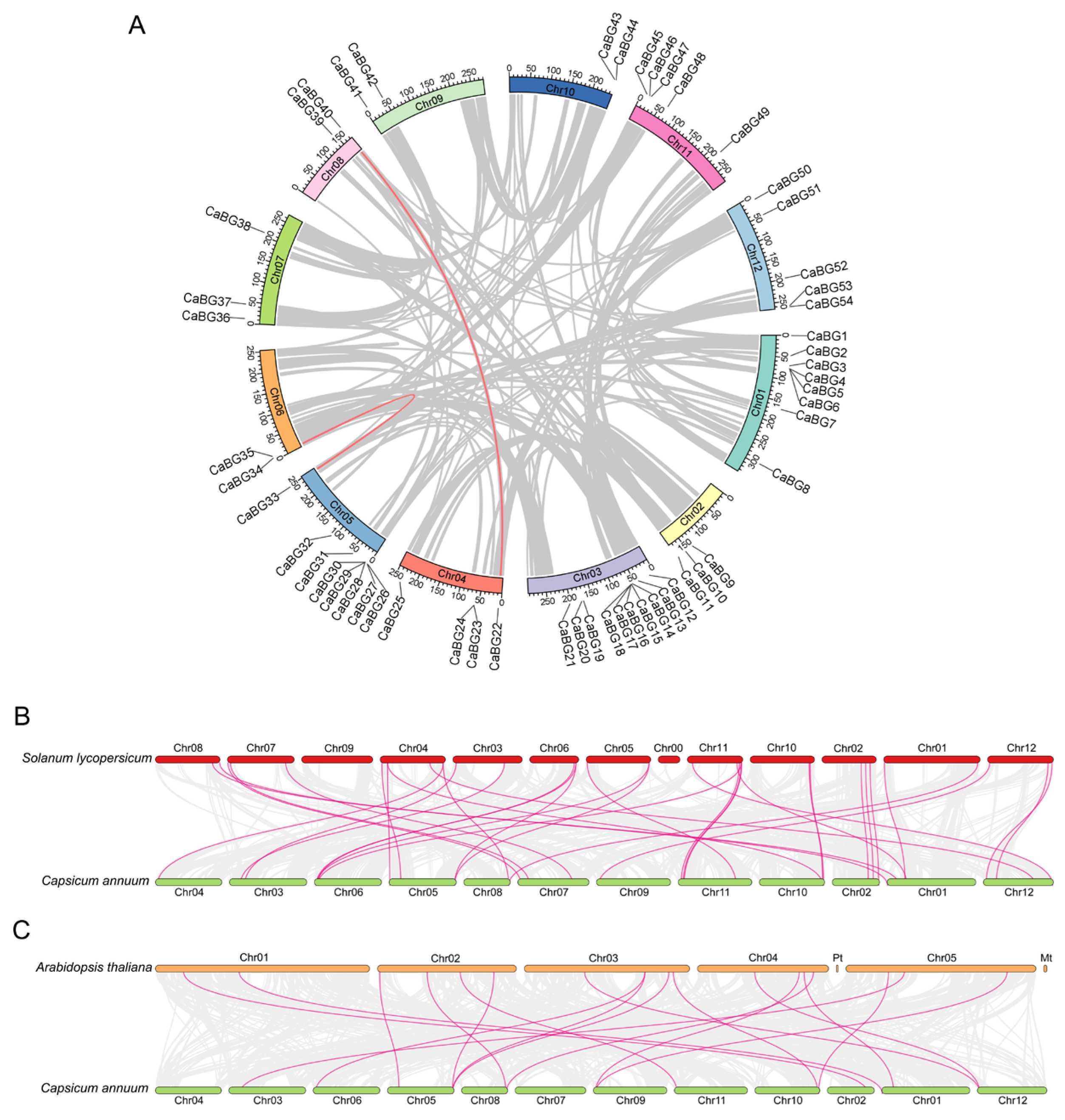
3.2. Chromosome Localization of Pepper BG Genes

We conducted the chromosomal localization of 54 BG genes in pepper, revealing their distribution across all 12 chromosomes (Figure 1). Chromosome 3 exhibited the highest gene density, with ten BG genes identified. Chromosomes 1 and 5 followed closely, each harboring eight CaBGs. In contrast, chromosomes 6, 8, 9, and 10 contained the fewest CaBG genes, with only two genes localized on each. This uneven distribution suggests that chromosomes 1, 3, and 5 may play a pivotal role in the evolutionary dynamics of CaBG genes in pepper.
A further analysis of gene positioning revealed the presence of gene clusters on specific chromosomes. These clusters, composed of genes with similar or related functions, likely contribute to coordinated biological roles within the organism. Notable examples include the clusters CaBG4, CaBG5, and CaBG6 on chromosome 1; CaBG14, CaBG15, CaBG16, CaBG17, and CaBG18 on chromosome 3; and CaBG28, CaBG29, and CaBG30 on chromosome 5. The clustering of these genes underscores their potential functional synergy and evolutionary significance.

3.3. Phylogenetic Tree Analysis of CaBG Family Genes

To study the evolutionary relationship between CaBGs, SlBGs and AtBGs, a phylogenetic analysis was performed (Figure 2). The results showed that the 54 CaBGs, 50 SlBGs [17], and 50 AtBGs [16] were classified into three subfamilies (α, β, and γ), consistent with previous studies in Arabidopsis [16]. β-1,3-glucanases are considered key regulators responsible for plant pollen and fruit development, seed production, and disease resistance. To identify candidate pepper orthologues, we analyzed the branching pattern of the phylogenetic tree. The results showed that in the α subfamily, CaBG31, 10, 8, and 3 clustered together with the plasma membrane proteins AT3G13560.1 (PdBG1), AT2G01630.1 (PdBG2), and AT1G66250.1 (PdBG3) from the Arabidopsis β-1,3-glucanase family. The proteins AtPdBG1, 2, and 3 are associated with plasmodesmal conductivity and callose turnover [37]. In the Arabidopsis β-1,3-glucanase family, AT1G64760.1 (ZET) and AT2G19440.1 (ZETH) are homologous genes. Studies have shown that the ZET gene is related to plant growth and development, and silencing the ZET gene leads to abnormal development in Arabidopsis plants [38]. In the β subfamily, the CaBG19, CaBG44, and CaBG52 genes cluster together with AT1G64760.1 (ZET) and AT2G19440.1 (ZETH), suggesting that these genes may share similar functions. In the γ subfamily, Solyc11g065280.1 (SlBG10) is a homolog of CaBG48, with functions primarily related to callose deposition in tomatoes and male sterility [39]. At4g16260.1, AT3G57270.1 (AtBG1), AT3G57260.1 (AtBG2), and AT3G57240.1 (AtBG3) are Arabidopsis virus defense proteins. Among them, At4g16260.1 shows significant expression responses to the highest number of pathogens (four species), and it is induced to the greatest extent under pathogen stress among all PR genes (induced 22-fold and 53-fold by Alternaria brassicicola and Phytophthora infestans) [16]. These genes primarily belong to the γ subfamily and show high homology with CaBG7, CaBG12, CaBG26, and CaBG43. We predict that CaBG7, CaBG12, CaBG26, and CaBG43 may be involved in the defense against pathogens in chili peppers.

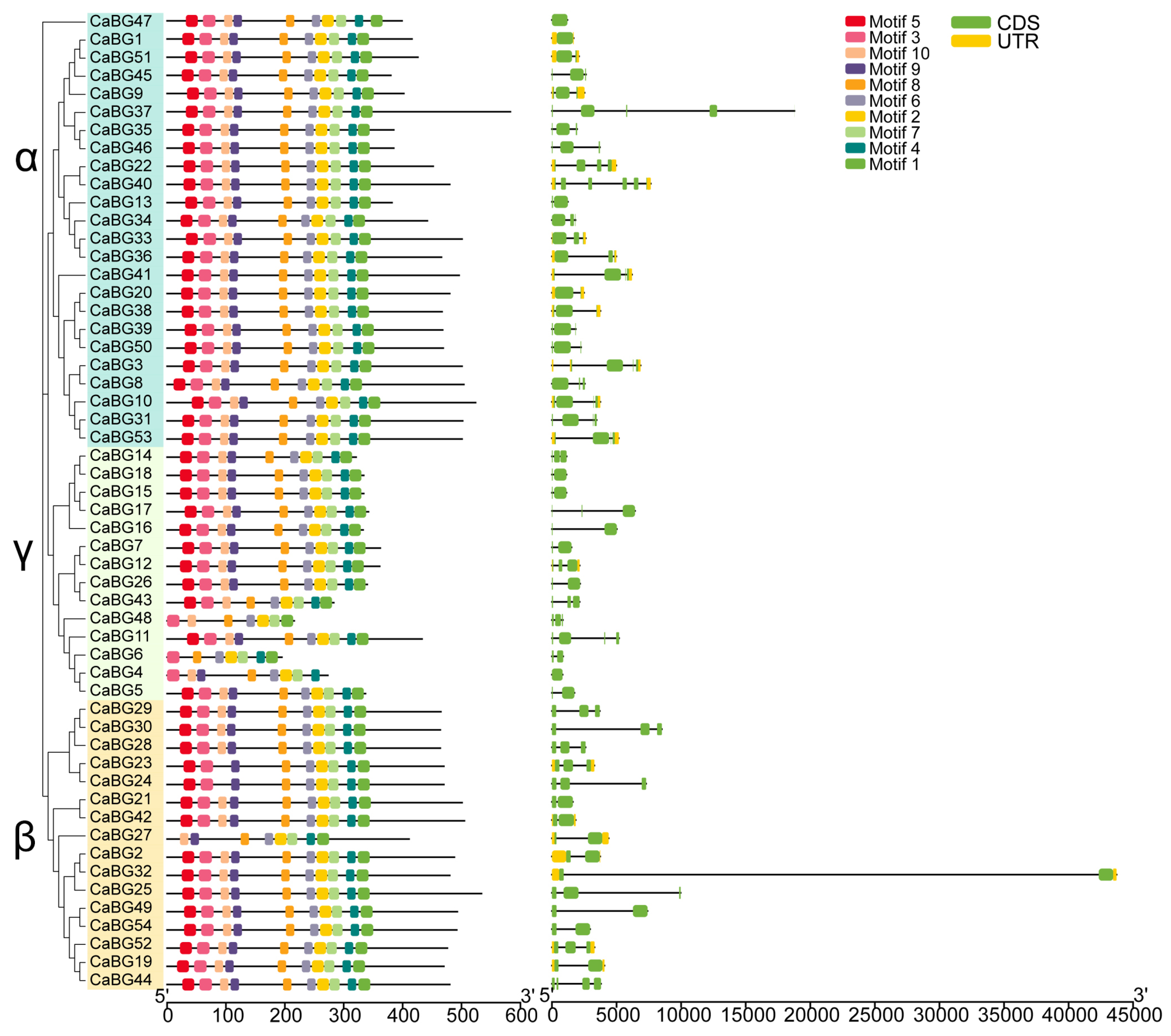
3.4. Gene Structure and Conserved Motif Analysis of Pepper BG Genes

We conducted a phylogenetic tree construction using the protein sequences from 54 pepper BG genes and analyzed these genes for gene motif and gene structure (intron–exon). Among them, the α subfamily contained the largest number of CaBGs, with 24 members, followed by the β subfamily, with 16 members, and finally, the γ subfamily, which contained 14 CaBGs (Figure 3). The motif analysis showed that the CaBG proteins exhibited strong conservation in the distribution pattern and quantity of motifs. An analysis revealed that motifs 2 and 7 correspond to conserved sequences of the β-1,3-glucanase GH-17 domain. As shown in Figure 3, all CaBG proteins contain these two motifs. Most of the CaBG proteins (47 in total) contain 10 motifs (maximum number), with only a few CaBGs having fewer than 10 motifs. Among them, CaBG6 and CaBG48 have the fewest motifs, with only seven motifs. An interesting observation is that the genes with fewer than 10 motifs are concentrated in the γ and β subfamilies, while the CaBG proteins in the α subfamily all possess the complete set of 10 motifs (Figure 3).
In the context of multi-gene families, the structural disparities in exon–introns within the same family serve as pivotal factors in the evolutionary process. We observed the gene structure of the pepper BG gene family and found that the number of exons is generally low. The number of exons in most CaBG genes is predominantly concentrated in the range of one to four. Among them, CaBG40 contains the most exons (six in total). Additionally, some genes lack intronic sequences, consisting of only a single exon, such as CaBG4 and CaBG47. Genes within the same branching of the evolutionary tree exhibit similarities in their exon–intron structure (Figure 3). The resemblance in these gene structures, coupled with a high sequence homology, suggests that gene duplication events transpired in the evolution of the pepper BG gene family.

3.5. Synteny Analysis of BG Genes Among Pepper, Arabidopsis, and Tomato

To better understand the evolutionary relationships of BG gene families in pepper, Arabidopsis, and tomato, we conducted a synteny analysis within and between species of those BG genes (Figure 4, Table S2). The results showed that there are only 2 pairs of syntenic genes (CaBG22/CaBG40 and CaBG33/CaBG34) within the pepper species (Figure 4A), which is significantly fewer compared to the 18 pairs between pepper and Arabidopsis (Figure 4B), and the 35 pairs between pepper and tomato (Figure 4C). This suggests that the pepper BG gene family is relatively conserved and has not undergone extensive whole-genome duplication events. The number of syntenic gene pairs between pepper and tomato is significantly higher, with 35 pairs, compared to the 18 pairs between pepper and Arabidopsis. This difference can be attributed to the relatively short divergence time between species like pepper and tomato, which results in fewer accumulated variations and the preservation of more ancestral traits. In contrast, species with a longer divergence time, such as pepper and Arabidopsis, have accumulated more variations, leading to fewer shared traits and shorter collinear segments [40].

3.6. Analysis of CaBG Promoter Cis-Elements

Through the analysis of cis-elements in the promoters, a total of 41 cis-elements were identified from the promoters of 54 CaBGs, and these elements were classified into 15 major categories (Figure 5). Among them, the transcription factor-related sites, such as MYB binding sites, accounted for the largest proportion (25.85%), followed by MYC binding sites (9.74%) and WRKY binding sites (2.97%). This indicates that CaBGs are regulated by transcription factors such as MYB, MYC, and WRKY, with MYB playing a dominant role in this regulation. In terms of environmental stress response elements, light response elements dominate, accounting for 24.09% of all elements, followed by defense and stress response elements (6.68%), low-temperature response elements (1.38%), and wound response elements (1.5%). This suggests that the expression of CaBGs may be largely influenced by light induction. Hormone-responsive elements include those for MeJA, abscisic acid, salicylic acid, and auxin, with MeJA having the largest proportion (7.29%) and auxin the smallest (1.06%). Additionally, there were growth and development-related response elements, such as anaerobic induction (3.82%), meristem expression (1.12%), and endosperm expression (0.65%).

3.7. Expression Pattern Analysis of CaBG Genes Under P. capsici Infestans Stress

We conducted an expression analysis of the stems and roots of peppers after infection with P. capsici. Pepper stem samples were compared between the susceptible pepper (S) and the resistant pepper (R) (Figure 6A). In pepper roots, we compared the changes in the expression of CaBGs after infestation at different times (0 hpi, 24 hpi, 48 hpi) (Figure 6B). The findings indicate that in the stem, only three genes were significantly upregulated across all four time points when comparing S and R, namely CaBG34, CaBG7, and CaBG12. Among these, CaBG34 generally exhibited a low expression level after infestation at different times; CaBG7 and CaBG12 expression was higher at 0 d (1 h) and 1 d of infestation, and then gradually decreased; and the expression of these genes was significantly higher in the disease-resistant variety R than in the susceptible variety S. In the roots, CaBG7 and CaBG12 were the two genes with the highest expression levels within the CaBGs family, and their expression increased with the duration of P. capsici infection at 12 and 24 h. From our previous evolutionary tree analysis, we found that disease resistance-related genes in Arabidopsis are primarily distributed in the γ subfamily. Both CaBG7 and CaBG12 belong to the γ subfamily and have a significant homology with Arabidopsis pathogen defense proteins At4g16260.1, AT3G57270.1 (AtBG1), AT3G57260.1 (AtBG2), and AT3G57240.1 (AtBG3). Therefore, we speculate that CaBG7 and CaBG12 are key genes in chili pepper resistance against P. capsici. Interestingly, after infection, the expression level of CaBG7 in the stem was lower than that of CaBG12, while its expression level in the roots was significantly higher than that of CaBG12. This suggests that CaBG7 may function specifically in the roots, whereas CaBG12 is more inclined to play a role in the stems.

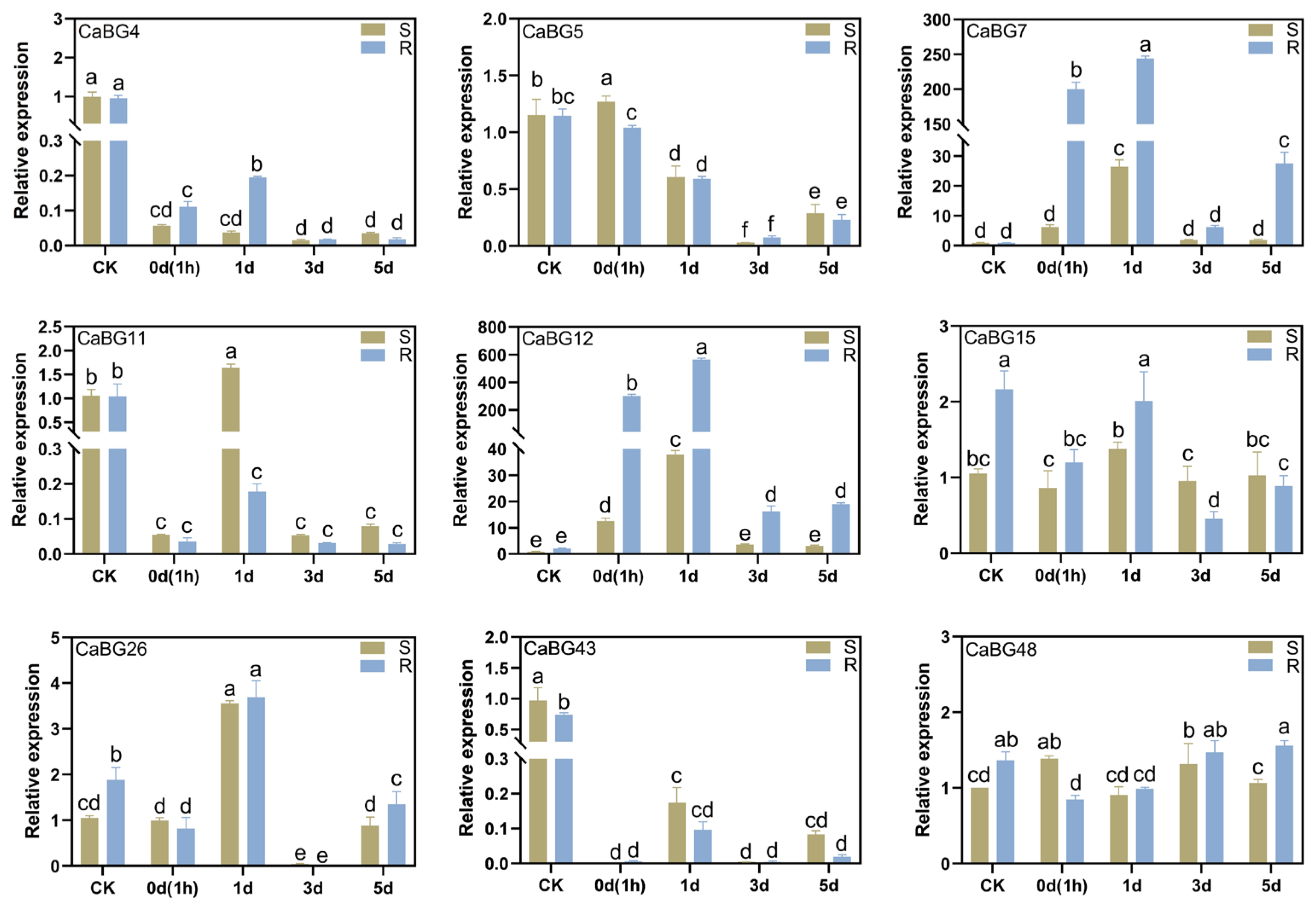
3.8. RT-qPCR Analysis of the Pepper BG Gene Family

To validate the accuracy of the transcriptome data, we selected all the γ subfamily of CaBG genes from the pepper BG gene family (Figure 7). According to our RT-qPCR test results, the expression patterns were generally consistent for the transcriptome data we focused on. Among these genes, CaBG6, CaBG14, CaBG16, CaBG17, and CaBG18 did not show any detectable expression levels. Additionally, except for CaBG7 and CaBG12, the expression levels of the other genes were generally low and did not exhibit any clear patterns. The expression levels of CaBG7 and CaBG12 significantly increased after infection with P. capsici, with the expression in the R pepper plants being notably higher than in the S pepper plants. However, the expression levels of both R and S plants significantly decreased at 3 d and 5 d post-infection. This suggests that CaBG7 and CaBG12 are genes responsive to P. capsici infection, primarily acting in the early stages post-infection (0 d and 1 d), with their expression levels significantly declining at 3d and 5d after infection.

4. Discussion

During the growth and development of peppers, they are subjected to various biotic and abiotic stresses, making them vulnerable to multiple diseases [41,42]. Among these, Phytophthora blight, caused by P. capsici, is a significant threat to global pepper production [23,43]. This disease is particularly prevalent in hot (25–28 °C) and humid conditions, and in severe cases, it can lead to total yield loss [23,44]. Phytophthora blight has occurred in numerous countries and regions worldwide, and it is estimated that the economic losses caused by this disease exceed one billion dollars annually [45,46]. In our previous study, we screened pepper plants resistant to P. capsici and performed transcriptome sequencing on both resistant and susceptible plants, with the aim of identifying key genes involved in the resistance to this pathogen.
β-1,3-glucanase is plant PR-2 enzyme belonging to the glycosyl hydrolase family 17 (GH-17) [47]. It plays a significant role in growth and development, reproduction, and resistance to both biotic and abiotic stresses in plants [14]. In fungi, β-1,3-glucanase is a major component of the cell wall [48,49], and β-1,3-glucanase can be expressed in plant–pathogen interactions to inhibit the growth of pathogens by disrupting their cell walls and improving plant defenses [7]. With advancements in sequencing technologies, more genomic and transcriptomic data are being published, and it has become particularly important to utilize these data to identify the genes and their family members that need to be studied. To date, a complex and diverse BG gene family has been found in tomato [17], tobacco [50], soybean [51], Arabidopsis [16], rice [52], and cotton [53]. In this study, we applied a similar research approach and used a gap-free chili pepper genome, Zunla v3.0 [28], to identify the BG gene family in peppers. A total of 54 BG genes were identified. In this study, we classified the pepper BG genes into three subfamilies, α, β, and γ, based on previous research [16,17]. In Arabidopsis, genes that have been functionally validated within the α subfamily, such as AtPdBG1, AtPdBG2, and AtPdBG3, are primarily associated with plasmodesmal conductivity and callose turnover [37]. In the β subfamily, AtZET and AtZETH are mainly associated with plant growth and development [38]. In the γ subfamily, At4g16260.1, AT3G57270.1 (AtBG1), AT3G57260.1 (AtBG2), and AT3G57240.1 (AtBG3) are Arabidopsis virus defense proteins [16]. Each β-1,3-glucanase gene in these subfamilies appears to have its specific function. Since this study primarily focuses on the role of β-1,3-glucanase in chili pepper’s resistance to P. capsici, our attention is specifically directed towards the β-1,3-glucanase genes in the γ subfamily of peppers.
Gene clusters are genomic regions composed of two or more adjacent and functionally related genes, typically generated through gene duplication events and retaining characteristics of their common ancestral gene. Genes within a cluster often encode functionally related enzymes, which can form multi-enzyme complexes that cooperatively catalyze key reactions in specific metabolic pathways [54]. This structure not only enhances the coordination of gene expression but may also improve the organism’s adaptability to environmental stresses [55,56]. In this study, through chromosomal localization analysis, we identified multiple gene clusters within the pepper CaBG gene family, such as CaBG4, CaBG5, and CaBG6 on chromosome 1; CaBG14, CaBG15, CaBG16, CaBG17, and CaBG18 on chromosome 3; and CaBG28, CaBG29, and CaBG30 on chromosome 5. The formation of these gene clusters may be closely related to the adaptive selection of pepper against diseases during its long-term evolution. From an evolutionary perspective, the formation of gene clusters may result from the combined effects of gene duplication and natural selection. Gene duplication events provide additional genetic material to the genome, while natural selection preserves gene combinations that enhance organismal adaptability. In the evolutionary history of pepper, diseases may have acted as a strong selective pressure, driving the formation of gene clusters through the duplication and recombination of disease resistance-related genes, such as CaBG genes. This structure not only facilitates coordinated gene expression but may also enhance pepper’s resistance to diseases through gene dosage effects. Furthermore, genes within a cluster may further optimize their disease resistance functions through subfunctionalization or neofunctionalization.
Whole-genome duplication, also known as polyploidy accompanied by gene loss, has long been a significant driving force in the genomic evolution of animals, fungi, and other organisms, particularly plants [57,58]. As whole-genome duplication progresses, significant changes in genome structure often occur, such as chromosomal rearrangements, gene inversions, and gene losses. The large number of duplicated genes generated by polyploidy becomes an important source of genetic innovation [59]. In this study, comparative genomics and synteny analysis were conducted on pepper, Arabidopsis, and tomato. It was found that there are only 2 pairs of syntenic gene pairs within pepper, 18 pairs between pepper and Arabidopsis, and 35 pairs between pepper and tomato. This indicates that the BG gene family is relatively conserved and has not undergone extensive whole-genome duplication events, leading to only two pairs of syntenic genes within pepper. Additionally, the highest number of syntenic pairs between pepper and tomato suggests that the variation accumulated between the source species is relatively low, thus retaining more ancestral traits, further evidence for the conservation of the BG gene family.
Different types of cis-acting elements on the promoter reveal the potential functions and regulatory differences of genes. In this study, we identified not only defense and stress-related cis-acting elements in the promoter regions of CaBGs but also multiple hormone-responsive elements, including those for methyl jasmonate (MeJA), abscisic acid (ABA), salicylic acid (SA), and auxin (IAA). Previous studies have demonstrated that these hormones can activate the expression of β-1,3-glucanase genes, thereby enhancing plant resistance to pathogen infection. For instance, in tobacco, researchers fused the promoter region of the β-1,3-glucanase gene with the GUS gene and found that it could respond to Tobacco Mosaic Virus (TMV), salicylate, ethylene, et al. [60]. The promoter of BjPR2 in Brassica juncea was induced by fungi, hormones, and wounds [61], and the BG gene promoter of sesame (Sesamum indicum L.) induced by ABA and drought [62]. We also found numerous light-responsive elements and binding sites for transcription factors such as MYB and MYC. These findings suggest that β-1,3-glucanase is not only activated by hormones but also influenced by photoperiods and regulated by transcription factors like MYB and MYC.
Previous studies have shown that the activity of β-1,3-glucanase can be upregulated in response to pathogens or abiotic stress, thereby enhancing the plant’s stress tolerance [63,64]. For example, in strawberry (Fragaria ananassa), the expression level of the β-1,3-glucanase gene FaBG2-1 increased 116.9-fold 48 h after infection with Colletotrichum fragariae [65]. The transcription level of the β-1,3-glucanase gene TaGlu in wheat increased 35-fold 24 h after inoculation with Puccinia striiformis [66]. ScGluD2 has been identified as the β-1,3-glucanase gene in sugarcane, and its expression was upregulated 14.77-fold in the disease-resistant sugarcane variety 1d after treatment with Sporisorium scitamineum. Moreover, the expression level of ScGluD2 was higher in the disease-resistant variety than in the susceptible variety during the early stages of interaction with the smut fungus (1 d or 3 d) [19]. This study also obtained similar results through transcriptome data analysis. By analyzing the expression levels of genes in the stems of both disease-resistant and susceptible pepper varieties, we found that the expression levels of the two genes, CaBG7 and CaB12, in the disease-resistant plants were significantly higher than those in the susceptible plants at 0 days (1 h), 1 d, 3 d, and 5 d after P. capsici inoculation. In addition, the expression of CaBG7 and CaB12 peaked at 1 d post-inoculation but declined after 2 days. RT-qPCR results also showed that the expression of peppers in both S and R varieties showed an increasing and then decreasing trend at different times after infestation with P. capsici, and compared to the susceptible pepper variety, the expression levels of CaBG7 and CaB12 in the resistant-variety pepper stems significantly increased after infection. CaBGs act as proteins that hydrolyze the cell wall of P. capsica, and the reason for the decrease in its expression after 2 d of infestation deserves to be studied and explored in the future; it is possible that it also plays an important role in early pathogen signaling. Additionally, the data indicated that CaBG7 and CaB12 levels were also dramatically elevated in the roots of pepper plants following P. capsici infection. Phylogenetic analysis revealed that CaBG7 and CaBG12 share a high homology with the disease resistance genes At4g16260.1, AT3G57270.1 (AtBG1), AT3G57260.1 (AtBG2), and AT3G57240.1 (AtBG3) in Arabidopsis thaliana [16]. Therefore, we speculate that CaBG7 and CaB12 are key genes involved in pepper resistance to P. capsici. Our subsequent work will focus on functionally validating these two genes, CaBG7 and CaB12.

5. Conclusions

In this study, we identified 54 BG genes in the pepper genome through whole-genome analysis, and these genes are located on 12 chromosomes of pepper. A phylogenetic analysis of the BG gene families from pepper, tomato, and Arabidopsis categorized the BG gene family into three subfamilies (α, β, and γ). The disease-resistant genes we focused on are located in the γ subfamily, and the disease resistance genes in Arabidopsis show a high degree of homology with CaBG7, CaBG12, CaBG26, and CaBG43 in pepper. Synteny analysis revealed synteny within pepper/pepper, with only two pairs of CaBG genes showing synteny. In contrast, there are 18 pairs of syntenic genes between pepper/Arabidopsis and 35 pairs between pepper/tomato. Promoter analysis indicates that CaBG genes are not only induced by hormones and environmental stress but may also be regulated by transcription factors such as MYB, MYC, and WRKY. Transcriptomic data showed that CaBG7 and CaBG12 are significantly activated in pepper stems after infection with P. capsici, and their expression levels are significantly higher in disease-resistant plants compared to susceptible ones. This result was further validated by RT-PCR experiments. In pepper roots, the expression of CaBG7 and CaBG12 also increased significantly after P. capsici infection. In summary, these findings provide target genes for further validation of the function of pepper CaBGs in resistance to P. capsici and lay the foundation for the molecular breeding of pepper resistance to P. capsici.

Supplementary Materials

The following supporting information can be downloaded at: https://www.mdpi.com/article/10.3390/agronomy15040802/s1, Figure S1: Reference gene stability by NormFinder; Figure S2: Melting curve analysis of each gene in RT-qPCR; Table S1: Information and physicochemical properties of 54 CaBG family members; Table S2: Analysis of synteny between pepper, Arabidopsis, and tomato; Table S3: Primers used for CaBG gene RT-qPCR analysis.

Author Contributions

Conceptualization, H.W., L.J. and H.J.; methodology, H.W., D.L., Y.Z., Y.W., T.S. and C.Y.; software, D.L., Y.Z. and Y.W.; validation, D.L., Y.Z., Y.W., T.S. and C.Y.; formal analysis, T.S. and C.Y.; investigation, D.L., Y.Z. and Y.W.; resources H.W., L.J. and H.J.; data curation, T.S. and C.Y.; writing—original draft preparation, H.W., L.J. and H.J.; writing—review and editing, L.J. and H.J.; visualization, H.W., D.L., Y.Z., Y.W., T.S. and C.Y.; project administration, L.J. and H.J.; funding acquisition, L.J. and H.J. All authors have read and agreed to the published version of the manuscript.

Funding

This research was funded by the Postdoctoral Research Program Support of Anhui Province (2024C863); China Agriculture Research System of MOF and MARA (CARS-23-G40, CARS-23-G49); Key Science and Technology Project of Anhui Province (202203a06020030); Anhui Province Vegetable industry Technology System (2021-711); and Anhui Province Improved Variety Joint Research Project of Pepper.

Data Availability Statement

All data are contained within the article and Supplementary Materials.

Conflicts of Interest

The authors declare no conflict of interest.

Abbreviations

The following abbreviations are used in this manuscript:
BGβ-1,3-glucanase
P. capsiciPhytophthora capsici
Rresistant
Ssusceptible
Dday
hpihour post-infection

References

  1. Li, Q.; Zhu, H.; Ai, G.; Yu, J.; Dou, D. Plant genes related to Phytophthora pathogens resistance. Phytopathol. Res. 2024, 6, 15. [Google Scholar] [CrossRef]
  2. Li, Y.; Yu, T.; Wu, T.; Wang, R.; Wang, H.; Du, H.; Xu, X.; Xie, D.; Xu, X. The dynamic transcriptome of pepper (Capsicum annuum) whole roots reveals an important role for the phenylpropanoid biosynthesis pathway in root resistance to Phytophthora capsici. Gene 2020, 728, 144288. [Google Scholar] [CrossRef] [PubMed]
  3. Pan, C.; Li, X.; Lu, X.; Hu, J.; Zhang, C.; Shi, L.; Zhu, C.; Guo, Y.; Wang, X.; Huang, Z.; et al. Identification and Functional Analysis of the Ph-2 Gene Conferring Resistance to Late Blight (Phytophthora infestans) in Tomato. Plants 2024, 13, 3572. [Google Scholar] [CrossRef] [PubMed]
  4. Lei, G.; Zhou, K.-H.; Chen, X.-J.; Huang, Y.-Q.; Yuan, X.-J.; Li, G.-G.; Xie, Y.-Y.; Fang, R. Transcriptome and metabolome analyses revealed the response mechanism of pepper roots to Phytophthora capsici infection. BMC Genom. 2023, 24, 626. [Google Scholar] [CrossRef]
  5. De Andrade, L.D.; Branco, I.; Choupina, A. Phytopathogenic oomycetes: A review focusing on Phytophthora cinnamomi and biotechnological approaches. Mol. Biol. Rep. 2020, 47, 9179–9188. [Google Scholar] [CrossRef]
  6. Volynchikova, E.; Kim, K.D. Biological Control of Oomycete Soilborne Diseases Caused by Phytophthora capsici, Phytophthora infestans, and Phytophthora nicotianae in Solanaceous Crops. Mycobiology 2022, 50, 269–293. [Google Scholar] [CrossRef]
  7. Gow, N.A.R.; Latge, J.P.; Munro, C.A. The Fungal Cell Wall: Structure, Biosynthesis, and Function. Microbiol. Spectr. 2017, 5, 28513415. [Google Scholar] [CrossRef]
  8. Edo, G.I.; Ndudi, W.; Makia, R.S.; Ainyanbhor, I.E.; Yousif, E.; Gaaz, T.S.; Zainulabdeen, K.; Jikah, A.N.; Opiti, R.A.; Akpoghelie, P.O.; et al. Beta-glucan: An overview in biological activities, derivatives, properties, modifications and current advancements in food, health and industrial applications. Process Biochem. 2024, 147, 347–370. [Google Scholar] [CrossRef]
  9. Islam, M.M.; El-Sappah, A.H.; Ali, H.M.; Zandi, P.; Huang, Q.; Soaud, S.A.; Alazizi, E.M.Y.; Wafa, H.A.; Hossain, M.A.; Liang, Y. Pathogenesis-related proteins (PRs) countering environmental stress in plants: A review. S. Afr. J. Bot. 2023, 160, 414–427. [Google Scholar] [CrossRef]
  10. Zhang, X.; Zheng, P.; Wen, X.; Bei, Z. Comprehensive Analysis of β-1,3-Glucanase Genes in Wolfberry and Their Implications in Pollen Development. Plants 2025, 14, 52. [Google Scholar]
  11. Hao, X.; Zhang, Y.; Zhang, H.; Yang, G.; Liu, Z.; Lv, H.; Zhou, X. Genome-Wide Identification, Expression and Interaction Analysis of GLN Gene Family in Soybean. Curr. Issues Mol. Biol. 2024, 46, 14154–14167. [Google Scholar] [CrossRef] [PubMed]
  12. Zhang, X.; Bei, Z.; Li, J.; Ma, H.; Wang, C.; Xu, W.; Ren, Y.; Zhou, J.; Yan, X. Regulatory Mechanisms of Pollen Development: Transcriptomic and Bioinformatic Insights into the Role of β-1,3 Glucanase Gene (LbGlu1) in Lycium barbarum. Horticulturae 2024, 10, 512. [Google Scholar] [CrossRef]
  13. Liu, H.; Lu, X.; Li, M.; Lun, Z.; Yan, X.; Yin, C.; Yuan, G.; Wang, X.; Liu, N.; Liu, D.; et al. Plant immunity suppression by an exo-β-1,3-glucanase and an elongation factor 1α of the rice blast fungus. Nat. Commun. 2023, 14, 5491. [Google Scholar] [CrossRef]
  14. Balasubramanian, V.; Vashisht, D.; Cletus, J.; Sakthivel, N. Plant β-1,3-glucanases: Their biological functions and transgenic expression against phytopathogenic fungi. Biotechnol. Lett. 2012, 34, 1983–1990. [Google Scholar] [CrossRef]
  15. Aimanianda, V.; Simenel, C.; Garnaud, C.; Clavaud, C.; Tada, R.; Barbin, L.; Mouyna, I.; Heddergott, C.; Popolo, L.; Ohya, Y.; et al. The Dual Activity Responsible for the Elongation and Branching of β-(1,3)-Glucan in the Fungal Cell Wall. mBio 2017, 8, e00619-17. [Google Scholar] [CrossRef]
  16. Doxey, A.C.; Yaish, M.W.; Moffatt, B.A.; Griffith, M.; McConkey, B.J. Functional divergence in the Arabidopsis beta-1,3-glucanase gene family inferred by phylogenetic reconstruction of expression states. Mol. Biol. Evol. 2007, 24, 1045–1055. [Google Scholar] [CrossRef]
  17. Paniagua, C.; Perry, L.; Benitez-Alfonso, Y. A phylogenetic and transcriptomic study of the β-1, 3-glucanase family in tomato identifies candidate targets for fruit improvement. bioRxiv 2021. [Google Scholar] [CrossRef]
  18. Cheong, Y.H.; Kim, C.Y.; Chun, H.J.; Moon, B.C.; Park, H.C.; Kim, J.K.; Lee, S.; Han, C.; Lee, S.Y.; Cho, M.J. Molecular cloning of a soybean class III beta-1,3-glucanase gene that is regulated both developmentally and in response to pathogen infection. Plant Sci. Int. J. Exp. Plant Biol. 2000, 154, 71–81. [Google Scholar] [CrossRef]
  19. Su, Y.; Wang, Z.; Liu, F.; Li, Z.; Peng, Q.; Guo, J.; Xu, L.; Que, Y. Isolation and Characterization of ScGluD2, a New Sugarcane beta-1,3-Glucanase D Family Gene Induced by Sporisorium scitamineum, ABA, H2O2, NaCl, and CdCl2 Stresses. Front. Plant Sci. 2016, 7, 1348. [Google Scholar] [CrossRef]
  20. Zhang, S.B.; Zhang, W.J.; Zhai, H.C.; Lv, Y.Y.; Cai, J.P.; Jia, F.; Wang, J.S.; Hu, Y.S. Expression of a wheat β-1,3-glucanase in Pichia pastoris and its inhibitory effect on fungi commonly associated with wheat kernel. Protein Exp. Purif. 2019, 154, 134–139. [Google Scholar] [CrossRef]
  21. Taif, S.; Zhao, Q.; Pu, L.; Li, X.; Liu, D.; Cui, X. A β-1,3-glucanase gene from Panax notoginseng confers resistance in tobacco to Fusarium solani. Ind. Crops Prod. 2020, 143, 111947. [Google Scholar] [CrossRef]
  22. Shen, Y.; Mao, L.; Zhou, Y.; Sun, Y.; Liu, Z.; Liang, C. Integrated Transcriptome and Metabolome Analysis Revealed the Molecular Mechanism of Anthocyanin Synthesis in Purple Leaf Pepper (Capsicum annuum L.) under Different Light Intensities. Agronomy 2023, 9, 814. [Google Scholar]
  23. Quesada-Ocampo, L.M.; Parada-Rojas, C.H.; Hansen, Z.; Vogel, G.; Smart, C.; Hausbeck, M.K.; Carmo, R.M.; Huitema, E.; Naegele, R.P.; Kousik, C.S.; et al. Phytophthora capsici: Recent Progress on Fundamental Biology and Disease Management 100 Years After Its Description. Annu. Rev. Phytopathol. 2023, 61, 185–208. [Google Scholar] [CrossRef]
  24. Guo, Y.; Qiu, X.; Lai, B.; Ou, C.; Wang, H.; Guo, H.; Li, L.; Lin, L.; Yu, D.; Liu, W.; et al. TBCC Domain-Containing Protein Regulates Sporulation and Virulence of Phytophthora capsici via Nutrient-Responsive Signaling. Int. J. Mol. Sci. 2024, 25, 12301. [Google Scholar] [CrossRef]
  25. López-Reyes, P.K.; De la Torre-Zavala, S.; Cortés-González, M.M.; Galán-Wong, L.J.; Avilés-Arnaut, H. Biological Control of Streptomyces sp. PR69 Against Phytophthora capsici and Its Growth-Promoting Effects on Plants. Horticulturae 2024, 10, 1365. [Google Scholar] [CrossRef]
  26. Zhou, J.; Liang, J.; Zhang, X.; Wang, F.; Qu, Z.; Gao, T.; Yao, Y.; Luo, Y. Trichoderma brevicompactum 6311: Prevention and Control of Phytophthora capsici and Its Growth-Promoting Effect. J. Fungi 2025, 11, 105. [Google Scholar] [CrossRef]
  27. Candole, B.L.; Conner, P.J.; Ji, P.J.H. Screening Capsicum annuum Accessions for Resistance to Six Isolates of Phytophthora capsici. HortScience 2010, 45, 254–259. [Google Scholar]
  28. Zhang, K.; Wang, X.; Chen, S.; Liu, Y.; Zhang, L.; Yang, X.; Yu, H.; Cao, Y.; Zhang, L.; Cai, C.; et al. The gap-free assembly of pepper genome reveals transposable-element-driven expansion and rapid evolution of pericentromeres. Plant Commun. 2025, 6, 101177. [Google Scholar] [CrossRef]
  29. Finn, R.D.; Clements, J.; Eddy, S.R. HMMER web server: Interactive sequence similarity searching. Nucleic Acids Res. 2011, 39, W29–W37. [Google Scholar] [CrossRef]
  30. Gasteiger, E.; Gattiker, A.; Hoogland, C.; Ivanyi, I.; Appel, R.D.; Bairoch, A. ExPASy: The proteomics server for in-depth protein knowledge and analysis. Nucleic Acids Res. 2003, 31, 3784–3788. [Google Scholar] [CrossRef]
  31. Horton, P.; Park, K.J.; Obayashi, T.; Fujita, N.; Harada, H.; Adams-Collier, C.J.; Nakai, K. WoLF PSORT: Protein localization predictor. Nucleic Acids Res. 2007, 35, W585–W587. [Google Scholar] [CrossRef] [PubMed]
  32. Chen, C.; Wu, Y.; Li, J.; Wang, X.; Zeng, Z.; Xu, J.; Liu, Y.; Feng, J.; Chen, H.; He, Y.; et al. TBtools-II: A “one for all, all for one” bioinformatics platform for biological big-data mining. Mol. Plant 2023, 16, 1733–1742. [Google Scholar] [CrossRef]
  33. Kumar, S.; Stecher, G.; Tamura, K. MEGA7: Molecular Evolutionary Genetics Analysis Version 7.0 for Bigger Datasets. Mol. Biol. Evol. 2016, 33, 1870–1874. [Google Scholar] [CrossRef] [PubMed]
  34. Cackett, L.; Luginbuehl, L.H.; Schreier, T.B.; Lopez-Juez, E.; Hibberd, J.M. Chloroplast development in green plant tissues: The interplay between light, hormone, and transcriptional regulation. New Phytol. 2022, 233, 2000–2016. [Google Scholar] [CrossRef] [PubMed]
  35. Livak, K.J.; Schmittgen, T.D. Analysis of relative gene expression data using real-time quantitative PCR and the 2(-Delta Delta C(T)) Method. Methods 2001, 25, 402–408. [Google Scholar] [CrossRef]
  36. Wan, H.; Yuan, W.; Ruan, M.; Ye, Q.; Wang, R.; Li, Z.; Zhou, G.; Yao, Z.; Zhao, J.; Liu, S.; et al. Identification of reference genes for reverse transcription quantitative real-time PCR normalization in pepper (Capsicum annuum L.). Biochem. Biophys. Res. Commun. 2011, 416, 24–30. [Google Scholar] [CrossRef]
  37. Benitez-Alfonso, Y.; Faulkner, C.; Pendle, A.; Miyashima, S.; Helariutta, Y.; Maule, A. Symplastic intercellular connectivity regulates lateral root patterning. Dev. Cell 2013, 26, 136–147. [Google Scholar] [CrossRef]
  38. Vaddepalli, P.; Fulton, L.; Schneitz, K. Asymmetric Redundancy of ZERZAUST and ZERZAUST HOMOLOG in Different Accessions of Arabidopsis thaliana. G3 2019, 9, 2245–2252. [Google Scholar] [CrossRef]
  39. Pei, Y.; Xue, Q.; Zhang, Z.; Shu, P.; Deng, H.; Bouzayen, M.; Hong, Y.; Liu, M. β-1,3-GLUCANASE10 regulates tomato development and disease resistance by modulating callose deposition. Plant Physiol. 2023, 192, 2785–2802. [Google Scholar] [CrossRef]
  40. Tang, H.; Bowers, J.E.; Wang, X.; Ming, R.; Alam, M.; Paterson, A.H. Synteny and collinearity in plant genomes. Science 2008, 320, 486–488. [Google Scholar] [CrossRef]
  41. Zhang, X.; Mo, Y.; Zhou, H.; Li, M.; Cheng, H.; Li, P.; Zhang, R.; Huang, Y.; Wang, Y.; Xu, J.; et al. CaHY5 Mediates UV-B Induced Anthocyanin Biosynthesis in Purple Pepper. Agronomy 2025, 15, 28. [Google Scholar]
  42. Liu, J.; Cheng, Y.; Ruan, M.; Ye, Q.; Wang, R.; Yao, Z.; Zhou, G.; Liu, C.; Wan, H. Phylogenetic, Structural, and Evolutionary Insights into Pepper NBS-LRR Resistance Genes. Int. J. Mol. Sci. 2025, 26, 1828. [Google Scholar] [CrossRef] [PubMed]
  43. Li, X.; Weng, Y.; Chen, Y.; Liu, K.; Liu, Y.; Zhang, K.; Shi, L.; He, S.; Liu, Z. CaARP1/CaSGT1 Module Regulates Vegetative Growth and Defense Response of Pepper Plants against Phytophthora capsici. Plants 2024, 13, 2849. [Google Scholar] [CrossRef]
  44. Foster, J.M.; Hausbeck, M.K. Resistance of Pepper to Phytophthora Crown, Root, and Fruit Rot Is Affected by Isolate Virulence. Plant Dis. 2010, 94, 24–30. [Google Scholar] [CrossRef]
  45. McGrath, M.T.; Strauss, J.; Dillard, H.R. First Report of Phytophthora Blight Caused by Phytophthora capsici on Snap Bean in New York. Plant Dis. 2011, 95, 1028. [Google Scholar] [CrossRef]
  46. Sanogo, S.; Lamour, K.; Kousik, C.S.; Lozada, D.N.; Parada-Rojas, C.H.; Quesada-Ocampo, L.M.; Wyenandt, C.A.; Babadoost, M.; Hausbeck, M.K.; Hansen, Z.; et al. Phytophthora capsici, 100 Years Later: Research Mile Markers from 1922 to 2022. Phytopathology 2023, 113, 921–930. [Google Scholar] [CrossRef]
  47. Enoki, S.; Fujimori, N.; Yamaguchi, C.; Hattori, T.; Suzuki, S. High Constitutive Overexpression of Glycosyl Hydrolase Family 17 Delays Floral Transition by Enhancing FLC Expression in Transgenic Arabidopsis. Plants 2017, 6, 31. [Google Scholar] [CrossRef]
  48. Adams, D.J. Fungal cell wall chitinases and glucanases. Microbiology 2004, 150, 2029–2035. [Google Scholar] [CrossRef]
  49. Roncero, C.; Vázquez de Aldana, C.R. Glucanases and Chitinases. Curr. Top. Microbiol. Immunol. 2020, 425, 131–166. [Google Scholar] [CrossRef]
  50. Linthorst, H.J.; Melchers, L.S.; Mayer, A.; van Roekel, J.S.; Cornelissen, B.J.; Bol, J.F. Analysis of gene families encoding acidic and basic beta-1,3-glucanases of tobacco. Proc. Natl. Acad. Sci. USA 1990, 87, 8756–8760. [Google Scholar] [CrossRef]
  51. Jin, W.; Horner, H.T.; Palmer, R.G.; Shoemaker, R.C. Analysis and mapping of gene families encoding beta-1,3-glucanases of soybean. Genetics 1999, 153, 445–452. [Google Scholar] [CrossRef] [PubMed]
  52. Wan, L.; Zha, W.; Cheng, X.; Liu, C.; Lv, L.; Liu, C.; Wang, Z.; Du, B.; Chen, R.; Zhu, L.; et al. A rice β-1,3-glucanase gene Osg1 is required for callose degradation in pollen development. Planta 2011, 233, 309–323. [Google Scholar] [CrossRef] [PubMed]
  53. Xu, X.; Feng, Y.; Fang, S.; Xu, J.; Wang, X.; Guo, W. Genome-wide characterization of the β-1,3-glucanase gene family in Gossypium by comparative analysis. Sci. Rep. 2016, 6, 29044. [Google Scholar] [CrossRef]
  54. Zhan, C.; Shen, S.; Yang, C.; Liu, Z.; Fernie, A.R.; Graham, I.A.; Luo, J. Plant metabolic gene clusters in the multi-omics era. Trends Plant Sci. 2022, 27, 981–1001. [Google Scholar] [CrossRef]
  55. Shoji, T.; Yuan, L. ERF Gene Clusters: Working Together to Regulate Metabolism. Trends Plant Sci. 2021, 26, 23–32. [Google Scholar] [CrossRef]
  56. Polturak, G.; Osbourn, A. Defense-related phenylpropanoid biosynthetic gene clusters in rice. Sci. Bull. 2022, 67, 13–16. [Google Scholar] [CrossRef]
  57. Van de Peer, Y.; Maere, S.; Meyer, A. The evolutionary significance of ancient genome duplications. Nat. Rev. Genet. 2009, 10, 725–732. [Google Scholar] [CrossRef]
  58. Cheng, F.; Wu, J.; Cai, X.; Liang, J.; Freeling, M.; Wang, X. Gene retention, fractionation and subgenome differences in polyploid plants. Nat. Plants 2018, 4, 258–268. [Google Scholar] [CrossRef]
  59. Feng, X.; Chen, Q.; Wu, W.; Wang, J.; Li, G.; Xu, S.; Shao, S.; Liu, M.; Zhong, C.; Wu, C.I.; et al. Genomic evidence for rediploidization and adaptive evolution following the whole-genome triplication. Nat. Commun. 2024, 15, 1635. [Google Scholar] [CrossRef]
  60. Van de Rhee, M.D.; Lemmers, R.; Bol, J.F. Analysis of regulatory elements involved in stress-induced and organ-specific expression of tobacco acidic and basic beta-1,3-glucanase genes. Plant Mol. Biol. 1993, 21, 451–461. [Google Scholar] [CrossRef]
  61. Ali, S.; Chandrashekar, N.; Rawat, S.; Nayanakantha, N.M.C.; Mir, Z.A.; Manoharan, A.; Sultana, M.; Grover, A. Isolation and molecular characterization of pathogenesis related PR2 gene and its promoter from Brassica juncea. Biol. Plant. 2017, 61, 763–773. [Google Scholar] [CrossRef]
  62. Parveen, S.; Mazumder, M.; Bhattacharya, A.; Mukhopadhyay, S.; Saha, U.; Mukherjee, A.; Mondal, B.; Debnath, A.J.; Das, S.; Sikdar, S.; et al. Identification of Anther-Specific Genes from Sesame and Functional Assessment of the Upstream Region of a Tapetum-Specific β-1,3-glucanase Gene. Plant Mol. Biol. Report. 2018, 36, 149–161. [Google Scholar] [CrossRef]
  63. Wang, L.; Li, R.; Li, K.; Qu, Z.; Zhou, R.; Lu, G.; Li, P.; Li, G. Genome-wide identification of the grapevine β-1,3-glucanase gene (VviBG) family and expression analysis under different stresses. BMC Plant Biol. 2024, 24, 911. [Google Scholar] [CrossRef] [PubMed]
  64. Mohammadizadeh-Heydari, N.; Tohidfar, M.; Maleki Zanjani, B.; Mohsenpour, M.; Ghanbari Moheb Seraj, R.; Esmaeilzadeh-Salestani, K. Co-overexpression of chitinase and β-1,3-glucanase significantly enhanced the resistance of Iranian wheat cultivars to Fusarium. BMC Biotechnol. 2024, 24, 35. [Google Scholar] [CrossRef]
  65. Shi, Y.; Zhang, Y.; Shih, D.S. Cloning and expression analysis of two β-1,3-glucanase genes from Strawberry. J. Plant Physiol. 2006, 163, 956–967. [Google Scholar] [CrossRef]
  66. Liu, B.; Xue, X.; Cui, S.; Zhang, X.; Han, Q.; Zhu, L.; Liang, X.; Wang, X.; Huang, L.; Chen, X.; et al. Cloning and characterization of a wheat beta-1,3-glucanase gene induced by the stripe rust pathogen Puccinia striiformis f. sp. tritici. Mol. Biol. Rep. 2010, 37, 1045–1052. [Google Scholar] [CrossRef]
Figure 1. Distribution of the pepper BG family genes on the chromosomes.
Agronomy 15 00802 g001
Figure 2. Phylogenetic relationships of pepper, tomato, and Arabidopsis BG proteins. The red star represents the protein in pepper. The white circle represents the protein in tomato. The green checkmark represents the protein in Arabidopsis.
Agronomy 15 00802 g002
Figure 3. Gene structures and protein motifs of the pepper BG gene family.
Agronomy 15 00802 g003
Figure 4. Analysis of synteny between pepper, Arabidopsis, and tomato. (A) Synteny between peppers. (B) Synteny between pepper and tomato. (C) Synteny between pepper and Arabidopsis. The gray lines represent the synteny relationships of the genes between pepper, Arabidopsis, and tomato. The pink lines represent the synteny relationships of the BG genes between pepper, Arabidopsis, and tomato.
Agronomy 15 00802 g004
Figure 5. Cis-elements in the promoter region of pepper BG genes. The data in the heatmaps represent the number of cis-elements.
Agronomy 15 00802 g005
Figure 6. The expression patterns of pepper BG genes in response to P. capsici infestans stress. (A) Heatmap of CaBG gene expression in susceptible (S) and resistant (R) pepper stems under P. capsici infestans stress. (B) Heatmap of CaBG gene expression in pepper roots under P. capsici infestans stress. hpi: hour post-infection. The data in the heatmaps are the original FPKM values.
Agronomy 15 00802 g006
Figure 7. Relative expression level of pepper BG genes in response to P. capsici infestans stress. S: susceptible pepper, R: resistant pepper. Error bars are the average error of three biological replicates. Different lowercase letters in the data column indicate significant differences (p < 0.05) according to Duncan’s test.
Agronomy 15 00802 g007
Disclaimer/Publisher’s Note: The statements, opinions and data contained in all publications are solely those of the individual author(s) and contributor(s) and not of MDPI and/or the editor(s). MDPI and/or the editor(s) disclaim responsibility for any injury to people or property resulting from any ideas, methods, instructions or products referred to in the content.

