Review

Effects and Mechanisms of Exosomes from Different Sources in Cerebral Ischemia

1
Hunan Provincial Key Laboratory for Prevention and Treatment of Integrated Traditional Chinese and Western Medicine on Cardio-Cerebral Diseases, Hunan University of Chinese Medicine, Changsha 410208, China
2
Hunan Provincial Key Laboratory of Integrated Traditional Chinese and Western Medicine, Hunan University of Chinese Medicine, Changsha 410208, China
*
Authors to whom correspondence should be addressed.
Cells 2022, 11(22), 3623; https://doi.org/10.3390/cells11223623
Submission received: 29 September 2022 / Revised: 11 November 2022 / Accepted: 11 November 2022 / Published: 15 November 2022

Abstract

:
Cerebral ischemia refers to the symptom of insufficient blood supply to the brain. Cells of many different origins participate in the process of repairing damage after cerebral ischemia occurs, in which exosomes secreted by the cells play important roles. For their characteristics, such as small molecular weight, low immunogenicity, and the easy penetration of the blood–brain barrier (BBB), exosomes can mediate cell-to-cell communication under pathophysiological conditions. In cerebral ischemia, exosomes can reduce neuronal damage and improve the brain microenvironment by regulating inflammation, mediating pyroptosis, promoting axonal growth, and stimulating vascular remodeling. Therefore, exosomes have an excellent application prospect for the treatment of cerebral ischemia. This article reviews the roles and mechanisms of exosomes from different sources in cerebral ischemia and provides new ideas for the prevention and treatment of cerebral ischemia.

1. Introduction

Cerebral ischemia is a life-threatening cerebral vascular disease [1]. As one of the leading causes of morbidity and mortality worldwide, cerebral ischemia is caused by the blockage of blood vessels due to thrombus or embolus [2]. The consequent insufficient blood supply can lead to severe metabolic and cellular symptoms, and eventually results in neuronal cell death and cerebral infarction. Therefore, injuries such as oxidative stress, intracellular calcium overload, excitatory amino acid accumulation, inflammation, blood–brain barrier (BBB) disruption, and peri-infarct depolarization are induced, which further aggravate the death of cells in the central nervous system (CNS) [3]. Meanwhile, along with the occurrence of these lesions, the processes of injury repair and functional recovery are hence triggered, including angiogenesis, neurogenesis, synaptogenesis, and oligodendrogenesis [4].
Extracellular vesicles (EVs) are diverse nanoscale membrane vesicles with diameters ranging from 30 to 1000 nm that are released by cells [5]. They are secreted via the outward budding of the plasma membrane, or the inward budding of the endosomal membrane [6,7]. Based on their biogenesis, size, and biophysical properties, EVs are classified into exosomes, microvesicles, and apoptotic bodies [8]. Exosomes are a subtype of EVs with a diameter of 40~160 nm, originating through the limiting membrane of late endosomes [9,10]. They were first found during an investigation of transferrin receptor fate during the maturation of sheep reticulocytes into red blood cells in 1983 [11,12], and were named as ‘exosomes’ in 1987 [13]. Found in many natural body fluids, such as blood, cerebral spinal fluid (CSF) [14], semen, saliva, plasma, serum, and bronchial fluid [15], exosomes have been shown to be secreted by almost all cell types [16]. In recent years, exosomes have gained extensive attention for their content composition [17]. They have a lipid bilayer structure, with cholesterol, sphingomyelin, ceramide, and other lipids being enriched on their surface [18]. Cell-derived exosomes contain various microRNAs (miRNAs), mRNAs, DNA [16], and proteins [19], which are the major mediators of cell–cell communication, playing indispensable roles in many different physiological processes [20]. The nanoscale sizes and low expression of membrane-bound proteins give exosomes some unique features, such as low immunogenicity, innate stability, and high delivery efficiency [21], which enables exosomes to participate in some important cellular processes, such as proliferation, differentiation, and immune regulation [22]. Furthermore, exosomes can pass across the BBB and take part in suppressing neuroinflammation and promoting neuron generation [23]. These characteristics have bought more and more attention to the therapeutic effects and potential applications of exosomes in the clinical treatment of ischemic stroke [24]. Evidence has shown that exosomes can provide a novel way to alleviate ischemic brain damage through the promotion of angiogenesis [25], suppressing cell apoptosis [26], reducing inflammation [27], and modulating the cell pyroptotic process [23]. Exosomes derived from both the central system and the peripheral system play an essential role in the mitigation [28] and repair of injury caused by ischemia stroke [29]. This paper reviews the effects and mechanisms of exosomes from different sources in cerebral ischemia, which offer a more profound understanding of the role of exosomes underlying ischemic strokes, and contributes to the exploration and development of new diagnostic methods and therapies.

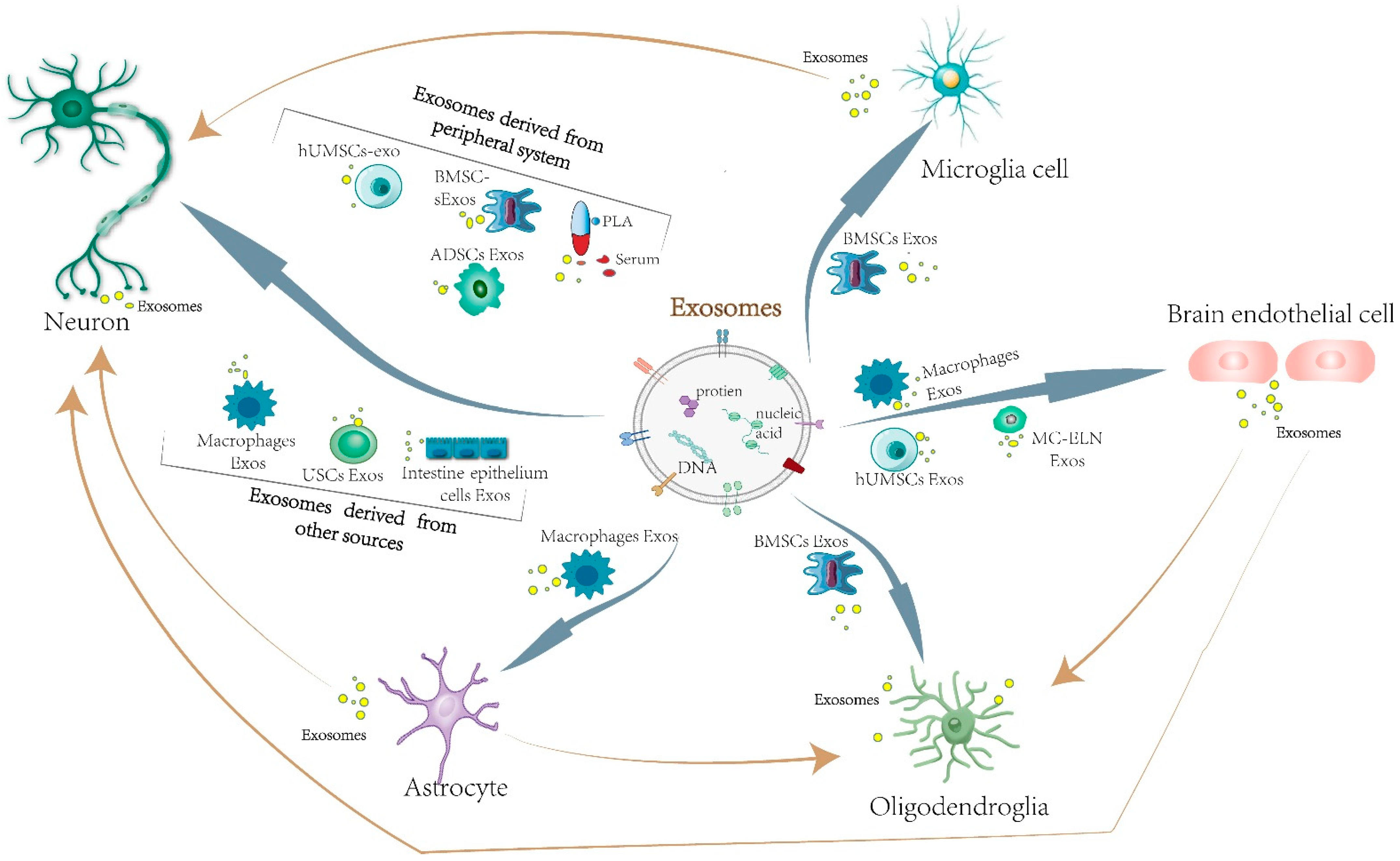
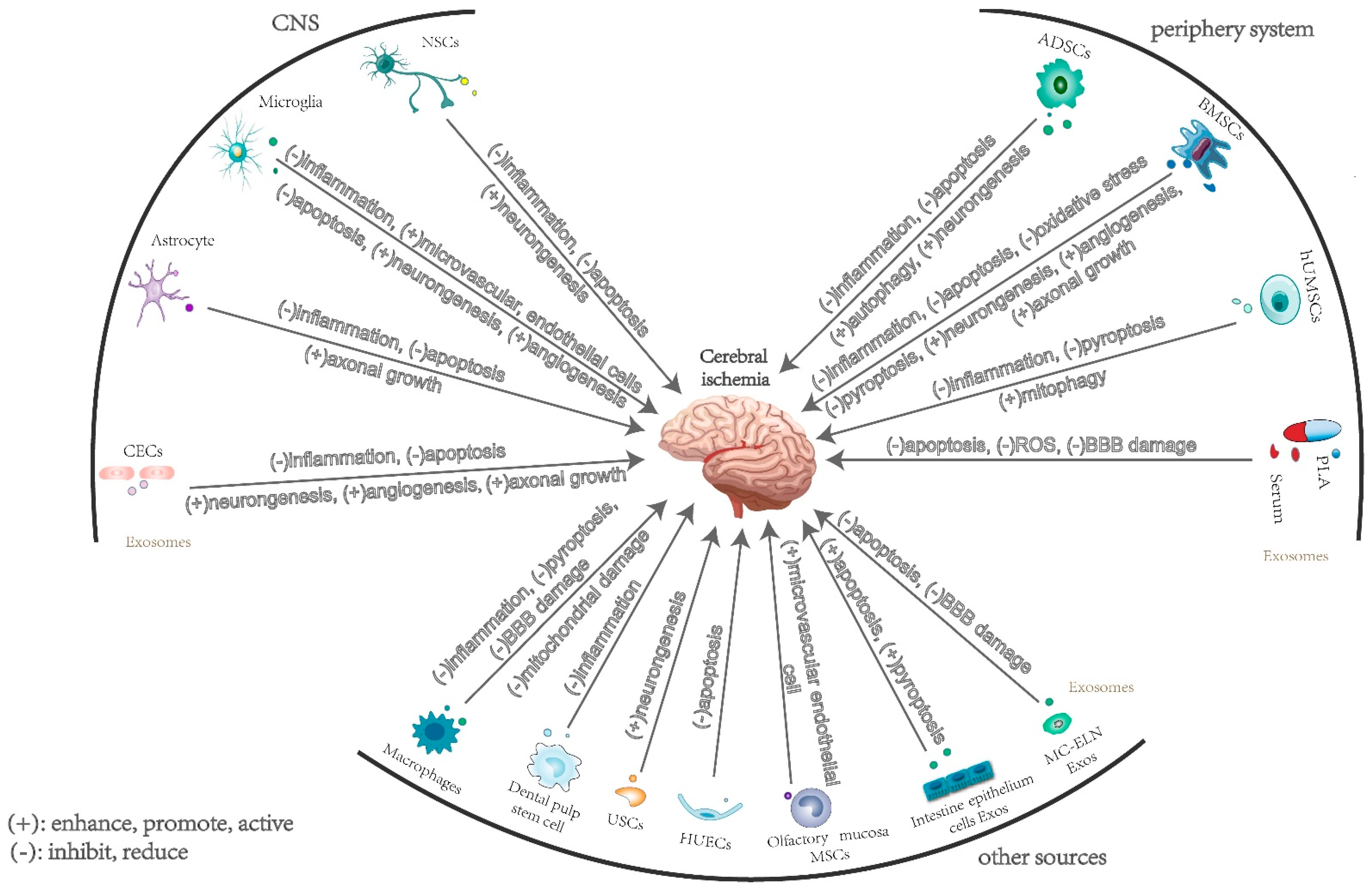
2. The Effects and Mechanisms of Exosomes Derived from Central Nervous System Cells in Cerebral Ischemia

Most types of cells in the CNS and peripheral system can secrete exosomes, including microglia, neurons, astrocytes [30], and endothelial cells [31]. The exosomes released from cells in the CNS contribute to cell-to-cell communication and intercellular signaling. Furthermore, they can pass BBB and blood–cerebrospinal fluid barriers, serving as communication between the periphery and the brain [32], which invests exosomes with enormous potential for clinical application.

2.1. Neuron-Derived Exosomes

Neural stem cells (NSCs) are a group of ectodermal progenitor cells that can differentiate into committed neural sub-types such as neurons, astrocytes, or oligodendrocytes [33]. Recent studies have shown that neural stem cell-derived exosomes (NSCs-exos) and cortical neuron-derived exosomes play injury-reducing and neuroprotective [34] roles in hypoxic-ischemic brain injury. Research indicates that NSCs-exos exert their neuroprotective effects via the transfer of miR-150-3p into injured cells, which inhibits the CASP2 signaling pathway, thus suppressing neuronal apoptosis and promoting neuron proliferation after brain injury [35]. miR-181c-3p contained in cortical neuron-derived exosomes is found to be an important molecule in the modulation of neuroinflammation after brain ischemia, as it can inhibit the expression of chemokine (C-X-C motif) ligand 1 (CXCL1) and the production of inflammatory factors by astrocytes, which reduces the inflammation burden and achieves the effect of protecting the nerves after ischemic brain injury (IBI). This finding may reveal a therapeutic approach for alleviating the symptoms of IBI through the regulation of astrocyte-induced inflammation [36]. Neural progenitor cell-derived extracellular vesicles (NPC-EVs) were shown to be effective against neuron inflammation, while their poor lesion region-targeting performance raises challenges to their clinical application in the therapy of ischemic stroke. Hence, researchers have managed to combine NPC-EVs with arginine-glycine-aspartic acid (RGD)-4C peptide, which has been proven to be a success for transferring therapeutic molecules to injured areas in the ischemic brain. The experimental results showed that RGD-treated NPC-EVs could improve the targeting of the lesion region of the ischemic brain and suppress poststroke inflammation [26], which leads to a potential therapeutic approach for ischemic stroke. Human induced pluripotent stem cell-derived neural progenitor cells (iPSC-NPCs) have many common points with cortical neurons in morphology and immunohistochemistry. In the microenvironment of ischemic stroke, iPSC-NPCs have a better potential for promoting cellular survival and proliferation, as well as differentiation into mature neurons or astrocytes [37,38]. Studies have shown that, through the regulation of the PTEN/AKT signaling pathway and neurite outgrowth, iPSC-NPC-derived exosomes could protect neurons against damage caused by oxygen–glucose deprivation (OGD) [39].

2.2. Microglia-Derived Exosomes

As the unique immune cells of the CNS, microglia play essential roles in many cerebral physiological processes, including regulating immune responses and promoting neuronal survival [40]. Brain damage induced by ischemia stroke can activate microglia and polarize them into either the “classically activated” M1 phenotype or the “alternatively activated” M2 phenotype [41]. Plenty of evidence suggests that M1 microglia promote the release of pro-inflammatory cytokines, inhibit synaptic growth, and aggravate acute brain damage [16]. On the contrary, the M2 phenotype is identified as a “healing” phenotype that establishes neuroprotective effects on the brain and improves long-term neurological outcomes after stroke [42]. Various kinds of miRNAs contained in microglia-derived exosomes are involved in the process of alleviating the inflammatory response, reducing damage, and promoting repair after ischemic hypoxic brain injury. Through the secretion of exosomes containing miRNA-26a, interleukin-4-polarized microglia cells can promote angiogenesis, thus alleviating ischemic stroke-induced damage [43]. Exosomes secreted by microglia in the M2 phenotype (BV2-exos) can transport the exosomal miRNA-137 targeting gene Notch1 to attenuate neuronal apoptosis, which contributes to the amelioration of ischemia–reperfusion (I/R) brain injury [44]. BV2-exos can moderate neuronal apoptosis after stroke occurs and promote the survival of neurons via the secretion of miR-124 enriched in exosomes and its downstream target USP14 [16]. Furthermore, a group of researchers have found that the neuroprotective effect of Vinpocetine could be associated with their actions on microglia cells. By inhibiting phosphodiesterase enzyme1-B (PDE1-B) in microglial cells, Vinpocetine could not only inhibit the polarization of the M1 microglial phenotype, but also enhance autophagic flux, which is associated with the alteration of exosomal contents and properties for protecting the survival and neurite structures of neurons against ischemic stroke [45]. Nevertheless, the effects of microglia-derived exosomes may not always be positive in the hypoxia state. Fibroblast growth factor2 (FGF2) can promote endothelial cell angiogenesis by activating STAT3, whereas upregulated miR-424-5p in microglial exosomes in hypoxia can inhibit the activation of the STAT3 pathway by targeting FGF2, thus resulting in brain microvascular endothelial cell (BMEC) injury. Therefore, the inhibition of miR-424-5p may promote endothelial cell angiogenesis and reduce neuronal damage [46].

2.3. Astrocyte-Derived Exosomes

The astrocyte is the largest type of glial cell. Astrocytes are widely distributed throughout the CNS. Their unique morphology can provide structural support to neurons. Furthermore, astrocytes have other functions, such as regulating neuroinflammatory responses, modulating synaptic activity, supplying energy to neurons, and maintaining the blood–brain barrier (BBB) [47]. Astrocytes also play a part in regulating autophagy level after brain injury occurs. Numerous experimental results have shown that insufficient or excessive levels of autophagy could aggravate cell death, while modest autophagy could protect cells against stressful circumstances and facilitate cell survival [48]. In vivo and in vitro experiments have confirmed that astrocyte-derived exosomes (ASC-exos) could ameliorate ischemia-induced neuronal damage via the inhibition of OGD-induced neuron autophagy [49]. By studying exosomes isolated from an OGD-based ischemia model in vitro, researchers have found that circular RNA circSHOC2 in ischemic-preconditioned astrocyte-derived exosomes suppress neuronal apoptosis and ameliorate cellular damage through the regulation of autophagy and its action on miR-7670-3p, thereby upregulating sirtuin1 (SIRT1) levels. The findings of this research may lead to a novel therapy for ischemic stroke treatment [50]. Through the suppression of histone demethylase KDM6B, downregulating bone morphogenetic proteins (BMP2), and silencing si-Bcl-2 modifying factor (BMF), astrocyte extracellular vesicle (ASC-EV)-derived miR-22-3p can significantly decrease apoptosis, mitigate the I/R brain injury in vivo, and enhance neuron viability in vitro [51]. Another study has suggested that ASC-exos that carry miR-17-5p could improve neurobehaviors, reduce neuronal apoptosis and cerebral infarction, and inhibit oxidative stress responses, as well as inflammation, in vivo and in vitro [52]. Except the miRNAs mentioned above, some other substances in astrocyte exosomes have also attracted attention. ASC-exos with prostaglandin D2 (PGD2) synthase expression can promote axonal outgrowth and functional recovery after stroke [53]. Astrocytes can also be linked to the mechanism of ischemic preconditioning (IPC). Neurons can take up exosomes released by OGD-preconditioned astrocytes. In this process, miR-92b-3p contained in exosomes is transferred from preconditioned astrocytes to neurons, which contributes to the attenuation of OGD-induced neuron death and apoptosis and protects neurons against OGD damage [54]. Another study demonstrated that, when using in vitro exosome treatment via delivering gap junction alpha 1 (GJA1), exosomes released by astrocytes can be taken up by neurons, leading to the downregulation of the apoptosis level and the upregulation of mitochondrial performance, which promotes the functional recovery of damaged neurons [55].

2.4. Brain Microvascular Endothelial Cell-Derived Exosomes

Exosomes derived from cerebral endothelial cells play essential roles in protecting neurons under a hypoxia state. Endothelial cell-derived exosomes (EC-exos) can protect neurons against I/R injury through the promotion of cell growth, migration, and invasion, and the inhibition of the apoptosis of SH-SY5Y nerve cells [56]. MiRNA-126-3p from EC-exos can protect PC12 cells against apoptosis and promote neurite outgrowth. This finding may serve as a therapeutic strategy for mitigating nerve damage and promoting function recovery [57]. Exosomes derived from cerebral endothelial cells (CECs) can enhance axonal growth by altering miRNAs and their target protein profiles, such as RhoA, in recipient neurons. The research also demonstrated that exosomes released by CECs from ischemic stroke-affected rats could promote axonal growth better than CEC-exos from a normal brain [58]. Vascular endothelial cell-derived exosomes (VEC-exos) can promote the survival viability of neural progenitor cells (NPC) and protect NSCs against I/R injury through the facilitation of neuron proliferation, migration, and the inhibition of apoptosis in vitro [59]. Endothelial progenitor cell-derived exosomes (EPC-exos) may alleviate ischemic injury by inhibiting apoptosis and promoting angiogenesis [60].
Based on above experiments and researches, it can be concluded that exosomes from CNS alleviate damage after cerebral ischemia and promote recovery through reducing inflammation, suppressing apoptosis, as well as enhancing neurogenesis and angiogenesis (Table 1).

3. The Effects and Mechanisms of Exosomes Derived from Peripheral Cells in Cerebral Ischemia

3.1. Mesenchymal Stem-Cell-Derived Exosomes

Mesenchymal stem cells, considered as “sentinel and safe-guards of injury” [61], have been proven to be able to release neurotrophic factors, including glial-derived neurotrophic factor, brain-derived neurotrophic factor, and nerve growth factor, which have shown their therapeutic potential in damage repair [32].

3.1.1. Bone Marrow Mesenchymal Stem-Cell-Derived Exosomes

Bone mesenchymal stem-cell-derived exosomes (BMSC) are a group of unique stem cells with great differential potential. The protective effect of BMSC-derived exosomes (BMSC-exos) is found to be linked to the inhibition of apoptosis in different studies. Various miRNAs can function in an ischemia-hypoxic brain environment taking BMSC-exos as carriers. BMSC-exo-miR-26a-5p mimics can dramatically reduce protein kinase CDK6 levels in BV-2 cells after oxygen–glucose deprivation/reoxygenation (OGD/R) treatment, as well as in brain tissues of the middle cerebral artery occlusion–reperfusion (MCAO/R) model. The downregulation of CDK6 mediated by exosomal miR-26a-5p might contribute to the attenuation of I/R injury in vivo, and the inhibition of microglia apoptosis in vitro [62]. By downregulating the gene PTEN, and therefore activating the PI3K/Akt/mTOR pathway, the miR-17-92 cluster enriched can increase corticospinal tract (CST) neuronal plasticity, axonal myelination, and axonal extension under the situation of rat cerebral stroke, which enhances the electrophysiological response and promotes neurological functional recovery in the poststroke phase [63]. Furthermore, BMSC-exos can facilitate angiogenic function and the cellular survival of the hypoxia/reoxygenation (H/R)-affected endothelial cell through the release of miR-126. The mechanism may be associated with the activation of the PI3K/Akt/eNOS pathway, which decreases cleaved caspase-3 expression, and promotes angiogenesis and the generation of growth factors [64]. Another study showed that the PI3K/Akt/eNOS pathway can also be activated by miR-132-3p-enriched BMSC-exos, thus mitigating H/R-induced oxidative stress and endothelial cell apoptosis [65]. By targeting tumor necrosis factor receptor associated factor 6 (TRAF6), microRNA-124-3p contained in BMSC-exos improves neurological functions, alleviates neuron pathological and structural damage, suppresses oxidative stress, and reduces neuronal apoptosis in newborn hypoxic-ischemic brain damage (HIBD) rats [66]. BMSC-exos carrying miR-455-3p can attenuate hippocampal neuronal injury in MCAO/R mice and OGD/R-induced N2a cell injury by increasing N2a cell activity and decreasing apoptosis through miR-455-3p targeting PDCD7 [67]. BMSC-exos can also inhibit apoptosis by targeting TGR5 [68]. miR-150-5p from BMSCs can enhance the therapeutic effects of BMSC-exos on cerebral I/R injury via a reduction in B-cell translocation gene 2 (TLR5) expression in MCAO rats, which contributes to a decrease in inflammatory factor levels, the inhibition of neuron apoptosis, the mitigation of pathological change, and an improvement in neurological function [69]. These studies provide a potential therapeutic strategy for the treatment of cerebral infarction.
Apart from the miRNA mentioned above, BMSC-exos are also found to function in protecting neurons through other mechanisms and pathways. It has been proven that the nucleotide-binding domain and leucine-rich repeat family protein 3 (NLRP3) inflammasome plays an essential role in neuronal damage induced by I/R [70]. BMSC-exos can promote microglial polarization toward M2, which suppresses NLRP3 inflammasome-mediated inflammation and pyroptosis, thus alleviating cerebral I/R injury [71]. Another study demonstrated that BMSC-exos could ameliorate NLRP3 inflammasome-mediated pyroptosis by promoting AMP-activated kinase (AMPK) dependent autophagic flux [72]. In addition, exosomes derived from CXC motif chemokine receptor type 4 (CXCR4) overexpressing BMSCs exhibit antiapoptotic effect via the Wnt-3a/β-catenin pathway, which facilitates the proliferation and tube formation of microvascular endothelial cells in cerebral I/R injury [73]. Researchers also noticed that cysteinyl leukotrienes (CysLTs), as potent inflammatory mediators, were largely produced with the decomposition of necrotic cells, while BMSC-exos could reverse CysLT2R-ERK1/2’s effect of inducing M1 microglia polarization, and promote the differentiation of microglia to the M2 phenotype, thus attenuating brain injury and ameliorating microglial inflammation meditated by M1 microglia [74]. An experiment intravenously administered BMSC-derived small extracellular vesicles (sEVs) obtained from bone marrow sample of a healthy donor (2 × 10⁶ or 2 × 10⁷ BMSC equivalents/kg) to young and aged mice. The results showed that sEVs at both doses promoted periinfarct angiogenesis in both types of rats. In addition, low-dose sEVs enhanced neurogenesis in the subventricular zone [75]. Additionally, BMSC-derived small extracellular vesicles (BMSC-sEVs) can decrease the infiltrates of inflammatory cells, such as leukocytes, monocytes, polymorphonuclear neutrophils, and macrophages, in the brains of aged mice with ischemic stroke. In peripheral blood, the number of monocytes and activated T cells are remarkably decreased by BMSC-sEVs, further reducing inflammation and mediating postischemic neuroprotection in brains of aged mice [76]. Molecular imaging technique showed that in the ischemic brains of mice, BMSC-exos treatment could significantly facilitate angiogenesis and neurogenesis and reduce the expression of IL-1β as well [77]. This research also showed that BMSC-exos could migrate into the brains of mice with ischemic stroke, which provides a new approach of clinical remission or therapy of ischemic stroke [77]. BMSC-exos treatment can attenuate OGD/R-induced oxidative stress and the dysregulation of mitochondrial function-associated genes in hippocampal neurons, which makes BMSC-exos treatment a potential therapeutic strategy to prevent neuronal damage induced by OGD/R [78]. Buyang Huanwu Decoction (BYHWD) treatment can promote angiogenetic miRNA and vascular endothelial growth factor (VEGF) expression in BMSC-exos, thus upregulating angiogenesis in the rat brain [79]. Another group of researchers observed that iron oxide nanoparticles (IONP) stimulated the expression of therapeutic growth factors in the MSC. They found that magnetic extracellular nanovesicles (MNV) derived from IONP-harboring BMSCs, supported by magnetic navigation, could improve the ischemic-lesion localization and the therapeutic effects of BMSC-exos, which enhances their abilities in anti-inflammation and anti-apoptosis, and promotes angiogenesis in ischemic brain lesions, thus contributing to a significant decrease in infarction volume and to the amelioration of motor function. The MNV injection may be applied to avoid the major defect of current BMSC-exos treatment or nanovesicles (NV) treatment in ischemic stroke [80].

3.1.2. Adipose-Derived Mesenchymal Stem Cells

Pigment epithelium-derived factor (PEDF) is a multifunctional protein that has neurotrophic, anti-inflammatory, and neuroprotective functions. Researchers have found that exosomes released by adipose-derived mesenchymal stem cells (ADSCs-exos) with increased PEDF content could further protect neurons against OGD-induced apoptosis in cerebral I/R through the activation of autophagy and the suppression of neuronal apoptosis. It is also observed that blocking autophagy could lead to a reduction in the effect of PEDF-containing exosomes [81]. Exosomes from hypoxic pre-treated ADSCs can alleviate brain injury caused by acute ischemic stroke via the delivery of circ-Rps5. Circ-Rps5 can induce the overexpression of its downstream targets miR-124-3p or the downregulation of SIRT7, which promotes the polarization of microglia from the M1 to M2 phenotype under lipopolysaccharide (LPS) conditions [82]. Another study showed that the systemic administration of ADSCs-exos remarkably promoted the expression of von Willebrand factor, an endothelia cell marker, and doublecortin, a neuroblasts marker, which reduced neuron cell death and enhanced cell proliferation in comparison with the control group. Moreover, researchers found that exosomes from miRNA-126-modified ADSCs could inhibit microglial activation and the expression of inflammatory factors in vivo and in vitro, thus improving functional recovery, inhibiting neuroinflammation, and enhancing neurogenesis. The result of this study suggested that the intravenous administration of miR-126+ exosomes in poststroke might represent a novel treatment for stroke [83].

3.1.3. Human Umbilical Cord Mesenchymal Stem Cell-Derived Exosomes

Multiple studies have shown that human umbilical cord mesenchymal stem cell-derived exosomes (hUMSC-exos) can reduce microglia-mediated neuroinflammation, protecting against brain injury. Exosomes derived from umbilical cord mesenchymal stem cells can interfere with the Toll-like receptor 4 (TLR4) signaling of BV-2 microglia, which contributes to reducing neuroinflammation induced by microglia in perinatal brain injury [84]. HUMSC-exos carrying miR-146a-5p can alleviate the neuroinflammatory response mediated by microglial via the suppression of the IRAK1/TRAF6 pathway [85]. Furthermore, hUMSC-exos can enhance mitophagy and alleviate subsequent neuronal injury by increasing FOXO3a expression, therefore attenuating OGD/R-induced microglial pyroptosis [86]. Researchers also explored the clinical application potential of hUMSC-exos. A study showed that hUMSC-exos injected into rats with deep vein thrombosis (DVT) could upregulate the delivery of miR-342-3p, which downregulated the expression of endothelin A receptor and eventually alleviated DVT. The above findings may provide a novel clinical treatment for DVT [87].

3.2. Plasma and Serum Exosomes

Research indicated that plasma-derived exosomes (PLA-exos) enriched with heat shock protein 70 (HSP70) showed better brain targeting and treatment effects. PLA-exos with inherited HSP70 can facilitate the migration and diapedesis of PLA-exos through an interaction with endothelial TLR4. Furthermore, PLA-exos-meditated HSP70 delivery can activate tight junction protein (TJP), which leads to the inhibition of mitochondria-meditated neuron cell apoptosis, the suppression of ROS accumulation, and the alleviation of BBB damage [88]. Edaravone-loaded PLA-exos could mitigate ischemic damage in brain tissue via the interaction between transferrin on the surface of PLA-exos and transferrin receptor (TfR) on the surface of brain endothelial cells, which reduces ROS generation [89]. Researchers have also found that melatonin-treated exosomes effectively reduced the infarct volume and improved function recovery via the regulation of the TLR4/Nuclear Factor kappa B (NF-kB) signaling pathway, thus further enhancing therapeutic effects of PLA-exos against inflammatory responses induced by ischemic stroke and inflammasome-mediated pyroptosis [90]. Another study showed that growth arrest and DNA damage-inducible protein 34 (GADD34) level were increased in PLA-exos of cerebral ischemic rats, which might be the consequence of the dephosphorylation of eukaryotic translation initiation factor 2α (eIF2α) and the phosphorylation of p53. Furthermore, GADD34 inhibitor treatment inhibited neuronal apoptosis, decreased the infarct volume, and improved functional outcomes in cortical penumbra after ischemic stroke [91]. Additionally, a study revealed that circulating plasma exosomes were related to the increased risk of stroke after varicella zoster virus reactivation caused herpes zoster (HZ, shingles). Compared to exosomes from patients without HZ, HZ exosomes can initiate platelets to form platelet–leukocyte aggregates and contain the proteins that can concert recipient cells to a prothrombotic state. Therefore, the application of antiplatelet agents for HZ may be a feasible clinical practice to decrease stroke risk [92].
Exosomes from remote ischemic preconditioning (RIPC) serum show neuroprotective effects via the upregulation of miRNA-126, thus reducing the expression of DNMTs3B in neurons and raising OGD tolerance in SH-SY5Y cells [93]. However, research has also proven that serum exosomal miR-27-3p could target PPARγ to stimulate the activation of microglia and the expression of inflammatory cytokines, thus aggravating acute cerebral infarction (ACI). Nevertheless, the root cause of upregulated exosomal miR-27-3p expression in serum after ACI requires further study [94]. Moyamoya disease (MMD) is a rare steno-occlusive and slowly progressing cerebrovascular disorder, and its pathogenesis mechanism is yet unknown. Researchers have found that the proliferation levels of mouse brain vascular EC cells were significantly increased, and that more ethynyl-2-deoxyuridine-positive cells were generated after treatment with MMD serum-derived exosomes (SDEs). However, it was also observed that SDEs from an ischemic MMD patient promoted neuroblastoma cell proliferation, and SDEs from hemorrhagic MMD patients induced the dysfunction of the mitochondria in cerebrovascular ECs. The finding may help with understanding the pathogenesis mechanisms of MMD and provide new therapeutic strategies for the disease [95].
According to the related studies we can summarize that the effects of exosomes from peripheral nervous system in brain ischemic stroke are mainly exerted through facilitating brain tissues functional recovery, inflammation reduction, as well as inhibition of cells pyroptosis and apoptosis (Table 2).

4. The Effects and Mechanisms of Exosomes Derived from Other Sources in Cerebral Ischemia

Apart from the above-mentioned sources, macrophages and some other cells can also react to brain injury via exosomes.
Through the regulation of microglial polarity from phenotype M1 to anti-inflammatory phenotype M2, LPS-stimulated macrophage-derived exosomes have functional improvements and neuroprotection effects after ischemic stroke [96]. Macrophage-derived exosomes can serve as carriers of various drug molecules to enhance or to facilitate the functions of drugs towards targeting pathways or cells. Exosomes from heptapeptide-loaded (Hep-loaded) macrophages can reduce mitochondrial injury in astrocytes via the suppression of dynamin-related protein-1 (Drp1)-fission 1 (Fis1) interaction after I/R, alleviating mitochondria-mediated neuronal damage [97]. Edaravone-loaded macrophage-derived exosomes enhance the targeting performance of Edaravone toward ischemic lesion areas in rat brains with permanent middle cerebral artery occlusion (PMCAO), which further reduces neuronal cell death and promotes the polarization of microglia from M1 to M2 [98]. Curcumin-laden exosomes from macrophages can alleviate cerebral I/R injury by downregulating ROS accumulation in lesions, thus reducing BBB damage and suppressing neuronal apoptosis mediated by mitochondria [99]. Another research team isolated exosomes from the supernatants of interleukin-4-induced M2-polarized macrophages, and discovered that M2-macrophages-derived exosomes can activate the nuclear factor erythroid related factor 2 (Nrf2)/heme-oxygenase-1 (HO-1) signaling pathway, which contributes to inhibiting the generation of ROS and malondialdehyde, reducing the release of lactate dehydrogenase, increasing cell activity, and eventually achieving the purpose of protecting HT22 neurons [100].
Human umbilical endothelial cells (HUECs) can reduce the apoptosis of neurons under OGD in a HUEC-neuron coculture assay. Researchers also found that Cav-1 upregulated by neurons during ischemia stroke can increase the neuron intake of extracellular vesicles derived from an endothelial cell, which attenuates apoptosis via exosomal miR-1290 and protects neurons [101]. Exosome release via stem cell-derived dental pulp can mitigate cerebral I/R damage by inhibiting the inflammatory response mediated by the HMGB1/TLR4/MyD88/NF-κB pathway [102]. Exosomes released by human urine-derived stem cells (USCs) can enhance neuronal differentiation and the proliferation of NSCs after OGD/R via the exosomal miR-26a/histone deacetylase 6 (HDAC6) axis [103]. Human USCs can also release exosomes enriched with miR-21-5p, which promotes early nerve formation by regulating the Eph receptor A(EPha4)/tyrosine kinase (TEK) axis [104]. EVs from human hypoxic olfactory mucosa MSCs (OM-MSCs) were proven to be able to promote the migration, proliferation, and angiogenic activities of human brain microvascular endothelial cells via the exosomal miR-612–TP53–hypoxia-inducible factor 1-alpha (HIF-1α)–vascular endothelial growth factor (VEGF) axis [105]. Another team performed an OGD-affected IEC-6-primary cortical neuron coculture system under normothermia (37 °C) and therapeutic hypothermia (TH) (32 °C) conditions. The researchers found that I/R-injured intestinal epithelium cells can induce cortical neuron death via the production of paracrine mediators such as exosomal miRNAs associated with necroptosis, apoptosis, and/or pyroptosis, whereas TH can counteract this process and protect cortical neurons in stroke patients [106]. Except for the exosomes from animal cells, researchers also isolated and characterized novel plant exosome-like nanoparticles (ELNs) from Momordica charantia (MC) and found that MC-ELNs attenuate ischemia–reperfusion-induced damage to the BBB and suppress neuronal apoptosis, probably via the modulation of the phosphoinositide-3-kinase (PI3Ks)/protein kinase B (AKT)/glycogen synthase kinase (GSK3β) signaling pathway [107]. Moreover, based on in vivo and in vitro experiments, researchers discovered the effects of extracted MC-exosomes (MCEs) in the inhibition of platelet activation, and the aggregation, adhesion, and platelet-mediated migration of HCT116 cells, which may reveal the potential of MCEs in therapies for stroke and tumor metastasis [108].
Therefore, we can conclude that exosomes from other sources also have protective effects in cerebral ischemic stroke, and possess great potential as therapy of brain ischemia (Table 3).

5. Conclusions and Prospects

It can be concluded from the above review that exosomes derived from diverse sources targeting at different brain cells (Figure 1) through different mechanism (Figure 2) result in corresponding protective (mainly) or damaging effects on the brain tissues after ischemia. However, the current studies are mostly limited to a single substance in exosomes from a single source, and whether there are synergistic effects of multiple substances in exosomes to function in the process of cerebral ischemia needs further exploration because the interactions between nerve cells are complex. Moreover, how do brain parenchymal cells or remote organs affect the secretion of exosomes and the changes of their internal substances through cell signaling pathways? How do exosomes regulate the expression of endogenous genes in recipient cells? These questions are waiting to be answered. Secondly, as molecular markers for disease diagnosis, exosomes have great potential in regulating neurological function recovery after ischemic stroke. Nevertheless, there is still room for improvement in the purity and quality of the extraction, although the method of extracting exosomes has gradually matured. Efficient and reliable exosome isolation technologies still require more study and investigation. Furthermore, exosomes can combine with drugs as carriers to play a synergistic role. There are many studies focusing on the combination of exosomes and drugs in the mitigation and treatment of ischemic stroke [26,45,53,80,89,90,97,98,99,105]. In addition, the association between exosomes and the formation of thrombus has been confirmed, as well as the accumulation of blood platelets, which may provide new ideas in the treatment of thrombolysis, thrombectomy, and anti-platelet in cerebral stroke. However, large-scale clinical trials are needed to verify their clinical feasibility. The potential adverse effects of exosomes applied in clinical therapy are as yet blurry. Improving targeting through the modification of exosomes to make them easier to reach the ischemic lesion is also a key direction for carrier-exosome therapy research, which is expected to provide strong support for the clinical treatment of cerebral ischemia. Overall, the clinical application of exosomes in the treatment of cerebral ischemia is a novel and promising therapeutic approach. The study of the mechanism of exosomes acting in the process of cerebral ischemia is of great significance for guiding the applications of exosomes in clinical treatment.

Author Contributions

Conceptualization, X.H., C.D.; Reference collection, R.X., X.Z., H.Y.; Draft Writing, R.X.; Manuscript polishing, X.H., R.X.; Picture and table drawing, R.X.; Supervision, X.H. All authors have read and agreed to the published version of the manuscript.

Funding

This study was funded by National Natural Science Foundation of China (No. 82274340), Natural Science Foundation of Hunan Province (2022JJ30432), Natural Science Foundation of Changsha (kq2014084), Key Project of Hunan Provincial Administration of Traditional Chinese Medicine (No. E2022001), The domestic first-class cultivation discipline project of Integrated Traditional Chinese and Western Medicine in Hunan Province (2021ZXYJH06), National College Students’ Innovation and Entrepreneurship Training Program in 2021 (S202110541053), Research Fund Project of Hunan University of Chinese Medicine (2021xjjj027), First-class Discipline Construction Project of basic medicine in 13th Five-Year Plan of Hunan University of Chinese Medicine (06), and Excellent Teaching Team of Postgraduate in Hunan Province (Teaching Team of Postgraduate in Basic Medicine, Grant No. (2019-370–118)).

Institutional Review Board Statement

Not applicable.

Conflicts of Interest

We confirm that there are no known conflict of interest associated with this publication and there has been no significant financial support for this work that could have influenced its outcome.

References

  1. Shin, T.H.; Lee, D.Y.; Basith, S.; Manavalan, B.; Paik, M.J.; Rybinnik, I.; Mouradian, M.M.; Ahn, J.H.; Lee, G. Metabolome Changes in Cerebral Ischemia. Cells 2020, 9, 1630. [Google Scholar] [CrossRef] [PubMed]
  2. Cuartero, M.I.; de la Parra, J.; Garcia-Culebras, A.; Ballesteros, I.; Lizasoain, I.; Moro, M.A. The Kynurenine Pathway in the Acute and Chronic Phases of Cerebral Ischemia. Curr. Pharm. Des. 2016, 22, 1060–1073. [Google Scholar] [CrossRef]
  3. Khoshnam, S.E.; Winlow, W.; Farzaneh, M.; Farbood, Y.; Moghaddam, H.F. Pathogenic mechanisms following ischemic stroke. Neurol. Sci. 2017, 38, 1167–1186. [Google Scholar] [CrossRef] [PubMed]
  4. Zhao, L.R.; Willing, A. Enhancing endogenous capacity to repair a stroke-damaged brain: An evolving field for stroke research. Prog. Neurobiol. 2018, 163–164, 5–26. [Google Scholar] [CrossRef] [PubMed]
  5. Abels, E.R.; Breakefield, X.O. Introduction to Extracellular Vesicles: Biogenesis, RNA Cargo Selection, Content, Release, and Uptake. Cell Mol. Neurobiol. 2016, 36, 301–312. [Google Scholar] [CrossRef] [Green Version]
  6. Minciacchi, V.R.; Freeman, M.R.; Di Vizio, D. Extracellular vesicles in cancer: Exosomes, microvesicles and the emerging role of large oncosomes. Semin. Cell Dev. Biol. 2015, 40, 41–51. [Google Scholar] [CrossRef] [Green Version]
  7. Théry, C.; Ostrowski, M.; Segura, E. Membrane vesicles as conveyors of immune responses. Nat. Rev. Immunol. 2009, 9, 581–593. [Google Scholar] [CrossRef]
  8. Shao, H.; Im, H.; Castro, C.M.; Breakefield, X.; Weissleder, R.; Lee, H. New Technologies for Analysis of Extracellular Vesicles. Chem. Rev. 2018, 118, 1917–1950. [Google Scholar] [CrossRef]
  9. Tan, S.; Xia, L.; Yi, P.; Han, Y.; Tang, L.; Pan, Q.; Tian, Y.; Rao, S.; Oyang, L.; Liang, J.; et al. Exosomal miRNAs in tumor microenvironment. J. Exp. Clin. Cancer Res. 2020, 39, 67. [Google Scholar] [CrossRef]
  10. Bebelman, M.P.; Smit, M.J.; Pegtel, D.M.; Baglio, S.R. Biogenesis and function of extracellular vesicles in cancer. Pharmacol. Ther. 2018, 188, 1–11. [Google Scholar] [CrossRef]
  11. Pan, B.T.; Teng, K.; Wu, C.; Adam, M.; Johnstone, R.M. Electron microscopic evidence for externalization of the transferrin receptor in vesicular form in sheep reticulocytes. J. Cell Biol. 1985, 101, 942–948. [Google Scholar] [CrossRef] [PubMed] [Green Version]
  12. Pan, B.T.; Johnstone, R.M. Fate of the transferrin receptor during maturation of sheep reticulocytes in vitro: Selective externalization of the receptor. Cell 1983, 33, 967–978. [Google Scholar] [CrossRef]
  13. Johnstone, R.M.; Adam, M.; Hammond, J.R.; Orr, L.; Turbide, C. Vesicle formation during reticulocyte maturation. Association of plasma membrane activities with released vesicles (exosomes). J. Biol. Chem. 1987, 262, 9412–9420. [Google Scholar] [CrossRef]
  14. Zhang, J.; Li, S.; Li, L.; Li, M.; Guo, C.; Yao, J.; Mi, S. Exosome and exosomal microRNA: Trafficking, sorting, and function. Genom. Proteom. Bioinform. 2015, 13, 17–24. [Google Scholar] [CrossRef] [PubMed] [Green Version]
  15. Doyle, L.M.; Wang, M.Z. Overview of Extracellular Vesicles, Their Origin, Composition, Purpose, and Methods for Exosome Isolation and Analysis. Cells 2019, 8, 727. [Google Scholar] [CrossRef] [Green Version]
  16. Song, Y.; Li, Z.; He, T.; Qu, M.; Jiang, L.; Li, W.; Shi, X.; Pan, J.; Zhang, L.; Wang, Y.; et al. M2 microglia-derived exosomes protect the mouse brain from ischemia-reperfusion injury via exosomal miR-124. Theranostics 2019, 9, 2910–2923. [Google Scholar] [CrossRef]
  17. Zhang, X.; Sai, B.; Wang, F.; Wang, L.; Wang, Y.; Zheng, L.; Li, G.; Tang, J.; Xiang, J. Hypoxic BMSC-derived exosomal miRNAs promote metastasis of lung cancer cells via STAT3-induced EMT. Mol. Cancer 2019, 18, 40. [Google Scholar] [CrossRef] [Green Version]
  18. Kalluri, R.; LeBleu, V.S. The biology, function, and biomedical applications of exosomes. Science 2020, 367, eaau6977. [Google Scholar] [CrossRef] [PubMed]
  19. Zhou, W.; Woodson, M.; Neupane, B.; Bai, F.; Sherman, M.B.; Choi, K.H.; Neelakanta, G.; Sultana, H. Exosomes serve as novel modes of tick-borne flavivirus transmission from arthropod to human cells and facilitates dissemination of viral RNA and proteins to the vertebrate neuronal cells. PLoS Pathog. 2018, 14, e1006764. [Google Scholar] [CrossRef] [PubMed] [Green Version]
  20. Wang, M.-M.; Feng, Y.-S.; Tan, Z.-X.; Xing, Y.; Dong, F.; Zhang, F. The role of exosomes in stroke. Mol. Biol. Rep. 2020, 47, 6217–6228. [Google Scholar] [CrossRef]
  21. de Abreu, R.C.; Fernandes, H.; da Costa Martins, P.A.; Sahoo, S.; Emanueli, C.; Ferreira, L. Native and bioengineered extracellular vesicles for cardiovascular therapeutics. Nat. Rev. Cardiol. 2020, 17, 685–697. [Google Scholar] [CrossRef]
  22. Aminzadeh, M.A.; Rogers, R.G.; Fournier, M.; Tobin, R.E.; Guan, X.; Childers, M.K.; Andres, A.M.; Taylor, D.J.; Ibrahim, A.; Ding, X.; et al. Exosome-Mediated Benefits of Cell Therapy in Mouse and Human Models of Duchenne Muscular Dystrophy. Stem Cell Rep. 2018, 10, 942–955. [Google Scholar] [CrossRef] [PubMed] [Green Version]
  23. Zhang, L.; Liu, H.; Jia, L.; Lyu, J.; Sun, Y.; Yu, H.; Li, H.; Liu, W.; Weng, Y.; Yu, W. Exosomes Mediate Hippocampal and Cortical Neuronal Injury Induced by Hepatic Ischemia-Reperfusion Injury through Activating Pyroptosis in Rats. Oxid. Med. Cell Longev. 2019, 2019, 3753485. [Google Scholar] [CrossRef]
  24. Jiang, L.; Chen, W.; Ye, J.; Wang, Y. Potential Role of Exosomes in Ischemic Stroke Treatment. Biomolecules 2022, 12, 115. [Google Scholar] [CrossRef]
  25. Xiao, Y.; Zhang, Y.; Li, Y.; Peng, N.; Liu, Q.; Qiu, D.; Cho, J.; Borlongan, C.V.; Yu, G. Exosomes Derived From Mesenchymal Stem Cells Pretreated With Ischemic Rat Heart Extracts Promote Angiogenesis via the Delivery of DMBT1. Cell Transpl. 2022, 31, 9636897221102898. [Google Scholar] [CrossRef] [PubMed]
  26. Tian, T.; Cao, L.; He, C.; Ye, Q.; Liang, R.; You, W.; Zhang, H.; Wu, J.; Ye, J.; Tannous, B.A.; et al. Targeted delivery of neural progenitor cell-derived extracellular vesicles for anti-inflammation after cerebral ischemia. Theranostics 2021, 11, 6507–6521. [Google Scholar] [CrossRef] [PubMed]
  27. Tian, Y.; Cheng, C.; Wei, Y.; Yang, F.; Li, G. The Role of Exosomes in Inflammatory Diseases and Tumor-Related Inflammation. Cells 2022, 11, 1005. [Google Scholar] [CrossRef]
  28. Li, J.Y.; Li, Q.Q.; Sheng, R. The role and therapeutic potential of exosomes in ischemic stroke. Neurochem. Int. 2021, 151, 105194. [Google Scholar] [CrossRef]
  29. Yang, D.; Li, Z.; Gao, G.; Li, X.; Liao, Z.; Wang, Y.; Li, W.; Zhange, Y.; Liu, W. Combined Analysis of Surface Protein Profile and microRNA Expression Profile of Exosomes Derived from Brain Microvascular Endothelial Cells in Early Cerebral Ischemia. ACS Omega 2021, 6, 22410–22421. [Google Scholar] [CrossRef]
  30. Wang, J.; Wang, J.; Li, X.; Shu, K. Cell-Derived Exosomes as Therapeutic Strategies and Exosome-Derived microRNAs as Biomarkers for Traumatic Brain Injury. J. Clin. Med. 2022, 11, 3223. [Google Scholar] [CrossRef]
  31. Davidson, S.M.; Riquelme, J.A.; Zheng, Y.; Vicencio, J.M.; Lavandero, S.; Yellon, D.M. Endothelial cells release cardioprotective exosomes that may contribute to ischaemic preconditioning. Sci. Rep. 2018, 8, 15885. [Google Scholar] [CrossRef] [PubMed] [Green Version]
  32. Zagrean, A.M.; Hermann, D.M.; Opris, I.; Zagrean, L.; Popa-Wagner, A. Multicellular Crosstalk Between Exosomes and the Neurovascular Unit After Cerebral Ischemia. Therapeutic Implications. Front. Neurosci. 2018, 12, 811. [Google Scholar] [CrossRef] [PubMed]
  33. Huang, L.; Zhang, L. Neural stem cell therapies and hypoxic-ischemic brain injury. Prog. Neurobiol. 2019, 173, 1–17. [Google Scholar] [CrossRef] [PubMed]
  34. Kos, A.; Loohuis, N.O.; Meinhardt, J.; van Bokhoven, H.; Kaplan, B.B.; Martens, G.J.; Aschrafi, A. MicroRNA-181 promotes synaptogenesis and attenuates axonal outgrowth in cortical neurons. Cell Mol. Life Sci. 2016, 73, 3555–3567. [Google Scholar] [CrossRef] [Green Version]
  35. Luo, H.; Ye, G.; Liu, Y.; Huang, D.; Luo, Q.; Chen, W.; Qi, Z. miR-150-3p enhances neuroprotective effects of neural stem cell exosomes after hypoxic-ischemic brain injury by targeting CASP2. Neurosci. Lett. 2022, 779, 136635. [Google Scholar] [CrossRef]
  36. Song, H.; Zhang, X.; Chen, R.; Miao, J.; Wang, L.; Cui, L.; Ji, H.; Liu, Y. Cortical Neuron-Derived Exosomal MicroRNA-181c-3p Inhibits Neuroinflammation by Downregulating CXCL1 in Astrocytes of a Rat Model with Ischemic Brain Injury. Neuroimmunomodulation 2019, 26, 217–233. [Google Scholar] [CrossRef]
  37. De La Rosa-Prieto, C.; Laterza, C.; Gonzalez-Ramos, A.; Wattananit, S.; Ge, R.; Lindvall, O.; Tornero, D.; Kokaia, Z. Stroke alters behavior of human skin-derived neural progenitors after transplantation adjacent to neurogenic area in rat brain. Stem Cell Res. Ther. 2017, 8, 59. [Google Scholar] [CrossRef] [Green Version]
  38. Hermanto, Y.; Sunohara, T.; Faried, A.; Takagi, Y.; Takahashi, J.; Maki, T.; Miyamoto, S. Transplantation of feeder-free human induced pluripotent stem cell-derived cortical neuron progenitors in adult male Wistar rats with focal brain ischemia. J. Neurosci. Res. 2018, 96, 863–874. [Google Scholar] [CrossRef]
  39. Hu, X.-Y.; Li, W.-Y.; Zhu, Q.-B.; Jin, L.-Y.; Yang, Y.; Xu, X.-Y. Exosomes derived from human induced pluripotent stem cell-derived neural progenitor cells protect neuronal function under ischemic conditions. Neural. Regen. Res. 2021, 16, 2064–2070. [Google Scholar] [CrossRef]
  40. Li, Q.; Barres, B.A. Microglia and macrophages in brain homeostasis and disease. Nat. Rev. Immunol. 2018, 18, 225–242. [Google Scholar] [CrossRef]
  41. Zhang, M.; Wu, X.; Xu, Y.; He, M.; Yang, J.; Li, J.; Li, Y.; Ao, G.; Cheng, J.; Jia, J. The cystathionine β-synthase/hydrogen sulfide pathway contributes to microglia-mediated neuroinflammation following cerebral ischemia. Brain Behav. Immun. 2017, 66, 332–346. [Google Scholar] [CrossRef] [PubMed]
  42. Liu, X.; Liu, J.; Zhao, S.; Zhang, H.; Cai, W.; Cai, M.; Ji, X.; Leak, R.K.; Gao, Y.; Chen, J.; et al. Interleukin-4 Is Essential for Microglia/Macrophage M2 Polarization and Long-Term Recovery After Cerebral Ischemia. Stroke 2016, 47, 498–504. [Google Scholar] [CrossRef] [PubMed] [Green Version]
  43. Tian, Y.; Zhu, P.; Liu, S.; Jin, Z.; Li, D.; Zhao, H.; Zhu, X.; Shu, C.; Yan, D.; Dong, Z. IL-4-polarized BV2 microglia cells promote angiogenesis by secreting exosomes. Adv. Clin. Exp. Med. 2019, 28, 421–430. [Google Scholar] [CrossRef] [PubMed] [Green Version]
  44. Zhang, D.; Cai, G.; Liu, K.; Zhuang, Z.; Jia, K.; Pei, S.; Wang, X.; Wang, H.; Xu, S.; Cui, C.; et al. Microglia exosomal miRNA-137 attenuates ischemic brain injury through targeting Notch1. Aging 2021, 13, 4079–4095. [Google Scholar] [CrossRef]
  45. Zang, J.; Wu, Y.; Su, X.; Zhang, T.; Tang, X.; Ma, D.; Li, Y.; Lui, Y.; Weng, Z.; Liu, X.; et al. Inhibition of PDE1-B by Vinpocetine Regulates Microglial Exosomes and Polarization Through Enhancing Autophagic Flux for Neuroprotection Against Ischemic Stroke. Front. Cell. Dev. Biol. 2020, 8, 616590. [Google Scholar] [CrossRef]
  46. Xie, L.; Zhao, H.; Wang, Y.; Chen, Z. Exosomal shuttled miR-424-5p from ischemic preconditioned microglia mediates cerebral endothelial cell injury through negatively regulation of FGF2/STAT3 pathway. Exp. Neurol. 2020, 333, 113411. [Google Scholar] [CrossRef]
  47. Ravi, K.; Paidas, M.J.; Saad, A.; Jayakumar, A.R. Astrocytes in rare neurological conditions: Morphological and functional considerations. J. Comp. Neurol. 2021, 529, 2676–2705. [Google Scholar] [CrossRef]
  48. Kang, C.; Avery, L. To be or not to be, the level of autophagy is the question: Dual roles of autophagy in the survival response to starvation. Autophagy 2008, 4, 82–84. [Google Scholar] [CrossRef] [Green Version]
  49. Pei, X.; Li, Y.; Zhu, L.; Zhou, Z. Astrocyte-derived exosomes suppress autophagy and ameliorate neuronal damage in experimental ischemic stroke. Exp. Cell Res. 2019, 382, 111474. [Google Scholar] [CrossRef]
  50. Chen, W.; Wang, H.; Zhu, Z.; Feng, J.; Chen, L. Exosome-Shuttled circSHOC2 from IPASs Regulates Neuronal Autophagy and Ameliorates Ischemic Brain Injury via the miR-7670-3p/SIRT1 Axis. Mol. Ther. Nucleic Acids 2020, 22, 657–672. [Google Scholar] [CrossRef]
  51. Zhang, Y.; Liu, J.; Su, M.; Wang, X.; Xie, C. Exosomal microRNA-22-3p alleviates cerebral ischemic injury by modulating KDM6B/BMP2/BMF axis. Stem Cell Res. Ther. 2021, 12, 111. [Google Scholar] [CrossRef] [PubMed]
  52. Du, L.; Jiang, Y.; Sun, Y. Astrocyte-derived exosomes carry microRNA-17-5p to protect neonatal rats from hypoxic-ischemic brain damage via inhibiting BNIP-2 expression. Neurotoxicology 2021, 83, 28–39. [Google Scholar] [CrossRef] [PubMed]
  53. Hira, K.; Ueno, Y.; Tanaka, R.; Miyamoto, N.; Yamashiro, K.; Inaba, T.; Urabe, T.; Okano, H.; Hattori, N. Astrocyte-Derived Exosomes Treated With a Semaphorin 3A Inhibitor Enhance Stroke Recovery via Prostaglandin D2 Synthase. Stroke 2018, 49, 2483–2494. [Google Scholar] [CrossRef] [PubMed]
  54. Xu, L.; Cao, H.; Xie, Y.; Zhang, Y.; Du, M.; Xu, X.; Ye, R.; Liu, X. Exosome-shuttled miR-92b-3p from ischemic preconditioned astrocytes protects neurons against oxygen and glucose deprivation. Brain Res. 2019, 1717, 66–73. [Google Scholar] [CrossRef]
  55. Chen, W.; Zheng, P.; Hong, T.; Wang, Y.; Liu, N.; He, B.; Zou, S.; Ren, D.; Duan, J.; Zhoa, L.; et al. Astrocytes-derived exosomes induce neuronal recovery after traumatic brain injury via delivering gap junction alpha 1-20 k. J. Tissue Eng. Regen. Med. 2020, 14, 412–423. [Google Scholar] [CrossRef]
  56. Xiao, B.; Chai, Y.; Lv, S.; Ye, M.; Wu, M.; Xie, L.; Fan, Y.; Zhu, X.; Gao, Z. Endothelial cell-derived exosomes protect SH-SY5Y nerve cells against ischemia/reperfusion injury. Int. J. Mol. Med. 2017, 40, 1201–1209. [Google Scholar] [CrossRef] [Green Version]
  57. Gao, B.; Zhou, S.; Sun, C.; Cheng, D.; Zhang, Y.; Li, X.; Zhang, L.; Zhao, J.; Xu, D.; Bai, Y. Brain Endothelial Cell-Derived Exosomes Induce Neuroplasticity in Rats with Ischemia/Reperfusion Injury. ACS Chem. Neurosci. 2020, 11, 2201–2213. [Google Scholar] [CrossRef]
  58. Zhang, Y.; Qin, Y.; Chopp, M.; Li, C.; Kemper, A.; Liu, X.; Wang, X.; Zhang, L.; Zhang, Z.G. Ischemic Cerebral Endothelial Cell-Derived Exosomes Promote Axonal Growth. Stroke 2020, 51, 3701–3712. [Google Scholar] [CrossRef]
  59. Zhou, S.; Gao, B.; Sun, C.; Bai, Y.; Cheng, D.; Zhang, Y.; Li, X.; Zhao, J.; Xu, D. Vascular Endothelial Cell-derived Exosomes Protect Neural Stem Cells Against Ischemia/reperfusion Injury. Neuroscience 2020, 441, 184–196. [Google Scholar] [CrossRef]
  60. Huang, R.; Cheng, T.; Lai, X. Mechanism of ischemic brain injury repair by endothelial progenitor cell-derived exosomes. Mol. Med. Rep. 2022, 26, 269. [Google Scholar] [CrossRef]
  61. Caplan, A.I. MSCs: The Sentinel and Safe-Guards of Injury. J. Cell Physiol. 2016, 231, 1413–1416. [Google Scholar] [CrossRef] [PubMed]
  62. Cheng, C.; Chen, X.; Wang, Y.; Cheng, W.; Zuo, X.; Tang, W.; Huang, W. MSCs-derived exosomes attenuate ischemia-reperfusion brain injury and inhibit microglia apoptosis might via exosomal miR-26a-5p mediated suppression of CDK6. Mol. Med. 2021, 27, 67. [Google Scholar] [CrossRef] [PubMed]
  63. Xin, H.; Liu, Z.; Buller, B.; Li, Y.; Golembieski, W.; Gan, X.; Wang, F.; Lu, M.; Ali, M.M.; Zhang, Z.G.; et al. MiR-17-92 enriched exosomes derived from multipotent mesenchymal stromal cells enhance axon-myelin remodeling and motor electrophysiological recovery after stroke. J. Cereb. Blood Flow Metab. 2021, 41, 1131–1144. [Google Scholar] [CrossRef] [PubMed]
  64. Pan, Q.; Wang, Y.; Lan, Q.; Wu, W.; Li, Z.; Ma, X.; Yu, L. Exosomes Derived from Mesenchymal Stem Cells Ameliorate Hypoxia/Reoxygenation-Injured ECs via Transferring MicroRNA-126. Stem Cells Int. 2019, 2019, 2831756. [Google Scholar] [CrossRef] [Green Version]
  65. Pan, Q.; Kuang, X.; Cai, S.; Wang, X.; Du, D.; Wang, J.; Wang, Y.; Chen, Y.; Bihl, J.; Chen, Y.; et al. miR-132-3p priming enhances the effects of mesenchymal stromal cell-derived exosomes on ameliorating brain ischemic injury. Stem Cell Res. Ther. 2020, 11, 260. [Google Scholar] [CrossRef] [PubMed]
  66. Min, W.; Wu, Y.; Fang, Y.; Hong, B.; Dai, D.; Zhou, Y.; Liu, J.; Li, Q. Bone marrow mesenchymal stem cells-derived exosomal microRNA-124-3p attenuates hypoxic-ischemic brain damage through depressing tumor necrosis factor receptor associated factor 6 in newborn rats. Bioengineered 2022, 13, 3194–3206. [Google Scholar] [CrossRef]
  67. Gan, C.; Ouyang, F. Exosomes Released from Bone-Marrow Stem Cells Ameliorate Hippocampal Neuronal Injury Through transferring miR-455-3p. J. Stroke Cerebrovasc. Dis. 2022, 31, 106142. [Google Scholar] [CrossRef]
  68. Liu, Z.; Li, X.; Ye, Z.; Lin, H. Neuroprotective effect of exosomes derived from bone marrow mesenchymal stem cells via activating TGR5 and suppressing apoptosis. Biochem. Biophys. Res. Commun. 2022, 593, 13–19. [Google Scholar] [CrossRef]
  69. Li, X.; Bi, T.; Yang, S. Exosomal microRNA-150-5p from bone marrow mesenchymal stromal cells mitigates cerebral ischemia/reperfusion injury via targeting toll-like receptor 5. Bioengineered 2022, 13, 3030–3043. [Google Scholar] [CrossRef]
  70. Gong, Z.; Pan, J.; Shen, Q.; Li, M.; Peng, Y. Mitochondrial dysfunction induces NLRP3 inflammasome activation during cerebral ischemia/reperfusion injury. J. Neuroinflamm. 2018, 15, 242. [Google Scholar] [CrossRef] [Green Version]
  71. Liu, X.; Zhang, M.; Liu, H.; Zhu, R.; He, H.; Zhou, Y.; Zhang, Y.; Li, C.; Liang, D.; Zeng, Q.; et al. Bone marrow mesenchymal stem cell-derived exosomes attenuate cerebral ischemia-reperfusion injury-induced neuroinflammation and pyroptosis by modulating microglia M1/M2 phenotypes. Exp. Neurol. 2021, 341, 113700. [Google Scholar] [CrossRef] [PubMed]
  72. Zeng, Q.; Zhou, Y.; Liang, D.; He, H.; Liu, X.; Zhu, R.; Zhang, M.; Luo, X.; Wang, Y.; Huang, G. Exosomes Secreted From Bone Marrow Mesenchymal Stem Cells Attenuate Oxygen-Glucose Deprivation/Reoxygenation-Induced Pyroptosis in PC12 Cells by Promoting AMPK-Dependent Autophagic Flux. Front. Cell Neurosci. 2020, 14, 182. [Google Scholar] [CrossRef] [PubMed]
  73. Li, X.; Zhang, Y.; Wang, Y.; Zhao, D.; Sun, C.; Zhou, S.; Xu, D.; Zhao, J. Exosomes Derived from CXCR4-Overexpressing BMSC Promoted Activation of Microvascular Endothelial Cells in Cerebral Ischemia/Reperfusion Injury. Neural Plast. 2020, 2020, 8814239. [Google Scholar] [CrossRef] [PubMed]
  74. Zhao, Y.; Gan, Y.; Xu, G.; Yin, G.; Liu, D. MSCs-Derived Exosomes Attenuate Acute Brain Injury and Inhibit Microglial Inflammation by Reversing CysLT2R-ERK1/2 Mediated Microglia M1 Polarization. Neurochem. Res. 2020, 45, 1180–1190. [Google Scholar] [CrossRef] [PubMed]
  75. Dumbrava, D.A.; Surugiu, R.; Börger, V.; Ruscu, M.; Tertel, T.; Giebel, B.; Hermann, D.M.; Popa-Wagner, A. Mesenchymal stromal cell-derived small extracellular vesicles promote neurological recovery and brain remodeling after distal middle cerebral artery occlusion in aged rats. Geroscience 2022, 44, 293–310. [Google Scholar] [CrossRef]
  76. Wang, C.; Börger, V.; Yusuf, A.M.; Tertel, T.; Stambouli, O.; Murke, F.; Freund, N.; Kleinschnitz, C.; Herz, J.; Gunzer, M.; et al. Postischemic Neuroprotection Associated With Anti-Inflammatory Effects by Mesenchymal Stromal Cell-Derived Small Extracellular Vesicles in Aged Mice. Stroke 2022, 53, e14–e18. [Google Scholar] [CrossRef]
  77. Xu, R.; Bai, Y.; Min, S.; Xu, X.; Tang, T.; Ju, S. In vivo Monitoring and Assessment of Exogenous Mesenchymal Stem Cell-Derived Exosomes in Mice with Ischemic Stroke by Molecular Imaging. Int. J. Nanomed. 2020, 15, 9011–9023. [Google Scholar] [CrossRef]
  78. Guo, X.-F.; Gu, S.-S.; Wang, J.; Sun, H.; Zhang, Y.-J.; Yu, P.-F.; Zhang, J.-S.; Jiang, L. Protective effect of mesenchymal stem cell-derived exosomal treatment of hippocampal neurons against oxygen-glucose deprivation/reperfusion-induced injury. World J. Emerg. Med. 2022, 13, 46–53. [Google Scholar] [CrossRef] [PubMed]
  79. Yang, J.; Gao, F.; Zhang, Y.; Liu, Y.; Zhang, D. Buyang Huanwu Decoction (BYHWD) Enhances Angiogenic Effect of Mesenchymal Stem Cell by Upregulating VEGF Expression After Focal Cerebral Ischemia. J. Mol. Neurosci. 2015, 56, 898–906. [Google Scholar] [CrossRef]
  80. Kim, H.Y.; Kim, T.J.; Kang, L.; Kim, Y.J.; Kang, M.K.; Kim, J.; Ryu, J.H.; Hyeon, T.; Yoon, B.-W.; Ko, S.-B.; et al. Mesenchymal stem cell-derived magnetic extracellular nanovesicles for targeting and treatment of ischemic stroke. Biomaterials 2020, 243, 119942. [Google Scholar] [CrossRef]
  81. Huang, X.; Ding, J.; Li, Y.; Liu, W.; Ji, J.; Wang, H.; Wang, X. Exosomes derived from PEDF modified adipose-derived mesenchymal stem cells ameliorate cerebral ischemia-reperfusion injury by regulation of autophagy and apoptosis. Exp. Cell. Res. 2018, 371, 269–277. [Google Scholar] [CrossRef] [PubMed]
  82. Yang, H.; Tu, Z.; Yang, D.; Hu, M.; Zhou, L.; Li, Q.; Yu, B.; Hou, S. Exosomes from hypoxic pre-treated ADSCs attenuate acute ischemic stroke-induced brain injury via delivery of circ-Rps5 and promote M2 microglia/macrophage polarization. Neurosci. Lett. 2022, 769, 136389. [Google Scholar] [CrossRef] [PubMed]
  83. Geng, W.; Tang, H.; Luo, S.; Lv, Y.; Liang, D.; Kang, X.; Hong, W. Exosomes from miRNA-126-modified ADSCs promotes functional recovery after stroke in rats by improving neurogenesis and suppressing microglia activation. Am. J. Transl. Res. 2019, 11, 780–792. [Google Scholar]
  84. Thomi, G.; Surbek, D.; Haesler, V.; Joerger-Messerli, M.; Schoeberlein, A. Exosomes derived from umbilical cord mesenchymal stem cells reduce microglia-mediated neuroinflammation in perinatal brain injury. Stem Cell Res. Ther. 2019, 10, 105. [Google Scholar] [CrossRef]
  85. Zhang, Z.; Zou, X.; Zhang, R.; Xie, Y.; Feng, Z.; Li, F.; Han, J.; Sun, H.; Ouyang, Q.; Hua, S.; et al. Human umbilical cord mesenchymal stem cell-derived exosomal miR-146a-5p reduces microglial-mediated neuroinflammation via suppression of the IRAK1/TRAF6 signaling pathway after ischemic stroke. Aging 2021, 13, 3060–3079. [Google Scholar] [CrossRef]
  86. Hu, Z.; Yuan, Y.; Zhang, X.; Lu, Y.; Dong, N.; Jiang, X.; Xu, J.; Zheng, D. Human Umbilical Cord Mesenchymal Stem Cell-Derived Exosomes Attenuate Oxygen-Glucose Deprivation/Reperfusion-Induced Microglial Pyroptosis by Promoting FOXO3a-Dependent Mitophagy. Oxid. Med. Cell. Longev. 2021, 2021, 6219715. [Google Scholar] [CrossRef] [PubMed]
  87. Pan, Z.; Chen, Q.; Ding, H.; Li, H. MicroRNA-342-3p loaded by human umbilical cord mesenchymal stem cells-derived exosomes attenuates deep vein thrombosis by downregulating EDNRA. J. Thromb. Thrombolysis 2022, 54, 411–419. [Google Scholar] [CrossRef] [PubMed]
  88. Jiang, Y.; He, R.; Shi, Y.; Liang, J.; Zhao, L. Plasma exosomes protect against cerebral ischemia/reperfusion injury via exosomal HSP70 mediated suppression of ROS. Life Sci. 2020, 256, 117987. [Google Scholar] [CrossRef] [PubMed]
  89. Guo, L.; Pan, J.; Li, F.; Zhao, L.; Shi, Y. A novel brain targeted plasma exosomes enhance the neuroprotective efficacy of edaravone in ischemic stroke. IET Nanobiotechnol. 2021, 15, 107–116. [Google Scholar] [CrossRef]
  90. Wang, K.; Ru, J.; Zhang, H.; Chen, J.; Lin, X.; Lin, Z.; Wen, M.; Huang, L.; Ni, H.; Zhuge, Q.; et al. Melatonin Enhances the Therapeutic Effect of Plasma Exosomes Against Cerebral Ischemia-Induced Pyroptosis Through the TLR4/NF-κB Pathway. Front. Neurosci. 2020, 14, 848. [Google Scholar] [CrossRef]
  91. Yang, T.; He, R.; Li, G.; Liang, J.; Zhao, L.; Zhao, X.; Li, L.; Wang, P. Growth arrest and DNA damage-inducible protein 34 (GADD34) contributes to cerebral ischemic injury and can be detected in plasma exosomes. Neurosci. Lett. 2021, 758, 136004. [Google Scholar] [CrossRef] [PubMed]
  92. Bubak, A.N.; Coughlan, C.; Posey, J.; Saviola, A.J.; Niemeyer, C.S.; Lewis, S.W.R.; Lopez, S.B.; Solano, A.; Tyring, S.K.; Delaney, C.; et al. Zoster-associated Prothrombotic Plasma Exosomes and Increased Stroke Risk. J. Infect Dis. 2022, jiac405. [Google Scholar] [CrossRef] [PubMed]
  93. Cui, J.; Liu, N.; Chang, Z.; Gao, Y.; Bao, M.; Xie, Y.; Xu, W.; Liu, X.; Jiang, S.; Liu, Y.; et al. Exosomal MicroRNA-126 from RIPC Serum Is Involved in Hypoxia Tolerance in SH-SY5Y Cells by Downregulating DNMT3B. Mol. Ther. Nucleic. Acids 2020, 20, 649–660. [Google Scholar] [CrossRef]
  94. Ye, Z.; Hu, J.; Xu, H.; Sun, B.; Jin, Y.; Zhang, Y.; Zhang, J. Serum Exosomal microRNA-27-3p Aggravates Cerebral Injury and Inflammation in Patients with Acute Cerebral Infarction by Targeting PPARγ. Inflammation 2021, 44, 1035–1048. [Google Scholar] [CrossRef]
  95. Wang, X.; Han, C.; Jia, Y.; Wang, J.; Ge, W.; Duan, L. Proteomic Profiling of Exosomes From Hemorrhagic Moyamoya Disease and Dysfunction of Mitochondria in Endothelial Cells. Stroke 2021, 52, 3351–3361. [Google Scholar] [CrossRef] [PubMed]
  96. Zheng, Y.; He, R.; Wang, P.; Shi, Y.; Zhao, L.; Liang, J. Exosomes from LPS-stimulated macrophages induce neuroprotection and functional improvement after ischemic stroke by modulating microglial polarization. Biomater. Sci. 2019, 7, 2037–2049. [Google Scholar] [CrossRef] [PubMed]
  97. Liu, W.; Su, C.; Qi, Y.; Liang, J.; Zhao, L.; Shi, Y. Brain-targeted heptapeptide-loaded exosomes attenuated ischemia-reperfusion injury by promoting the transfer of healthy mitochondria from astrocytes to neurons. J. Nanobiotechnol. 2022, 20, 242. [Google Scholar] [CrossRef] [PubMed]
  98. Li, F.; Zhao, L.; Shi, Y.; Liang, J. Edaravone-Loaded Macrophage-Derived Exosomes Enhance Neuroprotection in the Rat Permanent Middle Cerebral Artery Occlusion Model of Stroke. Mol. Pharm. 2020, 17, 3192–3201. [Google Scholar] [CrossRef]
  99. He, R.; Jiang, Y.; Shi, Y.; Liang, J.; Zhao, L. Curcumin-laden exosomes target ischemic brain tissue and alleviate cerebral ischemia-reperfusion injury by inhibiting ROS-mediated mitochondrial apoptosis. Mater. Sci. Eng. C Mater. Biol. Appl. 2020, 117, 111314. [Google Scholar] [CrossRef]
  100. Xiao, T.; Qu, H.; Zeng, Z.; Li, C.; Wan, J. Exosomes from M2-polarized macrophages relieve oxygen/glucose deprivation/normalization-induced neuronal injury by activating the Nrf2/HO-1 signaling. Arch. Biochem. Biophys. 2022, 721, 109193. [Google Scholar] [CrossRef]
  101. Yue, K.-Y.; Zhang, P.-R.; Zheng, M.-H.; Cao, X.-L.; Cao, Y.; Zhang, Y.-Z.; Wu, H.-N.; Lu, Z.-H.; Liang, L.; Xiang, X.-F.; et al. Neurons can upregulate Cav-1 to increase intake of endothelial cells-derived extracellular vesicles that attenuate apoptosis via miR-1290. Cell Death Dis. 2019, 10, 869. [Google Scholar] [CrossRef] [PubMed]
  102. Li, S.; Luo, L.; He, Y.; Li, R.; Xiang, Y.; Xing, Z.; Li, Y.; Albashari, A.A.; Liao, X.; Zhang, K.; et al. Dental pulp stem cell-derived exosomes alleviate cerebral ischaemia-reperfusion injury through suppressing inflammatory response. Cell Prolif. 2021, 54, e13093. [Google Scholar] [CrossRef] [PubMed]
  103. Ling, X.; Zhang, G.; Xia, Y.; Zhu, Q.; Zhang, J.; Li, Q.; Niu, X.; Hu, G.; Yang, Y.; Wang, Y.; et al. Exosomes from human urine-derived stem cells enhanced neurogenesis via miR-26a/HDAC6 axis after ischaemic stroke. J. Cell Mol. Med. 2020, 24, 640–654. [Google Scholar] [CrossRef] [PubMed] [Green Version]
  104. Pan, W.; Xu, X.; Zhang, M.; Song, X. Human urine-derived stem cell-derived exosomal miR-21-5p promotes neurogenesis to attenuate Rett syndrome via the EPha4/TEK axis. Lab. Investig. 2021, 101, 824–836. [Google Scholar] [CrossRef]
  105. Ge, L.; Xun, C.; Li, W.; Jin, S.; Liu, Z.; Zhuo, Y.; Da Duan, D.; Hu, Z.; Chen, P.; Lu, M. Extracellular vesicles derived from hypoxia-preconditioned olfactory mucosa mesenchymal stem cells enhance angiogenesis via miR-612. J. Nanobiotechnol. 2021, 19, 380. [Google Scholar] [CrossRef]
  106. Hsu, C.-C.; Huang, C.-C.; Chien, L.-H.; Lin, M.-T.; Chang, C.-P.; Lin, H.-J.; Chio, C.-C. Ischemia/reperfusion injured intestinal epithelial cells cause cortical neuron death by releasing exosomal microRNAs associated with apoptosis, necroptosis, and pyroptosis. Sci. Rep. 2020, 10, 14409. [Google Scholar] [CrossRef]
  107. Cai, H.; Huang, L.-Y.; Hong, R.; Song, J.-X.; Guo, X.-J.; Zhou, W.; Hu, Z.-L.; Wang, W.; Wang, Y.-L.; Shen, J.-G.; et al. Momordica charantia Exosome-Like Nanoparticles Exert Neuroprotective Effects Against Ischemic Brain Injury via Inhibiting Matrix Metalloproteinase 9 and Activating the AKT/GSK3β Signaling Pathway. Front. Pharmacol. 2022, 13, 908830. [Google Scholar] [CrossRef]
  108. Zhang, J.; Wang, W.; Liu, T.; Wang, Y.; Wei, X.; Qi, S.; Gu, B. Effects of Momordica charantia exosomes on platelet activation, adhesion, and aggregation. Blood Coagul. Fibrinolysis 2022, 33, 372–380. [Google Scholar] [CrossRef]
Figure 1. Target cells that exosomes from different sources influence in ischemic brain tissues. After ischemic stroke, exosomes can be released from cells of the CNS (neural stem cells, microglia, astrocyte, vascular endothelial cells), peripheral system, and other sources to intervene neurocyte. Exosomes secreted by peripheral mesenchymal stem cells can act on microglia. Macrophages, human umbilical cord mesenchymal stem cells, and momordica charantia exosomes can alleviate the injury of brain endothelial cells and protect the BBB. Astrocyte, brain endothelial cells and mesenchymal stem cells target at oligodendroglia, promoting remyelination. Moreover, exosomes derived from macrophages cells can affect astrocyte.
Cells 11 03623 g001
Figure 2. Mechanism of exosomes from different sources exert on ischemia brain tissues. The exosomes acting on cerebral ischemia mainly come from cells in the CNS and peripheral system. Exosomes from CNS can mitigate nerve cell damage through inhibition of neuronal apoptosis. Moreover, NSC-exos can promote neuronal proliferation in cerebral ischemia; microglia-derived exosomes have the effect of meditating inflammatory responses, as well as promoting neurogenesis and angiogenesis. ASC-exos function by inducing autophagy and regulating inflammation; exosomes from brain endothelial cells play a crucial role in protecting neurons from hypoxic injury, alleviating apoptosis, and promoting axonal growth. Exosomes from mesenchymal stem cells, including BMSCs, ADSCs, and hUMSCs, have the functions of reducing inflammation, mediating autophagy, regulating cell pyroptosis, promoting angiogenesis, and protecting neurons. Moreover, the researchers found that serum and plasma-derived exosomes, macrophage-derived exosomes, and exosomes of some other sources were also actively involved in the physiological regulation process after cerebral ischemia.
Cells 11 03623 g002
Table 1. Sources and components of central nervous system cell-derived exosomes, and their mechanism and functions.
Source of ExosomesComponentMechanismFunctionExperiment TypeReference
NSCs Transferred into ischemic cells, and repair tissueReduce injury and protect neuronsIn vitro[34]
NSCsmiR-150-3pInhibit CASP2 signaling pathwayAnti-apoptosis, promote neurogenesis In vitro and in vivo[35]
Cortical neuronmiR-181c-3pInhibit the expression CXCL1Anti-inflammationIn vitro[36]
NPC-EVs Anti-inflammation, suppressing cell apoptosisIn vitro and in vivo[26]
iPSC-NPCs PTEN/AKT signaling pathway and neurite outgrowthPromote neurogenesis In vitro[39]
Microglia Inhibition of PDE1-DAnti-inflammatory, enhance autophagic fluxIn vitro and in vivo[45]
MicrogliamiR-424-5pInhibit STAT3 pathwayInjure microvascular endothelial cellsIn vitro and in vivo[46]
M2 microgliamiR-124Target USP14Attenuate neuronal apoptosis and promote neurogenesisIn vitro[16]
M2 microgliamiR-137Target gene Notch1Attenuate neuronal apoptosisIn vitro and in vivo[44]
M2 microglia miR-26a Promote angiogenesisIn vitro and in vivo[43]
Astrocyte Inhibit autophagyIn vitro and in vivo[49]
Astrocytecircular RNA circSHOC2Regulate autophagy and miR-7670-3p/SIRT1Suppress neuronal apoptosisIn vitro and in vivo[50]
AstrocytemiR-22-3pSuppress KDM6B-mediated effects on the BMP2/BMF axisEnhance neuron viability, anti-apoptosisIn vitro and in vivo[51]
AstrocytemiR-17-5p Anti-apoptosis, anti-oxidation, anti-inflammationIn vitro and in vivo[52]
Astrocytes Axonal neuronal outgrowthIn vitro and in vivo[53]
AstrocytesmiR-92b-3p Attenuated neuron death and apoptosisIn vitro[54]
Astrocyte Deliver GJA1Anti-apoptosis In vitro and in vivo[55]
ECs Anti-apoptosis and promote neurogenesisIn vitro and in vivo[56]
ECsmiR-126-3p Anti-apoptosis and increase neurite outgrowthIn vivo[57]
CECsmiRNAsTarget RhoAFacilitate axonal growthIn vitro[58]
VECs Anti-apoptosis and promote neurogenesisIn vitro and in vivo[59]
EPCs Inhibit apoptosis and promote angiogenesisIn vivo[60]
NSCs, neural stem cells; NPC-EVs, neural progenitor cell-derived extracellular vesicles; iPSC-NPCs, human induced pluripotent stem cell-derived neural progenitor cells; ECs, endothelial cells; CECs, cerebral endothelial cells; VECs, vascular endothelial cells; EPCs, endothelial progenitor cells.
Table 2. Sources and components of peripheral system cell-derived exosomes, their mechanisms, and their functions.
Source of ExosomesComponentMechanismFunctionExperiment TypeReference
BMSCsmiR-26a-5pDownregulate CDK6Inhibit microglia apoptosis, reduce inflammationIn vitro and in vivo[62]
BMSCsmiR-17-92Downregulate gene PTEN, activate the PI3K/Akt/mTOR pathwayIncrease axonal extension and axonal myelinationIn vivo[63]
BMSCsmiR-126PI3K/Akt/eNOS pathwayPromote angiogenesis, downregulate caspase-3 of ECsIn vitro[64]
BMSCsmiR-132-3pPI3K/Akt/eNOS pathwayAmeliorate endothelial apoptosis and oxidative stressIn vitro and in vivo[65]
BMSCsmiR-124-3pTarget TRAF6Suppress oxidative stress and reduce neuronal apoptosisIn vivo[66]
BMSCsmiR-455-3pTarget PDCD7Anti-apoptosisIn vitro and in vivo[67]
BMSCs Target TGR5Anti-apoptosisIn vitro and in vivo[68]
BMSCsmiR-150-5pReduce TLR5 expressionRepress inflammation, block neuron apoptosisIn vivo[69]
BMSCs promote microglial polarization toward M2Inhibit NLRP3 inflammasome-mediated inflammation and pyroptosis In vitro and in vivo[71]
BMSCs Promote AMPK-dependent autophagic fluxAmeliorate NLRP3 inflammasome-mediated pyroptosisIn vitro[72]
BMSCs Wnt-3a/β-catenin pathwayAnti-apoptotic, facilitate the proliferation and tube formation of MSCsIn vitro and in vivo[73]
BMSCs Promote the differentiation of microglia to the M2Reverse CysLT2R-ERK1/2′s effect, reduce inflammationIn vitro and in vivo[74]
BMSCs Increase peri-infarct angiogenesisIn vivo[75]
BMSCs Decrease the infiltrates of inflammatory cellsAnti-inflammationIn vivo[76]
BMSCs Reduce the expression of IL-1βFacilitate angiogenesis and neurogenesisIn vivo[77]
BMSCs Alleviate oxidative stress and dysregulation of mitochondrial functionIn vitro[78]
BMSCsmiRNA and VEGF Promote angiogenesisIn vitro and in vivo[79]
BMSCs Inhibit inflammation and apoptosis, as well as promote angiogenesisIn vivo[80]
ADSCs Increase PEDF contentActivate autophagy and suppress neuronal apoptosisIn vitro and in vivo[81]
ADSCscirc-Rps5MiR-124-3p overexpression or SIRT7 downregulationAttenuate inflammationIn vitro and in vivo[82]
ADSCsmiR-126 Enhance neurogenesis and inhibit neuroinflammationIn vitro and in vivo[83]
hUMSCsmiR-146a-5pSuppress IRAK1/TRAF6 pathwayAnti-neuroinflammationIn vitro and in vivo[85]
hUMSCs Toll-like receptor 4 signaling of BV-2 microgliaReduce microglia-mediated neuroinflammationIn vitro and in vivo[84]
hUMSCs Increase FOXO3a expressionEnhance mitophagy, attenuate pyroptosisIn vitro[86]
hUMSCs Upregulate miR-342-3p and downregulate endothelin A receptor expressionAlleviate DVTIn vitro and in vivo[87]
PLAHSP70 Reduces ROS, apoptosis, and BBB damageIn vitro[88]
PLA Interaction between transferrin and transferrin receptorReduces ROS generationIn vivo[89]
PLA TLR4/NF-κB signaling pathwayEnhance plasma exosome against inflammatory responses and pyroptosisIn vivo[90]
PLA Dephosphorylation of eIF2α and phosphorylation of p53Induce neuronal apoptosisIn vivo[91]
HZ PLA Form platelet-leukocyte aggregatesHuman[92]
RIPC serummiR-126Downregulate DNMTs3B Reduce SH-SY5Y cells injure In vivo[93]
Serum exosomesmiR-27-3pTarget PPARγPromote inflammation, thereby aggravating ACIIn vitro and in vivo[94]
MMD serum Promote neuroblastoma cells proliferationIn vivo[95]
ADSCs, adipose-derived mesenchymal stem cells; BMSCs, bone mesenchymal stem cells; hUMSCs, human umbilical cord mesenchymal stem cells; DVT, deep vein thrombosis; HZ, herpes zoster; PLA, plasma; RIPC, remote ischemic preconditioning; ACI, aggravating acute cerebral infarction; MMD, moyamoya disease.
Table 3. Sources and components of other cell-derived exosomes, their mechanisms, and their functions.
Source of ExosomesComponentMechanismFunctionExperiment TypeReference
Macrophages Modulating microglial polarityAnti-inflammationIn vitro and in vivo[96]
Macrophages Drp1-Fis1 interactionReduce mitochondrial damage in astrocytesIn vitro and in vivo[97]
Macrophage Targets neuronal cells and microgliaReduce inflammation responseIn vivo[98]
Macrophages Downregulating ROSProtect BBB and antagonize neuronal apoptosisIn vitro and in vivo[99]
M2 macrophages Activate Nrf2/HO-1 signaling pathwayInhibit ROS, protect HT22 neuronsIn vitro[100]
Dental pulp stem cell Inhibit HMGB1/TLR4/MyD88/NF-κB pathwayAnti-inflammationIn vitro and in vivo[102]
USCs MiR-26a/HDAC6 axisPromote neurogenesisIn vitro and in vivo[103]
USCsmiR-21-5pEPha4/TEK axisPromote neurogenesisIn vitro and in vivo[104]
Olfactory mucosa MSCsmiR-612-TP53-HIF-1α-VEGF axis Promote the formation of HBMECsIn vitro and in vivo[105]
HUECsmiR-1290Cav-1 upregulates intake of HUECs-EVsAttenuates apoptosisIn vitro and in vivo[101]
Intestinal epithelium cellsmiRNA Promote apoptosis, necroptosis, and/or pyroptosis of cortical neuronsIn vitro[106]
MC-ELNs AKT/GSK3β signaling pathwayProtect BBB and anti-apoptosisIn vivo[107]
MCEs Inhibit platelet activation, aggregation, adhesion, and HCT116 cells migrationIn vitro and in vivo[108]
USCs, urine-derived stem cells; MSCs, mesenchymal stem cells; HUECs, human umbilical endothelial cell; MC, momordica charantia; ELNs, exosome-like nanoparticles; MCEs, MC-exosomes.
Publisher’s Note: MDPI stays neutral with regard to jurisdictional claims in published maps and institutional affiliations.

