Article

Sterol Regulation of Development and 20-Hydroxyecdysone Biosynthetic and Signaling Genes in Drosophila melanogaster

1
College of Biological Science and Agriculture, Qiannan Normal University for Nationalities, Duyun 558000, China
2
Guangdong Provincial Key Laboratory of Insect Development Biology and Applied Technology, Institute of Insect Science and Technology, School of Life Sciences, South China Normal University, Guangzhou 510631, China
3
Guangmeiyuan R&D Center, Guangdong Provincial Key Laboratory of Insect Developmental Biology and Applied Technology, South China Normal University, Meizhou 514779, China
*
Authors to whom correspondence should be addressed.
Cells 2023, 12(13), 1739; https://doi.org/10.3390/cells12131739
Submission received: 14 April 2023 / Revised: 19 June 2023 / Accepted: 27 June 2023 / Published: 28 June 2023
(This article belongs to the Special Issue Cellular Events in Insect Development, Immunity, and Reproduction)

Abstract

:
Ecdysteroids are crucial in regulating the growth and development of insects. In the fruit fly Drosophila melanogaster, both C27 and C28 ecdysteroids have been identified. While the biosynthetic pathway of the C27 ecdysteroid 20-hydroxyecdysone (20E) from cholesterol is relatively well understood, the biosynthetic pathway of C28 ecdysteroids from C28 or C29 dietary sterols remains unknown. In this study, we found that different dietary sterols (including the C27 sterols cholesterol and 7-dehydrocholesterol, the C28 sterols brassicasterol, campesterol, and ergosterol, and the C29 sterols β-sitosterol, α-spinasterol, and stigmasterol) differentially affected the expression of 20E biosynthetic genes to varying degrees, but similarly activated 20E primary response gene expression in D. melanogaster Kc cells. We also found that a single dietary sterol was sufficient to support D. melanogaster growth and development. Furthermore, the expression levels of some 20E biosynthetic genes were significantly altered, whereas the expression of 20E signaling primary response genes remained unaffected when flies were reared on lipid-depleted diets supplemented with single sterol types. Overall, our study provided preliminary clues to suggest that the same enzymatic system responsible for the classical C27 ecdysteroid 20E biosynthetic pathway also participated in the conversion of C28 and C29 dietary sterols into C28 ecdysteroids.

Graphical Abstract

1. Introduction

Ecdysteroids are assumed to be the principal steroid hormones regulating insect growth and development, especially molting and metamorphosis, including egg hatching, larval–larval molting, and larval–pupal–adult metamorphosis [1,2,3]. Sterols are precursors for steroid hormones. While vertebrates can produce steroid hormones from cholesterol that is synthesized endogenously de novo, insects are sterol auxotrophs, unable to biosynthesize cholesterol, and therefore must rely on dietary sterols for ecdysteroid biosynthesis [4]. Zoophagous insects can obtain cholesterol synthesized by other animals. But, for fungivorous insects, the dietary sterol is mainly ergosterol. And, for herbivorous insects, these dietary sterols are a variety of different phytosterols, such as campesterol, brassicasterol, β-sitosterol, stigmasterol, and α-spinasterol. These phytosterols typically differ from cholesterol in the number and position of double bonds in the tetracyclic sterol nucleus or in the side chain, or by having additional substituents, such as methyl or ethyl groups at C-24 in the side chain (Figure 1) [5].
It has been a long-held view that 20-hydroxyecdysone (20E) is the major and most active form of molting steroid hormone in insects, and a set of genes mediating 20E biosynthesis, includes four cytochrome P450 (CYP) enzymes that encoded by genes in the Halloween family [6]. Cholesterol is a C27 sterol which serves as the starting substrate in 20E biosynthesis. In the specialized ecdysteroidogenic organ that called the prothoracic glands, cholesterol is firstly converted into 7-dehydrocholesterol (7-dhC) by Neverland (Nvd) oxygenase (Figure 1A) [7]. Then, 7-dhC is converted into 2, 22, and 25-trideoxyecdysone (ketodiol) by a series of uncharacterized oxidative reactions known as the “Black Box”. 7-dhC is the precursor of uncharacterized intermediates in the Black Box [8]. Shroud (Sro), CYP307A1 (Spook, Spo), and CYP307A2 (Spookier, Spok) act as stage-specific components of the “Black Box”, but their substrates are still unknown so far [6,8,9,10,11]. The 25-hydroxylase CYP306A1 (Phantom, Phm), the 22-hydroxylase CYP302A1 (Disembodied, Dib), and the 2-hydroxylase CYP315A1 (Shadow, Sad) catalyze the three sequential hydroxylations yielding ecdysone (E) from ketodiol [12,13,14,15,16]. The synthesized E, the precursor of 20E, is then secreted into the hemolymph and converted to 20E by the 20-hydroxylase CYP314A1 (Shade, Shd) in the peripheral tissues, such as fat body and midgut (Figure 1A) [10,15]. Then, 20E binds to the ecdysone receptor (EcR) and ultraspiracle (Usp) to form the 20E-EcR-USP complex, leading to the initiation of the 20E-triggered transcriptional cascade. The 20E-EcR-USP complex first induces the expression of a small set of primary response genes that encode transcription factors such as Broad-Complex (Br), Ecdysone-induced protein 74 (E74), Ecdysone-induced protein 75 (E75), and Ecdysone-induced protein 93 (E93), which are responsible for the upregulation of a big set of downstream secondary response genes for successful molting or metamorphosis [17,18,19,20,21,22]. These transcription factor genes were termed the “primary response genes” because they are activated and transcribed within minutes after 20E stimulation, without the need for de novo protein synthesis [23].
Although zoophagous insects can obtain cholesterol directly from their diet, many phytophagous insects must dealkyate phytosterols to obtain cholesterol, as phytosterols are largely C28 and C29 compounds alkylated at the C-24 position (Figure 1B) [3,24,25,26]. In the meantime, some phytophagous insects also directly convert phytosterols into C28 or C29 ecdysteroids, such as makisterone A (MaA, C28) or makisterone C (MaC, C29) [3,24]. Previous studies have concluded that C27 ecdysteroid 20E is the major steroid hormone in the fruit fly Drosophila melanogaster [1]. However, according to a recent study, when D. melanogaster were fed with only a single kind of C28 or C29 sterol with a methyl or ethyl group at C-24, animals exclusively produce C28 ecdysteroid [3]. This study showed that the dealkylation of C28 sterols is not necessary for ecdysteroid biosynthesis in Drosophila [3]. So, insect ecdysteroids are not only C27 ecdysteroids (E and 20E) but also C28 ecdysteroids, such as Makisterone A (MaA), 24-epi-Makisterone A (24-epi-MaA), 24, 28-dehydromakisterone A (dhMaA), and so on (Figure 1C) [3].
The 20E biosynthesis and signaling pathway has been particularly well investigated in D. melanogaster [27]. Although it has been reported that only C28 ecdysteroids were produced when fed with different C28 or C29 dietary sterols in D. melanogaster, it remains unknown as to whether there are functional differences between different dietary sterols in D. melanogaster, whether the different dietary sterols synthesized ecdysteroids through classical 20E biosynthesis, and how the classical 20E signaling pathway is regulated by C28 ecdysteroid. Here, our study revealed that single dietary sterol was enough to support D. melanogaster growth and development. Further studies provided preliminary clues to suggest that different dietary sterols were converted into C28 ecdysteroids through classical 20E biosynthetic genes, although their expression levels showed some difference. Additionally, we found that C28 ecdysteroids might have similar inductive effects on 20E primary response genes.

2. Materials and Methods

2.1. Chemicals

Ecdysone (11711), 20-hydroxyecdysone (16145), makisterone A (11739), and α-spinasterol (14197) were purchased from Cayman Chemical, and cholesterol (C8667-5G), 7-dhC (30800-5G-F), ergosterol (E6510-5G), campesterol (C5157-10MG), brassicasterol (B4936-5MG), β-sitosterol (S1270-10MG), and stigmasterol (S2424-1G) were purchased from Sigma-Aldrich (St. Louis, MO, USA). 22-NBD cholesterol (HCQ000646) was purchased from BioFount Beijing Bio-Tech Co. Ltd., Beijing, China. Chloroform (10006818) was purchased from Sinopharm Chemical Reagent Co., Ltd., Shanghai, China. 2-Hydroxypropyl-β-cyclodextrin (HβC) (ST2114-5g) was purchased from Shanghai Beyotime Biotech. Inc. (Shanghai, China) DMSO (60313ES60) was purchased from Yeasen Biotechnology (Shanghai) Co., Ltd. (Shanghai, China).

2.2. Cell Culture

D. melanogaster Kc cells were maintained in complete medium (Schneider’s insect medium (Sigma-Aldrich, St. Louis, MO, USA) containing 5% fetal bovine serum (HyClone, Logan, UT, USA)) [28,29]. The cell lines were generously provided by Prof. Sheng Li of the South China Normal University, and were originally purchased from the Drosophila Genomics Resource Center (DGRC) (DGRC Stock 1; https://dgrc.bio.indiana.edu//stock/1 (accessed on 6 June 2022); RRID:CVCL_Z834). The day before sterol treatment, Kc cells were washed with PBS, inoculated into 12-well plates, and cultured with Schneider’s complete medium without fetal bovine serum. Each well contained one milliliter of culture medium. Sterols (including 22-NBD cholesterol) were stored in 10 mM concentrations in chloroform solution or in 45% HβC solution, 2 µL was added to culture medium, and the control was added in the same volume as that of chloroform. Ecdysone, 20-hydroxyecdyosne, and makisterone A were dissolved with DMSO to 10 mM. The treatment concentration of all of the chemicals was 200 µM. After 48 h treatment, cells were collected for total RNA extraction. The 22-NBD cholesterol-treated live Kc cells were nuclei-labeled with DAPI (40728ES03, Yeasen Biotechnology) and then directly observed using an Olympus Fluoview FV3000 confocal microscope at 100× magnifications.

2.3. Fly Stocks

Wild-type w1118 flies were available from the Bloomington Stock Center. The fly strain was kept on standard cornmeal/molasses/agar medium at 25 °C under a 12 h/12 h light/dark cycle with 50 ± 5% relative humidity.

2.4. D. melanogaster Diets

All experiments were performed at 25 °C. For experiments, flies were reared on lipid-depleted medium (LDM) (10% chloroform-extracted yeast autolysate (73145-500g-F, Sigma-Aldrich, USA), 10% glucose, 1% chloroform-extracted agarose, and 0.015% methyl paraben) which was made as previously described [30]. Sterols were dissolved into chloroform at 10 mM storage concentrations and were added to a final concentration of 200 µM in LDM; the control was added in the same volume as chloroform. Each bottle contained 5 mL of LDM or LDM with sterols.

2.5. Developmental Timing Analysis

Eggs were collected at 25 °C for 1 h on the grape-juice agar plates. After rinsing with distilled water for 5 min, 20 eggs were transferred into each bottle containing 5 mL of LDM or LDM with different sterols added (the concentration was 200 µM). Then, after 9 days, pupariation was recorded by counting pupa numbers every 24 h [28].

2.6. Total RNA Extraction and Quantitative Real-Time PCR

Kc cells after 48 h of treatment of sterols, and animals 10 days after egg laying or in the white prepupal stage upon treatment of sterols, were collected. Total RNA was extracted using TRIzol (Invitrogen, Waltham, MA, USA) according to the manufacturer’s instructions. Then, total RNA (2 µg) was reversely transcribed into cDNA using Reverse Transcriptase GoldenstarTM RT6 (Tsingke, China). Real-time quantitative PCR (qRT-PCR) was performed using T5 Fast SYBR®qPCRMix (Tsingke, Beijing, China) and the Applied BiosystemTM QuantStudioTM 6 Flex Real-Time PCR System. qRT-PCR was performed as described previously, using rp49 as an internal control [21,22,31]. The primers used for qRT-PCR are listed in Table 1. Three biological replicates were performed.

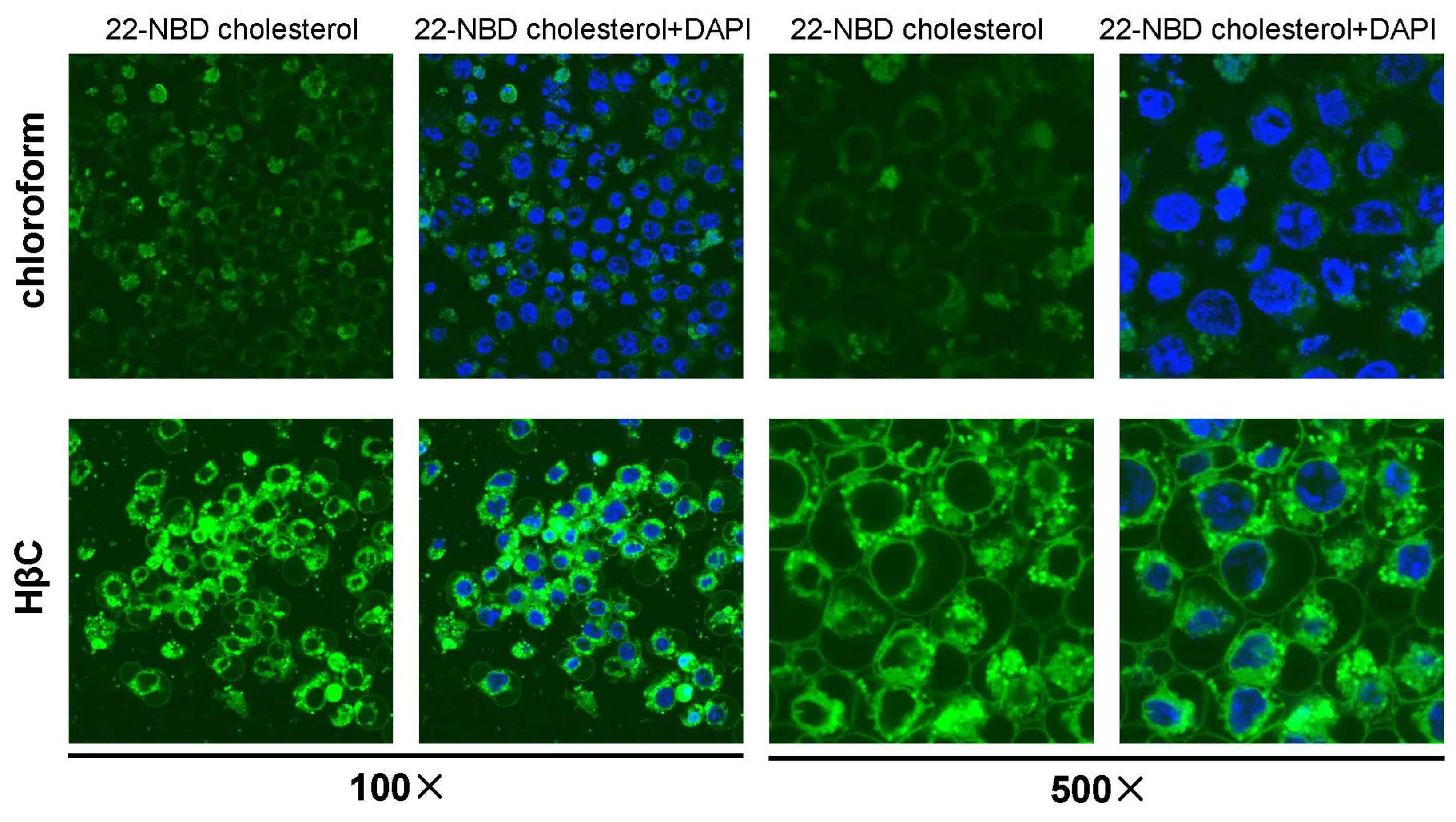
2.7. Immunocytochemistry of Drosophila Tissues

The brain-ring gland complex and fat body of different sterol treatments were dissected in the white prepupal stage in the PBS (pH 7.0). Dissected tissues were fixed and stained with antibodies according to standard procedures. The primary antibodies used included rabbit anti-Nvd (1:200), rabbit anti-Sad (1:200, SHW-101AP, FabGennix, Frisco, TX, USA), and rabbit anti-Shd (1:200, SHD-101AP, FabGennix). The secondary antibody was Alexa FlourTM 488 goat anti-rabbit IgG (1:200, A-11008, ThermoFisher, Waltham, MA, USA). The rabbit polyclonal antibody against Drosophila Nvd was generated via the immunization of rabbit with epitope YNHKRDTINRLRKLK by ChinaPeptides (Shanghai, China). DAPI was used for nuclei labeling. Fluorescence signals were detected using an Olympus Fluoview FV3000 confocal microscope at 40× magnifications.

2.8. Measurement of Free Cholesterol Level

Briefly, twenty white prepupal-stage animals were collected and total cholesterol was extracted with 200 μL of chloroform: isopropanol: IGEPAL CA-630 (7:11:0.1) using a microhomogenizer. Then, total cholesterol was measured via GC-MS according to a previously published GC-MS method for the quantification of free cholesterol [32].

2.9. Statistical Analysis of Data

All of the data were presented as the mean ± standard deviation of three independent experiments, and were analyzed using Student’s t-test and ANOVA. Asterisks indicate a significant difference as calculated using the two-tailed unpaired Student t-test (*, p < 0.05, **, p < 0.01). For ANOVA, the bars labeled with different lowercase letters are significantly different (p < 0.05).

3. Results

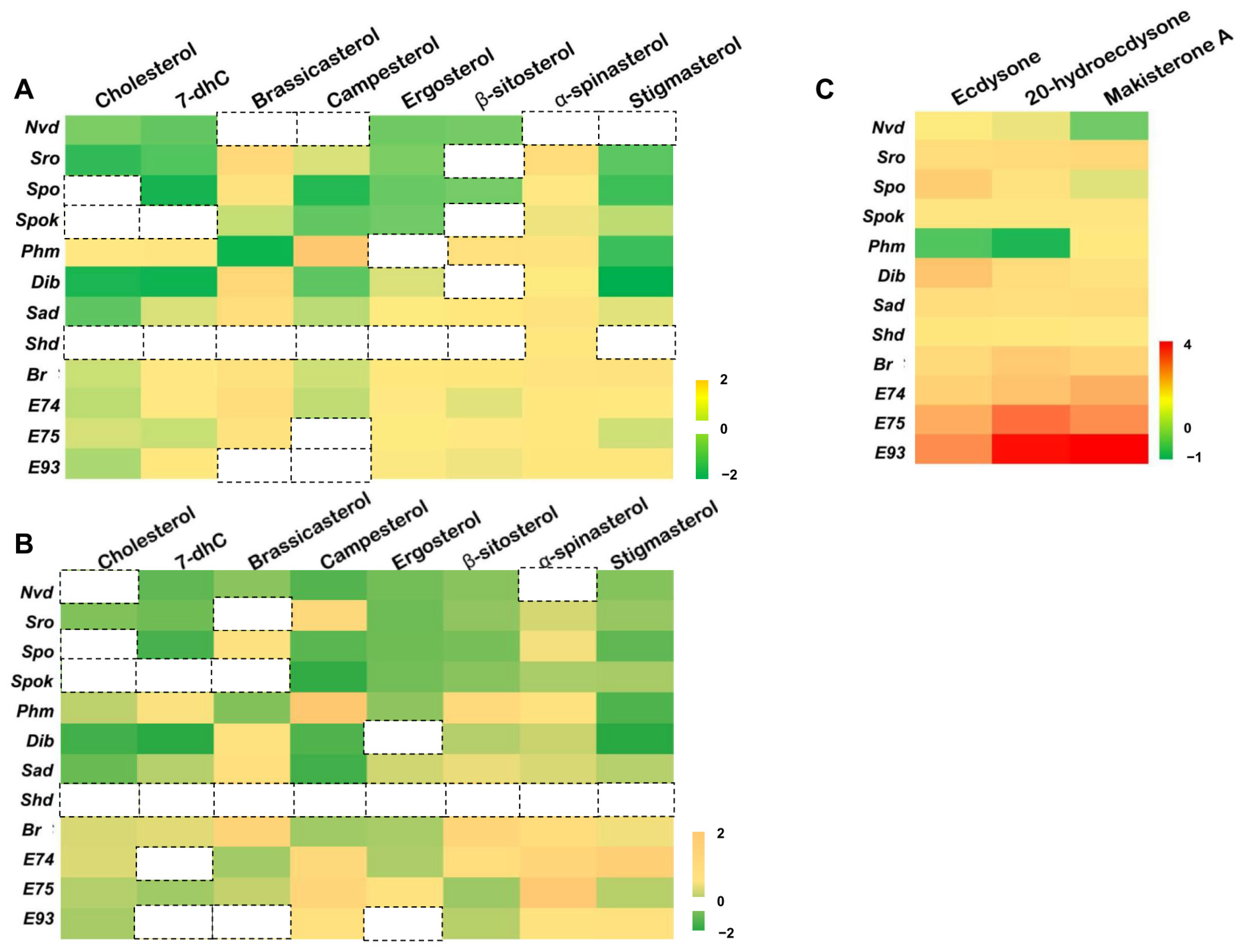
3.1. Dietary Sterols Inhibit the Most 20E Biosynthetic Genes but Activate the 20E Signaling Pathway in D. melanogaster Kc Cells

The D. melanogaster Kc cell line is derived from embryos and expresses many genes involved in the 20E biosynthesis and signaling pathway (http://flybase.org/ (accessed on 22 August 2020)). The cytochorome P450-18A1 (CYP18A1) inactivated 20E by catalyzing 26-hydroxylation of 20E. We further tested whether Kc cells could produce 20E by overexpressing Cyp18a1 and Shd. The overexpression of Cyp18a1 in the Kc cells reduced the expression levels of two 20E primary response genes E74 and E75; instead, the overexpression of Shd increased the expression of the above two genes (Figure S1). This indicates that Kc cells can produce 20E, perhaps in very small amounts. Dietary sterols, serving as precursors of ecdysteroids, may regulate ecdysteroidogenic genes and 20E primary response genes. First, two types of sterol solvents, chloroform and HβC, which can act as delivery vehicles, were compared for their efficiency in carrying sterols into cells. Using 22-NBD cholesterol as a fluorescent probe, the results showed that HβC worked better and more sterols in the HβC group were localized in the plasma membrane (Figure 2). Then, we investigated the effect of different single dietary sterols and three ecdysteroids (E, 20E, and MaA) on eight 20E biosynthetic genes (Nvd, Sro, Spo, Spok, Phm, Dib, Sad, and Shd) and four primary response genes (Br, E74, E75, and E93) in Kc cells via qRT-PCR; the control group was treated with chloroform, as seen in Figure 3A, 45% HβC, as seen in Figure 3B, and DMSO, as seen in Figure 3C. As expected, all three ecdysteroids promoted the expression of 20E primary response genes (>10 folds), and the effect of E was weaker than 20E and MaA (Figure 3C). Except for Nvd and Phm, the expression of the other 20E biosynthetic genes was activated by three ecdysteroids (>2.0 folds). Upon all eight forms of sterol treatment, the expressions of Nvd and Spok were decreased. The expression patterns of Sro, Spo, Dib, and Sad were similar: they were highly expressed (>1.5 folds) upon brassicasterol or α-spinasterol treatment, but were less expressed (<0.8 folds) upon other forms of sterol treatment. The expression of Phm was a little different from the above four genes: it was down-regulated in brassicasterol and stigmasterol, while it was induced by other forms of sterol treatment. It seemed that Shd expression was not significantly affected by these eight dietary sterols (Figure 3A,B).
For these four primary response genes of 20E signaling, different dietary sterols induced 20E response gene expression, and the induction effect of brassicasterol was the most pronounced, while the induction effects of cholesterol and campesterol were relatively weak. These cell treatment experiments preliminarily indicated that 20E biosynthetic genes in D. melanogaster were regulated by dietary sterols and different dietary sterols could be utilized by D. melanogaster to biosynthesize ecdysteroids. It is worth noting that there are slight differences between chloroform-dissolved sterols and HβC-dissolved sterols, probably because of the different amounts of sterols absorbed by the cells. As shown in Figure 2, the cells took up more sterols when HβC was used as a solvent.

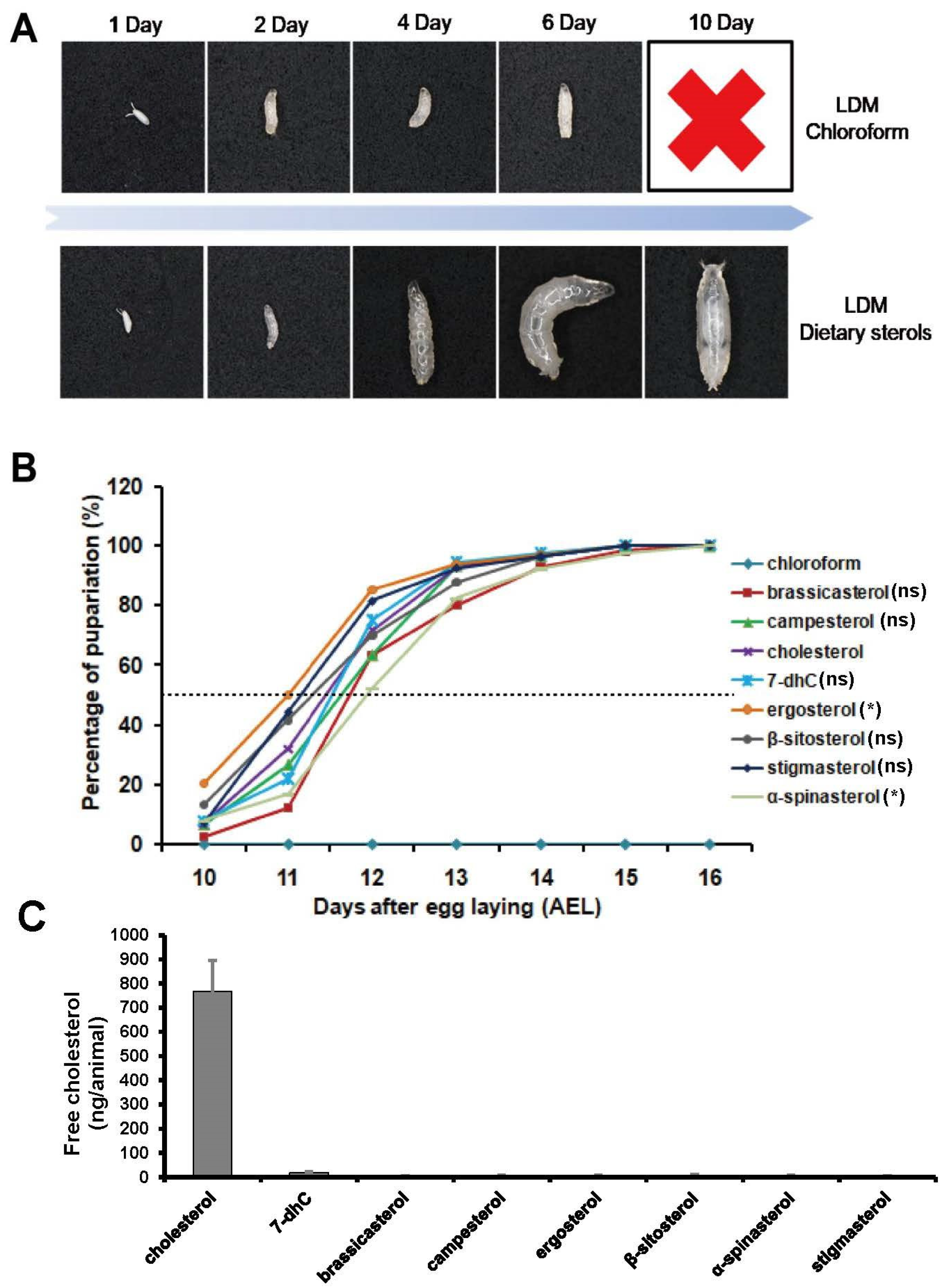
3.2. Single Dietary Sterol Is Enough to Support D. melanogaster Growth and Development

To answer whether different dietary sterols can support the growth and development of D. melanogaster, we observed their larval development on LDM or LDM supplemented with single dietary sterols. Most flies fed with LDM supplemented with dietary sterols completed their development from egg to adult. However, the development of those fed with LDM was arrested in the first instar larval stage (Figure 4A). Although an absence of dietary sterol arrested larval development, the larvae would live for more than six days without further development and continued to feed until they died. These results suggest that D. melanogaster might be capable of synthesizing ecdysteroids from many kinds of dietary sterols.
To better understand the effects of different dietary sterols on the growth and development of D. melanogaster, we compared the developmental timing and percentage of pupariation. As shown in Figure 3B, compared with the pupariation time of cholesterol-fed flies, ergosterol and α-spinasterol resulted in pupariation ∼12 h earlier and ∼12 h later, respectively, with a significantly difference (Figure 4B).
Recently, Lavrynenko et al. revealed that D. melanogaster only synthesized C28 ecdysteroids when they were reared on LDM with C28 or C29 dietary sterols [3]. This indicated that D. melanogaster cannot dealkylate C28 sterols to produce cholesterol. Our free cholesterol measurement experiments further confirmed that cholesterol was not produced when D. melanogaster were fed with C28 or C29 dietary sterols (less than 5 ng/animal) (Figure 4C). The content of free cholesterol is about 750 ng/animal in the cholesterol-fed group and is about 15 ng/animal in the 7-dhC-fed group. Compared to other dietary sterols, higher concentrations of free cholesterol can be detected in the 7-dhC-fed group, maybe coming from an impurity in the commercial 7-dhC. Taken together, these results suggest that single dietary sterol was sufficient to support D. melanogaster growth and development.

3.3. Effect of Dietary Sterols on 20E Biosynthetic Genes in D. melanogaster

Subsequently, we explored the effects of different dietary sterols on 20E biosynthetic genes in D. melanogaster reared on LDM spiked with different sterols in the white prepupal stage via qRT-PCR (Figure 5). The results showed that the expression levels of Phm and Dib were relatively constant in flies reared on diets with different dietary sterols. The expression levels of Nvd were significantly increased in campesterol- and α-spinasterol-reared flies, while they were significantly reduced in 7-dhC- and ergosterol-reared flies. The expression levels of Spok were significantly increased in β-sitosterol- and α-spinasterol-reared flies. The expression levels of Sad were significantly increased in campesterol-, α-spinasterol-, and stigmasterol-reared flies, while they were significantly decreased in brassicasterol-reared flies. The expression levels of Shd were significantly increased in α-spinasterol-reared flies. Interestingly, there was an overall down-regulation in biosynthetic gene expression above 20E in ergosterol-reared flies.
Considering the significant changes in Nvd and Sad upon different sterol treatments, we examined their protein levels in the prothoracic gland. The results showed that Nvd and Sad protein abundance became greater in campesterol-, α-spinasterol-, and stigmasterol-reared flies, and Nvd protein abundance became less in 7-dhC-reared files, which is consistent with the qRT-PCR results (Figure 6, left and middle columns, Figure S2). The Shd protein levels in the fat body became more abundant in α-spinasterol-reared flies and less abundant in ergosterol-reared flies, which is also consistent with the qRT-PCR results (Figure 6, right column, Figure S2).
These results suggest that, except for Phm and Dib, enzymes participating in the 20E biosynthesis in flies had different expression patterns after different sterol treatments, indicating that the catalytic ability of these enzymes to different sterol treatments was different. The expression of most 20E biosynthetic enzyme genes was decreased after ergosterol treatment but increased after α-spinasterol treatment, indicating that ergosterol might be a more suitable substrate for ecdysteroid biosynthesis compared with other dietary sterols.

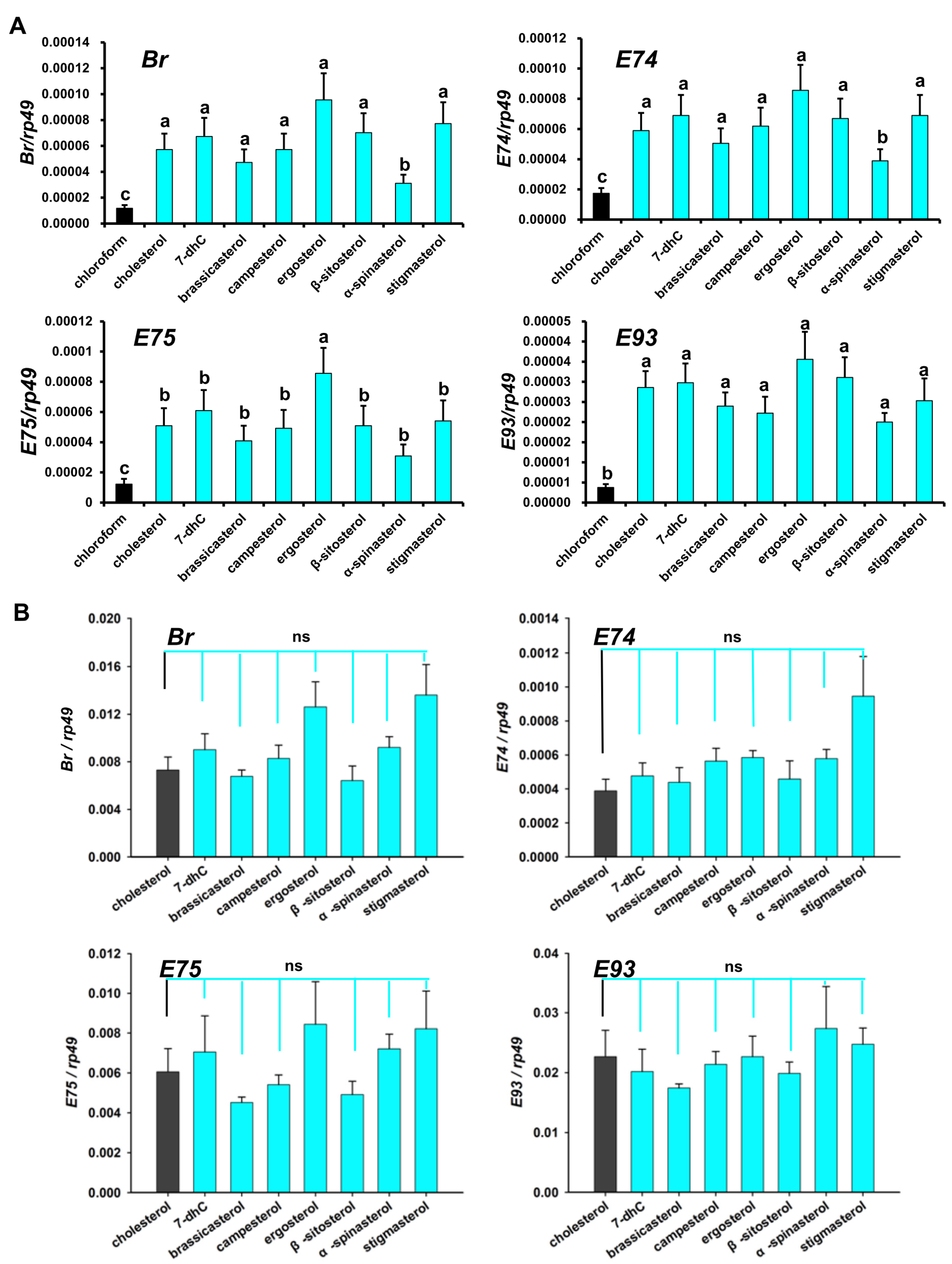
3.4. Regulation of Dietary Sterols on 20E Signaling Pathway in D. melanogaster

Our previous studies confirmed that dietary sterols induce the 20E signaling pathway in D. melanogaster Kc cells. To further investigate whether D. melanogaster reared on single dietary sterols need relatively highly expressed 20E signaling primary response genes to finish larval–larval molting and larval–pupal metamorphosis, we analyzed the relative expression of four 20E signaling primary response genes in first instar larvae (about 48 h after egg laying) and those in the white prepupal stage.
In the first instar larvae, compared with the chloroform-treated group of D. melanogaster, the expression levels of Br, E74, E75, and E93 in the sterol-treated groups were significantly higher. Moreover, compared with other sterol-treated groups, the ergosterol-treated group showed higher expression and the α-spinasterol-treated group showed lower expression. In the white prepupal stage, compared with the cholesterol-reared group, Br, E74, E75, and E93 expression in the other dietary sterol-treated groups did not show a significant difference (Figure 7B). These results imply that the C28 ecdysteroids synthesized from C28 and C29 sterols have similar functions with 20E, which is consistent with the above Kc cell results, showing that there were no obvious differences between 20E and MaA upon the induction of 20E primary response genes.

4. Discussion

In the present study, we have determined that dietary sterols are essential for D. melanogaster growth and development, and a single kind of dietary sterols can be utilized by flies to complete their growth and development. This is consistent with previous studies that have shown that insects are unable to synthesize sterols and must obtain dietary sterols from their food to complete their development [33,34]. As we know, cholesterol is not only a steroid hormone precursor but also an important component of cell membranes and is needed for lipid modifications on proteins [25,29]. When fed with LDM only, D. melanogaster larvae are arrested in the first instar larval stage. Conversely, those reared on LDM supplemented with one of eight different dietary sterols can complete adult development. Free cholesterol measurement experiments using GC/MS methods showed that almost no cholesterol was produced in the other seven dietary sterol-fed animals (Figure 4C), which demonstrates that these sterols are sufficient to undertake various functions of cholesterol in D. melanogaster. Moreover, we found that α-spinasterol-fed larvae had remarkably delayed development, and pupariated 12 h later than the pupariation time point of the cholesterol-fed flies. Thus, we hypothesized that α-spinasterol might not be a proper starting sterol for ecdysteroid biosynthesis. Consistent with this speculation, we found that most 20E biosynthetic genes were upregulated upon α-spinasterol treatment, both in Kc cells and in insects (Figure 3, Figure 5 and Figure 6). This may mean that more enzymes were required for α-spinasterol to convert into ecdysteroids. Our data also showed that, compared to other sterols, ergosterol, a C28 fungal sterol, induced a lower expression of 20E biosynthetic genes. We hypothesized that 20E synthetic enzymes may have high catalytic efficiency for converting ergosterol into C28 ecdysteroids, or there might be other unknown enzymes involved in the conversion process from ergosterol to C28 ecdysteroids.
According to our results, there were significant differences in the induction and reduction patterns of ecdysteroidogenic genes between in vitro and in vivo experiments. For example, the expression of phm was not reduced by 7-dhC in vitro but was reduced in vivo. These differences maybe arise due to two reasons. One is that Kc cells grew and proliferated normally under sterol deprivation [29,35], while D. melanogaster had to obtain sterols (Figure 4). This led to their different responses to sterol treatments. The other is that Kc cells only produced a very small amount of 20E, while D. melanogaster had specialized ecdysteroidogenic organs (the prothoracic glands) to produce relatively higher amounts of 20E. The amounts of sterol ingested by the cells also had an effect on the expression changes in ecdysteroidogenic genes, as shown in Figure 2 and Figure 3A,B.
It has been reported that D. melanogaster can produce four different active ecdysteroids, including 20E, MaA, epi-MaA, and dhMaA, using a new LC-MS/MS method [3,36]. Moreover, Drosophila only produced C28 ecdysteroids when they were reared on phytosterols or fungal sterols [3]. Although the biosynthetic pathway of the C27 ecdysteroid 20E from cholesterol is well understood, the process from the C28 or C29 dietary sterol as a precursor to C28 ecdysteroids has remained unknown. Our data showed that all classical 20E biosynthetic genes were expressed when D. melanogaster were reared on phytosterols or fungal sterols, implying that C28 ecdysteroids may be directly synthesized from C28 dietary sterols by the well-known 20E biosynthetic pathway. Considering that D. melanogaster fed on C29 dietary sterols also only produced C28 ecdysteroids, the demethylation must have happened in the 20E biosynthetic pathway, or maybe happened before they entered the classical 20E biosynthetic pathway (pathway ① in Figure 8). Alternatively, C29 dietary sterols may have entered into the 20E biosynthetic pathway first, and then the demethylation may have happened at an intermediate step (pathway ② in Figure 8). Above all, based on the above results, it can be preliminary concluded that C28 and C29 dietary sterols were converted into C28 ecdysteroids through the same ecdysteroidogenic gene network of the 20E biosynthetic pathway.
It has been suggested that some insects such as the hemiptera Oncopeltus fasciatus, Podisus maculiventirs, and the hymenopteran honey bee, Apis mellifera, can utilize the C28 ecdysteroid, MaA, as the major molting hormone rather than the more common C27 hormone, 20E [37]. A subsequent study examined that the EcR and Usp, which were critical for initiating the primary response gene expression, showed similar affinities for MaA and 20E [26]. MaA was found in D. melanogaster a few decades ago [38]; nevertheless, it is still unknown as to how C28 ecdysteroids regulate the molting and metamorphosis of D. melanogaster. Our research has demonstrated that the C28 ecdysteroids from phytosterol and fungal sterols as precursors can also activate the primary response genes of 20E signaling to regulate the growth and development of flies, and MaA showed indistinguishable physiological activity with 20E in inducing the expression of primary response genes Br, E74, E75, and E93 (Figure 2 and Figure 6).
In this study, it was found that brassicasterol is capable of supporting D. melanogaster larval growth to pupae, with a comparatively slow developmental speed (Figure 3B). But, as Lavrynenko et al. report, only a few individuals (about 5%) can survive to pupae [3]. This discrepancy may come from the different sterol concentrations in two experiments: it was 80 μg/mL in our assay and only 6 μg/mL in their assay. According to Martin’s results, the optimal concentration for cholesterol is up to 1.6 mg/mL [39]. Another study recommended that the cholesterol concentration in the medium should be 100–300 μg/mL [40]. So, the sterol concentration in this assay should be more suitable for D. melanogaster growth and development. Another possible reason is that other unknown sterols in commercial brassicasterol were utilized for the D. melanogaster to biosynthesize 20E; its purity is 98% according to the manufacturer’s instructions.
In summary, the present study is the first to find that there are differences related to the expression of 20E biosynthetic enzymes after different sterol treatments in vitro and in vivo given to D. melanogaster. Moreover, ecdysteroids synthesized from any kind of dietary sterols triggered almost the same 20E signaling activity. These results suggest that D. melanogaster possess a strong environment adaptation ability.

Supplementary Materials

The following supporting information can be downloaded at https://www.mdpi.com/article/10.3390/cells12131739/s1: Figure S1. The expression changes in 20E primary response genes under overexpression of Shd and Cyp18a1. Asterisks indicate a significant difference, as calculated using two-tailed unpaired Student’s t-test (* p < 0.05). Figure S2. The statistical analyses of relative fluorescence intensities of Nvd and Sad proteins in the prothoracic gland and Shd protein in the fat body. Asterisks indicate a significant difference, as calculated using two-tailed unpaired Student’s t-test (* p < 0.05; *** p < 0.001).

Author Contributions

Conceptualization, Q.J.; methodology, D.W. and Z.C.; validation, Z.C.; investigation, D.W. and J.W.; resources, Q.J.; writing—original draft, Q.J.; writing—review and editing, Z.C.; funding acquisition, D.W. All authors have read and agreed to the published version of the manuscript.

Funding

This research was supported by the General Project of Guangdong Natural Science Foundation (2022A1515010938), the National Science Foundation of China (32200398 (to Q.J.) and 31560609 (to D.W.)), the Natural Science Research Project of the Education Department of the Guizhou Province of China (KY [2018]419, KY [2020]071, [2015]68, QNYSKYTD2018006, KY [2017]347), the Scientific Research Project of Qiannan Normal University for Nationalities (QNSY2018BS018, Qnsyk201605, QNYSKYTD2018011, qnsy2018012, QNSY2018BS011), the Key Support Project for Biology in Qiannan Normal University for Nationalities (QNYSXXK2018005), the Qiannan Agricultural Science and Technology Program of the Qiannan Science and Technology Bureau (201721), and the Science and Technology Planning Project of the Science and Technology Department of Guizhou Province (20172853, 2018011).

Institutional Review Board Statement

Not applicable.

Data Availability Statement

The data presented in this study are available on request from the corresponding author. The data are not publicly available due to further analysis will be conducted.

Conflicts of Interest

The authors declare that they have no conflict of interest in their work.

References

  1. Yamanaka, N.; Rewitz, K.F.; O’Connor, M.B. Ecdysone control of developmental transitions: Lessons from Drosophila research. Annu. Rev. Entomol. 2013, 58, 497–516. [Google Scholar] [CrossRef] [Green Version]
  2. Enya, S.; Ameku, T.; Igarashi, F.; Iga, M.; Kataoka, H.; Shinoda, T.; Niwa, R. A Halloween gene noppera-bo encodes a glutathione s-transferase essential for ecdysteroid biosynthesis via regulating the behaviour of cholesterol in Drosophila. Sci. Rep. 2014, 4, 6586. [Google Scholar] [CrossRef] [PubMed] [Green Version]
  3. Lavrynenko, O.; Rodenfels, J.; Carvalho, M.; Dye, N.A.; Lafont, R.; Eaton, S.; Shevchenko, A. The ecdysteroidome of Drosophila: Influence of diet and development. Development 2015, 142, 3758–3768. [Google Scholar] [CrossRef] [PubMed] [Green Version]
  4. Igarashi, F.; Ogihara, M.H.; Iga, M.; Kataoka, H. Cholesterol internalization and metabolism in insect prothoracic gland, a steroidogenic organ, via lipoproteins. Steroids 2018, 134, 110–116. [Google Scholar] [CrossRef] [PubMed]
  5. Martin-Creuzburg, D.; Oexle, S.; Wacker, A. Thresholds for sterol-limited growth of Daphnia magna: A comparative approach using 10 different sterols. J. Chem. Ecol. 2014, 40, 1039–1050. [Google Scholar] [CrossRef] [Green Version]
  6. Rewitz, K.F.; O’Connor, M.B.; Gilbert, L.I. Molecular evolution of the insect Halloween family of cytochrome P450s: Phylogeny, gene organization and functional conservation. Insect Biochem. Mol. Biol. 2007, 37, 741–753. [Google Scholar] [CrossRef]
  7. Yoshiyama, T.; Namiki, T.; Mita, K.; Kataoka, H.; Niwa, R. Neverland is an evolutionally conserved rieske-domain protein that is essential for ecdysone synthesis and insect growth. Development 2006, 133, 2565–2574. [Google Scholar] [CrossRef] [PubMed] [Green Version]
  8. Saito, J.; Kimura, R.; Kaieda, Y.; Nishida, R.; Ono, H. Characterization of candidate intermediates in the Black Box of the ecdysone biosynthetic pathway in Drosophila melanogaster: Evaluation of molting activities on ecdysteroid-defective larvae. J. Insect Physiol. 2016, 93–94, 94–104. [Google Scholar] [CrossRef] [Green Version]
  9. Niwa, R.; Namiki, T.; Ito, K.; Shimadaniwa, Y.; Kiuchi, M.; Kawaoka, S.; Banno, Y.; Fujimoto, Y.; Shigenobu, S.; Kobayashi, S.; et al. Non-molting glossy/shroud encodes a short-chain dehydrogenase/reductase that functions in the ‘Black Box’ of the ecdysteroid biosynthesis pathway. Development 2010, 137, 1991–1999. [Google Scholar] [CrossRef] [Green Version]
  10. Ono, H.; Rewitz, K.F.; Shinoda, T.; Itoyama, K.; Petryk, A.; Rybczynski, R.; Jarcho, M.; Warren, J.T.; Marqués, G.; Shimell, M.J.; et al. Spook and spookier code for stage-specific components of the ecdysone biosynthetic pathway in Diptera. Dev. Biol. 2006, 298, 555. [Google Scholar] [CrossRef] [Green Version]
  11. Iga, M.; Kataoka, H. Recent studies on insect hormone metabolic pathways mediated by cytochrome P450 enzymes. Biol. Pharm. Bull. 2012, 35, 838–843. [Google Scholar] [CrossRef] [Green Version]
  12. Niwa, R.; Matsuda, T.; Yoshiyama, T.; Namiki, T.; Mita, K.; Fujimoto, Y.; Kataoka, H. CYP306A1, a cytochrome P450 enzyme, is essential for ecdysteroid biosynthesis in the prothoracic glands of Bombyx and Drosophila. J. Biol. Chem. 2004, 279, 35942–35949. [Google Scholar] [CrossRef] [Green Version]
  13. Warren, J.T.; Petryk, A.; Marques, G.; Jarcho, M.; Parvy, J.P.; Dauphin-Villemant, C.; O’Connor, M.B.; Gilbert, L. Molecular and biochemical characterization of two P450 enzymes in the ecdysteroidogenic pathway of Drosophila melanogaster. Proc. Natl. Acad. Sci. USA 2002, 99, 11043–11048. [Google Scholar] [CrossRef] [Green Version]
  14. Rewitz, K.F.; Rybczynski, R.; Warren, J.T.; Gilbert, L.I. The Halloween genes code for cytochrome P450 enzymes mediating synthesis of the insect moulting hormone. Biochem. Soc. Trans. 2006, 34, 1256–1260. [Google Scholar] [CrossRef]
  15. Wu, L.; Jia, Q.; Zhang, X.; Zhang, X.; Liu, S.; Park, Y.; Feyereisen, R.; Zhu, K.Y.; Ma, E.; Zhang, J.; et al. CYP303A1 has a conserved function in adult eclosion in Locusta migratoria and Drosophila melanogaster. Insect Biochem. Mol. Biol. 2019, 113, 103210. [Google Scholar] [CrossRef]
  16. Petryk, A.; Warren, J.T.; Marqués, G.; Jarcho, M.P.; Gilbert, L.I.; Kahler, J.; Parvy, J.P.; Li, Y.; Dauphin-Villemant, C.; O’Connor, M.B. Shade is the Drosophila P450 enzyme that mediates the hydroxylation of ecdysone to the steroid insect molting hormone 20-hydroxyecdysone. Proc. Natl. Acad. Sci. USA 2003, 100, 13773–13778. [Google Scholar] [CrossRef] [Green Version]
  17. Cherbas, L.; Hu, X.; Zhimulev, I.; Belyaeva, E.; Cherbas, P. EcR isoforms in Drosophila: Testing tissue-specific requirements by targeted blockade and rescue. Development 2003, 130, 271–284. [Google Scholar] [CrossRef] [Green Version]
  18. Jia, Q.; Liu, S.; Wen, D.; Chen, Y.; Bendena, W.G.; Wang, J.; Li, S. Juvenile hormone and 20-hydroxyecdysone coordinately control the developmental timing of matrix metalloproteinase-induced fat body cell dissociation. J. Biol. Chem. 2017, 292, 21504–21516. [Google Scholar] [CrossRef] [Green Version]
  19. Liu, X.; Dai, F.; Guo, E.; Li, K.; Ma, L.; Tian, L.; Cao, Y.; Zhang, G.; Palli, S.R.; Li, S. 20-hydroxyecdysone (20E) primary response gene E93 modulates 20E signaling to promote Bombyx larval-pupal metamorphosis. J. Biol. Chem. 2015, 290, 27370–27383. [Google Scholar] [CrossRef] [Green Version]
  20. Li, K.; Tian, L.; Guo, Z.; Guo, S.; Zhang, J.; Gu, S.-H.; Palli, S.R.; Cao, Y.; Li, S. 20-Hydroxyecdysone (20E) Primary Response Gene E75 Isoforms Mediate Steroidogenesis Autoregulation and Regulate Developmental Timing in Bombyx. J. Biol. Chem. 2016, 291, 18163–18175. [Google Scholar] [CrossRef] [Green Version]
  21. Li, K.; Guo, E.; Hossain, M.S.; Li, Q.; Cao, Y.; Tian, L.; Deng, X.; Li, S. Bombyx E75 isoforms display stage- and tissue-specific responses to 20-hydroxyecdysone. Sci. Rep. 2015, 5, 12114. [Google Scholar] [CrossRef] [PubMed] [Green Version]
  22. Liu, H.; Jia, Q.; Tettamanti, G.; Li, S. Balancing crosstalk between 20-hydroxyecdysone-induced autophagy and caspase activity in the fat body during Drosophila larval-prepupal transition. Insect Biochem. Mol. Biol. 2013, 43, 1068–1078. [Google Scholar] [CrossRef]
  23. Liu, H.; Wang, J.; Li, S. E93 predominantly transduces 20-hydroxyecdysone signaling to induce autophagy and caspase activity in Drosophila fat body. Insect Biochem. Mol. Biol. 2014, 45, 30–39. [Google Scholar] [CrossRef]
  24. Bahrami, S.; Drabløs, F. Gene regulation in the immediate-early response process. Adv. Biol. Regul. 2016, 62, 37–49. [Google Scholar] [CrossRef] [Green Version]
  25. Maurer, P.; Girault, J.P.; Larcheveque, M.; Lafont, R. 24-Epi-makisterone a (not makisterone A) is the major ecdysteroid in the leaf-cutting ant Acromyrmex octospinosus (reich) (hymenoptera, formicidae: Attini). Arch. Insect Biochem. Physiol. 1993, 23, 29–35. [Google Scholar] [CrossRef]
  26. Gilbert, L.I. Halloween genes encode P450 enzymes that mediate steroid hormone biosynthesis in Drosophila melanogaster. Mol. Cell Endocrinol. 2004, 215, 1–10. [Google Scholar] [CrossRef]
  27. Tohidi-Esfahani, D.; Graham, L.D.; Hannan, G.N.; Simpson, A.M.; Hill, R.J. An ecdysone receptor from the pentatomomorphan, nezaraviridula, shows similar affinities for moulting hormones makisterone A and 20-hydroxyecdysone. Insect Biochem. Mol. Biol. 2011, 41, 77–89. [Google Scholar] [CrossRef]
  28. Liu, S.; Li, K.; Gao, Y.; Liu, X.; Chen, W.; Ge, W.; Feng, Q.; Palli, S.R.; Li, S. Antagonistic actions of juvenile hormone and 20-hydroxyecdysone within the ring gland determine developmental transitions in Drosophila. Proc. Natl. Acad. Sci. USA 2018, 115, 139–144. [Google Scholar] [CrossRef] [Green Version]
  29. Jia, Q.; Liu, Y.; Liu, H.; Li, S. Mmp1 and mmp2 cooperatively induce Drosophila fat body cell dissociation with distinct roles. Sci. Rep. 2014, 4, 7535. [Google Scholar] [CrossRef] [Green Version]
  30. Carvalho, M.; Schwudke, D.; Sampaio, J.L.; Palm, W.; Riezman, I.; Dey, G.; Gupta, G.D.; Mayor, S.; Riezman, H.; Shevchenko, A.; et al. Survival strategies of a sterol auxotroph. Development 2010, 137, 3675. [Google Scholar] [CrossRef] [Green Version]
  31. Chen, X.; Yang, L.; Huang, R.; Li, S.; Jia, Q. Matrix metalloproteinases are involved in eclosion and wing expansion in the American cockroach, Periplaneta americana. Insect Biochem. Mol. Biol. 2021, 131, 103551. [Google Scholar] [CrossRef] [PubMed]
  32. Sarohia, G.S.; Garza, E.D.; Bhattacharya, S.K. Analyses of cholesterol metabolites of optic nerve using GC-MS methods. Methods Mol. Biol. 2019, 1996, 47–51. [Google Scholar]
  33. Janson, E.M.; Grebenok, R.J.; Behmer, S.T.; Abbot, P. Same host-plant, different sterols: Variation in sterol metabolism in an insect herbivore community. J. Chem. Ecol. 2009, 35, 1309–1319. [Google Scholar] [CrossRef] [PubMed]
  34. Cooke, J.; Sang, J.H. Utilization of sterols by larvae of Drosophila melanogaster. J. Insect Physiol. 1970, 16, 801–812. [Google Scholar] [CrossRef] [PubMed]
  35. Silberkang, M.; Havel, C.M.; Friend, D.S.; McCarthy, B.J.; Watson, J.A. Isoprene synthesis in isolated embryonic Drosophila cells. I. Sterol-deficient eukaryotic cells. J. Biol. Chem. 1983, 258, 8503–8511. [Google Scholar] [CrossRef]
  36. Blais, C.; Blasco, T.; Maria, A.; Dauphin-Villemant, C.; Lafont, R. Characterization of ecdysteroids in Drosophila melanogaster by enzyme immunoassay and nano-liquid chromatography-tandem mass spectrometry. J. Chromatogr. B 2010, 878, 925–932. [Google Scholar] [CrossRef]
  37. Feldlaufer, M.F.; Svoboda JAMakisterone, A. A 28-carbon insect ecdysteroid. Ecdysone 1986, 16, 45–48. [Google Scholar]
  38. Redfern, C.P. Evidence for the presence of makisterone A in Drosophila larvae and the secretion of 20-deoxymakisterone A by the ring gland. Proc. Natl. Acad. Sci. USA 1984, 81, 5643–5647. [Google Scholar] [CrossRef] [Green Version]
  39. Martin, A.M. The Effects of Sterols on Drosophila melanogaster: Physiology and Biochemistry. Master’s Thesis, Texas A&M University, College Station, TX, USA, 2015. [Google Scholar]
  40. Piper, M.D.; Blanc, E.; Leitão-Gonçalves, R.; Yang, M.; He, X.; Linford, N.J.; Hoddinott, M.P.; Hopfen, C.; Soultoukis, G.A.; Niemeyer, C. A holidic medium for Drosophila melanogaster. Nat. Methods 2014, 11, 100–105. [Google Scholar] [CrossRef] [Green Version]
Figure 1. Structures of different sterols and ecdysteroids. (A) Structures of C27 sterols and ecdysteroids. (B) Structures of C28 and C29 dietary sterols. (C) Structures of C28 ecdysteroids.
Cells 12 01739 g001
Figure 2. The comparison of the uptake efficiency of Kc cells to chloroform-dissolved 22-NBD cholesterol and HβC-dissolved 22-NBD cholesterol.
Cells 12 01739 g002
Figure 3. A heatmap of the relative expression of 20E biosynthetic and response genes in Kc cells treated with different sterols. Cholesterol, 7-dehydrocholesterol (7-dhC), brassicasterol, campesterol, ergosterol, β-sitosterol, stigmasterol, and α-spinasterol belong to dietary sterols. E, 20E, and MaA were used to compare the effects of different ecdysteroids. The control group in (A) was treated with chloroform, that in (B) was treated with 45% HβC, and that in (C) was treated with DMSO. Green indicates the lowest expression and red indicates the highest expression. The fold changes in gene expression levels were log2 transformed.
Cells 12 01739 g003
Figure 4. Development of Drosophila reared on diets with controlled composition of sterols. (A) Dietary sterol depletion arrests larvae growth and development. LDM with chloroform is the control group. The red cross means death. (B) Developmental timing and percentage of pupariation. In each group, 100 animals were measured. Asterisks indicate a significant difference, as calculated using two-tailed unpaired Student’s t-test (* p < 0.05). “ns” means “no significant difference”. (C) Total cholesterol content of whole body of Drosophila reared on LDM supplemented with individual sterols.
Cells 12 01739 g004
Figure 5. Expression changes in Drosophila 20E biosynthetic genes upon different dietary sterol treatments. Cholesterol was added as a control (black), while other dietary sterols (blue) were used to be compared with it. Data are reported as means ± standard deviation of three independent biological replications. The relative expression was calculated based on the value of the reference genes. Asterisks indicate a significant difference, as calculated using two-tailed unpaired Student’s t-test (* p < 0.05).
Cells 12 01739 g005
Figure 6. Immunostaining of 20E biosynthetic genes in the prothoracic gland (Nvd and Sad) and fat body (Shd).
Cells 12 01739 g006
Figure 7. Expression changes in Drosophila 20E signaling pathway genes upon different dietary sterol treatments. (A) The gene expression changes in the first instar larvae, about 48 h after egg laying. The chloroform-fed group was the control (black). The bars labeled with different lowercase letters are significantly different (p < 0.05) (B) The gene expression changes in the white prepupal stage. Cholesterol was added as a control (black), while other dietary sterols (green) were used to be compared with it. Data are reported as means ± standard deviation of three independent biological replications. The relative expression was calculated based on the value of the reference genes. “ns” means “no significant difference”.
Cells 12 01739 g007
Figure 8. Schematic description of hypothesis for C28 ecdysteroid biosynthesis and signaling pathway in Drosophila. Proposed pathway for biosynthesis of C28 ecdysteroid, catalyzed by Halloween genes.
Cells 12 01739 g008
Table 1. Primer used for qRT-PCR in this study.
Gene NameForward (5′-3′)Reverse (5′-3′)
NvdGGGGAGATTGACGATACATTTTTGAGCCCAACTGCCATTA
SroCCACAACATCAAGTCGGAAGGAGCACCAGGCGAATGGAATCGGG
SpoGCTGCGATATTCATCCTCCCGCTTTGTTCCTTTGACGGTTC
SpokATCAACTATTGGACCAGCTTACTAATTGAACCGAACTGAACAC
PhmGGATTTCTTTCGGCGCGATGTGTGCCTCAGTATCGAAAAGCCGT
DibTGCCCTCAATCCCTATCTGGTCACAGGGTCTTCACACCCATCTC
SadCCGCATTCAGCAGTCAGTGGACCTGCCGTGTACAAGGAGAG
ShdTACCCATTCCGACCTCCCACTTGGTCCTTGCTCGTTCACAATT
BrCCTCAACACGCACACCCAATGCTGAAGAGGGTCGAGGAG
E74GCCGGACATGAACTACGAGACTTGGGCACATCCACGAAC
E75CCTCAAGCAGCGCGAGTTGCGATTTCCTTGTGGGTCT
E93GCTGAAGAATGTATGGGTCGGGGATTGCTCTGGCTGAT
Rp49GACAGTATCTGATGCCCAACACTTCTTGGAGGAGACGCCGT
Disclaimer/Publisher’s Note: The statements, opinions and data contained in all publications are solely those of the individual author(s) and contributor(s) and not of MDPI and/or the editor(s). MDPI and/or the editor(s) disclaim responsibility for any injury to people or property resulting from any ideas, methods, instructions or products referred to in the content.

