

Biology of Cancer-Testis Antigens and Their Therapeutic Implications in Cancer


Abstract
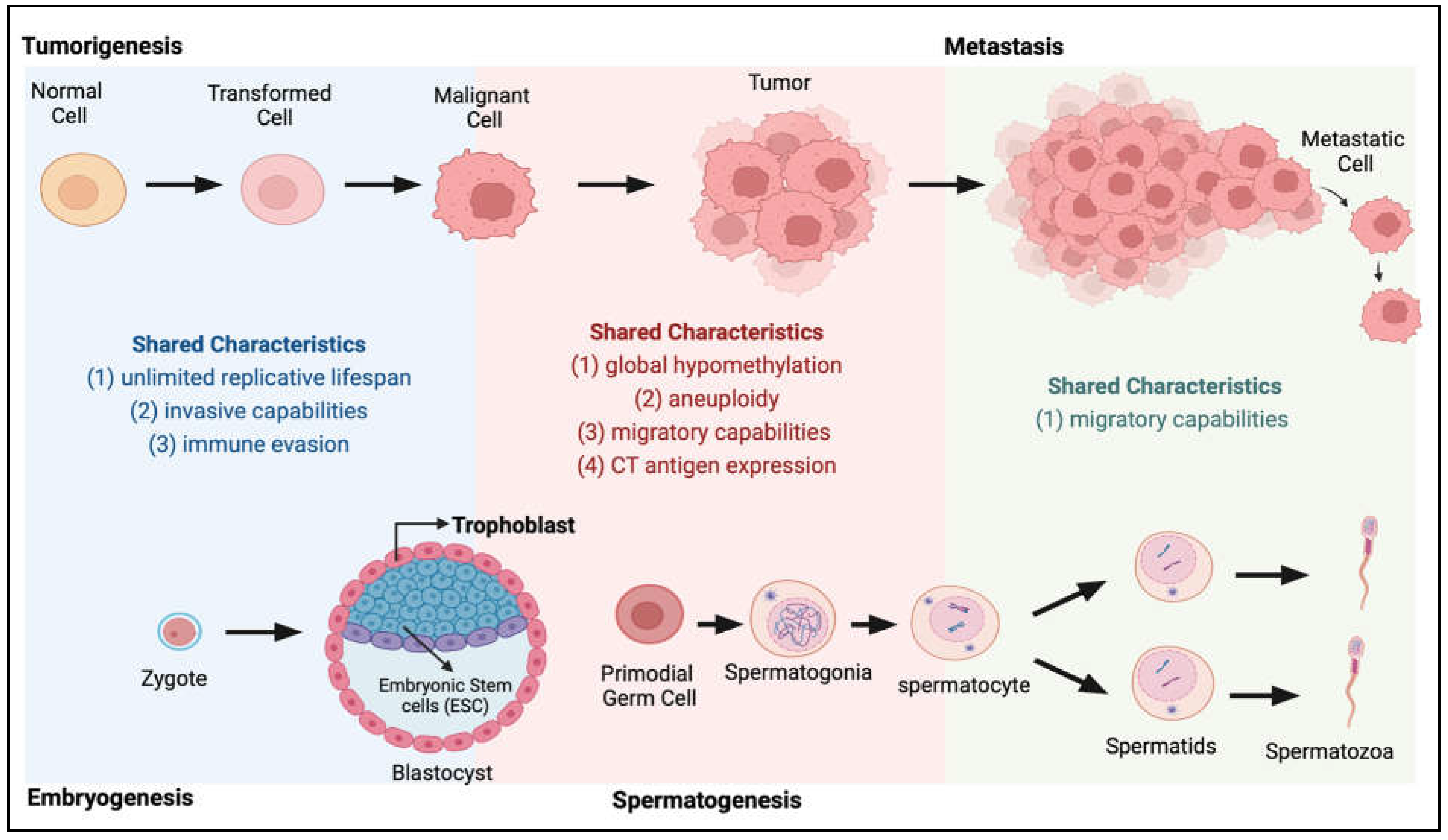
1. Introduction
2. Discovery of CTAs
3. CTA Biology in Carcinogenesis
3.1. CTAs in the Regulation of Transcriptional Programs Involved in Tumorigenesis
3.2. CTAs in Cell Division, Genomic Instability and the DNA Damage Response
3.3. Role of CTAs in the Evasion of Apoptosis
3.4. CTAs and Metastasis: Epithelial-to-Mesenchymal Transition
3.5. Preferential Expression of CTAs in Cancer Stem Cells (CSCs)
3.6. CTAs in Cancer Cell Energetics and Autophagy
3.7. CTAs and the Tumour Microenvironment
4. Regulation of CTAs
5. CTAs as Therapeutic Targets: Perspectives and Future Implications
6. Conclusions
Author Contributions
Funding
Institutional Review Board Statement
Informed Consent Statement
Data Availability Statement
Acknowledgments
Conflicts of Interest
References
- Gurchot, C. The Trophoblast Theory of Cancer (John Beard, 1857–1924) Revisited. Oncology 1975, 31, 310–333. [Google Scholar] [CrossRef]
- Acevedo, H.F.; Hartsock, R.J. Metastatic phenotype correlates with high expression of membrane-associated complete β-human chorionic gonadotropin in vivo. Cancer 1996, 78, 2388–2399. [Google Scholar] [CrossRef]
- Acevedo, H.F.; Tong, J.Y.; Hartsock, R.J. Human chorionic gonadotropin-beta subunit gene expression in cultured human fetal and cancer cells of different types and origins. Cancer 1995, 76, 1467–1475. [Google Scholar] [CrossRef]
- Scanlan, M.J.; GSimpson, A.J.; Old, L.J. The cancer/testis genes: Review, standardization, and commentary. Cancer Immun. Arch. 2004, 4, 1. [Google Scholar]
- Simpson, A.J.G.; Caballero, O.L.; Jungbluth, A.; Chen, Y.-T.; Old, L.J. Cancer/testis antigens, gametogenesis and cancer. Nat. Rev. Cancer 2005, 5, 615–625. [Google Scholar] [CrossRef] [PubMed]
- Bruggeman, J.W.; Koster, J.; Lodder, P.; Repping, S.; Hamer, G. Massive expression of germ cell-specific genes is a hallmark of cancer and a potential target for novel treatment development. Oncogene 2018, 37, 5694–5700. [Google Scholar] [CrossRef] [PubMed]
- Hanahan, D.; Weinberg, R.A. The Hallmarks of Cancer. Cell 2000, 100, 57–70. [Google Scholar] [CrossRef]
- Hanahan, D.; Weinberg, R.A. Hallmarks of Cancer: The Next Generation. Cell 2011, 144, 646–674. [Google Scholar] [CrossRef]
- Hanahan, D. Hallmarks of Cancer: New Dimensions. Cancer Discov. 2022, 12, 31–46. [Google Scholar] [CrossRef]
- Carey, T.E.; Takahashi, T.; Resnick, L.A.; Oettgen, H.F.; Old, L.J. Cell surface antigens of human malignant melanoma: Mixed hemadsorption assays for humoral immunity to cultured autologous melanoma cells. Proc. Natl. Acad. Sci. USA 1976, 73, 3278–3282. [Google Scholar] [CrossRef]
- Garrett, T.J.; Takahashi, T.; Clarkson, B.D.; Old, L.J. Detection of antibody to autologous human leukemia cells by immune adherence assays. Proc. Natl. Acad. Sci. USA 1977, 74, 4587–4590. [Google Scholar] [CrossRef] [PubMed]
- Foley, E.J. Antigenic properties of methylcholanthrene-induced tumors in mice of the strain of origin. Cancer Res 1953, 13, 835–837. [Google Scholar] [PubMed]
- Klein, G.; Sjogren, H.O.; Klein, E.; Hellstrom, K.E. Demonstration of resistance against methylcholanthrene-induced sarcomas in the primary autochthonous host. Cancer Res. 1960, 20, 1561–1572. [Google Scholar] [PubMed]
- Kripke, M.L. Antigenicity of murine skin tumors induced by ultraviolet light. J. Natl. Cancer Inst. 1974, 53, 1333–1336. [Google Scholar] [CrossRef]
- Prehn, R.T.; Main, J.M. Immunity to methylcholanthrene-induced sarcomas. J. Natl. Cancer Inst. 1957, 18, 769–778. [Google Scholar]
- Old, L.J. Cancer Immunology: The Search for Specificity—G. H. A. Clowes Memorial Lecture. Cancer Res. 1981, 41, 361. [Google Scholar]
- Old, L.J. Cancer/Testis (CT) antigens—A new link between gametogenesis and cancer. Cancer Immun. Arch. 2001, 1, 1. [Google Scholar]
- Pfreundschuh, M.; Shiku, H.; Takahashi, T.; Ueda, R.; Ransohoff, J.; Oettgen, H.F.; Old, L.J. Serological analysis of cell surface antigens of malignant human brain tumors. Proc. Natl. Acad. Sci. USA 1978, 75, 5122–5126. [Google Scholar] [CrossRef]
- Shiku, H.; Takahashi, T.; Oettgen, H.F. Cell surface antigens of human malignant melanoma. II. Serological typing with immune adherence assays and definition of two new surface antigens. J. Exp. Med. 1976, 144, 873–881. [Google Scholar] [CrossRef]
- Shiku, H.; Takahashi, T.; Resnick, L.A.; Oettgen, H.F.; Old, L.J. Cell surface antigens of human malignant melanoma. III. Recognition of autoantibodies with unusual characteristics. J. Exp. Med. 1977, 145, 784–789. [Google Scholar] [CrossRef]
- Ueda, R.; Shiku, H.; Pfreundschuh, M.; Takahashi, T.; Li, L.T.; Whitmore, W.F.; Oettgen, H.F.; Old, L.J. Cell surface antigens of human renal cancer defined by autologous typing. J. Exp. Med. 1979, 150, 564–579. [Google Scholar] [CrossRef] [PubMed]
- van der Bruggen, P.; Traversari, C.; Chomez, P.; Lurquin, C.; De Plaen, E.; Van den Eynde, B.; Knuth, A.; Boon, T. A Gene Encoding an Antigen Recognized by Cytolytic T Lymphocytes on a Human Melanoma. Science 1991, 254, 1643–1647. [Google Scholar] [CrossRef] [PubMed]
- Boël, P.; Wildmann, C.; Sensi, M.L.; Brasseur, R.; Renauld, J.-C.; Coulie, P.; Boon, T.; van der Bruggen, P. BAGE: A new gene encoding an antigen recognized on human melanomas by cytolytic T lymphocytes. Immunity 1995, 2, 167–175. [Google Scholar] [CrossRef] [PubMed]
- Van den Eynde, B.; Peeters, O.; De Backer, O.; Gaugler, B.; Lucas, S.; Boon, T. A new family of genes coding for an antigen recognized by autologous cytolytic T lymphocytes on a human melanoma. J. Exp. Med. 1995, 182, 689–698. [Google Scholar] [CrossRef]
- Rogner, U.C.; Wilke, K.; Steck, E.; Korn, B.; Poustka, A. The Melanoma Antigen Gene (MAGE) Family Is Clustered in the Chromosomal Band Xq28. Genomics 1995, 29, 725–731. [Google Scholar] [CrossRef]
- De Plaen, E.; Traversari, C.; Gaforio, J.J.; Szikora, J.-P.; De Smet, C.; Brasseur, F.; van der Bruggen, P.; Lethé, B.; Lurquin, C.; Chomez, P.; et al. Structure, chromosomal localization, and expression of 12 genes of the MAGE family. Immunogenetics 1994, 40, 360–369. [Google Scholar] [CrossRef]
- Chomez, P.; De Backer, O.; Bertrand, M.; De Plaen, E.; Boon, T.; Lucas, S. An overview of the MAGE gene family with the identification of all human members of the family. Cancer Res. 2001, 61, 5544–5551. [Google Scholar]
- Muscatelli, F.; Walker, A.P.; De Plaen, E.; Stafford, A.N.; Monaco, A.P. Isolation and characterization of a MAGE gene family in the Xp21.3 region. Proc. Natl. Acad. Sci. USA 1995, 92, 4987. [Google Scholar] [CrossRef]
- Lucas, S.; De Smet, C.; Arden, K.C.; Viars, C.S.; Lethé, B.; Lurquin, C.; Boon, T. Identification of a new MAGE gene with tumor-specific expression by representational difference analysis. Cancer Res. 1998, 58, 743. [Google Scholar]
- Lurquin, C.; De Smet, C.; Brasseur, F.; Muscatelli, F.; Martelange, V.; De Plaen, E.; Brasseur, R.; Monaco, A.P.; Boon, T. Two Members of the HumanMAGEBGene Family Located in Xp21.3 Are Expressed in Tumors of Various Histological Origins. Genomics 1997, 46, 397–408. [Google Scholar] [CrossRef]
- Lucas, S.; De Plaen, E.; Boon, T. MAGE-B5, MAGE-B6, MAGE-C2, and MAGE-C3: Four new members of the MAGE family with tumor-specific expression. Int. J. Cancer 2000, 87, 55–60. [Google Scholar] [CrossRef]
- Martelange, V.; De Smet, C.; De Plaen, E.; Lurquin, C.; Boon, T. Identification on a Human Sarcoma of Two New Genes with Tumor-specific Expression. Cancer Res. 2000, 60, 3848. [Google Scholar] [PubMed]
- Sahin, U.; Türeci, O.; Schmitt, H.; Cochlovius, B.; Johannes, T.; Schmits, R.; Stenner, F.; Luo, G.; Schobert, I.; Pfreundschuh, M. Human neoplasms elicit multiple specific immune responses in the autologous host. Proc. Natl. Acad. Sci. USA 1995, 92, 11810. [Google Scholar] [CrossRef] [PubMed]
- Chen, Y.-T. The journey from autologous typing to SEREX, NY-ESO-1, and Cancer/Testis antigens. Cancer Immun. 2012, 12, 8. [Google Scholar]
- Chen, Y.T.; Scanlan, M.J.; Sahin, U.; Türeci, O.; Gure, A.O.; Tsang, S.; Williamson, B.; Stockert, E.; Pfreundschuh, M.; Old, L.J. A testicular antigen aberrantly expressed in human cancers detected by autologous antibody screening. Proc. Natl. Acad. Sci. USA 1997, 94, 1914–1918. [Google Scholar] [CrossRef] [PubMed]
- Jungbluth, A.A.; Stockert, E.; Chen, Y.T.; Kolb, D.; Iversen, K.; Coplan, K.; Williamson, B.; Altorki, N.; Busam, K.J.; Old, L.J. Monoclonal antibody MA454 reveals a heterogeneous expression pattern of MAGE-1 antigen in formalin-fixed paraffin embedded lung tumours. Br. J. Cancer 2000, 83, 493–497. [Google Scholar] [CrossRef]
- Nelson, P.T.; Zhang, P.J.; Spagnoli, G.C.; Tomaszewski, J.E.; Pasha, T.L.; Frosina, D.; Caballero, O.L.; Simpson, A.J.G.; Old, L.J.; Jungbluth, A.A. Cancer/testis (CT) antigens are expressed in fetal ovary. Cancer Immun. 2007, 7, 1. [Google Scholar] [PubMed]
- Lahn, B.T.; Page, D.C. A human sex-chromosomal gene family expressed in male germ cells and encoding variably charged proteins. Hum. Mol. Genet. 2000, 9, 311–319. [Google Scholar] [CrossRef]
- Taguchi, A.; Taylor, A.D.; Rodriguez, J.; Çeliktaş, M.; Liu, H.; Ma, X.; Zhang, Q.; Wong, C.-H.; Chin, A.; Girard, L.; et al. A Search for Novel Cancer/Testis Antigens in Lung Cancer Identifies VCX/Y Genes, Expanding the Repertoire of Potential Immunotherapeutic Targets. Cancer Res. 2014, 74, 4694. [Google Scholar] [CrossRef]
- Skaletsky, H.; Kuroda-Kawaguchi, T.; Minx, P.J.; Cordum, H.S.; Hillier, L.; Brown, L.G.; Repping, S.; Pyntikova, T.; Ali, J.; Bieri, T.; et al. The male-specific region of the human Y chromosome is a mosaic of discrete sequence classes. Nature 2003, 423, 825–837. [Google Scholar] [CrossRef]
- Mueller, J.L.; Skaletsky, H.; Brown, L.G.; Zaghlul, S.; Rock, S.; Graves, T.; Auger, K.; Warren, W.C.; Wilson, R.K.; Page, D.C. Independent specialization of the human and mouse X chromosomes for the male germ line. Nat. Genet. 2013, 45, 1083–1087. [Google Scholar] [CrossRef] [PubMed]
- Ross, M.T.; Grafham, D.V.; Coffey, A.J.; Scherer, S.; McLay, K.; Muzny, D.; Platzer, M.; Howell, G.R.; Burrows, C.; Bird, C.P.; et al. The DNA sequence of the human X chromosome. Nature 2005, 434, 325–337. [Google Scholar] [CrossRef]
- Rajagopalan, K.; Mooney, S.M.; Parekh, N.; Getzenberg, R.H.; Kulkarni, P. A majority of the cancer/testis antigens are intrinsically disordered proteins. J. Cell. Biochem. 2011, 112, 3256–3267. [Google Scholar] [CrossRef] [PubMed]
- Türeci, Ö.; Sahin, U.; Zwick, C.; Koslowski, M.; Seitz, G.; Pfreundschuh, M. Identification of a meiosis-specific protein as a member of the class of cancer/testis antigens. Proc. Natl. Acad. Sci. USA 1998, 95, 5211. [Google Scholar] [CrossRef] [PubMed]
- Grizzi, F.; Chiriva-Internati, M.; Franceschini, B.; Hermonat, P.L.; Soda, G.; Lim, S.H.; Dioguardi, N. Immunolocalization of Sperm Protein 17 in Human Testis and Ejaculated Spermatozoa. J. Histochem. Cytochem. 2003, 51, 1245–1248. [Google Scholar] [CrossRef]
- Tapparel, C.; Reymond, A.; Girardet, C.; Guillou, L.; Lyle, R.; Lamon, C.; Hutter, P.; Antonarakis, S.E. The TPTE gene family: Cellular expression, subcellular localization and alternative splicing. Gene 2003, 323, 189–199. [Google Scholar] [CrossRef]
- Xu, H.-P.; Yuan, L.; Shan, J.; Feng, H. Localization and expression of TSP50 protein in human and rodent testes. Urology 2004, 64, 826–832. [Google Scholar] [CrossRef]
- Janic, A.; Mendizabal, L.; Llamazares, S.; Rossell, D.; Gonzalez, C. Ectopic Expression of Germline Genes Drives Malignant Brain Tumor Growth in Drosophila. Science 2010, 330, 1824. [Google Scholar] [CrossRef]
- Gibbs, Z.A.; Whitehurst, A.W. Emerging Contributions of Cancer/Testis Antigens to Neoplastic Behaviors. Trends Cancer 2018, 4, 701–712. [Google Scholar] [CrossRef]
- Ikeda, H.; Lethé, B.; Lehmann, F.; Van Baren, N.; Baurain, J.-F.; De Smet, C.; Chambost, H.; Vitale, M.; Moretta, A.; Boon, T.; et al. Characterization of an Antigen That Is Recognized on a Melanoma Showing Partial HLA Loss by CTL Expressing an NK Inhibitory Receptor. Immunity 1997, 6, 199–208. [Google Scholar] [CrossRef]
- Boon, K.; Edwards, J.B.; Siu, I.M.; Olschner, D.; Eberhart, C.G.; Marra, M.A.; Strausberg, R.L.; Riggins, G.J. Comparison of medulloblastoma and normal neural transcriptomes identifies a restricted set of activated genes. Oncogene 2003, 22, 7687–7694. [Google Scholar] [CrossRef]
- Oberthuer, A.; Hero, B.; Spitz, R.; Berthold, F.; Fischer, M. The tumor-associated antigen PRAME is universally expressed in high-stage neuroblastoma and associated with poor outcome. Clin. Cancer Res. 2004, 10, 4307. [Google Scholar] [CrossRef] [PubMed]
- Van’t Veer, L.J.; Dai, H.; van de Vijver, M.J.; He, Y.D.; Hart, A.A.M.; Mao, M.; Peterse, H.L.; van der Kooy, K.; Marton, M.J.; Witteveen, A.T.; et al. Gene expression profiling predicts clinical outcome of breast cancer. Nature 2002, 415, 530–536. [Google Scholar] [CrossRef] [PubMed]
- Van Baren, N.; Chambost, H.; Ferrant, A.; Michaux, L.; Ikeda, H.; Millard, I.; Olive, D.; Boon, T.; Coulie, P.G. PRAME, a gene encoding an antigen recognized on a human melanoma by cytolytic T cells, is expressed in acute leukaemia cells. Br. J. Haematol. 1998, 102, 1376–1379. [Google Scholar] [CrossRef]
- Epping, M.T.; Wang, L.; Edel, M.J.; Carlée, L.; Hernandez, M.; Bernards, R. The Human Tumor Antigen PRAME Is a Dominant Repressor of Retinoic Acid Receptor Signaling. Cell 2005, 122, 835–847. [Google Scholar] [CrossRef]
- Weon, J.L.; Potts, P.R. The MAGE protein family and cancer. Curr. Opin. Cell Biol. 2015, 37, 1–8. [Google Scholar] [CrossRef]
- Laduron, S.; Deplus, R.; Zhou, S.; Kholmanskikh, O.; Godelaine, D.; De Smet, C.; Hayward, S.D.; Fuks, F.; Boon, T.; De Plaen, E. MAGE-A1 interacts with adaptor SKIP and the deacetylase HDAC1 to repress transcription. Nucleic Acids Res. 2004, 32, 4340–4350. [Google Scholar] [CrossRef]
- Monte, M.; Simonatto, M.; Peche, L.Y.; Bublik, D.R.; Gobessi, S.; Pierotti, M.A.; Rodolfo, M.; Schneider, C. MAGE-A tumor antigens target p53 transactivation function through histone deacetylase recruitment and confer resistance to chemotherapeutic agents. Proc. Natl. Acad. Sci. USA 2006, 103, 11160. [Google Scholar] [CrossRef] [PubMed]
- Marcar, L.; MacLaine, N.J.; Hupp, T.R.; Meek, D.W. Mage-A Cancer/Testis Antigens Inhibit p53 Function by Blocking Its Interaction with Chromatin. Cancer Res. 2010, 70, 10362. [Google Scholar] [CrossRef]
- Bai, S.; He, B.; Wilson, E.M. Melanoma Antigen Gene Protein MAGE-11 Regulates Androgen Receptor Function by Modulating the Interdomain Interaction. Mol. Cell. Biol. 2005, 25, 1238. [Google Scholar] [CrossRef]
- Chen, C.D.; Welsbie, D.S.; Tran, C.; Baek, S.H.; Chen, R.; Vessella, R.; Rosenfeld, M.G.; Sawyers, C.L. Molecular determinants of resistance to antiandrogen therapy. Nat. Med. 2004, 10, 33–39. [Google Scholar] [CrossRef] [PubMed]
- Koivisto, P.; Kononen, J.; Palmberg, C.; Tammela, T.; Hyytinen, E.; Isola, J.; Trapman, J.; Cleutjens, K.; Noordzij, A.; Visakorpi, T.; et al. Androgen receptor gene amplification: A possible molecular mechanism for androgen deprivation therapy failure in prostate cancer. Cancer Res. 1997, 57, 314–319. [Google Scholar] [PubMed]
- Visakorpi, T.; Hyytinen, E.; Koivisto, P.; Tanner, M.; Keinänen, R.; Palmberg, C.; Palotie, A.; Tammela, T.; Isola, J.; Kallioniemi, O.P. In vivo amplification of the androgen receptor gene and progression of human prostate cancer. Nat. Genet. 1995, 9, 401–406. [Google Scholar] [CrossRef] [PubMed]
- Kim, Y.; Park, H.; Park, D.; Lee, Y.-S.; Choe, J.; Hahn, J.-H.; Lee, H.; Kim, Y.-M.; Jeoung, D. Cancer/Testis Antigen CAGE Exerts Negative Regulation on p53 Expression through HDAC2 and Confers Resistance to Anti-cancer Drugs. J. Biol. Chem. 2010, 285, 25957–25968. [Google Scholar] [CrossRef]
- Yang, X.; Potts, P.R. CSAG2 is a cancer-specific activator of SIRT1. EMBO Rep. 2020, 21, e50912. [Google Scholar] [CrossRef]
- Huang, J.; Wang, Y.; Liu, J.; Chu, M.; Wang, Y. TFDP3 as E2F Unique Partner, Has Crucial Roles in Cancer Cells and Testis. Front. Oncol. 2021, 11, 742462. [Google Scholar] [CrossRef]
- Massagué, J. TGFβ in Cancer. Cell 2008, 134, 215–230. [Google Scholar] [CrossRef]
- Bruna, A.; Greenwood, W.; Le Quesne, J.; Teschendorff, A.; Miranda-Saavedra, D.; Rueda, O.M.; Sandoval, J.L.; Vidakovic, A.T.; Saadi, A.; Pharoah, P.; et al. TGFβ induces the formation of tumour-initiating cells in claudinlow breast cancer. Nat. Commun. 2012, 3, 1055. [Google Scholar] [CrossRef]
- Maxfield, K.E.; Taus, P.J.; Corcoran, K.; Wooten, J.; Macion, J.; Zhou, Y.; Borromeo, M.; Kollipara, R.K.; Yan, J.; Xie, Y.; et al. Comprehensive functional characterization of cancer–testis antigens defines obligate participation in multiple hallmarks of cancer. Nat. Commun. 2015, 6, 8840. [Google Scholar] [CrossRef] [PubMed]
- Gibbs, Z.A.; Reza, L.C.; Cheng, C.C.; Westcott, J.M.; McGlynn, K.; Whitehurst, A.W. The testis protein ZNF165 is a SMAD3 cofactor that coordinates oncogenic TGFβ signaling in triple-negative breast cancer. eLife 2020, 9, e57679. [Google Scholar] [CrossRef] [PubMed]
- Debruyne, D.N.; Dries, R.; Sengupta, S.; Seruggia, D.; Gao, Y.; Sharma, B.; Huang, H.; Moreau, L.; McLane, M.; Day, D.S.; et al. BORIS promotes chromatin regulatory interactions in treatment-resistant cancer cells. Nature 2019, 572, 676–680. [Google Scholar] [CrossRef] [PubMed]
- Koo, S.J.; Fernández-Montalván, A.E.; Badock, V.; Ott, C.J.; Holton, S.J.; von Ahsen, O.; Toedling, J.; Vittori, S.; Bradner, J.E.; Gorjánácz, M. ATAD2 is an epigenetic reader of newly synthesized histone marks during DNA replication. Oncotarget 2016, 7, 70323–70335. [Google Scholar] [CrossRef]
- Nin, D.S.; Wujanto, C.; Tan, T.Z.; Lim, D.; Damen, J.M.A.; Wu, K.Y.; Dai, Z.M.; Lee, Z.W.; Idres, S.B.; Leong, Y.H.; et al. GAGE mediates radio resistance in cervical cancers via the regulation of chromatin accessibility. Cell Rep. 2021, 36, 109621. [Google Scholar] [CrossRef] [PubMed]
- Morozumi, Y.; Boussouar, F.; Tan, M.; Chaikuad, A.; Jamshidikia, M.; Colak, G.; He, H.; Nie, L.; Petosa, C.; de Dieuleveult, M.; et al. Atad2 is a generalist facilitator of chromatin dynamics in embryonic stem cells. J. Mol. Cell Biol. 2016, 8, 349–362. [Google Scholar] [CrossRef] [PubMed]
- Thaete, C.; Brett, D.; Monaghan, P.; Whitehouse, S.; Rennie, G.; Rayner, E.; Cooper, C.S.; Goodwin, G. Functional Domains of the SYT and SYT-SSX Synovial Sarcoma Translocation Proteins and Co-Localization with the SNF Protein BRM in the Nucleus. Hum. Mol. Genet. 1999, 8, 585–591. [Google Scholar] [CrossRef] [PubMed]
- dos Santos, N.R.; de Bruijn, D.R.H.; Balemans, M.; Janssen, B.; Gärtner, F.; Lopes, J.M.; de Leeuw, B.; Geurts van Kessel, A. Nuclear Localization of SYT, SSX and the Synovial Sarcoma-Associated SYT-SSX Fusion Proteins. Hum. Mol. Genet. 1997, 6, 1549–1558. [Google Scholar] [CrossRef]
- Gjerstorff, M.F.; Rösner, H.I.; Pedersen, C.B.; Greve, K.B.V.; Schmidt, S.; Wilson, K.L.; Mollenhauer, J.; Besir, H.; Poulsen, F.M.; Møllegaard, N.E.; et al. GAGE Cancer-Germline Antigens Are Recruited to the Nuclear Envelope by Germ Cell-Less (GCL). PLoS ONE 2012, 7, e45819. [Google Scholar] [CrossRef]
- Westbrook, V.A.; Schoppee, P.D.; Diekman, A.B.; Klotz, K.L.; Allietta, M.; Hogan, K.T.; Slingluff, C.L.; Patterson, J.W.; Frierson, H.F.; Irvin, W.P.; et al. Genomic Organization, Incidence, and Localization of the SPAN-X Family of Cancer-Testis Antigens in Melanoma Tumors and Cell Lines. Clin. Cancer Res. 2004, 10, 101. [Google Scholar] [CrossRef]
- Zeng, Y.; He, Y.; Yang, F.; Mooney, S.M.; Getzenberg, R.H.; Orban, J.; Kulkarni, P. The Cancer/Testis Antigen Prostate-associated Gene 4 (PAGE4) Is a Highly Intrinsically Disordered Protein. J. Biol. Chem. 2011, 286, 13985–13994. [Google Scholar] [CrossRef]
- DeLuca, J.; Moree, B.; Hickey, J.; Kilmartin, J.; Salmon, E. hNuf2 inhibition blocks stable kinetochore-microtubule attachment and induces mitotic cell death in HeLa cells. J. Cell Biol. 2002, 159, 549–555. [Google Scholar] [CrossRef]
- Jelluma, N.; Brenkman, A.B.; van den Broek, N.J.F.; Cruijsen, C.W.A.; van Osch, M.H.J.; Lens, S.M.A.; Medema, R.H.; Kops, G.J.P.L. Mps1 Phosphorylates Borealin to Control Aurora B Activity and Chromosome Alignment. Cell 2008, 132, 233–246. [Google Scholar] [CrossRef]
- Mondal, G.; Ohashi, A.; Yang, L.; Rowley, M.; Couch Fergus, J. Tex14, a Plk1-Regulated Protein, Is Required for Kinetochore-Microtubule Attachment and Regulation of the Spindle Assembly Checkpoint. Mol. Cell 2012, 45, 680–695. [Google Scholar] [CrossRef] [PubMed]
- Zhao W-m Seki, A.; Fang, G. Cep55, a Microtubule-bundling Protein, Associates with Centralspindlin to Control the Midbody Integrity and Cell Abscission during Cytokinesis. Mol. Biol. Cell 2006, 17, 3881–3896. [Google Scholar] [CrossRef]
- Cappell, K.M.; Sinnott, R.; Taus, P.; Maxfield, K.; Scarbrough, M.; Whitehurst, A.W. Multiple Cancer Testis Antigens Function To Support Tumor Cell Mitotic Fidelity. Mol. Cell. Biol. 2012, 32, 4131. [Google Scholar] [CrossRef]
- Mills, G.B.; Schmandt, R.; McGill, M.; Amendola, A.; Hill, M.; Jacobs, K.; May, C.; Rodricks, A.M.; Campbell, S.; Hogg, D. Expression of TTK, a novel human protein kinase, is associated with cell proliferation. J. Biol. Chem. 1992, 267, 16000–16006. [Google Scholar] [CrossRef] [PubMed]
- Abrieu, A.; Magnaghi-Jaulin, L.; Kahana, J.A.; Peter, M.; Castro, A.; Vigneron, S.; Lorca, T.; Cleveland, D.W.; Labbé, J.-C. Mps1 Is a Kinetochore-Associated Kinase Essential for the Vertebrate Mitotic Checkpoint. Cell 2001, 106, 83–93. [Google Scholar] [CrossRef]
- Daniel, J.; Coulter, J.; Woo, J.; Wilsbach, K.; Gabrielson, E. High levels of the Mps1 checkpoint protein are protective of aneuploidy in breast cancer cells. Proc. Natl. Acad. Sci. USA 2011, 108, 5384–5389. [Google Scholar] [CrossRef] [PubMed]
- Xu, Q.; Xu, Y.; Pan, B.; Wu, L.; Ren, X.; Zhou, Y.; Mao, F.; Lin, Y.; Guan, J.; Shen, S.; et al. TTK is a favorable prognostic biomarker for triple-negative breast cancer survival. Oncotarget 2016, 7, 81815–81829. [Google Scholar] [CrossRef] [PubMed]
- Kaistha, B.P.; Honstein, T.; Müller, V.; Bielak, S.; Sauer, M.; Kreider, R.; Fassan, M.; Scarpa, A.; Schmees, C.; Volkmer, H.; et al. Key role of dual specificity kinase TTK in proliferation and survival of pancreatic cancer cells. Br. J. Cancer 2014, 111, 1780–1787. [Google Scholar] [CrossRef]
- Liu, X.; Liao, W.; Yuan, Q.; Ou, Y.; Huang, J. TTK activates Akt and promotes proliferation and migration of hepatocellular carcinoma cells. Oncotarget 2015, 6, 34309–34320. [Google Scholar] [CrossRef]
- Zheng, L.; Chen, Z.; Kawakami, M.; Chen, Y.; Roszik, J.; Mustachio, L.M.; Kurie, J.M.; Villalobos, P.; Lu, W.; Behrens, C.; et al. Tyrosine Threonine Kinase Inhibition Eliminates Lung Cancers by Augmenting Apoptosis and Polyploidy. Mol. Cancer Ther. 2019, 18, 1775. [Google Scholar] [CrossRef] [PubMed]
- Dominguez-Brauer, C.; Thu Kelsie, L.; Mason Jacqueline, M.; Blaser, H.; Bray Mark, R.; Mak Tak, W. Targeting Mitosis in Cancer: Emerging Strategies. Mol. Cell 2015, 60, 524–536. [Google Scholar] [CrossRef] [PubMed]
- Whitehurst, A.W.; Xie, Y.; Purinton, S.C.; Cappell, K.M.; Swanik, J.T.; Larson, B.; Girard, L.; Schorge, J.O.; White, M.A. Tumor Antigen Acrosin Binding Protein Normalizes Mitotic Spindle Function to Promote Cancer Cell Proliferation. Cancer Res. 2010, 70, 7652. [Google Scholar] [CrossRef] [PubMed]
- Whitehurst, A.W.; Bodemann, B.O.; Cardenas, J.; Ferguson, D.; Girard, L.; Peyton, M.; Minna, J.D.; Michnoff, C.; Hao, W.; Roth, M.G.; et al. Synthetic lethal screen identification of chemosensitizer loci in cancer cells. Nature 2007, 446, 815–819. [Google Scholar] [CrossRef]
- Dang, E.; Yang, S.; Song, C.; Jiang, D.; Li, Z.; Fan, W.; Sun, Y.; Tao, L.; Wang, J.; Liu, T.; et al. BAP31, a newly defined cancer/testis antigen, regulates proliferation, migration, and invasion to promote cervical cancer progression. Cell Death Dis. 2018, 9, 791. [Google Scholar] [CrossRef]
- Kanehira, M.; Katagiri, T.; Shimo, A.; Takata, R.; Shuin, T.; Miki, T.; Fujioka, T.; Nakamura, Y. Oncogenic Role of MPHOSPH1, a Cancer-Testis Antigen Specific to Human Bladder Cancer. Cancer Res. 2007, 67, 3276. [Google Scholar] [CrossRef]
- Li, S.; Hu, X.; Cui, S.; He, D. Novel centrosome protein, TCC52, is a cancer-testis antigen. Cancer Sci. 2008, 99, 2274–2279. [Google Scholar] [CrossRef]
- Naetar, N.; Hutter, S.; Dorner, D.; Dechat, T.; Korbei, B.; Gotzmann, J.; Beug, H.; Foisner, R. LAP2α-binding protein LINT-25 is a novel chromatin-associated protein involved in cell cycle exit. J. Cell Sci. 2007, 120, 737. [Google Scholar] [CrossRef]
- Whitehurst, A.W. Cause and Consequence of Cancer/Testis Antigen Activation in Cancer. Annu. Rev. Pharmacol. Toxicol. 2014, 54, 251–272. [Google Scholar] [CrossRef]
- Sandhu, S.; Sou, I.F.; Hunter, J.E.; Salmon, L.; Wilson, C.L.; Perkins, N.D.; Hunter, N.; Davies, O.R.; McClurg, U.L. Centrosome dysfunction associated with somatic expression of the synaptonemal complex protein TEX12. Commun. Biol. 2021, 4, 1371. [Google Scholar] [CrossRef]
- Gao, Y.; Kardos, J.; Yang, Y.; Tamir, T.Y.; Mutter-Rottmayer, E.; Weissman, B.; Major, M.B.; Kim, W.Y.; Vaziri, C. The Cancer/Testes (CT) Antigen HORMAD1 promotes Homologous Recombinational DNA Repair and Radioresistance in Lung adenocarcinoma cells. Sci. Rep. 2018, 8, 15304. [Google Scholar] [CrossRef] [PubMed]
- Nichols, B.A.; Oswald, N.W.; McMillan, E.A.; McGlynn, K.; Yan, J.; Kim, M.S.; Saha, J.; Mallipeddi, P.L.; LaDuke, S.A.; Villalobos, P.A.; et al. HORMAD1 Is a Negative Prognostic Indicator in Lung Adenocarcinoma and Specifies Resistance to Oxidative and Genotoxic Stress. Cancer Res. 2018, 78, 6196–6208. [Google Scholar] [CrossRef] [PubMed]
- Lindsey, S.F.; Byrnes, D.M.; Eller, M.S.; Rosa, A.M.; Dabas, N.; Escandon, J.; Grichnik, J.M. Potential Role of Meiosis Proteins in Melanoma Chromosomal Instability. J. Ski. Cancer 2013, 2013, 190109. [Google Scholar] [CrossRef]
- Nielsen, A.Y.; Gjerstorff, M.F. Ectopic Expression of Testis Germ Cell Proteins in Cancer and Its Potential Role in Genomic Instability. Int. J. Mol. Sci. 2016, 17, 890. [Google Scholar] [CrossRef]
- Keeney, S.; Giroux, C.N.; Kleckner, N. Meiosis-Specific DNA Double-Strand Breaks Are Catalyzed by Spo11, a Member of a Widely Conserved Protein Family. Cell 1997, 88, 375–384. [Google Scholar] [CrossRef]
- Yang, F.; Eckardt, S.; Leu, N.A.; McLaughlin, K.J.; Wang, P.J. Mouse TEX15 is essential for DNA double-strand break repair and chromosomal synapsis during male meiosis. J. Cell Biol. 2008, 180, 673–679. [Google Scholar] [CrossRef]
- Lin, X.; Chen, Z.; Gao, P.; Gao, Z.; Chen, H.; Qi, J.; Liu, F.; Ye, D.; Jiang, H.; Na, R.; et al. TEX15: A DNA repair gene associated with prostate cancer risk in Han Chinese. Prostate 2017, 77, 1271–1278. [Google Scholar] [CrossRef]
- Hosoya, N.; Okajima, M.; Kinomura, A.; Fujii, Y.; Hiyama, T.; Sun, J.; Tashiro, S.; Miyagawa, K. Synaptonemal complex protein SYCP3 impairs mitotic recombination by interfering with BRCA2. EMBO Rep. 2012, 13, 44–51. [Google Scholar] [CrossRef] [PubMed]
- Watkins, J.; Weekes, D.; Shah, V.; Gazinska, P.; Joshi, S.; Sidhu, B.; Gillett, C.; Pinder, S.; Vanoli, F.; Jasin, M.; et al. Genomic Complexity Profiling Reveals That HORMAD1 Overexpression Contributes to Homologous Recombination Deficiency in Triple-Negative Breast Cancers. Cancer Discov. 2015, 5, 488. [Google Scholar] [CrossRef]
- Liu, K.; Wang, Y.; Zhu, Q.; Li, P.; Chen, J.; Tang, Z.; Shen, Y.; Cheng, X.; Lu, L.-Y.; Liu, Y. Aberrantly expressed HORMAD1 disrupts nuclear localization of MCM8–MCM9 complex and compromises DNA mismatch repair in cancer cells. Cell Death Dis. 2020, 11, 519. [Google Scholar] [CrossRef]
- Luo, S.; Wang, W.; Feng, J.; Li, R. TEX10 Promotes the Tumorigenesis and Radiotherapy Resistance of Urinary Bladder Carcinoma by Stabilizing XRCC6. J. Immunol. Res. 2021, 2021, 5975893. [Google Scholar] [CrossRef]
- Doyle, J.M.; Gao, J.; Wang, J.; Yang, M.; Potts, P.R. MAGE-RING Protein Complexes Comprise a Family of E3 Ubiquitin Ligases. Mol. Cell 2010, 39, 963–974. [Google Scholar] [CrossRef]
- Yang, B.; Herrin, S.M.; Wu, J.; Reagan-Shaw, S.; Ma, Y.; Bhat, K.M.R.; Gravekamp, C.; Setaluri, V.; Peters, N.; Hoffmann, F.M.; et al. MAGE-A, mMage-b, and MAGE-C Proteins Form Complexes with KAP1 and Suppress p53-Dependent Apoptosis in MAGE-Positive Cell Lines. Cancer Res. 2007, 67, 9954. [Google Scholar] [CrossRef] [PubMed]
- Gao, X.; Li, Q.; Chen, G.; He, H.; Ma, Y. MAGEA3 promotes proliferation and suppresses apoptosis in cervical cancer cells by inhibiting the KAP1/p53 signaling pathway. Am. J. Transl. Res. 2020, 12, 3596–3612. [Google Scholar] [PubMed]
- Nardiello, T.; Jungbluth, A.A.; Mei, A.; DiLiberto, M.; Huang, X.; Dabrowski, A.; Andrade, V.C.C.; Wasserstrum, R.; Ely, S.; Niesvizky, R.; et al. MAGE-A Inhibits Apoptosis in Proliferating Myeloma Cells through Repression of Bax and Maintenance of Survivin. Clin. Cancer Res. 2011, 17, 4309–4319. [Google Scholar] [CrossRef]
- Craig, A.J.; Garcia-Lezana, T.; Ruiz de Galarreta, M.; Villacorta-Martin, C.; Kozlova, E.G.; Martins-Filho, S.N.; von Felden, J.; Ahsen, M.E.; Bresnahan, E.; Hernandez-Meza, G.; et al. Transcriptomic characterization of cancer-testis antigens identifies MAGEA3 as a driver of tumor progression in hepatocellular carcinoma. PLoS Genet. 2021, 17, e1009589. [Google Scholar] [CrossRef] [PubMed]
- Gao, Y.; Mutter-Rottmayer, E.; Greenwalt, A.M.; Goldfarb, D.; Yan, F.; Yang, Y.; Martinez-Chacin, R.C.; Pearce, K.H.; Tateishi, S.; Major, M.B.; et al. A neomorphic cancer cell-specific role of MAGE-A4 in trans-lesion synthesis. Nat. Commun. 2016, 7, 12105. [Google Scholar] [CrossRef]
- Doghman-Bouguerra, M.; Granatiero, V.; Sbiera, S.; Sbiera, I.; Lacas-Gervais, S.; Brau, F.; Fassnacht, M.; Rizzuto, R.; Lalli, E. FATE1 antagonizes calcium- and drug-induced apoptosis by uncoupling ER and mitochondria. EMBO Rep. 2016, 17, 1264–1280. [Google Scholar] [CrossRef]
- Cilensek, Z.M.; Yehiely, F.; Kular, R.K.; Deiss, L.P. A Member of the GAGE Family of Tumor Antigens is an Anti-Apoptotic Gene that Confers Resistance to Fas/CD95/APO-1, Interferon-g, Taxol and g-irradiation. Cancer Biol. Ther. 2002, 1, 379–386. [Google Scholar] [CrossRef]
- Kular, R.K.; Yehiely, F.; Kotlo, K.U.; Cilensek, Z.M.; Bedi, R.; Deiss, L.P. GAGE, an Antiapoptotic Protein Binds and Modulates the Expression of Nucleophosmin/B23 and Interferon Regulatory Factor 1. J. Interferon Cytokine Res. 2009, 29, 645–656. [Google Scholar] [CrossRef]
- Zeng, Y.; Gao, D.; Kim, J.J.; Shiraishi, T.; Terada, N.; Kakehi, Y.; Kong, C.; Getzenberg, R.H.; Kulkarni, P. Prostate-associated gene 4 (PAGE4) protects cells against stress by elevating p21 and suppressing reactive oxygen species production. Am. J. Clin. Exp. Urol. 2013, 1, 39–52. [Google Scholar] [PubMed]
- Lv, C.; Fu, S.; Dong, Q.; Yu, Z.; Zhang, G.; Kong, C.; Fu, C.; Zeng, Y. PAGE4 promotes prostate cancer cells survive under oxidative stress through modulating MAPK/JNK/ERK pathway. J. Exp. Clin. Cancer Res. 2019, 38, 24. [Google Scholar] [CrossRef] [PubMed]
- Georgadaki, K.; Khoury, N.; Spandidos, D.A.; Zoumpourlis, V. The molecular basis of fertilization (Review). Int. J. Mol. Med. 2016, 38, 979–986. [Google Scholar] [CrossRef]
- Barrow, C.; Browning, J.; MacGregor, D.; Davis, I.D.; Sturrock, S.; Jungbluth, A.A.; Cebon, J. Tumor Antigen Expression in Melanoma Varies According to Antigen and Stage. Clin. Cancer Res. 2006, 12, 764. [Google Scholar] [CrossRef] [PubMed]
- Lüftl, M.; Schuler, G.; Jungbluth, A.A. Melanoma or not? Cancer testis antigens may help. Br. J. Dermatol. 2004, 151, 1213–1218. [Google Scholar] [CrossRef]
- Maine, E.A.; Westcott, J.M.; Prechtl, A.M.; Dang, T.T.; Whitehurst, A.W.; Pearson, G.W. The cancer-testis antigens SPANX-A/C/D and CTAG2 promote breast cancer invasion. Oncotarget 2016, 7, 14708–14726. [Google Scholar] [CrossRef]
- D’Arcy, P.; Maruwge, W.; Wolahan, B.; Ma, L.; Brodin, B. Oncogenic Functions of the Cancer-Testis Antigen SSX on the Proliferation, Survival, and Signaling Pathways of Cancer Cells. PLoS ONE 2014, 9, e95136. [Google Scholar] [CrossRef]
- Kim, Y.; Park, H.; Jeoung, D. CAGE, a cancer/testis antigen, induces c-FLIPL and Snail to enhance cell motility and increase resistance to an anti-cancer drug. Biotechnol. Lett. 2009, 31, 945–952. [Google Scholar] [CrossRef]
- Kim, Y.; Park, D.; Kim, H.; Choi, M.; Lee, H.; Lee, Y.S.; Choe, J.; Kim, Y.M.; Jeoung, D. miR-200b and Cancer/Testis Antigen CAGE Form a Feedback Loop to Regulate the Invasion and Tumorigenic and Angiogenic Responses of a Cancer Cell Line to Microtubule-targeting Drugs. J. Biol. Chem. 2013, 288, 36502–36518. [Google Scholar] [CrossRef]
- Al-Khadairi, G.; Naik, A.; Thomas, R.; Al-Sulaiti, B.; Rizly, S.; Decock, J. PRAME promotes epithelial-to-mesenchymal transition in triple negative breast cancer. J. Transl. Med. 2019, 17, 9. [Google Scholar] [CrossRef]
- Shang, B.; Gao, A.; Pan, Y.; Zhang, G.; Tu, J.; Zhou, Y.; Yang, P.; Cao, Z.; Wei, Q.; Ding, Y.; et al. CT45A1 acts as a new proto-oncogene to trigger tumorigenesis and cancer metastasis. Cell Death Dis. 2014, 5, e1285. [Google Scholar] [CrossRef]
- Yang, F.; Zhou, X.; Miao, X.; Zhang, T.; Hang, X.; Tie, R.; Liu, N.; Tian, F.; Wang, F.; Yuan, J. MAGEC2, an epithelial-mesenchymal transition inducer, is associated with breast cancer metastasis. Breast Cancer Res. Treat. 2014, 145, 23–32. [Google Scholar] [CrossRef]
- Wen, M.; Ren, H.; Zhang, S.; Li, T.; Zhang, J.; Ren, P. CT45A1 promotes the metastasis of osteosarcoma cells in vitro and in vivo through β-catenin. Cell Death Dis. 2021, 12, 650. [Google Scholar] [CrossRef] [PubMed]
- Ghosh, M.; Das, S. PRAMEF2-mediated dynamic regulation of YAP signaling promotes tumorigenesis. Proc. Natl. Acad. Sci. USA 2021, 118, e2105523118. [Google Scholar] [CrossRef]
- Liu, K.; Cheng, L.; Zhu, K.; Wang, J.; Shu, Q. The cancer/testis antigen HORMAD1 mediates epithelial–mesenchymal transition to promote tumor growth and metastasis by activating the Wnt/β-catenin signaling pathway in lung cancer. Cell Death Discov. 2022, 8, 136. [Google Scholar] [CrossRef] [PubMed]
- Koirala, S.; Klein, J.; Zheng, Y.; Glenn, N.O.; Eisemann, T.; Fon Tacer, K.; Miller, D.J.; Kulak, O.; Lu, M.; Finkelstein, D.B.; et al. Tissue-Specific Regulation of the Wnt/β-Catenin Pathway by PAGE4 Inhibition of Tankyrase. Cell Rep. 2020, 32, 107922. [Google Scholar] [CrossRef] [PubMed]
- Bai, Q.; Assou, S.; Haouzi, D.; Ramirez, J.M.; Monzo, C.; Becker, F.; Gerbal-Chaloin, S.; Hamamah, S.; De Vos, J. Dissecting the first transcriptional divergence during human embryonic development. Stem Cell Rev. Rep. 2012, 8, 150–162. [Google Scholar] [CrossRef] [PubMed]
- Gjerstorff, M.F.; Harkness, L.; Kassem, M.; Frandsen, U.; Nielsen, O.; Lutterodt, M.; Møllgård, K.; Ditzel, H.J. Distinct GAGE and MAGE-A expression during early human development indicate specific roles in lineage differentiation. Hum. Reprod. 2008, 23, 2194–2201. [Google Scholar] [CrossRef]
- Gjerstorff, M.F.; Terp, M.G.; Hansen, M.B.; Ditzel, H.J. The role of GAGE cancer/testis antigen in metastasis: The jury is still out. BMC Cancer 2016, 16, 7. [Google Scholar] [CrossRef]
- Lee, E.K.; Song, K.-A.; Chae, J.-H.; Kim, K.-M.; Kim, S.-H.; Kang, M.-S. GAGE12 mediates human gastric carcinoma growth and metastasis. Int. J. Cancer 2015, 136, 2284–2292. [Google Scholar] [CrossRef]
- Shi, D.-B.; Ma, R.-R.; Zhang, H.; Hou, F.; Guo, X.-Y.; Gao, P. GAGE7B promotes tumor metastasis and growth via activating the p38δ/pMAPKAPK2/pHSP27 pathway in gastric cancer. J. Exp. Clin. Cancer Res. 2019, 38, 124. [Google Scholar] [CrossRef]
- Saldanha-Araujo, F.; Haddad, R.; Zanette, D.L.; De Araujo, A.G.; Orellana, M.D.; Covas, D.T.; Zago, M.A.; Panepucci, R.A. Cancer/Testis Antigen Expression on Mesenchymal Stem Cells Isolated from Different Tissues. Anticancer Res. 2010, 30, 5023–5027. [Google Scholar] [PubMed]
- Wen, J.; Li, H.; Tao, W.; Savoldo, B.; Foglesong, J.A.; King, L.C.; Zu, Y.; Chang, C.-C. High throughput quantitative reverse transcription PCR assays revealing over-expression of cancer testis antigen genes in multiple myeloma stem cell-like side population cells. Br. J. Haematol. 2014, 166, 711–719. [Google Scholar] [CrossRef] [PubMed]
- Yamada, R.; Takahashi, A.; Torigoe, T.; Morita, R.; Tamura, Y.; Tsukahara, T.; Kanaseki, T.; Kubo, T.; Watarai, K.; Kondo, T.; et al. Preferential expression of cancer/testis genes in cancer stem-like cells: Proposal of a novel sub-category, cancer/testis/stem gene. Tissue Antigens 2013, 81, 428–434. [Google Scholar] [CrossRef] [PubMed]
- Miranda, A.; Hamilton, P.T.; Zhang, A.W.; Pattnaik, S.; Becht, E.; Mezheyeuski, A.; Bruun, J.; Micke, P.; de Reynies, A.; Nelson, B.H. Cancer stemness, intratumoral heterogeneity, and immune response across cancers. Proc. Natl. Acad. Sci. USA 2019, 116, 9020. [Google Scholar] [CrossRef]
- Nicholaou, T.; Ebert, L.; Davis, I.D.; Robson, N.; Klein, O.; Maraskovsky, E.; Chen, W.; Cebon, J. Directions in the immune targeting of cancer: Lessons learned from the cancer-testis Ag NY-ESO-1. Immunol. Cell Biol. 2006, 84, 303–317. [Google Scholar] [CrossRef]
- Raza, A.; Merhi, M.; Inchakalody, V.P.; Krishnankutty, R.; Relecom, A.; Uddin, S.; Dermime, S. Unleashing the immune response to NY-ESO-1 cancer testis antigen as a potential target for cancer immunotherapy. J. Transl. Med. 2020, 18, 140. [Google Scholar] [CrossRef]
- Costa, F.F.; Le Blanc, K.; Brodin, B. Concise Review: Cancer/Testis Antigens, Stem Cells, and Cancer. Stem Cells 2007, 25, 707–711. [Google Scholar] [CrossRef]
- Cronwright, G.; Le Blanc, K.; Götherström, C.; Darcy, P.; Ehnman, M.; Brodin, B. Cancer/testis antigen expression in human mesenchymal stem cells: Down-regulation of SSX impairs cell migration and matrix metalloproteinase 2 expression. Cancer Res. 2005, 65, 2207. [Google Scholar] [CrossRef]
- Soulez, M.; Saurin, A.J.; Freemont, P.S.; Knight, J.C. SSX and the synovial-sarcoma-specific chimaeric protein SYT-SSX co-localize with the human Polycomb group complex. Oncogene 1999, 18, 2739–2746. [Google Scholar] [CrossRef]
- Raaphorst, F.M. Self-renewal of hematopoietic and leukemic stem cells: A central role for the Polycomb-group gene Bmi-1. Trends Immunol. 2003, 24, 522–524. [Google Scholar] [CrossRef]
- Schultz-Thater, E.; Noppen, C.; Gudat, F.; Dürmüller, U.; Zajac, P.; Kocher, T.; Heberer, M.; Spagnoli, G.C. NY-ESO-1 tumour associated antigen is a cytoplasmic protein detectable by specific monoclonal antibodies in cell lines and clinical specimens. Br. J. Cancer 2000, 83, 204–208. [Google Scholar] [CrossRef] [PubMed]
- Fon Tacer, K.; Montoya, M.C.; Oatley, M.J.; Lord, T.; Oatley, J.M.; Klein, J.; Ravichandran, R.; Tillman, H.; Kim, M.; Connelly, J.P.; et al. MAGE cancer-testis antigens protect the mammalian germline under environmental stress. Sci. Adv. 2019, 5, eaav4832. [Google Scholar] [CrossRef] [PubMed]
- Lee, A.K.; Klein, J.; Fon Tacer, K.; Lord, T.; Oatley, M.J.; Oatley, J.M.; Porter, S.N.; Pruett-Miller, S.M.; Tikhonova, E.B.; Karamyshev, A.L.; et al. Translational Repression of G3BP in Cancer and Germ Cells Suppresses Stress Granules and Enhances Stress Tolerance. Mol. Cell 2020, 79, 645–659.e649. [Google Scholar] [CrossRef]
- Aurrière, J.; Goudenège, D.; Baris, O.R.; Boguenet, M.; May-Panloup, P.; Lenaers, G.; Khiati, S. Cancer/Testis Antigens into mitochondria: A hub between spermatogenesis, tumorigenesis and mitochondrial physiology adaptation. Mitochondrion 2021, 56, 73–81. [Google Scholar] [CrossRef] [PubMed]
- Makani, V.K.K.; Mendonza, J.J.; Edathara, P.M.; Yerramsetty, S.; Pal Bhadra, M. BORIS/CTCFL expression activates the TGFβ signaling cascade and induces Drp1 mediated mitochondrial fission in neuroblastoma. Free Radic. Biol. Med. 2021, 176, 62–72. [Google Scholar] [CrossRef]
- Aurrière, J.; Goudenege, D.; Baechler, S.A.; Huang, S.N.; Gueguen, N.; Desquiret-Dumas, V.; Chabrun, F.; Perrot, R.; Chevrollier, A.; Charif, M.; et al. Cancer/Testis Antigen 55 is required for cancer cell proliferation and mitochondrial DNA maintenance. Mitochondrion 2022, 64, 19–26. [Google Scholar] [CrossRef]
- Cheng, C.-C.; Wooten, J.; Gibbs, Z.A.; McGlynn, K.; Mishra, P.; Whitehurst, A.W. Sperm-specific COX6B2 enhances oxidative phosphorylation, proliferation, and survival in human lung adenocarcinoma. eLife 2020, 9, e58108. [Google Scholar] [CrossRef]
- Shuvalov, O.; Kizenko, A.; Petukhov, A.; Fedorova, O.; Daks, A.; Bottrill, A.; Snezhkina, A.V.; Kudryavtseva, A.V.; Barlev, N. SEMG1/2 augment energy metabolism of tumor cells. Cell Death Dis. 2020, 11, 1047. [Google Scholar] [CrossRef]
- Vander Heiden, M.G.; Cantley, L.C.; Thompson, C.B. Understanding the Warburg effect: The metabolic requirements of cell proliferation. Science 2009, 324, 1029–1033. [Google Scholar] [CrossRef]
- Hua, Y.; Liang, C.; Zhu, J.; Miao, C.; Yu, Y.; Xu, A.; Zhang, J.; Li, P.; Li, S.; Bao, M.; et al. Expression of lactate dehydrogenase C correlates with poor prognosis in renal cell carcinoma. Tumor Biol. 2017, 39, 1010428317695968. [Google Scholar] [CrossRef]
- Kong, L.; Du, W.; Cui, Z.; Wang, L.; Yang, Z.; Zhang, H.; Lin, D. Expression of lactate dehydrogenase C in MDA-MB-231 cells and its role in tumor invasion and migration. Mol. Med. Rep. 2016, 13, 3533–3538. [Google Scholar] [CrossRef] [PubMed]
- Pineda Carlos, T.; Ramanathan, S.; Fon Tacer, K.; Weon Jenny, L.; Potts Malia, B.; Ou, Y.-H.; White Michael, A.; Potts Patrick, R. Degradation of AMPK by a Cancer-Specific Ubiquitin Ligase. Cell 2015, 160, 715–728. [Google Scholar] [CrossRef] [PubMed]
- Goldsmith, J.; Levine, B.; Debnath, J. Autophagy and cancer metabolism. Methods Enzym. 2014, 542, 25–57. [Google Scholar]
- Liu, C.; Sun, L.; Yang, J.; Liu, T.; Yang, Y.; Kim, S.-M.; Ou, X.; Wang, Y.; Sun, L.; Zaidi, M.; et al. FSIP1 regulates autophagy in breast cancer. Proc. Natl. Acad. Sci. USA 2018, 115, 13075. [Google Scholar] [CrossRef]
- Liu, T.; Zhang, H.; Sun, L.; Zhao, D.; Liu, P.; Yan, M.; Zaidi, N.; Izadmehr, S.; Gupta, A.; Abu-Amer, W.; et al. FSIP1 binds HER2 directly to regulate breast cancer growth and invasiveness. Proc. Natl. Acad. Sci. USA 2017, 114, 7683. [Google Scholar] [CrossRef]
- Ding, L.-Y.; Chu, M.; Jiao, Y.-S.; Hao, Q.; Xiao, P.; Li, H.-H.; Guo, Q.; Wang, Y.-D. TFDP3 regulates the apoptosis and autophagy in breast cancer cell line MDA-MB-231. PLoS ONE 2018, 13, e0203833. [Google Scholar] [CrossRef]
- Yeon, M.; Byun, J.; Kim, H.; Kim, M.; Jung, H.S.; Jeon, D.; Kim, Y.; Jeoung, D. CAGE Binds to Beclin1, Regulates Autophagic Flux and CAGE-Derived Peptide Confers Sensitivity to Anti-cancer Drugs in Non-small Cell Lung Cancer Cells. Front. Oncol. 2018, 8, 599. [Google Scholar] [CrossRef]
- Yeon, M.; Kim, Y.; Pathak, D.; Kwon, E.; Kim, D.Y.; Jeong, M.S.; Jung, H.S.; Jeoung, D. The CAGE-MiR-181b-5p-S1PR1 Axis Regulates Anticancer Drug Resistance and Autophagy in Gastric Cancer Cells. Front. Cell Dev. Biol. 2021, 9, 666387. [Google Scholar] [CrossRef]
- Zhang, M.; Luo, J.; Luo, X.; Liu, L. SPAG6 silencing induces autophagic cell death in SKM-1 cells via the AMPK/mTOR/ULK1 signaling pathway. Oncol. Lett. 2020, 20, 551–560. [Google Scholar] [CrossRef]
- Wei, X.; Chen, F.; Xin, K.; Wang, Q.; Yu, L.; Liu, B.; Liu, Q. Cancer-Testis Antigen Peptide Vaccine for Cancer Immunotherapy: Progress and Prospects. Transl. Oncol. 2019, 12, 733–738. [Google Scholar] [CrossRef]
- Kothandan, V.K.; Kothandan, S.; Kim, D.H.; Byun, Y.; Lee, Y.-K.; Park, I.-K.; Hwang, S.R. Crosstalk between Stress Granules, Exosomes, Tumour Antigens, and Immune Cells: Significance for Cancer Immunity. Vaccines 2020, 8, 172. [Google Scholar] [CrossRef] [PubMed]
- Davis, I.D.; Chen, W.; Jackson, H.; Parente, P.; Shackleton, M.; Hopkins, W.; Chen, Q.; Dimopoulos, N.; Luke, T.; Murphy, R.; et al. Recombinant NY-ESO-1 protein with ISCOMATRIX adjuvant induces broad integrated antibody and CD4+ and CD8+ T cell responses in humans. Proc. Natl. Acad. Sci. USA 2004, 101, 10697. [Google Scholar] [CrossRef] [PubMed]
- Jäger, E.; Nagata, Y.; Gnjatic, S.; Wada, H.; Stockert, E.; Karbach, J.; Dunbar, P.R.; Lee, S.Y.; Jungbluth, A.; Jäger, D.; et al. Monitoring CD8 T cell responses to NY-ESO-1: Correlation of humoral and cellular immune responses. Proc. Natl. Acad. Sci. USA 2000, 97, 4760. [Google Scholar] [CrossRef] [PubMed]
- Stockert, E.; Jäger, E.; Chen, Y.T.; Scanlan, M.J.; Gout, I.; Karbach, J.; Arand, M.; Knuth, A.; Old, L.J. A survey of the humoral immune response of cancer patients to a panel of human tumor antigens. J. Exp. Med. 1998, 187, 1349–1354. [Google Scholar] [CrossRef]
- Akers, S.N.; Odunsi, K.; Karpf, A.R. Regulation of cancer germline antigen gene expression: Implications for cancer immunotherapy. Future Oncol. 2010, 6, 717–732. [Google Scholar] [CrossRef]
- Jäger, E.; Chen, Y.-T.; Drijfhout, J.W.; Karbach, J.; Ringhoffer, M.; Jäger, D.; Arand, M.; Wada, H.; Noguchi, Y.; Stockert, E.; et al. Simultaneous Humoral and Cellular Immune Response against Cancer–Testis Antigen NY-ESO-1: Definition of Human Histocompatibility Leukocyte Antigen (HLA)-A2–binding Peptide Epitopes. J. Exp. Med. 1998, 187, 265–270. [Google Scholar] [CrossRef]
- Yamaguchi, H.; Tanaka, F.; Ohta, M.; Inoue, H.; Mori, M. Identification of HLA-A24-Restricted CTL Epitope from Cancer-Testis Antigen, NY-ESO-1, and Induction of a Specific Antitumor Immune Response. Clin. Cancer Res. 2004, 10, 890. [Google Scholar] [CrossRef]
- Zeng, G.; Li, Y.; El-Gamil, M.; Sidney, J.; Sette, A.; Wang R-f Rosenberg, S.A.; Robbins, P.F. Generation of NY-ESO-1-specific CD4+ and CD8+ T cells by a single peptide with dual MHC class I and class II specificities: A new strategy for vaccine design. Cancer Res. 2002, 62, 3630–3635. [Google Scholar]
- Zeng, G.; Touloukian, C.E.; Wang, X.; Restifo, N.P.; Rosenberg, S.A.; Wang, R.-F. Identification of CD4+ T cell epitopes from NY-ESO-1 presented by HLA-DR molecules. J. Immunol. 2000, 165, 1153. [Google Scholar] [CrossRef]
- Cheever, M.A.; Allison, J.P.; Ferris, A.S.; Finn, O.J.; Hastings, B.M.; Hecht, T.T.; Mellman, I.; Prindiville, S.A.; Viner, J.L.; Weiner, L.M.; et al. The Prioritization of Cancer Antigens: A National Cancer Institute Pilot Project for the Acceleration of Translational Research. Clin. Cancer Res. 2009, 15, 5323. [Google Scholar] [CrossRef] [PubMed]
- Gajewski, T.F. The Next Hurdle in Cancer Immunotherapy: Overcoming the Non–T-Cell–Inflamed Tumor Microenvironment. Semin. Oncol. 2015, 42, 663–671. [Google Scholar] [CrossRef] [PubMed]
- Shukla, S.A.; Bachireddy, P.; Schilling, B.; Galonska, C.; Zhan, Q.; Bango, C.; Langer, R.; Lee, P.C.; Gusenleitner, D.; Keskin, D.B.; et al. Cancer-Germline Antigen Expression Discriminates Clinical Outcome to CTLA-4 Blockade. Cell 2018, 173, 624–633.e628. [Google Scholar] [CrossRef] [PubMed]
- Wang, Y.; Song, X.; Zheng, Y.; Liu, Z.; Li, Y.; Qian, X.; Pang, X.; Zhang, Y.; Yin, Y. Cancer/testis Antigen MAGEA3 Interacts with STAT1 and Remodels the Tumor Microenvironment. Int. J. Med. Sci. 2018, 15, 1702–1712. [Google Scholar] [CrossRef]
- Naik, A.; Thomas, R.; Al-Khadairi, G.; Bacha, R.; Hendrickx, W.; Decock, J. Cancer testis antigen PRAME: An anti-cancer target with immunomodulatory potential. J. Cell Mol. Med. 2021, 25, 10376–10388. [Google Scholar] [CrossRef]
- Vyas, J.M.; Van der Veen, A.G.; Ploegh, H.L. The known unknowns of antigen processing and presentation. Nat. Rev. Immunol. 2008, 8, 607–618. [Google Scholar] [CrossRef]
- Setiadi, A.F.; David, M.D.; Seipp, R.P.; Hartikainen, J.A.; Gopaul, R.; Jefferies, W.A. Epigenetic control of the immune escape mechanisms in malignant carcinomas. Mol. Cell Biol. 2007, 27, 7886–7894. [Google Scholar] [CrossRef]
- Roszik, J.; Wang, W.L.; Livingston, J.A.; Roland, C.L.; Ravi, V.; Yee, C.; Hwu, P.; Futreal, A.; Lazar, A.J.; Patel, S.R.; et al. Overexpressed PRAME is a potential immunotherapy target in sarcoma subtypes. Clin. Sarcoma Res. 2017, 7, 11. [Google Scholar] [CrossRef]
- Takata, K.; Chong, L.C.; Ennishi, D.; Aoki, T.; Li, M.Y.; Thakur, A.; Healy, S.; Viganò, E.; Dao, T.; Kwon, D.; et al. Tumor-associated antigen PRAME exhibits dualistic functions that are targetable in diffuse large B cell lymphoma. J. Clin. Investig. 2022, 132, e145343. [Google Scholar] [CrossRef]
- Yazarlou, F.; Mowla, S.; Kholghi Oskooei, V.; Motevaseli, E.; Farhady Tooli, L.; Afsharpad, M.; Nekoohesh, L.; Sanikhani, N.; Ghafouri-Fard, S.; Modarressi, M. Urine exosome gene expression of cancer-testis antigens for prediction of bladder carcinoma. Cancer Manag. Res. 2018, 10, 5373–5381. [Google Scholar] [CrossRef]
- Cui, Z.; Chen, Y.; Hu, M.; Lin, Y.; Zhang, S.; Kong, L.; Chen, Y. Diagnostic and prognostic value of the cancer-testis antigen lactate dehydrogenase C4 in breast cancer. Clin. Chim. Acta 2020, 503, 203–209. [Google Scholar] [CrossRef] [PubMed]
- Kannan, A.; Philley, J.V.; Hertweck, K.L.; Ndetan, H.; Singh, K.P.; Sivakumar, S.; Wells, R.B.; Vadlamudi, R.K.; Dasgupta, S. Cancer Testis Antigen Promotes Triple Negative Breast Cancer Metastasis and is Traceable in the Circulating Extracellular Vesicles. Sci. Rep. 2019, 9, 11632. [Google Scholar] [CrossRef]
- De Smet, C.; De Backer, O.; Faraoni, I.; Lurquin, C.; Brasseur, F.; Boon, T. The activation of human gene MAGE-1 in tumor cells is correlated with genome-wide demethylation. Proc. Natl. Acad. Sci. USA 1996, 93, 7149. [Google Scholar] [CrossRef]
- De Smet, C.; Lurquin, C.; Lethé, B.; Martelange, V.; Boon, T. DNA Methylation Is the Primary Silencing Mechanism for a Set of Germ Line- and Tumor-Specific Genes with a CpG-Rich Promoter. Mol. Cell. Biol. 1999, 19, 7327. [Google Scholar] [CrossRef] [PubMed]
- Illingworth, R.S.; Bird, A.P. CpG islands—‘A rough guide’. FEBS Lett. 2009, 583, 1713–1720. [Google Scholar] [CrossRef] [PubMed]
- Weber, J.; Salgaller, M.; Samid, D.; Johnson, B.; Herlyn, M.; Lassam, N.; Treisman, J.; Rosenberg, S.A. Expression of the MAGE-1 Tumor Antigen Is Up-Regulated by the Demethylating Agent 5-Aza-2′-Deoxycytidine. Cancer Res. 1994, 54, 1766. [Google Scholar]
- Karpf, A.R. A Potential Role for Epigenetic Modulatory Drugs in the Enhancement of Cancer/Germ-Line Antigen Vaccine Efficacy. Epigenetics 2006, 1, 116–120. [Google Scholar] [CrossRef]
- Karpf, A.R.; Bai, S.; James, S.R.; Mohler, J.L.; Wilson, E.M. Increased Expression of Androgen Receptor Coregulator MAGE-11 in Prostate Cancer by DNA Hypomethylation and Cyclic AMP. Mol. Cancer Res. 2009, 7, 523. [Google Scholar] [CrossRef]
- James, S.R.; Link, P.A.; Karpf, A.R. Epigenetic regulation of X-linked cancer/germline antigen genes by DNMT1 and DNMT3b. Oncogene 2006, 25, 6975–6985. [Google Scholar] [CrossRef]
- Woloszynska-Read, A.; James, S.R.; Link, P.A.; Yu, J.; Odunsi, K.; Karpf, A.R. DNA methylation-dependent regulation of BORIS/CTCFL expression in ovarian cancer. Cancer Immun. 2007, 7, 21. [Google Scholar]
- Zhang, J.; Zhang, F.; Zhang, F.; Wu, H.; Zhang, B.; Wu, X. Correlation between promoter methylation of the LDH-C4 gene and DNMT expression in breast cancer and their prognostic significance. Oncol. Lett. 2022, 23, 35. [Google Scholar] [CrossRef] [PubMed]
- Loriot, A.; De Plaen, E.; Boon, T.; De Smet, C. Transient Down-regulation of DNMT1 Methyltransferase Leads to Activation and Stable Hypomethylation of MAGE-A1 in Melanoma Cells. J. Biol. Chem. 2006, 281, 10118–10126. [Google Scholar] [CrossRef] [PubMed]
- Link, P.A.; Gangisetty, O.; James, S.R.; Woloszynska-Read, A.; Tachibana, M.; Shinkai, Y.; Karpf, A.R. Distinct Roles for Histone Methyltransferases G9a and GLP in Cancer Germ-Line Antigen Gene Regulation in Human Cancer Cells and Murine Embryonic Stem Cells. Mol. Cancer Res. 2009, 7, 851. [Google Scholar] [CrossRef]
- Wischnewski, F.; Pantel, K.; Schwarzenbach, H. Promoter demethylation and histone acetylation mediate gene expression of MAGE-A1, -A2, -A3, and -A12 in human cancer cells. Mol. Cancer Res. 2006, 4, 339. [Google Scholar] [CrossRef] [PubMed]
- Ellis, L.; Atadja, P.W.; Johnstone, R.W. Epigenetics in cancer: Targeting chromatin modifications. Mol. Cancer Ther. 2009, 8, 1409. [Google Scholar] [CrossRef] [PubMed]
- Tachibana, M.; Sugimoto, K.; Nozaki, M.; Ueda, J.; Ohta, T.; Ohki, M.; Fukuda, M.; Takeda, N.; Niida, H.; Kato, H.; et al. G9a histone methyltransferase plays a dominant role in euchromatic histone H3 lysine 9 methylation and is essential for early embryogenesis. Genes Dev. 2002, 16, 1779–1791. [Google Scholar] [CrossRef]
- Tachibana, M.; Ueda, J.; Fukuda, M.; Takeda, N.; Ohta, T.; Iwanari, H.; Sakihama, T.; Kodama, T.; Hamakubo, T.; Shinkai, Y. Histone methyltransferases G9a and GLP form heteromeric complexes and are both crucial for methylation of euchromatin at H3-K9. Genes Dev. 2005, 19, 815–826. [Google Scholar] [CrossRef]
- Dong, K.B.; Maksakova, I.A.; Mohn, F.; Leung, D.; Appanah, R.; Lee, S.; Yang, H.W.; Lam, L.L.; Mager, D.L.; Schübeler, D.; et al. DNA methylation in ES cells requires the lysine methyltransferase G9a but not its catalytic activity. EMBO J. 2008, 27, 2691–2701. [Google Scholar] [CrossRef]
- Tachibana, M.; Matsumura, Y.; Fukuda, M.; Kimura, H.; Shinkai, Y. G9a/GLP complexes independently mediate H3K9 and DNA methylation to silence transcription. EMBO J. 2008, 27, 2681–2690. [Google Scholar] [CrossRef]
- Sun, F.; Chan, E.; Wu, Z.; Yang, X.; Marquez, V.E.; Yu, Q. Combinatorial pharmacologic approaches target EZH2-mediated gene repression in breast cancer cells. Mol. Cancer Ther. 2009, 8, 3191. [Google Scholar] [CrossRef]
- Cheng, W.; Li, H.L.; Xi, S.Y.; Zhang, X.F.; Zhu, Y.; Le, X.; Mo, Y.X.; Li, M.M.; Kong, F.E.; Zhu, W.J.; et al. Growth differentiation factor 1-induced tumour plasticity provides a therapeutic window for immunotherapy in hepatocellular carcinoma. Nat. Commun. 2021, 12, 7142. [Google Scholar] [CrossRef] [PubMed]
- De Smet, C.; Courtois, S.J.; Faraoni, I.; Lurquin, C.; Szikora, J.-P.; De Backer, O.; Boon, T. Involvement of two Ets binding sites in the transcriptional activation of the MAGE1 gene. Immunogenetics 1995, 42, 282–290. [Google Scholar] [CrossRef] [PubMed]
- Serrano, A.; García, A.; Abril, E.; Garrido, F.; Ruiz-Cabello, F. Methylated CpG points identified within MAGE-1 promoter are involved in gene repression. Int. J. Cancer 1996, 68, 464–470. [Google Scholar] [CrossRef]
- Wischnewski, F.; Friese, O.; Pantel, K.; Schwarzenbach, H. Methyl-CpG Binding Domain Proteins and Their Involvement in the Regulation of the MAGE-A1, MAGE-A2, MAGE-A3, and MAGE-A12 Gene Promoters. Mol. Cancer Res. 2007, 5, 749. [Google Scholar] [CrossRef]
- Kang, Y.; Hong, J.A.; Chen, G.A.; Nguyen, D.M.; Schrump, D.S. Dynamic transcriptional regulatory complexes including BORIS, CTCF and Sp1 modulate NY-ESO-1 expression in lung cancer cells. Oncogene 2007, 26, 4394–4403. [Google Scholar] [CrossRef]
- Renaud, S.; Pugacheva, E.M.; Delgado, M.D.; Braunschweig, R.; Abdullaev, Z.; Loukinov, D.; Benhattar, J.; Lobanenkov, V. Expression of the CTCF-paralogous cancer-testis gene, brother of the regulator of imprinted sites (BORIS), is regulated by three alternative promoters modulated by CpG methylation and by CTCF and p53 transcription factors. Nucleic Acids Res. 2007, 35, 7372–7388. [Google Scholar] [CrossRef]
- Kwon, Y.; Kim, Y.; Jung, H.S.; Jeoung, D. Role of HDAC3-miRNA-CAGE Network in Anti-Cancer Drug-Resistance. Int. J. Mol. Sci. 2018, 20, 51. [Google Scholar] [CrossRef]
- Coiffier, B.; Lepage, E.; Brière, J.; Herbrecht, R.; Tilly, H.; Bouabdallah, R.; Morel, P.; Van Den Neste, E.; Salles, G.; Gaulard, P.; et al. CHOP Chemotherapy plus Rituximab Compared with CHOP Alone in Elderly Patients with Diffuse Large-B-Cell Lymphoma. N. Engl. J. Med. 2002, 346, 235–242. [Google Scholar] [CrossRef]
- Slamon, D.; Eiermann, W.; Robert, N.; Pienkowski, T.; Martin, M.; Press, M.; Mackey, J.; Glaspy, J.; Chan, A.; Pawlicki, M.; et al. Adjuvant trastuzumab in HER2-positive breast cancer. N. Engl. J. Med. 2011, 365, 1273–1283. [Google Scholar] [CrossRef]
- Vermorken, J.B.; Mesia, R.; Rivera, F.; Remenar, E.; Kawecki, A.; Rottey, S.; Erfan, J.; Zabolotnyy, D.; Kienzer, H.-R.; Cupissol, D.; et al. Platinum-Based Chemotherapy plus Cetuximab in Head and Neck Cancer. N. Engl. J. Med. 2008, 359, 1116–1127. [Google Scholar] [CrossRef]
- Zhang, H.; Chen, J. Current status and future directions of cancer immunotherapy. J. Cancer 2018, 9, 1773–1781. [Google Scholar] [CrossRef]
- Hay, K.A.; Turtle, C.J. Chimeric Antigen Receptor (CAR) T Cells: Lessons Learned from Targeting of CD19 in B-Cell Malignancies. Drugs 2017, 77, 237–245. [Google Scholar] [CrossRef]
- Hinrichs, C.S.; Rosenberg, S.A. Exploiting the curative potential of adoptive T-cell therapy for cancer. Immunol. Rev. 2014, 257, 56–71. [Google Scholar] [CrossRef] [PubMed]
- Robbins, P.F.; Morgan, R.A.; Feldman, S.A.; Yang, J.C.; Sherry, R.M.; Dudley, M.E.; Wunderlich, J.R.; Nahvi, A.V.; Helman, L.J.; Mackall, C.L.; et al. Tumor regression in patients with metastatic synovial cell sarcoma and melanoma using genetically engineered lymphocytes reactive with NY-ESO-1. J. Clin. Oncol. 2011, 29, 917–924. [Google Scholar] [CrossRef] [PubMed]
- Al-Khadairi, G.; Decock, J. Cancer Testis Antigens and Immunotherapy: Where Do We Stand in the Targeting of PRAME? Cancers 2019, 11, 984. [Google Scholar] [CrossRef]
- Thomas, R.; Al-Khadairi, G.; Roelands, J.; Hendrickx, W.; Dermime, S.; Bedognetti, D.; Decock, J. NY-ESO-1 Based Immunotherapy of Cancer: Current Perspectives. Front. Immunol. 2018, 9, 947. [Google Scholar] [CrossRef] [PubMed]
- Zajac, P.; Schultz-Thater, E.; Tornillo, L.; Sadowski, C.; Trella, E.; Mengus, C.; Iezzi, G.; Spagnoli, G.C. MAGE-A Antigens and Cancer Immunotherapy. Front. Med. 2017, 4, 18. [Google Scholar] [CrossRef] [PubMed]
- T Cells Targeting MAGE-A4 Shrink Tumors. Cancer Discov. 2020, 10, OF2. [CrossRef]
- Proto-Siqueira, R.; Figueiredo-Pontes, L.L.; Panepucci, R.A.; Garcia, A.B.; Rizzatti, E.G.; Nascimento, F.M.; Ishikawa, H.C.F.; Larson, R.E.; Falcão, R.P.; Simpson, A.J.; et al. PRAME is a membrane and cytoplasmic protein aberrantly expressed in chronic lymphocytic leukemia and mantle cell lymphoma. Leuk. Res. 2006, 30, 1333–1339. [Google Scholar] [CrossRef]
- Pankov, D.; Sjöström, L.; Kalidindi, T.; Lee, S.-G.; Sjöström, K.; Gardner, R.; McDevitt, M.R.; O’Reilly, R.; Thorek, D.L.J.; Larson, S.M.; et al. In vivo immuno-targeting of an extracellular epitope of membrane bound preferentially expressed antigen in melanoma (PRAME). Oncotarget 2017, 8, 65917–65931. [Google Scholar] [CrossRef]
- Yang, S.W.; Huang, X.; Lin, W.; Min, J.; Miller, D.J.; Mayasundari, A.; Rodrigues, P.; Griffith, E.C.; Gee, C.T.; Li, L.; et al. Structural basis for substrate recognition and chemical inhibition of oncogenic MAGE ubiquitin ligases. Nat. Commun. 2020, 11, 4931. [Google Scholar] [CrossRef] [PubMed]
- Mason, J.M.; Wei, X.; Fletcher, G.C.; Kiarash, R.; Brokx, R.; Hodgson, R.; Beletskaya, I.; Bray, M.R.; Mak, T.W. Functional characterization of CFI-402257, a potent and selective Mps1/TTK kinase inhibitor, for the treatment of cancer. Proc. Natl. Acad. Sci. USA 2017, 114, 3127. [Google Scholar] [CrossRef] [PubMed]


| Therapy | NCT Number | Title | Cancer Type | Target CTA | Sponsor | Location |
|---|---|---|---|---|---|---|
| T-Cell | NCT05430555 | A Phase 1/2, First-in-Human, Open-Label, Two-Part Clinical Trial of TK-8001 in Patients With HLA-A*02:01 Genotype and Advanced-Stage/Metastatic MAGE-A1+ Solid Tumors | Advanced Solid tumours | MAGEA1 | T-knife GmbH | Berlin, Germany |
| NCT03240861 | Genetically Engineered PBMC and PBSC Expressing NY-ESO-1 TCR After a Myeloablative Conditioning Regimen to Treat Patients With Advanced Cancer | Malignant Neoplasm/ Sarcoma | NY-ESO-1 | Jonsson Comprehensive Cancer Center | CA, USA | |
| NCT04752358 | ADP-A2M4CD8 in HLA-A2+ Subjects With MAGE-A4 Positive Esophageal or Esophagogastric Junction Cancers (SURPASS-2) | Esophageal Cancer/ Esophagogastric Junction Cancer | MAGEA4 | Adaptimmune | UK | |
| NCT04729543 | MAGE-C2 TCR T Cell Trial to Treat Melanoma and Head and Neck Cancer | Melanoma/ Head and Neck Cancer | MAGEC2 | Erasmus Medical Center | Rotterdam, Netherlands | |
| NCT05296564 | Anti-NY-ESO-1 TCR-Gene Engineered Lymphocytes Given by Infusion to Patients With NY-ESO-1 -Expressing Metastatic Cancers | Metastaic solid tumours | NY-ESO-1 | Hadassah Medical Organization | Jerusalem, Israel | |
| NCT04044859 | ADP-A2M4CD8 as Monotherapy or in Combination With Either Nivolumab or Pembrolizumab in HLA-A2+ Subjects With MAGE-A4 Positive Tumors (SURPASS) | Multiple Solid Tumors | MAGEA4 | Adaptimmune | UK | |
| NCT03652545 | Multi-antigen T Cell Infusion Against Neuro-oncologic Disease | Brain Tumor | WT1, PRAME | Catherine Bollard | WA, USA | |
| NCT04884282 | Efficacy of Tedopi Plus Docetaxel or Tedopi Plus Nivolumab as Second-line Therapy in Metastatic Non-small-cell Lung Cancer Progressing After First-line Chemo-immunotherapy (Combi-TED) | Metastatic Non-Small Cell Lung Cancer | MAGE2/3 | Fondazione Ricerca Traslazionale | Roma, Italy | |
| NCT05134740 | (TAA)-Specific Cytotoxic T-Lymphocytes to Pediatric Patients With Lymphomas (pediTACTAL). | Hodgkins Lymphoma, Non Hodgkins Lymphoma | NY-ESO-1, MAGEA4, PRAME, SSX. | Baylor College of Medicine | TX, USA | |
| NCT05620693 | Study of NY-ESO-1 TCR-T in Advanced Soft Tissue Sarcoma | Advanced Soft-tissue Sarcoma | NY-ESO-1 | Shenzhen University General Hospital | Shenzhen, China | |
| NCT03462316 | NY-ESO-1-specific T Cell Receptor (TCR) T Cell in Sarcoma | Bone Sarcoma, Soft Tissue Sarcoma | NY-ESO-1 | Sun Yat-sen University | Guangzhou, China | |
| NCT05549921 | Phase II Study of TAEST16001 in Soft Tissue Sarcoma | Soft Tissue Sarcoma | NY-ESO-1 | Sun Yat-sen University | Shenzhen, China | |
| NCT02494167 | Administration of Donor Multi TAA-Specific T Cells for AML or MDS (ADSPAM) | Acute Myeloid Leukemia, Myelodysplastic Syndrome | NY-ESO-1 | Baylor College of Medicine | TX, USA | |
| NCT04044768 | Spearhead 1 Study in Subjects With Advanced Synovial Sarcoma or Myxoid/Round Cell Liposarcoma | Synovial Sarcoma, Myxoid Liposarcoma | MAGEA4 | Adaptimmune | UK | |
| NCT01946373 | T Cell Transfer With or Without Dendritic Cell Vaccination in Patients With Melanoma | Melanoma | NY-ESO-1 | Karolinska University Hospital | Slona, Sweden | |
| NCT03686124 | ACTengine® IMA203/IMA203CD8 as Monotherapy or in Combination With Nivolumab in Recurrent and/or Refractory Solid Tumors | Solid Tumor, Recurrent Solid Tumors, Refractory Solid Tumors | PRAME | Immatics US, Inc. | TX, USA | |
| NCT05035407 | T Cell Receptor Gene Therapy Targeting KK-LC-1 for Gastric, Breast, Cervical, Lung and Other KK-LC-1 Positive Epithelial Cancers | Gastric, Breast, Cervical, Lung and Other KK-LC-1 Positive Epithelial Cancers | KK-LC-1 | National Cancer Institute (NCI) | MD, USA | |
| Vaccine | NCT04908111 | A Trial of ChAdOx1 and MVA Vaccines Against MAGE-A3 and NY-ESO-1 | Non-small Cell Lung Cancer | MAGEA3, NY-ESO-1 | Cancer Research UK | London, UK |
| NCT04739527 | Phase 1 Study to Evaluate the Safety, Feasibility and Immunogenicity of an Allogeneic, Cell-based Vaccine (DCP-001) in High Grade Serous Ovarian Cancer Patients After Primary Treatment | Ovarian Cancer | WT1, PRAME | University Medical Center Groningen | Groningen, Netherland | |
| NCT03970746 | Safety, Immunogenicity and Preliminary Clinical Activity Study of PDC*lung01 Cancer Vaccine in NSCLC | Non Small Cell Lung Cancer | NY-ESO-1, MAGEA3, MAGEA4, Multi-MAGE | PDC*line Pharma SAS | Brussels, Belgium | |
| NCT04751786 | Dose Escalation Study of Immunomodulatory Nanoparticles | Advanced Solid Tumor | NY-ESO-1 | Radboud University Medical Center | Nijmegen, Netherlands | |
| Others | NCT03973333 | Safety and Efficacy of IMC-C103C as Monotherapy and in Combination With Atezolizumab | Select Advanced Solid Tumors | MAGEA4 | Immunocore Ltd. | Oxfordshire, UK |
| NCT04262466 | Safety and Efficacy of IMC-F106C as a Single Agent and in Combination With Checkpoint Inhibitors | Select Advanced Solid Tumors | PRAME | Immunocore Ltd. | Oxfordshire, UK | |
| NCT05251714 | CFI-402257, a Potent and Selective TTK Inhibitor, in Solid Tumors and With Fulvestrant in Breast Cancer | Advanced Solid Tumors, Breast Cancer | TTK | Treadwell Therapeutics, Inc. | NY, USA |
| Therapy | NCT Number | Title | Cancer Type | Target CTA | Sponsor | Location |
|---|---|---|---|---|---|---|
| T-Cell | NCT03139370 | Safety and Efficacy of MAGE-A3/A6 T Cell Receptor Engineered T Cells (KITE-718) in HLA-DPB1*04:01 Positive Adults With Advanced Cancers | Multiple Solid Tumors | MAGEA3/A6 | Kite, A Gilead Company | CA, USA |
| NCT03132922 | MAGE-A4ᶜ¹º³²T for Multi-Tumor | Solid tumours | MAGEA4 | Adaptimmune | UK | |
| NCT02869217 | Study of TBI-1301 (NY-ESO-1 Specific TCR Gene Transduced Autologous T Lymphocytes) in Patients With Solid Tumors | NY-ESO-1 Expressing Solid Tumors | NY-ESO-1 | University Health Network, Toronto | Toronto, Canada | |
| NCT03093350 | TACTIC - TAA Specific Cytotoxic T Lymphocytes in Patients With Breast Cancer | Breast Cancer | NY-ESO-1, MAGEA4, PRAME, SSX2 | Baylor College of Medicine | TX, USA | |
| NCT02650986 | Gene-Modified T Cells With or Without Decitabine in Treating Patients With Advanced Malignancies Expressing NY-ESO-1 | Advanced Solid tumours | NY-ESO-1 | Roswell Park Cancer Institute | NY, USA | |
| NCT03192462 | TAA Specific Cytotoxic T Lymphocytes in Patients With Pancreatic Cancer | Pancreatic Cancer | NY-ESO-1, MAGEA4, PRAME, SSX2 | Baylor College of Medicine | TX, USA | |
| NCT03691376 | Genetically Engineered Cells (NY-ESO-1 TCR Engineered T Cells and HSCs) After Melphalan Conditioning Regimen in Treating Patients With Recurrent or Refractory Ovarian, Fallopian Tube, or Primary Peritoneal Cancer | Recurrent or Refractory Ovarian, Fallopian Tube, or Primary Peritoneal Cancer | NY-ESO-1 | Roswell Park Cancer Institute | NY, USA | |
| NCT04526509 | Master Protocol to Assess Safety and Dose of First Time in Human Next Generation Engineered T Cells in NY-ESO-1 and/or LAGE-1a Positive Advanced Solid Tumors | Neoplasms | NY-ESO-1and LAGE-1a | GlaxoSmithKline | London, UK | |
| NCT03967223 | Master Protocol to Assess the Safety and Antitumor Activity of Genetically Engineered T Cells in NY-ESO-1 and/or LAGE-1a Positive Solid Tumors | Neoplasms | NY-ESO-1 | GlaxoSmithKline | London, UK | |
| NCT03017131 | Genetically Modified T Cells and Decitabine in Treating Patients With Recurrent or Refractory Ovarian, Primary Peritoneal, or Fallopian Tube Cancer | Recurrent or Refractory Ovarian, Primary Peritoneal, or Fallopian Tube Cancer | NY-ESO-1 | Roswell Park Cancer Institute | NY, USA | |
| NCT04318964 | TAEST16001 in the Treatment of Soft Tissue Sarcoma | Soft Tissue Sarcoma | NY-ESO-1, LAGE-1 | Sun Yat-sen University | Guangzhou, China | |
| NCT03247309 | TCR-engineered T Cells in Solid Tumors (ACTengine IMA201-101) | Solid Tumor, Recurrent Solid Tumors, Refractory Solid Tumors | MAGEA4/A8 | Immatics US, Inc. | TX, USA | |
| NCT01333046 | Administration of TAA-Specific CTLs; Hodgkin or Non-Hodgkin Lymphoma; TACTAL | Hodgkin Lymphoma, Non-Hodgkin Lymphom, |Hodgkin Disease | NY-ESO-1, MAGEA4, PRAME, SSX. | Baylor College of Medicine | TX, USA | |
| NCT03441100 | TCR-engineered T Cells in Solid Tumors: IMA202-101 | Solid Tumor, Recurrent Solid Tumors, Refractory Solid Tumors | MAGEA1 | Immatics US, Inc. | TX, USA | |
| NCT03450122 | Modified T Cells, Chemotherapy, and Aldesleukin With or Without LV305 and CMB305 in Treating Participants With Advanced or Recurrent Sarcoma | Advanced or Recurrent Sarcoma | NY-ESO-1 | M.D. Anderson Cancer Center | TX, USA | |
| NCT04679194 | Study of Mana 312 (Multi Tumor-Associated Antigen T Cells) in Adults With AML/MDS After HSCT | AML/MDS | WT1, PRAME | Mana Therapeutics | VA, USA | |
| NCT03250325 | Study of TBI-1301 (NY-ESO-1 T Cell Receptor Gene Transduced Autologous T Lymphocytes) in Patients With Synovial Sarcoma | Synovial Sarcoma | NY-ESO-1 | Takara Bio Inc. | CA, USA | |
| NCT02475707 | Administration of Donor MultiTAA-Specific T Cells for ALL (STELLA) | Leukemia, Lymphoblastic (Acute) | WT1, NY-ESO-1, PRAME | Baylor College of Medicine | TX, USA | |
| Vaccine | NCT02737787 | A Phase I Study of WT1 or NY-ESO-1 Vaccine and Nivolumab For Recurrent Ovarian Cancer | Ovarian, Fallopian Tube, Primary Peritoneal Cancer, Recurrent Ovarian Cancer | WT1, NY-ESO-1 | Memorial Sloan Kettering Cancer Center | NY, USA |
| NCT01697527 | Gene and Vaccine Therapy in Treating Patients With Advanced Malignancies | Malignant Neoplasm | NY-ESO-1 | Jonsson Comprehensive Cancer Center | CA, USA | |
| NCT02410733 | Evaluation of the Safety and Tolerability of i.v. Administration of a Cancer Vaccine in Patients With Advanced Melanoma (Lipo-MERIT) | Melanoma | MAGEA3, NY-ESO-1 | BioNTech SE | Mainz, Germany | |
| NCT03206047 | Atezolizumab, Guadecitabine, and CDX-1401 Vaccine in Treating Patients With Recurrent Ovarian, Fallopian Tube, or Primary Peritoneal Cancer | Platinum-Resistant or Recurrent Fallopian Tube, Ovarian, Primary Peritoneal Carcinoma | NY-ESO-1 | National Cancer Institute (NCI) | MD, USA | |
| NCT01176474 | Vaccine Combining Multiple Class I Peptides and Montanide ISA 51VG With Escalating Doses of Anti-PD-1 Antibody Nivolumab or Ipilimumab With Nivolumab For Patients With Resected Stages IIIC/IV Melanoma | Melanoma (Skin) | NY-ESO-1 | H. Lee Moffitt Cancer Center and Research Institute | FL, USA | |
| Others | NCT02285816 | MG1 Maraba/MAGE-A3, With and Without Adenovirus Vaccine With Transgenic MAGE-A3 Insertion in Incurable MAGE-A3-Expressing Solid Tumours | Advanced or Metastatic Solid Tumours | MAGEA3 | Canadian Cancer Trials Group | ON, Canada |
| NCT04939701 | Study of ASP0739 Alone and With Pembrolizumab in Advanced Solid Tumors With NY-ESO-1 Expression Participants | Ovarian Cancer, Non Small Cell Lung Cancer, Esophageal Squamous-Cell Carcinomas, Solid Tumours | NY-ESO-1 | Astellas Pharma Global Development, Inc. | IL, USA | |
| NCT02792465 | A Study of Investigational Drug CFI-402257 in Patients With Advanced Solid Tumors | Breast Cancer | TTK | University Health Network, Toronto | Toronto, Canada | |
| NCT03568422 | CFI-402257 in Combination With Paclitaxel in Patients With Advanced/Metastatic HER2-Negative Breast Cancer | Breast Cancer | TTK | Canadian Cancer Trials Group | ON, Canada | |
| NCT05129280 | A Study to Evaluate Safety, Pharmacokinetics, and Preliminary Anti-tumor Activity of RO7444973 in Participants With Unresectable and/or Metastatic MAGE-A4-positive Solid Tumors | Solid tumours | MAGEA4 | Hoffmann-La Roche | Switzerland |
Disclaimer/Publisher’s Note: The statements, opinions and data contained in all publications are solely those of the individual author(s) and contributor(s) and not of MDPI and/or the editor(s). MDPI and/or the editor(s) disclaim responsibility for any injury to people or property resulting from any ideas, methods, instructions or products referred to in the content. |
© 2023 by the authors. Licensee MDPI, Basel, Switzerland. This article is an open access article distributed under the terms and conditions of the Creative Commons Attribution (CC BY) license (https://creativecommons.org/licenses/by/4.0/).
Share and Cite
Nin, D.S.; Deng, L.-W. Biology of Cancer-Testis Antigens and Their Therapeutic Implications in Cancer. Cells 2023, 12, 926. https://doi.org/10.3390/cells12060926
Nin DS, Deng L-W. Biology of Cancer-Testis Antigens and Their Therapeutic Implications in Cancer. Cells. 2023; 12(6):926. https://doi.org/10.3390/cells12060926
Chicago/Turabian StyleNin, Dawn Sijin, and Lih-Wen Deng. 2023. "Biology of Cancer-Testis Antigens and Their Therapeutic Implications in Cancer" Cells 12, no. 6: 926. https://doi.org/10.3390/cells12060926
APA StyleNin, D. S., & Deng, L.-W. (2023). Biology of Cancer-Testis Antigens and Their Therapeutic Implications in Cancer. Cells, 12(6), 926. https://doi.org/10.3390/cells12060926