Review

The Multiple Roles of Lactate in the Skeletal Muscle

Dipartimento di Scienze Biomediche, Sperimentali e Cliniche “M. Serio”, Università degli Studi di Firenze, 50134 Firenze, Italy
*
Author to whom correspondence should be addressed.
Cells 2024, 13(14), 1177; https://doi.org/10.3390/cells13141177
Submission received: 31 May 2024 / Revised: 27 June 2024 / Accepted: 9 July 2024 / Published: 10 July 2024
(This article belongs to the Special Issue Regulatory Programs of Skeletal Muscle Repair and Regeneration)

Abstract

:
Believed for a long time to be merely a waste product of cell metabolism, lactate is now considered a molecule with several roles, having metabolic and signalling functions together with a new, recently discovered role as an epigenetic modulator. Lactate produced by the skeletal muscle during physical exercise is conducted to the liver, which uses the metabolite as a gluconeogenic precursor, thus generating the well-known “Cori cycle”. Moreover, the presence of lactate in the mitochondria associated with the lactate oxidation complex has become increasingly clear over the years. The signalling role of lactate occurs through binding with the GPR81 receptor, which triggers the typical signalling cascade of the G-protein-coupled receptors. Recently, it has been demonstrated that lactate regulates chromatin state and gene transcription by binding to histones. This review aims to describe the different roles of lactate in skeletal muscle, in both healthy and pathological conditions, and to highlight how lactate can influence muscle regeneration by acting directly on satellite cells.

1. Lactate as a Metabolite and Signalling Molecule

Skeletal muscle is a dynamic tissue that plays a crucial role in the body’s locomotion, posture, and energy metabolism [1]. The tissue is primarily responsible for energy expenditure, metabolic adaptations, homeostasis, glucose metabolism, and lactate production [2]. The production of lactate in skeletal muscle is the result of lactic fermentation, which is activated during intense anaerobic exercise [3]. During this process, glucose is metabolised through glycolysis to produce pyruvate, which in hypoxic conditions or situations of high energy demand, is converted to lactic acid by lactate dehydrogenase (LDH). This reaction provides a fast alternative pathway for the regeneration of oxidised NAD to sustain glycolytic and ATP production [4]. LDH is a tetrameric enzyme formed by the association of two different subunits, M (muscle) and H (heart), differently expressed and associated in diverse tissues. Skeletal muscle expresses the M subunit that catalyses the conversion of pyruvate in lactic acid, while the heart contains the H subunit that is dependent on aerobic metabolism [5]. Lactate is extruded in a 1:1 ratio with hydrogen ions by facilitated diffusion through the monocarboxylate transporter 4 (MCT4). Once in the interstitial space or bloodstream, lactate can be co-transported with a hydrogen ion by monocarboxylate transporter 1 (MCT1) into the oxidative fibres, where it can be converted to pyruvate [6] (Figure 1). In rat L6 myotubes, MCT1 expression appears to be controlled by lactate that induces mct1 gene transcription through a mechanism involving reactive oxygen species formation [7].
The metabolic role of lactate can be summarised as two crucial functions: (i) it is a source of energy; (ii) it is the main gluconeogenic precursor. Lactate exchanges within and between cells are referred to as “intracellular” lactate shuttles, including cytosol–mitochondrial and cytosol–peroxisome exchanges and “cell-to-cell” lactate shuttles [8]. The role of lactate transport in metabolism has been extensively investigated. Although lactate production as an end point of aerobic glycolysis represents a critical point known as the Warburg Effect in cancer [9], scientific evidence shows that aerobic glycolysis leading to lactate production also occurs under high-stress conditions, such as high-altitude exposure, trauma, sepsis, pancreatitis, and heart failure [10]. Lactate is now rightly and strongly considered to be a valuable metabolite, replacing the traditional view that interpreted increased lactate levels as evidence of an anaerobic condition at the cellular level [8].
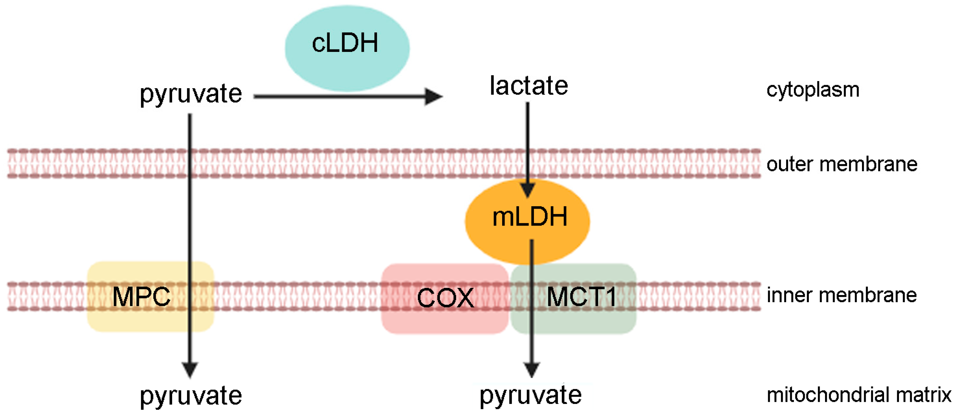
In skeletal muscle, lactate can be directly oxidised in mitochondria. In 1986, Brooks suggested the “lactate shuttle theory”, according to which lactate is up taken by cellular organelles, such as mitochondria or peroxisome, or secreted into the bloodstream and used by organs as a fuel [4,10]. The oxidation of lactate in mitochondria has been demonstrated by several approaches performed on cells and tissues (such as magnetic resonance spectrometry, confocal microscopy, and immunohistochemistry) [11,12]. Extensive studies in healthy human subjects show that the main lactate oxidation site is the mitochondrial lactate complex. Here, lactate is converted by mitochondrial LDH to pyruvate that is driven towards the Krebs cycle. This process is very useful in skeletal muscle since the ability to use lactate as an energy source provides an effective mechanism for ATP regeneration in the presence of high energy demand, such as during high-intensity exercise. The mitochondrial lactate oxidation complex is composed of mitochondrial LDH that is anchored to the outer side of the inner mitochondrial membrane and associated with cytochrome oxidase (COX) and mitochondrial MCT1. The exergonic redox change in cytochrome oxidase during mitochondrial electron transport is coupled with lactate oxidation. Finally, MCT1 transports pyruvate across the inner mitochondrial membrane [12] (Figure 2).
During muscle contraction, the metabolic rate of skeletal muscle rises exponentially, and the huge consumption of ATP is buffered by the modulation of glycolysis and glycogenolysis rates [13]. In the muscles and arterial blood of resting healthy humans, lactate concentration approximates 1.0 mM [13]. However, lactate concentration is subjected to considerable fluctuations with a significant change in the lactate/pyruvate ratio that dynamically affect the NAD+/NADH ratio. Therefore, lactate production has a significant impact on metabolic regulation and cellular redox. In this regard, lactate can affect the cellular production of reactive oxygen species (ROS) through both enzymatic and non-enzymatic pathways, primarily by affecting ROS production through mitochondrial respiration [14]. In this regard, mitochondrial lactate metabolism is involved in the generation of ROS, mainly hydrogen peroxide, via a flavin-dependent lactate oxidase (LOX) pathway.
Most of the lactate (75–80%) is disposed within the tissue or released and taken up by the working muscle [15], heart [16], brain [17], and liver for gluconeogenesis [18]. In addition to oxidative fuel, lactate is important in supplying carbon to gluconeogenesis. In this regard, the skeletal muscle and liver are linked by the so-called “Cori cycle”, an organ–organ lactate shuttle. The cycle ensures that the liver is supplied with lactate that is directed into gluconeogenesis after being converted to pyruvate by hepatic LDH.
Molecules produced during physiological cellular metabolism were for a long time considered exclusively to be a metabolic waste [19]. In recent years, their role has been reassessed, since these molecules can orchestrate a variety of biological processes through their binding to receptors or specific transporters. Lactate is among these multifunctional metabolites, acting as an intracellular signalling mediator and as an extracellular ligand [20,21]. Lactate displays a signalling function through binding to the lactate-activated G-protein-coupled receptor GPR81, also termed Hydroxycarboxylic Acid Receptor 1 (HCA1) [22]. This receptor is located in the plasma membrane, and its presence has been observed in other intracellular organelles, such as mitochondria, thus implying a role for GPR81 in the trafficking of lactate between the plasma membrane and intracellular compartments [23]. Predominantly expressed in the adipose tissue, GPR81 is present in the skeletal muscle, liver, spleen, kidney, and brain [24,25]. A lactate concentration between 0.2 and 1.0 mM results in the activation of the receptor, followed by cyclic AMP (cAMP) downregulation and the inhibition of protein kinase A (PKA)-mediated signalling [22]. Recent studies showed that lactate-induced GPR81 activation signals through a non-canonical, cAMP/PKA-independent pathway involving beta-arrestin, a GPR81 adaptor protein. This leads to the inhibition of TLR-4 and NOD-like receptor pyrin-domain-containing protein 3 (NLRP3) inflammasome, which mediates the production of proinflammatory mediators such as IL-1 beta and IL-18 [26,27]. Increasing evidence has shown that lactate plays different roles in various pathophysiological conditions, including inflammation and cancer. The GPR81 receptor mediates the non-metabolic effects of lactate, including the regulation of immune and inflammatory response, active participation in wound healing with increased angiogenesis, decreased adipocyte lipolysis, and neuroprotection [24]. These effects depend on several factors such as lactate concentration, extracellular acidity, and the amount of other nutrients in the cellular microenvironment [28]. Recent reports suggest that lactate produced by aerobic glycolysis has an immunosuppressive effect in the local environment in various disease conditions, including sepsis, cancer, chronic inflammation, and autoimmune diseases [29,30]. Lactate inhibits proinflammatory responses in macrophages in both cAMP-independent and GPR81-independent manner [31]. Lactate increases the production of anti-inflammatory interleukin-10 (IL-10) in dendritic cells and diminishes the production of proinflammatory IL-12 in response to Toll-like receptor (TLR) stimulators [32]. Moreover, lactate can also downregulate TNF alpha, NF-KB, and PTX-3, and upregulates IL-23 in monocytes, and transiently downregulates chemokines [33]. Lactate has also been shown to suppress proliferation and cytokine production in human cytotoxic T cells, thereby decreasing the cytotoxic effect [34,35]. Moreover, lactate is involved in reparative angiogenesis through the recruitment of endothelial progenitor cells, the stimulation of the endothelial cell migration, the activation of procollagen factors, and the enhancement of collagen deposition in the extracellular matrix [36,37]. It has been reported that the oral administration of the GPR81 agonist 3-Chloro-5-hydroxybenzoic acid (CHBA) to mice significantly increases the weight of plantaris and soleus muscles, and this effect is associated with the enhanced phosphorylation of ERK1-2 proteins [38]. In human skeletal muscle, GPR81 is significantly expressed mainly in type II glycolytic fibres, compared with type I oxidative fibres. Type II fibres produce the highest amounts of lactate, suggesting that lactate may function as an autocrine signalling molecule [39]. Upon ligand binding, GPR81 inhibits cAMP-dependent protein kinase (PKA) signalling and lipolysis (Figure 1). The lactate-induced inhibition of lipolysis, firstly shown in adipose tissue, has also been reported in mouse skeletal muscle [40]. One well-defined target of PKA is the cAMP-response element binding (CREB) protein. Consequently, the binding of lactate to GPR81 results in lower PKA activity, which leads to reduced CREB phosphorylation. Although exercise is known to increase lactate production, the effects on CREB phosphorylation are unclear so far [39]. In addition to regulating lipolysis, lactate has also been shown to stimulate the expression of genes involved in lactate and energy metabolism in skeletal muscle, as evidenced by Hashimoto et al. [7]. However, it is not yet known whether the effect of lactate in the regulation of gene expression is GPR81-dependent.

2. Lactate in Muscle Renewal

Skeletal muscle is characterised by a high regenerative capacity due to the presence of satellite cells (SC). Located between muscle basal lamina and sarcolemma, SC plays a key role in the regenerative process of adult skeletal muscle and in the maintenance of muscle mass. SC are normally in a quiescent state characterised by a relatively low energy demand. Following skeletal muscle damage, SC are rapidly activated to proliferate and differentiate to regenerate muscle tissue [41]. The metabolic profile undergoes significative modifications in quiescent, activated, and differentiated SC, suggesting that metabolic changes may influence the SC’s fate [42]. Recent data suggest that external factors in the muscle microenvironment, such as metabolites, could play a key role in SC activation. In this regard, lactate has been proposed as an excellent candidate. A lactate-dependent mechanism that indirectly controls SC’s fate has recently been reported. During ischemia, endothelial cells increase the secretion of lactate, exploiting their glycolytic activity. Extracellular lactate targets macrophages, inducing their polarisation towards an M2-like phenotype. These lactate-polarised macrophages enhance the expression of vascular endothelial growth factor (VEGF) and stimulate the proliferation and differentiation of muscle progenitor cells. These events support muscle revascularisation and regeneration after SC activation [43].
In vitro, lactate acts as a pro-myogenic factor, regulating the proliferative and differentiation rate of murine C2C12 myoblasts. The differentiation of myoblasts is induced by 10 mM lactate by increasing the expression of the myogenic determination protein (MyoD). In mice, the peritoneal injection of lactate provokes enhanced regeneration and fibre hypertrophy in regenerating muscles, showing that lactate also acts as myogenic factor in vivo [44]. Moreover, skeletal muscle regeneration is associated with the upregulation of the lactate transporter MCT1, which is useful for lactate uptake by muscle cells [45]. Extracellular lactate promotes C2C12 myotube hypertrophy by activating anabolic intracellular signals in a ROS-dependent pathway [46]. In mice, muscle regeneration in the cardiotoxin-treated tibialis anterior is promoted by the oral intake of lactate that induces the increase in Pax7-positive cells. The administration of 20 mM lactate during myogenesis induces the formation of myotubes characterised by increased diameter, length, and protein content in comparison with the control counterpart [38,47]. The positive effects of lactate on muscle differentiation occur in the early stage of differentiation, while the treatment of myoblasts with 20 mM lactate seems to delay the late phase of myogenesis [46,48]. This effect is due to the lactate-dependent downregulation of p38 MAPK phosphorylation, associated with decreased trimethylation of lysine 4 of histone 3 and the reduced expression of Myf5, myogenin, and myosin heavy chain (MHC) proteins. The same patterns have been observed in human skeletal muscle biopsies after training [48].

3. Protein Lactylation in Skeletal Muscle

In 2019, a novel type of protein post-translational modification was reported for the first time. The authors showed that the lysine residue of histones can bind to lactate, thus generating so-called “protein lactylation”. Histone lactylation acts as an epigenetic modulator that directly stimulates gene transcription, and a total of 28 lysine lactylation sites have been identified on the core histones in both human and mouse cells [49]. New lines of evidence have uncovered that lactate induces histone lactylation in the promoters of the profibrotic genes in macrophages, leading to the upregulation of epigenetic modifications in the fibrotic lungs [50]. Moreover, lysine lactylation in the brain is regulated by neural excitation and social stress [51]. In skeletal muscle, a correlation between histone lactylation and myogenesis has been reported both in vitro and in vivo. Lactate promotes the lactylation of lysine 9 in H3 histone, leading to increased Neu2 transcription and myogenesis [52]. Moreover, protein lactylation is involved in the maintenance of cell homeostasis in skeletal muscle during intense exercise. This process involves the lactylation of Vps34, a catalytic subunit of the Phosphatidylinositol 3 kinase (PI3K) complex, which phosphorylates phosphatidylinositol (PI) to produce phosphatidylinositol 3-phosphate [PtdIns(3)P]. Lactylated Vps34 increases lipid kinase activity to enhance autophagy and endolysosomal degradation, thus promoting muscle homeostasis [53]. Protein lactylation in skeletal muscle correlates with obesity and higher levels of plasma lactate. This effect appears more pronounced in obese females in comparison with lean females and male individuals [54]. Moreover, protein lactylation is associated with an increased phosphorylation on serine 636 in IRS-1, an effect already shown in insulin resistant conditions [55,56,57], as in the human skeletal muscle cells derived from type 2 diabetic donors [55]. In contrast, insulin sensitivity is negatively associated with skeletal muscle lactylation [54].

4. Lactate and Physical Exercise

In past years, skeletal muscle was primarily considered the site of lactate production during contraction, associated with poor muscle oxygenation and fatigue. However, skeletal muscle plays an important role not only in the production of lactate but also in its clearance [58]. Moreover, lactate generated during exercise is involved in the energy metabolism rate, mediating exercise adaptations and interorgan communication [59], and it can be considered an important source of carbohydrates in competition with glucose [60].
Skeletal muscle produces large amounts of lactate, mainly for two reasons: (i) at the start of moderate-intensity exercise, the rate of glycolysis is massive and faster in comparison to the oxidative phosphorylation pathway; (ii) the maximum glycolytic capacity exceeds the maximum oxidative ability [61]. Lactate production and consumption change during exercise, as shown in a typical blood lactate curve. Lactate is produced and used as fuel during skeletal muscle contraction, and most of the formed lactate is absorbed and oxidised by the producing fibre [61]. During the initial transition phase, between rest and exercise, a sudden rise in energy response occurs and blood lactate levels moderately increase. A rise in ATP demand by working muscles activates glycolysis, leading to increased pyruvate production. In this phase, most of the produced pyruvate is converted to lactate, thus enhancing circulating lactate levels, since no adequate increase in heart rate and capillary dilatation for the delivery of oxygen to contracting muscles occurs. When the cardiovascular response to exercise happens, more oxygen is delivered to skeletal muscle and blood lactate levels decrease and stabilise. In a subsequent phase of training, when exercise intensity increases, the pyruvate production exceeds the capacity of glycolytic fibres to oxidise pyruvate. As a consequence, pyruvate is reduced to lactate by LDH-A. An increase in circulating lactate during training is dependent on oxygen delivery, mitochondrial capacity, and the ability of other cells to take up and use lactate [10,62].
Intense physical exercise is associated with so-called “lactic acidosis”. Although the production of lactate is associated with hydrogen ion (H+) formation, the exact origin of this acidosis is still debated. It has been argued that non-mitochondrial ATP turnover could be the source of H+ [63], while others have hypothesised that the maintenance of electroneutrality occurs between lactate (negatively charged) and the H+ provided by water dissociation [64]. Acidosis seems to jeopardise muscle contraction, leading to fatigue, a rapid deterioration of contractile function, and eventually to cessation of exercise. The accumulation of lactic acid in muscles has been always recognised as the main cause of muscle fatigue. In humans, the concentration of intracellular lactate increases during intense exercise, up to 30 mM, in association with a small decrease in intracellular pH (about 0.5 units) [65]. However, acidification does not affect the reduction rate in fibre shortening due to muscle fatigue, since lactic acid plays a metabolic buffering role as well as oxidised NAD regeneration, rather than the onset of acidosis [59,66].
In sport medicine, the maximum level of physical effort not associated with lactate and hydrogen ion accumulation in the blood and muscles is called the “anaerobic threshold” or “lactate threshold”. This threshold refers to the intensity and duration of physical activity without changing from aerobic to anaerobic condition. The lactate threshold is considered an excellent predictor of performances, and it is enhanced by intense exercise, which in turn increases performance. As the muscle respiration rate decreases, lactate produced during muscle contraction is released into the blood, even after exercise. Subsequently, lactate is transported by blood circulation to the liver, where it becomes a preferable substrate for hepatic glucose synthesis via a process called the Cori cycle [67,68]. Most of the lactate produced from active muscle is released and taken up by exercising and non-exercising muscles, as well as the heart and brain, where it is oxidised and used as an energy substrate for the neurons [69,70]. Indeed, lactate produced during exercise is considered to influence brain cell function. Hence, the lactate accumulated during exercise is not merely a waste product due to oxygen deficit, since it acts in many processes. Lactate is exchanged between various tissues, such as between different muscles and between various fibre types within the contracting muscles, and it is transported to other organs. Among these, in the heart and brain, lactate is used for mitochondrial respiration [71], while in the kidney and liver it is directed to gluconeogenesis [65].

5. Lactate in the Skeletal Muscle Diseases

Lactate has been associated with various diseases, such as obesity and insulin resistance [72]. Regarding insulin resistance, it is considered one of the main challenges leading to the development of type 2 diabetes [73]. The involvement of lactate in the onset of insulin resistance has been reported in the skeletal muscles of rats, where elevated amounts of plasma lactate suppressed insulin-stimulated glycolysis. This effect was associated with decreased glucose uptake and the alteration of the insulin signalling cascade [74].
The increased production of lactate has been reported in cancer cachexia, a devastating pathology induced by oncologic disease that leads to pronounced weight loss and the worsening of the oncologic condition. Muscles of cachectic mice showed enhanced MCT1 levels and significantly increased lactate/pyruvate ratio, suggesting the high production and release of lactate by the tissue [75]. Myotubes, as well as cachectic mice, showed elevated amounts of lactate in cachectic conditions associated with decreased oxygen consumption [76,77]. The cachectic phenotype in myotubes is accompanied by a metabolic modification towards lactic fermentation. Lactate dehydrogenase inhibition by sodium oxamate impedes the formation of the cachectic condition, thus demonstrating that this metabolic conversion is useful for the onset of the cachectic condition [76]. Moreover, the lactate/pyruvate ratio appears to be decreased in muscles of cachectic mice, due to the increased production of lactate in this condition. The treatment with 2-deoxyglucose, that inhibits glycolysis and impedes the formation of pyruvate, blocks the induction of the cachectic characteristics in mice, thus demonstrating the significant involvement of lactate in the onset of cancer cachexia [78]. Moreover, the ablation of the lactate receptor GPR81 is sufficient to impede the onset of cancer cachexia in both the adipose tissue and skeletal muscle [79], thus highlighting the importance of lactate in the onset and progression of cancer cachexia.
Genetic mutations in gene-encoding lactate transporter MCT1 or an altered expression of the protein are observed in muscular pathologies. An altered MCT1 level has been reported in McArdle disease [Glycogen Storage Disease Type V; MD), a metabolic myopathy characterised by the absence of glycogenolysis due to a deficiency in muscle glycogen phosphorylase [80]. MD patients show increased MCT1 levels, probably as a compensatory mechanism for energy production in the absence of glycogenolysis [81]. MCT1 is encoded by the solute carrier family 16, with the member 1 [SLC16A1) gene organised as five exons interspersed by four introns [82]. Mutations in the SLC16A1 gene lead to clinical conditions. For example, patients with recurrent and severe ketoacidosis, a pathology that occurs when ketone formation exceeds ketone utilisation, show homozygous and heterozygous mutations in the SLC16A1 gene [83,84]. Moreover, a missense mutation in the MCT1 gene is responsible for altered lactate transport and muscle injury in humans [85]. Emerging evidence has shown that single-nucleotide polymorphism (SNP) can be used as a promising biomarker of individual genetic background to predict therapeutic responses and prognosis in cancer patients [86]. With regard to this, SNP in the MCT1 gene could be used as a prognostic marker in colorectal cancer patients and as a predicting marker in adjuvant chemotherapy [87].

6. Concluding Remarks

The point of view regarding the role of lactate in skeletal muscle has changed significantly over the years. As previously described, lactate has numerous and varied functions in skeletal muscle, in both pathological and healthy conditions. However, many aspects still need to be clarified. Firstly, the role of protein lactylation (not only regarding histones, but proteins in general) must be addressed in depth. An issue to be answered is whether lactate can influence the state of chromatin in all tissues, and whether this epigenetic effect is modified in pathological conditions. Moreover, the question of how and whether lactate binding to non-histone proteins can influence their function remains unresolved. Mass spectrometry combined with the use of primary antibodies that recognise lactate binding to proteins may be useful in this research. Moreover, the role played by the metabolite in skeletal muscle will need to be analysed widely. Increasing evidence highlights that the lactate receptor GPR81 could be involved in the onset and/or maintenance of muscle diseases (for example, in the onset of muscle wasting induced by a neoplastic pathology). No effective GPR81 antagonists, aiming to avoid the negative effects of lactate as a signalling molecule, are available so far. From this point of view, the discovery of new receptor inhibitors could contribute to the amelioration of those pathologies in which lactate plays an important role as signalling molecule.

Author Contributions

Conceptualisation, T.F.; writing—original draft preparation, T.F., B.B., T.G. and M.M.; writing—review and editing, T.F., B.B., T.G. and M.M.; supervision, T.F.; funding acquisition, T.F. All authors have read and agreed to the published version of the manuscript.

Funding

This work was supported by the Italian Ministry of University and Research (MIUR).

Data Availability Statement

No new data were created or analysed in this study. Data sharing is not applicable to this article.

Acknowledgments

The authors thank the Fondazione Italiana per la Ricerca sul Cancro (AIRC) for a one-year fellowship (Project Code: IG-25012).

Conflicts of Interest

The authors declare no conflicts of interest.

References

  1. Yadav, A.; Singh, A.; Phogat, J.; Dahuja, A.; Dabur, R. Magnoflorine prevent the skeletal muscle atrophy via Akt/mTOR/FoxO signal pathway and increase slow-MyHC production in streptozotocin-induced diabetic rats. J. Ethnopharmacol. 2021, 267, 113510. [Google Scholar] [CrossRef]
  2. Plotkin, D.L.; Roberts, M.D.; Haun, C.T.; Schoenfeld, B.J. Muscle Fiber Type Transitions with Exercise Training: Shifting Perspectives. Sports 2021, 9, 127. [Google Scholar] [CrossRef]
  3. Brooks, G.A. Lactate shuttles in Nature. Biochem. Soc. Trans. 2002, 30, 258–264. [Google Scholar] [CrossRef]
  4. Nalbandian, M.; Radak, Z.; Takeda, M. Lactate Metabolism and Satellite Cell Fate. Front. Physiol. 2020, 11, 610983. [Google Scholar] [CrossRef]
  5. Everse, J.; Kaplan, N.O. Lactate dehydrogenases: Structure and function. Adv. Enzymol. Relat. Areas Mol. Biol. 1973, 37, 61–133. [Google Scholar]
  6. Halestrap, A.P.; Wilson, M.C. The monocarboxylate transporter family—Role and regulation. IUBMB Life 2012, 64, 109–119. [Google Scholar] [CrossRef]
  7. Hashimoto, T.; Hussien, R.; Oommen, S.; Gohil, K.; Brooks, G.A. Lactate sensitive transcription factor network in L6 cells: Activation of MCT1 and mitochondrial biogenesis. FASEB J. 2007, 21, 2602–2612. [Google Scholar] [CrossRef]
  8. Brooks, G.A. Cell–cell and intracellular lactate shuttles. J. Physiol. 2009, 587, 5591–5600. [Google Scholar] [CrossRef]
  9. Warburg, O. On respiratory impairment in cancer cells. Science 1956, 124, 269–270. [Google Scholar] [CrossRef] [PubMed]
  10. Brooks, G.A.; Arevalo, J.A.; Osmond, A.D.; Leija, R.G.; Curl, C.C.; Tovar, A.P. Lactate in contemporary biology: A phoenix risen. J. Physiol. 2022, 600, 1229–1251. [Google Scholar] [CrossRef] [PubMed]
  11. Chen, Y.-J.; Mahieu, N.G.; Huang, X.; Singh, M.; A Crawford, P.; Johnson, S.L.; Gross, R.W.; Schaefer, J.; Patti, G.J. Lactate metabolism is associated with mammalian mitochondria. Nat. Chem. Biol. 2016, 12, 937–943. [Google Scholar] [CrossRef]
  12. Hashimoto, T.; Hussien, R.; Brooks, G.A. Colocalization of MCT1, CD147, and LDH in mitochondrial inner membrane of L6 muscle cells: Evidence of a mitochondrial lactate oxidation complex. Am. J. Physiol. Endocrinol. Metab. 2006, 290, E1237–E1244. [Google Scholar] [CrossRef]
  13. Brooks, G.A. The Science and Translation of Lactate Shuttle Theory. Cell Metab. 2018, 27, 757–785. [Google Scholar] [CrossRef]
  14. Passarella, S.; de Bari, L.; Valenti, D.; Pizzuto, R.; Paventi, G.; Atlante, A. Mitochondria and L-lactate metabolism. FEBS Lett. 2008, 582, 3569–3576. [Google Scholar] [CrossRef] [PubMed]
  15. Bergman, B.C.; Horning, M.A.; Casazza, G.A.; Wolfel, E.E.; Butterfield, G.E.; Brooks, G.A. Endurance training increases gluconeogenesis during rest and exercise in men. Am. J. Physiol. Endocrinol. Metab. 2000, 278, E244–E251. [Google Scholar] [CrossRef]
  16. Bergman, B.C.; Tsvetkova, T.; Lowes, B.; Wolfel, E.E. Myocardial glucose and lactate metabolism during rest and atrial pacing in humans. J. Physiol. 2009, 587, 2087–2099. [Google Scholar] [CrossRef]
  17. Glenn, T.C.; Martin, N.A.; Horning, M.A.; McArthur, D.L.; Hovda, D.A.; Vespa, P.; Brooks, G.A. Lactate: Brain Fuel in Human Traumatic Brain Injury: A Comparison with Normal Healthy Control Subjects. J. Neurotrauma 2015, 32, 820–832. [Google Scholar] [CrossRef]
  18. Emhoff, C.-A.W.; Messonnier, L.A.; Horning, M.A.; Fattor, J.A.; Carlson, T.J.; Brooks, G.A. Gluconeogenesis and hepatic glycogenolysis during exercise at the lactate threshold. J. Appl. Physiol. 2013, 114, 297–306. [Google Scholar] [CrossRef]
  19. Vander Heiden, M.G.; Cantley, L.C.; Thompson, C.B. Understanding the Warburg Effect: The Metabolic Requirements of Cell Proliferation. Science 2009, 324, 1029–1033. [Google Scholar] [CrossRef] [PubMed]
  20. Baltazar, F.; Afonso, J.; Costa, M.; Granja, S. Lactate Beyond a Waste Metabolite: Metabolic Affairs and Signaling in Malignancy. Front. Oncol. 2020, 10, 231. [Google Scholar] [CrossRef] [PubMed]
  21. Certo, M.; Llibre, A.; Lee, W.; Mauro, C. Understanding lactate sensing and signalling. Trends Endocrinol. Metab. 2022, 33, 722–735. [Google Scholar] [CrossRef] [PubMed]
  22. Sun, S.; Li, H.; Chen, J.; Qian, Q. Lactic Acid: No Longer an Inert and End-Product of Glycolysis. Physiology 2017, 32, 453–463. [Google Scholar] [CrossRef] [PubMed]
  23. Liu, C.; Wu, J.; Zhu, J.; Kuei, C.; Yu, J.; Shelton, J.; Sutton, S.W.; Li, X.; Yun, S.J.; Mirzadegan, T.; et al. Lactate Inhibits Lipolysis in Fat Cells through Activation of an Orphan G-protein-coupled Receptor, GPR81. J. Biol. Chem. 2009, 284, 2811–2822. [Google Scholar] [CrossRef] [PubMed]
  24. Lee, T.-Y. Lactate: A multifunctional signaling molecule. Yeungnam Univ. J. Med. 2021, 38, 183–193. [Google Scholar] [CrossRef] [PubMed]
  25. Brown, T.P.; Ganapathy, V. Lactate/GPR81 signaling and proton motive force in cancer: Role in angiogenesis, immune escape, nutrition, and Warburg phenomenon. Pharmacol. Ther. 2020, 206, 107451. [Google Scholar] [CrossRef] [PubMed]
  26. Hoque, R.; Ouyang, X.; Farooq, A.; Ghani, A.; Ahsan, K.; Guerra, M.; Mehal, W.Z. Activation of N-methyl-d-aspartate receptor downregulates inflammasome activity and liver inflammation via a β-arrestin-2 pathway. Am. J. Physiol. Gastrointest. Liver Physiol. 2014, 307, G732–G740. [Google Scholar] [CrossRef]
  27. Hoque, R.; Farooq, A.; Ghani, A.; Gorelick, F.; Mehal, W.Z. Lactate Reduces Liver and Pancreatic Injury in Toll-Like Receptor– and Inflammasome-Mediated Inflammation via GPR81-Mediated Suppression of Innate Immunity. Gastroenterology 2014, 146, 1763–1774. [Google Scholar] [CrossRef] [PubMed]
  28. Certo, M.; Tsai, C.-H.; Pucino, V.; Ho, P.-C.; Mauro, C. Lactate modulation of immune responses in inflammatory versus tumour microenvironments. Nat. Rev. Immunol. 2021, 21, 151–161. [Google Scholar] [CrossRef]
  29. Nolt, B.; Tu, F.; Wang, X.; Ha, T.; Winter, R.; Williams, D.L.; Li, C. Lactate and Immunosuppression in Sepsis. Shock 2018, 49, 120–125. [Google Scholar] [CrossRef]
  30. Pucino, V.; Bombardieri, M.; Pitzalis, C.; Mauro, C. Lactate at the crossroads of metabolism, inflammation, and autoimmunity. Eur. J. Immunol. 2017, 47, 14–21. [Google Scholar] [CrossRef]
  31. Errea, A.; Cayet, D.; Marchetti, P.; Tang, C.; Kluza, J.; Offermanns, S.; Sirard, J.-C.; Rumbo, M. Lactate Inhibits the Pro-Inflammatory Response and Metabolic Reprogramming in Murine Macrophages in a GPR81-Independent Manner. PLoS ONE 2016, 11, e0163694. [Google Scholar] [CrossRef] [PubMed]
  32. Nasi, A.; Fekete, T.; Krishnamurthy, A.; Snowden, S.; Rajnavölgyi, E.; Catrina, A.I.; Wheelock, C.E.; Vivar, N.; Rethi, B. Dendritic Cell Reprogramming by Endogenously Produced Lactic Acid. J. Immunol. 2013, 191, 3090–3099. [Google Scholar] [CrossRef]
  33. Peter, K.; Rehli, M.; Singer, K.; Renner-Sattler, K.; Kreutz, M. Lactic acid delays the inflammatory response of human monocytes. Biochem. Biophys. Res. Commun. 2015, 457, 412–418. [Google Scholar] [CrossRef]
  34. Fischer, K.; Hoffmann, P.; Voelkl, S.; Meidenbauer, N.; Ammer, J.; Edinger, M.; Gottfried, E.; Schwarz, S.; Rothe, G.; Hoves, S.; et al. Inhibitory effect of tumor cell–derived lactic acid on human T cells. Blood 2007, 109, 3812–3819. [Google Scholar] [CrossRef]
  35. Husain, Z.; Huang, Y.; Seth, P.; Sukhatme, V.P. Tumor-Derived Lactate Modifies Antitumor Immune Response: Effect on Myeloid-Derived Suppressor Cells and NK Cells. J. Immunol. 2013, 191, 1486–1495. [Google Scholar] [CrossRef]
  36. Hunt, T.K.; Aslam, R.S.; Beckert, S.; Wagner, S.; Ghani, Q.P.; Hussain, M.Z.; Roy, S.; Sen, C.K. Aerobically Derived Lactate Stimulates Revascularization and Tissue Repair via Redox Mechanisms. Antioxidants Redox Signal. 2007, 9, 1115–1124. [Google Scholar] [CrossRef]
  37. Porporato, P.E.; Payen, V.L.; De Saedeleer, C.J.; Préat, V.; Thissen, J.-P.; Feron, O.; Sonveaux, P. Lactate stimulates angiogenesis and accelerates the healing of superficial and ischemic wounds in mice. Angiogenesis 2012, 15, 581–592. [Google Scholar] [CrossRef]
  38. Ohno, Y.; Nakatani, M.; Ito, T.; Matsui, Y.; Ando, K.; Suda, Y.; Ohashi, K.; Yokoyama, S.; Goto, K. Activation of Lactate Receptor Positively Regulates Skeletal Muscle Mass in Mice. Physiol. Res. 2023, 72, 465–473. [Google Scholar] [CrossRef] [PubMed]
  39. Nordström, F.; Liegnell, R.; Apró, W.; Blackwood, S.J.; Katz, A.; Moberg, M. The lactate receptor GPR81 is predominantly expressed in type II human skeletal muscle fibers: Potential for lactate autocrine signaling. Am. J. Physiol. Cell Physiol. 2023, 324, C477–C487. [Google Scholar] [CrossRef] [PubMed]
  40. Chen, S.; Zhou, L.; Sun, J.; Qu, Y.; Chen, M. The Role of cAMP-PKA Pathway in Lactate-Induced Intramuscular Triglyceride Accumulation and Mitochondria Content Increase in Mice. Front. Physiol. 2021, 12, 709135. [Google Scholar] [CrossRef]
  41. Guilhot, C.; Catenacci, M.; Lofaro, S.; Rudnicki, M.A. The satellite cell in skeletal muscle: A story of heterogeneity. Curr. Top. Dev. Biol. 2024, 158, 15–51. [Google Scholar] [PubMed]
  42. Pala, F.; Di Girolamo, D.; Mella, S.; Yennek, S.; Chatre, L.; Ricchetti, M.; Tajbakhsh, S. Distinct metabolic states govern skeletal muscle stem cell fates during prenatal and postnatal myogenesis. J. Cell Sci. 2018, 131, jcs212977. [Google Scholar] [CrossRef] [PubMed]
  43. Zhang, J.; Muri, J.; Fitzgerald, G.; Gorski, T.; Gianni-Barrera, R.; Masschelein, E.; D’hulst, G.; Gilardoni, P.; Turiel, G.; Fan, Z.; et al. Endothelial Lactate Controls Muscle Regeneration from Ischemia by Inducing M2-like Macrophage Polarization. Cell Metab. 2020, 31, 1136–1153.e7. [Google Scholar] [CrossRef] [PubMed]
  44. Tsukamoto, S.; Shibasaki, A.; Naka, A.; Saito, H.; Iida, K. Lactate Promotes Myoblast Differentiation and Myotube Hypertrophy via a Pathway Involving MyoD In Vitro and Enhances Muscle Regeneration In Vivo. Int. J. Mol. Sci. 2018, 19, 3649. [Google Scholar] [CrossRef] [PubMed]
  45. Washington, T.A.; Brown, L.; Smith, D.A.; Davis, G.; Baum, J.; Bottje, W. Monocarboxylate transporter expression at the onset of skeletal muscle regeneration. Physiol. Rep. 2013, 1, e00075. [Google Scholar] [CrossRef] [PubMed]
  46. Willkomm, L.; Schubert, S.; Jung, R.; Elsen, M.; Borde, J.; Gehlert, S.; Suhr, F.; Bloch, W. Lactate regulates myogenesis in C2C12 myoblasts in vitro. Stem Cell Res. 2014, 12, 742–753. [Google Scholar] [CrossRef]
  47. Ohno, Y.; Ando, K.; Ito, T.; Suda, Y.; Matsui, Y.; Oyama, A.; Kaneko, H.; Yokoyama, S.; Egawa, T.; Goto, K. Lactate Stimulates a Potential for Hypertrophy and Regeneration of Mouse Skeletal Muscle. Nutrients 2019, 11, 869. [Google Scholar] [CrossRef] [PubMed]
  48. Willkomm, L.; Gehlert, S.; Jacko, D.; Schiffer, T.; Bloch, W. p38 MAPK activation and H3K4 trimethylation is decreased by lactate in vitro and high intensity resistance training in human skeletal muscle. PLoS ONE 2017, 12, e0176609. [Google Scholar] [CrossRef]
  49. Zhang, D.; Tang, Z.; Huang, H.; Zhou, G.; Cui, C.; Weng, Y.; Liu, W.; Kim, S.; Lee, S.; Perez-Neut, M.; et al. Metabolic regulation of gene expression by histone lactylation. Nature 2019, 574, 575–580. [Google Scholar] [CrossRef]
  50. Cui, H.; Xie, N.; Banerjee, S.; Ge, J.; Jiang, D.; Dey, T.; Mathews, Q.; Liu, R.-M.; Liu, G. Lung Myofibroblasts Promote Macrophage Profibrotic Activity through Lactate-induced Histone Lactylation. Am. J. Respir. Cell Mol. Biol. 2021, 64, 115–125. [Google Scholar] [CrossRef]
  51. Hagihara, H.; Shoji, H.; Otabi, H.; Toyoda, A.; Katoh, K.; Namihira, M.; Miyakawa, T. Protein lactylation induced by neural excitation. Cell Rep. 2021, 37, 109820. [Google Scholar] [CrossRef] [PubMed]
  52. Dai, W.; Wu, G.; Liu, K.; Chen, Q.; Tao, J.; Liu, H.; Shen, M. Lactate promotes myogenesis via activating H3K9 lactylation-dependent up-regulation of Neu2 expression. J. Cachex-Sarcopenia Muscle 2023, 14, 2851–2865. [Google Scholar] [CrossRef] [PubMed]
  53. Jia, M.; Yue, X.; Sun, W.; Zhou, Q.; Chang, C.; Gong, W.; Feng, J.; Li, X.; Zhan, R.; Mo, K.; et al. ULK1-mediated metabolic reprogramming regulates Vps34 lipid kinase activity by its lactylation. Sci. Adv. 2023, 9, eadg4993. [Google Scholar] [CrossRef] [PubMed]
  54. Maschari, D.; Saxena, G.; Law, T.D.; Walsh, E.; Campbell, M.C.; A Consitt, L. Lactate-induced lactylation in skeletal muscle is associated with insulin resistance in humans. Front. Physiol. 2022, 13, 951390. [Google Scholar] [CrossRef] [PubMed]
  55. Bouzakri, K.; Roques, M.; Gual, P.; Espinosa, S.; Guebre-Egziabher, F.; Riou, J.-P.; Laville, M.; Le Marchand-Brustel, Y.; Tanti, J.-F.; Vidal, H. Reduced Activation of Phosphatidylinositol-3 Kinase and Increased Serine 636 Phosphorylation of Insulin Receptor Substrate-1 in Primary Culture of Skeletal Muscle Cells from Patients With Type 2 Diabetes. Diabetes 2003, 52, 1319–1325. [Google Scholar] [CrossRef] [PubMed]
  56. Vlavcheski, F.; Tsiani, E. Attenuation of Free Fatty Acid-Induced Muscle Insulin Resistance by Rosemary Extract. Nutrients 2018, 10, 1623. [Google Scholar] [CrossRef] [PubMed]
  57. Vlavcheski, F.; Hartogh, D.J.D.; Giacca, A.; Tsiani, E. Amelioration of High-Insulin-Induced Skeletal Muscle Cell Insulin Resistance by Resveratrol Is Linked to Activation of AMPK and Restoration of GLUT4 Translocation. Nutrients 2020, 12, 914. [Google Scholar] [CrossRef] [PubMed]
  58. van Hall, G. Lactate kinetics in human tissues at rest and during exercise. Acta Physiol. 2010, 199, 499–508. [Google Scholar] [CrossRef]
  59. Hargreaves, M.; Spriet, L.L. Skeletal muscle energy metabolism during exercise. Nat. Metab. 2020, 2, 817–828. [Google Scholar] [CrossRef]
  60. Park, J.; Kim, J.; Mikami, T. Exercise-Induced Lactate Release Mediates Mitochondrial Biogenesis in the Hippocampus of Mice via Monocarboxylate Transporters. Front. Physiol. 2021, 12, 736905. [Google Scholar] [CrossRef]
  61. Lee, S.; Choi, Y.; Jeong, E.; Park, J.; Kim, J.; Tanaka, M.; Choi, J. Physiological significance of elevated levels of lactate by exercise training in the brain and body. J. Biosci. Bioeng. 2023, 135, 167–175. [Google Scholar] [CrossRef] [PubMed]
  62. Hall, M.M.; Rajasekaran, S.; Thomsen, T.W.; Peterson, A.R. Lactate: Friend or Foe. PM&R 2016, 8, S8–S15. [Google Scholar] [CrossRef]
  63. Robergs, R.A.; Ghiasvand, F.; Parker, D. Biochemistry of exercise-induced metabolic acidosis. Am. J. Physiol. Regul. Integr. Comp. Physiol. 2004, 287, R502–R516. [Google Scholar] [CrossRef] [PubMed]
  64. Lindinger, M.I.; Kowalchuk, J.M.; Heigenhauser, G.J.F. Applying physicochemical principles to skeletal muscle acid-base status. Am. J. Physiol. Regul. Integr. Comp. Physiol. 2005, 289, R891-4–R904-10. [Google Scholar] [CrossRef] [PubMed]
  65. Proia, P.; Di Liegro, C.M.; Schiera, G.; Fricano, A.; Di Liegro, I. Lactate as a Metabolite and a Regulator in the Central Nervous System. Int. J. Mol. Sci. 2016, 17, 1450. [Google Scholar] [CrossRef] [PubMed]
  66. Baker, J.S.; McCormick, M.C.; Robergs, R.A. Interaction among Skeletal Muscle Metabolic Energy Systems during Intense Exercise. J. Nutr. Metab. 2010, 2010, 905612. [Google Scholar] [CrossRef]
  67. Yang, W.H.; Park, H.; Grau, M.; Heine, O. Decreased Blood Glucose and Lactate: Is a Useful Indicator of Recovery Ability in Athletes? Int. J. Environ. Res. Public Health 2020, 17, 5470. [Google Scholar] [CrossRef] [PubMed]
  68. Iepsen, U.W.; Plovsing, R.R.; Tjelle, K.; Foss, N.B.; Meyhoff, C.S.; Ryrsø, C.K.; Berg, R.M.G.; Secher, N.H. The role of lactate in sepsis and COVID-19: Perspective from contracting skeletal muscle metabolism. Exp. Physiol. 2022, 107, 665–673. [Google Scholar] [CrossRef] [PubMed]
  69. Adeva-Andany, M.; López-Ojén, M.; Funcasta-Calderón, R.; Ameneiros-Rodríguez, E.; Donapetry-García, C.; Vila-Altesor, M.; Rodríguez-Seijas, J. Comprehensive review on lactate metabolism in human health. Mitochondrion 2014, 17, 76–100. [Google Scholar] [CrossRef]
  70. Mächler, P.; Wyss, M.T.; Elsayed, M.; Stobart, J.; Gutierrez, R.; Von Faber-Castell, A.; Kaelin, V.; Zuend, M.; San Martín, A.; Romero-Gómez, I.; et al. In Vivo Evidence for a Lactate Gradient from Astrocytes to Neurons. Cell Metab. 2016, 23, 94–102. [Google Scholar] [CrossRef]
  71. Kasparov, S. Are Astrocytes the Pressure-Reservoirs of Lactate in the Brain? Cell Metab. 2016, 23, 1–2. [Google Scholar] [CrossRef]
  72. Lin, Y.; Bai, M.; Wang, S.; Chen, L.; Li, Z.; Li, C.; Cao, P.; Chen, Y. Lactate Is a Key Mediator That Links Obesity to Insulin Resistance via Modulating Cytokine Production From Adipose Tissue. Diabetes 2022, 71, 637–652. [Google Scholar] [CrossRef] [PubMed]
  73. DeFronzo, R.A.; Tripathy, D. Skeletal Muscle Insulin Resistance Is the Primary Defect in Type 2 Diabetes. Diabetes Care 2009, 32 (Suppl. S2), S157–S163. [Google Scholar] [CrossRef] [PubMed]
  74. Choi, C.S.; Kim, Y.-B.; Lee, F.N.; Zabolotny, J.M.; Kahn, B.B.; Youn, J.H. Lactate induces insulin resistance in skeletal muscle by suppressing glycolysis and impairing insulin signaling. Am. J. Physiol. Endocrinol. Metab. 2002, 283, E233–E240. [Google Scholar] [CrossRef] [PubMed]
  75. Visavadiya, N.P.; Rossiter, H.B.; Khamoui, A.V. Distinct glycolytic pathway regulation in liver, tumour and skeletal muscle of mice with cancer cachexia. Cell Biochem. Funct. 2021, 39, 802–812. [Google Scholar] [CrossRef] [PubMed]
  76. Mannelli, M.; Gamberi, T.; Magherini, F.; Fiaschi, T. A Metabolic Change towards Fermentation Drives Cancer Cachexia in Myotubes. Biomedicines 2021, 9, 698. [Google Scholar] [CrossRef] [PubMed]
  77. Mannelli, M.; Gamberi, T.; Garella, R.; Magherini, F.; Squecco, R.; Fiaschi, T. Pyruvate prevents the onset of the cachectic features and metabolic alterations in myotubes downregulating STAT3 signaling. FASEB J. 2022, 36, e22598. [Google Scholar] [CrossRef] [PubMed]
  78. Wei, L.; Wang, R.; Wazir, J.; Lin, K.; Song, S.; Li, L.; Pu, W.; Zhao, C.; Wang, Y.; Su, Z.; et al. 2-Deoxy-D-glucose Alleviates Cancer Cachexia-Induced Muscle Wasting by Enhancing Ketone Metabolism and Inhibiting the Cori Cycle. Cells 2022, 11, 2987. [Google Scholar] [CrossRef] [PubMed]
  79. Liu, X.; Li, S.; Cui, Q.; Guo, B.; Ding, W.; Liu, J.; Quan, L.; Li, X.; Xie, P.; Jin, L.; et al. Activation of GPR81 by lactate drives tumour-induced cachexia. Nat. Metab. 2024, 6, 708–723. [Google Scholar] [CrossRef]
  80. Wu, Y.; Weber, J.; Vladutiu, G.; Tarnopolsky, M. Six novel mutations in the myophosphorylase gene in patients with McArdle disease and a family with pseudo-dominant inheritance pattern. Mol. Genet. Metab. 2011, 104, 587–591. [Google Scholar] [CrossRef]
  81. Kitaoka, Y.; Ogborn, D.I.; Mocellin, N.J.; Schlattner, U.; Tarnopolsky, M.A. Monocarboxylate transporters and mitochondrial creatine kinase protein content in McArdle disease. Mol. Genet. Metab. 2013, 108, 259–262. [Google Scholar] [CrossRef]
  82. Cuff, M.A.; Shirazi-Beechey, S.P. The Human Monocarboxylate Transporter, MCT1: Genomic Organization and Promoter Analysis. Biochem. Biophys. Res. Commun. 2002, 292, 1048–1056. [Google Scholar] [CrossRef]
  83. van Hasselt, P.M.; Ferdinandusse, S.; Monroe, G.R.; Ruiter, J.P.; Turkenburg, M.; Geerlings, M.J.; Duran, K.; Harakalova, M.; van der Zwaag, B.; Monavari, A.A.; et al. Monocarboxylate Transporter 1 Deficiency and Ketone Utilization. N. Engl. J. Med. 2014, 371, 1900–1907. [Google Scholar] [CrossRef]
  84. Balasubramaniam, S.; Lewis, B.; Greed, L.; Meili, D.; Flier, A.; Yamamoto, R.; Bilić, K.; Till, C.; Sass, J.O. Heterozygous Monocarboxylate Transporter 1 (MCT1, SLC16A1) Deficiency as a Cause of Recurrent Ketoacidosis. JIMD Rep. 2016, 29, 33–38. [Google Scholar]
  85. Merezhinskaya, N.; Fishbein, W.N.; Davis, J.I.; Foellmer, J.W. Mutations in MCT1 cDNA in patients with symptomatic deficiency in lactate transport. Muscle Nerve 2000, 23, 90–97. [Google Scholar] [CrossRef]
  86. Ulrich, C.M.; Robien, K.; McLeod, H.L. Cancer pharmacogenetics: Polymorphisms, pathways and beyond. Nat. Rev. Cancer 2003, 3, 912–920. [Google Scholar] [CrossRef]
  87. Fei, F.; Guo, X.; Chen, Y.; Liu, X.; Tu, J.; Xing, J.; Chen, Z.; Ji, J.; He, X. Polymorphisms of monocarboxylate transporter genes are associated with clinical outcomes in patients with colorectal cancer. J. Cancer Res. Clin. Oncol. 2015, 141, 1095–1102. [Google Scholar] [CrossRef]
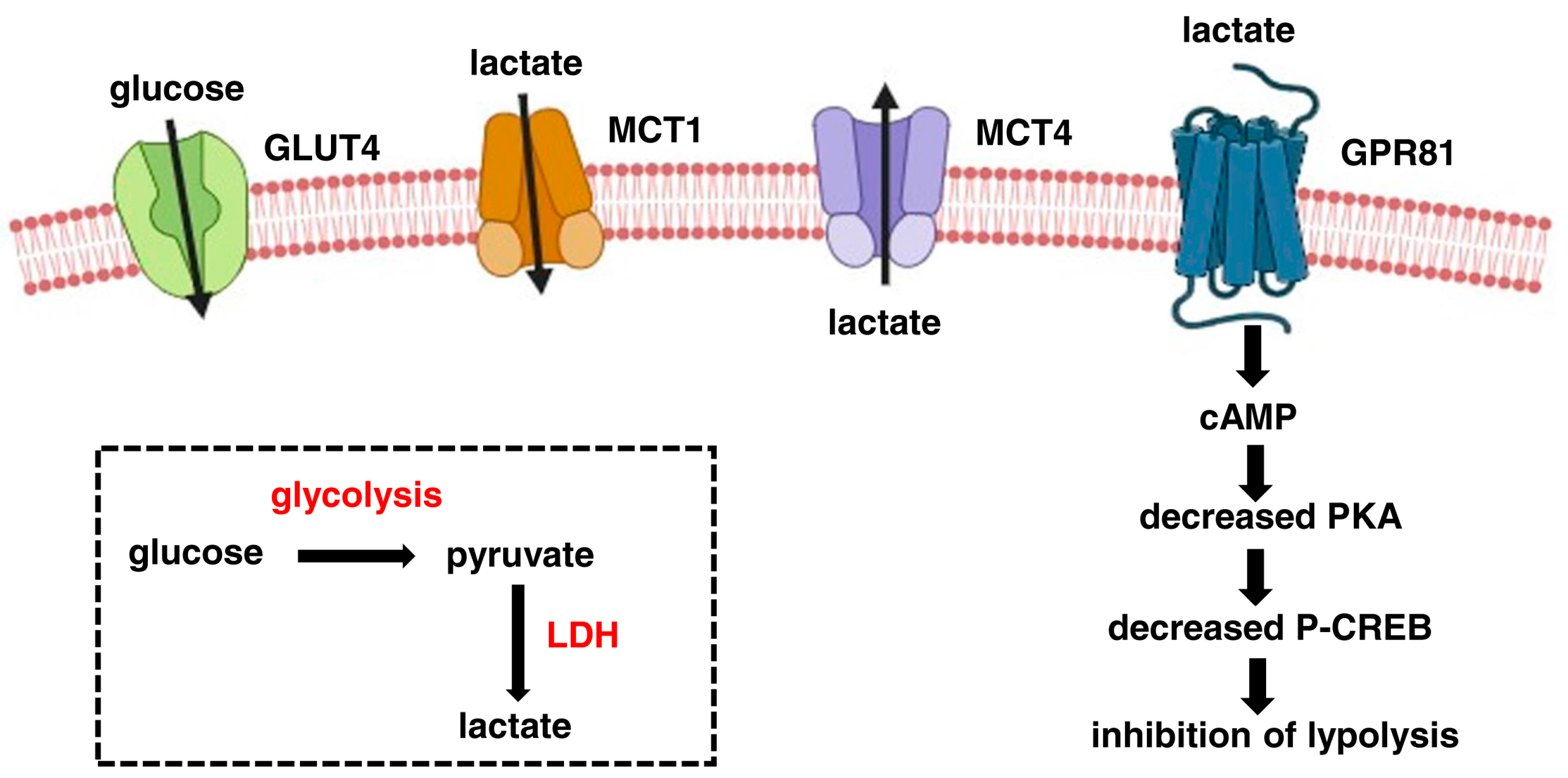
Figure 1. The route of lactate in the skeletal muscle. Glucose (which enters in the muscle through the GLUT4 transporter) is oxidised in two molecules of pyruvate during glycolysis. In anaerobic and in energy-demand conditions, lactate dehydrogenase (LDH) transforms pyruvate into lactate (as shown in the rectangle). Lactate can shuttle between the cytoplasm and the extracellular environment through the two transporters, MCT4 (out of the cell) and MCT1 (within the cell). Extracellular lactate can bind to the specific receptor GPR81. Ligand binding leads to the activation of inhibitory G proteins and the decreased production of cyclic AMP (cAMP). In turn, Protein Kinase A (PKA) activity and phosphorylation of CREB transcription factor are reduced, thus leading to decreased lipolysis. The figure was created with biorender.com.
Cells 13 01177 g001
Figure 2. The lactate in the mitochondria. Lactate produced in the cytoplasm by cytoplasmic LDH (cLDH) enters the intermembrane space, freely crossing the outer mitochondrial membrane. Here, the mitochondrial LDH (mLDH) transforms lactate in pyruvate, which enters the mitochondrial matrix through the MCT1 transporter. MCT1, localised in the inner mitochondrial membrane, together with cytochrome oxidase (COX) and mLDH constitute the so-called “lactate mitochondrial oxidation complex”. Cytoplasmic pyruvate directly enters the mitochondrial matrix, passing through the mitochondrial pyruvate complex (MCP) located in the inner mitochondrial membrane. In the mitochondrial matrix, pyruvate becomes the substrate of the pyruvate dehydrogenase complex that catalyses the conversion of the pyruvate into acetyl CoA. The figure was created with biorender.com.
Cells 13 01177 g002
Disclaimer/Publisher’s Note: The statements, opinions and data contained in all publications are solely those of the individual author(s) and contributor(s) and not of MDPI and/or the editor(s). MDPI and/or the editor(s) disclaim responsibility for any injury to people or property resulting from any ideas, methods, instructions or products referred to in the content.