Article

Ploidy Status, Nuclear DNA Content and Start Codon Targeted (SCoT) Genetic Homogeneity Assessment in Digitalis purpurea L., Regenerated In Vitro

1
Cellular Differentiation and Molecular Genetics Section, Department of Botany, Jamia Hamdard, New Delhi 110062, India
2
Genomic and Biotechnology Unit, Department of Biology, Faculty of Science, University of Tabuk, Tabuk 71491, Saudi Arabia
*
Author to whom correspondence should be addressed.
Genes 2022, 13(12), 2335; https://doi.org/10.3390/genes13122335
Submission received: 2 November 2022 / Revised: 28 November 2022 / Accepted: 7 December 2022 / Published: 11 December 2022
(This article belongs to the Special Issue Genome-Wide Identifications: Recent Trends in Genomic Studies)

Abstract

:
Digitalis purpurea L. is a therapeutically important plant that synthesizes important cardiotonics such as digitoxin and digoxin. The present work reports a detailed and efficient propagation protocol for D. purpurea by optimizing various PGR concentrations in Murashige and Skoog (MS) medium. The genetic homogeneity of in vitro regenerants was assessed by the flow cytometric method (FCM) and Start Codon Targeted (SCoT) marker technique. Firstly, the seeds inoculated in full MS medium added with 0.5 mg/L GA3 produced seedlings. Different parts such as hypocotyl, nodes, leaves and apical shoots were used as explants. The compact calli were obtained on BAP alone or in combinations with 2, 4-D/NAA. The hypocotyl-derived callus induced somatic embryos which proliferated and germinated best in 0.75 mg/L BAP-fortified MS medium. Scanning electron microscopic (SEM) images confirmed the presence of various developmental stages of somatic embryos. Shoot regeneration was obtained in which BAP at 1.0 mg/L and 2.0 mg/L BAP + 0.5 mg/L 2,4-D proved to be the best treatments of PGRs in inducing direct and indirect shoot buds. The regenerated shoots showed the highest rooting percentage (87.5%) with 24.7 ± 1.9 numbers of roots/shoot in 1.0 mg/L IBA augmented medium. The rooted plantlets were acclimatized in a greenhouse at a survival rate of 85–90%. The genome size and the 2C nuclear DNA content of field-grown, somatic embryo-regenerated and organogenic-derived plants were estimated and noted to be 3.1, 3.2 and 3.0 picogram (pg), respectively; there is no alteration in ploidy status and the DNA content, validating genetic uniformity. Six SCoT primers unveiled 94.3%–95.13% monomorphic bands across all the plant samples analyzed, further indicating genetic stability among in vitro clones and mother plants. This study describes for the first time successful induction of somatic embryos from hypocotyl callus; and flow cytometry and SCoT marker confirmed the genetic homogeneity of regenerated plants.

1. Introduction

Digitalis purpurea L., a member of the Plantaginaceae family, is a biennial/perennial herbaceous plant [1]. It is popularly known as foxglove (lady’s glove). D. purpurea is an indigenous species of Europe which has now been widely cultivated in various parts of the world, such as China, New Zealand, USA and Canada. It has conspicuous bell-shaped flowers of different colors ranging from purple to yellow, pink, gray or white and hence, it is being grown as an ornamental plant across the world [2]. Apart from ornamental values, the plant also holds great medicinal importance in the pharmaceutical sector due to the presence of cardiac glycosides. The members of this cardenolide group include digitoxin, digoxin, digitoxigenin, digoxigenin, medigoxin, strophanthins and lanthosides [3]. Digitoxin, digoxin and lanthosides are the major bioactive compounds found in D. purpurea [4]. These cardenolides have been extensively utilized in treating congestive heart failure, myocardial infarction, oedema, coronary artery disease, hypertrophy and hypertension [5]. In recent times, digitoxin and digoxin have proved to be effective as anti-cancer and anti-viral agents [6].
Digitalis is the natural source of glycosides, and it is quite difficult to synthesize under laboratory conditions owing to structural intricacy. The unregulated utilization of this plant by the pharmaceutical industry has been a major cause of the decline of the plant population in its native environment [7]. The delayed seed-set formation, low seed germination and viability limit the growth in the wild [8,9]. Under such circumstances, plant tissue culture can serve as a better alternative for rapid multiplication of microbe-free plants in a short period of time, which is otherwise not possible to achieve under natural conditions [10]. The in vitro culture technique offers several advantages over conventional breeding approaches such as fast propagation, preservation of germplasm, production of polyploids, genetic transformation and crop advancement [11,12]. In addition, it provides continual and enhanced yields of medically important bioactives found in plants [13].
Somatic embryogenesis and organogenesis are the two intriguing biotechnological approaches to produce in vitro regenerated plantlets [14]. However, due to prolonged exposure to various Plant Growth Regulators (PGRs), continuous passaging and genotypic make-up, the regenerated plants often accumulate genetic and epigenetic alterations, commonly termed as somaclonal variations [15,16]. Therefore, the genetic uniformity evaluation of plants raised in vitro is crucial to ensure commercial uses on a global level [17,18]. In this regard, flow cytometric method proves to be a handy technique for measuring nuclear 2C DNA content of plant cells [19], making it simple, fast and easy to determine the ploidy of micropropagated plants [20]. Recent investigations have been done to examine the ploidy levels in various plants, such as Allium sativum [21], different species of Zephyranthes [22] and Catharanthus roseus [23]. Additionally, the genetic homogeneity of tissue culture plants can also be carried out by using PCR-based DNA markers such as Random Amplified Polymorphic DNA (RAPD), Start Codon Targeted (SCoT), Inter Simple Sequence Repeat (ISSR) and Amplified Fragment Length Polymorphism (AFLP) [24]. The SCoT marker technique ascertains polymorphism in the genome of micropropagated plants, thereby screening out possible somaclonal variations with high efficacy [25], as reported successfully in different plants [26,27].
In this current study, a sustainable in vitro plant propagation protocol via somatic embryogenesis and shootlet organogenesis in D. purpurea has been devised. To date, there are no reports of somatic embryo formation in D. purpurea; thus, for the first time, this article describes an efficient embryogenesis method from hypocotyl callus. Different morphological developments of somatic embryos have also been described using scanning electron microscopy (SEM). Moreover, this is the first ever report of assessing genetic homogeneity of in vitro-derived plantlets in D. purpurea with respect to the mother plant using flow cytometry and SCoT molecular marker approaches.

2. Materials and Methods

2.1. Collection of Seed Materials and Explant Preparation

The seeds of Digitalis purpurea L. were collected from the Herbal Research and Development Institute (HRDI), Mandal (coordinates: 30°27′13.3″ N 79°16′17.8″ E), Chamoli district of Uttarakhand, India. Initially, the collected seeds were immersed in a 20% teepol solution for 10 min, then washed under running tap water for several minutes. Afterwards, the seeds were surface-sterilised with 70% ethanol and 0.1% HgCl2 for 2 min each, and then thoroughly rinsed with sterilised double-distilled water three times to eliminate remnants of the sterilising agents.

2.2. Seed Germination and Culture Conditions

For the effectual germination, the disinfected seeds were then aseptically placed onto full- and half-strength Murashige and Skoog (MS) basal medium [28] comprised of 3% (w/v) sucrose and 0.8% (w/v) agar. The pH calibration of the medium was done at 5.7 using 1N HCl and/or 1N NaOH preparatory to autoclaving at 121 °C for 15 min. All culture tubes were maintained at 25 ± 2 °C under white fluorescent light (irradiance at 50 μmol/m2/s−1) for a 16 h photoperiod at a relative humidity of 60%. The frequency of seed germination was noted after a three-week period. Node, leaf, hypocotyl and apical shoot segments from 21-day-old germinated seedlings were taken as explants for further experimentation.

2.3. Callus Induction and Proliferation

Different explants, such as node, leaf and hypocotyl, were inoculated on MS medium enriched with 0.5–2.0 mg/L BAP (6-benzylaminopurine) alone/in combination with varied 2,4-Dconcentrations (2,4-dichlorophenoxyacetic acid)/NAA (α-naphthalene acetic acid) for callus induction. The cultures were maintained by sub culturing the callus in the same PGR added MS medium after 3–4 weeks The callus induction frequencies of different explants used were recorded three weeks after the culture period. The texture of the callus was also noted for each explant used in the experiments.
Callus   induction   frequency   ( % ) = Number   of   explants   showing   callusing ÷ Total   number   of   explants   inoculated × 100

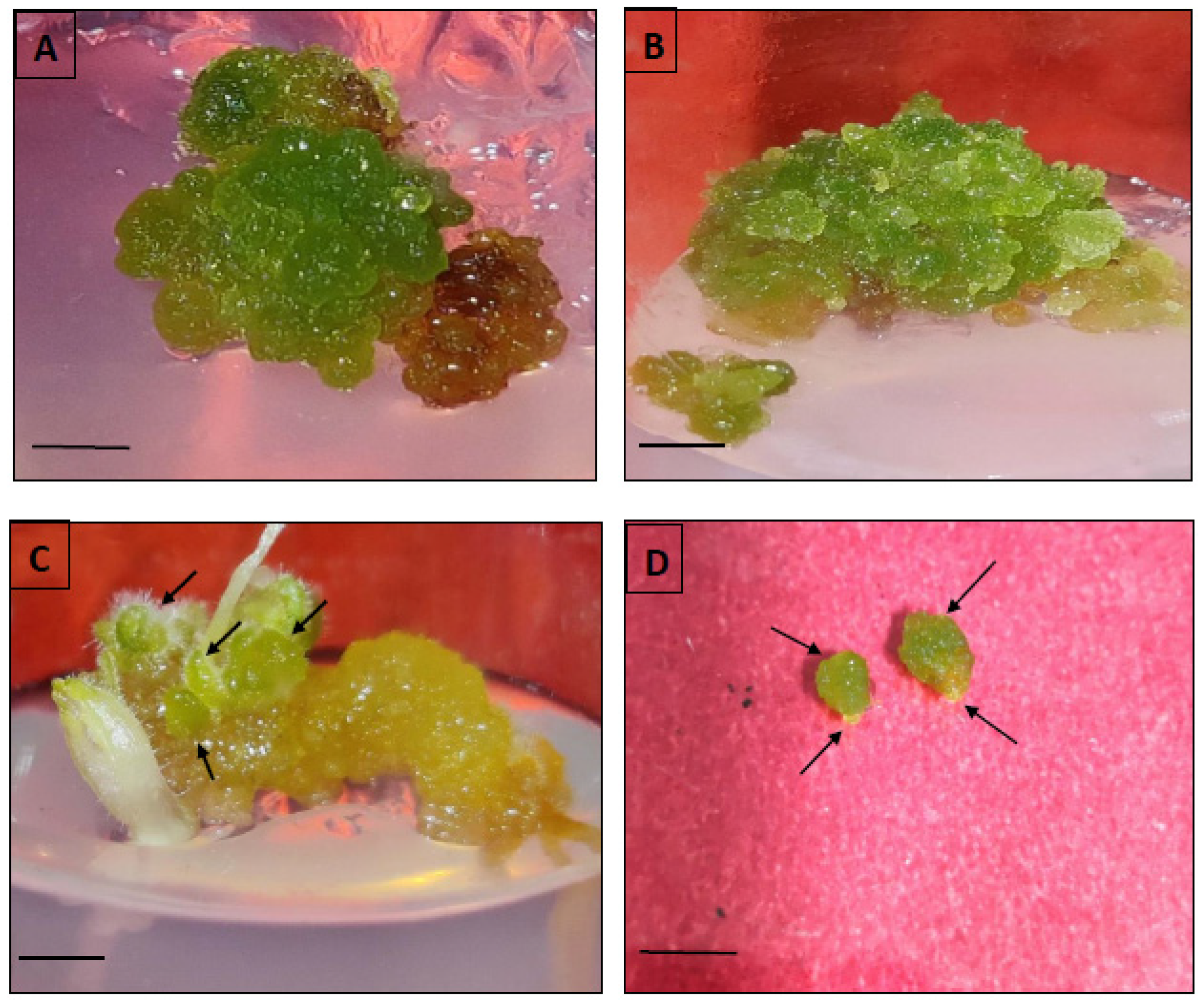
2.4. Indirect Somatic Embryogenesis

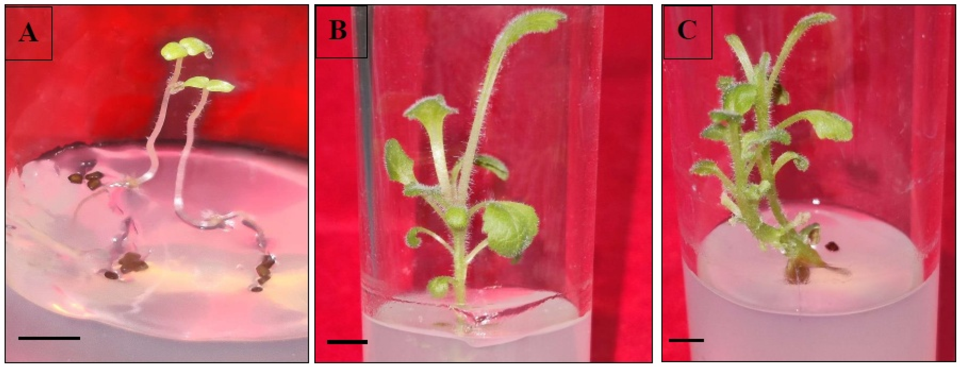
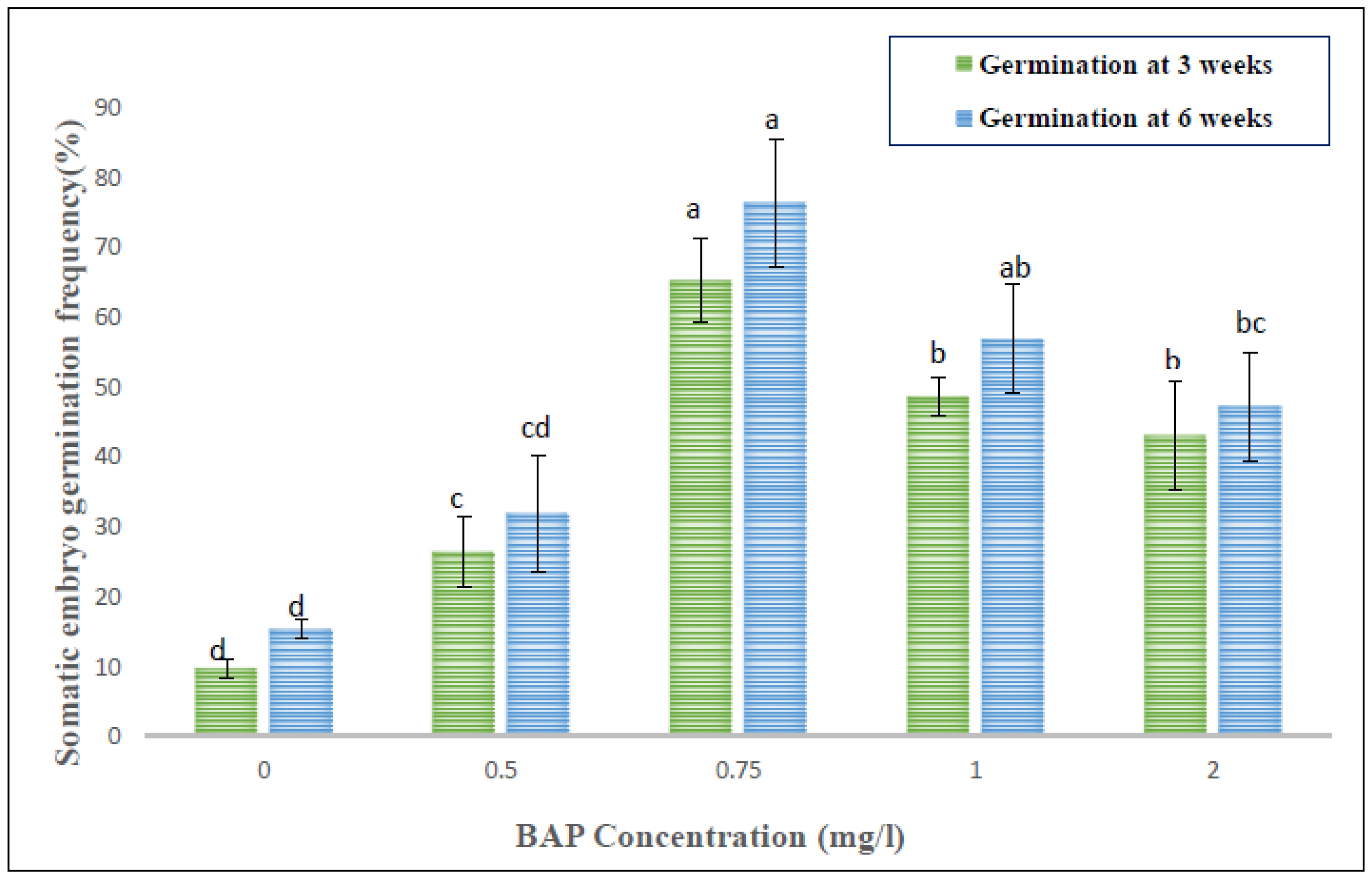
Within 4–5 weeks of continuous sub-culturing of hypocotyl-derived callus on MS, the non-embryogenic calli were transformed into greenish embryogenic calli, distinguished by the presence of different embryo stages, such as globular, heart- or torpedo-shaped. The somatic embryogenesis percentage and the obtained somatic embryos per hypocotyl-derived callus were noted after five weeks of culture. The germination of somatic embryos was accomplished either on full MS fortified with low levels of BAP or without any PGR, and the germination frequency was recorded after five weeks of culture.

2.5. Scanning Electron Microscopy (SEM)

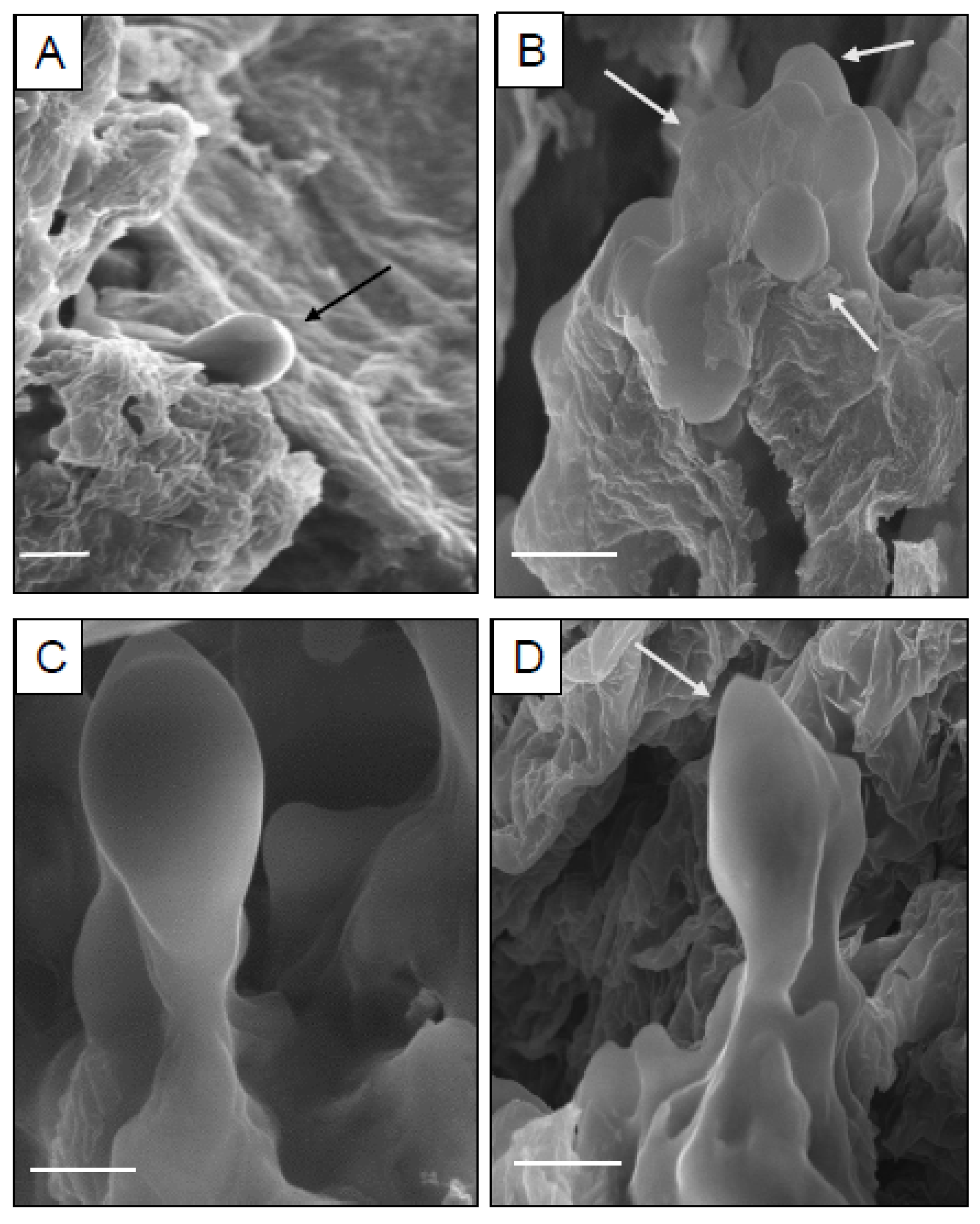
The origin and different developmental stages of somatic embryos were analyzed using scanning electron microscopy. For this purpose, the embryonic calli were prefixed with 2.5% (v/v) glutaraldehyde and 0.1 M of phosphate buffer (pH 6.8) and incubated for 24 h at 4 °C. The samples were then rinsed with the same buffer and postfixed with 0.1% fresh osmium tetroxide for 2 h. Thereafter, the calli were subject to graded ethanol dehydration series [25% (v/v), 50% (v/v), 75% (v/v), 90% (v/v)] for 15 min each and 100% ethanol twice for 12–15 min. Later, the dried tissues underwent sputter coating with a gold-palladium alloy and finally viewed under a LEO 435 VP scanning electron microscope (Zeiss, Oberkochen, Germany) operated at 20 kV.

2.6. Shoot Organogenesis via Direct and Indirect Method

Apical buds and cotyledonary nodal segments from 21-day-old in vitro raised seedlings were taken as explants for direct shoot regeneration. The explants were inoculated onto shoot multiplication MS medium augmented with varied concentrations of BAP (0.5–2.0 mg/L) or kinetin (Kn) (0.5–2.0 mg/L). The passaging of the cultures in the same medium was repeatedly done every three weeks. The percentage of shoot proliferation (total number of shoots emerged/total explants × 100) and the average regenerated shoot length per treatment were counted after five weeks.
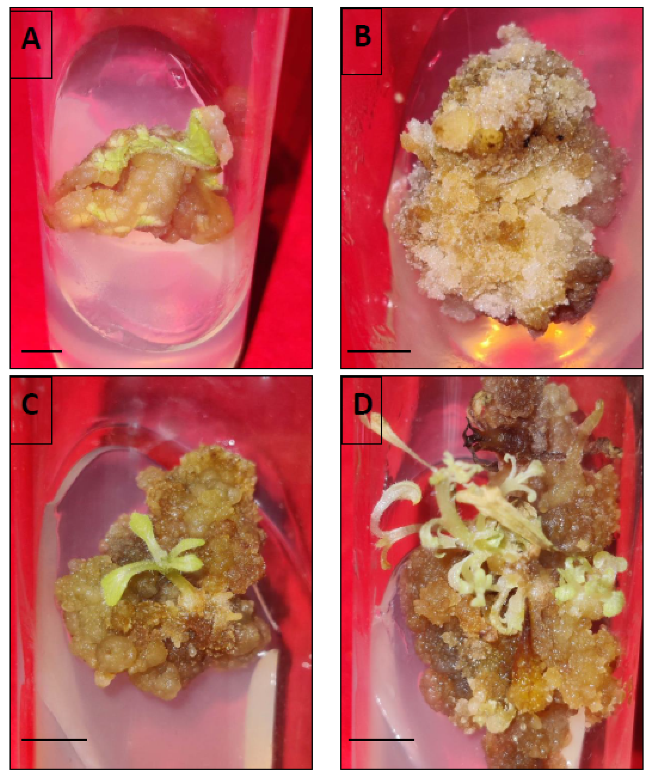
To induce indirect shoots, proliferative calli obtained from leaf and hypocotyl explants were transferred to MS added with different concentrations and combinations of BAP (0.5–2.0 mg/L) and 2,4-D (0.5–1.0 mg/L). The shoot induction rate (%) and the mean number of shoots/callus obtained were counted after 5 weeks.

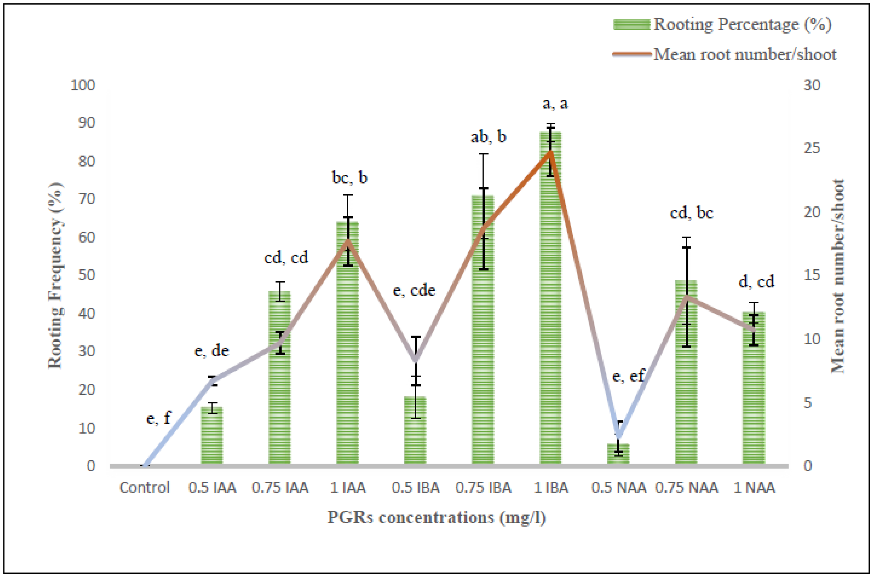
2.7. Root Initiation and Acclimatization

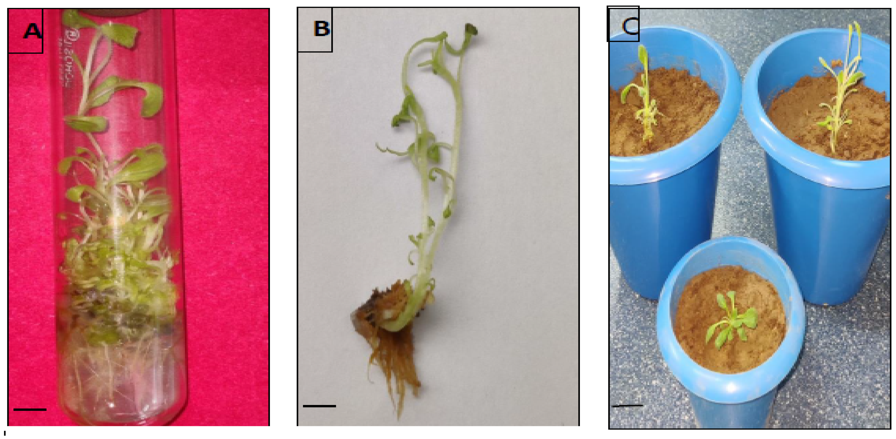
In vitro regenerated shoots (3–4 cm long), derived from somatic embryos and organogenic callus, were excised and transferred onto root-inducing MS medium incorporated with various concentration ranges of indole-3-acetic acid (IAA)/indole-3-butyric acid (IBA)/NAA. Data pertaining to the effects of different auxin treatments were noted down as root induction frequency (%) and the average number of roots generated by each shoot after three weeks of culture. The rooted plantlets were subsequently washed with autoclaved double-distilled water to clear away the adhering culture medium, and shifted to plastic pots carrying an autoclaved mixture of sand, soil and soilrite (1:1:1). Initially, these potted plants were wrapped with transparent polybags and maintained in a culture room (temperature 25 ± 2 °C; humidity 70 ± 10%; light 60 μmolm−2s−1) for two weeks. Afterwards, the plants were allowed to grow under greenhouse conditions with an optimum temperature of 27 ± 2 °C, 60–70% relative humidity and a 10–12 h photoperiod.
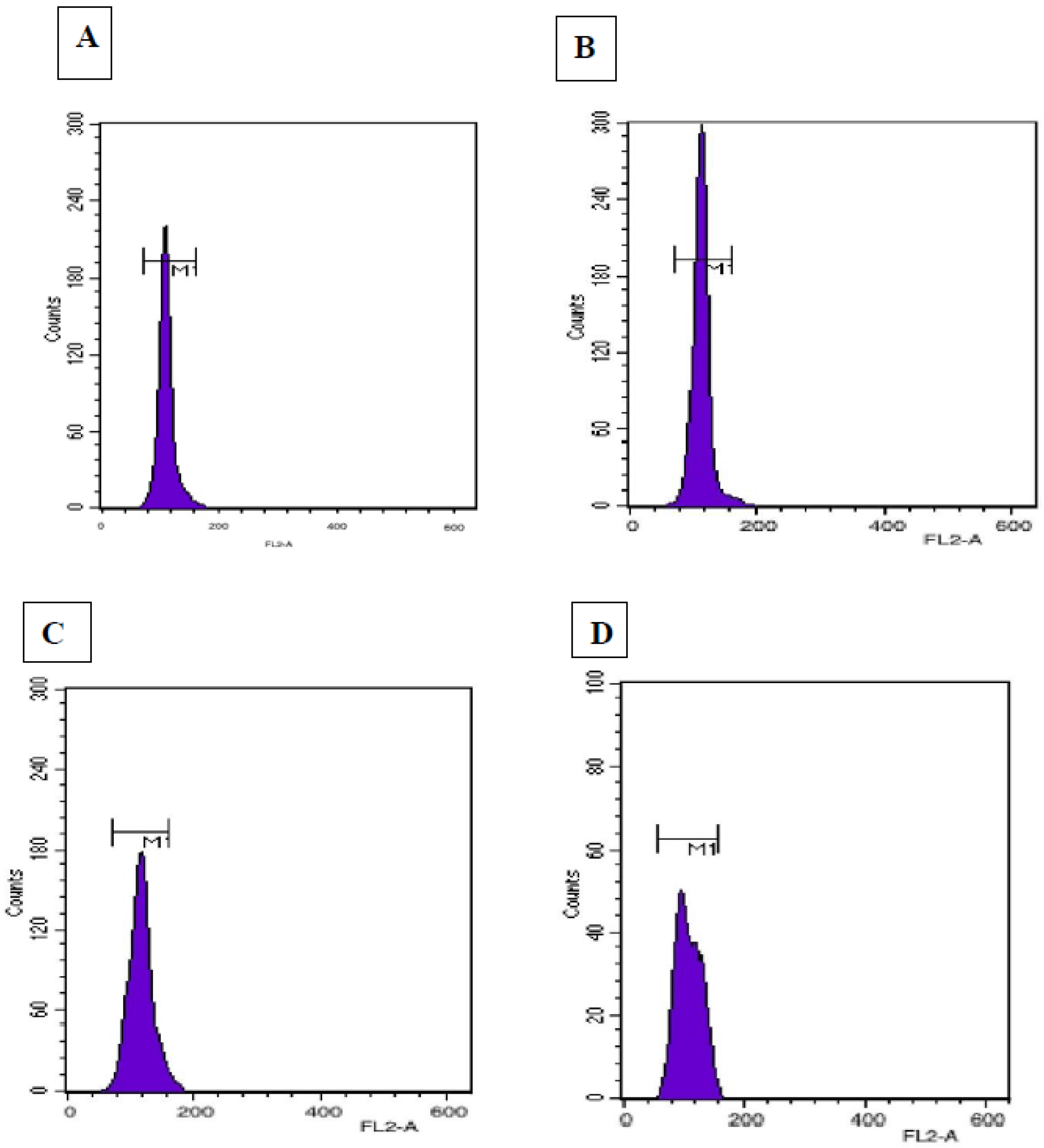
2.8. Flow Cytometry

Flow cytometric analyses were conducted to determine the genetic stability and the genome size of in vitro regenerated plantlets according to the method described by Galbraith [29]. Fresh young leaves were randomly selected from field-grown Digitalis (control) and plants regenerated via somatic embryogenesis and indirect organogenesis for ploidy level analysis. Digitalis lanata Ehrh. was used as a reference standard with known 2C nuclear DNA content of 3.0 pg [30]. Approximately 100 mg of each leaf sample was finely chopped using a fresh surgical blade in a pre-chilled Petri-dish containing 1.5 mL ice-cold Galbraith’s buffer (45 mM MgCl2, 30 mM sodium citrate, 20 mM MOPS and 0.1% Triton-X) for nuclei extraction. Afterwards, the suspensions were filtered using a double-layered nylon mesh of 50 μm pore size to remove larger cellular debris and were stained with 50 μg/mL of PI RNase (Propidium iodide RNase) (Sigma-Aldrich, St. Louis, MO, USA) for 10 min. The samples were then put in dark conditions at 4 °C for about 1 h. Finally, the incubated samples were analyzed on a BD FACS (Calibur) flow cytometer (BD Biosciences, Franklin Lakes, NJ, USA). The 2C DNA content of regenerated plants of D. purpurea was estimated by using the formula [31]:
2C DNA content of sample (pg) =
2C DNA content of standard (pg) × mean position of G0/G1 peak of sample
mean position of G0/G1 peak of standard

2.9. SCoT Marker Analysis

The total genomic DNA extraction was done from 500 mg of each leaf sample of twelve randomly selected in vitro regenerants (six derived from somatic embryos and six from indirectly induced shoots) along with the mother plant by following the modified cetyltrimethyl ammonium bromide (CTAB) methodology [32]. The quantitative and qualitative parameters of isolated DNA were assessed by agarose gel electrophoresis (0.8%).
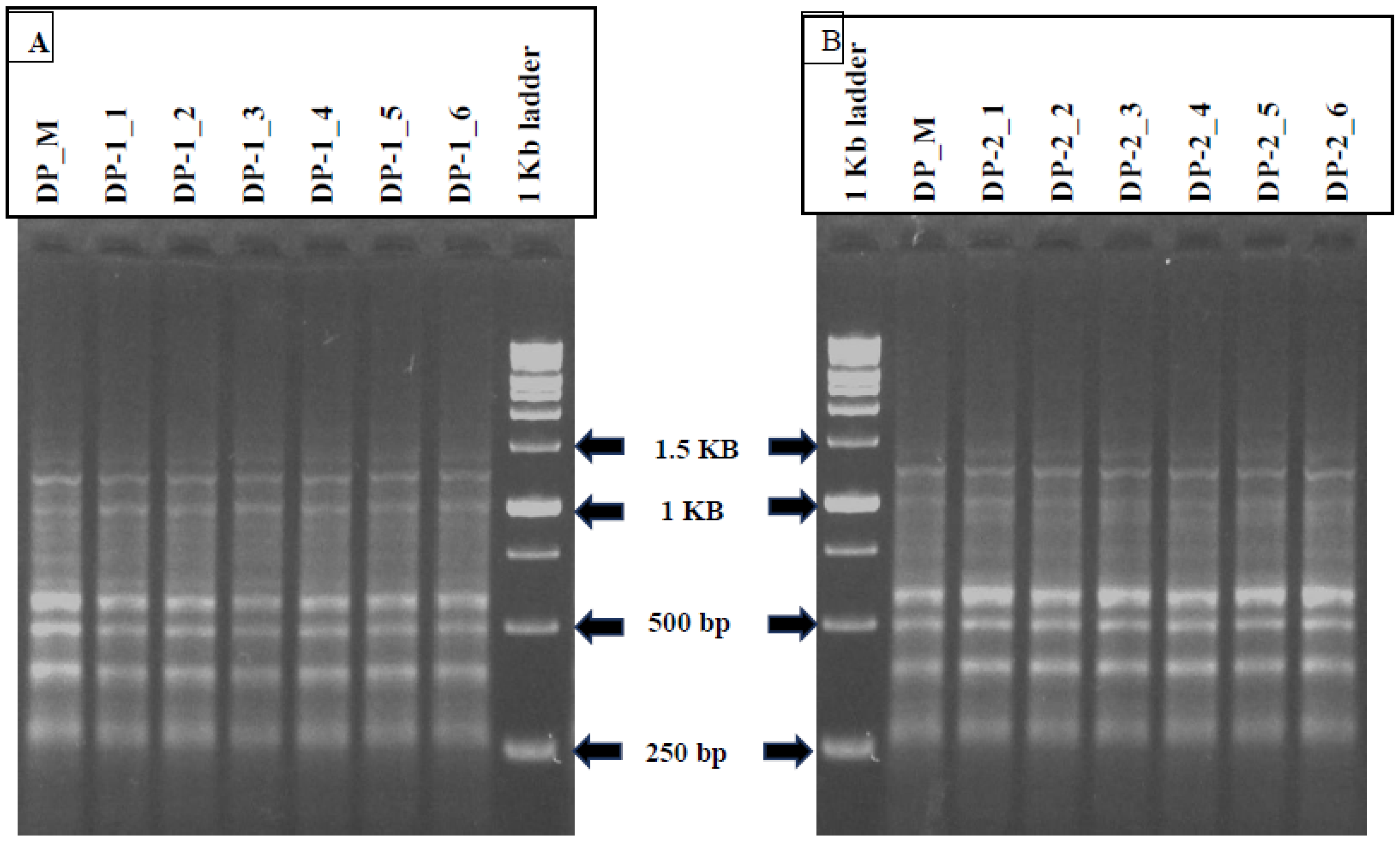
For genetic homogeneity studies, a total of 12 SCoT primers were examined, out of which six produced desirable and reproducible amplified bands, which were finally used for further amplification reactions). The SCoT-PCR reaction mixture of 15 μL contained 50–60 ng of genomic DNA, 10× Taq polymerase buffer, 2.5 mM MgCl2, 10 mM dNTPs, forward and reverse primer (18 nucleotides long), 5 units/μL of Taq DNA Polymerase (Sigma-Aldrich) and deionised water. The PCR technique was operated in a thermal cycler (GeneAmp PCR 9700) with an initial denaturation at 94 °C for 5 min, followed by 35 cycles of 30 s denaturation at 94 °C, 30 s annealing of primers at 50 °C and 1 min extension at 72 °C with the final extension for 5 min at 72 °C. The amplified products were then cooled down at 4 °C (holding temperature) and resolved through agarose gel electrophoresis (1.5%) by using 1X TBE (Tris Borate EDTA) buffer. All the amplification reactions with SCoT primers were replicated thrice to confirm the reproducibility. The gel images were captured using a gel documentation system (Azure Biosystem, Dublin, CA, USA). The sizes of the PCR amplicons were determined by 1 kb DNA ladder (Gene DireX, Inc., Taoyuan, Taiwan).
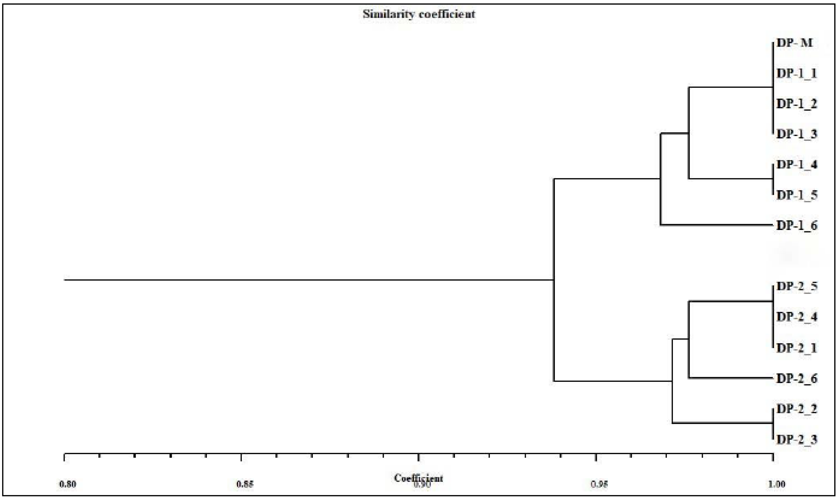
The distinguishable and reproducible bands produced by SCoT primers were scored manually for their presence and absence. The genetic similarity (GS) values between mother plant and in vitro-derived plantlets were computed based on the Jaccard’s similarity coefficient. The obtained similarity coefficients were then utilized to construct a dendrogram through NTSYSpc software (version 2.02, Rohlf, New York, USA) [33] using the UPGMA (Unweighted Pair Group Method of Arithmetic Averages) method [34].

2.10. Statistical Analysis

The in vitro experiments were performed in a completely randomized design (CRD). The data influencing the role of PGRs on explants in inducing callus, somatic embryogenesis and direct/indirect organogenesis were expressed as mean ± standard error. All the experiments were carried out with three replicates, and each experiment was done twice. In the flow cytometric study, three replicates from each regenerated plant group as well as the control were chosen and subjected to flow cytometric analysis. The statistical analyses of data were done with one-way ANOVA using SPSS software (version 15, Chicago, IL, USA). The mean comparisons were determined by DMRT (Duncan’s Multiple Range Test) at p < 0.05 [35].

3. Results

3.1. In Vitro Seed Germination

The in vitro seed germination was successfully obtained on all the three different kinds of MS media (half MS, full MS and full MS with GA3). Within 21 days of inoculation, the seedlings started to emerge with the highest frequency in full MS augmented with 0.5 mg/L GA3 (73.3%) (Table 1), followed by full-strength MS medium (62.2%) as compared to seeds inoculated in half MS medium (48.9%). The grown seedlings had long hypocotyls in all the three germinating media tested (Figure 1A).

3.2. Callus Induction and Proliferation

Three explants (leaf, node and hypocotyl) were cultured in MS having different concentrations as well as combinations of BAP with 2,4-D or NAA. Among these explants, the leaf tissues generated a high incidence (93.1%) of callus formation when a combination of BAP (2.0 mg/L) and NAA (0.5 mg/L) was used (Table 2, Figure 2A). In contrast, the hypocotyl explants showed maximum callusing (90.3%) in MS, amended with 0.75 mg/L BAP, followed by nodal tissues (84.7%) on 2.0 mg/L BAP + 0.5 mg/L 2,4-D. The nodal and leaf produced friable callus of a pale-yellow colour, whereas green and yellow compact calli were developed from hypocotyl explants (Figure 2B).
NOTE :   Callus   induction   frequency   ( % ) = Number   of   explants   showing   callusing ÷ Total   number   of   explants   inoculated × 100

3.3. Somatic Embryo Formation

The subculturing of hypocotyl callus on MS medium added with BAP alone or with 2,4-D/NAA resulted in embryogenic calli within 4–5 weeks of sub-culturing (Figure 3), with a frequency range of 9.7% to 81.9% (Table 3). BAP at 0.75 mg/L showed maximum embryogenesis frequency (81.9 ± 3.7%) with 13.7 ± 2.3 mean numbers of somatic embryos. A gradual decrease in both the frequency (18.1% to 0%) and average count of somatic embryos (2.3 to 0) was observed with increased BAP and 2,4-D (0.5–1.0 mg/L) added medium. Later, the somatic embryos were kept in full MS or with BAP (0.5–2.0 mg/L) for germination. The highest somatic embryo germination (65.3%) was noted on 0.75 mg/L BAP-amended MS medium, which further increased to 76.4% after six weeks of culture (Figure 4). The plantlets grown via somatic embryogenesis were morphologically identical to their parent plantlets.
Figure 3. Callus induction and indirect somatic embryogenesis from hypocotyl callus in D. purpurea. (A,B) Callus induction and proliferation (bars (A) = 0.3 cm, (B) = 0.5 cm), (C) Somatic embryos formation with different developmental stages (bar = 0.5 cm), (D) Isolated bipolar stages of early and late globular embryos with shoot and root ends (arrowheads) (bar = 0.3 cm).
Genes 13 02335 g003

3.4. Scanning Electron Microscopy (SEM)

Scanning electron microscopic studies were carried out to determine the origin of somatic embryos from hypocotyl calli. The SEM images revealed the presence of different stages of somatic embryos (globular and heart-shaped) on callus surfaces. The globular embryos were more prominently observed throughout the investigation as compared to the other embryonic stages (Figure 5).

3.5. Shoot Organogenesis via Direct and Indirect Method

The shoot regeneration ability of apical buds and cotyledonary nodes cultured on BAP- or kinetin-added MS medium is presented in Table 4. The PGR-free MS medium was ineffective, while incorporation of cytokinins (BAP or Kn) in media led to the induction of shoots in both the two explants. The high incidence of shoot proliferation was observed in apical meristem (93.1%), followed by cotyledonary node (87.5%) on 1.0 mg/L BAP-added MS medium (Figure 1B,C). Relatively low shooting efficiency was noted on kinetin-fortified (0.5 mg/L) MS (11.1% in apical bud and 8.3% in cotyledonary node). The average length of regenerated shoots was 1.9–4.1 cm on BAP-fortified MS medium, whereas in kinetin treatments shoots had a mean length of 1.2–3.4 cm after five weeks of culture (Table 4).
The subculturing of friable callus (leaf- and hypocotyl-derived) on BAP- (0.5–2.0 mg/L) and 2,4-D-added (0.5–1.0 mg/L) MS medium produced successful induction of shoots (indirect) within 5–6 weeks (Figure 2C). The leaf-derived calli were more responsive in developing shoots compared to the hypocotyl callus. Of the several BAP and 2,4-D concentrations examined, 2.0 mg/L BAP + 0.5 mg/L 2,4-D was found to be the best treatment in producing a maximum number of shoots in both the two callus sources (Table 5). The leaf-derived callus showed 70.8% shoot regeneration ability with 10.3 average shoot number/callus mass (Figure 2D), followed by hypocotyls callus having 40.3% shoot induction ability with a 4.3 ± 1.9 average count of shoots after six weeks of culture. At a lower level of BAP (<2 mg/L), a significant reduction in shoot formation was noted in two calli sources. The regenerated shoots were then finally transferred to a medium for root induction.

3.6. Rooting and Plantlets Acclimatization

To attain rooting of regenerants, the MS medium was supplied with three different auxins, viz., IBA, IAA and NAA at three concentrations (0.5, 0.75 and 1.0 mg/L) (Figure 6). Except for the control, the roots appeared from the cut ends of the shoots in all the rooting treatments within three weeks. IBA treatments had more impact than the IAA and NAA in terms of root induction percentage and average root numbers per shoot (Figure 7). The highest rooting percentage (87.5%) was recorded on 1.0 mg/L IBA with 24.7 numbers of roots per shoot, whereas the lowest rooting (5.6%) was observed at 0.5 mg/L NAA with 2.3 mean root number/shoot. Those plants with IBA treatments had thicker roots, whereas in IAA and NAA treated plants had narrow and fine roots. About 85–90% of tissue culture raised plants were successfully potted in greenhouse conditions (Figure 7C).

3.7. Flow Cytometric Analysis

The determination of ploidy status of in vitro regenerated plants is a prerequisite for establishing true-to-type nature. In the current study, this has been studied by estimating and comparing the nuclear 2C DNA content of in vivo grown and tissue culture raised D. purpurea plants. The comparison of flow cytometric histograms showed similar fluorescence peaks (Figure 8), and the estimated 2C DNA contents of in vivo and in vitro raised plants (via somatic embryogenesis and shoot organogenesis) was 3.1, 3.2 and 3.0 pg, respectively (Table 6). No visible changes have been detected between mother and tissue cultured-derived plants, thus confirming genetic stability.

3.8. SCoTmarker Analysis

The genetic homogeneity assessment of tissue culture propagated D. purpurea with their mother plant was done using SCoT marker (a DNA-based PCR method). Among the 12 SCoT primers tested, six primers amplified 35 and 37 monomorphic bands with a base pair (bp) size ranging from 200–1600 in somatic embryo-regenerated and organogenic-derived plants, respectively (Table 7; Figure 9A,B). An average of six scorable bands per primer was noted. The maximum number (10 bands) of monomorphic bands was produced by SCoT3 primers, while the lowest band number (four) was produced by SCoT33 primer. The banding profiles validated the genetic uniformity of in vitro-derived plantlets with their field-grown mother plants. Dendrogram based on Jaccard’s similarity index of tissue culture-derived and mother plant is presented in Figure 10.

4. Discussion

The present investigation aimed to lay down an efficient in vitro plant regeneration protocol via embryogenesis and organogenesis, a prerequisite step to produce elite clonal medicinally important plants which further aids in providing a high yield of pharmaceutically important bio-actives. In the current study, in vitro seed germination, callus induction, direct shoot regeneration, somatic embryogenesis and organogenesis have been successfully conducted in D. purpurea by utilizing and optimizing various PGRs (2,4-D, NAA, BAP, kinetin). Maximum in vitro seed germination of D. purpurea was observed in 0.5 mg/L GA3 amended full MS medium, and the germinated seedlings were found to possess longer hypocotyls. Seed dormancy is a common problem encountered in various plant species that leads to delayed fruit set formation and reduced production of viable plants. GA3 is widely known to break seed dormancy, and it has also been used in promoting the seed germination rate under in vitro conditions in other species of Digitalis, such as D. ferruginea [37] and D. cariensis [8].
For direct shoot regeneration, the apical meristems and cotyledonary nodal parts of in vitro grown seedlings were then cultured onto shoot proliferation medium. The cytokinins BAP and kinetin were proved to be effective in inducing shoots in both the explants in D. purpurea. However, the employment of BAP at 1.0 mg/L was more productive in shoot proliferation as compared to other BAP treatments. An increase or decrease in BAP level lowered shoot induction rate and shoot length. Similar results have also been described in different plant species [19,38]). On the other hand, the incorporation of kinetin showed moderate shoot growth in both the explants with the highest shooting frequency of 58.3%. Thus, in our study, BAP was found to be a superior cytokinin in regenerating shoots than kinetin. Earlier reports also suggested that BAP is a better cytokinin for shoot regeneration in several plants, such as Chenopodium quinoa [39], Eurycomalongifolia [40] and Cicer arietinum [41].
Callogenesis has been proved to be a valuable tool to obtain in vitro regenerated plants via embryogenesis/organogenesis. In Digitalis, callus induction was achieved from different explants in several species [42,43,44]. Here, in this study, all the tested explants (leaf, node and hypocotyls) were able to induce callus in BAP-amended MS medium with or without 2,4-D/NAA at a concentration level of 0.5–2.0 mg/L. The obtained results are in accordance with earlier findings reported by Rad et al. [4] and Mamgain et al. [45]. The organogenic potential was also examined wherein the leaf and hypocotyls-derived calli produced shoots in BAP- and 2,4-D-amended MS medium at different concentrations. These findings are in good agreement with Flores et al. [46], who found that the combination of BAP and 2,4-D was found to be proficient in inducing shoot organogenesis. In the current study, cytokinin at higher concentrations stimulated shoot-bud formation from callus more efficiently than auxin. The results are in accordance with Hesami and Dhaneshvar [47], who found that 1.5 mg/L BAP along with 0.15 mg/L IBA showed 100% shoot regeneration frequency. Contrary to our observation, BAP with NAA was proved to be an efficient combination for indirect shoot regeneration in Digitalis ferruginea [37], Rauvolfia serpentina [48] and Aspilia africana [49].
Within five weeks, embryogenic calli were obtained from hypocotyl callus in the same medium. Generally, the auxins (2,4-D, IAA, NAA) alone or in combination with cytokinins (BAP, Kn, TDZ) are widely known to induce somatic embryos (directly or indirectly) and are noted in several plants. In our study, BAP alone was more effective in somatic embryo development (also in combination with auxins). However, the mean number of somatic embryos developed per callus was quite low in different tested treatments. Our observations are very similar and consistent with previous reports, investigating Chrysanthemum spp. [50] and Metabriggsia ovalifolia [51]. SEM analyses confirmed the origin of somatic embryos and demonstrated the progression through different developmental stages (globular, heart) of embryogenesis. The successful conversion of somatic embryos into healthy plantlets was achieved more efficiently in MS fortified with varied levels of BAP as compared to PGR-free MS medium. Similar somatic embryo germination responses were recorded in PGR-added media in various plant species [52,53]. Somatic embryo germination is, however, frequently regulated by a number of factors including plant genotype, PGRs, culture conditions, photoperiod, and in interaction with endogenous levels of hormones [54]. The root-induction ability of tissue culture raised plants was tested with three different auxins, namely IAA, IBA and NAA. Among those, IBA at 1.0 mg/L exhibited the best response in producing roots in comparison to the other two auxins i.e., IAA and NAA. Sinchana et al. [55] attributed this root-promoting phenomenon towards efficient absorption and metabolisation of IBA by the cultured shoots. Similar rooting ability with IBA has been reported in several plants such as Curcuma zedoaria [56] and Solanum khasianum [57].
Plantlets obtained via an intervening callusing stage (embryogenesis/organogenesis) are a good source of somaclonal variation showing ploidy level changes, epigenetic variations and other mutations [15]. To ensure true-to-type plant propagation, flow cytometry and SCoT marker-based PCR techniques have been attempted and showed genetic homogeneity with field-grown mother plants. The similarity in histogram peaks confirmed no genetic variation in laboratory-grown plants. Similarly, the 2C nuclear DNA was estimated recently in plants like Curcuma angustifolia [58], Cucumis melo [59], Disanthus cercidifolius [60] and in other species of Digitalis, i.e., D. lanata [30].
The genetic uniformity of Digitalis was further confirmed by SCoT-PCR analysis, and this is the first report where genetic homogeneity was assessed in D. purpurea by using SCoT. Here, six different SCoT primers (SCoT 3, SCoT 7, SCoT 14, SCoT 16, SCoT 26 and SCoT 33) were employed and all in vitro raised plants (randomly selected) produced monomorphic bands revealing 94.3% similarity in somatic embryo-derived plants and 95.13% similarity in organogenic-derived plantlets with the parent plant. In recent years, SCoT marker-based molecular analysis of genetic uniformity of in vitro grown plants has been conducted in multiple plant varieties [61,62,63,64]. Perez-Alonso et al. [9] investigated genetic homogeneity of D. purpurea micropropagated plants by using RAPD analyses earlier. These results suggest that SCoT marker-assisted genetic evaluation of tissue culture raised plants can also be applied in other Digitalis species.

5. Conclusions

The present investigation demonstrates an efficient in vitro regeneration protocol of Digitalis purpurea for mass propagation of plantlets which can be exploited pharmaceutically for medicinal uses. Flow cytometry and SCoT marker analysis clearly confirmed the genetic uniformity of regenerated populations. Due to greater applicability, reliability and reproducibility, flow cytometry and SCoT marker-based approaches have proved to be robust techniques in detecting genetic homogeneity/somaclonal variations of regenerated plants with high commercial values.

Author Contributions

Conceptualization, A.M. and Y.B.; methodology, Y.B.; data curation and writing, Y.B., Z.H.S., R.S., J.M. and B.E.; editing, A.M.; supervision, A.M.; All authors have read and agreed to the published version of the manuscript.

Funding

The research work is funded by the Department of Biotechnology (DBT), New Delhi, India.

Institutional Review Board Statement

Not applicable.

Informed Consent Statement

Not applicable.

Data Availability Statement

Not applicable.

Acknowledgments

The first author is thankful to the Department of Biotechnology (DBT) for financial support given as Junior Research Fellowship; and HRDI, Uttarakhand for providing seeds and to Arvind Kumar for providing BD FACS Flow Cytometry facility at Delhi University, South Campus, New Delhi. The authors are grateful to the laboratory facilities provided by Department of Botany, Jamia Hamdard, New Delhi.

Conflicts of Interest

The authors declare no conflict to interest.

Abbreviations

PGRsPlant Growth Regulators
MSMurashige and Skoog
BAP6-benzylaminopurine
2,4-D2,4- dichlorophenoxyacetic acid
NAAα-Naphthaleneacetic acid
KnKinetin
IAAIndole-3-Acetic Acid
IBAIndole-3-butyric Acid
SEMScanning Electron Microscopy
SCoTStart Codon Targeted
DMRTDuncan’s Multiple Range Test

References

  1. Patil, J.G.; Ahire, M.L.; Nitnaware, K.M.; Panda, S.; Bhatt, V.P.; Kishor, P.B.K.; Nikam, T.D. In Vitro propagation and production of cardiotonic glycosides in shoot cultures of Digitalis purpurea L. by elicitation and precursor feeding. Appl. Microbiol. Biotechnol. 2013, 97, 2379–2393. [Google Scholar] [CrossRef]
  2. Wu, B.; Li, Y.; Yan, H.; Ma, Y.; Luo, H.; Yuan, L.; Chen, S.; Lu, S. Comprehensive transcriptome analysis reveals novel genes involved in cardiac glycoside biosynthesis and mlnc RNAs associated with secondary metabolism and stress response in Digitalis purpurea. BMC Genom. 2012, 13, 15. [Google Scholar] [CrossRef] [PubMed] [Green Version]
  3. Verma, S.K.; Das, A.K.; Cingoz, G.S.; Gurel, E. In Vitro culture of Digitalis L. (Foxglove) and the production of cardenolides: An up-to-date review. Ind. Crops Prod. 2016, 94, 20–51. [Google Scholar] [CrossRef]
  4. Rad, M.M.; Abdossi, V.; Moradi, P.; Rakhshandehroo, F.; Mehrafarin, A. Phytochemical changes of Digitalis purpurea L. in response to polyamines and methyl jasmonate application in callus culture. J. Plant Biochem. Biotechnol. 2022, 31, 310–319. [Google Scholar] [CrossRef]
  5. Verma, S.K.; Sahin, G.; Yucesan, B.; Eker, I.; Sahbaz, N.; Gurel, S.; Gurel, E. Direct somatic embryogenesis from hypocotyl segments of Digitalis trojana Ivan and subsequent plant regeneration. Ind. Crops Prod. 2012, 40, 76–80. [Google Scholar] [CrossRef]
  6. Bhusare, B.P.; John, C.K.; Bhatt, V.P.; Nikam, T.D. In Vitro propagation of Digitalis lanata Ehrh. through direct shoot regeneration–A source of cardiotonic glycosides. Ind. Crops Prod. 2018, 121, 313–319. [Google Scholar] [CrossRef]
  7. Verma, S.K.; Das, A.K.; Gantait, S.; Gurel, S.; Gurel, E. Influence of auxin and its polar transport inhibitor on the development of somatic embryos in Digitalis trojana. 3 Biotech 2018, 8, 99. [Google Scholar] [CrossRef] [PubMed] [Green Version]
  8. Mohammed, A.; Yücesan, B.; Demir-Ordu, Ö.; Cihangir, C.; Eker, İ.; Kreis, W.; Gürel, E. In Vitro regeneration and cardenolide determination of an endemic foxglove, Digitalis cariensis (Aegean Foxglove). Vitr. Cell. Dev. Biol. -Plant. 2015, 51, 438–444. [Google Scholar] [CrossRef]
  9. Pérez-Alonso, N.; Martín, R.; Capote, A.; Pérez, A.; Hernández-Díaz, E.K.; Rojas, L.; Jiménez, E.; Quiala, E.; Angenon, G.; Garcia-Gonzales, R.; et al. Efficient direct shoot organogenesis, genetic stability and secondary metabolite production of micropropagated Digitalis purpurea L. Ind. Crops Prod. 2018, 116, 259–266. [Google Scholar] [CrossRef]
  10. Syeed, R.; Mujib, A.; Malik, M.Q.; Gulzar, B.; Zafar, N.; Mamgain, J.; Ejaz, B. Direct somatic embryogenesis and flow cytometric assessment of ploidy stability in regenerants of Caladium × hortulanum ‘Fancy’. J. Appl. Genet. 2022, 63, 199–211. [Google Scholar] [CrossRef]
  11. Niazian, M. Application of genetics and biotechnology for improving medicinal plants. Planta 2019, 249, 953–973. [Google Scholar] [CrossRef] [PubMed]
  12. Gulzar, B.; Mujib, A.; Mushtaq, Z.; Malik, M.Q. Old Catharanthus roseus culture (14 years) produced somatic embryos and plants and showed normal genome size; demonstrated an increased antioxidant defense mechanism; and synthesized stress proteins as biochemical, proteomics, and flow-cytometry studies reveal. J. Appl. Genet. 2021, 62, 43–57. [Google Scholar] [CrossRef] [PubMed]
  13. Mujib, A.; Ali, M.; Isah, T. Somatic embryo mediated mass production of Catharanthus roseus in culture vessel (bioreactor)–A comparative study. Saudi J. Biol. Sci. 2014, 21, 442–449. [Google Scholar] [CrossRef] [PubMed] [Green Version]
  14. García-Gonzáles, R.; Quiroz, K.; Carrasco, B.; Caligari, P. Plant tissue culture: Current status, opportunities and challenges. Cien. Inv. Agr. 2010, 37, 5–30. [Google Scholar] [CrossRef] [Green Version]
  15. Escobedo-GraciaMedrano, R.M.; Maldonado-Borges, J.I.; Burgos-Tan, M.J.; Valadez-González, N.; Ku-Cauich, J.R. Using flow cytometry and cytological analyses to assess the genetic stability of somatic embryo-derived plantlets from embryogenic Musa acuminata Colla (AA) ssp. malaccensis cell suspension cultures. Plant Cell Tiss. Organ Cult. 2014, 116, 175–185. [Google Scholar] [CrossRef]
  16. Kairuz, E.; Pérez-Alonso, N.; Angenon, G.; Jiménez, E.; Chong-Pérez, B. Shoot organogenesis, genetic stability, and secondary metabolite production of micropropagated Digitalis purpurea. In Plant Cell and Tissue Differentiation and Secondary Metabolites; Ramawat, K.G., Ekiert, H.M., Goyal, S., Eds.; Springer: Cham, Switzerland, 2021; pp. 169–186. [Google Scholar] [CrossRef]
  17. Faisal, M.; Alatar, A.A.; Hegazy, A.K.; Alharbi, S.A.; El-Sheikh, M.; Okla, M.K. Thidiazuron induced in Vitro multiplication of Menthaarvensis and evaluation of genetic stability by flow cytometry and molecular markers. Ind. Crops Prod. 2014, 62, 100–106. [Google Scholar] [CrossRef]
  18. Ali, M.; Mujib, A.; Tonk, D.; Zafar, N. Plant regeneration through somatic embryogenesis and genome size analysis of Coriandrum sativum L. Protoplasma 2017, 254, 343–352. [Google Scholar] [CrossRef]
  19. Zafar, N.; Mujib, A.; Ali, M.; Tonk, D.; Gulzar, B.; Malik, M.; Sayeed, R.; Mamgain, J. Genome size analysis of field grown and tissue culture regenerated Rauvolfia serpentina (L) by flow cytometry: Histology and scanning electron microscopic study for in vitro morphogenesis. Ind. Crops Prod. 2019, 128, 545–555. [Google Scholar] [CrossRef]
  20. Çördük, N.; Yücel, G.; Akıncı, N.; Tuna, M. Assessment of the genetic stability of indirect shoot organogenesis-derived plantlets of Digitalis trojana Ivanina by flow cytometry and cytological analyses. Tekirdağ Ziraat Fakültesi Derg. 2017, 14. [Google Scholar]
  21. Malik, M.Q.; Mujib, A.; Gulzar, B.; Zafar, N.; Syeed, R.; Mamgain, J.; Ejaz, B. Genome size analysis of field grown and somatic embryo regenerated plants in Allium sativum L. J. Appl. Genet. 2020, 61, 25–35. [Google Scholar] [CrossRef]
  22. Syeed, R.; Mujib, A.; Malik, M.Q.; Mamgain, J.; Ejaz, B.; Gulzar, B.; Zafar, N. Mass propagation through direct and indirect organogenesis in three species of genus Zephyranthes and ploidy assessment of regenerants through flow cytometry. Mol. Biol. Rep. 2021, 48, 513–526. [Google Scholar] [CrossRef] [PubMed]
  23. Mujib, A.; Malik, M.Q.; Bansal, Y.; Syeed, R.; Ejaz, B.; Mamgain, J. Somatic Embryogenesis in Catharanthus Roseus: Proteomics of embryogenic and non-embryogenic tissues; and genome size analysis of regenerated plant. In The Catharanthus Genome; Kole, C., Ed.; Springer: Cham, Switzerland, 2022; pp. 85–100. [Google Scholar] [CrossRef]
  24. Ajithan, C.; Vasudevan, V.; Sathish, D.; Sathish, S.; Krishnan, V.; Manickavasagam, M. The influential role of polyamines on the in vitro regeneration of pea (Pisum sativum L.) and genetic fidelity assessment by SCoT and RAPD markers. Plant Cell Tiss. Organ Cult. 2019, 139, 547–561. [Google Scholar] [CrossRef]
  25. Erişen, S.; Kurt-Gür, G.; Servi, H. In Vitro propagation of Salvia sclarea L. by meta-Topolin, and assessment of genetic stability and secondary metabolite profiling of micropropagated plants. Ind. Crops Prod. 2020, 157, 112892. [Google Scholar] [CrossRef]
  26. Ejaz, B.; Mujib, A.; Mamgain, J.; Malik, M.Q.; Syeed, R.; Gulzar, B.; Bansal, Y. Comprehensive in vitro regeneration study with SCoT marker assisted clonal stability assessment and flow cytometric genome size analysis of Carthamus tinctorius L.: An important medicinal plant. Plant Cell Tiss. Organ Cult. 2022, 148, 403–418. [Google Scholar] [CrossRef]
  27. Mamgain, J.; Mujib, A.; Ejaz, B.; Gulzar, B.; Malik, M.Q.; Syeed, R. Flow cytometry and start codon targeted (SCoT) genetic fidelity assessment of regenerated plantlets in Tylophora indica (Burm. f.) Merrill. Plant Cell Tiss. Organ Cult. 2022, 150, 129–140. [Google Scholar] [CrossRef]
  28. Murashige, T.; Skoog, F. A revised medium for rapid growth and bio assays with tobacco tissue cultures. Physiol. Plant. 1962, 15, 473–497. [Google Scholar] [CrossRef]
  29. Galbraith, D.W. Simultaneous flow cytometric quantification of plant nuclear DNA contents over the full range of described angiosperm 2C values. Cytom Part A J. Int. Soc. Adv. Cytom. 2009, 75, 692–698. [Google Scholar] [CrossRef]
  30. Bhusare, B.P.; John, C.K.; Bhatt, V.P.; Nikam, T.D. Induction of somatic embryogenesis in leaf and root explants of Digitalis lanata Ehrh.: Direct and indirect method. S. Afr. J. Bot. 2020, 130, 356–365. [Google Scholar] [CrossRef]
  31. Doležel, J.; Greilhuber, J.; Suda, J. Estimation of nuclear DNA content in plants using flow cytometry. Nat. Protoc. 2007, 2, 2233–2244. [Google Scholar] [CrossRef] [PubMed]
  32. Doyle, J.J. Isolation of plant DNA from fresh tissue. Focus 1990, 12, 13–15. [Google Scholar]
  33. Rohlf, F.J. NTSYSpc: Numerical Taxonomy and Multivariate Analysis System, Version 2.02; Setauket: New York, NY, USA, 2000. [Google Scholar]
  34. Sneath, P.H.; Sokal, R.R. Numerical Taxonomy. The Principles and Practice of Numerical Classification; W. H. Freeman and Company: New York, NY, USA, 1973. [Google Scholar]
  35. Duncan, D.B. Multiple range and multiple F tests. Biometrics 1955, 11, 1–42. [Google Scholar] [CrossRef]
  36. Dolezel, J. Nuclear DNA content and genome size of trout and human. Cytom. Part A 2003, 51, 127–128. [Google Scholar]
  37. Verma, S.K.; Yucesan, B.; Sahin, G.; Gurel, E. Embryogenesis, plant regeneration and cardiac glycoside determination in Digitalis ferruginea subsp. ferruginea L. Plant Cell Tiss. Organ Cult. 2014, 119, 625–634. [Google Scholar] [CrossRef]
  38. Hsieh, Y.F.; Jain, M.; Wang, J.; Gallo, M. Direct organogenesis from cotyledonary node explants suitable for Agrobacterium-mediated transformation in peanut (Arachis hypogaea L.). Plant Cell Tiss. Organ Cult. 2017, 128, 161–175. [Google Scholar] [CrossRef]
  39. Hesami, M.; Naderi, R.; Yoosefzadeh-Najafabadi, M. Optimizing sterilization conditions and growth regulator effects on in vitro shoot regeneration through direct organogenesis in Chenopodium quinoa. BioTechnologia 2018, 99, 49–57. [Google Scholar] [CrossRef]
  40. Alttaher, A.G.A.; Yusof, Z.N.B.; Mahmood, M.; Shaharuddin, N.A. High-frequency induction of multiple shoots and plant regeneration from cotyledonary node explants of Tongkat Ali (Eurycoma longifolia Jack). Appl. Ecol. Environ. Res. 2020, 18, 6321–6333. [Google Scholar] [CrossRef]
  41. Sadhu, S.; Jogam, P.; Thampu, R.K.; Abbagani, S.; Penna, S.; Peddaboina, V. High efficiency plant regeneration and genetic fidelity of regenerants by SCoT and ISSR markers in chickpea (Cicer arietinum L.). Plant Cell Tiss. Organ Cult. 2020, 141, 465–477. [Google Scholar] [CrossRef]
  42. Fatima, Z.; Mujib, A.; Fatima, S.; Arshi, A.; Umar, S. Callus induction, biomass growth, and plant regeneration in Digitalis lanata Ehrh.: Influence of plant growth regulators and carbohydrates. Turk. J. Bot. 2009, 33, 393–405. [Google Scholar] [CrossRef]
  43. Yücesan, B.; Müller-Uri, F.; Kreis, W.; Gürel, E. Cardenolide estimation in callus-mediated regenerants of Digitalis lamarckii Ivanina (dwarf foxglove). Vitr. Cell. Dev. Biol.-Plant 2014, 50, 137–142. [Google Scholar] [CrossRef]
  44. Verma, S.K.; Sahin, G.; Gurel, E. Somatic embryogenesis, pigment accumulation, and synthetic seed production in Digitalis davisiana Heywood. Indian J. Exp. Biol. 2016, 54, 245–253. [Google Scholar] [PubMed]
  45. Mamgain, J.; Mujib, A.; Syeed, R.; Ejaz, B.; Malik, M.Q.; Bansal, Y. Genome size and gas chromatography-mass spectrometry (GC–MS) analysis of field-grown and in vitro regenerated Pluchea lanceolata plants. J. Appl. Genet. 2022, 1–21. [Google Scholar] [CrossRef] [PubMed]
  46. Flores, R.; Maldaner, J.; Brondani, D.; Cezarotto, V.; Giacomelli, S.; Garlet, T.; Nicoloso, F. Direct and indirect regeneration from Pfaffia tuberosa (Spreng.) Hicken calli and its β-ecdysone production. Rev. Bras. Pl. Med. 2017, 19, 266–273. [Google Scholar]
  47. Hesami, M.; Daneshvar, M.H. Indirect Organogenesis through Seedling-Derived Leaf Segments of Ficus Religiosa—A Multipurpose Woody Medicinal Plant. J. Crop Sci. Biotechnol. 2018, 21, 129–136. [Google Scholar] [CrossRef]
  48. Banu, T.A.; Khan, S.; Goswami, B.; Afrin, S.; Habib, A.; Akter, S. Indirect organogenesis and somatic embryogenesis for regeneration of Rauvolfia serpentina L. from root explants. Bangladesh J. Bot. 2020, 49, 1021–1027. [Google Scholar] [CrossRef]
  49. Okello, D.; Yang, S.; Komakech, R.; Chung, Y.; Rahmat, E.; Gang, R.; Omujal, F.; Lamwaka, A.V.; Kang, Y. Indirect in vitro regeneration of the medicinal plant, Aspilia africana, and histological assessment at different developmental stages. Front. Plant Sci. 2021, 12, 2928. [Google Scholar] [CrossRef] [PubMed]
  50. Mani, T.; Senthil, K. Multiplication of Chrysanthemum through somatic embryogenesis. Asian J. Pharma Technol. 2011, 1, 13–16. [Google Scholar]
  51. Ouyang, Y.; Chen, Y.; Lü, J.; Teixeira da Silva, J.A.; Zhang, X.; Ma, G. Somatic embryogenesis and enhanced shoot organogenesis in Metabriggsia ovalifolia W. T. Wang. Sci. Rep. 2016, 6, 1–9. [Google Scholar] [CrossRef] [Green Version]
  52. MusfirMehaboob, V.; Faizal, K.; Thilip, C.; Raja, P.; Thiagu, G.; Aslam, A.; Shajahan, A. Indirect somatic embryogenesis and Agrobacterium-mediated transient transformation of ginger (Zingiber officinale Rosc.) using leaf sheath explants. J. Hortic. Sci. Biotechnol. 2019, 94, 753–760. [Google Scholar] [CrossRef]
  53. Mujib, A.; Bansal, Y.; Malik, M.Q.; Syeed, R.; Mamgain, J.; Ejaz, B. Internal and external regulatory elements controlling somatic embryogenesis in Catharanthus: A model medicinal plant. In Somatic Embryogenesis; Ramírez-Mosqueda, M.A., Ed.; Humana: New York, NY, USA, 2022; Volume 2527, pp. 11–27. [Google Scholar] [CrossRef]
  54. Mazri, M.A.; Naciri, R.; Belkoura, I. Maturation and conversion of somatic embryos derived from seeds of Olive (Olea europaea L.) cv. Dahbia: Occurrence of secondary embryogenesis and adventitious bud formation. Plants 2020, 9, 1489. [Google Scholar] [CrossRef] [PubMed]
  55. Sinchana, N.S.; Kattimani, K.N.; Prabhuling, G.; Sudesh, K.; Jagadeesha, N. Standardization of tissue culture protocol for turmeric (Curcuma longa L.) Cv. Salem. Int. J. Chem. Stud. 2020, 8, 2721–2726. [Google Scholar]
  56. Jena, S.; Ray, A.; Sahoo, A.; Sahoo, S.; Dash, B.; Kar, B.; Nayak, S. Rapid plant regeneration in industrially important Curcuma zedoaria revealing genetic and biochemical fidelity of the regenerants. 3 Biotech 2020, 10, 1–13. [Google Scholar] [CrossRef] [PubMed]
  57. Chirumamilla, P.; Gopu, C.; Jogam, P.; Taduri, S. Highly efficient rapid micropropagation and assessment of genetic fidelity of regenerants by ISSR and SCoT markers of Solanum khasianum Clarke. Plant Cell Tiss. Organ Cult. 2021, 144, 397–407. [Google Scholar] [CrossRef]
  58. Jena, S.; Ray, A.; Sahoo, A.; Sahoo, S.; Kar, B.; Panda, P.C.; Nayak, S. High-frequency clonal propagation of Curcuma angustifolia ensuring genetic fidelity of micropropagated plants. Plant Cell Tiss. Organ Cult. 2018, 135, 473–486. [Google Scholar] [CrossRef]
  59. Raji, M.R.; Farajpour, M. Genetic fidelity of regenerated plants via shoot regeneration of muskmelon by inter simple sequence repeat and flow cytometry. J. Saudi Soc. Agric. Sci. 2020, 20, 88–93. [Google Scholar] [CrossRef]
  60. Ulvrova, T.; Vitamvas, J.; Cepkova, P.H.; Eliasova, K.; Janovska, D.; Bazant, V.; Viehmannova, I. Micropropagation of an ornamental shrub Disanthus cercidifolius Maxim. and assessment of genetic fidelity of regenerants using ISSR and flow cytometry. Plant Cell Tiss. Organ Cult. 2021, 144, 555–566. [Google Scholar] [CrossRef]
  61. Sathish, D.; Vasudevan, V.; Theboral, J.; Elayaraja, D.; Appunu, C.; Siva, R.; Manickavasagam, M. Efficient direct plant regeneration from immature leaf roll explants of sugarcane (Saccharum officinarum L.) using polyamines and assessment of genetic fidelity by SCoT markers. Vitr. Cell Dev. Biol. Plant 2018, 54, 399–412. [Google Scholar] [CrossRef]
  62. Sharma, U.; Rai, M.K.; Shekhawat, N.S.; Kataria, V. Genetic homogeneity revealed in micropropagated Bauhinia racemosa Lam. using gene targeted markers CBDP and SCoT. Physiol. Mol. Biol. Plants 2019, 25, 581–588. [Google Scholar] [CrossRef]
  63. Tikendra, L.; Potshangbam, A.M.; Dey, A.; Devi, T.R.; Sahoo, M.R.; Nongdam, P. RAPD, ISSR, and SCoT markers based genetic stability assessment of micropropagated Dendrobium fimbriatum Lindl. var. oculatum Hk. f.-an important endangered orchid. Physiol. Mol. Biol. Plants 2021, 27, 341–357. [Google Scholar] [CrossRef]
  64. Mood, K.; Jogam, P.; Sirikonda, A.; Shekhawat, M.S.; Rohela, G.K.; Manokari, M.; Allini, V.R. Micropropagation, morpho-anatomical characterization, and genetic stability studies in Lippia javanica (Burm. f.) Spreng: A multipurpose medicinal plant. Plant Cell Tiss. Organ Cult. 2022, 150, 427–437. [Google Scholar] [CrossRef]
Figure 1. In vitro germination of seeds and direct shoot regeneration in D. purpurea. (A) Germinated seedlings having elongated hypocotyls (bar = 0.5 cm). (B,C) In vitro direct shoot proliferation (bars = 1.0 cm).
Genes 13 02335 g001
Figure 2. Callus induction and callus-mediated shoot organogenesis from leaf explant in D. purpurea. (A) Callus initiation (bar = 1.0 cm), (B) Callus proliferation after postweeks (bar = 0.5 cm), (C,D) Shoot regeneration from leaf derived callus (bars = 0.5 cm).
Genes 13 02335 g002
Figure 4. Effect of BAP-fortified MS medium on somatic embryo germination after a three- and six-week period, respectively. Values indicate the Mean ± SE of three replicates per treatment. Mean values with different superscripts within a column are significantly different from each other as per DMRT at p ≤ 0.05.
Genes 13 02335 g004
Figure 5. Scanning electron microscopic (SEM) images of somatic embryos in D. purpurea. (A) Globular shaped somatic embryo (bar = 20 μm), (B) Cluster of globular shaped somatic embryos (bar = 2 μm), (C) Globular shaped embryo with suspensor (bar = 2 μm), (D) Heart shaped somatic embryo (bar = 2 μm).
Genes 13 02335 g005
Figure 6. Effect of various PGRs concentration on root induction frequency and mean number of roots/shoot. Values indicate the mean ± SE of 3 replicates per treatment. Mean values with different superscripts within a column are significantly different from each other as per DMRT at p ≤ 0.05.
Genes 13 02335 g006
Figure 7. (A,B) Root induction of in vitro regenerated D. purpurea plants (bars = 1.0 cm). (C) Tissue culture raised D. purpurea plants in pots (bar = 3 cm).
Genes 13 02335 g007
Figure 8. Flow cytometric histogram peaks of relative 2C DNA content of nuclei obtained from (A) Digitalis lanata (standard), (B) Field-grown (mother) plant, (C) somatic embryo regenerated plant, and (D) organogenic-derived plant of Digitalis purpurea.
Genes 13 02335 g008
Figure 9. SCoT banding profiles of in vitro propagated plants with mother plant (DP_M) of D. purpurea with SCoT 14 primer. (A) DP-1_1 to DP-1_6 (somatic embryo-derived plants), (B) DP-2_1 to DP-2_6 (organogenic-derived plants).
Genes 13 02335 g009
Figure 10. Dendrogram of SCoT analysis depicting the genetic similarity between the mother plant (DP-M) and the in vitro regenerated plants of D. purpurea.
Genes 13 02335 g010
Table 1. In vitro seed germination frequency of Digitalis purpurea after three weeks.
MediumGermination Rate (%)
Half MS48.9 ± 4 b
Full MS62.2 ± 1.1 a
Full MS + 0.5 mg/L GA373.3 ± 3.8 a
Values indicate mean of 25 seeds inoculated in different media. Total seeds observed (25 × 3 = 75). Germinate Rate % = No. of germinated seedlings/Total numbers of seeds inoculated × 100. Mean values with the different superscripts within a column are significantly different from each other as per DMRT at p ≤ 0.05.
Table 2. Effects of different concentrations as well as combinations of PGRs on callus induction frequency from hypocotyl, node and leaf in D. purpurea.
PGRsConcentration (mg/L)Explants
HypocotylNodeLeaf
Control00 g0 i0 i
BAP0.551.4 ± 10.8 c9.7 ± 2.8 h,i12.5 ± 2.4 f,g,h,i
0.7590.3 ± 1.4 a19.4 ± 3.7 g,h20.8 ± 4.2 e,f,g,h
158.3 ± 4.2 b,c11.1 ± 1.4 h,i6.9 ± 3.7 g,h,i
230.6 ± 2.8 d,e,f12.5 ± 2.4 h,i5.6 ± 1.4 h,i
BAP + 2,4-D0.5 + 1.018.1 ± 6.1 f,g22.2 ± 3.7 g,h13.9 ± 3.7 f,g,h,i
0.5 + 2.040.3 ± 10 c,d,e26.4 ± 3.7 f,g,h25 ± 6.4 d,e,f,g,h
1.0 + 0.545.8 ± 13.4 c,d36.1 ± 5 f,g29.2 ± 2.4 d,e,f
1.0 + 2.050 ± 10.5 c43.1 ± 5 e,f37.5 ± 4.8 c,d,e
2.0 + 0.50 ± 0 g84.7 ± 6.9 a77.8 ± 6.1 a
2.0 + 1.00 ± 0 g58.3 ± 4.2 c,d,e54.2 ± 10.5 b,c
BAP + NAA0.5 + 1.025 ± 2.4 e,f54.2 ± 6.4 d,e26.4 ± 1.4 d,e,f,g
0.5 + 2.070.8 ± 4.2 b56.9 ± 9.7 c,d,e43.1 ± 10.8 b,c,d
1.0 + 0.512.5 ± 2.4 f,g68.1 ± 7.3 a,b,c,d29.2 ± 4.8 d,e,f
1.0 + 2.00 ± 0 g65.3 ± 10.8 b,c,d59.7 ± 13.2 b
2.0 + 0.50 ± 0 g73.6 ± 7.3 a,b,c93.1 ± 1.4 a
2.0 + 1.00 ± 0 g81.9 ± 1.4 a,b84.7 ± 5 a
Values indicate means ± standard errors of three replicates of two experiments. Mean values with different superscripts within a column are significantly different from each other as per DMRT at p ≤ 0.05. The superscripts denote the ranking as per Duncan test i.e ‘a’ represents the best treatment and ‘i’ represents the lowest value.
Table 3. Effect of different concentrations as well as combinations of PGRs on somatic embryogenesis from hypocotyl callus in D. purpurea.
PGRsConcentration (mg/L)Frequency (%) of Somatic EmbryogenesisMean Number of SEs/Callus Mass (250 mg)
Control00 g0 f
BAP0.556.9 ± 7.3 b11.3 ± 0.9 a,b
0.7581.9 ± 3.7 a13.7 ± 2.3 a
1.045.8 ± 6.4 b,c8.3 ± 1.9 b,c
BAP + 2,4-D0.5 + 1.018.1 ± 3.7 e,f2.3 ± 0.3 d,e,f
0.75 + 0.59.7 ± 2.8 f,g0.7 ± 0.3 e,f
1.0 + 0.50 ± 0 g0 ± 0 f
BAP + NAA0.5 + 1.034.7 ± 9.7 c,d5.7 ± 1.5 c,d
0.75 + 0.555.6 ± 3.7 b11.7 ± 1.3 a,b
1.0 + 0.526.4 ± 3.7 d,e4.3 ± 0.9 d,e
Values indicate means ± standard errors of three replicates of two experiments. Mean values with different superscripts within a column are significantly different from each other as per DMRT at p ≤ 0.05. SEs: Somatic Embryos. The superscripts denote the ranking as per Duncan test i.e ‘a’ represents the best treatment and ‘i’ represents the lowest value.
Table 4. Effect of BAP and Kn on direct shoot regeneration in D. purpurea..
PGRsConcentration (mg/L)Shoot Proliferation Frequency (%)Mean No. of shoots/explantLength of Shoots (cm)
Apical BudCotyledonary NodeApical BudCotyledonary NodeApical BudCotyledonary Node
Control00 f0 e0 d0 e0 e0e
BAP0.545.8 ± 4.8 c,d18.1 ± 6.1 d4.7 ± 0.9 ab2.7 ± 0.3 bc3.7 ± 0.3 a2.1 ± 0.2 b,c
0.7577.8 ± 5.6 b81.9 ± 6.9 a5.3 ± 0.3 a4.7 ± 0.9 ab3.0 ± 0.5 a,b3.7 ± 0.2 a
1.093.1 ± 1.4 a87.5 ± 2.4 a5.3 ± 0.7 a5.7 ± 0.9 a3.8 ± 0.4 a4.1 ± 0.3 a
2.026.4 ± 1.4 e37.5 ± 2.4 c3.3 ± 0.3 bc3.7 ± 0.7 abc1.9 ± 0.1 c,d3.8 ± 0.2 a
Kn0.511.1 ± 1.4 f8.3 ± 4.2 d,e2.0 ± 1.2 c0.3 ± 0.3 de1.2 ± 0.2 d1.2 ± 0.2 d
0.7558.3 ± 8.7 c44.4 ± 5 b,c4.3 ± 0.3 ab4.3 ± 0.9 abc2.2 ± 0.2 b,c2.3 ± 0.3 b,c
1.034.7 ± 5 d,e58.3 ± 4.2 b3.7 ± 0.3 abc5.0 ± 0.6 a3.4 ± 0.3 a2.7 ± 0.3 b
2.029.2 ± 2.4 e20.8 ± 6.4 d2.3 ± 0.3 c2.3 ± 0.9 cd2.0 ± 0.2 c,d2.0 ± 0.1 c
Values indicate means ± standard errors of three replicates of two experiments. Mean values with different superscripts within a column are significantly different from each other as per DMRT at p ≤ 0.05. The superscripts denote the ranking as per Duncan test i.e ‘a’ represents the best treatment and ‘i’ represents the lowest value.
Table 5. Effect of BAP and 2,4-D on shoot organogenesis from leaf and hypocotyl callus in D. purpurea.
PGRsConcentration (mg/L)Frequency of Organogenesis (%)Mean No. of Shoots/Callus Mass
LeafHypocotylLeafHypocotyl
Control00 d0 c0 e0 b
BAP + 2,4-D0.5 +1.018.1 ± 6.1 c,d8.3 ± 4.2 c2.3 ± 0.3 d2.3 ± 1.2 a,b
0.75 + 1.043.1 ± 10 b6.9 ± 1.4 c4.3 ± 0.7 c,d1.3 ± 0.3 a,b
1.0 + 0.556.9 ± 3.7 a,b22.2 ± 1.4 b7.3 ± 0.9 b4.3 ± 1.9 a
1.5 + 0.7537.5 ± 4.8 b,c29.2 ± 2.4 b4.7 ± 0.9 c2.3 ± 0.9 a,b
2.0 + 0.570.8 ± 8.7 a40.3 ± 3.7 a10.3 ± 0.9 a3.7 ± 1.2 a,b
Values indicate means ± standard errors of three replicates of two experiments. Mean values with different superscripts within a column are significantly different from each other as per DMRT at p ≤ 0.05. The superscripts denote the ranking as per Duncan test i.e ‘a’ represents the best treatment and ‘i’ represents the lowest value.
Table 6. Comparison of nuclear 2C DNA content and genome size of in vitro propagated plants (somatic embryo regenerated and callus-mediated plants) and donor plants of D. purpurea.
Plant Sample Types2C DNA Content (pg *)1C DNA Content (pg)Genome Size (Mbp)
In vivo (donor) plant3.1 ± 0.01 a1.55 ± 0.0 a3031.8 a
Somatic embryo regenerated plant 3.2± 0.01 a1.6 ± 0.01 a3129.6 a
Callus-mediated plant3.0 ± 0.01 a1.5 ± 0.0 a2934 a
Values indicate means ± standard errors of three replicates of two experiments. Mean values with different superscripts within a column are significantly different from each other as per DMRT at p ≤ 0.05. * 1 pg = 978 Mbp as per Dolezel [36].
Table 7. List of SCoT primers, their sequences, %G/C, Tm, number of bands and their approximate band length (bp) obtained in micropropagated plantlets of D. purpurea.
S. NoPrimer NamePrimer Sequences (5′-3′)%G/CTm (°C)No. of Bands AmplifiedApproximate Band Length (bp)
DP_1 *DP_2 *
1SCoT3CAACAATGGCTACCACCG5648 °C1010300–1400
2SCoT7CAACAATGGCTACCACGG5648 °C55300–1500
3SCoT14ACGACATGGCGACCACGC5648 °C66300–1250
4SCoT16ACCATGGCTACCACCGAC5648 °C76300–1400
5SCoT26ACCATGGCTACCACCGTC6150 °C54500–1500
6SCoT33CCATGGCTACCACCGCAG6750 °C44200–1600
* DP_1 and DP_2 represent somatic embryo regenerated plantlets and callus-mediated plantlets, respectively.
Publisher’s Note: MDPI stays neutral with regard to jurisdictional claims in published maps and institutional affiliations.

