Article

Molecular Evolution and Protein Structure Variation of Dkk Family

1
College of Animal Science and Technology, Inner Mongolia Minzu University, Tongliao 028000, China
2
College of Life Science, Inner Mongolia Minzu University, Tongliao 028000, China
3
College of Animal Science, Inner Mongolia Agricultural University, Hohhot 010018, China
4
College of Veterinary Medicine, Inner Mongolia Agricultural University, Hohhot 010018, China
*
Authors to whom correspondence should be addressed.
These authors contributed equally to this work.
Genes 2023, 14(10), 1863; https://doi.org/10.3390/genes14101863
Submission received: 24 August 2023 / Revised: 17 September 2023 / Accepted: 22 September 2023 / Published: 25 September 2023
(This article belongs to the Special Issue Livestock Genetic Breeding and Quantitative Genomics)

Abstract

:
Dkks have inhibitory effects on the Wnt signaling pathway, which is involved in the development of skin and its appendages and the regulation of hair growth. The nucleotide sequences were compared and analyzed to further investigate the relationship between the structure and function of the Dkk gene family and vertebrate epidermal hair. The analysis of the molecular evolution of the Dkk family revealed that the evolution rate of the genes changed significantly after speciation, with the Aves and Reptilia branches showing accelerated evolution. Additionally, positive selection was observed at specific sites. The tertiary structure of the protein was also predicted. The analysis of the functional divergence of the Dkk family revealed that the functional divergence coefficient of each gene was greater than 0, with most of the functional divergence sites were located in the Cys-2 domain and a few in the Cys-1 domain. This suggests that the amino acid and functional divergence sites may play a role in regulating the binding of the Dkk family to LRP5/6, and thus affect the inhibition of Wnt signaling, leading to different functions of Dkk1, Dkk2, and Dkk4 in the development of skin hair follicles. In addition, the Dkk families of Aves and Reptilia may have undergone adaptive evolution and functional divergence.

1. Introduction

The Dkk gene encodes a protein that acts as an inducer in the head of Xenopus laevis, and Dkk was found to be a potent antagonist of Wnt signaling [1]. After that, Dkk genes were found in vertebrates and invertebrates one after another [2,3,4,5]. The Dkk gene family encodes secreted proteins, such as Dkk1–4 and soggy (Sgy), which are predominantly found in vertebrates. Four Dkk proteins (Dkk1–4) exist in humans, and all contain two cysteine-rich domains (CRDs), designated CRD1 and CRD2, each of which contains five disulfide bonds [6]. Members of the Dkk protein family can bind to the co-receptor of Wnt, namely low-density lipoprotein receptor-related protein (LRP), and regulate its activity, thus controlling the transduction of Wnt signals. Sgy is a novel secreted protein related to Dkk-3 but which lacks the cysteine-rich domains [2]. The function of Sgy is different from that of the other Dkk family members. The Dkk family of proteins is known to play a role in regulating the activity of the Wnt signaling pathway, which is involved in the development and regeneration of hair follicles. By modulating the activity of the Wnt signaling pathway, the Dkk family can influence the development and periodic regeneration of hair.
Studies have found that the blocking effect of Dkk1 occurs at the beginning of epidermal morphological changes or cell differentiation caused by molecular signals [7]. A study found that ectopic expression of Dkk1 in the epidermis leads to defects in tentacles, hair, teeth, and mammary glands [8]. These studies suggest that Dkk1 negatively regulates hair follicle development and follicle number by antagonizing the canonical Wnt signaling pathway. Dkk2 inhibits the formation of plantar hair follicles and maintains the normal shape of plantar skin in embryonic development by blocking the Wnt/β-catenin signaling pathway [7]. When Dkk2 is overexpressed, the activity of the dermal papilla is reduced; when Dkk2 is knocked out, pulp formation is reduced, both of which lead to delayed feather regeneration [9]. These indicate that knockout and overexpression of Dkk2 could not maintain the periodic regeneration of hair follicles. Dkk3 has not been reported to be related to hair follicles. The expression of Dkk4 was higher in primary hair follicles, but significantly decreased in secondary hair follicles and growing hair follicles [10]. Cui prepared skin-specific Dkk4 transgenic mice to study the role of Dkk4 in hair follicle development. In another experiment, the introduction of Dkk4 into cats and wild-type mice had no effect on primary hair, but the induction of secondary hair and hair follicles was completely prevented [11].
Current research has revealed that the Dkk gene family plays a critical role in skin morphology, hair follicle development and growth cycle, and embryonic development of tissue and organs. Vertebrate skin appendages include scales, feathers, and hair; although they look different, they are regulated by the Wnt signaling pathway. This study focused on the functional divergence and molecular evolution of the Dkk gene family in the context of skin appendage evolution. In order to gain a better understanding of the genetic variation and evolutionary relationship of the Dkk gene family in vertebrates, we retrieved the coding region sequences of Dkk gene family members from the Genebank database on the NCBI website for a selection of representative species. Because the structure and function of sgy is different from that of Dkk1–4, we only analyze Dkk1–4. The gene sequences of Dkk1–4 were then analyzed using molecular evolution techniques to gain insights into their genetic variation and evolutionary relationship. Additionally, the protein structures of these genes were also analyzed to gain further insights into their function and role in vertebrate evolution.

2. Results

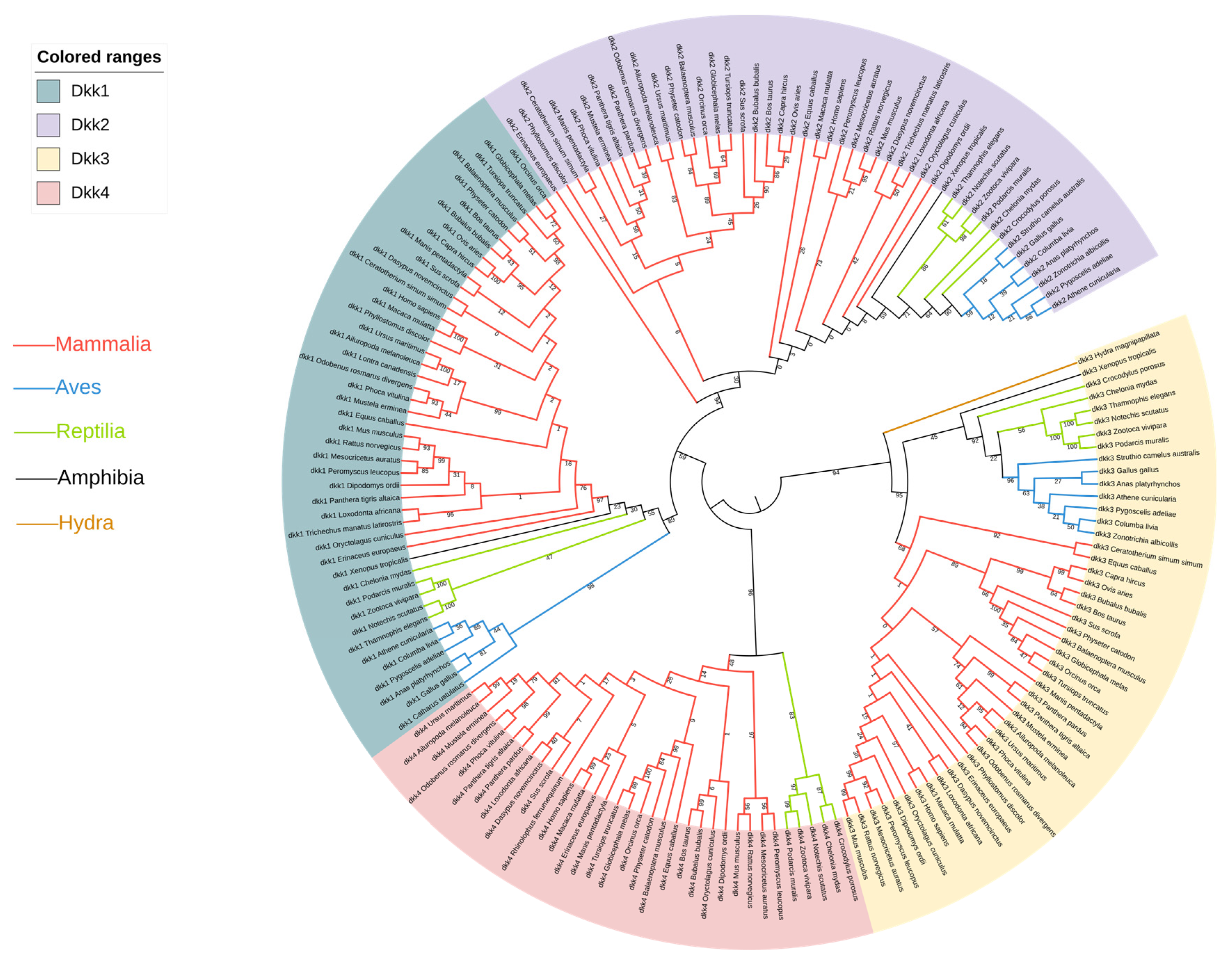
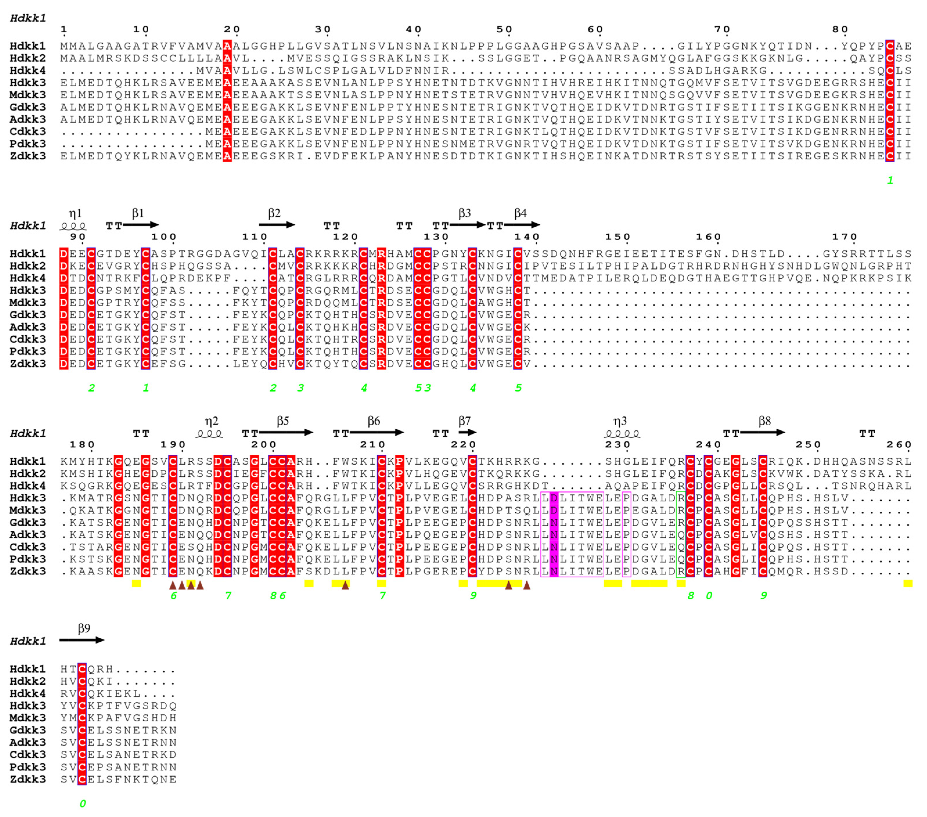
2.1. Sequence Alignment and Phylogenetic Analyses

To study the phylogenetic relationship between the different genes, a phylogenetic tree was constructed using the Maximum Likelihood method. From the topology of the tree, it can be seen that Dkk1, Dkk2, and Dkk4 all originated from Dkk3 (Figure 1). Multiple sequence alignments were performed for the Dkk1–4 genes. There are obvious differences in the gene sequence of Dkk1 in vertebrates; Aves lack 10 amino acids in the middle region (Figure S1A). This study also found that the protein structure in Aves is different from that of humans and mice (Figure 2). From the tertiary structure of proteins, it can be observed that avian protein sequences are shorter and lack a significant amount of α-helices. In the multiple sequence alignment of Dkk2, there were differences between Aves and Reptilia: 14 amino acids were inserted in Aves and Reptilia (Figure S1B). In the multiple sequence alignment of Dkk3, there are differences among Aves, Reptilia, Anura, and Chiroptera; 11 amino acids were inserted in Aves and Reptilia (Figure S1C). The results of the multiple sequence alignment of Dkk4 showed no significant differences.

2.2. Variation in Molecular Evolution Rate of Dkk

To examine the changes in selection pressure on the four genes of the Dkk gene family during the evolution of vertebrates, we calculated the ω values using the model M0 in PAML (Phylogenetic Analysis by Maximum Likelihood): ωDkk1 = 0.10793, ωDkk2 = 0.06302, ωDkk3 = 0.20621, and ωDkk4 = 0.26750. The results show that the synonymous substitution rate is much higher than the non-synonymous substitution rate, indicating that the entire Dkk gene family has generally undergone purifying selection during the evolution of vertebrates, showing the functional conservation of this gene family.
Although the overall Dkk gene sequence was strongly purified and selected, it could not be ruled out that some amino acids at specific sites were under positive selection. Therefore, we used Site models (M3 vs. M0 and M2 vs. M1) to detect the selection pressure at all sites. The LRT difference between M3 and M0 of Dkk1 was significant (2Δl = 10858.42, df = 4, p < 0.001), indicating that discrete selection pressures were applied to different sites of Dkk1, but none of the sites were in a positive selection state. The LRT between M1 and M2 of Dkk1 does not support the hypothesis that model M2 is superior to model M1 (2Δl = 0, df = 2, p > 0.05), and there is no positive selection site. The results of each model are shown in Table S2. The results showed that all Dkk genes were under purifying selection, and that no positive sites were found.
Although all four genes showed purifying selection, a free-ratio model was developed in order to better reflect the selection pressure of different species in evolution. The results showed that in Dkk1, the ω value of Reptilia (ω = 21.85) was significantly higher than that of other branches. In Dkk3, Aves and Reptilia have ω = 3.04. However, in Aves species, ω = 999 for the Dkk1 and Dkk3 genes. In Dkk2 and Dkk4, the ω-value of the branch of Pholidota and Carnivora was significantly higher than that of the other branches (ω > 1). A ω ratio significantly greater than one is a convincing indicator of positive selection. These results suggest that positive selection may have played a potential role in the early evolution of Reptilia, Aves, Pholidota, and Carnivora.
To further determine if any amino acid sites in the Dkk gene family’s accelerated evolution branch are under selection pressure, we ran Model A (Model = 2, NSsites = 2) in the branch site model, which takes into account not only the ω value between sites but also the ω value between branches. The test results (Table 1) are all based on the probability calculated by the BEB method (*: p > 95%; **: p > 99%).

2.3. Dkks Functional Divergence Analysis Results

According to the previously constructed species evolution tree, the vertebrates were divided into three groups: A for Mammalia, B for Reptilia, and C for Aves (Table 2 and Table 3). Most of these functional divergence sites are distributed in the Cys-2 domain, and very few are distributed in the Cys-1 domain. In the Dkk gene family, Dkk2, Dkk3, and Dkk4 had obvious type I functional divergence; the value of θI was between 0.23 and 0.63, while the θI coefficient of Dkk1 was relatively small. The θII coefficients of Dkk1–4 are all relatively small (Table 2 and Table 3).

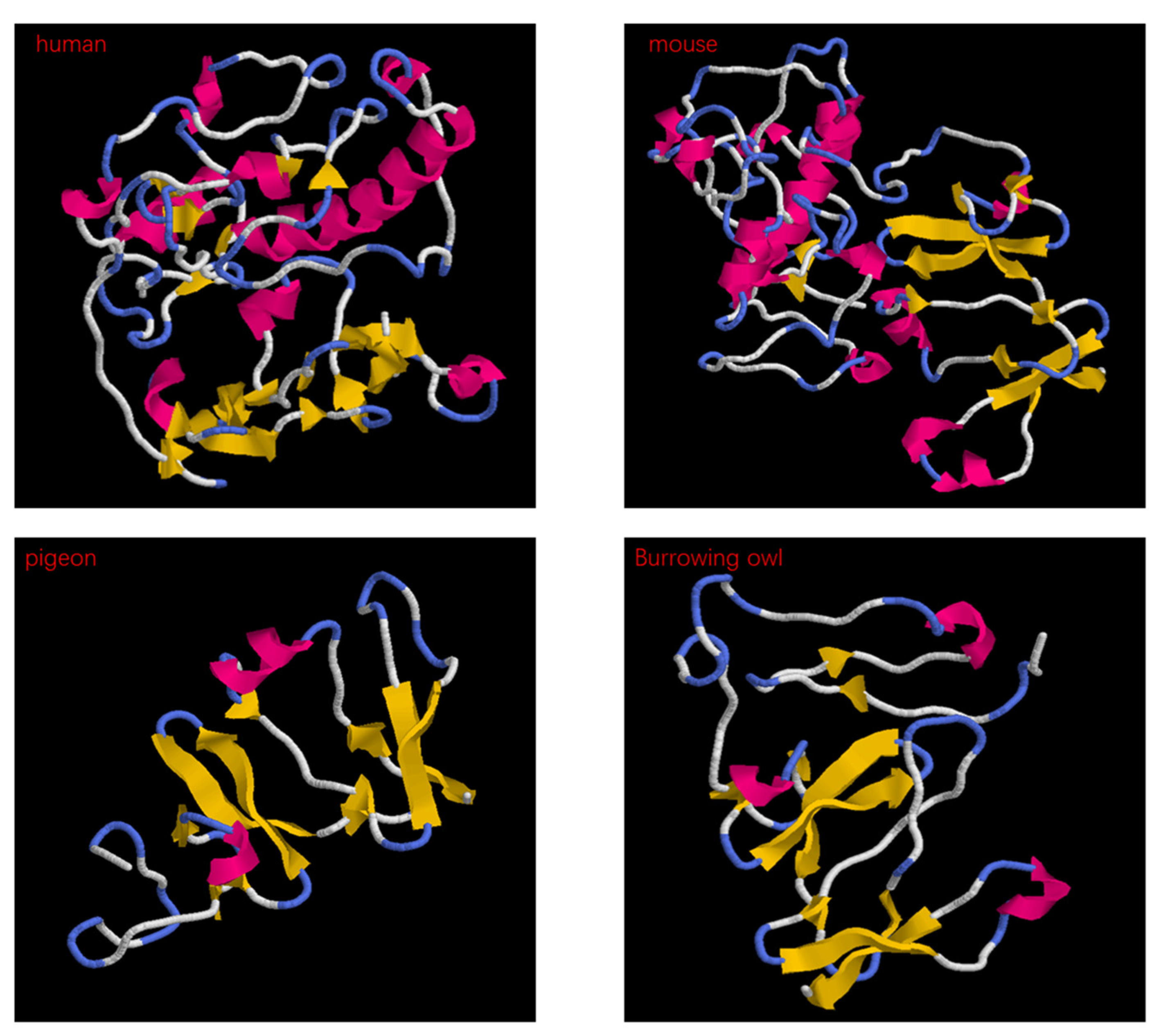
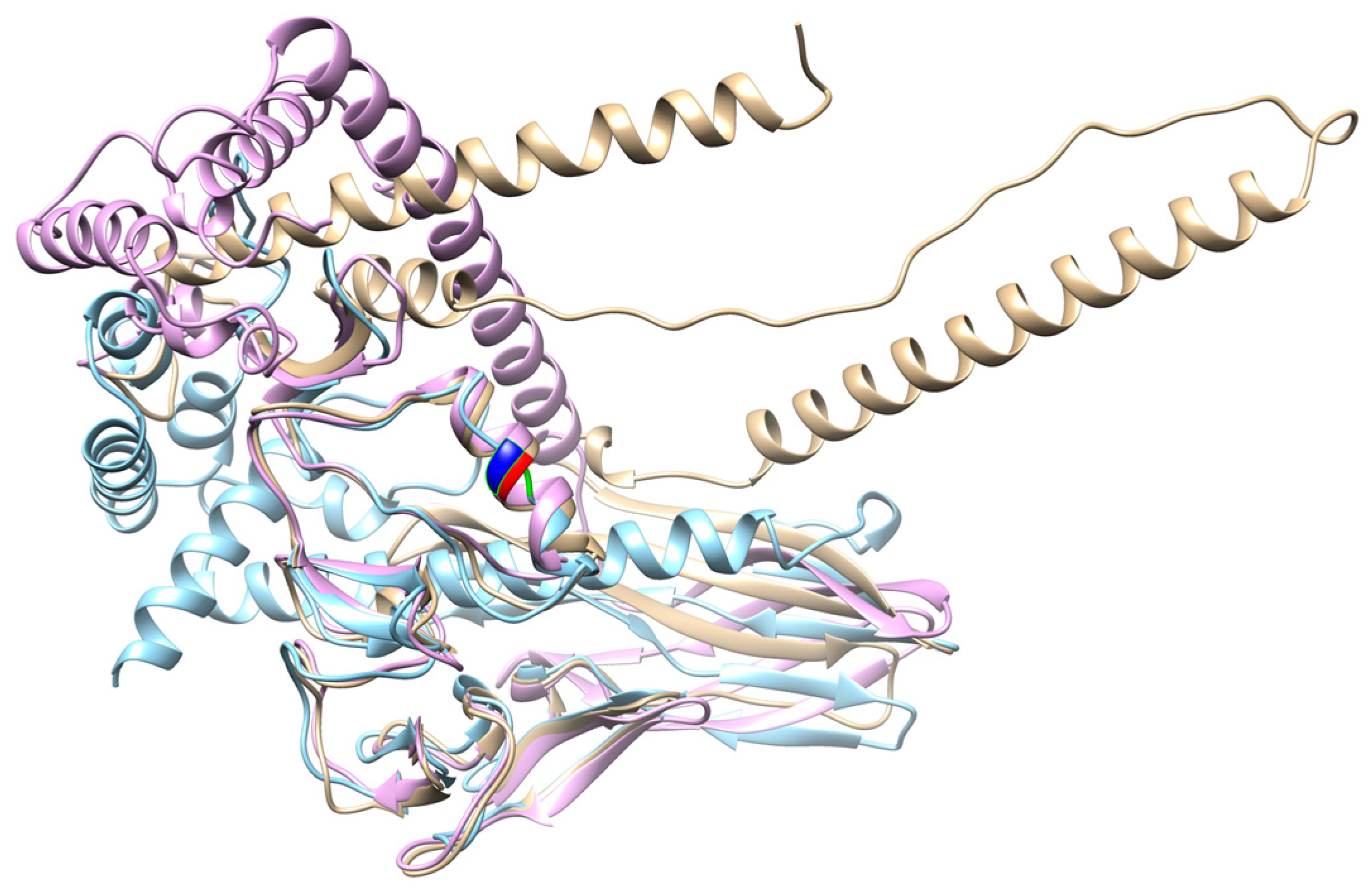
2.4. Dkk Protein Analysis Results

To infer structure–function correlations, the sequences of the positive selection sites were detected using the PAML software and are based on the human amino acid sequence. This is because some amino acids will be deleted when the PAML software is running, so it is necessary to determine the position of the detected amino acid position in the complete amino acid sequence. The pictures of the involved sites are shown in Figure S3. We found that there was a change in the protein sequence of Homo sapiens (255S) and Anas platyrhynchos (223P) Dkk1, and the protein structure changed from a turn to a coil. This amino acid site is located in the Cys-2 domain. In Dkk2, the amino acid site (27V) of H. sapiens and the corresponding amino acid site of Manis pentadactyla (86M) changed, and the protein structure changed from a turn to a coil. This amino acid site is located in the Cys-1 domain. In Dkk3, the amino acid site (264R) of H. sapiens and the corresponding amino acid site of Zonotrichia albicollis (209L) changed, and the protein structure changed from a turn to a coil. In Dkk4, the amino acid site (132K) of H. sapiens and the corresponding amino acid site of Podarcis muralis (131Q) changed, and the protein structure changed from a coil to a turn.

3. Discussion

In this paper, we investigated the evolutionary relationship of Dkk proteins in vertebrates. We discuss the nature of Dkk’s interactions with its partner Krm1 and the E3E4 region of LRP5/6, which have been widely established, and the functional divergence of Dkk proteins in vertebrate evolution, which has not been reported. We also explore the surprising lack of understanding of the Dkk family in terms of molecular evolution. Our findings provide insight into the evolution of Dkk proteins and their role in Wnt signaling.
In the multiple sequence alignment, Dkk1, Dkk2, and Dkk3 showed obvious differences. The inserted amino acids have an irregular curl in the protein structure. The insertion of these amino acids may cause the Dkk genes to differ in skin phenotype between different species. Through the construction of a phylogenetic tree, the phylogenetic relationship between species or genes can be displayed clearly. Dkk1, Dkk2, and Dkk4 all originated from Dkk3. A study confirmed that vertebrate Dkk-1, 2, and 4 may have originated from a common ancestor gene of Dkk3 [12]. But, in another study, Dkk3 appears to be a divergent member of the Dkk family [13]. The origin of the Dkk family is currently debated, but our results confirm that they originate from Dkk3. Due to structural differences, Dkk3 proteins exhibit biological characteristics that are different from those of the other family members. Most of the reports on Dkk3 gene are closely related to the occurrence, development, metastasis, and prognosis of common tumors [14]. Dkk1, Dkk2, and Dkk4 have all been reported to be involved in hair follicle development. The Dkk family may play an important role in hair follicle variation in vertebrates. To test this hypothesis, we used CodeML estimates of synonymous and non-synonymous substitutions.
The results of the M0 model indicate that the Dkk gene family is under purifying selection in most vertebrates. The M0 model indicates that this gene family has important functions. In addition, the free-ratio model evolution studies have shown that, in vertebrates, the selection pressure of the Dkk family changes. The adaptive evolution of the four genes occurred primarily in single branches of the phylogenetic tree, including Reptilia, Aves, Cetaceans, Lepidoptera, and Carnivora. For Reptilia and Aves Dkk1 and Dkk3 genes, ω > 1. An ω ratio significantly higher than 1 is convincing evidence of diversification [15]. Aves Dkk1 and Dkk3 showed the strongest positive selection signal (ω  =  999) based on the branch mode. Reptilian scales and avian feathers are considered homologous structures [16]. The complex topology of bird feathers may be responsible for the accelerated evolution of birds during feather development. The topology of bird feathers is more complex than that of reptile scales [17]. Feathers consist of many tiny structures that form complex interactions and liaisons between each other. This complex structure allows birds to be more adaptable in terms of flight and protecting themselves. However, in the evolution of vertebrates, the hair phenotypes of Reptilia and Aves are unique. Given that diversity in hair development can occur through multiple pathways, this lack of a parallel signature is perhaps not surprising [18]. The results of the Branch site model of the Dkk gene family showed that there were adaptive evolution and purifying selection sites in Reptilia in Dkk1, Dkk3, and Dkk4. There are also purifying selection sites in birds in Dkk1 and Dkk3. Moreover, the amino acid sites we found almost all existed in the CRD. It was found that the CRD domain is both necessary and sufficient for the binding to Wnt [19,20]. However, the Wnt signaling pathway plays an important role in many genes or signaling pathways that regulate hair follicle growth and development [21,22].
To further determine whether the Dkk gene family had functional divergence among species, family members were tested for type I and type II functional divergence. The degree of functional divergence of type I is greater than that of type II. This indicates that the functional constraints between replicative genes have changed [23]. Our results also proved that the Dkk gene family underwent accelerated evolution during species evolution, and some residues may have undergone functional restriction changes after speciation. Mammals, Aves, and Reptilia have different hair phenotypes, which may cause the genes to have different functions. At the same time, one purifying selection site (281V) in the Dkk3 gene was also identified as functional divergence site by the pressure selection analysis.
Most of the purifying selection sites and functional divergence sites screened in this study are in the middle and downstream regions, and some of them are in the Cys-2 domain. Dkk1, Dkk2, and Dkk4 have been identified as effective inhibitors of Wnt signaling and bind to the Wnt coreceptor LRP5/6 [24,25,26]. However, some studies have found that CRD2 is essential for suppressing Wnt signals [27]. The binding site of Dkk1 CRD2 to LRP5/6 has been reported [28,29]. The binding sites of Dkk1 and Kremen1 have also been reported [30,31]. Neutral sites were found near the binding sites. Mohammadpour performed in silico analyses and established that Dkk3, similar to other Dkk family members, can bind to the third PE pair of LRP5/6 through its CRD2 [32]. In another in silico study, Fujii reported that the insertion of seven amino acids (L249-E255 in human Dkk3) and P258 reduced the binding affinity between DKK3 and LRP5/6 [33]. Interestingly, the D250 residue in the Dkk3 protein sequence was mutated to N in Aves, Reptilia, and Amphibia (Figure 3). This mutation leads to a decrease in amino acid hydrophilicity. Comparing the tertiary structures of three species (H. sapiens, Zootoca vivipara, and Gallus gallus), the secondary structure of D250N changed from a turn to a random coil (Figure 4). When the hydrophilicity of an amino acid is weakened, it may have an effect on the structure and function of the protein [34].
A study designed and improved several small peptides based on the LRP6-binding site of the CRD2 of Dkk3 [35]. These peptides were highly capable of binding to LRP6 in silico, and may prevent the formation of an active Wnt-LRP6-Fz complex [35]. In the experiment conducted by Poorebrahim, several small peptides did not alter 264R, an amino acid that forms a salt bridge with Asp811 of LRP6. In our experiment, we found a mutation in 264R in the Aves species, where Arg was replaced by Gln, resulting in decreased hydrophilicity (Figure 3). In the protein structure, two amino acid residues with opposite charges form an ion pair; when the distance between the charged groups of the two amino acid side chains in the ion pair (that is, any oxygen atom in the negatively charged residue carboxylate and the positively charged residue side or the distance between any nitrogen atom in the chain) is less than 4 Å, the ion pair is considered a salt bridge [36,37]. Gln is a polar uncharged residue. The Arg264Gln substitution would abolish a salt bridge with Asp811 in LRP6. The selected amino acids found in CRD2 may affect the binding of the Dkk gene family to LRP5/6, thus affecting the inhibition mechanism of Wnt signals and making Dkk1, Dkk2, and Dkk4 show different functions in hair follicle development. Vertebrate habitats range from the deepest parts of the ocean to the highest peaks of mountain ranges, from the tropics to the Arctic. The environmental variations trigger divergent natural selection, leading to the emergence of niche specialists [27]. The living environment of Aves and Reptilia may also cause these genes to differentiate in function during the evolutionary process.

4. Conclusions

Dkk1–4 all underwent accelerated evolution and purifying sites were detected. The changes in the Dkk gene family in vertebrates under selection pressure and functional divergence were tested. However, the evolution rates of purifying selection sites and functional divergence sites are different. These amino acid sites will affect the tertiary structure of proteins and make genes differentiate functionally. The current study shows that the Dkk gene family underwent changes in selection patterns during vertebrate evolution and may have acquired additional functional constraints in different branches.

5. Materials and Methods

5.1. Sequence Acquisition

Sequence data of the CDS of the Dkk1–4 genes of different species were retrieved from the GenBank database. For the Dkk1 gene, a total of 45 species were selected, 47 species were selected for the Dkk2 gene, 47 species for the Dkk3 gene, and 34 species for the Dkk4 gene (Table S1). These data were then used to analyze the genetic variation and evolutionary relationship of the Dkk gene family in vertebrates. Hydra magnipapillata is an invertebrate. We chose Hydra as an outgroup because we wanted to study the origin of the Dkk gene family. The Dkk gene family has been found in vertebrates and some invertebrate phyla but Dkk4 appears to only be present in mammals and Reptilia.

5.2. Nucleotide Sequence Analysis

Using MEGA-X [38], the Maximum Likelihood tree of the amino acid sequences of the Dkk gene family of vertebrates was constructed, and the confidence value of each branch was calculated with 500 repetitions of the Bootstrap test. Additionally, the nucleotide sequences of the Dkk1–4 genes of the above species were aligned using the ClustaW method using MEGA-X. Itol (http://itol.embl.de/ accessed on 1 May 2023) was used to beautify the evolutionary trees. The multiple sequence alignment diagram of the amino acid sequence was generated using ESPript 3 (https://espript.ibcp.fr/ESPript/ESPript/index.php accessed on 20 May 2023).

5.3. Molecular Evolution Analysis

To detect changes in the selection pressure of four genes in the Dkk gene family during vertebrate evolution, the CodeML program in PAMLX [39] was used to calculate the ω (dN/dS) values of the Dkk1–4 sequences, where dN represents the rate of non-synonymous substitutions, and dS represents the rate of synonymous substitutions. dN/dS < 1 means purifying selection, dN/dS = 1 means neutral evolution, and dN/dS > 1 means positive Darwinian selection.
To test whether there is positive selection at specific amino acid sites, we compared four models: M3 vs. M0 and M2 vs. M1. The rates along specific branches of the tree were estimated, and the topology is based on published studies [40,41], as shown in Figure S2. We performed a one-ratio model for each gene separately, which assumes equal ω values for all branches of the phylogenetic tree [42]. Secondly, a free-ratio model was developed, which assumes that the ω value is different for all branches in the phylogenetic tree [43], and analyzes the selection pressure of this gene at the branch level of different species. Finally, Model A of the branch site model (Model = 2, NSsites = 2) was developed, which analyzes the amino acid sites on the branch with high selection pressure where members of the Dkk gene family are positively selected [43]. All species comparisons were made using H. sapiens as a reference.
To determine whether the Dkk gene family has functional divergence among the various branches of the phylogenetic tree, DIVERGE3.0 [44] was used to test the functional divergence of each gene. Functional divergence was measured by the functional divergence coefficient (θ). The value of θ is between 0 and 1, and the closer the value of θ is to 1, the more significant the functional divergence of the gene clusters is. At least three species are needed to form a group in species functional divergence analysis. Among amniotes, stem reptiles were basal to extant reptiles, birds, and mammals [45]. In this study, it was found that there is significant divergence between Reptilia and Aves, and vertebrates were divided into three groups, namely, A for Mammalia, B for Reptilia, and C for Aves. Any two groups were compared and analyzed, and type I and type II functional divergences were analyzed.

5.4. Protein Analysis

To infer structure–function correlations, positively selected amino acid residues were mapped to the three-dimensional structure of Dkk proteins. The H. sapiens protein structure was from the AlphaFold database. However, the tertiary structures of proteins of the Dkk gene family were not available in protein databases (https://espript.ibcp.fr/ESPript/ESPript/index.php accessed on 15 Jan 2023). The tertiary structures of Dkk1, Dkk2, Dkk3, and Dkk4 proteins from different species were predicted by Robetta (https://robetta.bakerlab.org/ accessed on 16 Apr 2023). We used Rasmol to display the protein tertiary structure of Dkks. Models were superimposed using MatchMaker from CHIMERA 1.16 (https://www.cgl.ucsf.edu/chimera/ accessed on 25 May 2023).

Supplementary Materials

The following supporting information can be downloaded at: https://www.mdpi.com/article/10.3390/genes14101863/s1, Figure S1: Multiple sequence alignment. (A) In Dkk1, Aves lack 10 amino acids in the middle region, while 10 amino acids are inserted in other vertebrates. (B) In Dkk2, mammals and Anura lacked 13 amino acids in the middle segment, and 14 amino acids were inserted in Aves and Reptilia. (C) In Dkk3, 12 amino acids were inserted in Xenopus tropicalis and Phyllostomus discolor and Amphibia, and 11 amino acids were inserted in Aves and Reptilia; Figure S2: Phylogenetic tree of Dkk gene family. (A) The type I (θI) and type II (θII) functional divergences of the Dkk1 gene were estimated between mammals and Reptilia, mammals and Aves, and Reptilia and Aves. (B) The type I (θI) and type II (θII) functional divergences of the Dkk2 gene were estimated between mammals and Reptilia, mammals and Aves, and Reptilia and Aves. (C) The type I (θI) and type II (θII) functional divergences of the Dkk3 gene were estimated between mammals and Reptilia, mammals and Aves, and Reptilia and Aves. (D) The type I (θI) and type II (θII) functional divergences of the Dkk4 gene were estimated between mammals and Reptilia; Figure S3. The amino acids under positive selection are mapped onto the 3D structure of the protein. (A) In Dkk1, a is H. sapiens, the positively selected amino acid (255S) is shown in green, and the structure is the corner. b is Anas platyrhynchos, the corresponding amino acid (223P) is shown in green, and the structure is a coil. (B) In Dkk2, a is H. sapiens, the positively selected amino acid (86V) is shown in green, and the structure is the corner. b is Manis pentadactyla, the corresponding amino acid (86M) is shown in green, and the structure is a coil. (C) In Dkk3, a is H. sapiens, the positively selected amino acid (264R) is shown in green, and the structure is the corner. b is Zonotrichia albicollis, the corresponding amino acid (209L) is shown in green, and the structure is not a regular coil. (D) In Dkk4, a is H. sapiens, the positively selected amino acid (132K) is shown in green, and the structure is an irregular coil. b is Podarcis muralis, the corresponding amino acid (131Q) is shown in green, and the structure is the corner; Table S1: Species, scientific names, and accession numbers of the Dkk gene family; Table S2: Parameters and results of evolutionary model of Dkk gene family.

Author Contributions

J.W. and W.G. conceived and designed the experiments. B.W. and S.H. performed the experiments. B.W. analyzed the data. B.W. wrote the paper. J.Y. edited and revised the paper. All authors have read and agreed to the published version of the manuscript.

Funding

This work was supported by the National Natural Science Foundation of China (32260812, 3211101853) to Jianghong Wu and Jun Yin, and the Inner Mongolia Autonomous Region’s Key Technology Tackling Plan (2021GG0008, 2020GG0069), and the Program for Young Talents of Science and Technology in Universities of Inner Mongolia Autonomous Region (2023NJYT23015), and the Youth Program for Grassland Elite in Inner Mongolia Autonomous Region to Jianghong Wu, and the Doctoral Scientific Research Foundation of Inner Mongolia Minzu University (BS527, BS526) to Jianghong Wu and Sile Hu.

Institutional Review Board Statement

Not applicable.

Informed Consent Statement

Not applicable.

Data Availability Statement

All data generated or analyzed during this study shall be made available upon reasonable request.

Acknowledgments

Zaccheaus Pazamilala Akonyani was helpful in revising our manuscript, and we are very grateful to him.

Conflicts of Interest

The authors declare no conflict of interest.

References

  1. Glinka, A.; Wu, W.; Delius, H.; Monaghan, A.P.; Blumenstock, C.; Niehrs, C. Dickkopf-1 is a member of a new family of secreted proteins and functions in head induction. Nature 1998, 391, 357–362. [Google Scholar] [CrossRef]
  2. Krupnik, V.E.; Sharp, J.D.; Jiang, C.; Robison, K.; Chickering, T.W.; Amaravadi, L.; Brown, D.E.; Guyot, D.; Mays, G.; Leiby, K.; et al. Functional and structural diversity of the human Dickkopf gene family. Gene 1999, 238, 301–313. [Google Scholar] [CrossRef]
  3. Guder, C.; Pinho, S.; Nacak, T.G.; Schmidt, H.A.; Hobmayer, B.; Niehrs, C.; Holstein, T.W. An ancient Wnt-Dickkopf antagonism in Hydra. Development 2006, 133, 901–911. [Google Scholar] [CrossRef]
  4. Hino, K.; Satou, Y.; Yagi, K.; Satoh, N. A genomewide survey of developmentally relevant genes in Ciona intestinalis. VI. Genes for Wnt, TGFbeta, Hedgehog and JAK/STAT signaling pathways. Dev. Genes Evol. 2003, 213, 264–272. [Google Scholar] [CrossRef]
  5. Yoshida, S.; Satoh, H.; Mitsuya, T.; Tate, G. Assignment1 of the human dickkopf (Xenopus) homolog 4 (DKK4) to chromosome 8p11.2→p11.1 by fluorescence in situ hybridization. Cytogenet. Cell Genet. 2001, 94, 88–89. [Google Scholar] [CrossRef]
  6. Patel, S.; Barkell, A.M.; Gupta, D.; Strong, S.L.; Bruton, S.; Muskett, F.W.; Addis, P.W.; Renshaw, P.S.; Slocombe, P.M.; Doyle, C.; et al. Structural and functional analysis of Dickkopf 4 (Dkk4): New insights into Dkk evolution and regulation of Wnt signaling by Dkk and Kremen proteins. J. Biol. Chem. 2018, 293, 12149–12166. [Google Scholar] [CrossRef]
  7. Song, Y.; Boncompagni, A.C.; Kim, S.S.; Gochnauer, H.R.; Zhang, Y.; Loots, G.G.; Wu, D.; Li, Y.; Xu, M.; Millar, S.E. Regional Control of Hairless versus Hair-Bearing Skin by Dkk2. Cell Rep. 2018, 25, 2981–2991. [Google Scholar] [CrossRef]
  8. Andl, T.; Reddy, S.T.; Gaddapara, T.; Millar, S.E. WNT signals are required for the initiation of hair follicle development. Dev. Cell 2002, 2, 643–653. [Google Scholar] [CrossRef]
  9. Chu, Q.; Cai, L.; Fu, Y.; Chen, X.; Yan, Z.; Lin, X.; Zhou, G.; Han, H.; Widelitz, R.B.; Chuong, C.M.; et al. Dkk2/Frzb in the dermal papillae regulates feather regeneration. Dev. Biol. 2014, 387, 167–178. [Google Scholar] [CrossRef]
  10. Hiramitsu, S.; Terauchi, M.; Kubota, T. The effects of Dickkopf-4 on the proliferation, differentiation, and apoptosis of osteoblasts. Endocrinology 2013, 154, 4618–4626. [Google Scholar] [CrossRef]
  11. Cui, C.Y.; Kunisada, M.; Piao, Y.; Childress, V.; Ko, M.S.; Schlessinger, D. Dkk4 and Eda regulate distinctive developmental mechanisms for subtypes of mouse hair. PLoS ONE 2010, 5, e10009. [Google Scholar] [CrossRef] [PubMed]
  12. Fedders, H.; Augustin, R.; Bosch, T.C. A Dickkopf-3-related gene is expressed in differentiating nematocytes in the basal metazoan Hydra. Dev. Genes Evol. 2004, 214, 72–80. [Google Scholar] [CrossRef] [PubMed]
  13. Niehrs, C. Function and biological roles of the Dickkopf family of Wnt modulators. Oncogene 2006, 25, 7469–7481. [Google Scholar] [CrossRef] [PubMed]
  14. Veeck, J.; Dahl, E. Targeting the Wnt pathway in cancer: The emerging role of Dickkopf-3. Biochim. Biophys. Acta 2012, 1825, 18–28. [Google Scholar] [CrossRef]
  15. Yang, Z.; Bielawski, J.P. Statistical methods for detecting molecular adaptation. Trends Ecol. Evol. 2000, 15, 496–503. [Google Scholar] [CrossRef]
  16. Maderson, P.F.A. When? Why? and How? Some Speculations on the Evolution of the Vertebrate Integument. Am. Zool. 2015, 12, 159–171. [Google Scholar] [CrossRef]
  17. Yu, M.; Wu, P.; Widelitz, R.B.; Chuong, C.M. The morphogenesis of feathers. Nature 2002, 420, 308–312. [Google Scholar] [CrossRef]
  18. Winter, H.; Langbein, L.; Krawczak, M.; Cooper, D.N.; Jave-Suarez, L.F.; Rogers, M.A.; Praetzel, S.; Heidt, P.J.; Schweizer, J. Human type I hair keratin pseudogene phihHaA has functional orthologs in the chimpanzee and gorilla: Evidence for recent inactivation of the human gene after the Pan-Homo divergence. Hum. Genet. 2001, 108, 37–42. [Google Scholar] [CrossRef]
  19. Bhanot, P.; Brink, M.; Samos, C.H.; Hsieh, J.C.; Wang, Y.; Macke, J.P.; Andrew, D.; Nathans, J.; Nusse, R. A new member of the frizzled family from Drosophila functions as a Wingless receptor. Nature 1996, 382, 225–230. [Google Scholar] [CrossRef]
  20. Dann, C.E.; Hsieh, J.C.; Rattner, A.; Sharma, D.; Nathans, J.; Leahy, D.J. Insights into Wnt binding and signalling from the structures of two Frizzled cysteine-rich domains. Nature 2001, 412, 86–90. [Google Scholar] [CrossRef]
  21. Lei, M.X.; Chuong, C.M.; Widelitz, R.B. Tuning Wnt signals for more or fewer hairs. J. Investig. Dermatol. 2013, 133, 7–9. [Google Scholar] [CrossRef] [PubMed]
  22. Tsai, S.Y.; Sennett, R.; Rezza, A.; Clavel, C.; Grisanti, L.; Zemla, R.; Najam, S.; Rendl, M. Wnt/β-catenin signaling in dermal condensates is required for hair follicle formation. Dev. Biol. 2014, 385, 179–188. [Google Scholar] [CrossRef] [PubMed]
  23. Gu, X. Maximum-likelihood approach for gene family evolution under functional divergence. Mol. Biol. Evol. 2001, 18, 453–464. [Google Scholar] [CrossRef] [PubMed]
  24. Fedi, P.; Bafico, A.; Nieto Soria, A.; Burgess, W.H.; Miki, T.; Bottaro, D.P.; Kraus, M.H.; Aaronson, S.A. Isolation and biochemical characterization of the human Dkk-1 homologue, a novel inhibitor of mammalian Wnt signaling. J. Biol. Chem. 1999, 274, 19465–19472. [Google Scholar] [CrossRef]
  25. Mao, B.; Wu, W.; Li, Y.; Hoppe, D.; Stannek, P.; Glinka, A.; Niehrs, C. LDL-receptor-related protein 6 is a receptor for Dickkopf proteins. Nature 2001, 411, 321–325. [Google Scholar] [CrossRef]
  26. Bafico, A.; Liu, G.; Yaniv, A.; Gazit, A.; Aaronson, S.A. Novel mechanism of Wnt signalling inhibition mediated by Dickkopf-1 interaction with LRP6/Arrow. Nat. Cell Biol. 2001, 3, 683–686. [Google Scholar] [CrossRef]
  27. Caffrey, B.E.; Williams, T.A.; Jiang, X.; Toft, C.; Hokamp, K.; Fares, M.A. Proteome-wide analysis of functional divergence in bacteria: Exploring a host of ecological adaptations. PLoS ONE 2012, 7, e35659. [Google Scholar] [CrossRef]
  28. Cheng, Z.; Biechele, T.; Wei, Z.; Morrone, S.; Moon, R.T.; Wang, L.; Xu, W. Crystal structures of the extracellular domain of LRP6 and its complex with DKK1. Nat. Struct. Mol. Biol. 2011, 18, 1204–1210. [Google Scholar] [CrossRef]
  29. Ahn, V.E.; Chu, M.L.; Choi, H.J.; Tran, D.; Abo, A.; Weis, W.I. Structural basis of Wnt signaling inhibition by Dickkopf binding to LRP5/6. Dev. Cell 2011, 21, 862–873. [Google Scholar] [CrossRef]
  30. Nakamura, T.; Nakamura, T.; Matsumoto, K. The functions and possible significance of Kremen as the gatekeeper of Wnt signalling in development and pathology. J. Cell Mol. Med. 2008, 12, 391–408. [Google Scholar] [CrossRef]
  31. Wang, K.; Zhang, Y.; Li, X.; Chen, L.; Wang, H.; Wu, J.; Zheng, J.; Wu, D. Characterization of the Kremen-binding site on Dkk1 and elucidation of the role of Kremen in Dkk-mediated Wnt antagonism. J. Biol. Chem. 2008, 283, 23371–23375. [Google Scholar] [CrossRef]
  32. Mohammadpour, H.; Pourfathollah, A.A.; Nikougoftar Zarif, M.; Khalili, S. Key role of Dkk3 protein in inhibition of cancer cell proliferation: An in silico identification. J. Theor. Biol. 2016, 393, 98–104. [Google Scholar] [CrossRef] [PubMed]
  33. Fujii, Y.; Hoshino, T.; Kumon, H. Molecular simulation analysis of the structure complex of C2 domains of DKK family members and β-propeller domains of LRP5/6: Explaining why DKK3 does not bind to LRP5/6. Acta Med. Okayama 2014, 68, 63–78. [Google Scholar]
  34. David, A.; Razali, R.; Wass, M.N.; Sternberg, M.J. Protein-protein interaction sites are hot spots for disease-associated nonsynonymous SNPs. Hum. Mutat. 2012, 33, 359–363. [Google Scholar] [CrossRef]
  35. Poorebrahim, M.; Sadeghi, S.; Rahimi, H.; Karimipoor, M.; Azadmanesh, K.; Mazlomi, M.A.; Teimoori-Toolabi, L. Rational design of DKK3 structure-based small peptides as antagonists of Wnt signaling pathway and in silico evaluation of their efficiency. PLoS ONE 2017, 12, e0172217. [Google Scholar] [CrossRef]
  36. Barlow, D.J.; Thornton, J.M. Ion-pairs in proteins. J. Mol. Biol. 1983, 168, 867–885. [Google Scholar] [CrossRef]
  37. Kumar, S.; Nussinov, R. Close-range electrostatic interactions in proteins. Chembiochem 2002, 3, 604–617. [Google Scholar] [CrossRef] [PubMed]
  38. Kumar, S.; Stecher, G.; Li, M.; Knyaz, C.; Tamura, K. MEGA X: Molecular Evolutionary Genetics Analysis across Computing Platforms. Mol. Biol. Evol. 2018, 35, 1547–1549. [Google Scholar] [CrossRef]
  39. Xu, B.; Yang, Z. PAMLX: A graphical user interface for PAML. Mol. Biol. Evol. 2013, 30, 2723–2724. [Google Scholar] [CrossRef]
  40. Murphy, W.J.; Eizirik, E.; Johnson, W.E.; Zhang, Y.P.; Ryder, O.A.; O’Brien, S.J. Molecular phylogenetics and the origins of placental mammals. Nature 2001, 409, 614–618. [Google Scholar] [CrossRef]
  41. Liang, D.; Wu, R.; Geng, J.; Wang, C.; Zhang, P. A general scenario of HOX gene inventory variation among major sarcopterygian lineages. BMC Evol. Biol. 2011, 11, 25. [Google Scholar] [CrossRef] [PubMed]
  42. Yang, Z.; Nielsen, R.; Goldman, N.; Pedersen, A.M. Codon-substitution models for heterogeneous selection pressure at amino acid sites. Genetics 2000, 155, 431–449. [Google Scholar] [CrossRef]
  43. Zhang, J.; Nielsen, R.; Yang, Z. Evaluation of an improved branch-site likelihood method for detecting positive selection at the molecular level. Mol. Biol. Evol. 2005, 22, 2472–2479. [Google Scholar] [CrossRef] [PubMed]
  44. Gu, X.; Vander Velden, K. DIVERGE: Phylogeny-based analysis for functional-structural divergence of a protein family. Bioinformatics 2002, 18, 500–501. [Google Scholar] [CrossRef] [PubMed]
  45. Chang, C.; Wu, P.; Baker, R.E.; Maini, P.K.; Alibardi, L.; Chuong, C.M. Reptile scale paradigm: Evo-Devo, pattern formation and regeneration. Int. J. Dev. Biol. 2009, 53, 813–826. [Google Scholar] [CrossRef] [PubMed]
Figure 1. Phylogenetic tree of Dkk gene family. The numbers on the evolutionary tree are bootstrap values.
Genes 14 01863 g001
Figure 2. Protein structure of human, mouse, pigeon, and burrowing owl Dkk1. Deep red represents α-helix, yellow represents β-fold, light blue represents coil, and white represents other residues.
Genes 14 01863 g002
Figure 3. Dkk multiple sequence alignment. Highly similar residues are colored in red and framed in blue. The green number indicates the disulfide bond. The yellow box at the bottom of the sequence represents the residue involved in the interaction between Dkk1 and LRP6. The brown triangle is the residue involved in the interaction between Dkk1 and Kremen proteins. Seven amino acids (L249-E255 in human Dkk3, LDLITWE) and P258 are represented in a purple box. In the purple box, the black letter is the site of a Dkk3 mutation (D250N). The green box is the Arg264Gln of Dkk3 gene. Abbreviations: H = H. sapiens, M = Mus musculus, G = G. gallus, A = Anas platyrhynchos, C = Columba livia, P = Pygoscelis adeliae, and Z = Z. vivipara.
Genes 14 01863 g003
Figure 4. Overlay structure of Dkk3 proteins from three species. Species: H. sapiens (brown), Zootoca vivipara (light blue), and G. gallus (pink). Red, green, and dark blue represent the relative positions of the D250N mutation. The secondary structure of 250N in Z. vivipara is a random coil.
Genes 14 01863 g004
Table 1. Results of branch site model. *: p > 95%; **: p > 99%.
GeneForeground BranchPossible Positive Selection Sites
Dkk1Aves17 H 0.997 **, 75 Y 0.998 **, 91 S 0.981 *
Aves and Reptilia7 H 0.979, 75 Y 1.000 **
Dkk2Lepidoptera (pangolin)27 V 0.998 **
Dkk3Aves and Reptilia24 R 0.997 **, 41 R 0.998 **, 52 P 1.000 **,
58 V 0.999 **
Dkk4Reptilia103 S 0.975 *, 105 K 0.970 *, 108 Q 0.961 *
Table 2. Functional divergence of type I Dkk gene family.
GeneClustersθIMFE SEPossible Positive Selection Sites (Qk > 0.90)
Dkk1A/B−0.0647540.145614
A/C0.1431040.155194
B/C0.0685580.216823
Dkk2A/B0.2392650.140598
A/C0.4034010.100621171G
B/C0.5148110.144675
Dkk3A/B0.4271490.301757367T, 377H, 410R, 414H, 416L, 423T, 443V, 450G, 495E
A/C0.6302690.134886399R, 407L, 411H
B/C0.6235370.348995399R
Dkk4A/B0.3324770.090430185Q
Table 3. Functional divergence of type II Dkk gene family.
GeneClustersθIIMFE SEPossible Positive Selection Sites (Qk > 0.90)
Dkk1A/B−0.0386240.086041
A/C0.0599110.081074263Y, 268Q, 271S, 312S, 324G, 328S, 341N, 342S
B/C0.0100270.088840252Y, 261K, 324G
Dkk2A/B0.0358920.052704165H, 188H
A/C0.0973380.04358336A, 38L, 44S, 46G, 92Q, 97S, 98S, 114G, 161D, 165H, 166R, 170H, 174S, 176N, 177H, 188H, 212F, 266Y, 270A
B/C−0.0205930.043545
Dkk3A/B0.1411600.144634360G, 371V, 373G, 375L, 377H, 380A, 385D, 404S, 410P, 411H, 416L, 417V, 418Y, 421K, 422P, 423T, 443V, 444G, 446R, 447D, 450G, 472D, 477G, 484E, 487R, 488Q, 492D, 495E, 496R, 507P, 516G
A/C0.2251940.105301360G, 371V, 375L, 380A, 381S, 385D, 398D, 411H, 416L, 417V, 418Y, 421K, 422P, 423T, 443V, 444G, 447D, 448Q, 450G, 471P, 472D, 484E, 488Q, 496R, 507P, 516G
B/C−0.0161330.132199373G, 471P, 477G
Dkk4A/B−0.0813730.135288
Disclaimer/Publisher’s Note: The statements, opinions and data contained in all publications are solely those of the individual author(s) and contributor(s) and not of MDPI and/or the editor(s). MDPI and/or the editor(s) disclaim responsibility for any injury to people or property resulting from any ideas, methods, instructions or products referred to in the content.

