Article

A Molecular Genetic Analysis of RPE65-Associated Forms of Inherited Retinal Degenerations in the Russian Federation

Research Centre for Medical Genetics, Moscow 115478, Russia
*
Author to whom correspondence should be addressed.
Genes 2023, 14(11), 2056; https://doi.org/10.3390/genes14112056
Submission received: 13 September 2023 / Revised: 26 October 2023 / Accepted: 1 November 2023 / Published: 9 November 2023
(This article belongs to the Special Issue Molecular Diagnosis and Disease Mechanisms in Eye Disorders)

Abstract

:
Pathogenic variants in the RPE65 gene cause the only known form of inherited retinal degenerations (IRDs) that are prone to gene therapy. The current study is aimed at the evaluation of the prevalence of RPE65-associated retinopathy in the Russian Federation, the characterization of known variants in the RPE65 gene, and the establishment of the specificities of the mutation spectrum in Russian patients. Methods: The analysis was carried out on blood samples obtained from 1053 non-related IRDs patients. The analysis, which consisted of 211 genes, was carried out based on the method of massive parallel sequencing (MPS) for all probands. Variant validation, as well as biallelic status verification, were carried out using direct automated Sanger sequencing. The number of copies of RPE65 exons 1–14 was analyzed with quantitative MLPA using an MRC-Holland SALSA MLPA probemix. Results: Out of 1053 non-related patients, a molecular genetic diagnosis of IRDs has been confirmed in 474 cases, including 25 (5.3%) patients with RPE65-associated retinopathy. We detected 26 variants in the RPE65 gene, nine of which have not been previously described in the literature. The most common mutations in the Russian population were c.304G>T/p.(Glu102*), c.370C>T/p.(Arg124*), and c.272G>A/p.(Arg91Gln), which comprised 41.8% of all affected chromosomes. Conclusions: The current study shows that pathogenic variants in the RPE65 gene contribute significantly to the pathogenesis of IRDs and comprise 5.3% of all patients with a confirmed molecular genetic diagnosis. This study allowed for the formation of a cohort for target therapy of the disorder; such therapy has already been carried out for some patients.

1. Introduction

Inherited retinal degenerations (IRDs) are a group of heterogeneous genetic disorders leading to visual impairment caused by progressive retinal degeneration.
The prevalence of IRDs in Europe is approximately 1:3000 [1,2,3]. IRDs comprise multiple rare disorders with varying ages of manifestation, rates of progression, primary retinal damage levels, and inheritance types. The symptoms of retinal dystrophy may vary from congenital blindness, in cases of more severe forms of retinal degeneration, to mild retinal dysfunction manifesting as night blindness. As of date, there are more than 340 known genes causing nonsyndromic and syndromic retinal diseases with autosomal recessive, autosomal dominant, and X-linked types of inheritance (according to https://sph.uth.edu/retnet, 12 September 2023).
Considering the prevalence of IRDs, in the Russian Federation (population of approximately 145 million), there are more than 48,000 people suffering from various forms of RP. Although the majority of the disorders are quite rare, in aggregate, they are a common cause of blindness or significant vision impairment in infants and adults of working age.
RPE65-associated retinopathies became the first group of hereditary ophthalmological disorders with approved gene therapy; therefore, the RPE65 gene came under the close attention of researchers from around the world.
The RPE65 gene is located on the 1p31 chromosome, contains 14 exons, and encodes proteins with a mass of 65 kDa, which consists of 533 amino acid residues [4]. RPE65 is a membrane-associated protein, which is expressed primarily in the pigment epithelium and plays a vital role in the regeneration of 11-cis-retinol in the visual cycle [4]. Light activates the visual pigment, causing it to break down hydrolytically with the formation of a free all-trans-retinal, which regenerates in the external segments of photoreceptors using retinol dehydrogenase. The resulting all-trans-retinol is transferred to the pigment epithelium, where it is transformed into all-trans-retinyl esters with retinol acyltransferase [5]. The all-trans-retinyl esters can be stored in retinosomes or be further converted [6]. In the retinal pigment epithelium, RPE65 converts all-trans-retinyl esters into 11-cis-retinol and a free fatty acid via simultaneous hydrolysis and isomerization reactions, thereby being named isomerohydrolase. After that, in the pigment epithelium, the 11-cis-retinol is oxidized using 11-cis-retinol dehydrogenase (RDH5) to 11-cis-retinal [7], then returns to the external segment of the photoreceptor to regenerate main visual pigments, thus completing the cycle [8].
RPE65 alterations lead to the accumulation of all-trans-retinyl esters and a decrease in or absence of visual pigment in the photoreceptors.
A fully active RPE65 protein is a dimer of two symmetrical enzymatically independent subunits. A monomer of RPE65 is a seven-bladed β-propeller and an iron-dependent enzyme, in which four histidine residues (His180, His241, His313, and His527) and three glutamic acid residues (Glu148, Glu417, and Glu469) coordinate an Fe2+ cation [9].
Biallelic pathogenic variants in the RPE65 gene cause Leber congenital amaurosis (LCA) (OMIM 204100) and severe early-onset retinitis pigmentosa (RP20; OMIM 613794) [10]. In various populations around the world, 0.6% to 16% of IRD cases are caused by pathogenic variants in the RPE65 gene [4,11]. The most common pathogenic RPE65 variants differ from population to population; quite often, there are unique variants for specific geographic regions. The most common pathogenic variant among Spanish patients was c.292_311del (p.(Ile98Hisfs*26)) with an allelic frequency of 11% [11]; Greek—c.304G>T (p.(E102*)); Dutch—c.1102T>C (p.(Y368H)); Saudi Arabian and Tunisian—c.271C>T (p.(R91W)); and Danish—c.329A>G (p.(D110G)) [12]. Previously, there were no global studies on the RPE65 mutation spectrum in the Russian Federation.
Establishing the exact genetic cause of the disorder allows us to predict the course of the disease, evaluate the risk for other family members, and make an offspring prognosis. Moreover, identifying the causative gene is a key step in the possibility of using gene therapy.

2. Materials and Methods

An analysis was carried out on blood samples obtained from 1053 non-related patients with a referral diagnosis of “retinitis pigmentosa”, “cone-rod dystrophy”, or “LCA” from various regions of the Russian Federation. In total, 524 (49.8%) patients were female and 529 (50.2%) were male.
All patients underwent standard ophthalmic examinations by ophthalmologists at their place of residence, including Snellen best corrected visual acuity (BCVA), slit lamp biomicroscopy, Goldmann visual field tests, optical coherence tomography (OCT), and full-field electroretinography (ffERG). The criteria for “retinitis pigmentosa”, “cone-rod dystrophy”, or “LCA” diagnosis included decreased vision from early childhood, nystagmus, nyctalopia, atrophic changes in the outer layers of the retina, and electroretinography abnormalities. Probands with any lesions of other systems suggesting a syndromic disease or having affected parents were excluded from the study.
The DNA was extracted from peripheral blood leukocytes using a QIAamp DNA Mini Kit, Qiagen, according to the manufacturer’s protocol.
All proband DNA was analyzed using a custom AmpliSeq™ panel «Ophthalmo» on an Ion Torrent S5 next generation sequencer. The panel «Ophthalmo» includes coding sequences of the 211 genes: ABCA4, ADAMTSL4, COL8A2, CRB1, EPHA2, FOXE3, GJA8, GNAT2, HMCN1, MYOC, NMNAT1, PLA2G5, PRPF3, RD3, RPE65, SEMA4A, UBIAD1, USH2A, ZNF644, ARMS2, CDHR1, HTRA1, OAT, OPTN, PAX2, PDE6C, PITX3, RAB18, RBP3, RBP4, RGR, SLC16A12, VIM, ZEB1, BEST1, CABP4, CAPN5, CEP164, CRYAB, FZD4, LRP5, MFRP, PAX6, ROM1, TEAD1, TMEM126A, ZNF408, CEP290, DCN, GDF3, KERA, KIF21A, KRT3, MIP, PDE6H, RDH5, GJA3, GRK1, RB1, LTBP2, NRL, OTX2, RDH12, SIX6, SMOC1, SPATA7, TTC8, VSX2, ALDH1A3, NR2E3, OCA2, POLG, SLC24A1, STRA6, TRPM1, ABCC6, ARL2BP, BBS2, CHST6, CLN3, CNGB1, HSF4, MAF, SLC38A8, TUBB3, AIPL1, CA4, CRYBA1, FSCN2, GPR179, GUCY2D, KRT12, PDE6G, PITPNM3, POLG2, PRCD, PRPF8, RGS9, UNC45B, RAX, TCF4, CRX, LIM2, NTF4, OPA3, PRPF31, RGS9BP, SIPA1L3, ABCB6, C2orf71, CERKL, CHN1, CNGA3, CNNM4, CRYBA2, CRYGC, CRYGD, CRYGB, CYP1B1, EFEMP1, FAM161A, IFT172, KCNJ13, MERTK, PIKFYVE, PRSS56, RAB3GAP1, SAG, SNRNP200, TTC21B, ZNF513, CHMP4B, IDH3B, PRPF6, SLC4A11, VSX1, CRYAA, LSS, CRYBA4, CRYBB1, CRYBB2, CRYBB3, TIMP3, ARL6, BFSP2, CLRN1, CRYGS, FYCO1, GNAT1, IMPG2, OPA1, RHO, SLC7A14, SOX2, CNGA1, CYP4V2, LRAT, LRIT3, PDE6B, PITX2, PROM1, RAB28, SLC25A4, TENM3, WFS1, GRM6, PDE6A, WDR36, COL11A2, ELOVL4, EYS, FOXC1, GCNT2, GUCA1A, IMPG1, LCA5, MAK, PRPH2, RIMS1, TULP1, AGK, IMPDH1, KLHL7, OPN1SW, RP9, SHH, TSPAN12, ADAM9, C8orf37, CNGB3, GDF6, HGSNAT, RP1, RP1L1, KCNV2, PRPF4, TDRD7, TOPORS, CACNA1F, CHM, CHRDL1, FRMD7, GPR143, NDP, NHS, OFD1, OPN1LW, OPN1MW, RP2, RPGR, RLBP1, and RS1. The sequencing results were processed using the «NGS-Data» software v.1.1 [13].
Variant validation and biallelic status verification were carried out using direct automated Sanger sequencing.
The copies of RPE65 exons 1–14 was analyzed with quantitative MLPA using an MRC-Holland SALSA MLPA probemix P221 LCA mix-1 kit. The results were evaluated in Coffalyser (MRC Holland, Netherlands) (https://www.mlpa.com).
The detected alterations in the RPE65 gene were named according to the international HGVS nomenclature (http://varnomen.hgvs.org/recommendations/DNA, v.20.05) using the reference cDNA sequence presented on the NCBI portal (http://www.ncbi.nlm.nih.gov/nuccore): NM_000329.3.
The clinical significance of previously non-described nucleotide sequence variants was evaluated based on the Russian MPS data interpretation guidelines [14].
Written informed consent was obtained from the legal representatives of all patients under 18 years of age. Written informed consent from patients over 18 years of age was obtained. This study was conducted according to the guidelines of the Declaration of Helsinki and approved by the Ethics Committee of the Research Centre for Medical genetics.

3. Results

During this study, we established a molecular genetic diagnosis for 474 patients. The diagnosis was considered to be confirmed if (a) two pathogenic or likely pathogenic variants that have not been described previously in a cis state in genes with the autosomal recessive inheritance type were detected; a pathogenic or likely pathogenic variant was detected in a homozygous or hemizygous state; (b) a pathogenic or likely pathogenic variant was detected in a heterozygous state in a gene with the autosomal dominant inheritance type; (c) a pathogenic or likely pathogenic variant was detected in a hemizygous state in a gene with the X-linked inheritance type.
Among patients with a confirmed diagnosis, in 25 cases, the disorder was caused by biallelic mutations in the RPE65 gene.
Four patients with RP caused by previously detected pathogenic variants in the RPE65 gene were referred to our laboratory for segregation analysis. The trans state of the variants was confirmed for all four probands.
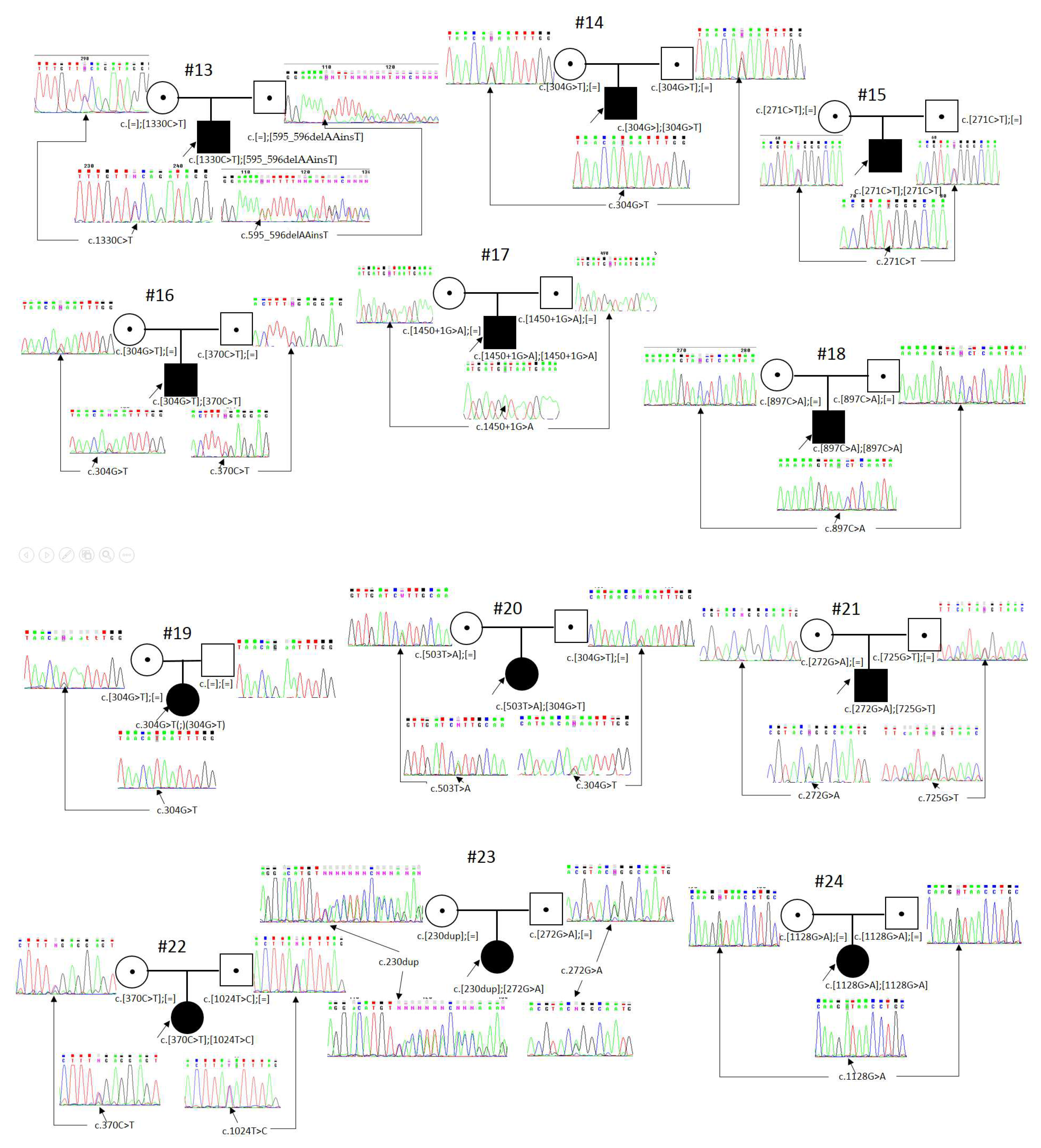
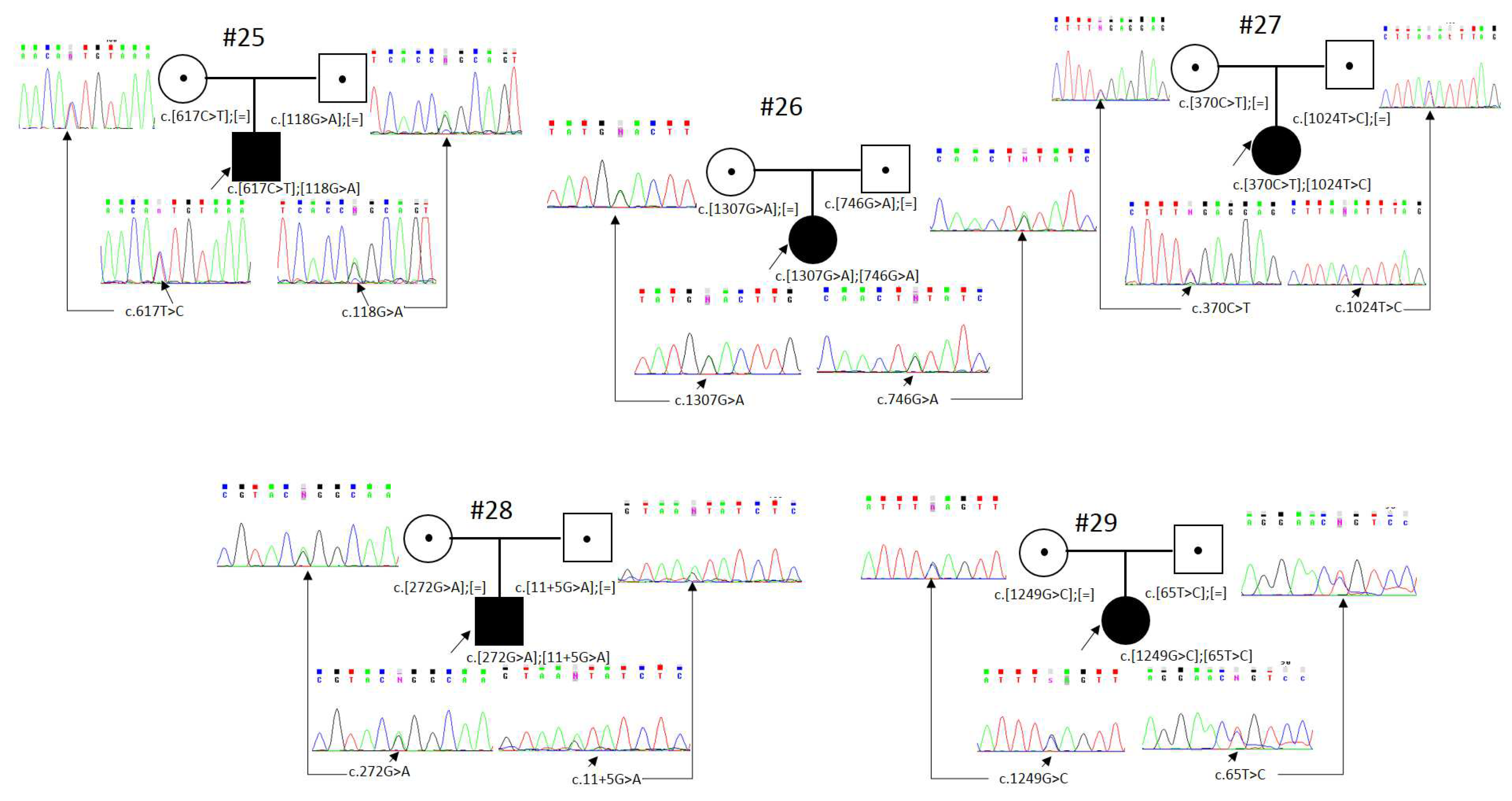
Thus, segregation analysis and confirmation of biallelic status were carried out for 29 patients with detected pathogenic or likely pathogenic variants in the RPE65 gene (Figure 1). The patients’ age at the moment of their molecular genetic diagnosis of “RPE65-associated retinopathy” varied from 4 months to 50 years (Table 1).
We detected 26 different variants in the RPE65 gene on 58 chromosomes; nine variants were novel—(c.1450+1G>A, c.1128+1G>A, c.897C>A/p.(Tyr299*), c.725G>T/p.(Ser242Ile) c.230dup/p.(Thr78Hisfs*10), c.1330C>T/p.(Pro444Ser), c.595_596delAAinsT p.(Asn199Phefs*9), c.1565T>A/p.(Ile522Asn), and c.503T>A/p.(Leu168His))—and two were previously described by us [15] (Figure 2, Table 2).
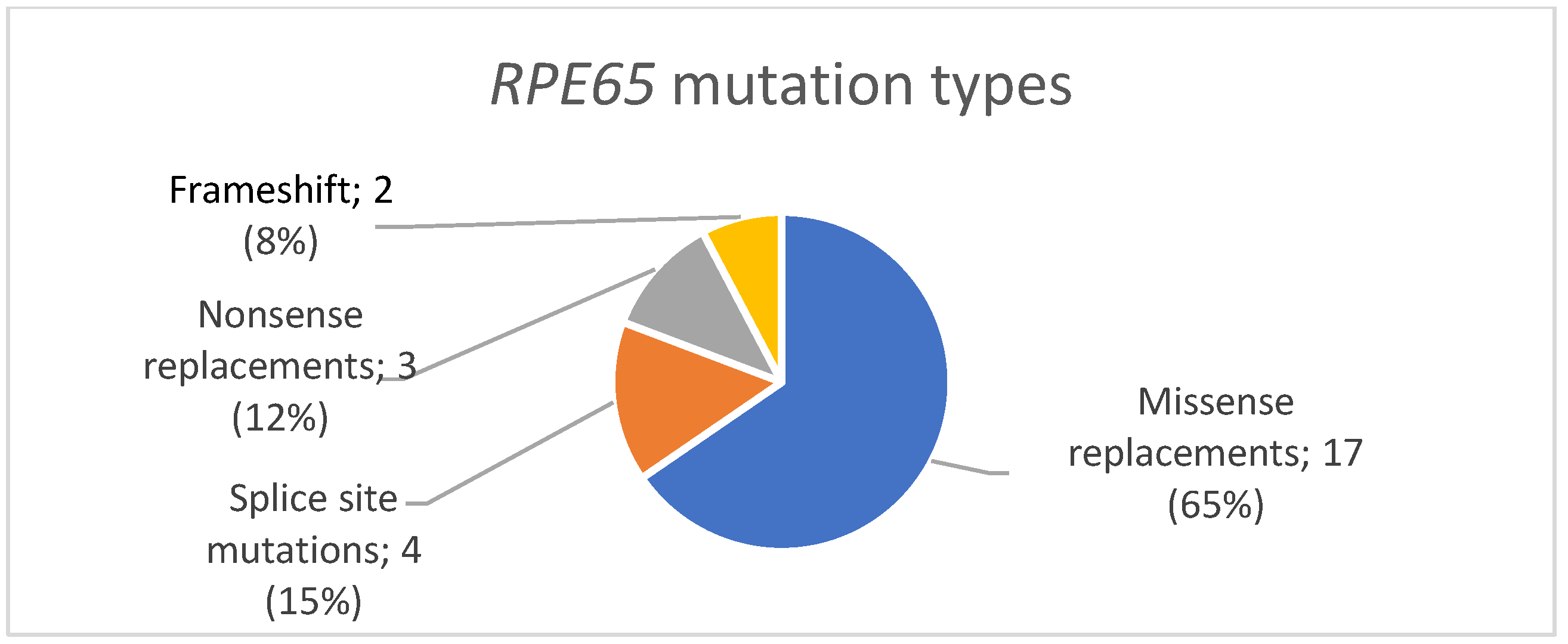
We detected 17 missense replacements in the RPE65 gene: (c.272G>A/p.(Arg91Gln), c.65T>C/p.(Leu22Pro), c.1024T>C/p.(Tyr342His), c.1451G>A/p.(Gly484Asp), c.271C>T/p.(Arg91Trp), c.725G>T/p.(Ser242Ile), c.1307G>A/p.(Gly436Glu), c.746A>G/p.(Tyr249Cys), c.1330C>T/p.(Pro444Ser), c.1565T>A/p.(Ile522Asn), c.1451G>T/p.(Gly484Val), c.1340T>C/p.(Leu447Pro), c.982C>T/p.(Leu328Phe), c.617T>C/p.(Ile206Thr), c.118G>A/p.(Gly40Ser), c.503T>A/p.(Leu168His), c.1249G>C/p.(Glu417Gln)). We also detected four splice site mutations (c.11+5G>A, c.1450+1G>A, c.1128+1G>A, c.1451-G>A), three nonsense replacements (c.304G>T/p.(Glu102*), c.370C>T/p.(Arg124*), c.897C>A/p.(Tyr299*)), and two variants leading to a frameshift and the formation of a premature stop codon (c.230dup/p.(Thr78Hisfs*10) and c.595_596delAAinsT/p.(Asn199Phefs*9)) (Figure 3).
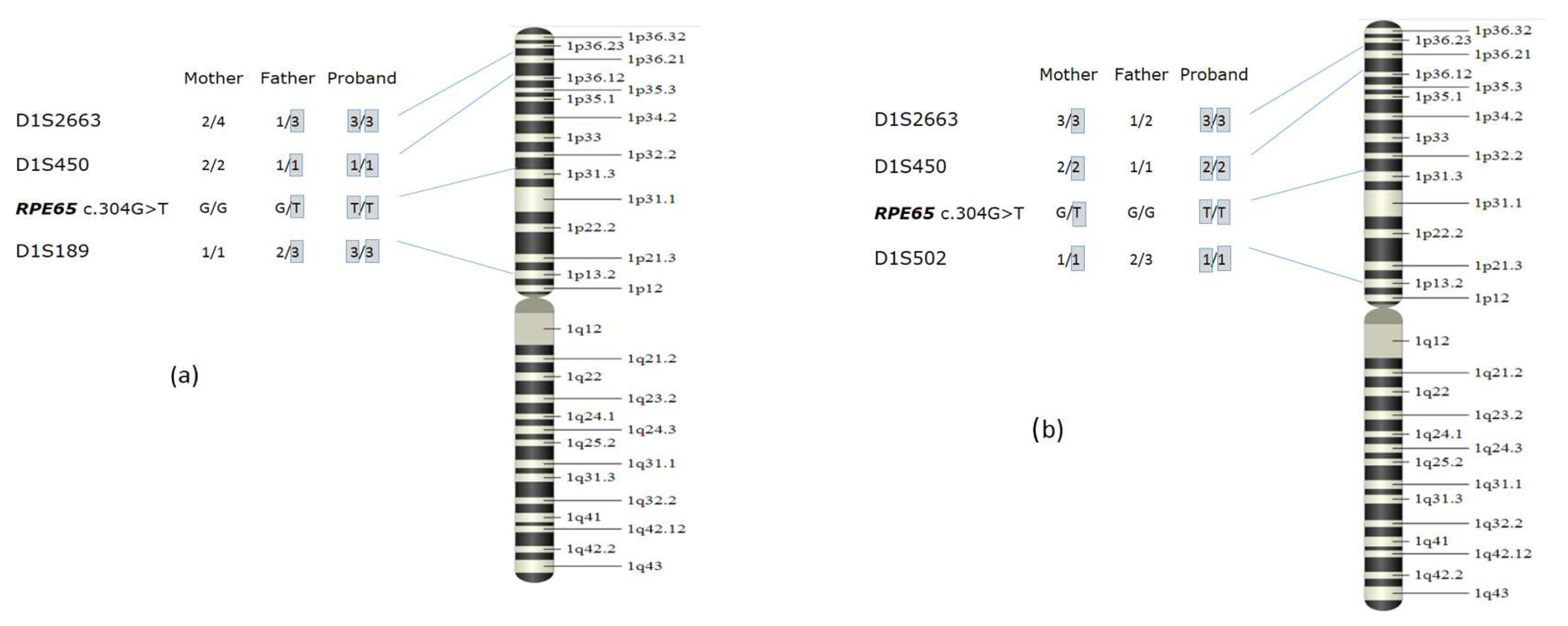
Two non-related patients (№8 and №19) had the c.304G>T/p.(Glu102*) variant in a homo-/hemizygous state, detected using MPS. When determining the zygosity, we discovered that one of each patient’s parents did not have the c.304G>T variant (in the first case, the variant was absent in the mother, and in the second case, in the father). The number of copies of RPE65 exons 1 to 14 was analyzed using quantitative MLPA for both probands and their parents. Two copies of the RPE65 gene were detected in the parents and probands themselves. In both cases, the relation was confirmed via an analysis of the microsatellite markers from 12 different chromosomes. To prove the hypothesis of uniparental isodisomy, we carried out an analysis using polymorphic markers from chromosome 1. As a result, in the first family, we established that at markers D1S450, D1S2663, and D1S189, the proband does not have maternal alleles, which signifies paternal uniparental disomy in the region containing the RPE65 gene. In the second family, at markers D1S2663, D1S450, and D1S502, the proband did not have a paternal allele (Figure 4). Thus, in two families, the probands had a c.304G>T/p.(Glu102*) variant in a homozygous state because of uniparental isodisomy. The patients did not have any phenotypic abnormalities other than LCA, confirming previous works showing the absence of imprinted genes on chromosome 1 that have a significant effect on the phenotype [27,28]. A case of RPE65-associated retinopathy due to paternal uniparental isodisomy on chromosome 1 was first described by Thompson, D.A. et al. in 2002 [27]. Later, another case of RPE65-associated retinopathy caused by maternal uniparental isodisomy was described in the literature [28]. Moreover, multiple cases of various retinitis pigmentosa types were reported to be a result of alterations in other genes and uniparental isodisomy on chromosome 1: in the ABCA4 gene [29], in the USH2A gene [30], and in the CRB1 gene [31]. The established inheritance mechanism plays an essential role in medical genetic counseling for all family members.

4. Discussion

In the current study, we established a molecular genetic diagnosis of “RPE65-associated retinopathy” in 5.3% of patients with a confirmed IRD diagnosis. The obtained data mostly correspond well with the literature, according to which the prevalence of RPE65-associated retinopathy among patients with different retinitis pigmentosa types (confirmed via molecular genetic means) in Europe and North America varied from 3% in the USA and Spain to 9.98% in the Netherlands [32]. However, the prevalence of RPE65-associated retinopathy in Denmark and India differs from the worldwide average, reaching 16% and 16.6%, respectively [12,33].
Missense replacements were the most common nucleotide sequence variants in the current study, comprising 65% (17), followed by variants affecting the splice site—15% (4), then nonsense replacements—12% (3), then deletions and insertions leading to a frameshift and the formation of a premature stop codon—8% (2) (Figure 2). Thus, the obtained results confirm the data presented in previous studies: missense replacements in the RPE65 gene are a common cause of RPE65-associated retinopathies [11,34].
According to the data presented in the literature, the most common pathogenic variants worldwide were c.271C>T/p.(Arg91Trp) (detected at 100 out of 864 pathogenic alleles (11.6%)), c.1102T>C/p.(Tyr368His) (69 out of 864 (8%)), and c.11+5G>A (62 out of 864 (7.2%)) [12]. Only two of these variants were detected in patients from the Russian Federation: c.271C>T and c.11+5G>A, being repeated, but not major. The prevalence of c.271C>T and c.11+5G>A in Russian patients with RPE65-associated retinopathy was 3.45% and 5.17%, respectively. The most common pathogenic variants encountered in the current study were c.304G>T/p.(Glu102*)—21.2%, c.370C>T/p.(Arg124*)—12%, and c.272G>A/p.(Arg91Gln)—8.6% of all affected chromosomes.
The most common variant in the examined cohort, c.304G>T/p.(Glu102*), was first described by Dharmaraj, S R et al. in 2000 in patients with LCA from the USA. In our cohort, this variant was detected in nine probands on thirteen chromosomes, with eleven of them being non-related (21.2%). It was initially described as major in the Netherlands [12]; however, it was never as common as in the current study. Patients carrying the c.304G>T variant were referred from various regions of the Russian Federation (Kursk, Irkutsk, Rostov, Yekaterinburg, Stavropol, Magadan); one of them was born in Turkmenistan and was ethnically Turkmen. All patients except for him were Russian.
A pathogenic variant, c.370C>T (p.Arg124*), was detected on seven chromosomes (12%). Two patients had the variant in a homozygous state, with three in a compound heterozygous state. This variant was initially described in 1998 by Morimura H et al. in patients from the USA and Canada [4]. It was present in various populations worldwide, never being major or common [35,36,37].
A nucleotide sequence variant, c.272G>A, leading to a p.(Arg91Gln) missense replacement and previously described as pathogenic, was detected on five chromosomes. It was initially described in 2000 by Thompson D.A. et al. in a compound heterozygous state [17]. A study by Philp AR et al. shows that the isomerase activity of RPE65 molecules with the p.(Arg91Gln) replacement comprised less than 6% of wild-type RPE65 activity [38]. The prevalence of this variant was 8.6%. It has been detected in Brazilian and Chinese patients [36,39]. This variant was also common in the study by Sallum JMF. et al.; however, its prevalence was significantly lower: 1.1%.
Variants c.11+5G>A and c.65T>C/p.(Leu22Pro) were detected on three chromosomes, each with the same prevalence of 5.17%.
The c.11+5G>A variant was detected in three patients in a compound heterozygous state. In the cohort of patients with RPE65-associated retinopathy from the USA and Europe, this variant was the most common, with a prevalence of 22.5%. The authors proved at least two independent origins of this variant [17]. The c.11+5G>A variant has been described multiple times as pathogenic in various worldwide populations; however, there were no more data on its prevalence among other alterations in the RPE65 gene [22,37,40]. A study by Vázquez-Domínguez I. et al. shows that the c.11+5G>A variant does not affect splicing as expected according to prediction programs, but leads to a significant decrease in the expression of RPE65, specific for the pigment epithelium, with an undetermined mechanism [41].
The c.65T>C/p.(Leu22Pro) variant was detected in two patients in a homozygous and compound heterozygous state. It was initially described in 1998 in a compound heterozygous state in a patient with a “mild” disease course [19]. The p.(Leu22Pro) replacement was located in the inactive center of the enzyme. A study by Jin M et al. shows that the isomerase activity of RPE65 with the p.(Leu22Pro) mutation increases significantly at 30 °C. The researchers believe that a combination of a peroral intake of 4-phenylbutyrate (PBA) and a low-temperature eye mask provides “protein regeneration therapy”, which may increase the effectiveness of gene therapy [42].
It is worth noting that the five pathogenic variants listed above comprise more than half of all mutations in the RPE65 gene in the examined cohort (31/58, 53.4%).
Six variants—c.1024T>C, c.1450+1G>A, c.1451G>A, c.271C>T, c.897C>A, and c.1128+1G>A—were detected on two chromosomes (3.45%) each. The c.1024T>C variant was detected in a compound heterozygous state, and others in a homozygous state.
The remaining 15 mutations, c.725G>T, c.230dup, c.1307G>A, c.746A>G, c.1330C>T, c.595_596delAAinsT, c.1565T>A, c.1451G>T, c.1451-G>A, c.1340T>C, c.982C>T, c.617T>C, c.118G>A, c.503T>A, and c.1249G>C, were detected once each.
The previously non-described variants—c.1128+1G>A and c.1450+1G>A—were located in the canonic splice site; c.230dup and c.595_596delAAinsT led to a frameshift with the formation of a premature stop codon, while the c.897C>A/p.(Tyr299*) variant led to the formation of a premature translation termination site in codon 299 (PVS1). These nucleotide sequence variants were not registered in gnomAD (The Genome Aggregation Database) control cohorts (PM2). The c.230dup variant was detected in a compound heterozygous state with a previously described pathogenic variant (PM3). Thus, according to the data interpretation guidelines [14], the detected nucleotide sequence variants should be regarded as likely pathogenic.
All previously non-described missense variants were also classified as likely pathogenic according to the data interpretation guidelines [14]: PM2—the variants were not registered in the gnomAD (The Genome Aggregation Database) control cohorts; PM3—the variants were detected in a compound heterozygous state with variants previously described as pathogenic; PP3—the pathogenicity prediction programs MutationTaster [43], FATHMM [44], LRT [45], and DEOGEN2 [46] evaluated these variants to be pathogenic; PP2—pathogenic missense variants in the RPE65 gene are a common cause of RPE65-associated retinopathy [11,29].
We did not note any “hot” exons in the RPE65 gene: pathogenic variants were detected in nine different exons—every exon except 1 and 11. Exons 4, 6, 12, and 14 had three variants each; exons 3 and 9 had two variants each; and the remaining exons had one pathogenic variant each (Figure 1). The location of the mutation in the gene has no influence on the effectiveness of target therapy.
In 2017, the Food and Drug Administration (FDA) approved a medication for gene therapy in patients with autosomal recessive retinitis pigmentosa caused by biallelic pathogenic variants in the RPE65 gene. The therapy is based on a recombinant adeno-associated viral vector (AAV2) carrying a functioning copy of normal human RPE65. Gene therapy using AAV2 injects a normal copy of the gene into the cell, not repairing or deleting the defective gene [47]. When placed in sustainable cells of pigment epithelium via a singular subretinal injection, the medication provides the potential for vision cycle reparation [48]. Thus, an accurate molecular diagnosis is essential for the approval of target therapy.
All patients with confirmed biallelic variants in the RPE65 gene, as well as the patient with uniparental isodisomy, are suitable for this target therapy.
The current study shows that pathogenic variants in the RPE65 gene contribute significantly to the pathogenesis of IRDs and comprise 5.3% of all patients with a confirmed molecular genetic diagnosis. The distinctive specificity of the spectrum was the high prevalence of a single variant—c.304G>T/p.(Glu102*)—of 21.2% among all probands. The most common pathogenic variants in patients with RPE65-associated retinopathy from the Russian Federation were c.304G>T/p.(Glu102*), c.370C>T/p.(Arg124*), and c.272G>A/p.(Arg91Gln), comprising 41.8% of all affected chromosomes. Two out of the three most common pathogenic variants worldwide—c.271C>T and c.11+5G>A—were detected in the Russian cohort on two and three non-related chromosomes, respectively, but were not the most common.
Seeing as the sooner the patient undergoes treatment, the more cells react to therapy, and that the presence of biallelic variants in the RPE65 gene is a necessary condition for gene therapy, molecular genetic diagnostics need to be carried out as soon as possible in cases of suspected RP. The current study allowed us to form a cohort for target therapy for the disorder; such therapy has already been carried out for some patients.

Author Contributions

Conceptualization, A.S. and A.P.; methodology, A.S., O.S. and A.P.; software, A.S.; validation, A.S., N.O. and O.S.; formal analysis, A.S.; investigation, A.S. and N.O.; resources, V.K. and A.P.; data curation, O.S. and A.P.; writing—original draft preparation, A.S.; writing—review and editing, A.S. and A.P.; visualization, A.S.; supervision, O.S., S.K. and A.P.; project administration, O.S., S.K. and A.P.; All authors have read and agreed to the published version of the manuscript.

Funding

This work was supported by a state assignment of the Ministry of Science and Higher Education of the Russian Federation for RCMG.

Institutional Review Board Statement

This study was conducted according to the guidelines of the Declaration of Helsinki, and approved by the Ethics Committee of the Research Centre for Medical genetics (protocol number 2, dated 14 April 2023).

Informed Consent Statement

Informed consent was obtained from all subjects involved in the study. Informed consent was obtained from the legal representatives of all patients under 18 years of age. Written informed consent was obtained from the patients to publish this paper.

Data Availability Statement

Not applicable.

Acknowledgments

The authors are grateful for the participation of all patients and their parents in this study. We thanks Alyona Chukhrova, Nina Ryadninskaya, Svetlana Braslavskaya, and Gulnara Bayazutdinova for their help in the sequencing analysis. We also thank Oxana Ryzhkova for her help in the sequencing analysis.

Conflicts of Interest

The authors declare no conflict of interest.

References

  1. Bertelsen, M.; Jensen, H.; Bregnhøj, J.F.; Rosenberg, T. Prevalence of generalized retinal dystrophy in Denmark. Ophthalmic Epidemiol. 2014, 21, 217–223. [Google Scholar] [CrossRef] [PubMed]
  2. Liew, G.; Michaelides, M.; Bunce, C. A comparison of the causes of blindness certifications in England and Wales in working age adults (16–64 years), 1999–2000 with 2009–2010. BMJ Open 2014, 4, e004015. [Google Scholar] [CrossRef] [PubMed]
  3. Puech, B.; Kostrubiec, B.; Hache, J.C.; François, P. Epidémiologie et prévalence des principales dystrophies rétiniennes héréditaires dans le Nord de la France [Epidemiology and prevalence of hereditary retinal dystrophies in the Northern France]. J. Fr. Ophtalmol. 1991, 14, 153–164. [Google Scholar]
  4. Morimura, H.; Fishman, G.A.; Grover, S.A.; Fulton, A.B.; Berson, E.L.; Dryja, T.P. Mutations in the RPE65 gene in patients with autosomal recessive retinitis pigmentosa or leber congenital amaurosis. Proc. Natl. Acad. Sci. USA 1998, 95, 3088–3093. [Google Scholar] [CrossRef] [PubMed]
  5. Moiseyev, G.; Chen, Y.; Takahashi, Y.; Wu, B.X.; Ma, J.X. RPE65 is the isomerohydrolase in the retinoid visual cycle. Proc. Natl. Acad. Sci. USA 2005, 102, 12413–12418. [Google Scholar] [CrossRef]
  6. Imanishi, Y.; Batten, M.L.; Piston, D.W.; Baehr, W.; Palczewski, K. Noninvasive two-photon imaging reveals retinyl ester storage structures in the eye. J. Cell Biol. 2004, 164, 373–383. [Google Scholar] [CrossRef] [PubMed]
  7. Jang, G.-F.; Van Hooser, J.P.; Kuksa, V.; McBee, J.K.; He, Y.-G.; Janssen, J.J.M.; Driessen, C.A.G.G.; Palczewski, K. Characterization of a dehydrogenase activity responsible for oxidation of 11-cis-retinol in the retinal pigment epithelium of mice with a disrupted RDH5 gene. A model for the human hereditary disease fundus albipunctatus. J. Biol. Chem. 2001, 276, 32456–32465. [Google Scholar] [CrossRef]
  8. Kiser, P.D. Retinal pigment epithelium 65 kDa protein (RPE65): An update. Prog. Retin. Eye Res. 2022, 88, 101013. [Google Scholar] [CrossRef]
  9. Kiser, P.D.; Farquhar, E.R.; Shi, W.; Sui, X.; Chance, M.R.; Palczewski, K. Structure of RPE65 isomerase in a lipidic matrix reveals roles for phospholipids and iron in catalysis. Proc. Natl. Acad. Sci. USA 2012, 109, E2747–E2756. [Google Scholar] [CrossRef]
  10. Testa, F.; Murro, V.; Signorini, S.; Colombo, L.; Iarossi, G.; Parmeggiani, F.; Falsini, B.; Salvetti, A.P.; Brunetti-Pierri, R.; Aprile, G.; et al. RPE65-Associated Retinopathies in the Italian Population: A Longitudinal Natural History Study. Investig. Opthalmol. Vis. Sci. 2022, 63, 13. [Google Scholar] [CrossRef]
  11. Lopez-Rodriguez, R.; Lantero, E.; Blanco-Kelly, F.; Avila-Fernandez, A.; Merida, I.M.; del Pozo-Valero, M.; Perea-Romero, I.; Zurita, O.; Jiménez-Rolando, B.; Swafiri, S.T.; et al. RPE65-related retinal dystrophy: Mutational and phenotypic spectrum in 45 affected patients. Exp. Eye Res. 2021, 212, 108761. [Google Scholar] [CrossRef] [PubMed]
  12. Astuti, G.D.N.; Bertelsen, M.; Preising, M.N.; Ajmal, M.; Lorenz, B.; Faradz, S.M.H.; Qamar, R.; Collin, R.W.J.; Rosenberg, T.; Cremers, F.P.M. Comprehensive genotyping reveals RPE65 as the most frequently mutated gene in Leber congenital amaurosis in Denmark. Eur. J. Hum. Genet. 2016, 24, 1071–1079. [Google Scholar] [CrossRef]
  13. Available online: https://ngs-data-ccu.epigenetic.ru/ (accessed on 12 September 2023).
  14. Ryzhkova, O.P.; Kardymon, O.L.; Prohorchuk, E.B.; Konovalov, F.A.; Maslennikov, A.B.; Stepanov, V.A.; Afanasyev, A.A.; Zaklyazminskaya, E.V.; Kostareva, A.A.; Pavlov, A.E.; et al. Guidelines for the interpretation of massive parallel sequencing variants (update 2018, v2). Med. Genet. 2019, 18, 3–23. (In Russian) [Google Scholar] [CrossRef]
  15. Stepanova, A.A.; Kadyshev, V.V.; Shchagina, O.A.; Polyakov, A.V. Selective screening of patients with hereditary retinal degeneration to identify the target group for gene therapy with voretigene neparvovec. Med. Genet. 2022, 21, 51–55. (In Russian) [Google Scholar] [CrossRef]
  16. Dharmaraj, S.; Silva, E.; Pina, A.L.; Li, Y.Y.; Yang, J.M.; Carter, R.C.; Loyer, M.; El-Hilali, H.; Traboulsi, E.; Sundin, O.; et al. Mutational analysis and clinical correlation in Leber congenital amaurosis. Ophthalmic Genet. 2000, 21, 135–150. [Google Scholar] [CrossRef] [PubMed]
  17. Thompson, D.A.; Gyürüs, P.; Fleischer, L.L.; Bingham, E.L.; McHenry, C.L.; Apfelstedt–Sylla, E.; Zrenner, E.; Lorenz, B.; Richards, J.E.; Jacobson, S.G.; et al. Genetics and phenotypes of RPE65 mutations in inherited retinal degeneration. Investig. Ophthalmol. Vis. Sci. 2000, 41, 4293–4299. [Google Scholar]
  18. Gu, S.-M.; Thompson, D.A.; Srikumari, C.S.; Lorenz, B.; Finckh, U.; Nicoletti, A.; Murthy, K.; Rathmann, M.; Kumaramanickavel, G.; Denton, M.J.; et al. Mutations in RPE65 cause autosomal recessive childhood-onset severe retinal dystrophy. Nat. Genet. 1997, 17, 194–197. [Google Scholar] [CrossRef]
  19. Marlhens, F.; Griffoin, J.M.; Bareil, C.; Arnaud, B.; Claustres, M.; Hamel, C.P. Autosomal recessive retinal dystrophy associated with two novel mutations in the RPE65 gene. Eur. J. Hum. Genet. 1998, 6, 527–531. [Google Scholar] [CrossRef]
  20. Weleber, R.G.; Michaelides, M.; Trzupek, K.M.; Stover, N.B.; Stone, E.M. The phenotype of Severe Early Childhood Onset Retinal Dystrophy (SECORD) from mutation of RPE65 and differentiation from Leber congenital amaurosis. Investig. Opthalmol. Vis. Sci. 2011, 52, 292–302. [Google Scholar] [CrossRef]
  21. Henderson, R.H.; Waseem, N.; Searle, R.; van der Spuy, J.; Russell-Eggitt, I.; Bhattacharya, S.S.; Thompson, D.A.; Holder, G.E.; Cheetham, M.E.; Webster, A.R.; et al. An assessment of the apex microarray technology in genotyping patients with Leber congenital amaurosis and early-onset severe retinal dystrophy. Investig. Opthalmol. Vis. Sci. 2007, 48, 5684–5689. [Google Scholar] [CrossRef]
  22. Weisschuh, N.; Obermaier, C.D.; Battke, F.; Bernd, A.; Kuehlewein, L.; Nasser, F.; Zobor, D.; Zernner, E.; Weber, E.; Wissinger, B.; et al. Genetic architecture of inherited retinal degeneration in Germany: A large cohort study from a single diagnostic center over a 9-year period. Hum. Mutat. 2020, 41, 1514–1527. [Google Scholar] [CrossRef]
  23. Li, S.; Xiao, X.; Yi, Z.; Sun, W.; Wang, P.; Zhang, Q. RPE65 mutation frequency and phenotypic variation according to exome sequencing in a tertiary centre for genetic eye diseases in China. Acta Ophthalmol. 2020, 98, e181–e190. [Google Scholar] [CrossRef] [PubMed]
  24. Yang, G.; Liu, Z.; Xie, S.; Li, C.; Lv, L.; Zhang, M.; Zhao, J. Genetic and phenotypic characteristics of four Chinese families with fundus albipunctatus. Sci. Rep. 2017, 7, 46285. [Google Scholar] [CrossRef] [PubMed]
  25. Zampaglione, E.; Kinde, B.; Place, E.M.; Navarro-Gomez, D.; Maher, M.; Jamshidi, F.; Nassiri, S.; Mazzone, J.A.; Finn, C.; Schlegel, D.; et al. Copy-number variation contributes 9% of pathogenicity in the inherited retinal degenerations. Genet. Med. 2020, 22, 1079–1087. [Google Scholar] [CrossRef] [PubMed]
  26. Simovich, M.J.; Miller, B.; Ezzeldin, H.; Kirkland, B.T.; McLeod, G.; Fulmer, C.; Nathans, J.; Jacobson, S.G.; Pittler, S.J. Four novel mutations in the RPE65 gene in patients with Leber congenital amaurosis. Hum. Mutat. 2001, 18, 164. [Google Scholar] [CrossRef] [PubMed]
  27. Thompson, D.A.; McHenry, C.L.; Li, Y.; Richards, J.E.; Othman, M.I.; Schwinger, E.; Vollrath, D.; Jacobson, S.G.; Gal, A. Retinal dystrophy due to paternal isodisomy for chromosome 1 or chromosome 2, with homoallelism for mutations in RPE65 or MERTK, respectively. Am. J. Hum. Genet. 2002, 70, 224–229. [Google Scholar] [CrossRef]
  28. Motta, F.L.; Filippelli-Silva, R.; Kitajima, J.P.; Batista, D.A.; Wohler, E.S.; Sobreira, N.L.; Martin, R.P.; Sallum, J.M.F. Analysis of an NGS retinopathy panel detects chromosome 1 uniparental isodisomy in a patient with RPE65-related leber congenital amaurosis. Ophthalmic Genet. 2021, 42, 553–560. [Google Scholar] [CrossRef]
  29. Fingert, J.H.; Eliason, D.A.; Phillips, N.C.; Lotery, A.J.; Sheffield, V.C.; Stone, E.M. Case of stargardt disease caused by uniparental isodisomy. Arch. Ophthalmol. 2006, 124, 744. [Google Scholar] [CrossRef]
  30. Rivolta, C.; Berson, E.L.; Dryja, T.P. Paternal uniparental heterodisomy with partial isodisomy of chromosome 1 in a patient with retinitis pigmentosa without hearing loss and a missense mutation in the Usher syndrome type II gene USH2A. Arch. Ophthalmol. 2002, 120, 1566–1571. [Google Scholar] [CrossRef]
  31. Stone, E.M. Leber congenital amaurosis–a model for efficient genetic testing of heterogeneous disorders: LXIV Edward Jackson memorial lecture. Am. J. Ophthalmol. 2007, 144, 791–811.e6. [Google Scholar] [CrossRef]
  32. Sallum, J.M.F.; Kaur, V.P.; Shaikh, J.; Banhazi, J.; Spera, C.; Aouadj, C.; Viriato, D.; Fischer, M.D. Epidemiology of Mutations in the 65-kDa Retinal Pigment Epithelium (RPE65) Gene-Mediated Inherited Retinal Dystrophies: A Systematic Literature Review. Adv. Ther. 2022, 39, 1179–1198. [Google Scholar] [CrossRef] [PubMed]
  33. Verma, A.; Perumalsamy, V.; Shetty, S.; Kulm, M.; Sundaresan, P. Mutational screening of LCA genes emphasizing RPE65 in South Indian cohort of patients. PLoS ONE 2013, 8, e73172. [Google Scholar] [CrossRef] [PubMed]
  34. Motta, F.L.; Martin, R.P.; Porto, F.B.O.; Wohler, E.S.; Resende, R.G.; Gomes, C.P.; Pesquero, J.B.; Sallum, J.M.F. Pathogenicity Reclasssification of RPE65 Missense Variants Related to Leber Congenital Amaurosis and Early-Onset Retinal Dystrophy. Genes 2019, 11, 24. [Google Scholar] [CrossRef] [PubMed]
  35. Colombo, L.; Maltese, P.E.; Castori, M.; El Shamieh, S.; Zeitz, C.; Audo, I.; Zulian, A.; Marinelli, C.; Benedetti, S.; Costantini, A.; et al. Molecular Epidemiology in 591 Italian Probands With Nonsyndromic Retinitis Pigmentosa and Usher Syndrome. Investig. Opthalmology Vis. Sci. 2021, 62, 13. [Google Scholar] [CrossRef]
  36. Zhong, Z.; Rong, F.; Dai, Y.; Yibulayin, A.; Zeng, L.; Liao, J.; Wang, L.; Huang, Z.; Zhou, Z.; Chen, J. Seven novel variants expand the spectrum of RPE65-related Leber congenital amaurosis in the Chinese population. Mol. Vis. 2019, 25, 204–214. [Google Scholar] [PubMed]
  37. Chung, D.C.; Bertelsen, M.; Lorenz, B.; Pennesi, M.E.; Leroy, B.P.; Hamel, C.P.; Pierce, E.; Sallum, J.; Larsen, M.; Stieger, K.; et al. The Natural History of Inherited Retinal Dystrophy Due to Biallelic Mutations in the RPE65 Gene. Am. J. Ophthalmol. 2019, 199, 58–70. [Google Scholar] [CrossRef] [PubMed]
  38. Philp, A.; Jin, M.; Li, S.; Schindler, E.; Iannaccone, A.; Lam, B.; Weleber, R.; Fishman, G.; Jacobson, S.; Mullins, R.; et al. Predicting the pathogenicity of RPE65 mutations. Hum. Mutat. 2009, 30, 1183–1188. [Google Scholar] [CrossRef]
  39. Sallum, J.M.F.; Motta, F.L.; Arno, G.; Porto, F.B.O.; Resende, R.G.; Belfort, R., Jr. Clinical and molecular findings in a cohort of 152 Brazilian severe early onset inherited retinal dystrophy patients. Am. J. Med. Genet. C Semin. Med. Genet. 2020, 184, 728–752. [Google Scholar] [CrossRef]
  40. Zenteno, J.C.; García-Montaño, L.A.; Cruz-Aguilar, M.; Ronquillo, J.; Rodas-Serrano, A.; Aguilar-Castul, L.; Matsui, R.; Vencedor-Meraz, C.I.; Arce-González, R.; Graue-Wiechers, F.; et al. Extensive genic and allelic heterogeneity underlying inherited retinal dystrophies in Mexican patients molecularly analyzed by next-generation sequencing. Mol. Genet. Genom. Med. 2020, 8. [Google Scholar] [CrossRef]
  41. Vázquez-Domínguez, I.; Duijkers, L.; Fadaie, Z.; Alaerds, E.C.W.; Post, M.A.; van Oosten, E.M.; O’gorman, L.; Kwint, M.; Koolen, L.; Hoogendoorn, A.D.M.; et al. The Predicted Splicing Variant c.11+5G>A in RPE65 Leads to a Reduction in mRNA Expression in a Cell-Specific Manner. Cells 2022, 11, 3640. [Google Scholar] [CrossRef]
  42. Jin, M.; Li, S.; Hu, J.; Jin, H.H.; Jacobson, S.G.; Bok, D. Functional Rescue of Retinal Degeneration-Associated Mutant RPE65 Proteins. Adv. Exp. Med. Biol. 2016, 854, 525–532. [Google Scholar] [CrossRef] [PubMed]
  43. Schwarz, J.M.; Rodelsperger, C.; Schuelke, M.; Seelow, D. Mutation Taster evaluates disease-causing potential of sequence alterations. Nat. Methods 2010, 7, 575–576. [Google Scholar] [CrossRef] [PubMed]
  44. Shihab, H.A.; Gough, J.; Cooper, D.N.; Stenson, P.D.; Barker, G.L.; Edwards, K.J.; Day, I.N.; Gaunt, T.R. Predicting the functional, molecular, and phenotypic consequences of amino acid substitutions using hidden Markov models. Hum. Mutat. 2013, 34, 57–65. [Google Scholar] [CrossRef] [PubMed]
  45. Chun, S.; Fay, J.C. Identification of deleterious mutations within three human genomes. Genome Res. 2009, 19, 1553–1561. [Google Scholar] [CrossRef] [PubMed]
  46. Raimondi, D.; Tanyalcin, I.; Ferté, J.; Gazzo, A.; Orlando, G.; Lenaerts, T.; Rooman, M.; Vranken, W. DEOGEN2: Prediction and interactive visualization of single amino acid variant deleteriousness in human proteins. Nucleic Acids Res. 2017, 45, W201–W206. [Google Scholar] [CrossRef]
  47. Smith, A.J.; Bainbridge, J.W.; Ali, R.R. Gene supplementation therapy for recessive forms of inherited retinal dystrophies. Gene Ther. 2012, 19, 154–161. [Google Scholar] [CrossRef]
  48. Kang, C.; Scott, L.J. Voretigene Neparvovec: A Review in RPE65 Mutation-Associated Inherited Retinal Dystrophy. Mol. Diagn. Ther. 2020, 24, 487–495. [Google Scholar] [CrossRef]
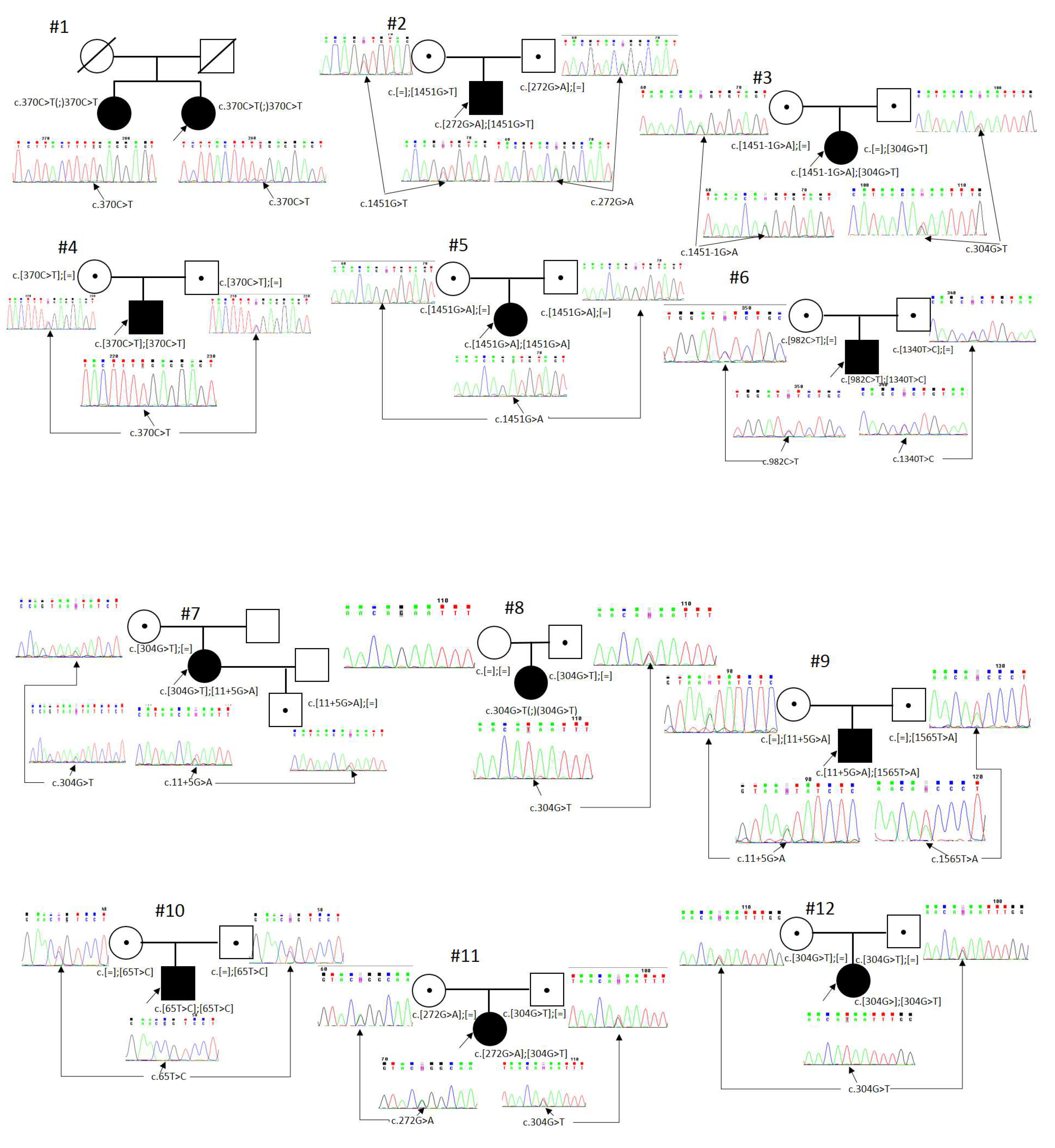
Figure 1. Segregation analysis. Black—affected, white—unaffected, dot—carrier, square—male, circle—female, arrow—proband, slash—deceased. #1, #2, …, #29—Patient ID.
Genes 14 02056 g001aGenes 14 02056 g001bGenes 14 02056 g001c
Figure 2. The structure of the RPE65 gene with the detected nucleotide sequence variants. Previously non-described variants are placed above the gene structure.
Genes 14 02056 g002
Figure 3. The distribution of RPE65 mutation types.
Genes 14 02056 g003
Figure 4. Microsatellite marker analysis on chromosome 1 in families with uniparental isodisomy. (a) Paternal uniparental isodisomy, patient #8; (b) maternal uniparental isodisomy, patient #19.
Genes 14 02056 g004
Table 1. Patients and genotypes.
Patient IDAge at the Moment of Molecular Genetic DiagnosisSexPlace of ResidenceEthnic GroupGenotypeNystagmusNyctalopiaSnellen BCVACentral Foveal Retinal Thickness, µmColor Vision AnomalyffERG
ODOSODOSOUOU
150 yearsfChita regionBuryatc.[370C>T]; [370C>T]YesFull0.0010.0018090MChND
25 yearsmMoscow regionBelarusianc.[1451G>T]; [272G>A]YesFull0.160.2202185DeuExt
35 yearsfKursk regionRussianc.[1451-1G>A]; [304G>T]Noinc0.20.2134129NoND
426 yearsmBuryatiaBuryatc.[370C>T]; [370C>T]YesFull0.010.0190102MChND
513 yearsfDagestanLezginc.[1451G>A]; [1451G>A]YesFull0.010.01137124DeuExt
612 years Yamalo-Nenets Autonomous OkrugRussianc.[982C>T]; [1340T>C]Noinc0.30.16230247NoSubN
731 yearfIrkutsk regionRussianc.[304G>T]; [11+5G>A]YesFull0.050.05134133MChND
86 yearsmRostov regionRussianc.304G>T(;) (304G>T)Noinc0.40.4153158TriExt
93 yearsmLeningrad regionRussianc.[11+5G>A]; [1565T>A]YesFull0.20.2176176TriSubN
109 yearsmDagestanKumykc.[65T>C]; [65T>C]Yesinc0.60.7170185AnTSubN
116 yearsfSverdlovsk regionRussianc.[272G>A]; [304G>T]Yesinc0.050.05182221DichSubN
1236 yearsfMoscow regionTurkmenc.[304G>]; [304G>T]YesFull0.0010.00110090MChExt
1328 yearsmMoscow regionRussianc.[1330C>T]; [595_596delAAinsT]YesFull0.150.05204195MChND
1424 yearsmStavropol regionRussianc.[304G>]; [304G>T]YesFull0.010.01199216DichND
1518 yearsmBashkortostanBashkirc.[271C>T]; [271C>T]YesFull0.30.2165158AnTND
1612 yearsmMagadan RegionRussianc.[304G>T]; [370C>T]Yesinc0.20.2157152AnTSubN
1712 yearsmSverdlovsk regionTajikc.[1450+1G>A]; [1450+1G>A]Yesinc0.10.05213271MChSubN
1811 yearsmStavropol regionDarginc.[897C>A]; [897C>A]YesFull0.050.2234215TriSubN
194 yearsmBryansk regionRussianc.304G>T(;) (304G>T)YesFull0.010.01144132AnTExt
204 monthsfSmolensk regionRussianc.[503T>A]; [304G>T]Noincsvsv----
211 yearmVologda regionInterethnic—Uzbek-Russianc.[272G>A]; [725G>T]Yesincsvsv----
225 yearsfTuva regionTuvanc.[370C>T]; [1024T>C]Yesinc0.350.4203200DeuExt
2318 yearsfMoscow regionRussianc.[230dup]; [272G>A]YesFull0.10.16271245AnTND
2411 yearsfLeningrad regionTajikc.[1128G>A]; [1128G>A]YesFull0.20.1213210DeuExt
2528 yearsmOmsk regionRussianc.[617C>T]; [118G>A]YesFull0.10.1140120MChND
264 yearsfMoscow regionRussianc.[1307G>A]; [746G>A]YesFull0.20.2170168AnTExt
2713 yearsfTuva regionTuvanc.[370C>T]; [1024T>C]NoFull0.30.3172180MChSubN
2843 yearsmIvanovo regionRussianc.[272G>A]; [11+5G>A]YesFull0.0010.001186134MChND
297 yearsfBuryatiaKumykc.[1249G>C]; [65T>C]YesFull0.40.4189179AnTND
Abbreviations: f—female, m—male, inc—incomplete, sv—Subject vision, MCh—monochromasia, Deu—deuteranopia, Tri—tritanopia, AnT—anomalous trichromat, Dich—dichromat, ND—non-detectable, Ext—Extinguished, SubN—subnormal.
Table 2. Detected variants in the RPE65 gene.
VariantEffectExon/Intron№ of chr.Prevalence, %Allele Frequency in gnomADReferences/Pathogenicity Criteria
1c.304G>Tp.(Glu102*)ex 413 (11)21.20.00003580[16]
2c.370C>Tp.(Arg124*)ex 5712.00.00005674[4]
3c.272G>Ap.(Arg91Gln)ex 458.60.00004600[17]
4c.11+5G>Asplicingin 135.170.000078[18]
5c.65T>Cp.(Leu22Pro)ex 235.170.000028[19]
6c.1024T>Cp.(Tyr342His)ex 1023.45n/a[15]
7c.1450+1G>Asplicingin 1323.45n/aPVS1, PM2
8c.1128+1G>Asplicingin 1023.45n/aPVS1, PM2
9c.1451G>Ap.(Gly484Asp)ex 1423.450.000008047[20]
10c.271C>Tp.(Arg91Trp)ex 423.450.000053[4]
11c.897C>Ap.(Tyr299*)ex 923.45n/dPVS1, PM2
12c.725G>Tp.(Ser242Ile)ex 711.72n/dPM2, PP3, PP2, PM3
13c.230dupp.(Thr78Hisfs*10)ex 311.72n/dPVS1, PM2,PM3
14c.1307G>Ap.(Gly436Glu)ex 1211.72n/d[17]
15c.746A>Gp.(Tyr249Cys)ex 811.720.00001773[21]
16c.1330C>Tp.(Pro444Ser)ex 1211.72n/dPM2, PP3, PP2, PM3
17c.595_596delAAinsTp.(Asn199Phefs*9)ex 611.72n/dPVS1, PM2
18c.1565T>Ap.(Ile522Asn)ex 1411.72n/dPM2, PP3, PP2, PM3
19c.1451G>Tp.(Gly484Val)ex 1411.720.00001207[22]
20c.1451-G>Asplicingin 1411.720.000004024[23]
21c.1340T>Cp.(Leu447Pro)ex 1311.72n/d[15]
22c.982C>Tp.(Leu328Phe)ex 911.720.039[24]
23c.617T>Cp.(Ile206Thr)ex 611.720.000012[25]
24c.118G>Ap.(Gly40Ser)ex 311.720.000028[4]
25c.503T>Ap.(Leu168His)ex 611.72n/dPM2, PP3, PP2, PM3
26c.1249G>Cp.(Glu417Gln)ex 1211.720.000004[26]
Disclaimer/Publisher’s Note: The statements, opinions and data contained in all publications are solely those of the individual author(s) and contributor(s) and not of MDPI and/or the editor(s). MDPI and/or the editor(s) disclaim responsibility for any injury to people or property resulting from any ideas, methods, instructions or products referred to in the content.