Article

Alternative Splicing of NAC Transcription Factor Gene CmNST1 Is Associated with Naked Seed Mutation in Pumpkin, Cucurbita moschata

1
College of Horticulture, Shanxi Agricultural University, Taiyuan 030031, China
2
USDA-ARS Vegetable Crops Research Unit, Horticulture Department, University of Wisconsin, Madison, WI 53706, USA
*
Authors to whom correspondence should be addressed.
Genes 2023, 14(5), 962; https://doi.org/10.3390/genes14050962
Submission received: 15 March 2023 / Revised: 16 April 2023 / Accepted: 21 April 2023 / Published: 23 April 2023

Abstract

:
In pumpkin (Cucurbita moschata), the naked or hull-less seed phenotype has great benefits for breeding this crop for oil or snack use. We previously identified a naked seed mutant in this crop. In this study, we report genetic mapping, identification, and characterization of a candidate gene for this mutation. We showed that the naked seed phenotype is controlled by a single recessive gene (N). The bulked segregant analysis identified a 2.4 Mb region on Chromosome 17 with 15 predicted genes. Multiple lines of evidence suggested that CmoCh17G004790 is the most probable candidate gene for the N locus which encodes a NAC transcription factor WALL THICKENING PROMOTING FACTOR 1 (CmNST1). No nucleotide polymorphism or structural variation was found in the genomic DNA sequences of CmNST1 between the mutant and the wildtype inbred line (hulled seed). However, the cDNA sequence cloned from developing seed coat samples of the naked seed mutant was 112 bp shorter than that from the wildtype which is due to seed coat-specific alternative splicing in the second exon of the mutant CmNST1 transcript. The expression level of CmNST1 in the developing seed coat was higher in the mutant than in the wildtype during early seed coat development which was reversed later. Transcriptomic profiling with RNA-Seq at different stages of seed development in the mutant and wildtype revealed a critical role of CmNST1 as a master regulator for the lignin biosynthesis pathway during seed coat development while other NAC and MYB transcription factors were also involved in forming a regulatory network for the building of secondary cell walls. This work provides a novel mechanism for the well-characterized NST1 transcription factor gene in regulating secondary cell wall development. The cloned gene also provides a useful tool for marker-assisted breeding of hull-less C. moschata varieties.

1. Introduction

The pumpkin and squash in the genus Cucurbita (mainly C. pepo, C. maxima, and C. moschata, 2n = 2x = 24) are economically important crops worldwide. In addition to their primary uses as vegetables (immature fruit) or staple foods (mature fruit), and ornamentals, their seeds are also consumed as both seed snacks and culinary oil [1,2]. Pumpkin/squash seeds are rich in oil, protein, unsaturated fatty acids, and antioxidants that have many health benefits (e.g., [3,4,5,6,7,8]). One drawback in the use of cucurbit seeds is their thick and leathery seed coat that is hard to decorticate. One way to address this is to use naked or hull-less seed mutants which lack a complete seed coat, and thus are preferred for snacking and oil production because they eliminate the need for manual de-hulling prior to use. Naked pumpkin seeds are a popular ingredient in many snacks, breads, breakfast cereals, soups, and other edible goods [9,10]. Thus, the development of oil-or snack-use pumpkin/squash varieties is also an important objective for breeding programs [3,11,12].
The first spontaneous hull-less mutant called Styrian (hull-less) was reported in C. pepo subsp. pepo var. Styriaca almost 80 years ago in Austria [3]. Since then, several more hull-less mutants have been described in all three major Cucurbita crops, which were all shown to be controlled by a single recessive gene n (for naked seed) or h (for hull-less seed) [2,12,13,14] although some modifiers having a minor influence on testa development are also possible [3,12]. There were a few early studies on molecular mapping of the n or h locus in C. pepo (e.g., [15,16,17]). More recently, three studies on fine genetic mapping of the hull-less mutation in C. pepo [18,19,20] suggested that the hull-less seed phenotype in the pumpkin line HLP36 is due to a single recessive gene (cphl-1). Linkage mapping identified a candidate gene region that was 2.1 Mbp on chromosome 12 including secondary cell wall and lignin biosynthesis-related transcriptional factors viz., “NAC” (Cp4.1LG12g04350) and “MYB” (Cp4.1LG12g03120) that were suggested as possible candidates for the cphl-1 locus. Meru et al. [20] conducted linkage mapping in the segregating populations derived from a cross between two C. pepo inbred lines, Kakai (hull-less) and Table Gold Acorn (hulled), and identified two SNPs that were significantly associated with the hull-less trait in cultivars and accessions of diverse genetic backgrounds. From this research, several candidate genes were proposed including a NAC domain-containing protein gene and a Fiber Protein fb11 gene involved in lignin accumulation and cell wall deposition across plant species, respectively. By BSA (bulked segregant analysis) and fine mapping, Lv et al. [19] found that mutation of a single gene, NAC SECONDARY WALL THICKENING PROMOTING FACTOR 1 (NST1), accounts for the hull-less trait in the C. pepo line P-HL. They further proposed that a 14-bp sequence insertion in the CpNST1 gene causes premature termination of CpNST1 translation, leading to a lack of secondary cell wall (SCW) biosynthesis in hull-less seed coats. From these studies, it seems the hull-less mutations in three C. pepo lines share the same candidate gene (CpNST1, a NAC transcription factor gene).
In the regular, hulled seed plants (wildtype or WT hereinafter), during the initial seed coat development, the seed testa exhibits five distinct layers including epidermis (E), hypodermis (H), sclerenchyma (S), parenchyma (P), and innermost chlorenchyma (C) [21,22]. In hull-less seeds, four seed tissue layers (E, H, S, and P) collapse forming a papery thin hyaline hull that may reveal the green color of the underlying C layer [12,21,23,24]. The changes in the seed testa of the mutant are associated with reduced biosynthesis of cellulose and lignin in the secondary cell walls which starts approximately 10–15 days after pollination (DAP) (e.g., [21,22,25]). Lignin deposition diminution also coincides with reduced expression of the genes or enzyme activities in the lignin biosynthesis pathways or secondary cell wall development. For example, the expressions of lignin/cellulose synthesis genes for cellulose synthase (CES), phenylalanine ammonia lyase (PAL), cinnamoyl CoA reductase, 4-coumaric acid CoA ligase (4CL), cinnamyl alcohol dehydrogenase (CAD), cinnamic acid-4-hydroxylase (C4H), glutathione reductase, and abscisic acid-responsive protein E exhibited significantly lower expression in the C. pepo hull-less mutant than in the WT [19,25,26].
While we now have a better understanding of the genetic and molecular basis of the hull-less mutation in C. pepo, the work of such mutants in the other two Cucurbita species (C. moschata and C. maxima) is limited. We previously characterized a hull-less seed (thin-coated or naked) C. moschata mutant that was first identified in China [13,27]. From the mutant, a hull-less variety (65-1-8) and two hull-less inbred lines were developed with large naked seeds that are rich in fat and proteins with good commercial potential [13,28]. We also investigated the anatomical and biochemical bases of this hull-less mutant and found that the degeneration or absence of the typical seed testa structure in hull-less seeds of C. moschata is due to the reduced activity of key enzymes in the lignin biosynthesis pathway [25]. However, the genetic basis and underlying candidate gene for this mutation are unknown which hinders its efficient use in breeding pumpkin varieties through marker-assisted selection. Thus, the objectives of the present study were to (1) investigate the inheritance of hull-less seed phenotype in the mutant line (HLS-B); (2) conduct genetic mapping and map-based cloning to identify the candidate gene for the mutant allele (n locus); (3) understand the regulatory network for seed coat development in C. moschata. Here we report mapping and identification of a candidate gene of the n locus through BSA-Seq. We performed RNA-Seq with seed coat samples collected from different development stages to reveal important genes and pathways for seed coat development. We showed the NAC transcription factor CmNST1 was a candidate gene for the n locus, and alternative splicing in the CmNST1 gene in the seed coat was responsible for the hull-less phenotype.

2. Materials and Methods

2.1. Plant Materials and Phenotyping of Seed Coat

The hulled seed (HS-A, wildtype or WT hereinafter) and hull-less or naked seed mutant HLS-B (mutant hereinafter) inbred lines of C. moschata (Figure 1A,B) were obtained from the College of Horticulture, Shanxi Agricultural University (SAU), Taiyuan, China. A recombinant inbred line (RIL) population with 189 F8 RILs was developed from the cross between the two inbred lines through single seed descent (SSD), which was used for linkage analysis and candidate gene identification. Reciprocal F1’s and additional segregating populations (F2, BC1P1, and BC1P2) from the same cross were used to investigate the inheritance of the mutant. All plants were grown in plastic greenhouses in the Dongyang Innovation Base of SAU (Jinzhong, China) and grown with trellis support (1.0 m × 0.5 m spacing). Management of plants followed standard local cultivation practices. Seed coat phenotypes were visually assessed on mature seeds as either hulled (WT) or hull-less (naked) which were sampled from fruits at least 45 days after pollination (DAP).

2.2. Bulked Segregant Analysis (BSA) and Resequencing (BSA-Seq)

We used BSA-Seq to locate the naked seed (CmN) mutant allele in a subchromosomal region. Young leaf samples of RILs were collected and kept in a −80 °C freezer. Once the seed coat phenotype of each RIL was determined, two bulks, HS-A and HLS-B, were constructed by pooling equal amounts of leaf samples from 42 WT and 42 mutant RILs, respectively. Genomic DNAs from the two bulks and two parental lines were extracted using the CTAB (cetyl trimethylammonium bromide) method. The four DNA samples were then sent for high throughput resequencing at Biomarker Technologies Corporation (Beijing, China) using the Illumina Hi-SeqTM 2500 platform following service provider’s procedures. High-quality read sequences were aligned to the C. moschata reference genome (var. Rifu) (https://www.cucurbitgenomics.org/, accessed on 10 April 2023) by BWA with default parameters [29]. SNP calling between two pools and two parental lines was performed with the GATK pipeline [30]. Homozygous SNPs were employed for calculating SNP index in each sample, and ΔSNP-index was calculated from SNP-index (HS-A bulk) minus SNP-index (HLS-B bulk).

2.3. Linkage Mapping and Identification of Candidate Gene for the CmN Locus

BSA-Seq delimited the CmN locus into a ~2.2 Mbp region. To narrow the candidate interval, SNPs and polymorphic InDel markers that were detected in both mixed pools and two parental lines in the target region were selected. The SNPs were converted to CAPS or dCAPS for SNP genotyping. Primers design used the dCAPS Finder 2.0 and Primer Premier 5.0 software. Once the polymorphism of a marker was validated between the pools and two parental lines, it was applied to the RIL population. The polymorphic SNPs were used in KASP (Kompetitive allele-specific PCR) genotyping in the RILs. KASP primers were designed with Primer Premier 5.0 (https://primer-premier-5.software.informer.com/, accessed on 10 April 2023). Primer information for all markers used in this study is provided in Supplemental Table S1.
For CAPS/dCAPS assays, the PCR was carried out in a 25 μL reaction containing 2.5 μM MgCl2, 0.25 μM of each dNTP, 2 units of Taq polymerase (Takara) and 0.5 μM of each primer. For InDel markers, PCR was carried out using 10 μL samples containing ~40 ng of genomic DNA, 0.4 μM of each primer, 400 μM dNTPs, 1× reaction buffer, and 0.5 U Taq polymerase (Takara). The PCR amplifications were performed with the following conditions: 94 °C for 5 min; 35 cycles of 94 °C for 30 s, 55–60 °C for 30 s, and 72 °C for 30 s; and a final extension at 72 °C for 5 min. PCR products for indel markers or restriction enzyme-digested PCR products from CAPS assays were resolved via Native PAGE (Native Polyacrylamide Gel Electrophoresis) and visualized with silver staining.
For KASP assays, the PCR was performed in a BioRad CFX-96 Thermal Cycler with 10 μL reaction mixture containing 1× KASP master mix, 0.17 μM KASP assay mix (two forward allele-specific primers and a common reverse primer), and 20 ng of genomic DNA. PCR was performed with the following cycling conditions: an initial denaturation at 94 °C for 10 min, 10 cycles of denaturation at 94 °C for 20 s, annealing at 61–65 °C for 45 s, 35 cycles of denaturation at 94 °C for 20 s, annealing at 55 °C for 45 s.
Genotypic data for all markers were scored as A, B, and H representing homozygous WT, mutant and heterozygous genotypes, respectively. Genetic mapping was carried out with the 189 RILs using JoinMap 4.0. The recombination values were converted into map distances (centimorgan or cM) using the Kosambi mapping function.

2.4. Sequence Analysis of CmN Candidate Gene

Linkage analysis suggested that CmN is located in a ~390 kb physical interval on C. moschata Chr17. Predicted genes in this region were extracted from the C. moschata reference genome (http://www.cucurbitgenomics.org/, accessed on 10 April 2023). We also performed manual annotation of this region with FGENESH (http://linux1.softberry.com/berry.phtml/, accessed on 10 April 2023). Genomic DNA, full-length cDNA, and promoter sequences of the candidate gene (CmNST1) were cloned from two parental lines and selected RILs. The intron-exon structure was further validated by alignment of transcripts against the cDNA sequence of the candidate gene from public pumpkin/squash transcriptome data.
Since alternative splicing was found to be responsible for the naked seed phenotype in HLS-B, we verified this by cloning of cDNA sequences from different organs and seed coat tissues of WT and mutant RILs at different fruit development stages. For cDNA sequencing, total RNA was extracted using the TransZolUp kit from Transgen Biotech Co., Ltd. (Beijing, China) (https://www.transgenbiotech.com/, accessed on 10 April 2023). cDNA was synthesized with GoScript™ Reverse Transcription kit from Promega Co. (Madison, WI, USA) (https://www.promega.com.cn/, accessed on 10 April 2023).
The predicted genomic DNA and mRNA sequences of C. moschata reference genome (cv Rifu), its homologs in C. maxima and C. pepo were extracted from the cucurbit genome database (https://cucurbitgenomic.org/, accessed on 10 April 2023), which were aligned with gDNA and cDNA sequences of HS-A and HLS-B obtained from the present study using Clustal W2 (https://www.ebi.ac.uk/Tools/msa/clustalw2/, accessed on 10 April 2023).

2.5. Quantitative Real-Time PCR (qPCR)

For expression analysis of candidate gene, equal amounts of seed coat samples at 10, 20, 30, and 40 DAP of five WT and five mutant RILs were pooled. Total RNA was extracted by the TRIzol method (Invitrogen, Carlsbad, CA, USA). cDNA was synthesized using GoScript™ Reverse Transcription Mix (Promega, Madison, WI, USA). qPCR was performed using TranStart®Tip Green qPCR Super Mix under the following conditions: initial denaturation at 94 °C for 30 s, followed by 45 cycles of denaturation at 94 °C for 5 s, annealing at 60 °C for 31 s, and final extension at 60 °C for 31 s. There were three biological replicates for each sample. The actin gene was used as the internal reference. Relative expression levels were quantified using the 2−ΔΔCT method [31], and significance analysis was performed using SPSS 23.0 software.

2.6. Bulked Segregant RNA-Seq (BSR-Seq)

We investigated the transcriptomes of WT and mutant bulks from RILs with RNA-Seq. Fresh seed coat tissues were collected from F8 RILs at 10, 20, 30, and 40 DAP. At each time point, the WT and mutant pools were constructed by pooling equal amounts of seed tissues from five WT and naked seed mutant RILs, respectively. Each pool had three biological replications. Total RNA for the 24 bulks was extracted using RNAprep Pure Plant Kit (Tiangen Co., Beijing, China). cDNA library preparation and high throughput Illumina sequencing was performed in Biomarker Biotechnology Inc. following service provider’s protocols. After initial data quality assessment and filtering, Spearman’s correlation coefficients were calculated to evaluate reproducibility among three biological replicates of each sample. The clean reads filtered from raw data were mapped onto the C. moschata reference genome (http://cucurbitgenomics.org/, accessed on 10 April 2023) with HISAT2 [32]. Assembly of mapped reads was performed with StringTie [33]. After filtering low-quality reads (unknown nucleotides > 5% or low Q value ≤ 20%), FPKM (fragments per kilobase of transcript per million mapped reads) values were calculated to estimate gene expression levels by Cufflinks software. Differentially expressed genes (DEGs) between the WT and mutant at each time point were determined using the DESeq [34], and the false discovery rate (FDR) ≤ 0.05 and |log2(fold change)| ≥ 1 were used as the thresholds to determine statistically significant differences in gene expression. The DEGs at 10 DAP between the mutant and WT were further subjected to various enrichment analyses including GO (Gene Ontology) analysis, COG (Cluster of Orthologous Groups of proteins) analysis, KEGG (Kyoto Encyclopedia of Genes and Genomes) pathway analysis. Finally, SNPs and Indels (insertion-deletion) between mutant and WT in the transcripts from RNA-Seq were detected with GATK4 [30] and annotated with SnpEff [35].

3. Results

3.1. Phenotypic Characterization and Inheritance of Naked Seed Mutation

In multiple years of observations, the naked or hull-less seed mutant line (HLS-B) always exhibited a thin layer of seed coat on the seed kernel, and the wildtype (WT) line had a thick seed coat (Figure 1A). No other visual differences could be seen in the developing seeds at different development stages (Figure 1B).
We observed seed coat development in the mutant and WT under a scanning electron microscope (SEM) (Figure 1C). The seed testa of WT seeds gradually formed five clearly distinguishable tissue layers including from outside to inside the epidermis (E), hypodermis (H), sclerenchyma (S), parenchyma (P), and chlorenchyma (C). The five-layer cell wall in the WT started thickening at approximately 20 days after pollination (DAP) by lignin deposition, especially in the S layer until seed maturity at ~50 DAP. In the mutant, although the five-layer structure was also formed in the seed testa at ~20 DAP, lignin deposition in the testa was lacking, and the successive degradation of the five-layer structure was evident (Figure 1C). These observations suggest that the naked seed mutant is probably defective in lignin biosynthesis and secondary cell wall formation.
To analyze the inheritance of the naked seed phenotype, we examined the segregation of this trait in populations derived from the cross between WT and HLS-B inbred lines. The F1’s from mutant (female) × WT (male) (n = 283) and from WT (female) × mutant (male) (n = 272) all had naked and hulled seeds, respectively. All 253 BC1P1 plants displayed the WT hulled seed phenotype. Among 228 BC1P2 plants, 116 and 112 were hull-seeded and had naked seeds, respectively, corresponding to the expected 1:1 segregation ratio (χ2 = 0.317, p = 0.852). Among 165 F2, there were 121 hull-seeded and 44 hull-less seeded individuals, which was consistent with a 3:1 segregation ratio (χ2 = 0.317, p = 0.188). Finally, of the 189 F8 RILs, 96 and 93 exhibited hulled seeds, and hull-less seeds, respectively, fitting a 1:1 tested ratio in the χ2 test (p = 0.8272). These data were consistent with previous studies on the inheritance of naked seed phenotype in different pumpkin/squash varieties (see Introduction) that the naked seed mutation is controlled by a single recessive gene, which was designated as n (for naked seed) per gene nomenclature rules in pumpkin/squash [14,36].

3.2. BSA-Seq Analysis

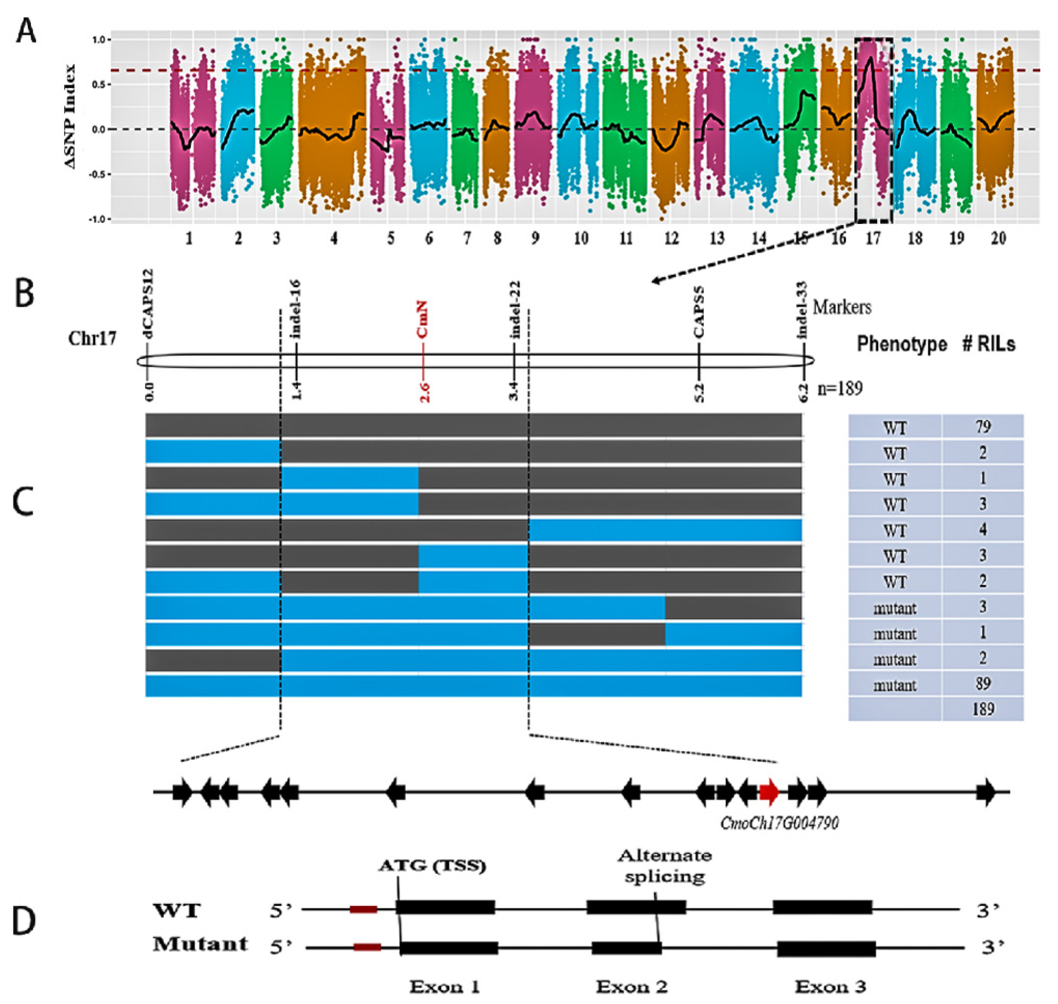
Mutant and WT bulks were constructed from 42 mutant and 42 WT RILs, respectively. Illumina sequencing of the two bulks and two parental lines generated ~27.47 Gb clean reads (Q30 > 80%) including 11.07 Gb from the parents and 16.4 Gb from the two bulks. The main statistics of BSA-Seq are presented in Supplementary Table S2. The average sequencing depths for the two parents and the two F8 RIL pools were 13.5× and 21×, respectively. These reads were mapped onto the C. moschata reference genome with an average mapping rate of 97.7%. From 1,019,929 SNPs and small Indels detected, after serial filtering processes (biallelic, single copy, read depth > 4, consistent between two bulks and two parental lines), 377,185 high-quality SNPs were used for calculating ΔSNP/Indel-index and the Euclidean distance (ED) estimation. The genome-wide ΔSNP-index plot is shown in Figure 2A. The ED plot is illustrated in Supplementary Figure S1. The ΔSNP-index association analysis mapped the N locus to a 2.38 Mbp physical interval on Chromosome 17 (2,810,000 bp to 5,190,000 bp) in the reference genome. BSA with Indels and ED resulted in a slightly larger interval that overlapped with the one identified with the ΔSNP-index method suggesting the N locus is located in this region.

3.3. Linkage Mapping and Candidate Gene Identification for the N Locus

To narrow down the candidate gene region, we developed new molecular markers in the 2.38 Mbp region. From BSA seq, 10,433 SNPs and 2434 Indel polymorphisms were identified in this region, from which 59 non-synonymous SNPs and 47 Indels were targeted for primer design. Both CAPS and KASP assays were employed for SNP genotyping. Polymorphisms of these markers were first verified between the two bulks and parental lines, then applied in the RIL population. Finally, three Indels and two KASP/CAPS markers were successfully mapped in the RIL population. Primer information of these mapped marker loci is shown in Supplementary Table S1. The resulting linkage map and nine haplotypes defined by these markers among 189 RILs are presented in Figure 2B. Through linkage analysis, the N locus was placed into a 389.5-kb region delimited by two flanking markers indel-16 and indel-22.
Fifteen genes (#1–15) were annotated in the C. moschata reference genome (Figure 2C), which are listed in Supplementary Table S3. To identify a potential candidate gene for the N locus, we first examined the sequence variation in this region between two parental lines. From BSA-seq, 1121 SNPs and 28 Indels were identified against the reference genome. The complete list of the 1159 variants is provided in Supplementary Table S4. Many SNPs/Indels were polymorphic between the WT or mutant and the reference genome, but not between the WT and the mutant. We scanned all these variants to find the association of nucleotide variation with the naked seed vs. hulled seed phenotypes using the following criteria: high quality (nucleotide accuracy and coverage), polymorphic between WT and mutant, located in promoter or coding regions, and non-synonymous mutations. As the line of the reference genome is also a wildtype, any causal SNP must also carry the same allele as the WT/HS-A line used in this study. When a polymorphism was located inside an exonic region, we further examined its variation in cDNA sequences extracted from the RNA-Seq data (see below). None of 1159 SNPs/Indels met these criteria suggesting the causal polymorphism may not be present at the genomic DNA (gDNA) level.
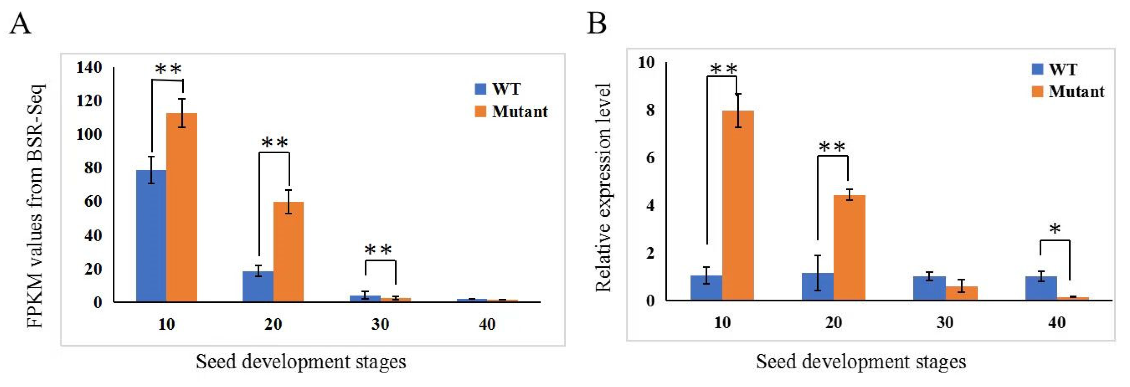
We further investigated the expression dynamics of the 15 genes from the RNA-Seq data (see below for more details). FPKM (fragments per kilobase of transcript per million mapped reads) values were extracted from WT and mutant samples at four time points (10, 20, 30, and 40 DAP, three biological reps each), which are provided in Supplementary Table S3. The bar graphs of FPKM values are plotted and illustrated in Figure 3A (for Gene #12), and Supplementary Figure S2 (the remaining 14 genes). We calculated the log2 (fold change) (WT vs. mutant) value for each gene, which is also presented in Table S3. Among all 15 genes, only the 12th gene, CmoCh17G004790 that was predicted to encode NAC domain-containing protein 43 (NAC43) showed a statistically significant difference in expression between the mutant and WT at 30 DAP using the criteria of FDR < 0.05 and |log2(FC)| ≤ 1. The expression dynamics of CmoCh17G004790 in the developing seed coat of the two lines were further validated by qPCR, which is presented in Figure 3B. Its expression was higher in the seed coat of the mutant than in the WT at 10 and 20 DAP, which showed the opposite at 30 and 40 DAP. CmoCh17G004790 is a homolog of Arabidopsis gene AT2G46770 that encodes a NAC transcription factor WALL THICKENING PROMOTING FACTOR 1 (NST1) [37]. In C. pepo, its homolog CpNST1 has been shown to be the candidate gene for the hull-less seed mutation [19]. These data suggested that CmoCh17G004790 (CmNST1 hereinafter) is a possible candidate for the N locus in C. moschata.

3.4. Sequence Analysis Suggests Alternative Splicing in the CmNST1 Transcript May Contribute to Defective Seed Coat Development in the Mutant

Data from BSA-Seq identified no causal polymorphisms in gDNA sequences between the mutant and WT in any of the 15 genes. With Sanger sequencing, we further cloned the gDNA and cDNA sequences of CmoCh17G004790 from the different organs/tissues in the mutant and WT including young leaves, root, stem at seedling stage, seed coat of developing seeds at 10, 20, 30, and 40 DAP and leaf tissues from adult plants. The gDNA and cDNA sequences and their alignment from the seed coat and young leaves together with the gDNA and mRNA sequences from the reference genome (cv Rifu) are shown in Supplemental File S1. The alignment of part of the gDNA and cDNA sequences of CmNST1 in WT, mutant, and the reference genome is presented in Figure 4. Annotation of this gene in the C. moschata genome suggested three exons and two introns of CmNST1 (Figure 2D; Supplemental File S1), which was consistent with the cloning of cDNA sequences and manual annotation, as well as that in Arabidopsis. Consistent with BSA-Seq, no sequence variation at the gDNA level was found between WT and the mutant. However, a 112 bp deletion was found in the cDNA cloned from seed coat tissue as compared with cDNA cloned from young leaves, which started from inside the second exon and ended at the start of the second intron (Figure 4). We wondered if this was due to a sequencing error and therefore we cloned gDNAs and cDNAs from different organs at different development stages including cotyledons, root, stem, and young leaves from seedlings, as well as seed coat samples collected at 10, 20, 30, and 40 DAP in both the mutant and WT. In each case, multiple clones were Sanger sequenced, and the results were the same. That is, all cDNA clones from the seed coat of the mutant at any time point had the 112 bp deletion as compared with cDNA from vegetative organs of the mutant and any organs/tissues from the WT or the reference genome (Supplemental File S1) supporting alternative splicing (AS) in the developing seed coat of the mutant may be associated with the naked seed mutation.
We also compared the gene structure and annotation of the CmNST1 homolog genes in C. pepo (Cp4.1LG12g04350) and C. maxima (CmaCh17G005080). The gDNA and mRNA sequences of C. maxima and C. pepo were downloaded from the cucurbit genomics database. The sequences and their alignment among the three species are shown in Supplemental File S2. The gDNA and mRNA sequences from the C. pepo reference genome were shorter than the other two homologs including missing the translation start codon. Only two exons were annotated in the C. pepo gene which may represent a misannotation of Cp4.1LG12g04350. Lv et al. [19] suggested that the hull-less seed mutation in C. pepo is due to an insertion of a 16 bp sequence in the first exon of CpNST1 (Cp4.1LG12g04350). We found that this was a simple sequence repeat (SSR) with a CA motif which was presented in both the mutant and WT of C. moschata of our study (Figure 4) thus excluding it as a possible contributing mutation to naked seed in HLS-B inbred. Interestingly, in C. moschata, the SSR was 5bp upstream of TSS of CmNST1 whereas, in C. pepo, the start codon (ATG) was proposed to be ~60 bp upstream of the second proposed start codon in C. moschata ([19]; also Figure 4). While this discrepancy needs further clarification, these observations may suggest a different mechanism associated with naked seed mutation in C. pepo [19] and C. moschata (this study).

3.5. Transcriptome Profiling Reveals Regulatory Gene Network for Seed Coat Development in C. moschata

To understand the gene regulatory network controlled by the CmNST1 gene, we conducted BSR-Seq to study the transcriptomes of the seed coat of the mutant and WT. Seed coat samples were collected from developing seeds at four stages: 10, 20, 30, and 40 DAP with three biological replications per sample (total of 24). Main RNA-seq statistics are presented in Supplemental Table S5. The complete transcriptome datasets have been deposited into the CNGB Sequence Archive (CNSA) of the China National GeneBank Database (CNGBdb) with accession number CNP0003716. Nearly 93.4% of bases in each sample had a Q-score no less than Q30. Of the ~510 million high-quality paired-end reads (~152 Gbp) generated from 24 cDNA libraries, ~92.5% of reads could be mapped to the C. moschata reference genome (Supplemental Table S5). The high quality of the RNA-Seq datasets could be seen from the high Spearman’s rank correlation coefficient plot among replicates of each sample (Supplemental Figure S3). We explored SNPs and Indels in transcripts between WT and mutant from BSR-Seq. Consistent with results from BSA-Seq and Sanger sequencing results, no polymorphisms were detected in cDNAs in any of the 15 genes in the 389.5 kb candidate gene region further confirming that CmNST1 was the most likely candidate for the n locus. In addition, as compared with the reference genome, 2073 new genes not present in the reference genome were annotated. However, the annotation of the 15 genes in the target 389.5 kb region remained the same.
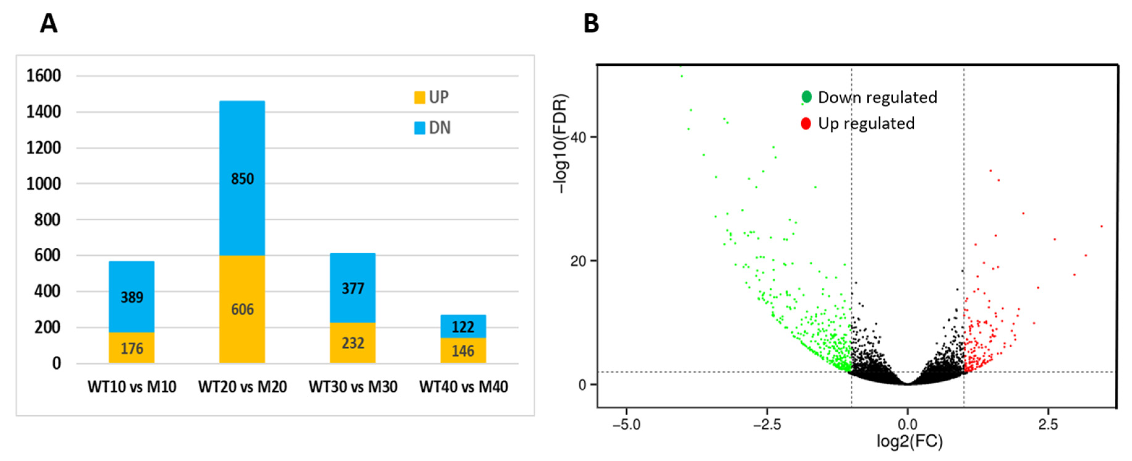
Differentially expressed genes (DEGs) were identified using FDR ≤ 0.05 and |log2(fold change)| ≥ 1 as the criteria in various comparisons. Total numbers of DEGs and up- or down-regulated genes in each comparison are presented in Supplementary Table S6. Specifically, 565, 1456, 609, and 268 DEGs were identified between the WT and the mutant at 10, 20, 30, and 40 DAP, respectively. Among them, 176, 606, 232, and 146 DEGs showed higher expression (up-regulated), and 389, 850, 377, and 122 showed lower expression (down-regulated) in the mutant than in the WT seed coat, respectively, which are illustrated in Figure 5A. These data indicated that significant changes in the mutant transcriptomes occurred in early seed coat development, and more genes were down-regulated than those up-regulated.
Since the WT and mutant showed significant differences in seed coat development that were observed at a very early stage, we focused on DEGs at 10 DAP for further analysis. Of the 565 DEGs at this time point, 429 had been functionally annotated with predicted functions. The gene IDs, predicted functions, GO, COG, and KEGG terms, and their expression dynamics at four seed development stages of the 429 genes are provided in Supplemental Table S7. The candidate gene CmNST1 and 25 additional DEGs in the lignin biosynthesis pathway were also included in Supplemental Table S7 (total 455). Of the 455 genes, at 10 DAP, many more genes (313 or 68.8%) showed lower expression in the mutant; only 142 (31.2%) had higher expression in the WT. This could clearly be seen from the volcano plot of these DEGs shown in Figure 5B.
We performed GO enrichment analysis on 429 DEGs between the HS-A and HLS-B at 10 DAP (Figure 6A). The most enriched GO terms in ‘Biological Process’ included metabolic processes, cellular processes, and biological regulation. The most enriched ‘Molecular function’ terms were binding, catalytic activity, transporter, and transcriptional factor activity. COG (Cluster of Orthologous Groups of proteins) function classification analysis of these DEGs (Figure 6B) revealed that the top functional groups were carbohydrate transport and metabolism, cell wall/membrane/envelope genesis, secondary metabolite biosynthesis, transport, and catabolism, as well as signal transduction mechanisms. In KEGG pathway enrichment analysis, the top enriched pathways associated with these DEGs included phenylpropanoid biosynthesis, flavonoid biosynthesis, and phenylalanine metabolism (Figure 6C). These data strongly suggested that genes for enzymes of secondary metabolism, transcription factors, and phytohormones are involved in seed coat development in pumpkin.
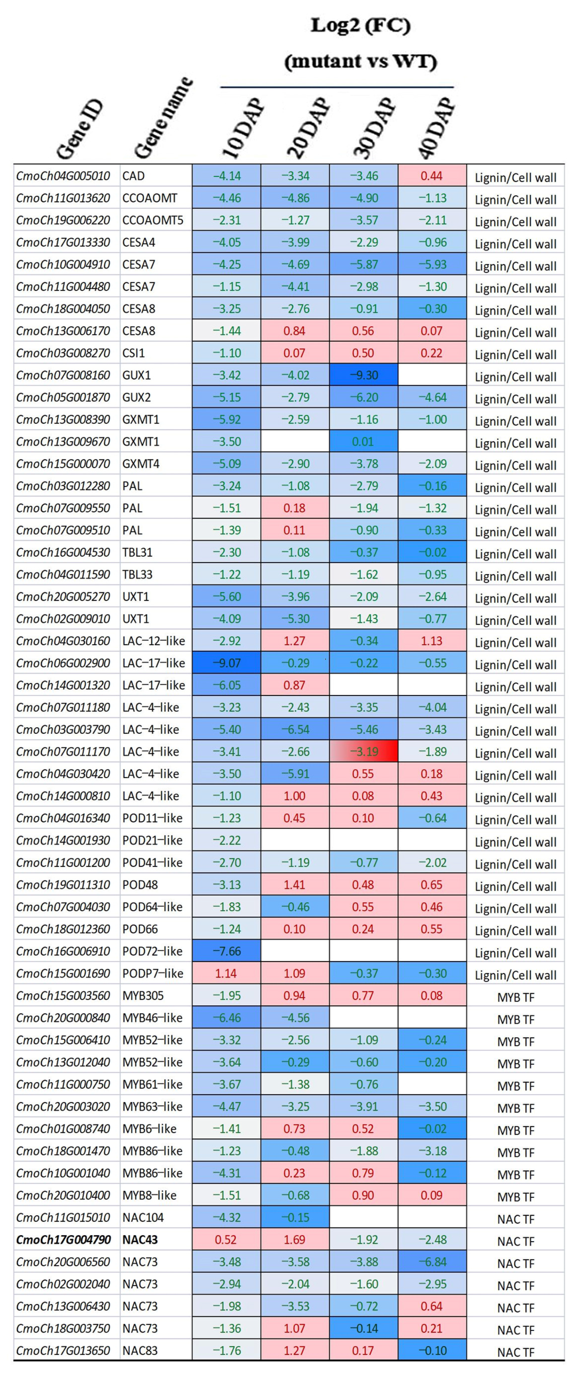
In our previous study, we found a continued decrease of lignin content in the testa of the mutant but a continued increase in the WT during seed development [25]. We also found that the activities of several enzymes in the lignin biosynthesis pathway were significantly lower in the mutant than in the WT, which included the phenylalanine ammonia lyase (PAL), 4-coumaric acid coenzyme A ligase (4CL), cinnamate-4-hydroxylase (C4H), and cinnamyl alcohol dehydrogenase (CAD). We inferred that the degeneration or absence of the typical seed testa structure in the mutant was due to the reduced activity of key enzymes in the lignin biosynthesis pathway [25]. Lignin is an important component of the secondary cell wall during seed coat development. The biosynthesis of lignin is tightly controlled by a complex regulatory network containing many transcription factors especially members of the NAC and MYB families, as well as phytohormones (reviewed in [38,39,40,41,42]; also see Introduction and Discussion sections). We manually examined all DEGs in the transcriptomes of the mutant and WT at 10 DAP and DEGs of the lignin biosynthesis pathway at 20, 30, or 40 DAP (Table S7). Among the 455 DEGs, 85 (18.7%) encoded enzymes that were directly involved in the lignin biosynthesis pathway or secondary cell wall formation; 45 (10%) were transcription factor genes including seven members of the NAC TF family (NAC43/73/83/104) and 10 members of the MYB TF family (MYB 6/8/46/52/61/63/86/305); 16 were involved in phytohormone biosynthesis/signaling (auxin/IAA, ethylene, BR, GA, and JA) (Table S7). A heatmap of the expression level of 54 selected genes from 85 DEGs involved in the lignin biosynthesis pathway or secondary cell wall formation in terms of log2 (fold change) at the four time points of seed coat development is illustrated in Figure 7. The majority of these genes were down-regulated in the naked seed mutant as compared with hulled seed WT. We specifically examined the key genes/enzymes in the lignin biosynthesis pathway which is presented in Figure 8. Most genes-encoding enzymes in this pathway showed reduced expression in the mutant. Some examples were genes for PAL, 4CL, C4H, CAD, CCoAOMT (caffeoyl-CoA O-methyltransferase), and CCR (cinnamoyl-CoA reductase). Multiple members in POD (peroxidases) and laccase (LAC) gene families that play important roles in the last step of lignin biosynthesis were also down-regulated in the mutant (Figure 8).
Taken together, transcriptome profiling in the mutant and WT during seed coat development clearly supported that the lignin biosynthesis pathway and secondary cell wall formation were compromised in the HLS-B, which was likely due to the alternative splicing mutation in the CmNST1 (n or NAC3) gene transcript. Many other transcription factors and hormone biosynthesis/signaling pathways were also involved in the regulation of the secondary cell wall development in C. moschata.

4. Discussion

4.1. CmNST1 Is a Candidate Gene for the Naked Seed (n) Locus

In this study, we show that the naked seed phenotype in HLS-B C. moschata inbred line was controlled by a single recessive gene (n). BSA-Seq and linkage analysis placed the N locus into a 389.5 kb region on Chromosome 17 containing 15 predicted genes (Figure 2; Table S3). We suggest CmoCh17G004790 as the most possible candidate gene for the N locus which encodes a transcription factor (NAC domain-containing protein 43 or NAC43). Our conclusion was based on multiple lines of evidence. First, from BSA-Seq and resequencing data, no polymorphisms were detected in the coding region or promoter region in any of the 15 genes between the two parental lines, and no consistent haplotypes defined by SNPs or Indels were associated with the phenotypes (the WT of HS-A, Rifu vs. HSL-B mutant) (Table S4). From both BSR-Seq and qPCR, among the 15 genes, CmoCh17G004790 is the only one showing differential expression between the WT and the mutant (Figure 3; Table S3). More importantly, we found alternative splicing in the mRNA of CmoCh17G004790 that only occurred in the developing seed coat of the mutant but not in other organs or tissues of the mutant, nor in the mRNA from any tissues or organs of the WT (Figure 4; Supplemental File S1).
Hull-less or naked seed mutants have been identified in all three Cucurbita species, which were all controlled by a single recessive gene (n or h) [2,12,13,14]. The n locus in C. pepo has been fine-mapped, and the results from three independent studies all support the NAC transcription gene Cp4.1LG12g04350 (CpNST1) on Chromosome 12 of C. pepo as the most possible candidate gene [19,20,43]. CmoCh17G004790 (CmNST1) and Cp4.1LG12g04350 (CpNST1) are homologs in the C. pepo and C. moschata genomes (Supplemental File S2). Thus, our study represents the first report of cloning of a candidate gene for the hull-less mutation in C. moschata. Although there is no report on cloning of the hull-less locus in C. maxima, the data from the present work may suggest that the hull-less seed phenotypes in the three Cucurbita species probably share a similar genetic basis and common candidate gene.
The causal mutations in these mutants may be different though. Lv et al. [19] proposed that a 14-bp insertion in CpNST1 was the causal polymorphism in the hull-less C. pepo mutant which would result in premature termination of CpNST1 translation. The 14 bp sequence includes AA followed by six repeats of a dinucleotide (CA) SSR motif: ((CA)6). In the mutant and WT lines used in this study and the C. moschata reference genome, the SSRs were (CA)11, (CA)11, and (CA)3, respectively (Figure 4). This suggests a variation of this SSR in natural populations of Cucurbita species although the scope of this variation and its association with the hull-less phenotype requires further investigation. This observation also eliminates the possibility that the SSR is the causal mutation for the hull-less phenotype in the HLS-B inbred line of this study. Another interesting difference is the translation start position in CpNST1 and CmNST1. There are two start codons (ATG) before and after the SSR (Figure 4; Supplemental File S2). Which is the actual TSS may need additional studies.
One common feature between CpNST1 and CmNST1 ([19] and this study) was the expression dynamics in the mutant and WT. In this study, CmNST1 showed higher expression in the seed coats of the mutant than in the WT up to 20 DAP. After that, its expression at 30 and 40 DAP was lower in the mutant than in the WT (Figure 7). The higher expression in the mutant than in the WT was consistent with that in the C. pepo hull-less mutant [19] although its expression after 20 DAP was not reported in the early study in C. pepo. The higher expression of NST1 homologs in the mutant was also observed in Medicago truncatula [44] supporting a conserved function of NST1 as a negative regulator in secondary cell wall/seed coat development (see more discussions below).

4.2. Alternative Splicing Contributes to Naked Seed Mutation in C. moschata

We found that the CmNST1 cDNA cloned from seed coat tissues of the mutant was 112 bp shorter than that from the WT suggesting alternative splicing (AS) in the mutant. No AS was found in cDNA sequences of this gene cloned from vegetative organs (root, stem, cotyledon, young and old leaves) of the mutant, nor from cDNAs from any organs or tissues in the WT (Figure 4; Supplemental File S1). This suggests that the AS of CmNST1 only occurs in the developing seed coat of the hull-less mutant. This represents a different mechanism from C. pepo [19] for the hull-less mutation despite the mutants in both crops having the same candidate gene.
AS refers to the generation of multiple splice isoforms (mRNA transcripts) from a single gene due to different splicing sites which is a critical mechanism for gene expression regulation at the post-transcriptional level that significantly expands the coding capacity of genomes and improves transcriptome plasticity and proteome diversity [45]. AS is a common phenomenon in plants that plays an important role in plant adaptation and evolution. For example, early studies estimated that at least 42% Arabidopsis and 48% rice intron-containing genes are alternatively spliced [46,47]. In silico analysis of transcriptomic data from high throughput transcriptome sequencing suggested > 70% of plant genes may have AS in a tissue-specific, developmental, or signal transduction-dependent manner (e.g., [48,49,50]). AS in many genes playing important roles in plant growth and development and adaptation to environments has been documented (reviewed in [51]). To the best of our knowledge, AS has not been reported in any genes that play a role in seed coat development.
Several types of AS have been observed which include exon skipping (ES), intron retention (IR), alternative 5′ splice site (alt 5′SS), alternative 3′ splice site (alt 3′SS), variable first exon (VFE), variable last exon (VLE) and mutually exclusive exons (MEE) [52,53,54]. In the present study, AS occurred in the second exon of CmNST1 (Figure 4; Supplemental File S1), which could be classified as an alternative 5′ splice site. Both the mutant and WT seem to fit the GT-AG rule (Figure 4). The reason why the splicing site of the second intron in the mutant started from the first GT is unknown. How does the 112-bp shorter isoform of the mutant transcript result in reduced lignin biosynthesis and secondary cell wall formation, thus a defective seed coat development is an interesting question that merits additional studies.

4.3. CmNST1-Regulated Seed Coat Development Involves a Complex Regulatory Network for Lignin Biosynthesis and Secondary Cell Wall Formation

We show that CmNST1 is a candidate gene for the n locus in C. moschata which is a homolog of Arabidopsis NAC SECONDARY WALL THICKENING PROMOTING FACTOR 1 (NST1). Plant secondary cells are the building blocks for seed coat development, which are composed primarily of cellulose, lignin, and hemicelluloses. The biosynthesis of cellulose, xylan, and lignin is under elegant transcriptional regulation [55,56,57]. Many members in the NAC transcription factor families have been identified as master regulators by activating different layers of downstream secondary wall-related TFs such as MYBs [44,55,58]. For example, in Arabidopsis, important NAC master TF regulators may include NST1/NST2/NST3, VND6 (VASCULAR-RELATED NAC-DOMAIN 6 (VND6) and VND5 (reviewed in [39]). In this hierarchical network, NACs target MYB TFs such as MYB46 and MYB83, which in turn will activate the expression of genes in the lignin biosynthesis pathways such as PAL, C4H, 4CL, CCoAOMT, CCR, and CAD genes via binding to the promoters of these genes [38,59]. Downstream of MYB46/MYB83, multiple MYB TFs (e.g., MYB4, 7, 32, 58, 63, and 85) have been shown to be specific regulators of lignin biosynthesis in Arabidopsis (reviewed in [40,41,42]).
BSR-Seq in the present study revealed many DEGs between the mutant and WT including seven NAC and 10 MYB TF genes (Table S7, Figure 7). Many of these DEGS are involved in the biosynthesis pathways for lignin and xylan, which were down-regulated in the mutant from a very early stage of seed development. Many of these DEGs were also identified in the transcriptomes of C. pepo hull-less mutant and WT [19,24,26]. Thus, our data support a similar mechanism identified in Arabidopsis and other plant species that NST1 is a master transcription regulator for secondary cell wall and seed coat development (Figure 7).
Recent discoveries of new transcription factors in Arabidopsis, and other plant species suggest that the regulatory network of lignin biosynthesis in plants may extend beyond the NAC-MYB network [41,60]. This could also be true in C. moschata from the data of the present study. The expression of CmNST1 was higher in the mutant than in the WT before 20 DAP (Figure 3) whereas most genes involved in lignin biosynthesis or secondary cell wall development were down-regulated in the mutant throughout seed development (Figure 7; Table S7). Histological characterization of hull-less pumpkin seeds reveals that seed coat development is similar in both hulled and hull-less seeds in the first 10–15 days after pollination [21,25,26]. There were no significant differences in the seed coats between the mutant and WT in lignin content, and enzymatic activities for PAL, 4CL, C4H, and CAD [25]. Why does NST1 show higher expression in the hull-less mutant of the present study or the C. pepo hull-less mutant [19] and the non-lignification mutant in M. truncatula [44] than in the respective wildtype plant is an interesting question. NST1 is a master TF to regulate secondary wall synthesis and its expression must be under strict developmental regulation [38,59,60]. For example, in Arabidopsis, the SND1 (for secondary wall-associated NAC domain protein) is a homolog of NST1 [55]. Wang et al. (2011) reasoned that the expression of SND1 is under both positive regulation by its own translation product and negative regulation by downstream MYB TFs (Figure 8). Thus, these effects are presumably balanced until the balance is broken by a mutation or changing environmental factors. The elevated NST1 transcription in the mutant may suggest that the positive autoregulation is overridden by negative regulators under normal circumstances. Fine tuning of NST1 expression by balancing activation and repression may enable plants to adapt to ever changing environmental conditions [55]. However, other regulatory mechanisms independent of CmNST1 may also be present. How alternative splicing results in transcription changes and downstream targets, in particular, will be interesting subjects of future research.

Supplementary Materials

The following supporting information can be downloaded at https://www.mdpi.com/article/10.3390/genes14050962/s1. Figure S1: Genome-wide plotting of Euclidean distance (ED) values. Figure S2: FKPM values of 14 genes in 389.5 kb region at different seed coat development stages. Figure S3: Spearman’s rank correlation coefficient (rs) plot among three replications of RNA-Seq samples. Supplemental Table S1: Information on molecular markers and primers used in this study for various purposes. Supplemental Table S2: BSA-Seq statistics. Supplemental Table S3: Annotated genes in 389.5 kb target region of the N locus. Supplemental Table S4: SNP allele distribution among reference region, mutant and WT bulks, and parental lines in 389.5 kb candidate gene region on Chr17. Supplemental Table S5: BSR-Seq statistics of 24 seed coat samples at four development stages from WT and mutant. Supplemental Table S6: DEGs in transcriptomes from different comparisons between the mutant and WT at four development stages. Supplemental Table S7: Differential expressed genes (DEGs) between mutant and WT seed coats at 10 days after pollination from RNA-Seq data. Supplemental File S1: Genomic and cDNA sequences of CmNST1 in C. moschata and alignment of these sequences. Supplemental File S2: Genomic and mRNA sequences of CmNST1 in C. maxima (Cmax), C. pepo (Cp), and C. moschata and alignment of these sequences.

Author Contributions

Q.S.: performed the majority of the research, data analysis, draft preparation, and revision. Y.W.: experimental design, data analysis, manuscript writing. All authors have read and agreed to the published version of the manuscript.

Funding

This project was financially supported by the National Natural Science Foundation of China (32202490), the Youth Science and Technology Research Fund project of Shanxi Province (201901D211566), and the Doctoral Research Fund of Shanxi Academy of Agricultural Sciences (YBSJJ2007).

Institutional Review Board Statement

Not applicable.

Informed Consent Statement

Not applicable.

Data Availability Statement

All data pertinent to the reported work have been provided in the manuscript or in the supplemental online materials.

Conflicts of Interest

The authors declare no conflict of interest.

References

  1. Ferriol, M.; Picó, B. Pumpkin and winter squash. In Vegetables I: Asteraceae, Brassicaceae, Chenopodicaceae, and Cucurbitaceae; Springer: Berlin/Heidelberg, Germany, 2008; pp. 317–349. [Google Scholar]
  2. Paris, H.S. Germplasm enhancement of Cucurbita pepo (pumpkin, squash, gourd: Cucurbitaceae): Progress and challenges. Euphytica 2016, 208, 415–438. [Google Scholar] [CrossRef]
  3. Lelley, T.; Loy, B.; Murkovic, M. Hull-less oil seed pumpkin. Oil Crops 2010, 4, 469–492. [Google Scholar]
  4. Stevenson, D.G.; Eller, F.J.; Wang, L.; Jane, J.L.; Wang, T.; Inglett, G.E. Oil and tocopherol content and composition of pumpkin seed oil in 12 cultivars. J. Agric. Food Chem. 2007, 55, 4005–4013. [Google Scholar] [CrossRef]
  5. Fruhwirth, G.O.; Hermetter, A. Seeds and oil of the Styrian oil pumpkin: Components and biological activities. Eur. J. Lipid Sci. Technol. 2007, 109, 1128–1140. [Google Scholar] [CrossRef]
  6. Bergantin, C.; Maietti, A.; Tedeschi, P.; Font, G.; Manyes, L.; Marchetti, N. HPLC–UV/Vis–APCI–MS/MS determination of major carotenoids and their bioaccessibility from “Delica”(Cucurbita maxima) and “Violina”(Cucurbita moschata) pumpkins as food traceability markers. Molecules 2018, 23, 2791. [Google Scholar] [CrossRef]
  7. Kim, E.Y.; Kwon, C.W.; Chang, P.S. Purification and characterization of a novel acid-tolerant and heterodimeric β-glucosidase from pumpkin (Cucurbita moschata) seed. J. Biosci. Bioeng. 2021, 132, 125–131. [Google Scholar] [CrossRef]
  8. Vinayashree, S.; Vasu, P. Biochemical, nutritional and functional properties of protein isolate and fractions from pumpkin (Cucurbita moschata var. Kashi Harit) seeds. Food Chem. 2021, 340, 128177. [Google Scholar] [CrossRef]
  9. Baxter, G.; Murphy, K.; Paech, A. The Potential to Produce Pumpkim Seed for Processing in North East Victoria; Rural Industries Development Corporation: Kingston, Australia, 2012; Volume 11. [Google Scholar]
  10. Loy, J.B. Morpho-physiological aspects of productivity and quality in squash and pumpkins (Cucurbita spp.). Crit. Rev. Plant Sci. 2004, 23, 337–363. [Google Scholar] [CrossRef]
  11. Wenzl, T.; Prettner, E.; Schweiger, K.; Wagner, F.S. An improved method to discover adulteration of Styrian pumpkin seed oil. J. Biochem. Biophys. Methods 2002, 53, 193–202. [Google Scholar] [CrossRef]
  12. Zraidi, A.; Pachner, M.; Lelley, T.; Obermayer, R. On the genetics and histology of the hull-less character of Styrian oil-pumpkin (Cucurbita pepo L.). Mol. Breed. 2003, 26, 57–61. [Google Scholar]
  13. Zhou, X.L. Study on the breeding of naked kernel pumpkin and its genetic behavior. Acta Hortic. Sin. 1987, 14, 115–118. [Google Scholar]
  14. Paris, H.S.; Brown, R.N. The genes of pumpkin and squash. HortScience 2005, 40, 1620–1630. [Google Scholar] [CrossRef]
  15. Gong, L.; Stift, G.; Kofler, R.; Pachner, M.; Lelley, T. Microsatellites for the genus Cucurbita and an SSR-based genetic linkage map of Cucurbita pepo L. Theor. Appl. Genet. 2008, 117, 37–48. [Google Scholar] [CrossRef]
  16. Zraidi, A.; Stift, G.; Pachner, M.; Shojaeiyan, A.; Gong, L.; Lelley, T. A consensus map for Cucurbita pepo. Mol. Breed. 2007, 20, 375–388. [Google Scholar] [CrossRef]
  17. Inan, N.; Yildiz, M.; Sensoy, S.; Kafkas, S.; Abak, K. Efficacy of ISSR and SRAP techniques for molecular characterization of some Cucurbita genotypes including naked (hull-less) seed pumpkin. J. Anim. Plant Sci. 2012, 22, 126–136. [Google Scholar] [CrossRef]
  18. Chahal, G.K.; Kaur, A.; Dhatt, A.S. A single-gene mutation changed the architecture of pumpkin seed: A review. J. Plant Growth Regul. 2022, 41, 113–118. [Google Scholar] [CrossRef]
  19. Lv, X.; Shi, L.; Zhao, M.; Li, Z.; Liao, N.; Meng, Y.; Ma, Y.; Zhou, Y.; Xue, Q.; Hu, Z. A natural mutation of the NST1 gene arrests secondary cell wall biosynthesis in the seed coat of a hull-less pumpkin accession. Hortic. Res. 2022, 9, uhac136. [Google Scholar] [CrossRef]
  20. Meru, G.; Fu, Y.; Shrestha, S.; Michael, V.N.E.; Dorval, M.; Mainviel, R. Genomic position and markers associated with the hull-less seed trait in pumpkin. Plants 2022, 11, 1238. [Google Scholar] [CrossRef]
  21. Stuart, S.G.; Loy, J.B. Comparison of testa development in normal and hull-less seeded strains of Cucurbita pepo L. Bot. Gaz. 1983, 144, 491–500. [Google Scholar] [CrossRef]
  22. Bezold, T.N.; Loy, J.B.; Minocha, S.C. Changes in the cellular content of polyamines in different tissues of seed and fruit of a normal and a hull-less seed variety of pumpkin during development. Plant Sci. 2003, 164, 743–752. [Google Scholar] [CrossRef]
  23. Kreft, M.; Zorec, R.; Janeš, D.; Kreft, S. Histolocalisation of the oil and pigments in the pumpkin seed. Ann. Appl. Biol. 2009, 154, 413–418. [Google Scholar] [CrossRef]
  24. Xue, Y.; Shen, Z.; Tao, F.; Zhou, J.; Xu, B. Transcriptomic analysis reveal the molecular mechanisms of seed coat development in Cucurbita pepo L. Front. Plant Sci. 2022, 13, 51. [Google Scholar] [CrossRef]
  25. Shen, Q.; Wu, L.; Zhao, X.; Li, Y. Comparative analysis of electron microscopic structure of seed testa, lignin and biosynthesis-related enzyme activities in hulled and hull-less seeds of Cucurbita moschata. Sci. Hortic. 2019, 245, 137–143. [Google Scholar] [CrossRef]
  26. Bezold, T.N.; Mathews, D.; Loy, J.B.; Minocha, S.C. Molecular analysis of the hull-less seed trait in pumpkin: Expression profiles of genes related to seed coat development. Seed Sci. Res. 2005, 15, 205–217. [Google Scholar] [CrossRef]
  27. Zhou, X.L. Study on the breeding and genetic rules of naked pumpkin. Shanxi Agric. Sci. 1983, 12, 11–12. [Google Scholar]
  28. Wu, J.X.; Chang, Z.J. Study on seed characteristics of two naked kernel Cucurbita moschata. Shanxi Agric. Sci. 2011, 39, 230–231. [Google Scholar] [CrossRef]
  29. Li, Q.; Li, L.; Yang, X.; Warburton, M.L.; Bai, G.H.; Dai, J.R.; Li, J.S.; Yan, J.B. Relationship, evolutionary fate and function of two maize co-orthologs of rice GW2 associated with kernel size and weight. BMC Plant Biol. 2010, 10, 143. [Google Scholar] [CrossRef]
  30. McKenna, A.; Hanna, M.; Banks, E.; Sivachenko, A.; Cibulskis, K.; Kernytsky, A.; Garimella, K.; Altshuler, D.; Gabriel, S.; Daly, M. The Genome Analysis Toolkit: A MapReduce framework for analyzing next-generation DNA sequencing data. Genome Res. 2010, 20, 1297–1303. [Google Scholar] [CrossRef]
  31. Livak, K.J.; Schmittgen, T.D. Analysis of relative gene expression data using real-time quantitative PCR and the 2−ΔΔCT method. Methods Cell Sci. 2001, 25, 402–408. [Google Scholar] [CrossRef]
  32. Kim, D.; Langmead, B.; Salzberg, S.L. HISAT: A fast spliced aligner with low memory requirements. Nat. Methods 2015, 12, 357–360. [Google Scholar] [CrossRef]
  33. Pertea, M.; Pertea, G.M.; Antonescu, C.M.; Chang, T.C.; Mendell, J.T.; Salzberg, S.L. StringTie enables improved reconstruction of a transcriptome from RNA-seq reads. Nat. Biotechnol. 2015, 33, 290–295. [Google Scholar] [CrossRef] [PubMed]
  34. Love, M.I.; Huber, W.; Anders, S. Moderated estimation of fold change and dispersion for RNA-seq data with DESeq2. Genome Biol. 2014, 15, 550. [Google Scholar] [CrossRef] [PubMed]
  35. Cingolani, P.; Platts, A.; Wang, L.L.; Coon, M.; Nguyen, T.; Wang, L.; Land, S.J.; Lu, X.; Ruden, D.M. A program for annotating and predicting the effects of single nucleotide polymorphisms, SnpEff: SNPs in the genome of Drosophila melanogaster strain w1118; iso-2; iso-3. Fly 2012, 6, 80–92. [Google Scholar] [CrossRef]
  36. Paris, H.S.; Kabelka, E. Gene list for Cucurbita species, 2009. Cucurbit Genet. Coop. Rep. 2009, 31, 44–69. [Google Scholar]
  37. Mitsuda, N.; Seki, M.; Shinozaki, K.; Ohme-Takagi, M. The NAC transcription factors NST1 and NST2 of Arabidopsis regulate secondary wall thickenings and are required for anther dehiscence. Plant Cell 2005, 17, 2993–3006. [Google Scholar] [CrossRef] [PubMed]
  38. Zhong, R.; Ye, Z. Complexity of the transcriptional network controlling secondary wall biosynthesis. Plant Sci. 2014, 229, 193–207. [Google Scholar] [CrossRef]
  39. Nakano, Y.; Yamaguchi, M.; Endo, H.; Rejab, N.A.; Ohtani, M. NAC-MYB-based transcriptional regulation of secondary cell wall biosynthesis in land plants. Front. Plant Sci. 2015, 6, 288. [Google Scholar] [CrossRef]
  40. Liu, Q.; Luo, L.; Zheng, L. Lignins: Biosynthesis and biological functions in plants. Int. J. Mol. Sci. 2018, 19, 335. [Google Scholar] [CrossRef]
  41. Xie, M.; Zhang, J.; Tschaplinski, T.J.; Tuskan, G.A.; Chen, J.; Muchero, W. Regulation of lignin biosynthesis and its role in growth-defense tradeoffs. Front. Plant Sci. 2018, 9, 1427. [Google Scholar] [CrossRef]
  42. Pratyusha, D.S.; Sarada, D.V. MYB transcription factors–master regulators of phenylpropanoid biosynthesis and diverse developmental and stress responses. Plant Cell Rep. 2022, 41, 2245–2260. [Google Scholar] [CrossRef]
  43. Kaur, B.; Garcha, K.S.; Bhatia, D.; Khosa, J.S.; Sharma, M.; Mittal, A.; Verma, N.; Dhatt, A.S. Identification of single major QTL and candidate gene(s) governing hull-less seed trait in pumpkin. Front. Plant Sci. 2022, 13, 948106. [Google Scholar] [CrossRef] [PubMed]
  44. Wang, H.; Zhao, Q.; Chen, F.; Wang, M.; Dixon, R.A. NAC domain function and transcriptional control of a secondary cell wall master switch. Plant J. 2011, 68, 1104–1114. [Google Scholar] [CrossRef]
  45. Yu, H.; Tian, C.; Yu, Y.; Jiao, Y. Transcriptome survey of the contribution of alternative splicing to proteome diversity in Arabidopsis thaliana. Mol. Plant Pathol. 2016, 9, 749–752. [Google Scholar] [CrossRef] [PubMed]
  46. Filichkin, S.A.; Priest, H.D.; Givan, S.A.; Shen, R.; Bryant, D.W.; Fox, S.E.; Wong, W.K.; Mockler, T.C. Genome–wide mapping of alternative splicing in Arabidopsis thaliana. Genome Res. 2010, 20, 45–58. [Google Scholar] [CrossRef] [PubMed]
  47. Lu, T.; Lu, G.; Fan, D.; Zhu, C.; Li, W.; Zhao, Q.; Feng, Q.; Zhao, Y.; Guo, Y.; Li, W. Function annotation of the rice transcriptome at single–nucleotide resolution by RNA–seq. Genome Res. 2010, 20, 1238–1249. [Google Scholar] [CrossRef]
  48. Huang, J.; Lu, X.; Wu, H.; Xie, Y.; Peng, Q.; Gu, L.; Wu, J.; Wang, Y.; Reddy, A.S.; Dong, S. Phytophthora effectors modulate genome–wide alternative splicing of host mRNAs to reprogram plant immunity. Mol. Plant Pathol. 2020, 13, 1470–1484. [Google Scholar] [CrossRef]
  49. Martín, G.; Márquez, Y.; Mantica, F.; Duque, P.; Irimia, M. Alternative splicing landscapes in Arabidopsis thaliana across tissues and stress conditions highlight major functional differences with animals. Genome Biol. 2021, 22, 35. [Google Scholar] [CrossRef] [PubMed]
  50. Syed, N.H.; Kalyna, M.; Marquez, Y.; Barta, A.; Brown, J.W. Alternative splicing in plants–coming of age. Trends Plant Sci. 2012, 17, 616–623. [Google Scholar] [CrossRef]
  51. Tognacca, R.S.; Rodríguez, F.S.; Aballay, F.E.; Cartagena, C.M.; Servi, L.; Petrillo, E. Alternative splicing in plants: Current knowledge and future directions for assessing the biological relevance of splice variants. J. Exp. Bot. 2022, 74, 2251–2272. [Google Scholar] [CrossRef]
  52. Black, D.L. Mechanisms of alternative pre–messenger RNA splicing. Annu. Rev. Biochem. 2003, 72, 291–336. [Google Scholar] [CrossRef]
  53. Barbazuk, W.B.; Fu, Y.; McGinnis, K.M. Genome–wide analyses of alternative splicing in plants: Opportunities and challenges. Genome Res. 2008, 18, 1381–1392. [Google Scholar] [CrossRef]
  54. Ule, J.; Blencowe, B.J. Alternative splicing regulatory networks: Functions, mechanisms, and evolution. Mol. Cell 2019, 76, 329–345. [Google Scholar] [CrossRef]
  55. Zhong, R.; Demura, T.; Ye, Z. SND1, a NAC domain transcription factor, is a key regulator of secondary wall synthesis in fibers of Arabidopsis. Plant Cell 2006, 18, 3158–3170. [Google Scholar] [CrossRef] [PubMed]
  56. Wang, H.; Dixon, R.A. On–off switches for secondary cell wall biosynthesis. Mol. Plant Pathol. 2012, 5, 297–303. [Google Scholar] [CrossRef]
  57. Barros, J.; Serk, H.; Granlund, I.; Pesquet, E. The cell biology of lignification in higher plants. Ann. Bot. 2015, 115, 1053–1074. [Google Scholar] [CrossRef] [PubMed]
  58. Yoon, J.; Choi, H.; An, G. Roles of lignin biosynthesis and regulatory genes in plant development. J. Integr. Plant Biol. 2015, 57, 902–912. [Google Scholar] [CrossRef]
  59. Kim, W.C.; Kim, J.Y.; Ko, J.H.; Kang, H.; Han, K.H. Identification of direct targets of transcription factor MYB46 provides insights into the transcriptional regulation of secondary wall biosynthesis. Plant Mol. Biol. Rep. 2014, 85, 589–599. [Google Scholar] [CrossRef]
  60. Rao, X.; Dixon, R.A. Current models for transcriptional regulation of secondary cell wall biosynthesis in grasses. Front. Plant Sci. 2018, 9, 399. [Google Scholar] [CrossRef] [PubMed]
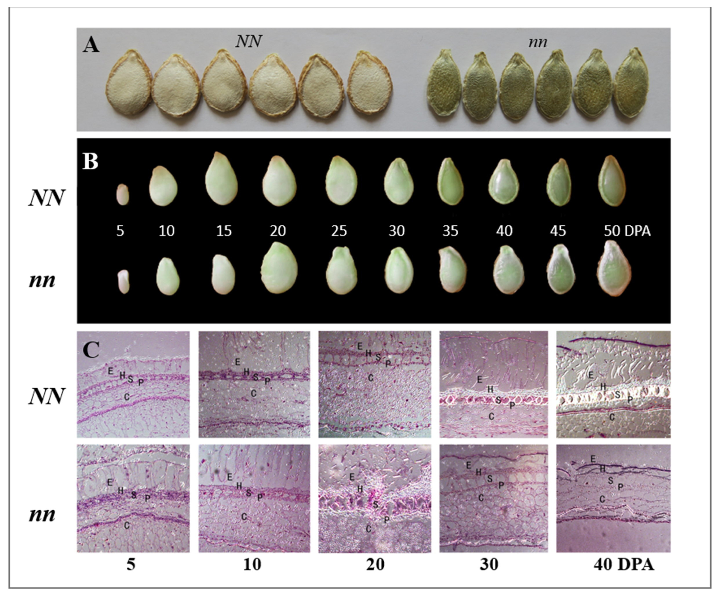
Figure 1. Seed development and scanning electronic microscopic (SEM) observations of seed testa in wildtype (HS-A, NN) and naked seed (HLS-B, nn) pumpkin lines. (A) Appearance of mature dry seeds of WT (left) and mutant (right). (B) Morphology of fresh seed coat at different development stages in WT and mutant. (C) SEM view of seed testa structure in WT and mutant at different developmental stages. E = epidermis, H = hypodermis, S = sclerenchyma, P = parenchyma, C = chlorenchyma. DAP, days after pollination. Scale bars, 25 μm.
Genes 14 00962 g001
Figure 2. Identification of candidate gene for the N locus in C. moschata. (A) Genomewide ΔSNP-index plot using SNPs between the mutant and WT bulk shows that the N locus is located in a 2.42 Mbp region on Chr17. (B) Linkage mapping in RILs narrowed down the N locus into a 389.5-kb region with 15 predicted genes. (C) Multiple lines of evidence support CmoCh17G004790 as the most possible candidate gene for the N locus (Red in (C)). (D) Sequence analysis indicates that the naked seed mutation is due to an alternative splicing in seed coat in the mutant. In (A), the X-axis shows chromosomes. The Y-axis is ΔSNP-index value. Red line is 99% significance threshold. In (B), the grey and blue bars represent different haplotypes defined by molecular markers. The genotype and # of RILs carrying each haplotype in the RIL population are shown to the right. In (D), the red block upstream of the translation start site (TSS) indicates SSR (simple sequence repeat) that was proposed to be the causal variant of naked seed in C. pepo [19].
Genes 14 00962 g002
Figure 3. Expression of CmNST1 in seed coat from RNA-Seq (A) and qPCR (B). The expression level of the CmNST1 at 10, 20, 30, and 40 DAP from BSR-Seq is expressed in FPKM (fragments per kilobase of transcript per million mapped reads) values. ** p < 0.01; * p < 0.05. Error bar represents mean ± SD (n = 3 biological replications).
Genes 14 00962 g003
Figure 4. Alignment of genomic DNA (gDNA) and cDNA/mRNA of part of the CmNST1 gene showing translation start (TSS) and alternative splicing (AS) in the seed coat of naked seed mutant. The SSR in 5′ UTR and the AS site in the second exon are highlighted in blue and green colors, respectively. The proposed start codon (ATG, boldface typed) by Lv et al. [19] and current study (boldface typed and highlighted in yellow) is also shown. M = mutant (nn), WT = wildtype (NN), yl = young leaves, sc = seed coat, Ref = reference genome (Rifu from https://cucurbitgenomics.org/, accessed on 10 April 2023).
Genes 14 00962 g004
Figure 5. Differentially expressed genes (DEGs) between WT and mutant seed coat transcriptomes revealed with BSR-Seq. (A) Bar graphs show number of DEGs in WT vs mutant (M) seed coat transcriptomes at 10, 20, 30, and 40 days after pollination (DAP). DN = down-regulated and UP = up-regulated for genes in mutant as compared with those in WT at threshold of log2(fold change) ≥ 1 and FDR < 0.05. (B) volcano plot shows DEGs between WT and mutant at 10 DAP M vs. WT. Green dots are down-regulated genes, while red dots are up-regulated ones, and black dots are genes without significant differences.
Genes 14 00962 g005
Figure 6. Transcriptome profiling in the WT and mutant pumpkin seed coat with DEGs at 10 days after pollination. (A) GO enrichment analysis of DEGs between WT and mutant seed coats. BP, biological process; CC, cellular component; MF, molecular function. (B) COG (Cluster of Orthologous Groups of proteins) function classification analysis of DEGs. (C) KEGG pathway enrichment analysis of DEGs.
Genes 14 00962 g006
Figure 7. Heatmap of expression level of selected genes involved in lignin biosynthesis pathway or secondary cell wall formation. Value in each cell is log2 (fold change) in the mutant in comparison with the WT from RNA-seq data. The boldface types gene is the candidate for the n locus. Cool (light blue, blue) and warm (pink/red) colors indicate down- and up-regulated genes in the mutant as compared to the WT, respectively. See Table S7 for full names of genes.
Genes 14 00962 g007
Figure 8. Roles of CmNST1 in regulating key genes in the lignin biosynthesis pathway and their expression in the naked seed (nn) mutant as compared with its level in the WT of C. moschata from this study. NAC and MYB transcription factor (TF) genes showing differential expression in the two lines are also listed. Dashed arrows indicate the hierarchical network in which CmNST1 acts as the master regulator that activates other NAC or MYB TFs, which in turn regulate genes in the lignin biosynthesis pathway. Green and red arrows indicate the gene is down- or up-regulated in the mutant as compared with that in the WT. PAL, phenylalanine ammonia lyase; C4H, cinnamate 4-hydroxylase; 4CL, 4-hydroxycinnamoyl-CoA ligase; C3H, p-coumarate 3-hydroxylase; CCoAOMT, caffeoyl-CoA O-methyltransferase; CCR, cinnamoyl-CoA reductase; F5H, ferulate 5-hydroxylase; COMT, caffeic/5-hydroxyferulic acid O-methyltransferase; CAD, cinnamyl-alcohol dehydrogenase; POD, peroxidase; LAC, laccase. The pathway diagram follows [40,41,42].
Genes 14 00962 g008
Disclaimer/Publisher’s Note: The statements, opinions and data contained in all publications are solely those of the individual author(s) and contributor(s) and not of MDPI and/or the editor(s). MDPI and/or the editor(s) disclaim responsibility for any injury to people or property resulting from any ideas, methods, instructions or products referred to in the content.