Article

Efficacy of Radiant Catalytic Ionization in Reduction of Enterococcus spp., Clostridioides difficile and Staphylococcus aureus in Indoor Air

by
Krzysztof Skowron
1,*,
Katarzyna Grudlewska-Buda
1,
Sylwia Kożuszko
1,
Natalia Wiktorczyk
1,
Karolina Jadwiga Skowron
2,
Agnieszka Mikucka
1,
Zuzanna Bernaciak
1 and
Eugenia Gospodarek-Komkowska
1
1
Department of Microbiology, Nicolaus Copernicus University in Toruń, Ludwik Rydygier Collegium Medicum, 9 M. Skłodowskiej-Curie Street, 85-094 Bydgoszcz, Poland
2
Institute of Telecommunications and Computer Science, UTP University of Science and Technology, Al. prof. S. Kaliskiego 7, 85-796 Bydgoszcz, Poland
*
Author to whom correspondence should be addressed.
Atmosphere 2020, 11(7), 764; https://doi.org/10.3390/atmos11070764
Submission received: 16 June 2020 / Revised: 15 July 2020 / Accepted: 17 July 2020 / Published: 20 July 2020

Abstract

:
(1) Background: An aerogenic way is one of main rout of spreading microorganisms (including antibiotic resistant), that cause healthcare-associated infections. The source of microorganisms in the air can be patients, personnel, visitors, outdoor air, hospital surfaces and equipment, and even sink drains. (2) Methods: The standardized suspensions (0.5 McFarland) of the examined strains (Enterococcus spp., Clostridioides difficile, Staphylococcus aureus) were nebulized in sterile chamber. Then the Induct 750 (ActivTek) device, generating RCI (radiant catalytic ionization) phenomenon, was used for 20 min. Next, the number of bacteria in the air was calculated using collision method. The percentage of reduction coefficient (R) was calculated. (3) Results: In case of enterococci, the R value was >90% and there are no statistically significant differences among tested strains. For C. difficile strains the R value range from 64–95%. The R value calculated for hypervirulent, antibiotic resistant CDI PCR 27 strain was statistically significantly lower than for other examined strains. For S. aureus non-MRSA the R value was 99.87% and for S. aurues MRSA the R value was 95.61%. (4) Conclusions: The obtained results indicate that the use of RCI may contribute to reducing the occurrence of dangerous pathogens in the air, and perhaps transmission and persistence in the hospital buildings environment.

1. Introduction

Healthcare-associated infections (HAIs) are a very serious medical problem. HAIs pose a threat to hospitalized patients. They mainly affect person with a weak immune system. Hospital infections most often affect people in intensive care units. They are usually caused by multi-resistant bacteria with high spreading potential. One of the directions of action is striving to limit patients’ contact with pathogenic microorganisms in the air. The problem of nosocomial infections can be minimized through appropriate control and monitoring systems [1].
Many actions are taken to prevent spread of microorganism. These activities include: antibiotic prevention and therapies, cleaning and disinfection of the surface, hygiene of the hands and clothing of the staff, separation of special areas, isolation of patients with diarrhea, etc., [1].
However, one of the main ways of spreading microorganisms that cause HAI—an aerogenic path—is often overlooked. Unfortunately, for many years, the transmission of pathogenic microorganisms by air was not considered a serious health risk [2]. However, there is a lot of evidence that suggests that both Gram-negative and Gram-positive bacteria are often spread by air in the hospital environment [1]. The hospital environment is a very dynamic environment from the microbiological point of view. The composition of bioaerosol generated in hospital rooms is usually very diverse. The source of microorganisms present in the air can be patients, personnel, visitors, outdoor air, hospital surfaces and equipment, and even sink drains [3,4]. Factors affecting the presence of microbes in hospital air include seasons, weather conditions (e.g., temperature, humidity), efficient ventilation system, humidity, number of patients and guests, as well as activities such as how often the door is opened or staff and guests move [5,6]. Microorganisms can get into the air during medical treatments and even during simple maintenance activities, such as changes in bedding or clothing [7]. It was also shown that contaminated air-conditioning devices were the source of P. aeruginosa infection [1]. It was shown that in the environment of patients infected with methicillin-resistant Staphylococcus aureus (MRSA), 4.7 CFU m3 of MRSA, and during bed turn down the number of bacteria increased up to 116 CFU m3 [7]. The problem of bioaerosol formation by Gram-negative bacteria is particularly dangerous in relation to medical equipment, e.g., humidifiers, nebulizers, respirators, which is associated with respiratory infections [1]. The main route of infectious bioaerosol spread in the hospital are ventilation systems. There are two main types of ventilation systems—natural and mechanical [8], which choice depends on the type of room. Mechanical fans can be installed directly in windows or walls or installed in air ducts to supply air to or extract air from the room [9,10]. Through ventilation system, pathogenic microorganisms from one patient can spread to other rooms or even other floors of the building, and can also sediment on various surfaces in other rooms, causing their contamination [9]. Proper maintenance of ventilation systems can help to reduce infection [10].
Among the microorganisms spreading through the aerogenic pathway in hospitals, multi-drug resistant (MDR) enterococci, C. difficile and MRSA are important.
Enterococcus spp. are a natural microbiota of the digestive tract of humans and animals. Enterococcus spp. may be the etiological factor of various forms of clinical infections, especially urinary tract, endocarditis, peritonitis, and burn wound infections. Most nosocomial infections are caused by biofilm forming bacteria, which allows them to survive in the urinary tract, avoiding the host’s immune response. Increasingly, MDR strains of Enterococcus spp. are isolated in the hospital environment. Currently, enterococci are classified as alarm pathogens. They are among the most dangerous multi-drug resistant pathogens, called the ESKAPE acronym (Enterococcus faecium, Staphylococcus aureus, Klebsiella pneumoniae, Acinetobacter baumannii, Pseudomonas aeruginosa, Enterobacter spp.), responsible for infections associated with healthcare (HAIs) [11].
The majority of enterococcal infections are endogenous, however, exogenous infections are more frequently found in hospitalized patients and result from transmission of strains from other patients or the hospital environment. The primary mode of spread from patient-to-patient occurs through the hands of healthcare workers [12]. In recent decades, an increase in isolation of Enterococcus spp. resistant to ampicillin and vancomycin from the hospital environment was noticed. This tendency applies in particular Enterococcus faecium strains. This applies mainly to the acquisition of vancomycin resistance among these strains (Vancomycin Resistant Enterococcus, VRE) [11]. A frequent among enterococci is the simultaneous occurrence of several mechanisms of resistance to antibiotics from different chemical groups [13].
C. difficile is currently one of the most important pathogens responsible for antibiotic-associated diarrhea and pseudomembranous colitis, mainly in hospital patients, but increasingly also in non-hospital patients, including persons without risk factors [14]. Pathogenic C. difficile strains produce toxin A (TcdA, encoded by the tcdA gene) and/or toxin B (TcdB, encoded by the tcdB gene). Toxins cause, among others, destruction of the cytoskeleton, apoptosis of epithelial cells, induction of proinflammatory cytokine production, recruitment of inflammatory cells, contributing to the destruction of intercellular connections, which, with the participation of hydrolytic enzymes, leads to the development of colitis, pseudomembrane, and diarrhea [14,15]. Since the emergence of hypervirulent ribotype PCR 027 in Europe, there has been an increase in C. difficile infection (CDI) cases, mortality, and further evolution of strains to increase virulence, the possibility of spreading in the environment and resistance to antibiotics [15]. Resistance to moxifloxacin may be a marker of increased virulence of C. difficile strains [16]. Roberts et al. [17] and Best et al. [18] demonstrated that C. difficile can easily spread in the hospital environment through aerogenic pathways. Spores play a major role in the spread and maintenance of C. difficile strains in the hospital environment [19]. Spores are formed after 15 min of exposure of vegetative forms to oxygen, they are resistant to many hygienization and disinfection procedures [20]. Evidence for this is provided by the frequent isolation of C. difficile spores from ventilation ducts in hospitals and high horizontal surfaces [21]. The mean spore length is 1–1.5 mm and the mean diameter is 0.5–0.7 μm, although there is documented significant variation in individual sizes of spores both within and between strains. Fallout time of C. difficile spores in a still room from 1 m height range from 2.1 to 13.9 h [22]. Furthermore, it was suggested that aerial dissemination could play a role in the persistence of C. difficile in hospitals [19].
S. aureus is an opportunistic human pathogen [23,24], which can cause, among others, wound infections, pneumonia in immunocompromised individuals, chest abscess and bacteraemia [25]. S. aureus is listed as one of the most common pathogens responsible for nosocomial infections [23]. About a third of the population is carriers of S. aureus. It usually colonizes wet areas, such as armpits, groin, and nose, although it can also be found on other parts of the body, for example on the hands [23]. In contrast, MRSA (Methicillin-resistant Staphylococcus aureus) strains are of particular concern because of their resistance to β-lactam antibiotics, which makes treatment more difficult [25]. This pathogen is currently responsible for about 61% of staphylococcal infections [25]. The basic mode of transmission of MRSA strains within the hospital are temporarily colonized hands of hospital staff. In addition, it is believed that MRSA in the form of bioaerosol can pollute the air. Although the transmission of MRSA in the air is generally considered less frequent than the transmission by direct contact, it is considered that air is an important factor to be considered in hospital wards [26]. Both S. aureus and MRSA in the air are present in the form of particles with an aerodynamic diameter that can accumulate in the human upper respiratory tract, primary, secondary, and final bronchi and alveoli. The spherical cells of S. aureus are up to maximal 1 μm in diameter [23,24,25,26].
Therefore, it is important to look for the effective methods to maintain microbiological purity of the air and ventilation systems. One of them is radiant catalytic ionization (RCI). RCI [27] is an active method of air and surface cleaning. The RCI cell consists of matrices of elongated polycarbonate components, arranged in a parallel orientation resembling a honeycomb. A coating of matrices comprises a grouping of the materials: titanium dioxide, rhodium, silver, and copper. On the opposite site a broad-spectrum UV light source is located. The UV lamp utilizes argon gas with mercury and carbide filaments with a spectrum of 100 and 367 nm [28,29]. It works by creating a proper wavelength and using the photo-oxidation effect with the participation of UV light and appropriate photocatalysers such as TiO2, which are placed in the hydrophilic coverage of the RCI chamber [28]. The result is generation of biocidal reactive oxygen species (ROS), hydroxyl radicals (OH), superoxide radicals (O2−•), hydro-peroxyl radical (HO2), and hydrogen peroxide (H2O2) [28,29]. The manufacturer declares that the total number of generated ions is about 5.0 × 10⁵ ions cm−3 of air. In addition, in photocatalytic oxidation also other secondary impurities aldehydes: acetaldehyde and formaldehyde are produced. Also aldehydes have harmful health effects [30,31]. Photocatalysis has been found to not only effectively eliminate Gram-positive and Gram-negative bacteria, spores, viruses, fungi, and protozoa but also inactivate prions and bacterial toxins [32]. Gram-positive bacteria have been shown to be more resistant to photocatalytic disinfection than Gram-negative [28]. Their action is related to the oxidation of coenzyme A molecules, which inhibits the respiratory pathway, oxidation of unsaturated phospholipids, interactions with extracellular polymeric substances (EPS), and causes the accumulation of DNA and RNA damage in the bacterial cell [32,33,34]. Despite the ozone generation during the operation of the device, it has been shown that it is not a main bactericidal agent, because at the time of air exchange its level does not exceed 0.05 ppm [28,33,35]. It is important that WHO Air Quality Quidelines 2005 gives for ozone 8-h mean limit of 100 μg/m3. WHO points out that sensitive individual may have health effects also in lower concentrations than the limit. It is very important that the producer of the equipment not only says, but also offers some reliably tested proofs about the measured levels of the produced ozone. According to USA EPA ozone generators have many harmful health effects [36]. In case of tested RCI device, its producer declares ozone production at a level below 0.04 ppm [35]. Currently, new version of RCI devices does not generate ozone.
The aim of the study is to assess the efficiency of RCI in eliminating enterococci resistant to selected antibiotics in the air compared to the antibiotic-susceptible strain, antibiotic-resistant, toxinogenic C. difficile in comparison with the non-toxinogenic, antibiotic-susceptible strain and elimination of S. aureus non-MRSA and MRSA strain.

2. Materials and Methods

2.1. Materials

The research material consisted of three Enterococcus spp. standard strains, three C. difficile strains isolated, and two S. aureus strains from a clinical specimen from the collection of the Department of Microbiology, Ludwik Rydygier Collegium Medicum. in Bydgoszcz, Nicolaus Copernicus University in Toruń:
Enterococcus faecalis PCM 1861, isolated from clinical material, susceptible to antibiotics.
Enterococcus faecalis ATCC 51299, isolated from a peritoneal fluid sample from a patient from Saint Louis, Missouri, USA, resistant to vancomycin, gentamicin, streptomycin, and erythromycin, with confirmed presence of genes: vanB i ant(6)-I acc(6′) aph(2′′′′).
Enterococcus faecium ATCC 51559, isolated from a patient from Brooklyn, New York, USA, resistant to vancomycin, teicoplanin, ampicillin, gentamycin, ciprofloxacin, and rifampicin, with confirmed presence of vanA gene.
C. difficile non-toxigenic strain (CDI tox(−)).
C. difficile strain producing A, B, and binary toxins and resistant to moxifloxacin (CDI MXF-R/tox A/B/bin(+)).
C. difficile PCR-ribotype 027 strain (CDI PCR 027).
S. aureus MRSA isolated from wound infection.
S. aureus non-MRSA isolated from wound infection.
All strains were plated on Columbia Agar with 5% sheep blood (Becton-Dickinson) and incubated at 37 °C for 24 h. After this time, the grown strains were transfer on the same type of medium, and the grown colonies were used in the next stages of the study. C. difficile was incubated under anaerobic conditions (Genbag anaer atmosphere generator (bioMérieux) at 37 °C for 48 h.

2.2. RCI Efficiently

For the purpose of the study, standardized suspensions of the examined strains were prepared from fresh culture in physiological saline (Polpharma) with an optical density of 0.5 McFarland standard using a densitometer (DEN-1B, Biosan). The bacterial concentration in the suspension was respectively for Enterococcus spp. 2.38 × 10⁸ CFU cm−3 (±5.26 × 10⁷); C. difficile 7.25 × 10⁷ (±1.66 × 10⁷) CFU cm−3; S. aureus 1.72 × 10⁸ (±4.17 × 10⁷) CFU cm–3. In case of C. difficile, the suspensions of vegetative cells of the tested strains were used. However, further handling with suspension lasting about 50 min was not conducted in anaerobic conditions, which could lead to the formation of spores. As a result, the spontaneously arose mixtures of vegetative forms and spores of C. difficile were RCI treated during the experiment. This reflected the actual conditions for the spread of C. difficile in hospitals.
Then, 4 mL of each suspension was placed individually in a sterile nebulizer chamber of the MONSUN MP1 (Medbryt) pneumatic inhaler. Nebulization was carried out until the inhaler chamber was completely emptied (about 15 min). The capacity of the nebulizer compressor is 15.5 L/min and the maximal aerosol capacity is 0.48 mL/min with a particle diameter of 1.4–2.4 μm.
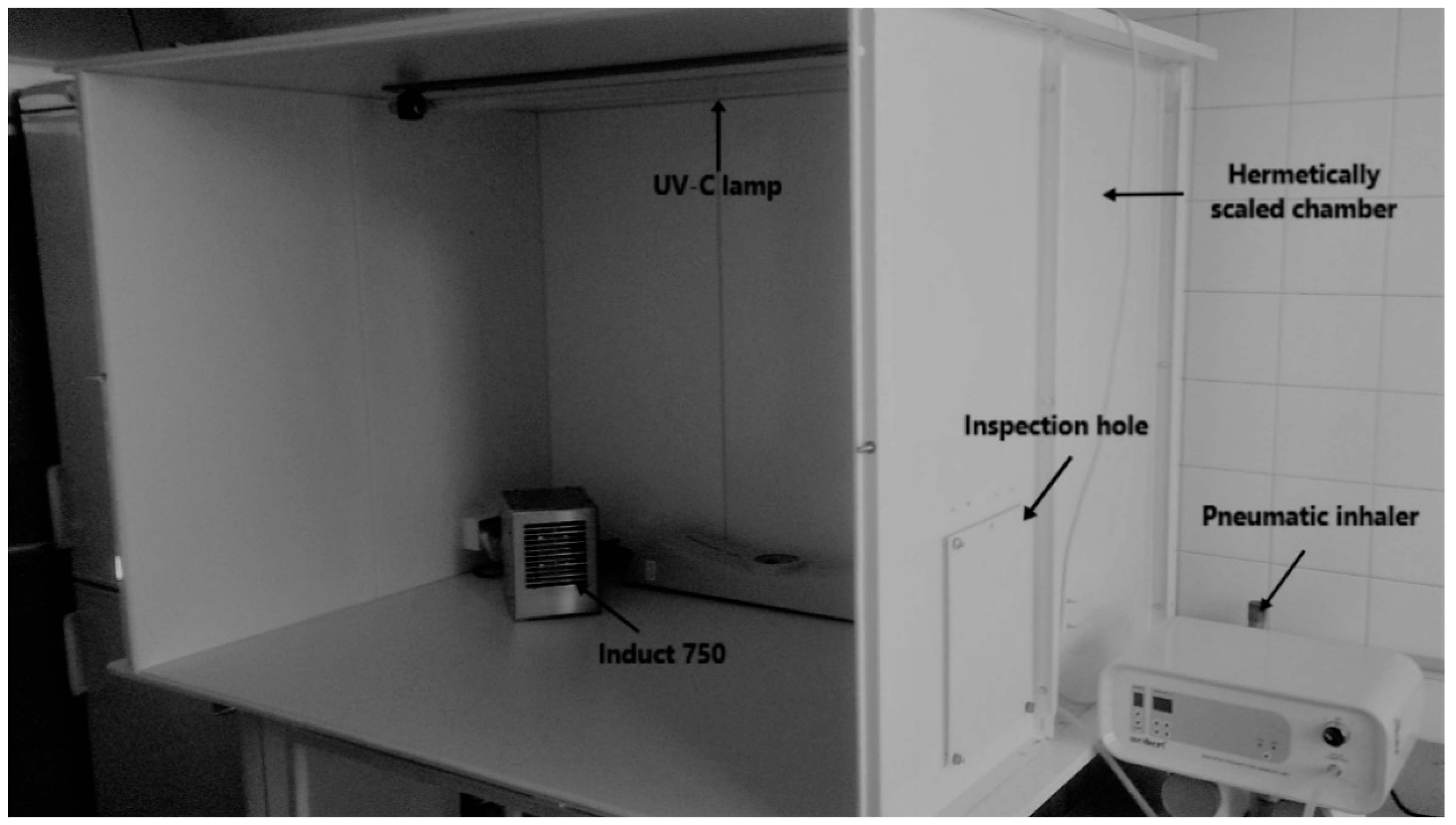
The nebulizer chamber was placed in the test room, which was a hermetic chamber, with a volume of 1.4 m3 made of steel plates. The chamber is closed with a front wall made of polycarbonate placed in a metal frame. This wall is screwed and closed with side closures. All connections are sealed. The hermetic test was carried out using colored smoke generated by Björnax smoke candles. Before each subsequent nebulization, the walls of the chamber were disinfected chemically with an agent intended for disinfection of solid surfaces, and the air contained in it was subjected to UV-C lamp Philips TUV 36W/G36 T8 for 20 min. After this time, the chamber was opened for about 20 min to remove accumulated ozone. During ventilation the chamber, in the test room the airflow UV-C lamp, which does not generate ozone, was turning on. Moreover, the air and surfaces in the test room were subjected to UV-C radiation for 2 h, before the start the experiment. Before commencing nebulization a control assessment of microbiological purity of the air was carried out in order to check the so-called microbiological background level. A detailed experimental design is shown in Figure 1, and the appearance of research set is presented in Figure 2. Each experiment was conducted in triplicate for each strains.
The air samples were sampled using the collision method with MAS-100 Eco (Merck) device. The nominal air flow through the sampler head is 100 L/min. In order to assess the microbiological purity of the air after using Philips TUV 36W/G36 T8 UV-C lamps and Induct 750 devices, 0.2 and 0.5 m3 were taken. However, in order to assess the level of bacterial contamination in the chamber after nebulization of the bacterial suspension, 0.01 and 0.05 m3 were sampled. Enterococcus spp. bacteria were cultured on Enterococcosel Agar (Becton-Dickinson) at 35 °C for 24 h. C. difficile were grown on chromID® C. difficile (bioMérieux) under anaerobic conditions generated with Genbag anaer atmosphere generator (bioMérieux) at 37 °C for 48 h. S. aureus strains were cultured on mannitol salt agar (Becton Dickinson). The colonies growing on the agar were counted and converted into colony forming units (CFU) m−3 of air. Effectiveness was expressed by giving the number of CFU before and after using the Induct 750, and calculating the percentage reduction (R [%]) according to the formula:
R RCI [ % ] = A B A × 100
where: A—the initial number of microorganisms [CFU m−3], B—the number of microorganisms after using the device [CFU m−3].
The positive control in the study was suspensions of the microorganisms tested, which were nebulized and not exposed to RCI. Samples in the volume of 0.2 and 0.5 m3 were taken after 20 min. In this way, spontaneous precipitate overtime was evaluated. Precipitate factor was expressed by giving the number of CFU directly after neubulization and 20 min after nebulization, and calculating the percentage reduction Rwitout RCI [%] according to the formula presented above, where B was the number of microorganisms after 20 min from nebulization without RCI action [CFU m−3].

2.3. Experimental Environment Conditions

In addition, negative ion concentration was measured in the chamber air before and after the RCI technology usage. The measurement was made using air negative ion measuring instrument KT-401 AIR (VKTECH)—measurement range 104–106 ions cm−3. The air temperature and humidity in the chamber was also measured using the thermo-hygrometer LB-710AL (Lab-El).

2.4. Statistical Analysis

The obtained results were subjected to statistical analysis in the STATISTICA 13.0 PL (TIBCO Software, Palo Alto Networks, Inc., Santa Clara, CA, USA). The significance of differences between the values of R coefficients calculated for the strains of a given species was checked based on Tukey’s test at the significance level of 0.05.

3. Results

3.1. Experimental Environment Coditions

The experimental environment conditions are presented in Table 1.

3.2. Changes in the Number of Enterococcus spp. in the Air

The obtained results showed some fluctuations of the CFU number of tested enterococci present in the air after nebulization of suspensions (Table 2). For this reason, it was decided to introduce an absolute reduction measure in the form of R [%].
After the nebulization, it was found that the tested strains of Enterococcus spp. in a similar number spread in the form of an aerosol (Table 2).
The use of RCI resulted in a significant decrease in the CFU number both E. faecalis and E. faecium strains in the air. CFU number of all tested strains decreased by over 90%. The highest decrease was observed in the E. faecalis PCM 1861 strain, susceptible to antibiotics, and the lowest in the E. faecalis ATCC 51299 strain. The differences between the strains were not statistically significant (Table 2).

3.3. Changes in the Number of C. difficile in the Air

The obtained results showed, similarly as in the case of Enterococcus spp., some fluctuations in the CFU number of tested bacilli present in the air after the suspension nebulization (Table 3). For this reason, it was decided to introduce an absolute reduction measure in the form of R [%].
After the nebulization, it was found that the CDI strain MXF-R/tox A/B/bin (+) most intensively formed bioaerosol, and the CDI PCR 027 strain—the worst (Table 3).
The use of RCI resulted in a decrease in the CFU number of all C. difficile strains tested in the air. The highest decrease, amounting to almost 95%, was found for the CDI MXF-R/tox A/B/bin(+) strain, and the lowest (over 64%) for the CDI PCR 027 strain (Table 3). The non-toxigenic CDI tox(−) strain was more resistant than the CDI MXF-R/tox A/B/bin(+) strain producing all toxins. Percentage reduction coefficients for the number of CDI tox(−) and CDI MXF-R/tox A/B/bin(+) strains were statistically significantly higher compared to the coefficient calculated for the CDI PCR 027 strain (Table 3).

3.4. Changes in the Number of Staphylococcus aureus in the Air

Like in the case of species mentioned above, some fluctuations in the CFU number of tested S. aureus strains in the air after the suspensions nebulization were observed (Table 4). For this reason, it was decided to introduce an absolute reduction measure in the form of R [%].
The number of S. aureus reisolated from bioaerosol, after nebulization, range from 4.70 × 10⁵ CFU m−3 for MRSA strain to 5.10 × 10⁵ CFU m−3 for non-MRSA strain (Table 4).
The use of RCI resulted in a decrease in the CFU number of both S. aureus strains tested in the air. The greater number reduction (99.87%) was observed in case of non-MRSA strain. The statistically significant differences in R[%] were not shown among the tested strains (Table 4).

4. Discussion

Microbiological air pollution in hospitals plays an important role in the spread of healthcare-associated infections [38,39,40]. Despite the methods of air purification used so far, e.g., ultraviolet germicidal irradiation (UVGI), HEPA filters, it is necessary to look for new, more effective technologies that enable the fight against increasingly virulent microbial strains that can spread through the aerogenic route. It is also important that new techniques are safe for people, because we spend over 90% of our time indoors and can be used while people are inside. RCI technology, unlike passive air purification methods, is an active method that purifies the air not only inside, but also outside the device. RCI has been shown to be an effective method for eliminating microbial contaminants, including viruses, vegetative forms, and persistent bacteria, from the surface [37,41,42,43,44,45]. It was found that this technology can be successfully used in many industries.
RCI method is quite a new approach to the indoor air disinfection issue. There are only very few publications in the available literature regarding the use of this method, and especially in relation to the experimental layout adopted in this research. Therefore, there are difficulties with regard to the results of own research to the work of other authors.
In the conducted experiment, the effectiveness of RCI against different strains of Enterococcus spp. and vegetative forms of C. difiicile were evaluated, paying attention to their virulence and resistance to antibiotics. The study showed the highest efficacy of RCI against E. faecalis PCM 1861 susceptible to antibiotics (R [%] = 99.76), but for all tested strains of Enterococcus spp., more than 90% efficiency in reducing the number of microorganisms in air after RCI has been demonstrated. In contrast, the lowest RCI efficacy was demonstrated for the C. difficile strain ribotype 027 (R [%] = 64.45). The use of RCI resulted in a decrease in the CFU number of both S. aureus strains tested in the air. The greater number reduction (99.87%) was observed in case of non-MRSA strain. Skowron et al. [37], higher efficacy of RCI against other spore forming bacteria, such as Bacillus subtilis, has been demonstrated (R [%] = 98.92). The lower susceptibility of C. difficile to RCI may be related to the virulence factors of these microorganisms, whereas this mechanism should be explained in subsequent studies. C. difficile rybotype 027 is an epidemic, high virulent strain that is characterized by more intense sporulation and production of large amounts of toxin A and B (16–23 times higher in vitro concentration than in case of other strains) [19,46,47,48]. The risk of aerogenic transfer of these microorganisms has been demonstrated by Roberts et al. [17], where the presence of these microorganisms in air samples collected in a hospital environment in Great Britain was demonstrated. Skowron et al. [37] showed the lowest RCI efficacy against Clostridium sporogenes spores (R [%] = 71.73). In other studies [44], we showed a reduction of Klebsiella pneumoniae NDM strains in the air at the level of 1.80 log CFU m−3 after using RCI technology. Skowron et al. [37] for S. aureus and S. epidermidis found a decrease in number amounted to 4–5 logarithmic units, and the percentage reduction rate was 99.9%. Also Grinshpun et al. [27] indicated that RCI technology caused a reduction of the B. subtilis spores in the 2.75 m3 chamber. In the study, a chamber with a cubic capacity of 1.4 m3 was used. The device manufacturer declares the device’s efficiency to 70 m3 (ActivTek, instruction of use Induct 750).
Skowron et al. [37] also demonstrated the differential efficacy of RCI on microorganisms in the air. The highest reduction coefficient (R [%] = 100) was demonstrated for Escherichia coli and Candida albicans. Effective elimination from the air was observed for E. faecalis ATCC 29212 (R [%] = 99.99), which, like E. faecalis PCM 1861, is sensitive to antibiotics [37]. Barabasz et al. [48] indicated that RCI was effective in rooms of cubature 20 and 45 m3. The percentage reduction rate for the total number of Staphylococcus spp. and fungi ranged from 73.1% to 82.0% [42]. In turn, Wiktorczyk et al. [49] used the RCI module as a built-in element of the cabinet for storing endoscopes. The treatment influenced the meeting of microbiological criteria of air in the wardrobe [49].
In this study, the chamber nebulization time was 20 min, this is one of the parameters that can affect the effectiveness of eliminating pathogens from the air. In Skowron et al. study [37], the exposure time was also 20 min. However, in another study, we evaluated different exposure times [50] for the effectiveness of RCI technology. Skowron et al. [50], showed that the number of bacteria decreased with RCI exposure over time, which confirms previous studies. Skowron et al. [50] showed that the reduction of S. aureus from a stainless steel surface after 20 min exposure to RCI was 5.20 log CFU cm−2. At the same time, Skowron et al. [50] found that S. aureus was the most resistant to RCI on the rubber surface. Grinshpun et al. [27] found that increasing the exposure time from 10 to 30 min increased the reduction in B. subtilis from 75 to 90%. Ortega et al. [34] showed that the 2 h RCI activity allowed a reduction of 90% of bacterial stainless steel plankton cells. The duration of RCI technology is crucial at the site of its application, especially in hospitals.
RCI technology based on reactions of photooxidation may in the near future become one of the most popular methods of air and surface cleaning, which is already in use in hospitals, museums, and schools [51]. Skorwon et al. [44] demonstrated the effectiveness of eliminating K. pneumoniae NDM from the materials used as hospital room equipment, including bedding. The effectiveness of this technology seems to be particularly important for highly virulent and antimicrobial resistant organisms that pose the greatest challenge for modern microbiology. In addition, Dimitrakopoulou et al. (2012), showed that the use of photocatalysis methods using UV-A/TiO2 plays a role in the degradation of antibiotics present in the environment [52]. It is possible to use this technology in ventilation systems as support for existing solutions, e.g., HEPA filters, which will enable inactivation and removal of dead microorganisms from the air at the same time. RCI technology is mainly intended for cleaning inside the rooms and preventing the spread of microorganisms present in the hospital environment, e.g., between rooms, by personnel. RCI technology is based on photocatalysis. The emission of harmful substances has been proven, such as, ozone, aldehydes—formaldehyde and acetaldehyde, during photocatalysis, if this process occurs in an environment where a high concentration of volatile organic compounds (VOCs) is stated [30]. However, there is no research in the available literature on the production of such substances during the operation of RCI in rooms where there are people. A safe solution would be monitoring the concentrations of hazardous substances in places where RCI technology is used continuously when people are present in the room.

5. Conclusions

The obtained results indicate that the use of RCI may contribute to reducing the occurrence of dangerous pathogens (including MRSA) in the indoor air, and perhaps transmission and persistence in the environment. It is worth noticing that the RCI device should be taken into account in case of ventilation systems designing. However, in the future, more strains from different species should be examined. In addition, research is needed in real indoor environments.

Author Contributions

Conceptualization, K.S., K.G.-B., and S.K.; methodology, K.S., K.G.-B., and N.W.; validation, K.S. and N.W.; formal analysis, K.S. and K.J.S.; investigation, K.G.-B., S.K., and A.M.; resources, K.G.-B., S.K., and A.M.; writing—original draft preparation, N.W. and Z.B.; writing—review and editing, K.S., K.J.S., and Z.B.; visualization, N.W. and K.J.S.; supervision, K.S.; project administration, K.S. and E.G.-K.; funding acquisition, E.G.-K. All authors have read and agreed to the published version of the manuscript.

Funding

This research was financially supported by the Nicolaus Copernicus University with funds from the maintenance of the research potential of the Department of Microbiology PDB WF 536.

Conflicts of Interest

The authors declare no conflict of interest.

References

  1. Beggs, C.; Knibbs, L.D.; Johnson, G.R.; Morawska, L. Environmental contamination and hospital-acquired infection: Factors that are easily overlooked. Indoor Air 2015, 25, 462–474. [Google Scholar] [CrossRef] [PubMed] [Green Version]
  2. Jung, C.C.; Wu, P.C.; Tseng, C.H.; Su, H.J. Indoor air quality varies with ventilation types and working areas in hospitals. Build. Environ. 2015, 85, 190–195. [Google Scholar] [CrossRef]
  3. Gilbert, Y.; Veillette, M.; Duchaine, C. Airborne bacteria and antibiotic resistance genes in hospital rooms. Aerobiologia 2010, 26, 185–194. [Google Scholar] [CrossRef]
  4. Park, D.U.; Yeom, J.K.; Lee, W.J.; Lee, K.M. Assessment of the levels of airborne bacteria, gram-negative bacteria and fungi in hospital lobbies. Int. J. Environ. Res. Publ. Health 2013, 10, 541–555. [Google Scholar] [CrossRef] [PubMed]
  5. Scaltriti, S.; Cencetti, S.; Rovesti, S.; Marchesi, I.; Bargellini, A.; Borella, P. Risk factors for particulate and microbial contamination of air in operating theatres. J. Hosp. Infect. 2007, 66, 320–326. [Google Scholar] [CrossRef]
  6. Wan, G.H.; Chung, F.F.; Tang, C.S. Long-term surveillance of air quality in medical center operating rooms. Am. J. Infect. Control 2011, 39, 302–308. [Google Scholar] [CrossRef]
  7. Shiomor, T.; Miyamoto, H.; Makishima, K.; Yoshida, M.; Fujiyoshi, T.; Udaka, T.; Inaba, T.; Hiraki, N. Evaluation of bedmaking-related airborne and surface methicillin-resistant Staphylococcus aureus contamination. J. Hosp. Infect. 2002, 50, 30–35. [Google Scholar] [CrossRef] [Green Version]
  8. Atkinson, J.; Chartier, Y.; Pessoa-Silva, C.L.; Jensen, P.; Li, Y.; Seto, W.H. Natural Ventilation for Infection Control in Health-Care Settings; World Health Organization: Geneva, Switzerland, 2009. Available online: https://www.ncbi.nlm.nih.gov/books/NBK143277/ (accessed on 8 April 2020).
  9. Lim, T.; Chob, J.; Kim, B.S. The predictions of infection risk of indoor airborne transmission of diseases in high-rise hospitals: Tracer gas simulation. Energy Build. 2010, 42, 1172–1181. [Google Scholar] [CrossRef]
  10. Wallner, P.; Munoz, U.; Tappler, P.; Wanka, A.; Kundi, M.; Shelton, J.F.; Hutter, H.P. Indoor environmental quality in mechanically ventilated, energy-efficient buildings vs. conventional buildings. Int. J. Environ. Res. Public Health 2015, 12, 14132–14147. [Google Scholar] [CrossRef]
  11. Gao, M.; An, T.; Li, G.; Nie, X.; Yip, H.L.; Zhao, H.; Wong, P.K. Genetic studies of the role of fatty acid and coenzyme A in photocatalytic inactivation of Escherichia coli. Water Res. 2012, 46, 3951–3957. [Google Scholar] [CrossRef]
  12. Hayden, M.K. Insights into the epidemiology and control of infection with vancomycin-resistant enterococci. Clin. Infect. Dis. 2000, 31, 1058–1065. [Google Scholar] [CrossRef] [PubMed] [Green Version]
  13. Rathnayake, I.U.; Hargreaves, M.; Huygens, F. Antibiotic resistance and virulence traits in clinical and environmental Enterococcus faecalis and Enterococcus faecium isolates. Syst. Appl. Microbiol. 2012, 35, 326–333. [Google Scholar] [CrossRef] [PubMed]
  14. Collins, J.; Auchtung, J.M. Control of Clostridium difficile infection by defined microbial communities. Microbiol. Spectr. 2017, 5, 1–25. [Google Scholar]
  15. Awad, M.M.; Johanesen, P.A.; Carter, G.P.; Rose, E.; Lyras, D. Clostridium difficile virulence factors: Insights into an anaerobic spore-forming pathogen. Gut Microbes 2014, 5, 579–593. [Google Scholar] [CrossRef] [PubMed] [Green Version]
  16. Quesada-Gómez, C.; López-Ureña, D.; Acuña-Amador, L.; Villalobos-Zúñiga, M.; Du, T.; Freire, R.; Guzmán-Verri, C.; del Mar Gamboa-Coronado, M.; Lawley, T.D.; Moreno, E.; et al. Emergence of an outbreak-associated Clostridium difficile variant with increased virulence. J. Clin. Microbiol. 2015, 53, 1216–1226. [Google Scholar] [CrossRef] [PubMed] [Green Version]
  17. Roberts, K.; Smith, C.F.; Snelling, A.M.; Kerr, K.G.; Banfield, K.R.; Sleigh, P.A.; Beggs, C.B. Aerial dissemination of Clostridium difficile spores. BMC Infect. Dis. 2008, 8, 1–6. [Google Scholar] [CrossRef] [Green Version]
  18. Best, E.L.; Fawley, W.N.; Parnell, P.; Wilcox, M.H. The potential for airborne dispersal of Clostridium difficile from symptomatic patients. Clin. Infect. Dis. 2010, 50, 1450–1457. [Google Scholar] [CrossRef] [Green Version]
  19. Carlson, P.E.; Kaiser, A.M.; McColm, S.A.; Bauer, J.M.; Young, V.B.; Aronoff, D.M.; Hanna, P.C. Variation in germination of Clostridium difficile clinical isolates correlates to disease severity. Anaerobe 2015, 33, 64–70. [Google Scholar] [CrossRef] [Green Version]
  20. Gerding, D.N.; Muto, C.A.; Owens, R.C. Measures to control and prevent Clostridium difficile infection. Clin. Infect. Dis. 2008, 46, S43–S49. [Google Scholar] [CrossRef] [Green Version]
  21. Fawley, W.N.; Parnell, P.; Verity, P.; Freeman, J.; Wilcox, M.H. Molecular epidemiology of endemic Clostridium difficile infection and the significance of subtypes of the United Kingdom epidemic strain (PCR ribotype 1). J. Clin. Microbiol. 2005, 43, 2685–2696. [Google Scholar] [CrossRef] [Green Version]
  22. Snelling, A.M.; Beggs, C.B.; Kerr, K.G.; Shepherd, S.J. Spores of Clostridium difficile in Hospital Air. Clin. Infect. Dis. 2010, 51, 1104–1105. [Google Scholar] [CrossRef] [PubMed]
  23. Aires de Sousa, M.; Lencastre, H. Bridges from hospitals to the laboratory: Genetic portraits of methicillin-resistant Staphylococcus aureus clones. Pathog. Dis. 2004, 40, 101–111. [Google Scholar]
  24. Berning, C.; Lanckohr, C.; Baumgartner, H.; Drescher, M.; Becker, K.; Peters, G.; Köck, R.; Kahl, B.C. Fatal infections caused by methicillin-resistant Staphylococcus aureus of clonal complex 398: Case presentations and molecular epidemiology. JMM Case Rep. 2015, 2, e000024. [Google Scholar] [CrossRef]
  25. Shirmori, T.; Miyamoto, H.; Makishima, K. Significance of airbone transmission of methicillin-resistant Staphylococus aureus in an otolaryngology-head and neck surgery unit. Arch. Otolaryngol. Head Neck Surg. 2001, 127, 644–648. [Google Scholar] [CrossRef] [PubMed] [Green Version]
  26. Space Fundation, Radiant Catalytic Ionization Air & Water Purification. Available online: http://www.spacefoundation.org/programs/space-certification/certified-products/space-technology/radiant-catalytic-ionization-air (accessed on 10 February 2020).
  27. Grinshpun, S.A.; Adhikari, A.; Honda, T.; Kim, K.Y.; Toivola, M.; Rao, K.S. Reponen, Control of aerosol contaminants in indoor air: Combining the particle concentration reduction with microbial inactivation. Environ. Sci. Technol. 2007, 41, 606–612. [Google Scholar] [CrossRef] [PubMed]
  28. Binas, V.; Venieri, D.; Kotzias, D.; Kiriakidis, G. Modified TiO2 based photocatalysts for improved air and health quality. J. Materiomics 2017, 3, 3–16. [Google Scholar] [CrossRef]
  29. Gao, W.; Howden, B.P.; Stinear, T.P. Evolution of virulence in Enterococcus faecium, a hospital-adapted opportunistic pathogen. Curr. Opin. Microbiol. 2017, 41, 76–82. [Google Scholar] [CrossRef]
  30. Hodgson, A.I.; Destaillats, H.; Sullivan, D.P.; Fisk, W.J. Performance of ultraviolet photocatalytic oxidation for indoor air cleaning applications. Indoor Air 2007, 17, 305–316. [Google Scholar] [CrossRef]
  31. Mo, J.; Zhang, Y.; Xu, Q.; Lamson, J.J.; Zhao, R. Photocatalytic purification of volatile organic compounds in indoor air: A literature review. Atmos. Environ. 2009, 43, 2229–2246. [Google Scholar] [CrossRef]
  32. Huang, G.; Xia, D.; An, T.; Ng, T.W.; Yip, H.Y.; Li, G.; Zhao, H.; Wong, P.K. Dual roles of capsular extracellular polymeric substances in photocatalytic inactivation of Escherichia coli: Comparison of E. coli BW25113 and isogenic mutants. Appl. Environ. Microbiol. 2015, 81, 5174–5183. [Google Scholar] [CrossRef] [Green Version]
  33. Yang, X.; Wang, Y. Photocatalytic effect on plasmid DNA damage under different UV irradiation time. Build. Environ. 2008, 43, 253–257. [Google Scholar] [CrossRef]
  34. Ortega, M.T.; Franken, L.J.; Hatesohl, P.R.; Marsden, L.J. Efficacy of ecoquest radiant catalytic Ionization cell and breeze at ozone generator at reducing microbial populations on stainless steel surfaces. J. Rapid Meth. Aut. Mic. 2007, 5, 359–368. [Google Scholar] [CrossRef]
  35. ActivTek. Instruction of Use Induct 750 Product. Available online: http://activtek.pl/wp-content/uploads/2014/12/INDUCT-750-Spec-Sheet.pdf (accessed on 10 February 2020).
  36. WHO. Air Quality Guidelines for Particulate Matter, Ozone, Nitrogen Dioxide and Sulfur Dioxide Global Update 2005 Summary of Risk Assessment; World Health Organization: Geneva, Switzerland, 2005. [Google Scholar]
  37. Skowron, K.; Grudlewska, K.; Kwiecińska-Piróg, J.; Gryń, G.; Śrutek, M.; Gospodarek-Komkowska, E. Efficacy of radiant catalytic ionization to reduce bacterial populations in air and on different surfaces. Sci. Total Environ. 2018, 610–611, 111–120. [Google Scholar] [CrossRef] [PubMed]
  38. Zhang, Y.H.; Leung, N.H.; Cowling, B.J.; Yang, Z.F. Role of viral bioaerosols in nosocomial infections and measures for prevention and control. J. Aerosol Sci. 2018, 117, 200–211. [Google Scholar]
  39. Fletcher, L.A.; Noakes, C.J.; Beggs, C.B.; Sleigh, P.A. The importance of bioaerosols in hospital infections and the potential for control using germicidal ultraviolet irradiation. In Proceedings of the 1st Seminar on Applied Aerobiology, Murcia, Spain, 20 May 2004. [Google Scholar]
  40. Lai, K.; Nasir, Z.A.; Taylor, J. Bioaerosols and hospital infections. Aerosol Sci. 2014, 117, 271–289. [Google Scholar]
  41. Matsunaga, T.; Tamoda, R.; Nakajima, T.; Wake, H. Photoelectrochemical sterilization of microbial cells by semiconductor powers. FEMS Microbiol. Lett. 1985, 29, 211–214. [Google Scholar] [CrossRef]
  42. Gogniat, G.; Thyssen, M.; Denis, M.; Pulgarin, C.; Dukan, S. The bactericidal effect of TiO2 photocatalysis in volves adsorption on to catalyst and the loss of membrane integrity. FEMS Microbiol. Lett. 2006, 258, 18–24. [Google Scholar] [CrossRef] [Green Version]
  43. Paspaltsis, I.; Kotta, K.; Lagoudaki, R.; Grigoriadis, N.; Poulios, I.; Sklaviadis, T. Titanium dioxide photocatalytic inactivation of prions. J. Gen. Virol. 2006, 87, 3125–3130. [Google Scholar] [CrossRef]
  44. Skowron, K.; Wiktorczyk, N.; Kwiecińska-Piróg, J.; Sękowska, A.; Wałecka-Zacharska, E.; Gospodarek-Komkowska, E. Elimination of Klebsiella pneumoniae NDM from the air and selected surfaces in hospital using radiant catalytic ionization. Lett. Appl. Microbiol. 2019, 69, 333–338. [Google Scholar] [CrossRef]
  45. McDonald, L.C.; Killgore, G.E.; Thompson, A.; Owens, R.C.; Kazakova, S.V.; Sambol, S.P.; Johnson, S.; Gerding, D.N. An epidemic, toxin gene-variant strain of Clostridium difficile. N. Engl. J. Med. 2005, 353, 2433–2441. [Google Scholar] [CrossRef] [Green Version]
  46. Smits, W.K.; Lyras, D.; Lacy, D.B.; Wilcox, M.H.; Kuijper, E.J. Clostridium difficile infection. Nat. Rev. Dis. Primers 2012, 2, 16–20. [Google Scholar] [CrossRef] [PubMed] [Green Version]
  47. Warny, M.; Pepin, J.; Fang, A.; Killgore, G.; Thompson, A.; Brazier, J.; Frost, E.; McDonald, L.C. Toxin production by an emerging strain of Clostridium difficile associated with outbreaks of severe disease in North America and Europe. Lancet 2005, 36, 1079–1084. [Google Scholar] [CrossRef]
  48. Barabasz, W. The Results of the Research Carried out in University of Agriculture in Krakow about Efficacy of RCI Technology. 2014. Available online: http://activtek.pl/wp-content/uploads/2014/07/Uniwersytet-Rolniczy.pdf (accessed on 10 February 2020).
  49. Wiktorczyk, N.; Kwiecińska-Piróg, J.; Skowron, K.; Michalska, A.; Zalas-Więcek, P.; Białucha, A.; Budzyńska, A.; Grudlewska-Buda, K.; Prażyńska, M.; Gospodarek-Komkowska, E. Assessment of endoscope cleaning and disinfection efficacy, and the impact of endoscope storage on the microbiological safety level. J. App. Microb. 2019, 128, 1503–1513. [Google Scholar] [CrossRef] [PubMed]
  50. Skowron, K.; Wałecka-Zacharska, E.; Grudlewska, K.; Kwiecińska-Piróg, J.; Wiktorczyk, N.; Kowalska, M.; Paluszak, Z.; Kosek-Paszkowska, K.; Brożek, K.; Korkus, J.; et al. Effect of selected environmental factors on the microbicidal effectiveness of radiant catalytic ionization. Front. Microbiol. 2020, 10. [Google Scholar] [CrossRef] [PubMed]
  51. ActivTek. Documents About Use of Product. Available online: http://activtek.pl/dokumenty/ (accessed on 10 February 2020).
  52. Dimitrakopoulou, D.; Rethemiotaki, I.; Frontistis, Z.; Xekoukoulotakis, N.P.; Venieri, D.; Mantzavinos, D. Degradation, mineralization and antibiotic inactivation of amoxicillin by UV-A/TiO2 photocatalysis. J. Environ. Manag. 2012, 98, 168–174. [Google Scholar] [CrossRef] [PubMed]
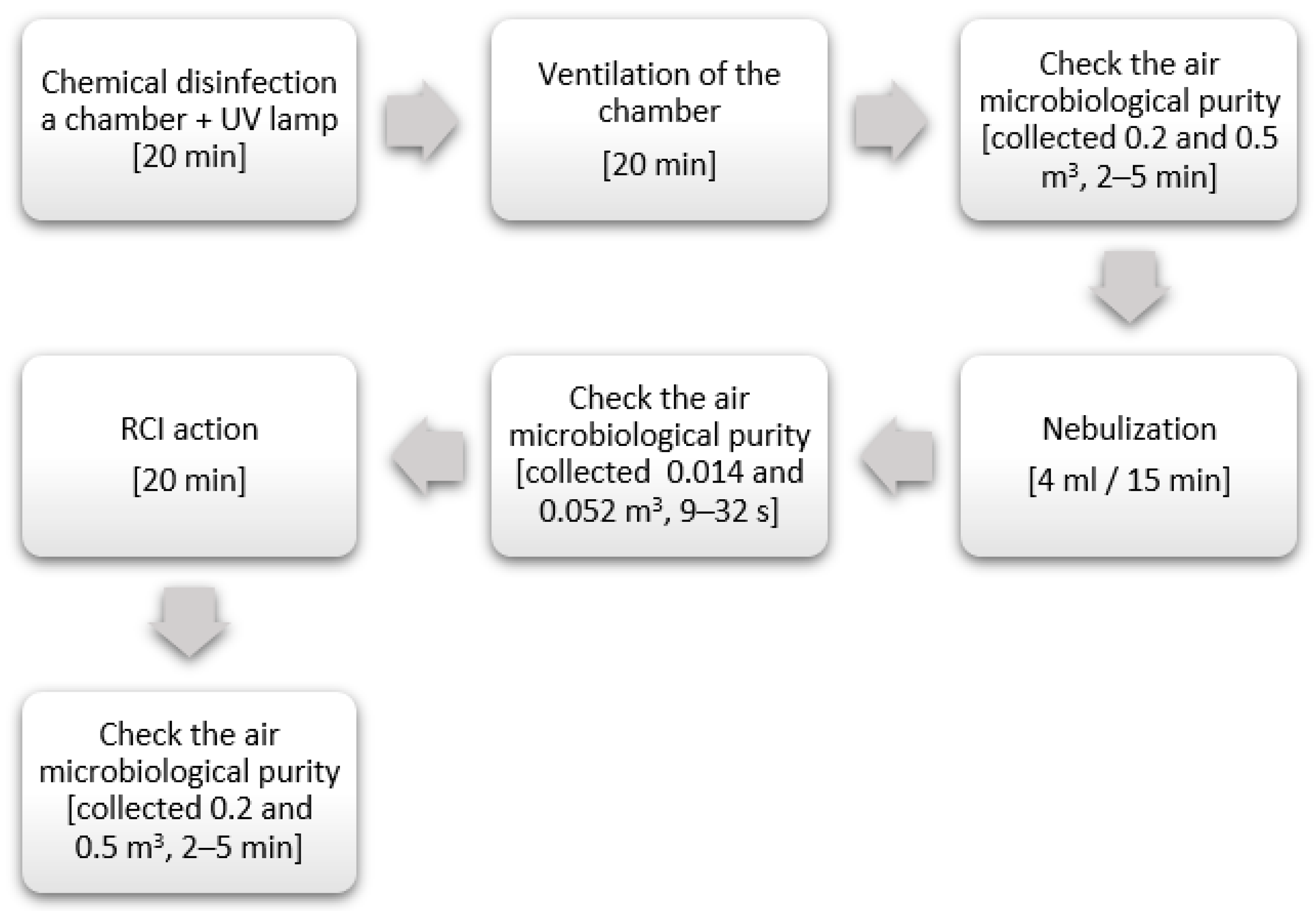
Figure 1. Detailed experimental design according [37].
Atmosphere 11 00764 g001
Figure 2. Appearance of research set according [37].
Atmosphere 11 00764 g002
Table 1. Basic experimental environment conditions.
ParameterRCI Technology Usage
BeforeAfter
Negative ions concentration [ion cm−3]<1.0 × 1047.2 × 105
(±0.7 × 10⁵)
Temperature [°C]25.3
(±0.4)
24.8
(±0.2)
Relative humidity [%]50.4
(±1.1)
49.2
(±0.8)
Table 2. The number of Enterococcus spp. recovered from air and the percentage reduction coefficient R [%].
StrainThe Average Number of Bacteria after Nebulization [CFU m−3]The Average Number of Bacteria after 20 min without RCIPrecipitate Factor RwithoutRCI (K+) [%]The Average Number of Bacteria after Using the Induct 750 [CFU m−3]Percentage of Reduction in the Number of Bacteria RRCI [%]
Enterococcus faecalis PCM 18613.64 × 105
(±5.31 × 104) *
2.89 × 105
(±2.31 × 10⁴)
20.60 a8.72 × 102
(±7.05 × 10³)
99.76 b
Enterococcus faecalis ATCC 512993.91 × 105
(±7.13 × 104) *
3.04 × 105
(±3.16 × 10⁴)
22.25 a3.89 × 104
(±7.05 × 10³)
90.05 b
Enterococcus faecium ATCC 515593.18 × 105
(±8.23 × 10⁴)
2.58 × 10⁵
(±5.31 × 10⁴)
18.99 a1.25 × 103
(±9.63 × 10²)
99.61 b
*—standard deviation. a,b—values marked with different letters differ statistically significant (p ≤ 0.05).
Table 3. Number of Clostridioides difficle recovered from the air and the percentage reduction coefficient R [%].
StrainThe Average Number of Bacteria after Nebulization [CFU m−3]The Average Number of Bacteria after 20 min without RCIPrecipitate Factor RwithoutRCI (K+) [%]The Average Number of Bacteria after Using the Induct 750 [CFU m−3]Percentage of Reduction in the Number of Bacteria RRCI [%]
CDI tox(−)3.14 × 10⁴
(±3.54 × 102) *
2.28 × 104
(±1.11 × 10²)
27.36 a5.15 × 103
(±7.21 × 10²)
83.57 b
CDI MXF-R/ tox A/B/bin(+)2.43 × 105
(±2.47 × 10⁴)
1.74 × 10⁵
(±2.26 × 10⁴)
28.42 a1.30 × 104
(±1.85 × 10³)
94.65 b
CDI PCR 0278.20 × 103
(±4.24 × 10²)
6.03 × 10⁴
(±3.81 × 10²)
26.46 a2.92 × 103
(±1.87 × 10²)
64.45 b
CDI tox(−)—C. difficile non-toxinogenic strain; CDI MXF-R/tox A/B/bin(+)—C. difficile strain producing A, B and binary toxins and resistant to moxifloxacin; CDI PCR 027—C. difficile strain PCR-rybotype 027; *—standard deviation; a,b—values marked with different letters differ statistically significant (p ≤ 0.05).
Table 4. The number of Staphylococcus aureus recovered from the air and the percentage reduction coefficient R [%].
StrainThe Average Number of Bacteria after Nebulization [CFU m−3]The Average Number of Bacteria after 20 min without RCIPrecipitate Factor RwithoutRCI (K+) [%]The Average Number of Bacteria after Using the Induct 750 [CFU m−3]Percentage of Reduction in the Number of Bacteria RRCI [%]
Staphylococcus aureus MRSA4.70 × 105
(±7.16 × 104) *
3.65 × 105
(±3.44 × 10⁴)
22.34 a2.06 × 104
(±1.72 × 10⁴)
95.61 b
Staphylococcus aureus non-MRSA5.10 × 105
(±1.12 × 10⁵)
3.90 × 10⁵
(±1.89 × 10⁵)
23.60 a6.50 × 102
(±9.22 × 101)
99.87 b
*—standard deviation; a,b—values marked with different letters differ statistically significant (p ≤ 0.05).