Article

Differential Susceptibility to Particulate Matter-Induced Cardiac Remodeling and Senescence: A Comparative Study in Young and Aged Mice

by
Dunia Waked
1,
Gabriel Henrique Rodella Guedes
2,
Raissa Macedo
2,
Paulo Hilário Nascimento Saldiva
3,
Mariana Matera Veras
1 and
Ana Paula Cremasco Takano
2,*
1
Laboratory of Environmental and Experimental Pathology, LIM05—Hospital das Clinicas HCFMUSP, Faculdade de Medicina, Universidade de Sao Paulo, Sao Paulo 01246-903, Brazil
2
Department of Anatomy, Institute of Biomedical Sciences, Universidade de Sao Paulo, Sao Paulo 05508-000, Brazil
3
Department of Pathology, Faculdade de Medicina FMUSP, Universidade de Sao Paulo, Sao Paulo 01246-903, Brazil
*
Author to whom correspondence should be addressed.
Atmosphere 2025, 16(1), 109; https://doi.org/10.3390/atmos16010109
Submission received: 21 December 2024 / Revised: 16 January 2025 / Accepted: 17 January 2025 / Published: 19 January 2025
(This article belongs to the Special Issue Research on Air Pollution and Human Exposures)

Abstract

:
Background: Epidemiological and toxicological studies have shown that inhalation of particulate matter (PM), a major component of air pollution, is associated with the development of cardiovascular diseases (CVDs). Cellular senescence and other aging mechanisms are also key factors in the development and progression of CVD. This study aims to investigate age-related susceptibility to cardiac remodeling and senescence due to PM exposure. Methods: Young and old male C57BL/6 mice were exposed to filtered or polluted air for six months using an ambient particle concentrator. Cardiac hypertrophy, fibrosis, and markers of cellular senescence (p53, p21, p-H2AX, and lipofuscin) in the myocardium were evaluated in the experimental groups. Results: PM exposure induces signs of cardiac remodeling, including cardiomyocyte enlargement and increased fibrosis, in young mice, along with elevated p53 expression. However, no significant alterations in cardiac structure or senescence markers were observed between aged mice exposed or not to PM. Conclusions: Our study indicates that younger individuals may be more vulnerable to the cardiovascular effects of chronic PM than older individuals exposed later. Further studies are needed to explore detailed mechanisms of this age-dependent response.

1. Introduction

Almost all of the global population breathes air containing high levels of pollutants, with low- and middle-income countries suffering from the highest exposures [1]. Particulate matter (PM), a significant pollutant with strong evidence for being of public health concern, consists of inhalable particles containing sulfate, nitrates, ammonia, sodium chloride, black carbon, mineral dust, or water [2]. PM particles vary in size and are typically classified by their aerodynamic diameter into coarse (2.5–10 μm), fine (0.1–2.5 μm), and ultrafine particles (0.01–0.1 μm). Fine (PM2.5) and ultrafine (PM0.1) particles are the most harmful to human health [3,4,5], as they can lodge in the alveoli and cross the capillary wall, entering the circulatory system [6]. This can lead to negative cardiovascular outcomes, as extensively demonstrated by previous studies [7,8,9,10,11,12].
Various molecular and cellular mechanisms, such as oxidative stress, inflammation, Ca2+ signaling dysregulation, autophagy disturbances, and apoptosis induction, are involved in the cardiac responses to fine PM exposure [13]. These mechanisms also overlap with the cellular changes observed during aging [14,15,16], suggesting a connection between pollution and the aging process. Kuntic et al. (2023) also support this by reviewing the effects of air pollution on the cardiovascular manifestation of aging, as well as suggesting potential underlying genetic and metabolic aging mechanisms [17].
Myocardial aging affects the composition and structural arrangement of its components, manifested by the loss of cardiomyocytes, hypertrophy of remaining cells, and accumulation of interstitial fibrosis [16,18,19,20]. Over time, the heart also shows an increase in senescent cells, identified by the accumulation of lipofuscin pigments [21,22,23], mitochondrial damage [24], increased expression of molecular markers such as p53 and p21, and shortened telomeres [25,26,27]. These changes impair cardiac function and increase morbidity and mortality from cardiovascular diseases [19,28,29].
Regarding this context, a recent in vitro study provided important findings by exploring the extractable organic matter from PM2.5, inciting rat embryonic cardiac cell line H9c2 cardiomyoblast senescence through the mediation of aryl hydrocarbon receptor (AhR)-induced oxidative stress [30].
Although numerous studies focus on the impact of short-term and long-term exposure to air pollution on age-related cardiovascular outcomes and disease progression, the relationship between air pollution and these cardiac aging-associated senescence mechanisms remains underexplored. Additionally, the possible involvement of PM in anticipating cardiac responses typically observed in the aging process remains unclear. This study aims to investigate age-dependent differences in cardiac remodeling and senescence in response to chronic air pollution, specifically urban PM2.5. The experimental approach conducted in Sao Paulo, Brazil, reflects conditions found in large urban centers. By addressing this gap in the literature, we aim to provide insights into the role of environmental pollutants in accelerating cardiovascular aging and potentially contributing to the increasing burden of age-related cardiovascular diseases.

2. Materials and Methods

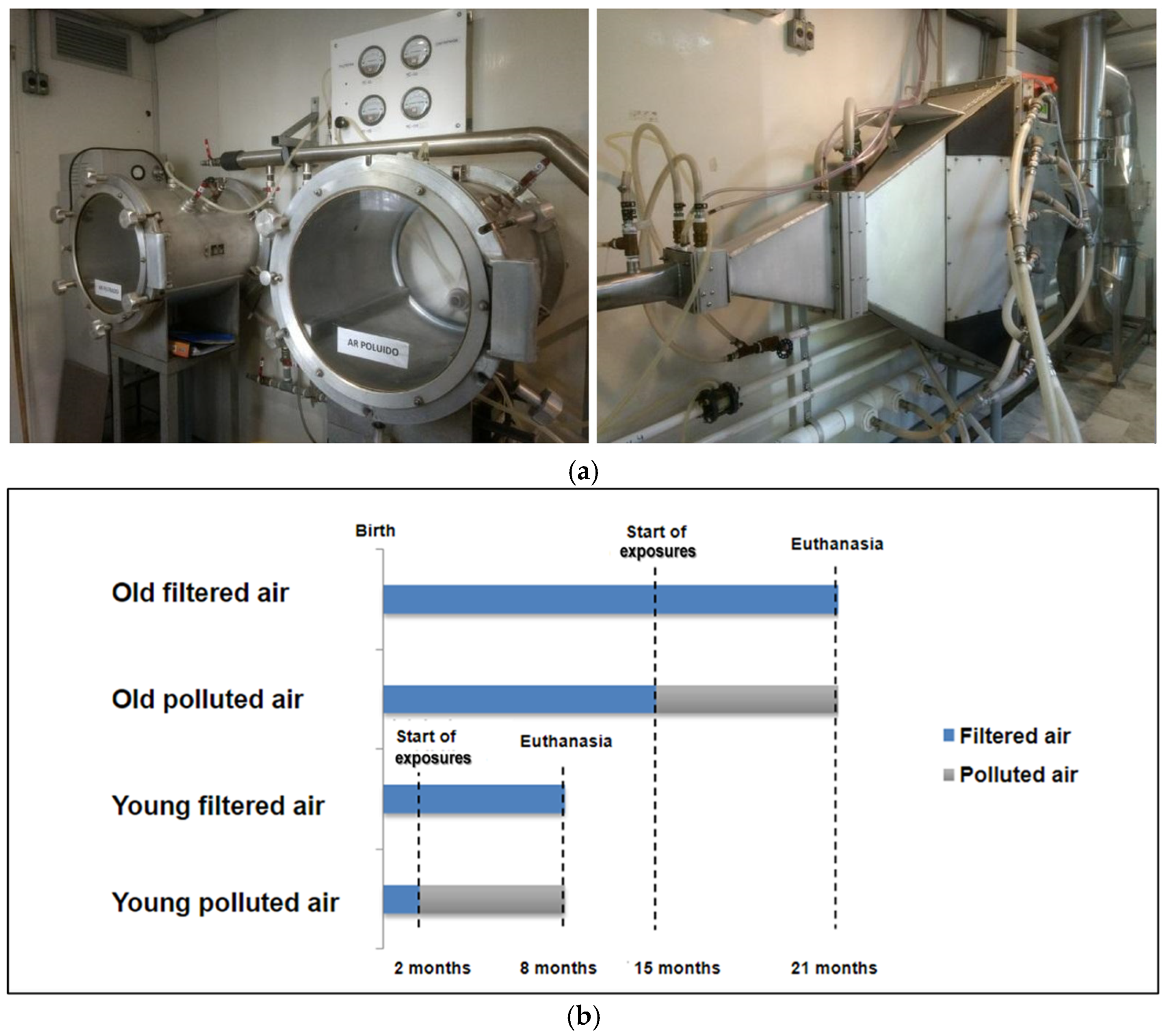
2.1. Animals and Experimental Protocol

This project was approved by the Ethic Committee on Animal Use of the Medical School of the University of Sao Paulo (protocol code 1080/2018), and the experiments were ethically carried out in accordance with the approved protocols. Male C57BL/6 mice, a model previously used for studies of cardiac cellular senescence [31] and for evaluating the effects of exposure to air pollution [32], were used in this research. The mice were provided by the Animal Facility of the Medical School at the University of Sao Paulo. Young and old animals were exposed or not to pollution (PM2.5) for a period of 6 months. Body weight monitoring was performed monthly. The young animals began their exposure at 2 months of age, while the old group began their exposure at 15 months of age. In summary (Figure 1b), the experimental groups used were a young group exposed to filtered air (YFA), a young group exposed to polluted air (YPA), an old group exposed to filtered air (OFA), and an old group exposed to polluted air (OPA). All of the animals in the study were exposed simultaneously, with each group placed in its respective cage and chamber, either with filtered air (control groups) or polluted air (exposed groups), under identical conditions, having free access to water and food. At the end of the exposure period, the mice were weighed and euthanized by an overdose of inhaled anesthetic Isoflurane®, and the carcasses were disposed of according to the institution’s biosafety regulations. The heart and left tibia were obtained from each animal for the analyses of heart weight to tibia length ratio. The heart was then split into an upper part for morphological experiments, and the apical part was kept at −80 °C for molecular analysis.

2.2. Exposure to Air Pollution

The exposure to air pollution was conducted using the PM2.5 ambient particle concentrator (APC) (Figure 1a), previously developed by researchers from Harvard University [33] and used by our research group [34,35,36]. The APC has impactor technology, which allows the concentration of PM2.5 up to 27 times the ambient concentration, directing these particles to an exposure chamber where the animals to be exposed (PA) were placed. The equipment also has a filtered air chamber, equipped with a HEPA filter that retains PM2.5, where the animals in the filtered air group (FA) were placed. The APC is located on the campus of the Medical School at the University of Sao Paulo, approximately 100 m from a CETESB monitoring station and 20 m from a high-traffic avenue. The animals were taken to the APC for exposure 5 times a week for a period of 6 months. They were placed in their respective chambers (FA and PA) in boxes with free movement and access to water and food. APC only concentrates ambient levels of PM2.5. Therefore, to guarantee that animals receive the same daily dose (600 µg/m3 per day, which is the estimated daily mean exposure of a dweller from Sao Paulo city), the exposure duration varied depending on the ambient levels of PM2.5. To estimate how long animals will be exposed, we measured the environmental concentration of PM2.5 for 5 min before exposure. Based on this measurement and considering the concentration capacity of our equipment, we calculated the time needed to expose the animals using the following formula: “Time = [DD/CC] × 60 min”, where DD is the daily exposure dose of PM2.5 (600 µg/m3 per day) and CC is the concentration inside the chambers, achieved using the concentrator (varies according to the PM2.5 concentration at the time). Therefore, on days with lower concentrations, the animals stayed in the exposure chambers for a longer period. The control group spent the same amount of time in the clean chamber. Between exposures, the animals were kept in plastic cages at a controlled temperature with a 12 h light–dark cycle.

2.3. Exposure Characterization

Elemental characterization of PM2.5 was conducted using an energy-dispersive X-ray fluorescence spectrometer (EDX 700 Shimadzu, Analytical Instruments Division, Tokyo, Japan) as previously described [37,38]. For this, once per week, we used an air outlet from the animal exposure chamber to which a polycarbonate filter was connected to collect particles.

2.4. Histological Analysis

The collected materials were fixed in 10% formaldehyde, dehydrated in alcohol solutions (70%, 85%, 95%, and absolute), cleared in xylene (I, II, and III), and embedded in paraffin blocks. Sections of 5 µm were cut and stained using hematoxylin-eosin (HE) and Picro Sirius for morphometric analysis of myocardial composition. Images of 20 fields per slide at 20× (Picro Sirius) or 40× (HE) magnification were captured using a camera attached to a Nikon light microscope (Eclipse E200—Nikon Corporation, Tokyo, Japan). Quantitative analysis of interstitial fibrosis was performed using NIS-Elements AR 3.2 software (Nikon Corporation), which maps color differences by pixels. Thus, areas stained red (collagen deposits) versus light yellow (cardiomyocytes) in Picro Sirius-stained slides were identified, allowing the calculation of the fibrosis fraction expressed as a percentage of a given tissue area [39]. Regarding the analysis of cardiac hypertrophy, in addition to measuring heart weight, individual cardiomyocyte areas in cross-section with central nuclei were quantified in HE-stained slides using ImageJ software (https://imagej.net/ij/index.html, accessed on 9 December 2024).

2.5. Lipofuscin Measurements

Lipofuscin, also referred to as an ‘age pigment’, has autofluorescence properties, and this pigment can be detected using fluorescence or confocal microscopy [21,22,40]. Lipofuscin deposition was analyzed using the same HE-stained slides utilized for histological examination. Ten images were captured from each sample at 20× magnification using a confocal microscope. The imaging conditions included a laser excitation wavelength of 640 nm (4.51%) and a detection range of 656–700 nm. A machine learning method was trained on these images using the Ilastik application and integrated with CellProfiler. This combination allowed for the quantification of lipofuscin deposition by calculating the ratio of lipofuscin to myocardial tissue.

2.6. Analysis of Protein Expression of Cellular Senescence Markers

Total proteins from the frozen cardiac tissue were extracted using a specific buffer, following the protocol detailed in a previous study [41]. The total proteins were separated on an acrylamide gel and transferred to a nitrocellulose membrane. Each membrane was then incubated with the appropriate antibody. The primary antibodies used included anti-p21, anti-p53, and anti-phospho histone H2AX (Ser 139) from Santa Cruz Biotechnologies (Santa Cruz, CA, USA). After incubation with the peroxidase-conjugated secondary antibody, the signal was detected using the ECL Western Blotting reagent (Thermo Scientific, Rockford, IL, USA). The band corresponding to each protein of interest was quantified using the ImageJ program, with values expressed as a percentage relative to the respective control group (filtered air).

2.7. Statistical Analysis

Statistical analysis was performed using GraphPad Prism 8 (GraphPad Software Inc., San Diego, CA, USA). The results are presented as mean ± standard deviation. Data were primarily analyzed and compared using the student’s t-test to compare data between filtered and polluted air groups of the same age. Two-way analysis of variance was also employed to verify aging effects. Values of p < 0.05 were considered statistically significant.

3. Results

3.1. Experimental Exposure

The exposures took place over a period of 132 days. During this time, on days when air humidity levels were high or it was raining, the exposures were not conducted, as these conditions caused pollutant concentrations to be very low. As a result, a total of 93 exposures were carried out, while 39 days (29.5%) had no exposures due to unfavorable weather conditions. This adjustment to the schedule was necessary to maintain optimal conditions to avoid interference with the results. The fine PM concentration was measured daily, and the weekly average of the values obtained was calculated, resulting in a weekly average concentration of 19.47 µg/m3 (Figure 2).
In addition, we analyzed the filters collected during the experiment by EDX. In Table 1, we depict the elements found, including those with very low contributions.

3.2. Chronic PM Exposure Did Not Affect Body Weight and Cardiac Weight Ratio

The body weight of both young and old mice was monitored over the six months of exposure to either filtered or polluted air. There were no significant differences in body weight between the groups exposed to filtered air (Figure 3a) and those exposed to polluted air (Figure 3b) during the same evaluated period, indicating that chronic PM exposure did not affect body weight.

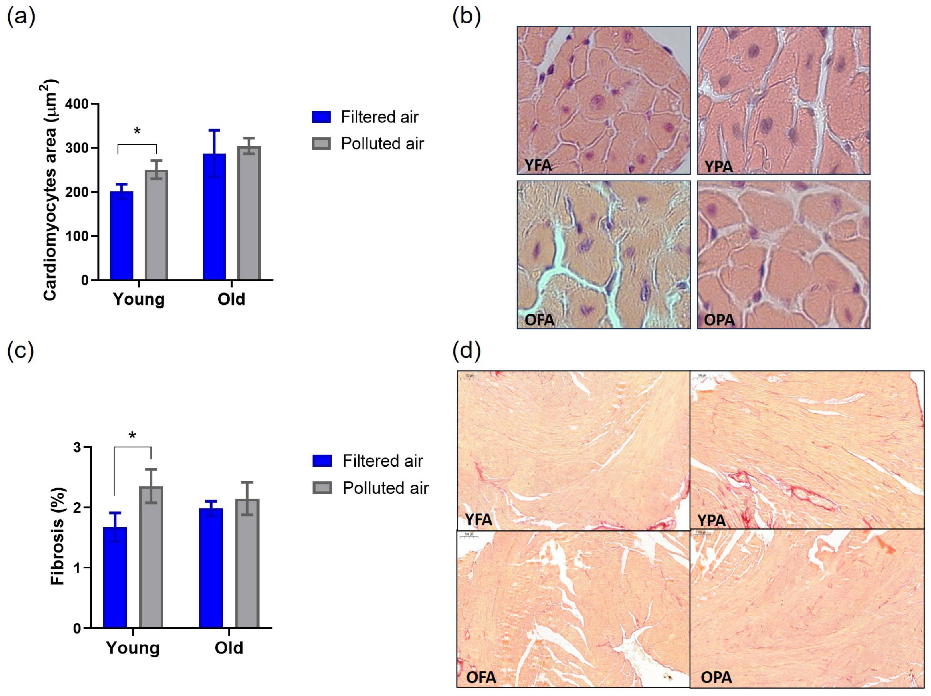
3.3. Chronic PM Exposure Affects Cardiac Remodeling in Young Mice

The heart weight to tibia length ratio (HW/TL) and the heart weight to body weight ratio (HW/BW) were evaluated. Although we observed an increased mean value of both ratios in the young polluted air group compared to the respective filtered air group, the difference was not statistically significant (p = 0.24, Figure 3c; and p = 0.22, Figure 3d). While these findings suggest that chronic PM exposure did not affect cardiac weight ratios, the detailed myocardium morphology evaluation demonstrated interesting results. Cardiomyocytes were increased in the hearts of the young polluted air group compared to the young filtered group (Figure 4a,b). Additionally, cardiac fibrosis was significantly increased by exposure to polluted air in young mice (Figure 4c,d). When comparing the later-exposed groups, we observed that the exposure to PM did not result in alterations in cardiac remodeling in aged mice.

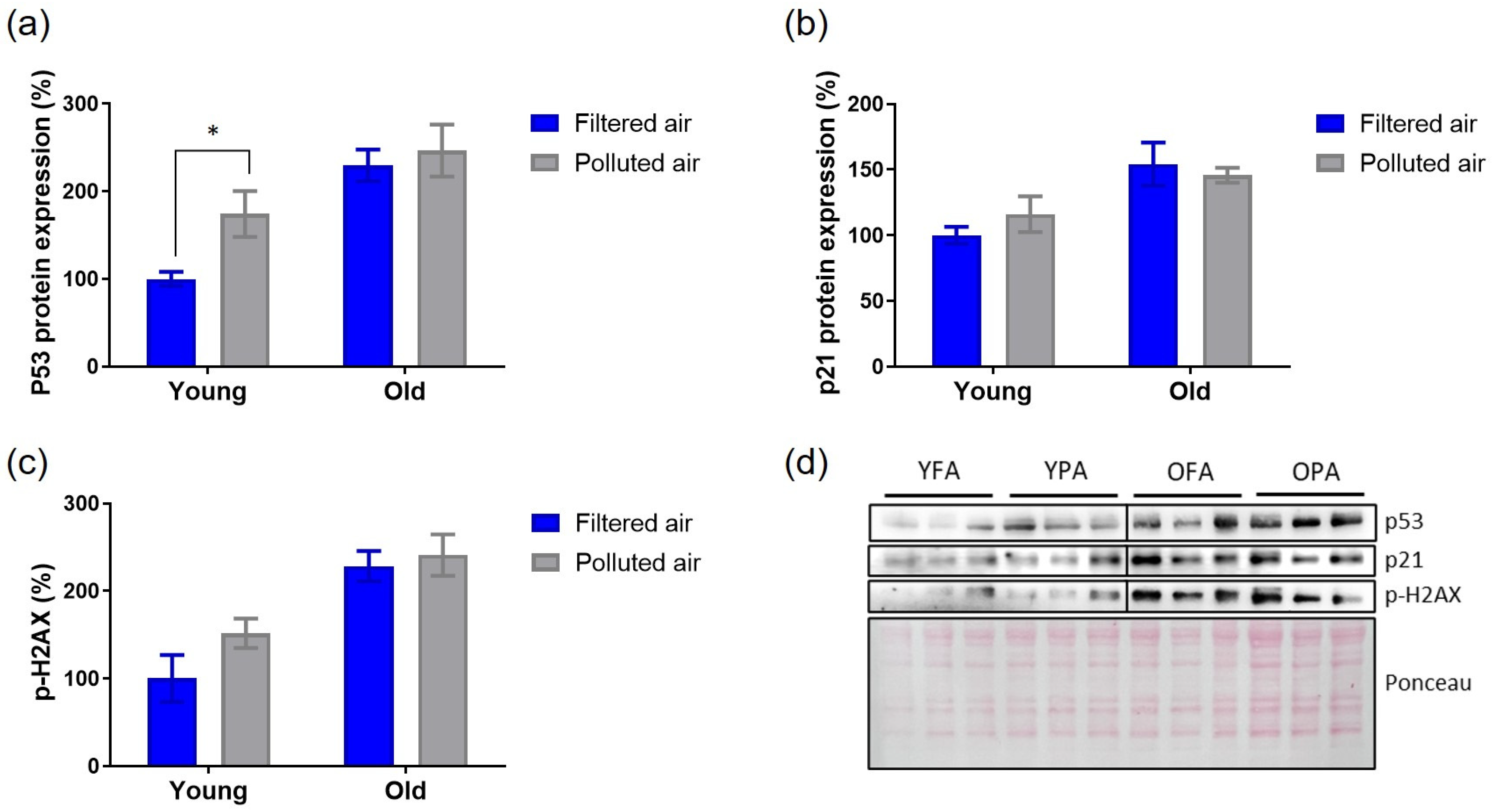
3.4. Cardiac Senescence Parameters Are Partially Triggered by Chronic Exposure to PM in Young Mice

Subsequently, parameters of cellular senescence were examined. There was a significant increase in the protein expression of p53 in the cardiac tissue of young mice exposed to PM compared to young mice exposed to filtered air (Figure 5a). However, the expression of p21 (Figure 5b) and p-H2AX (Figure 5c) was not altered by chronic exposure to pollutants in either the young or old groups compared to their respective filtered air groups.
Lipofuscin deposition was significantly notable in both aged groups compared to the young ones, as expected (p < 0.01; two-way analysis of variance). However, exposure to pollution did not intensify this response compared to the filtered air exposure condition within the same age groups (Figure 6).

4. Discussion

In this study, we sought to investigate the effects of chronic exposure to PM2.5 on cardiac remodeling and cellular senescence during the early adult phase and late stage of life in an experimental murine model. Our results demonstrate that chronic PM exposure significantly affects cardiac remodeling in young mice and appears to partially activate cardiac senescence mechanisms in these mice. Interestingly, in contrast to the young mice, old mice exposed to polluted air exhibit responses similar to those observed under filtered air conditions. These findings highlight the increased vulnerability of younger individuals to urban air pollution and suggest that age-related differences in cardiac responses to environmental stressors could have important implications for developing age-specific therapeutic strategies.
Our initial hypothesis was that exposure to air pollution would induce cardiac hypertrophy, fibrosis, and cellular senescence in the hearts of both young and old mice, with a more pronounced effect in the aged group due to the accumulation of age-related cellular damage. However, we found that the detrimental impact of PM exposure on the evaluated outcomes was observed exclusively in the young mice. This suggests that PM2.5 accelerates aging-associated changes in cardiac tissue rather than exacerbating pre-existing alterations in older mice.
It is important to consider that PM2.5 composition varies according to its source. Air pollution in Sao Paulo primarily originates from its vehicle fleet, with additional contributions from industrial and civil construction emissions. Although previous studies have demonstrated the composition of PM2.5 [38,42], as well as the evolution of PM2.5 over the last 30 years in this megacity [43], our research group routinely monitors the PM2.5 elemental composition in all experimental protocols using the APC system. This practice is crucial because the season significantly influences PM2.5 composition and concentration in Sao Paulo, with a dry winter and a very rainy summer. Whenever possible, our studies using the APC system are conducted during the driest period of the year, from June to September. In the present study, due to the long-term exposure period, we exposed the mice during the indicated period and extended it to the beginning of the summer season, concluding the exposure protocol and collecting tissues for analysis. Among other factors, the mixed composition of PM2.5, which varies considerably depending on regional sources and seasonal factors related to the exposure protocols, may influence some of the diversity of cardiovascular outcomes evaluated in experimental studies [44].
Previous studies have demonstrated cardiac remodeling in mice exposed to PM2.5 for varying durations, and our results align with these observations. For instance, Wold and colleagues reported cardiac remodeling in young mice subjected to 9 months of concentrated ambient PM exposure [32]. Although the exposure conditions differed, the extended exposure to urban air particles from Buenos Aires also led to vascular dilation and inflammatory infiltration in BALB/c mice cardiac tissue [45]. Chronic exposure to the same polluted environment for 12 weeks also impairs cardiac oxygen metabolism and mitochondrial function, along with pro-inflammatory cytokine release from the lungs into systemic circulation [46]. These factors can contribute to cardiac remodeling and the progression of cardiovascular disease in mice following chronic exposure to polluted urban air.
Furthermore, Qin et al. (2018) found increased cardiac fibrosis in both very young female mice (4 weeks old) and older mice (10 months old) after oropharyngeal aspiration of suspended PM2.5, with the fibrosis being reversible upon cessation of exposure [47].
A study examining short-term exposure to vehicular-derived nanoparticles revealed that young female mice exhibit elevated levels of stress-responsive proteins, which, with age, reach a plateau [48]. Specifically, in 21-month-old females, the response to pollutant exposure did not exceed aged basal levels, suggesting that the ability to mount a stress response may be compromised in older individuals, leading to a so-called “aging ceiling” effect [48,49]. This concept may help explain our findings, where aged male mice showed elevated markers of cellular senescence compared to their younger counterparts, yet their response to PM2.5 exposure did not differ significantly from the filtered air group. The age-related changes in cardiac tissue may have already reached a threshold where the capacity to respond to additional stressors, like air pollution, is limited. This suggests that in the aging heart, which is already undergoing molecular and cellular alterations contributing to structural and functional deterioration, environmental pollutants may no longer effectively stimulate additional mechanisms involved in cardiac remodeling and senescence.
Cellular senescence in cardiomyocytes occurs during aging and in response to various stresses, including hypoxia/reoxygenation, ischemia/reperfusion, myocardial infarction, and pressure overload [50]. Persistent activation and accumulation of senescent cardiomyocytes lead to cardiac dysfunction, adverse remodeling, and, ultimately, heart failure [51]. Although no single marker definitively indicates cellular senescence, it can be identified by a combination of characteristics, such as increased expression of p53 and p21, lysosomal expansion detectable by SA-β-Gal, elevated DNA damage response, a senescence-associated secretory phenotype, telomere shortening, and lipofuscin accumulation [15,23,52,53]. The last two mechanisms may take longer to be impacted by certain stress stimuli [54]. Therefore, although lipofuscin is widely accepted as a biomarker of cellular aging in long-lived post-mitotic cells such as cardiomyocytes, this pigment accumulates gradually over time, and even the long-term exposure to PM used in our study was not able to influence this response.
Studies investigating cardiac senescence usually analyze some of these mentioned markers, as we did in our study evaluating a set of combined markers. As expected, aged mice groups showed activation of senescence mechanisms. The increased p53 expression in young mice exposed to PM compared to the filtered condition may represent the onset of the senescence process. Possibly, more intense exposure to pollution could lead to a more significant increase in these evaluated cellular senescence markers, or other markers might be altered but were not evaluated. The in vitro study conducted by Liu et al. (2024) demonstrated that extractable organic matter from PM2.5 exposure led to cellular senescence in H9c2 cardiomyoblast cells, characterized by an increase in the percentage of β-galactosidase-positive cells, elevated expression levels of p16 and p21, and significant elevation in DNA damage evaluated by γ-H2A.X staining [30]. In conjunction with our in vivo findings, it is suggested that exposure to atmospheric pollutants may contribute to the senescence response of cardiac tissue. However, further studies should explore this context more comprehensively and consider all tissues and cells within the cardiovascular system. Although there is growing evidence supporting the relationship between air pollution and organismal accelerated aging and cellular senescence [55], our study contributes to the understanding of specific cardiac aging processes in early adult life exposure.
The study also has limitations. Our experimental model of exposure to inhaled PM closely mimics the real scenario of pollutant exposure in megacities compared to PM administered via gavage or other methods. However, our findings were based on the toxicity of PM2.5 only instead of the combined effect with other urban atmospheric pollutants of Sao Paulo, and it does not account for climate humidity conditions, although these days were less damaging for all individuals in that area. We also limited our groups to only two specific ages and did not include female groups, considering possible sex differences in response to PM-induced cardiac senescence and remodeling. Furthermore, although exploring particle deposition in the nasal cavity or other parts of the upper respiratory airways is relevant to toxicology applications [56,57], our study did not evaluate this aspect, which could have an influence on systemic responses and may have interfered with the obtained results. Other complementary senescence markers and associated aging responses related to the inflammatory context, for example, could further enhance our findings. However, due to the limited sample size available during the study period, we were unable to analyze them. Finally, the use of animal models, while informative, may not fully replicate the complexity of human responses to PM2.5 exposure. Future research should aim to include a more diverse range of ages, sexes, and environmental conditions to provide a more comprehensive understanding of the effects of PM2.5 on cardiac aging and senescence.

5. Conclusions

This study examined the effects of chronic PM2.5 exposure on cardiac remodeling and cellular senescence in both young and old mice. Our findings demonstrate that long-term PM2.5 exposure significantly impacts cardiac remodeling in young mice, with increased cardiomyocyte size and enhanced cardiac fibrosis. Moreover, exposure to this pollutant partially triggered cardiac senescence mechanisms in the young mice, as evidenced by elevated p53 expression. In contrast, older mice exposed later to PM2.5 did not show significant changes in cardiac remodeling or senescence markers compared to those exposed to filtered air.
These results underscore the heightened vulnerability of younger individuals to environmental stressors like air pollution, potentially accelerating aging processes in cardiac tissue. In contrast, the aging heart appears to reach a threshold beyond which its capacity to respond to additional stressors is limited. This finding has important implications for understanding cardiovascular health across the lifespan, and it highlights the need for age-specific strategies in managing the effects of environmental pollutants on heart health, considering the complex interplay between age and susceptibility to air pollution.

Author Contributions

Conceptualization, A.P.C.T.; methodology, P.H.N.S., M.M.V., and A.P.C.T.; formal analysis, D.W., G.H.R.G., and A.P.C.T.; investigation, D.W., G.H.R.G., R.M., and A.P.C.T.; resources, P.H.N.S., M.M.V., and A.P.C.T.; data curation, D.W. and A.P.C.T.; writing—original draft preparation, D.W. and G.H.R.G.; writing—review and editing, A.P.C.T. and M.M.V.; supervision, M.M.V. and A.P.C.T.; funding acquisition, P.H.N.S. and A.P.C.T. All authors have read and agreed to the published version of the manuscript.

Funding

This research was funded by The São Paulo Research Foundation—FAPESP, grants 19/06435-5; 13/21728-2.

Institutional Review Board Statement

The animal study protocol was approved by the Ethic Committee on Animal Use of the Medical School of the University of Sao Paulo (protocol code 1080/2018).

Informed Consent Statement

Not applicable.

Data Availability Statement

Data are presented in the manuscript. Additional information obtained during the experiments is available upon request.

Acknowledgments

The authors acknowledge Mario Costa Cruz for technical support.

Conflicts of Interest

The authors declare no conflicts of interest.

References

  1. World Health Organization (WHO). Available online: https://www.who.int/health-topics/air-pollution#tab=tab_1 (accessed on 9 December 2024).
  2. World Health Organization (WHO). Available online: https://www.who.int/teams/environment-climate-change-and-health/air-quality-and-health/health-impacts/types-of-pollutants (accessed on 9 December 2024).
  3. Krewski, D.; Rainham, D. Ambient air pollution and population health: Overview. J. Toxicol. Environ. Health Part A 2007, 70, 275–283. [Google Scholar] [CrossRef] [PubMed]
  4. Thangavel, P.; Park, D.; Lee, Y.C. Recent Insights into Particulate Matter (PM2.5)-Mediated Toxicity in Humans: An Overview. Int. J. Environ. Res. Public Health 2022, 19, 7511. [Google Scholar] [CrossRef]
  5. Pryor, J.T.; Cowley, L.O.; Simonds, S.E. The Physiological Effects of Air Pollution: Particulate Matter, Physiology and Disease. Front. Public Health 2022, 10, 882569. [Google Scholar] [CrossRef] [PubMed]
  6. Brook, R.D. Cardiovascular effects of air pollution. Clin. Sci. 2008, 115, 175–187. [Google Scholar] [CrossRef] [PubMed]
  7. Akinaga, L.; Lichtenfels, A.; Oliveira, R.; Caldini, E.; Dolhnikoff, M.; Silva, L.; Bueno, H.; Pereira, L.; Saldiva, P.; Garcia, M. Effects of Chronic Exposure to Air Pollution from Sao Paulo City on Coronary of Swiss Mice, from Birth to Adulthood. Toxicol. Pathol. 2009, 37, 306–314. [Google Scholar] [CrossRef]
  8. Damaceno-Rodrigues, N.R.; Veras, M.M.; Negri, E.M.; Zanchi, A.C.; Rhoden, C.R.; Saldiva, P.H.; Dolhnikoff, M.; Caldini, E.G. Effect of pre- and postnatal exposure to urban air pollution on myocardial lipid peroxidation levels in adult mice. Inhal. Toxicol. 2009, 21, 1129–1137. [Google Scholar] [CrossRef]
  9. Hamanaka, R.B.; Mutlu, G.M. Particulate Matter Air Pollution: Effects on the Cardiovascular System. Front. Endocrinol. 2018, 9, 680. [Google Scholar] [CrossRef]
  10. Dwivedi, A.K.; Vishwakarma, D.; Dubey, P.; Reddy, S.Y. Air Pollution and the Heart: Updated Evidence from Meta-analysis Studies. Curr. Cardiol. Rep. 2022, 24, 1811–1835. [Google Scholar] [CrossRef]
  11. Krittanawong, C.; Qadeer, Y.K.; Hayes, R.B.; Wang, Z.; Virani, S.; Thurston, G.D.; Lavie, C.J. PM2.5 and Cardiovascular Health Risks. Curr. Probl. Cardiol. 2023, 48, 101670. [Google Scholar] [CrossRef]
  12. Abdul-Rahman, T.; Roy, P.; Bliss, Z.S.B.; Mohammad, A.; Corriero, A.C.; Patel, N.T.; Wireko, A.A.; Shaikh, R.; Faith, O.E.; Arevalo-Rios, E.C.E.; et al. The impact of air quality on cardiovascular health: A state of the art review. Curr. Probl. Cardiol. 2024, 49, 102174. [Google Scholar] [CrossRef]
  13. Feng, S.; Huang, F.; Zhang, Y.; Feng, Y.; Zhang, Y.; Cao, Y.; Wang, X. The pathophysiological and molecular mechanisms of atmospheric PM2.5 affecting cardiovascular health: A review. Ecotoxicol. Environ. Saf. 2023, 249, 114444. [Google Scholar] [CrossRef] [PubMed]
  14. López-Otín, C.; Blasco, M.A.; Partridge, L.; Serrano, M.; Kroemer, G. The hallmarks of aging. Cell 2013, 153, 1194–1217. [Google Scholar] [CrossRef] [PubMed]
  15. López-Otín, C.; Blasco, M.A.; Partridge, L.; Serrano, M.; Kroemer, G. Hallmarks of aging: An expanding universe. Cell 2023, 186, 243–278. [Google Scholar] [CrossRef] [PubMed]
  16. Hastings, M.H.; Zhou, Q.; Wu, C.; Shabani, P.; Huang, S.; Yu, X.; Singh, A.P.; Guseh, J.S.; Li, H.; Lerchenmüller, C.; et al. Cardiac aging: From hallmarks to therapeutic opportunities. Cardiovasc. Res. 2024, cvae124. [Google Scholar] [CrossRef]
  17. Kuntic, M.; Kuntic, I.; Hahad, O.; Lelieveld, J.; Münzel, T.; Daiber, A. Impact of air pollution on cardiovascular aging. Mech. Ageing Dev. 2023, 214, 111857. [Google Scholar] [CrossRef]
  18. Yang, X.; Sreejayan, N.; Ren, J. Views from within and beyond: Narratives of cardiac contractile dysfunction under senescence. Endocrine 2005, 26, 127–137. [Google Scholar] [CrossRef]
  19. Hua, Y.; Robinson, T.J.; Cao, Y.; Shi, G.P.; Ren, J.; Nair, S. Cathepsin K knockout alleviates aging-induced cardiac dysfunction. Aging Cell 2015, 14, 345–351. [Google Scholar] [CrossRef]
  20. Nakou, E.S.; Parthenakis, F.I.; Kallergis, E.M.; Marketou, M.E.; Nakos, K.S.; Vardas, P.E. Healthy aging and myocardium: A complicated process with various effects in cardiac structure and physiology. Int. J. Cardiol. 2016, 209, 167–175. [Google Scholar] [CrossRef]
  21. Terman, A.; Brunk, U.T. The aging myocardium: Roles of mitochondrial damage and lysosomal degradation. Heart Lung Circ. 2005, 14, 107–114. [Google Scholar] [CrossRef]
  22. Kakimoto, Y.; Okada, C.; Kawabe, N.; Sasaki, A.; Tsukamoto, H.; Nagao, R.; Osawa, M. Myocardial lipofuscin accumulation in ageing and sudden cardiac death. Sci. Rep. 2019, 9, 3304. [Google Scholar] [CrossRef]
  23. Li, W.W.; Wang, H.J.; Tan, Y.Z.; Wang, Y.L.; Yu, S.N.; Li, Z.H. Reducing lipofuscin accumulation and cardiomyocytic senescence of aging heart by enhancing autophagy. Exp. Cell Res. 2021, 403, 112585. [Google Scholar] [CrossRef] [PubMed]
  24. Martín-Fernández, B.; Gredilla, R. Mitochondria and oxidative stress in heart aging. Age 2016, 38, 225–238. [Google Scholar] [CrossRef]
  25. Bernardes de Jesus, B.; Blasco, M.A. Assessing cell and organ senescence biomarkers. Circ. Res. 2012, 111, 97–109. [Google Scholar] [CrossRef] [PubMed]
  26. Marín-Aguilar, F.; Lechuga-Vieco, A.V.; Alcocer-Gómez, E.; Castejón-Vega, B.; Lucas, J.; Garrido, C.; Peralta-Garcia, A.; Pérez-Pulido, A.J.; Varela-López, A.; Quiles, J.L.; et al. NLRP3 inflammasome suppression improves longevity and prevents cardiac aging in male mice. Aging Cell 2020, 19, e13050. [Google Scholar] [CrossRef] [PubMed]
  27. Rossiello, F.; Jurk, D.; Passos, J.F.; d’Adda di Fagagna, F. Telomere dysfunction in ageing and age-related diseases. Nat. Cell Biol. 2022, 24, 135–147. [Google Scholar] [CrossRef] [PubMed]
  28. Ma, Y.; Chiao, Y.A.; Clark, R.; Flynn, E.R.; Yabluchanskiy, A.; Ghasemi, O.; Zouein, F.; Lindsey, M.L.; Jin, Y.F. Deriving a cardiac ageing signature to reveal MMP-9-dependent inflammatory signalling in senescence. Cardiovasc. Res. 2015, 106, 421–431. [Google Scholar] [CrossRef]
  29. Yeh, J.K.; Wang, C.Y. Telomeres and Telomerase in Cardiovascular Diseases. Genes 2016, 7, 58. [Google Scholar] [CrossRef]
  30. Liu, T.; Jiang, B.; Fu, B.; Shang, C.; Feng, H.; Chen, T.; Jiang, Y. PM2.5 Induces Cardiomyoblast Senescence via AhR-Mediated Oxidative Stress. Antioxidants 2024, 13, 786. [Google Scholar] [CrossRef]
  31. Cheng, Z.; Ito, S.; Nishio, N.; Thanasegaran, S.; Fang, H.; Isobe, K. Characteristics of cardiac aging in C57BL/6 mice. Exp. Gerontol. 2013, 48, 341–348. [Google Scholar] [CrossRef]
  32. Wold, L.E.; Ying, Z.; Hutchinson, K.R.; Velten, M.; Gorr, M.W.; Velten, C.; Youtz, D.J.; Wang, A.; Lucchesi, P.A.; Sun, Q.; et al. Cardiovascular remodeling in response to long-term exposure to fine particulate matter air pollution. Circ. Heart Fail. 2012, 5, 452–461. [Google Scholar] [CrossRef]
  33. Sioutas, C.; Koutrakis, P.; Burton, R.M. A technique to expose animals to concentrated fine ambient aerosols. Environ. Health Perspect. 1995, 103, 172–177. [Google Scholar] [CrossRef] [PubMed]
  34. Di Domenico, M.; Benevenuto, S.G.M.; Tomasini, P.P.; Yariwake, V.Y.; de Oliveira Alves, N.; Rahmeier, F.L.; da Cruz Fernandes, M.; Moura, D.J.; Nascimento Saldiva, P.H.; Veras, M.M. Concentrated ambient fine particulate matter (PM2.5) exposure induces brain damage in pre and postnatal exposed mice. Neurotoxicology 2020, 79, 127–141. [Google Scholar] [CrossRef] [PubMed]
  35. Yariwake, V.Y.; Torres, J.I.; Dos Santos, A.R.P.; Freitas, S.C.F.; De Angelis, K.; Farhat, S.C.L.; Câmara, N.O.S.; Veras, M.M. Chronic exposure to PM2.5 aggravates SLE manifestations in lupus-prone mice. Part. Fibre Toxicol. 2021, 18, 15. [Google Scholar] [CrossRef] [PubMed]
  36. Waked, D.; Rodrigues, A.C.B.; Silva, T.M.; Yariwake, V.Y.; Farhat, S.C.L.; Veras, M.M. Effect of chronic exposure to fine particulate matter on cardiac tissue of NZBWF1 mice. Int. J. Exp. Pathol. 2023, 104, 177–187. [Google Scholar] [CrossRef]
  37. Andrade, S.J. Investigação Sobre a Composição Química e Avaliação da Mutagenicidade do Material Particulado Atmosférico sob a Influência da Fuligem da Queima de Cana-De-Açúcar. Ph.D. Thesis, Instituto de Química/UNESP, Araraquara-Sao Paulo, Brazil, 2004. [Google Scholar]
  38. de Castro, K.R.; Almeida, G.H.D.R.; Matsuda, M.; de Paula Vieira, R.; Martins, M.G.; Rici, R.E.G.; Saldiva, P.H.N.; Veras, M.M. Exposure to urban ambient particles (PM2.5) before pregnancy affects the expression of endometrial receptive markers to embryo implantation in mice: Preliminary results. Tissue Cell 2024, 88, 102368. [Google Scholar] [CrossRef]
  39. Takano, A.P.C.; de André, C.D.S.; de Almeida, R.; Waked, D.; Veras, M.M.; Saldiva, P.H.N. Association of pulmonary black carbon accumulation with cardiac fibrosis in residents of Sao Paulo, Brazil. Environ. Res. 2024, 248, 118380. [Google Scholar] [CrossRef]
  40. Tohma, H.; Hepworth, A.R.; Shavlakadze, T.; Grounds, M.D.; Arthur, P.G. Quantification of ceroid and lipofuscin in skeletal muscle. J. Histochem. Cytochem. 2011, 59, 769–779. [Google Scholar] [CrossRef]
  41. Takano, A.P.C.; Senger, N.; Munhoz, C.D.; Barreto-Chaves, M.L.M. AT1 receptor blockage impairs NF-κB activation mediated by thyroid hormone in cardiomyocytes. Pflügers Arch. Eur. J. Physiol. 2018, 470, 549–558. [Google Scholar] [CrossRef]
  42. Andrade, M.D.; de Miranda, R.M.; Fornaro, A.; Kerr, A.; Oyama, B.; de Andre, P.A.; Saldiva, P. Vehicle emissions and PM2.5 mass concentrations in six Brazilian cities. Air Qual. Atmos. Health 2012, 5, 79–88. [Google Scholar] [CrossRef]
  43. Andrade, M.F.; Kumar, P.; de Freitas, E.D.; Ynoue, R.Y.; Martins, J.; Martins, L.D.; Nogueira, T.; Perez-Martinez, P.; de Miranda, R.M.; Albuquerque, T.; et al. Air quality in the megacity of São Paulo: Evolution over the last 30 years and future perspectives. Atmos. Environ. 2017, 159, 66–82. [Google Scholar] [CrossRef]
  44. Jia, Y.Y.; Wang, Q.; Liu, T. Toxicity Research of PM2.5 Compositions In Vitro. Int. J. Environ. Res. Public Health 2017, 14, 232. [Google Scholar] [CrossRef] [PubMed]
  45. Maglione, G.A.; Kurtz, M.L.; Orona, N.S.; Astort, F.; Brites, F.; Morales, C.; Berra, A.; Tasat, D.R. Changes in extrapulmonary organs and serum enzyme biomarkers after chronic exposure to Buenos Aires air pollution. Environ. Sci. Pollut. Res. 2020, 27, 14529–14542. [Google Scholar] [CrossRef] [PubMed]
  46. Marchini, T.; Magnani, N.; Garces, M.; Kelly, J.; Paz, M.; Caceres, L.; Calabro, V.; Lasagni Vitar, R.; Caltana, L.; Contin, M.; et al. Chronic exposure to polluted urban air aggravates myocardial infarction by impaired cardiac mitochondrial function and dynamics. Environ. Pollut. 2022, 295, 118677. [Google Scholar] [CrossRef]
  47. Qin, G.; Xia, J.; Zhang, Y.; Guo, L.; Chen, R.; Sang, N. Ambient fine particulate matter exposure induces reversible cardiac dysfunction and fibrosis in juvenile and older female mice. Part. Fibre Toxicol. 2018, 15, 27. [Google Scholar] [CrossRef] [PubMed]
  48. Pomatto, L.C.D.; Cline, M.; Woodward, N.; Pakbin, P.; Sioutas, C.; Morgan, T.E.; Finch, C.E.; Forman, H.J.; Davies, K.J.A. Aging attenuates redox adaptive homeostasis and proteostasis in female mice exposed to traffic-derived nanoparticles (‘vehicular smog’). Free Radic. Biol. Med. 2018, 121, 86–97. [Google Scholar] [CrossRef]
  49. Pomatto, L.C.D.; Davies, K.J.A. The role of declining adaptive homeostasis in ageing. J. Physiol. 2017, 595, 7275–7309. [Google Scholar] [CrossRef]
  50. Zhai, P.; Sadoshima, J. Cardiomyocyte senescence and the potential therapeutic role of senolytics in the heart. J. Cardiovasc. Aging 2024, 4, 18. [Google Scholar] [CrossRef]
  51. Mehdizadeh, M.; Aguilar, M.; Thorin, E.; Ferbeyre, G.; Nattel, S. The role of cellular senescence in cardiac disease: Basic biology and clinical relevance. Nat. Rev. Cardiol. 2022, 19, 250–264. [Google Scholar] [CrossRef]
  52. Kumar, A.; Bano, D.; Ehninger, D. Cellular senescence in vivo: From cells to tissues to pathologies. Mech. Ageing Dev. 2020, 190, 111308. [Google Scholar] [CrossRef]
  53. Chen, M.S.; Lee, R.T.; Garbern, J.C. Senescence mechanisms and targets in the heart. Cardiovasc. Res. 2022, 118, 1173–1187. [Google Scholar] [CrossRef]
  54. Terman, A.; Brunk, U.T. Lipofuscin. Int. J. Biochem. Cell Biol. 2004, 36, 1400–1404. [Google Scholar] [CrossRef] [PubMed]
  55. Wang, S.N.; Shi, Y.C.; Lin, S.; He, H.F. Particulate matter 2.5 accelerates aging: Exploring cellular senescence and age-related diseases. Ecotoxicol. Environ. Saf. 2024, 284, 116920. [Google Scholar] [CrossRef]
  56. Gu, X.; Wen, J.; Wang, M.; Jian, G.; Zheng, G.; Wang, S. Numerical investigation of unsteady particle deposition in a realistic human nasal cavity during inhalation. Exp. Comput. Multiph. Flow 2019, 1, 39–50. [Google Scholar] [CrossRef]
  57. Amjadimanesh, H.; Faramarzi, M.; Sadrizadeh, S.; Abouali, O. Micro-particle deposition in maxillary sinus for various sizes of opening in a virtual endoscopic surgery. Exp. Comput. Multiph. Flow 2023, 5, 262–271. [Google Scholar] [CrossRef]
Figure 1. Ambient particle concentrator (APC) and representative scheme of the experimental protocol. (a) The APC consists of the filtered air (FA) and polluted air (PA) chambers (left), which are connected to the impactors that concentrate the particulate matter—PM2.5 (right). (b) Young groups with onset of exposure at two months old and older groups with onset of exposure at 15 months of age; all groups were euthanized six months after the start of the exposure.
Atmosphere 16 00109 g001
Figure 2. Weekly average of daily PM2.5 concentration (ug/m3) throughout the experimental period.
Atmosphere 16 00109 g002
Figure 3. Body weight monitoring and heart weight evaluation. (a) Body weight (BW) in young animals over the six months of exposure to filtered air (YFA) or polluted air (YPA); (b) BW in old animals over the six months of exposure to filtered air (OFA) or polluted air (OPA). (c) Heart weight (HW) to tibia length (TL) ratio and (d) HW to BW ratio of all experimental groups; n = 6 per group.
Atmosphere 16 00109 g003
Figure 4. Myocardium remodeling evaluation. (a) Quantification (* p = 0.0094, n = 4 per group) and (b) representative images of cardiomyocytes area. Crops of entire original images were made to highlight and better represent the corresponding quantitative findings. (c) Quantification (* p = 0.0399, n = 5 per group) and (d) representative images of the myocardium used for fibrosis quantification. YFA: young filtered air; YPA: young polluted air; OFA: old filtered air; OPA: old polluted air.
Atmosphere 16 00109 g004
Figure 5. Analysis of protein expression of cellular senescence markers. Quantitative analysis of p53 (a), p21 (b), and phospho-histone H2AX (Ser 139) (c) in experimental groups. Representative image (d); YFA: young filtered air; YPA: young polluted air; OFA: old filtered air; OPA: old polluted air. * p < 0.05, n = 3–6.
Atmosphere 16 00109 g005
Figure 6. Lipofuscin evaluation. (a) Quantification and (b) representative images of myocardium histological sections used for lipofuscin proportion deposition. Arrows indicate evident lipofuscin in aged mice from both experimental exposure conditions. & p <0.01 vs. young filtered air group, and # p < 0.01 vs. young polluted air group, evaluated by two-way ANOVA.
Atmosphere 16 00109 g006
Table 1. Descriptive analysis of the elemental composition present in PM2.5 samples from the exposure site.
Element%SD1
C55.87076.8503
S27.48821.6269
Mg11.70789.4825
Ba1.67940.5529
Cl0.96220.2746
P0.90930.4804
Ca0.55810.1705
Fe0.40500.1363
K0.21560.0514
Si0.08080.1452
Zn0.03900.0192
W0.03350.0654
Cu0.03200.0473
Ti0.00610.0113
Cr0.00520.0002
Rb0.00460.0033
V0.00130.0002
Sr0.00060.0011
SD1 = standard deviation.
Disclaimer/Publisher’s Note: The statements, opinions and data contained in all publications are solely those of the individual author(s) and contributor(s) and not of MDPI and/or the editor(s). MDPI and/or the editor(s) disclaim responsibility for any injury to people or property resulting from any ideas, methods, instructions or products referred to in the content.

