Abstract
The outbreak of Coronavirus Disease 2019 (COVID-19), a severe respiratory disease caused by betacoronavirus SARS-CoV-2, in 2019 that further developed into a pandemic has received an unprecedented response from the scientific community and sparked a general research interest into the biology and ecology of Coronaviridae, a family of positive-sense single-stranded RNA viruses. Aquatic environments, lakes, rivers and ponds, are important habitats for bats and birds, which are hosts for various coronavirus species and strains and which shed viral particles in their feces. It is therefore of high interest to fully explore the role that aquatic environments may play in coronavirus spread, including cross-species transmissions. Besides the respiratory tract, coronaviruses pathogenic to humans can also infect the digestive system and be subsequently defecated. Considering this, it is pivotal to understand whether wastewater can play a role in their dissemination, particularly in areas with poor sanitation. This review provides an overview of the taxonomy, molecular biology, natural reservoirs and pathogenicity of coronaviruses; outlines their potential to survive in aquatic environments and wastewater; and demonstrates their association with aquatic biota, mainly waterfowl. It also calls for further, interdisciplinary research in the field of aquatic virology to explore the potential hotspots of coronaviruses in the aquatic environment and the routes through which they may enter it.
1. Introduction
The outbreak of severe acute respiratory syndrome caused by betacoronavirus SARS-CoV-2 (provisionally known as 2019nCoV) began at the end of 2019 in China and further spread to other countries [1] and across different continents, forcing the World Health Organization (WHO) to first declare a Public Health Emergency of International Concern at the end of January 2020 [2] and to later announce a pandemic of COVID-19 in March 2020 [3]. This is the first time that any coronavirus has sparked such an epidemiological situation, although some other coronaviruses were already known to reveal pathogenicity to humans. SARS-CoV-2 and COVID-19 have received unprecedented research interest encompassing fields of molecular biology [4], mechanism of cell entry [5], diagnostics [6], epidemiological modeling [7], immunology [8], experimental therapies and vaccine development [9,10], clinical medicine [11,12], prejudice and xenophobia [13] and the psychological effects of the pandemic [14,15]. It has also renewed a general interest in coronavirus biology and ecology.
The aquatic environment can be a source of an uncountable number of microorganisms pathogenic to different aquatic and terrestrial animals, as well as humans [16,17,18]. Lakes and rivers are an important habitat for bats and birds (including waterfowl), which represent one of the main reservoirs for various coronaviruses [19,20]. It is established that these animals can shed coronaviral RNA through feces, although it remains to be explored whether this is related to the presence of infectious viral particles [21,22]. Nevertheless, it is of high interest to investigate the association between aquatic environments and this viral group. It would add to the understanding of the role that these ecosystems may potentially play in infections within species as well as in cross-species transmission.
Understanding the role of aquatic ecosystems in this context is also essential from a human health perspective, since untreated water, a well-established source of various pathogens, is used in various areas, particularly those with poor sanitation. It would also help to understand whether aquatic biota could play a potential role as an intermediate host from which humans could contract the coronavirus. Such a process would, however, require the spike glycoprotein that mediates coronavirus entry into cells [23,24] to be first optimized in such a host for binding to human-like angiotensin-converting enzyme 2 (ACE2), alanyl aminopeptidase (CD13), dipeptidyl peptidase 4 (CD26) or other entry receptors through natural selection [5,25].
It has been demonstrated that the discharge of inadequately treated sewage effluents is a significant source of enteric viral pathogens in lakes and rivers, including those which are the leading causes of recreational waterborne illnesses [26,27,28]. All seven coronaviruses pathogenic to humans (SARS-CoV, SARS-CoV-2, MERS-CoV, HCoV-229E, HCoV-NL63, HCoV-OC43 and HCoV-HKU1) can infect the respiratory tract and digestive system, and their RNA material can be present in stool [29]. One should note that the detection of the genetic material in fecal material does not necessarily indicate that infectious virions are defecated. However, the presence of cultivable SARS-CoV in stool was already reported, and some preliminary observations, based on a small number of patients [30], suggest that this may also be a case for SARS-CoV-2 [31,32]. How frequently the infectious virus can be present in the human stool and what viral loads can be expected are yet to be explored. Such data would enable understanding whether wastewater, particularly untreated, may serve as a route of their dissemination to the aquatic environment. The understanding of the scale of this process first requires an exploration of the survival of coronaviruses in sewage and its treatment with various methods as well as a monitoring of human pathogenic coronaviruses in untreated and treated wastewater.
In the present review, we provide a brief overview of coronaviruses, their taxonomy, molecular biology, natural reservoirs and pathogenicity; outline their potential to survive in aquatic environments and wastewater; and demonstrate their association with aquatic biota, mainly waterfowl, and other animal species related to aquatic ecosystems. Future research prospects regarding the association between selected coronaviruses and water-related issues are put forward with a call for interdisciplinary research in the field of aquatic virology.
2. General Characteristics of the Coronaviridae Family
The subsequent sections give a brief overview of the taxonomy of coronaviruses, their general molecular features, main natural reservoirs and, finally, the pathogenicity of selected strains to humans.
2.1. Taxonomy of Coronaviruses
The Coronaviridae family is classified into the realm Riboviria, which includes all RNA viruses and viroids that replicate by means of RNA-dependent RNA polymerases. The Coronaviridae belongs to the order Nidovirales and suborder Coronavirineae (Figure 1). It is further divided into two subfamilies—Orthocoronavirinae and Letovirinae [33,34]. The latter was recently proposed and currently consists of a single Alphaletovirus genus with a single subgenus of Milecovirus and one representative species known so far—Microhyla letovirus 1 (MLeV), isolated from the ornamented pygmy frog Microhyla fissipes Boulenger [35].
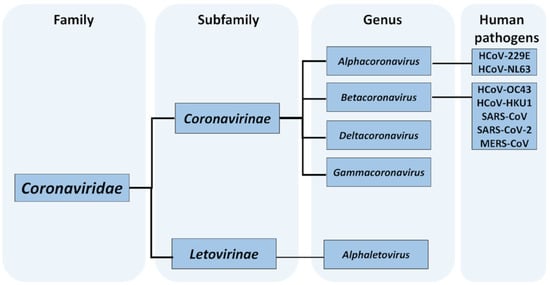
Figure 1.
Taxonomy of Coronaviridae family with an indication of species known to be pathogenic to humans and cause respiratory diseases [33].
The Orthocoronavirinae subfamily, whose members are typically referred to as coronaviruses, is currently divided into four genera: Alphacoronavirus (alphacoronaviruses (α-CoVs)), Betacoronavirus (betacoronaviruses (β-CoVs)), Deltacoronavirus (deltacoronaviruses (δ-CoVs)) and Gammacoronavirus (gammacoronaviruses (γ-CoVs)) (Figure 1). The members of this subfamily are currently classified by means of a threshold level of sequence identity of replicase regions: the ORF1ab gene and the pp1ab polyprotein [36]. Members that belong to a similar species display over 90% amino acid uniqueness in the seven conserved domains of the 1ab protein [37]. The genus Alphacoronavirus is the most taxonomically diversified and is represented by seventeen identified species across twelve genera: Colacovirus, Decacovirus, Duvinacovirus, Luchacovirus, Minacovirus, Minunacovirus, Myotacovirus, Nyctacovirus, Pedacovirus, Rhinacovirus, Setracovirus and Tegacovirus. The β-CoVs are divided into five subgenera—Embecovirus, Hibecovirus, Merbecovirus, Nobecovirus and Sarbecovirus—with a total of twelve species known so far. The δ-CoVs and γ-CoVs are taxonomically divided into a respective four (Andecovirus, Buldecovirus, Herdecovirus and Moordecovirus) and two (Cegacovirus and Igacovirus) subgenera and contain a total of seven and two species, respectively [33].
2.2. Molecular Biology of Coronaviruses
The coronaviruses have a characteristic spiky or crown-like (corona) appearance. Their virions are spherical and usually range between 50 and 200 nm in diameter [38,39]. The Coronaviridae are enveloped, positive single-stranded RNA (+ssRNA) viruses; the size of their genomes, coiled inside a helical nucleocapsid of 9–11 nm diameter, ranges between 26.2 and 31.7 kb, making them the largest enveloped RNA viruses [40]. The RNA strand is capped at the 5′ end and contains a 3′ poly(A) tail, therefore being very similar to mRNA during translation [41]. Nevertheless, the translation of coronaviruses varies from the canonic one. It includes intricate mechanisms such as leaky scanning, ribosomal frameshifting, in-build internal ribosome entry and non-AUG initiation events [42]. The only protein identified to be encoded by genome is replicase-transcriptase, while the rest appear in the form of subgenomic mRNAs [43]. At the 5′ end, two-thirds of the coronavirus genome consists of the replicase gene, which has two open reading frames, ORF1a and ORF1b, within. The frameshifting during translation allows for the production of polyprotein 1a/1ab that further enables sixteen nonstructural proteins in the host cell to be obtained [44]. Inversely, the 3′ third part of the genome consists of ORFs encoding structural and accessory proteins [45].
The order of the essential structural proteins in the genome is as follows: spike (S), envelope (E), membrane (M) and nucleocapsid (N) [46]. Contrary to this, the accessory genes are placed in between: two between S and E (3a, 3b); five between the M and N (6, 7a, 7b, 8a, 8b); and one included in the N gene (9b) [47]. Although the accessory proteins are not necessary for replication in vitro, they may take part in the pathogenesis of coronaviruses [41]
The N protein is a phosphoprotein that forms a helical nucleocapsid for the genomic RNA. Moreover, it plays a significant role in the virus assembly, transcription and replication [48]. The structure of the protein is composed of two detached domains, the N-terminal and C-terminal, both of which are required for optimum RNA binding [49]. The nucleocapsid is surrounded by a lipid bilayer. This envelope is acquired by budding at membranes of endoplasmic reticulum, intermediate compartment and/or Golgi complex. The S, M and E proteins are embedded in this envelope [50,51].
The spike (S) glycoprotein, which has a rod-like shape and length of approximately 10–20 nm, is essential in facilitating viral entry to the host cell [52,53,54]. It consists of a trimer of two proteins—S1 and S2. The S1 protein is an ectodomain, with a signal peptide for an endoplasmic reticulum at its N-terminal, while the S2 protein comprises heptad repeat regions, putative fusion peptide, transmembrane domain and endodomain at the C-terminus [55]
The envelope (E) protein is a small protein with a size of 8–12 kDa, and its amount is limited in viruses. This protein may be necessary for assembly and to generate an accurate virion [41,55]. It consists of a hydrophilic N-terminus, a hydrophobic transmembrane domain and the hydrophilic C-terminus. It is suggested that the secondary envelope structure is a motif that operates as a transport signal to the Golgi complex [51].
The M glycoprotein, whose preglycosylated form has a size between 25 and 30 kDa, is the most abundant envelope component. In general, this protein has three transmembrane regions. The tiny part of the amino-terminal domain is localized outside of the virion, whereas the carboxyterminal is situated inside [55]. The glycosylation of the M protein may be responsible for the induction of interferon in host cells [48,56]. Along with the N protein, the M protein contributes to the packing of the genomic RNA along with assembling and interacting with virions [57].
2.3. Pathogenicity in Humans
Overall, seven coronaviruses are currently known to be pathogenic to humans, and all of them have a zoonotic origin [58]. The α-CoVs NL63 and HCoV-229E, as well as the β-CoVs HCoV-O43 and HCoV-HKU1, are known to be cause mild upper respiratory tract disease in otherwise healthy subjects [59]. However, more severe cases, characterized by pneumonia and bronchiolitis, can occur in elderly and immunocompromised subjects as well as children and immunocompromised patients [60,61,62,63,64]. Neuroinvasion and gastrointestinal infections were also reported for these coronaviruses [65,66].
Severe respiratory infections in humans can be induced by three β-CoVs: SARS-CoV, MERS-CoV and, most recently, SARS-CoV-2. The SARS-CoV and SARS-CoV-2, two strains of the same species (Sarbecovirus subgenus), are the causative factors for severe acute respiratory syndrome (SARS) and COVID-19, respectively. In turn, MERS-CoV (Merbecovirus subgenus) is a cause of the Middle East respiratory syndrome (MERS). All of these strains have emerged from natural hosts and later spread through human–human transmission, predominantly by droplet and contact routes. The fecal–oral route is also plausible since, in the case of some patients, the viral RNA was detected in fecal samples. Although it appears that this route of transmission did not generally play a significant role during reported outbreaks, it may have greater implications in areas with poor sanitation [67,68,69,70]. However, it remains unanswered whether the infection of SARS-CoV-2 could occur via ingestion and whether the virus can survive passage through the stomach [71]. Typical clinical manifestations commonly include fever, cough and breathing difficulties with pneumonia, although they are not obligatorily present. Gastrointestinal signs such as nausea, abdominal pain, diarrhea and vomiting can also occur [72,73,74]. Additionally, prevalent olfactory and gustatory dysfunctions were observed over the course of SARS-CoV-2 infection [75].
SARS-CoV, MERS-CoV and SARS-CoV-2 are responsible for three major coronavirus-related outbreaks in humans within the past two decades. The SARS-CoV was responsible for the epidemic of SARS that originated in Guangdong Province in China in November 2002 [76]. Although the majority of cases were reported in China, the virus spread to 29 countries across Asia, North America, Europe and Oceania [77]. Most of the cases were confirmed in 2003, and strict control measures were implemented to contain the outbreak [78]. The last cases were reported in May 2004, and all were instances of infection at a microbiological laboratory where research on SARS-CoV was also conducted [79]. A total of 8096 confirmed cases were reported, with a mortality rate of 9.6% [77]. The first human cases of MERS-CoV infection, known as the Middle East respiratory syndrome, emerged in September 2012 in Saudi Arabia, and the vast majority of the nearly 2500 confirmed cases have been associated with the Arabian Peninsula [80]. However, as a result of travel, MERS-CoV was exported across the Middle East, Europe, North Africa and Asia and reported by 27 countries to date. As of late 2019, the mortality rate was 34.4% [81,82]. The most recent coronavirus-associated outbreak in humans emerged in late 2019 in the city of Wuhan, China, and was caused by SARS-CoV-2 [39,83]. The COVID-19 outbreak quickly evolved into the first pandemic caused by any human coronavirus [3]; the total confirmed cases amounted to 3,299,603 at the end of April 2020, with a 7.1% mortality rate. The total number of worldwide cases exceeded 4,700,000 on 18 May 2020, according to an online interactive dashboard developed by the Johns Hopkins University [84].
The foremost factor for entering the host cell is S glycoprotein, which is responsible for receptor-recognition and fusion with a membrane and which likewise allows for cross-species transmission [85]. The α-CoV HCoV-NL63 and SARS-associated coronaviruses, SARS-CoV and SARS-CoV-2, employ an ACE2 receptor for cell entry [5,86,87,88]), although its action is complemented by heparin sulfate proteoglycans which are involved in the adhesion of virions to the cell surface [89,90]. The MERS-CoV uses DPP4 for cellular entry (Figure 2) [91]. In the case of α-CoV HCoV-229E, the CD13/aminopeptidase N is bound by S protein [92], while β-CoVs HCoV-OC43 and HCoV-HKU1 can utilize human leukocyte antigen class I (HLA-I) or O-acetylated sialic acids [93,94,95,96]. Moreover, in the case of all coronaviruses pathogenic to humans, cellular entry can be mediated via type II transmembrane serine protease TMPRSS2 serine protease, which acts as an activator for S glycoprotein [5,97,98,99,100,101]. The respiratory and enteric tracts are the main sites of infection due to the expression of molecules involved in coronaviruses adhesion, activation and entry on the apical membranes of epithelial cells, although other types of cells and organs can also be a potential target, as experimentally demonstrated for selected strains [102,103,104,105].
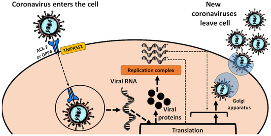
Figure 2.
A general scheme of cellular entry of coronaviruses that pose the highest human health risks, namely SARS-CoV, SARS-CoV-2 (entering via ACE2) and MERS-CoV (facilitated by DPP4), and further virus propagation.
2.4. Natural Reservoirs of Coronaviruses
In general, avian and mammalian species, along with humans, serve as hosts for viruses belonging to the Orthocoronavirinae subfamily [106]. In cases of the Letovirinae subfamily, the only host for the single species (MLeV) known so far is the ornamented pygmy frog Microhyla fissipes [35]. It is highly plausible that there are other hosts, at least amphibian, for MLeV, although this issue requires further studies. Most generally, selected species of α-CoVs and β-CoVs are causative factors of human and domestic animal infections. Otherwise, γ-CoVs and δ-CoVs are more associated with avian hosts, but selected species have also been discovered in marine mammals [20,107].
Bats are known as the largest group of natural reservoirs for coronaviruses, not only in Asia but also in Europe, Africa and South and North America [19,108], and may reveal a relatively high prevalence of viral RNA shedding in their feces [21]. A survey in China has reported that over 6% of bats, distributed throughout the region, can harbor coronaviruses, with α-CoV strains BtCoV/701/05 (Myotis ricketti Peters), BtCoV/512/05, BtCoV/527/05 and BtCoV/515/05 (Scotophilus kuhlii Leach), and β-CoVs strains BtCoV/273/04 (Rhinolophus ferrumequinum Schreber), BtCoV/279/04 (Rhinolophus macrotis Blyth), BtCoV/133/05 (Tylonycteris pachypus Temminck), BtCoV/434/05 (Pipistrellus pipistrellus Schreber) and BtCoV/355/05 (Pipistrellus abramus Temminck) having been identified [109]. Therefore, bats are most likely to contribute to cross-species transmissions, including transmission to humans, highlighting the continuous need to explore coronaviruses in bats of different geographical origin, identify the potential hotspots and take safety measures, particularly in regions where bats are hunted for food, ornate decorations or alleged medicinal properties, as well as areas where their guano is mined [110,111]. Importantly, bats are also the primary hosts of human infectious coronaviruses. The MERS-CoV most likely originated from Taphozous perforatus Geoffroy, Rhinopoma hardwickii Gray and Pipistrellus kuhlii Kuhl [112,113]. In turn, the SARS-CoV was suggested to have evolved from horseshoe bats, Rhinolophus spp. [114], and SARS-CoV-2 is also most likely to be originally linked with a bat host, with one study demonstrating 96% identity at the whole-genome level to β-CoV BatCoV RaTG13 detected in Rhinolophus affinis Horsfield [115]. In the case of these three coronaviruses, the role of an intermediate host for human transmission has been suggested. The dromedary camel Camelus dromedarius L. was suggested to serve as such a host for MERS-CoV, as individuals in Egypt were found to harbor MERS-CoV-like NRCE-HKU205 and NRCE-HKU270 [116]. One study has also identified novel β-CoV Erinaceus coronavirus strains in the European hedgehog Erinaceus europaeus L., namely EriCoV/2012-68/GER/2012, EriCoV/2012-216/GER/2012, EriCoV/2012-174/GER/2012 and EriCoV/2012-51/GER/2012, all of which were demonstrated to be phylogenetically related to MERS-CoV [117]. In the case of SARS-CoV, the Himalayan palm civet Paguma larvata Hamilton-Smith or raccoon dog Nyctereutes procyonoides Gray were likely to have been involved in transmission after SARS-CoV-like isolates SZ3 and SZ16 were detected in these animals [118]. The transmission of SARS-CoV-2 to humans may also involve an intermediate host. Although no specific species is yet known, it is suggested that this strain may evolve in pangolins [119]. The other hypothesis assumes that, following the initial zoonotic event, an adaptive process involving the acquisition of the polybasic cleavage site was occurring during unrecognized transmission between humans [4]. In turn, α-CoVs HCoV-229E and HCoV-NL63 probably have a common ancestor and may be associated with HCoV-NL63-like viruses circulating in Triaenops sp. bats and HCoV-229E-like particles identified in Hipposideros sp. bats [120]. Two studies suggest that D. dromedarius may play a potential role as an intermediate host in the case of HCoV-229E [121,122].
On the other hand, both α-CoVs HCoV-OC43 and HCoV-HKU1 are unlikely to have direct ancestral links with bats, and it appears that rodents represent their primordial hosts, while human transmissions may occur via domestic animals such as pigs or cattle [123]. For a long time, rodents, which constitute approximately 40% of mammalian species (compared to the 20% share of bats), were not considered to be an important reservoir of coronaviruses, since only murine coronavirus M-CoV species had been identified in these animals [124]. However, a rodent survey conducted in China identified novel coronaviruses species in Apodemus agrarius Pallas, Niviventer confucianus Milne-Edwards, Rattus norvegicus Berkenhout, R. losea Swinhoe and R. tanezumi Temminck: an α-CoV, Lucheng Rn rat coronavirus, and two β-CoVs, Longquan Aa mouse coronavirus (LAMV) and Longquan R1 rat coronavirus (LRLV) [125]. Moreover, R. norvegicus has also been shown to be a reservoir for a novel China Rattus coronavirus HKU24 (ChrCoV HKU24), a representative of lineage A of β-CoVs [123]. More recently, an Asian house shrew Suncus murinus L. has been shown to naturally harbor α-CoV denoted as Wénchéng shrew virus (WESV) [126]. All in all, rodents appear to represent an important group of mammalian reservoirs for α-CoVs and β-CoVs and require further exploration in this respect.
Birds are the hosts for γ-CoVs and δ-CoVs. Within the former, the avian coronavirus is the only species recognized and includes infectious bronchitis viruses (IBVs) which are responsible for an acute and highly contagious respiratory disease in chickens [127] as well as a number of analogous viruses which can infect domestic birds: guinea fowls, quails, peafowls, teals and turkeys [128,129,130,131]. However, IBVs have also been identified in wild birds, and their infections are rather asymptomatic [132,133]. According to the International Committee on Taxonomy of Viruses, seven species associated with birds are classified within the Deltacoronavirus genus: bulbul coronavirus HKU11, common moorhen coronavirus HKU21, munia coronavirus HKU13, night heron coronavirus HKU19, thrush coronavirus HKU12, white-eye coronavirus HKU16 and wigeon coronavirus HKU20 [33]. However, thrush coronavirus HKU12 and magpie-robin coronavirus HKU18 were also suggested as a separate species [20,134]. The γ-CoVs and δ-CoVs associated with aquatic birds are discussed in detail in Section 3.2.
Apart from the above-mentioned IBV-associated viruses in chickens, guinea fowls, quails, peafowls, teals and turkeys, coronaviruses have also been found to infect other domesticated animals. Pigs can be infected with β-CoVs, namely porcine epidemic diarrhea virus (PEDV) and transmissible gastroenteritis coronavirus (TGEV) [135,136,137], as well as δ-CoV HKU15 [134,138]. All of these viruses can cause vomiting and diarrhea. The α-CoV feline coronavirus has been identified in cats, and some strains can replicate in domestic cats [139]. The α-CoV ferret coronavirus (FECV) in known to cause epizootic catarrhal enteritis in domestic ferrets [140]. The β-CoV canine coronavirus can cause gastroenteritis symptoms, such as diarrhea, vomiting and anorexia [141]. In turn, β-CoV RbCoV HKU14 has been recognized in domestic rabbits [142].
β-CoVs associated with dromedary camels [116], hedgehogs [117] and raccoon dogs [118] have also been identified. There is a limited number of reports on α-CoVs and γ-CoVs in marine mammals, as further elaborated in Section 3.2.
3. Association of Coronaviruses with the Aquatic Environment and Wastewater
3.1. Survival in Water and Wastewater
The survival of selected coronaviruses outside the host has been subject to a number of experimental investigations [143,144,145]. Although such studies have certain limitations in mimicking the realistic settings (e.g., the environmental parameters, such as temperature and humidity, are stable and the surfaces with the virus particles are immobilized), they provide an overview of extracellular virus viability which is often essential in understanding the dynamics of infection spread and routes of transmission. Unfortunately, the existing data are limited only to human coronaviruses, namely SARS-CoV, SARS-CoV-2, MERS-CoV, HCoV-229E and HCoV-OC43, and selected strains associated with domestic animals, namely MHV and TGEV [143]. Considering that all coronaviruses are enveloped and share similar structural features, it is highly plausible that available information can be extrapolated from all representatives of the Coronaviridae family, although we encourage research on species not associated with humans and domestic animals to fully explore this issue. Considering that active infection of the digestive system has been demonstrated for selected species and strains, including human pathogens, and that the infectious viral particles can be present in bat, avian and human feces, their survivability in water can potentially contribute to viral spread and cross-species transmission. It would, therefore, be of particular interest to focus on γ-CoVs and δ-CoVs associated with waterfowls and present in their feces to understand whether the aquatic environment may play a role in transmission of these viruses. The presence of coronaviruses in wastewater can result in the discharge of viral material to aquatic ecosystems, particularly in areas with poor sanitation. This may potentially increase the risk of infection for individuals involved in sewage management. Moreover, if water and wastewater can contain viable coronavirus, their aerosolization may pose a significant risk of infection of a potentially high number of people. One should, however, note that so far this has not been evidenced to play any role in human infections with coronaviruses.
In general, the survival of coronaviruses in natural water resources is likely to depend on four key conditions: (i) water temperature; (ii) light availability; (iii) level of organic matter; and (iv) predation. Higher temperature decreases the survivability of enveloped RNA viruses [146,147]. Therefore, the extracellular survival of coronaviruses in lakes and rivers will differ depending on geographical location, with potentially higher persistence in temperate areas as compared to subtropical and tropical zones [148]. Depth should also have an impact on survival, since shallow aquatic ecosystems tend to have higher mean water temperatures and weak or no stratification [149].
Exposure to UV light can also decrease the viral titer. However, this could likely be attributed only to UV-B, since UV-A was shown experimentally to be ineffective in coronavirus inactivation, at least in the case of SARS-CoV [150]. It is plausible that particular species vary in susceptibility to UV light. For example, UV-C was shown to cause a significant and rapid decrease in infectious SARS-CoV, while it had no such effect on canine coronavirus, the other representative of β-CoVs, despite exposition for 3 days [151]. Similarly to UV-C, pyrimidine dimers can be induced by UV-B, although at much lower rate [152]. The effectiveness of UV-B in the inactivation of different coronaviruses is yet to be explored. However, if such a phenomenon occurs, one should take into account seasonal and geographical variations in UV-B availability [153,154], which may differentiate the effect of UV light on coronaviruses in the aquatic environment.
Adsorption of viral particles to the suspended organic matter may, in turn, provide shielding from light and affect settling behavior. It may also influence the viral diffusion coefficient [155] and potentially result in clusters of viruses, particularly in waters with high levels of suspended solids. Eventually, the presence of antagonistic microorganisms that inactivate the virus may also modify the survival of coronavirus in water. It is known that some protozoans graze on viruses, and antiviral factors can be released by algae and actinomycetes, while extracellular bacterial enzymes can effectively inactivate selected viruses [156]. One should, however, note that the potential interactions between aquatic microorganisms and coronaviruses remain yet to be explored and at this point any extrapolations should be performed with appropriate caution.
Experiments on coronavirus survival, which may at least partially mimic the conditions of the natural environment, have so far considered only the effect of water temperature. For example, at 25 °C, the α-CoV transmissible gastroenteritis virus (TGEV), which infects pigs and reveals high mortality in piglets, required 13 and 22 days for a 99% reduction of infectious titer in lake water and reagent-grade water, respectively. Similarly, the infectious mouse hepatitis virus (MHV) decreased by 99% during 10 and 17 days in lake water and reagent-grade water, respectively. However, at 4 °C, no significant decrease of infectious titers was observed at the experimental endpoint (maximum 49 days). It was predicted that, at 4 °C, a 99% reduction of infectious titer of TGEV and MHV would require 220 days and more than one year, respectively [157]. It has been shown that HCoV-229E and feline infectious peritonitis virus (FIPV) are far less stable in either filtered or unfiltered tap water at 23 °C compared to poliovirus-1. A 99.9% decrease in coronavirus titer was observed after 10–12 days, whereas poliovirus-1 was demonstrated to survive 43–47 days. However, at 4 °C, both HCoV-229E and FIPV revealed a significantly extended persistence—the predicted time required for a 99% decrease in the virus titer for HCoV-229E and FIPV was 392 and 87 days, respectively [158]. SARS-CoV persisted only two days in dechlorinated tap water at 20 °C but persisted at least 14 days (the study endpoint) at 4 °C [159]. One should note that the only experimental study so far on the survival of coronaviruses in lake water used samples taken from an impoundment that serves as the drinking water source [157]. It may, therefore, not fully reflect the survival in surface freshwaters that present significantly different chemical and biological features. Further studies on survival under realistic conditions are required to fully understand the dynamics of coronavirus survival in aquatic ecosystems characterized by differing conditions as regards temperature, organic matter availability, pH and trophic state.
On the other hand, analysis of coronavirus survival in untreated and treated wastewater is important to understand whether they may play a role in the dissemination of human-associated strains to the aquatic environment as well as to elucidate if wastewater leakage and discharge may represent a potential role in human infections during outbreaks. One should note that a number of physicochemical parameters of wastewater could possibly influence the survival of coronaviruses, e.g., temperature, pH, organic matter content and composition. However, in this case, temperature is likely to be a crucial factor due to the sensitivity of coronaviruses to its increased levels. The temperature of wastewater ranges from 10 °C in the winter to over 20 °C in the summer. Therefore, the potentially higher survival of coronaviruses in sewage coincides with a period of increased coronavirus infections in the human population [160]. On the other hand, it has been shown that the addition of 20% fetal calf serum (FCS) decreased the effect of high temperature (56 °C) on reducing the SARS-CoV titer [161]. Authors associated this phenomenon with the presence of proteins in FCS [161], which typically are in the 3.8–4.4 g/dL range [162]. Wastewater usually also contains an increased protein content [163], and it can be hypothesized that a protective effect on virus survival could be seen under such conditions. One should, however, note that FCS is a chemically rich matrix [162]. Therefore, the protective effect of other factors present in FCS on the survival at higher temperatures cannot be entirely excluded at this moment.
Nevertheless, the experimentally observed survival of studied coronaviruses in wastewater was much lower than in the case of water. One should note that untreated wastewater is a source of microorganisms, e.g., bacteria, the presence of which may, at least to some extent, decrease the presence of viable viruses [156]. In unfiltered and filtered primary effluent, the time taken for the virus titer to decrease by 99% was 1.6 and 2.3 days for HCoV-229E, respectively, and 1.7 and 1.6 days for FIPV, respectively. In the case of secondary effluent, the 99% reduction occurred after 1.8 days for HCoV-229E and 1.6 days for FIPV [158]. The effect of wastewater temperature was clearly stated in studies investigating the survival of SARS-CoV—at 20 °C the virus could persist in domestic and hospital sewage only for 2–3 days, while at 4 °C it remained detectable at least for 14 days, the endpoint of the study [159,164]. The detection of viral RNA does not equate to infectibility, as clearly shown in another study in which genomic SARS-CoV material was detected in untreated hospital wastewater, although it was evidenced not to be viable in an in vitro cell line model [165]. At this moment, there are no data on the survival of SARS-CoV-2 in wastewater, although it is likely that it will be similar to SARS-CoV. In other words, one can expect that wastewater, including domestic and hospital sewage, may not represent a route of transport of viable viral material to an aquatic environment. However, the observations for selected coronaviruses should be used cautiously to predict the survival of species not tested in this respect. As demonstrated, TGEV and MHV required 9 and 7 days, respectively, until their infectivity in pasteurized settled sewage was reduced by 99% [157]. In the case of outbreaks, the disinfection of wastewater can be considered as a precautionary measure. The addition of chlorine or chlorine dioxide (SARS-CoV) and hydrogen peroxide (TGEV) were demonstrated to be useful in this regard [159,166]. One should, however, note that chlorine disinfectants are a threat to aquatic plants and wildlife; as recently highlighted, their widespread use during the COVID-19 pandemic may pose a significant ecological risk [167].
It is yet to be studied how long coronaviruses can survive in animal and human feces to elucidate whether the deposition of feces may play an essential role in viral dissemination to the environment, including aquatic ecosystems. Observations for SARS-CoV demonstrated that the virus was capable of surviving in human feces for at least 96 h at room temperature [168], but no studies have addressed this issue for wildlife.
3.2. Association of Coronaviruses with Aquatic Biota
Aquatic ecosystems such as lakes, rivers and ponds, are an important habitat for bats, the largest group of the mammalian reservoirs for coronaviruses, as they provide insect food and support diverse bat communities and bat activity during the night and throughout an active season [169,170]. Many bat species are mostly nocturnal and forage and defecate at night; thus, they may potentially make a relevant contribution in the direct deposition of fecal matter in water. Furthermore, various avian species that harbor coronaviruses are more or less associated with the aquatic environment and can also deposit their feces in the water. This may potentially contribute to viral spread and cross-species transmission—a hypothesis yet to be verified. Further research focusing on the surveillance of coronaviruses in water birds, monitoring levels of viral particles in deposited feces and considering the survival and infectivity of coronaviruses associated with bat and avian hosts under realistic conditions of the aquatic ecosystem would be necessary to address this issue.
The sum of aquatic animals, represented mostly by waterfowls that can harbor coronaviruses, is presented in Table 1. Most of identified coronaviruses belonged to Delta- or Gammacoronavirus, with only one representative of Alphacoronavirus and no Betacoronavirus identified so far.
The presence of the viral material was confirmed in different types of biological samples: feces; cloacal, tracheal, oropharyngeal swabs; and tissue material from the liver and kidneys. This clearly indicates that the coronaviruses associated with aquatic birds can infect both respiratory and digestive systems and can be potentially defecated. The excretion of the virus and further deposition in the terrestrial and aquatic environment may represent a potential route of intra- and interspecies transmission. However, no studies have addressed the viability of coronaviruses in fecal material from birds and their infectivity.
It is known that a variety of bird species can serve as hosts for δ-CoVs and γ-CoVs [20]. Limited and more extensive surveillance of water birds was conducted in Australia [171], Beringia area [172], Brazil [173], Cambodia [22], Chile [174], Hong Kong [22], England [132], Korea [175], Sweden [176], Finland [177], Norway [178] and the USA [179,180,181], and the following species were identified as hosts: Anas crecca L., A. gracilis Buller, A. platyrhynchos L., A. superciliosa Gmelin, A. acuta L., Anser albifrons Scopoli, Anser anser L., Anser canagicus Sevastianov, Anser caerulescens L. Anser cygnoides L., Ardea cinerea L., Ardeola bacchus Bonaparte, Ardeola speciosa Horsfield, Arenaria interpres L., Aythya fuligula L., Aythya marila L., Branta bernicla L., Calidris alba Pallas, C. alpina L., C. canutus L., C. ferruginea Pontoppidan, C. mauri Cabanis, C. pusilla L., C. pygmaea L., C. ruficollis Pallas, Cepphus columba Pallas, Chroicocephalus ridibundus L., Clangula hyemalis L., Cygnus cygnus L., Dendrocygna javanica Horsfield, Egretta picata Gmelin, Haematopus ostralegus L., Larus argentatus Pontoppidan, Larus fuscus L., L. glaucescens Naumann, L. hyperboreus Gunnerus, L. vegae Palmen Mareca americana Gmelis, Mareca penelope L., Phalacrocorax carbo L., Phalacrocorax brasilianus Gmelin, Phalaropus fulicarius L., Phalaropus lobatus L., Platalea minor Temminck & Schlegel, Radjah radjah Lesson, Rynchops niger L., Somateria mollissima L., Spatula clypeata L. and Tadorna tadorna L. [22,132,171,172,175,176,177,178,179,180,181]. In a larger survey, the prevalence of coronaviruses in studied bird species varied from very low in Brazil up to 12% in Asia, 15% in Australia and 19% Scandinavia [173].
The above-mentioned findings clearly indicate that different species of waterfowl from various geographical locations represent important hosts for δ-CoVs and γ-CoVs. The presence of the virus in cloaca and feces highlights that defecation is a shedding route and can contribute to the deposition of coronaviruses in the aquatic environment. This may particularly concern the species that form large nesting and breeding colonies on inshore islands, river forks and in areas nearby lakes, along with waterfowl that flock in large numbers. For example, Phalacrocorax carbo can excrete approximately 25–50 g of fecal matter per day per individual, some of which is deposited directly to the water column, whereas loads deposited in colonized areas can be partially mobilized and transferred to aquatic ecosystems via surface runoff [16]. The cormorant colonies can significantly contribute to microbial pollution of aquatic ecosystems, although this has been shown so far only for enteric bacteria, protozoa and parasitic fungi [182,183,184]. Moreover, the terrestrial deposition of feces of birds that colonize areas nearby aquatic ecosystems, such as cormorants, may potentially lead to their aerosolization, and this may represent a route of infection for other birds. However, such a phenomenon has never been subject to any study. On the other hand, waterfowl such as ducks and geese are known to flock in large concentrations and defecate an even larger amount of fecal matter compared to cormorants [185]. The feces of these birds are documented to affect water quality and increase microbial pollution, particularly with enteric bacteria [186,187]. Considering that some studies have shown a high rate of prevalence (in some cases exceeding even 50%) of coronaviruses in ducks and geese (e.g., Anser canagica, Anas gracilis, Aythya marila, Anas platyrhynchos) [176], the water reservoirs on which they can form large flocks may represent significant hotspots of these viruses.
The pathogenicity of coronaviruses in birds, including waterfowls, is not clear and has been subject to only a limited number of studies. However, it is plausible that, depending on bird species, individual characteristics and virus strain, an infection can have an asymptomatic, mild or severe course. For example, the γ-CoV denoted as Canada goose coronavirus has been implicated in the massive die-off of Branta canadensis L. and Anser caerulescens in Canada [188]. On the other hand, a report on coronaviruses associated with Phalacrocorax brasilianus suggested asymptomatic infections [174].
Genomic sequence analyses confirmed that coronaviruses associated with birds share a common avian ancestor [134]. None of the identified avian coronaviruses are known to be pathogenic to humans. The risk of cross-species transmission, including birds to humans, cannot be fully ruled out. One should note that selected avian coronaviruses appear to be very closely related to coronaviruses identified in mammals [189]. Moreover, some studies have already suggested a recent bird-swine transmission of δ-CoV. Recombination is frequent in coronaviruses, and within avian δ-CoVs such recombination has frequently concerned the spike region, involved in receptor binding. Such recombination may, therefore, lead to the emergence of coronaviruses in novel hosts [190].

Table 1.
Summary of confirmed associations of coronaviruses with aquatic biota.
Table 1.
Summary of confirmed associations of coronaviruses with aquatic biota.
| Host Species | Detected Coronavirus | Coronavirus Genus | Material of Detection | Reference |
|---|---|---|---|---|
| Aquatic birds | ||||
| Anas acuta | Anas/UK/p71/2005, p33/2005, p71/2005; AcoV12WB180; J1404,J1407,J1435,J1451/Anas acuta/091230; J1616/Anas acuta/100112; J1375/Anas acuta/100123; Northern Pintail CoV-PBA124, PBA37,PBA16, PBA25, PBA15, PBA10 | γ-CoV | Fecal, cloacal and oropharyngeal swab | [22,132,172,175] |
| Anas clypeata | J1300,K589,K547,K554,K561/Anas clypeata/091223; J0590,J0554,JO807/Anas clypeata/091217; J1491/Anas clypeata/100112; J0901/Anas clypeata/09121; | γ-CoV, δ-CoV | Cloacal and oropharyngeal swab | [22] |
| Anas crecca | Anas/UK/p20/2005; Avian Coronavirus/Anas crecca/Finland strains; J0126/Anas crecca/091106; J055/Anas crecca/091127; J0579/Anas crecca/091127; J1420/Anas crecca/091230 | γ-CoV, δ-CoV | Feces, cloacal and oropharyngeal swab | [22,132,177] |
| Anas gracilis | Grey Teal CoV-10214-2016/6/13-CR/VIC; Grey Teal CoV-10230-2016/10/28-MM/NSW; Grey Teal CoV-10228-2016/10/28-MM/NSW | γ-CoV | Oropharyngeal and cloacal swabs | [171] |
| Anas penelope | J0588/Anas penelope/091127; K596/Anas penelope/091223; J1561/Anas penelope/100112; AvCoV_Mallard Sweden strains | γ-CoV | Feces, cloacal and oropharyngeal swab | [22,176] |
| Anas platyrhynchos | Duck CoV D03/1094; Anas/UK/p20/2005, p33/2005; Avian Coronavirus/Anas platyrhynchos/Finland strains; Mallard CoV/Ottenby strains | γ-CoV | Cloacal swab, tracheal swab, oropharyngeal swab, or tissue (not specified) | [132,176,177,178] |
| Anas poecilorhyncha | AcoV12WB14, 16,18, 19, 49,52, 53, 55, 62, 63, 69, 70, 71; IBV-Snu8067;IBV-KM91 | γ-CoV | Oropharyngeal swab | [175] |
| Anas superciliosa | Pacific Black Duck CoV-10196-2016/6/13-CR/VIC; Pacific Black Duck CoV-9710-2016/12/21-LC/VIC; Pacific Black Duck DeltaCoV-G0001-2016/12 /21-LC/VIC | δ-CoV, γ-CoV | Oropharyngeal and cloacal swabs | [171] |
| Anser albifrons | Avian-CoV(ns) | γ-CoV | Pharyngeal swabs | [172] |
| Anser anser | G03/586-50, G03/586-77 | γ-CoV | Liver, kidney, fecal, cloacal swab | [178] |
| Anser caerulescens | Snow Goose CoV/Arkansas/0009/2015, 0012/2015, 0014/2015, 0017/2015; Canada Goose CoV; Snow Goose CoV WIR159 | δ-CoV, γ-CoV | Feces, Cloacal, pharyngeal swabs | [172,181,188] |
| Anser canagica | Avian-CoV(ns) | γ-CoV | Pharyngeal swabs | [172] |
| Anser cygnoides | DPV_5, DPV_16, DPV_10/ Anser_cygnoides/coronavirus/Brazil/ 2013 | δ-CoV, γ-CoV | Oropharyngeal and cloacal swabs | [173] |
| Ardea cinerea | K513, K581/Ardea cinerea/091223 | δ-CoV, | Cloacal and oropharyngeal swab | [22] |
| Ardea picata | Pied Heron DeltaCoV-9522-2016/5/1-HD/NT; Pied Heron DeltaCoV-9523-2016/5/1-HD/NT; Pied Heron DeltaCoV-9524-2016/5/1-HD/NT; Pied Heron DeltaCoV-9518-2016/4/30-HD/NT; Pied Heron DeltaCoV-9s21-2016/4/30-HD/NT | δ-CoV | Oropharyngeal and cloacal swabs | [171] |
| Ardeola bacchus/ speciosa | KH08-1475, KH08-1474/Ardeola sp/081107 | δ-CoV | Cloacal and oropharyngeal swab | [22] |
| Arenaria interpres | Ruddy Turnstone Duck CoV DK/CH/HN/ZZ2004-like; Ruddy Turnstone CoV Australia strains; Ruddy Turnstone CoV (JX548304) | δ-CoV, γ-CoV | Feces, cloacal and oropharyngeal swabs | [171,179,180] |
| Aythya fuligula | J1482/Aythya fuligula/100112; Avian-CoV (ns) | γ-CoV | Feces, cloacal and oropharyngeal swabs | [22,176] |
| Aythya marila, | Scaup CoV Sweden strains | γ-CoV | Feces, cloacal, oropharyngeal swabs | [176] |
| Branta bernicla | Avian CoV (ns); Brent Goose CoV-KR70, KR88, KR69 | γ-CoV | Feces, pharyngeal and cloacal swabs | [172,176] |
| Branta canadensis | Canada Goose CoV | γ-CoV | Cloacal and pharyngeal swabs, lungs | [188] |
| Caladris ferrugine | Curlew Sandpiper CoV-9776-2016/12/28-WS/VIC; Curlew Sandpiper CoV-9819-2016/12/30-WS/VIC; Curlew Sandpiper CoV-9822-2016/12/30-WS/VIC; Curlew Sandpiper DeltaCoV-9825-2016/12/30-WS/VIC | δ-CoV, γ-CoV | Oropharyngeal and cloacal swabs | [171] |
| Caladris ruficolis | Red-Necked Stint CoV/Australia strains | δ-CoV, γ-CoV | Oropharyngeal and cloacal swabs | [171] |
| Calidris fuscicollis | PNLP159/Calidris_fuscicollis/coronavirus Brazil/2009 | γ-CoV | Oropharyngeal and cloacal swabs | [173] |
| Calidris alba | PNLP100/Calidris_alba/coronavirus/ Brazil/2009 | γ-CoV | Oropharyngeal and cloacal swabs | [173] |
| Calidris alpina | Avian CoV (ns) | γ-CoV | Pharyngeal swabs | [172] |
| Calidris mauri | Western Sandpiper CoV-KR28 | γ-CoV | Pharyngeal swabs | [172] |
| Calidris ruficollis | Avian CoV (ns) | γ-CoV | Pharyngeal swabs | [172] |
| Calidris pusilla | Avian CoV (ns) | γ-CoV | Pharyngeal swabs | [172] |
| Calitris canutus | Red Knot/UK/p60/2006 | γ-CoV | Feces, oropharyngeal swabs | [132] |
| Cepphus columba | Avian CoV (ns) | γ-CoV | Pharyngeal swabs | [172] |
| Chroicocephalus ridibundus | Avian Coronavirus/ Chroicocephalus ridibundus/ Finland/10083/ 2013 | δ-CoV, | Cloacal swab, tracheal swab, oropharyngeal swab, or tissue (ns) | [177] |
| Clangula hyemalis | Avian Coronavirus/ Clangula hyemalis/Finland strains | γ-CoV | Cloacal swab, tracheal swab, oropharyngeal swab, tissue (ns) | [177] |
| Columba sp. | Avian Coronavirus/Columba sp./Finland/6709/2012; Avian Coronavirus/Columba sp./Finland/11782/2013 | γ-CoV | Cloacal swab, tracheal swab, oropharyngeal swab, or tissue (ns) | [177] |
| Columbia livia | Pigeon CoV P03/653 | γ-CoV | Liver, lungs, spleen, tracheal swab | [178] |
| Cygnus cygnus | Whooper Swan/UK/p3/2005; Avian Coronavirus/Cygnus cygnus/Finland/4983/2013 | γ-CoV | Feces, cloacal swab, tracheal swab, oropharyngeal swab, tissue (ns) | [132,177] |
| Dendrocygna javanica | KH08-0852/Dendrocygna javanica/080506 | γ-CoV | Cloacal and oropharyngeal swab | [22] |
| Duck (ns) | DK/CH/ZJ2012; DK/CH/HN/ZZ2004 | γ-CoV | Feces | [191] |
| Eurynorhynchus pygmeus | Avian CoV (ns) | γ-CoV | Pharyngeal swabs | [172] |
| Haematohpeus spp. | Oystercatcher/UK/p17/2006 | γ-CoV | Feces oropharyngeal swab | [132] |
| Larus argentatus | Avian Coronavirus/Larus argentatus/Finland/9211/2010; Avian Coronavirus/Larus argentatus/Finland/9211/2010; Avian Coronavirus/Larus argentatus/Finland/10877/2013; Avian Coronavirus/Larus argentatus/Finland/10879/2013; Avian Coronavirus/Larus argentatus/Finland/13125/2013; Avian Coronavirus/Larus argentatus/Finland/12822/2012 | γ-CoV | Cloacal swab, tracheal swab, oropharyngeal swab, tissue (ns) | [177] |
| Larus fuscus | Avian Coronavirus/Larus fuscus/Finland/10059/2013 | δ-CoV | Cloacal swab, tracheal swab, oropharyngeal swab, or tissue (ns) | [177] |
| Larus glaucescens | Glaucous-Winged Gull CoV-CIR66002 | γ-CoV | Pharyngeal swabs | [172] |
| Larus hyperboreus | Glaucous Gull CoV-PBA173 | γ-CoV | Pharyngeal swabs | [172] |
| Larus ridibundus | Black-Headed Gull CoV-CIRS6162, CIRS6187, CIR66185, CIRS6183, CIRS6146, CIR66144 | γ-CoV | Pharyngeal swabs | [172] |
| Larus vegae | Avian CoV(ns) | γ-CoV | Pharyngeal swabs | [172] |
| Mareca americana | Anas/UK/p20/2005,/p71/2005,/p42/2005,/p42/2005 | γ-CoV | Fecal, oropharyngeal swab | [132] |
| Phalacrocorax brasilianus | 16087/NeotropicCormorant, 16090/NeotropicCormorant, 16094/NeotropicCormorant, 16095/NeotropicCormorant, 16099/NeotropicCormorant, 16100/NeotropicCormorant | γ-CoV | Cloacal swabs | [174] |
| Phalacrocorax carbo | J1517/Phalacrocorax carbo/100112; J0982/Phalacrocorax carbo/091217 | δ-CoV | Cloacal and oropharyngeal swab | [22] |
| Phalacrocorax spp. | Avian CoV (ns) | γ-CoV | Pharyngeal swabs | [172] |
| Phalaropus fulicarius | Avian CoV (ns) | γ-CoV | Pharyngeal swabs | [172] |
| Phalaropus labatus | Avian CoV (ns) | γ-CoV | Pharyngeal swabs | [172] |
| Platalea minor | J0569/Platalea minor/091127 | δ-CoV | Cloacal and oropharyngeal swab | [22] |
| Rynchops niger | PNLP115/Rynchops_niger/coronavirus/Brasil | δ-CoV, | Oropharyngeal and cloacal swabs | [173] |
| Somateria mollissima | Avian CoV (ns) | ns | Feces, cloacal swab | [176] |
| Tadorna radjah | Radjah Shelduck CoV-9515-2016/11/19-BB/TAS; Radjah Shelduck CoV -9515-2016/4/28-HD/NT | γ-CoV | Oropharyngeal and cloacal swabs | [171] |
| Tadorna tadorna | Avian CoV (ns) | ns | Feces, cloacal swab | [176] |
| Marine mammals | ||||
| Delphinapterus leucas | Beluga Whale CoV SW1 | γ-CoV | Liver | [192] |
| Phoca vitulina | Harbor Seal CoV | α-CoV | Lungs, spleen | [193] |
| Tursiops aduncus | Bottlenose Dolphin CoV HKU22 | γ-CoV | Feces | [107] |
α-CoV, alphacoronavirus; δ-CoV, deltacoronavirus; γ-CoV, gammacoronavirus; ns, not specified.
One should note that little is known about the mechanism of cell entry of γ-CoVs and δ-CoVs. If these viruses were to use ACE-2, similarly to SARS-CoV, SARS-CoV-2 and HCoV-NL63, the optimization to a human version of this receptor would be a rather improbable event or would require an intermediate host(s). This is because the comparison of human and bird (shown with the example of chickens) versions of ACE-2 reveals only 66% identity, although hydrophilicity plots are highly similar [194]. However, the exact cellular receptors employed by avian coronaviruses are yet to be elucidated. Some studies have demonstrated that spike protein M41 S1 of the infectious bronchitis virus (IBV) displays a high affinity to α2,3-linked sialic acid, especially Neu5Acα2,3Galβ1,3(Neu5Acα2,3Galβ1,4)-GlcNAc [195,196], while its endocytosis requires low pH [197]. However, other information on viral mechanisms remains scare, and therefore it is presently challenging to estimate the risk of transmission of coronaviruses associated with birds to other animal groups, including aquatic biota, e.g., fish. To the best of our knowledge, there has been no surveillance on coronaviruses in fish. We encourage such study, particularly in combination with virus monitoring in waterfowl feces.
Additionally, coronaviruses have also been detected in marine mammals. Single studies have identified them in fecal swabs collected from Indo-Pacific bottlenose dolphins Tursiops aduncus Ehrenberg kept in an aquatic park [107], a dead beluga whale Delphinapterus leucas Pallas kept in captivity in an aquatic park [192] and a dead harbor seal Phoca vitulina L. found on the California coast [193]. One of these studies also investigated respiratory and fecal swabs collected from California sea lions Zalophus californianus Lesson, but all samples were negative [107]. The strain identified in T. aduncus was shown to be associated with the one found in D. leucas, and both were classified in the Gammacoronavirus genus [107,192]. On the contrary, the stain identified in P. vitulina was found to belong to α-CoVs. This is an important finding if one considers that α-CoVs are pathogenic to domestic animals and that select ones, namely HCoV229E and HCoV-NL63, frequently infect humans. The mechanisms of cell entry of the coronaviruses identified in marine mammals remain entirely unknown. It is highly plausible that these animals harbor a diverse, hitherto unknown, range of coronaviruses and frequently serve as hosts. Further surveillance, in dead marine mammals and those in aquatic parks, is required to explore this issue.
One should also note that recently identified Pacific salmon nidovirus (PsNV), a currently unclassified representative of the Nidovirales order, is most closely related to MLeV isolated from the pygmy frog Microhyla fissipes, so far the only representative of the Letovirinae subfamily [35,198]. PsNV was found in wild and aquacultured specimens of Oncorhynchus tshawytscha. In the hatchery fish, it was primarily located in the gills and was suggested to be a causative factor for branchial proliferation [198]. The exact mechanisms of cellular infection of MLeV and PsNV remain unknown and require further studies. However, their phylogenetic relationship puts forward a question for cross-species potential. The distribution of PsNV in fish species other than O. tshawytscha and the potential effect of this virus on fish also require further research attention.
4. Conclusions
Considering that seven strains of coronaviruses are already known to infect humans, some of which can cause severe respiratory disease, and in light of the SARS-CoV-2-caused pandemic with global economic consequences, it is of high interest to further explore all possible routes and intermediate hosts via which further strains pathogenic to humans may emerge. Although the body of work on coronaviruses is extensive, there are numerous knowledge gaps that require further studies. This paper gives an overview of coronaviruses, their survival in the aquatic environment, their association with aquatic biota and their potential to enter aquatic ecosystems via wastewater. Further research is required to explore γ-CoVs and δ-CoVs associated with aquatic birds inhabiting different geographical locations. Considering that waterfowls, as well as bats, can deposit a large mass of droppings directly into the water, it is of high interest to investigate the survival and infectivity of various coronavirus strains related to these animals under realistic conditions of aquatic ecosystems. This is pivotal to understanding whether aquatic environments inhabited by large populations of flock-forming or colony-forming waterfowls, and frequently visited by bats, can represent hotspots of coronaviruses with potential for cross-species transmission. Moreover, there is a need to further explore the understudied Letovirinae subfamily, which is currently represented by only one known species (associated with an amphibian species). It is plausible that marine mammalian species can constitute a significant reservoir for coronaviruses of different genera, but this requires surveillance in wild animals found dead and/or individuals kept in captivity in aquatic parks. Finally, not much is known on the mechanisms of infection of coronaviruses associated with aquatic birds and marine mammals. Any studies identifying potential receptors employed in this process would be valuable to estimate the risks of interspecies transmissions, including those to humans.
Author Contributions
Conceptualization, A.W. and P.R.; writing—Original draft preparation, A.W. and P.R.; supervision, P.R. All authors have read and agreed to the published version of the manuscript.
Funding
This research received no external funding.
Conflicts of Interest
The authors declare no conflict of interest.
References
- Wu, D.; Wu, T.; Liu, Q.; Yang, Z. The SARS-CoV-2 outbreak: What we know. Int. J. Infect. Dis. 2020, 94, 44–48. [Google Scholar] [CrossRef] [PubMed]
- Sohrabi, C.; Alsafi, Z.; O’Neill, N.; Khan, M.; Kerwan, A.; Al-Jabir, A.; Iosifidis, C.; Agha, R. World Health Organization declares global emergency: A review of the 2019 novel coronavirus (COVID-19). Int. J. Surg. 2020, 76, 71–76. [Google Scholar] [CrossRef]
- Cucinotta, D.; Vanelli, M. WHO Declares COVID-19 a Pandemic. Acta Bio Med. Atenei Parm. 2020, 91, 157–160. [Google Scholar] [CrossRef]
- Andersen, K.G.; Rambaut, A.; Lipkin, W.I.; Holmes, E.C.; Garry, R.F. The proximal origin of SARS-CoV-2. Nat. Med. 2020, 26, 450–452. [Google Scholar] [CrossRef] [PubMed]
- Hoffmann, M.; Kleine-Weber, H.; Schroeder, S.; Kruger, N.; Herrler, T.; Erichsen, S.; Schiergens, T.S.; Herrler, G.; Wu, N.H.; Nitsche, A.; et al. SARS-CoV-2 Cell entry depends on ACE2 and TMPRSS2 and is blocked by a clinically proven protease inhibitor. Cell 2020, 181, 271–280. [Google Scholar] [CrossRef] [PubMed]
- Ai, T.; Yang, Z.; Hou, H.; Zhan, C.; Chen, C.; Lv, W.; Tao, Q.; Sun, Z.; Xia, L. Correlation of Chest CT and RT-PCR testing in coronavirus disease 2019 (COVID-19) in China: A report of 1014 cases. Radiology 2020, 200642. [Google Scholar] [CrossRef] [PubMed]
- Chen, T.M.; Rui, J.; Wang, Q.P.; Zhao, Z.Y.; Cui, J.A.; Yin, L. A mathematical model for simulating the phase-based transmissibility of a novel coronavirus. Infect. Dis. Poverty 2020, 9, 1–8. [Google Scholar] [CrossRef] [PubMed]
- Chen, G.; Wu, D.; Guo, W.; Cao, Y.; Huang, D.; Wang, H.; Wang, T.; Zhang, X.; Chen, H.; Yu, H.; et al. Clinical and immunologic features in severe and moderate Coronavirus Disease 2019. J. Clin. Investig. 2020, 130, 2620–2629. [Google Scholar] [CrossRef]
- Rismanbaf, A. Potential treatments for COVID-19; a narrative literature review. Arch. Acad. Emerg. Med. 2020, 8, e29. [Google Scholar]
- Jin, Y.; Yang, H.; Ji, W.; Wu, W.; Chen, S. Virology, epidemiology, pathogenesis, and control of COVID-19. Viruses 2020, 12, 372. [Google Scholar] [CrossRef]
- Sun, D.; Li, H.; Lu, X.X.; Xiao, H.; Ren, J.; Zhang, F.R.; Liu, Z.S. Clinical features of severe pediatric patients with coronavirus disease 2019 in Wuhan: A single center’s observational study. World J. Pediatr. 2020. [Google Scholar] [CrossRef] [PubMed]
- Wang, D.; Hu, B.; Hu, C.; Zhu, F.; Liu, X.; Zhang, J.; Wang, B.; Xiang, H.; Cheng, Z.; Xiong, Y.; et al. Clinical characteristics of 138 hospitalized patients with 2019 novel coronavirus-infected pneumonia in Wuhan, China. JAMA 2020, 323, 1061–1069. [Google Scholar] [CrossRef] [PubMed]
- Rzymski, P.; Nowicki, M. Preventing COVID-19 prejudice in academia. Science 2020, 368, 1313. [Google Scholar] [CrossRef]
- Li, S.; Wang, Y.; Xue, J.; Zhao, N.; Zhu, T. The impact of COVID-19 epidemic declaration on psychological consequences: A study on active Weibo users. Int. J. Environ. Res. Public Health 2020, 17, 2032. [Google Scholar] [CrossRef] [PubMed]
- Wang, C.; Pan, R.; Wan, X.; Tan, Y.; Xu, L.; Ho, C.S. Immediate psychological responses and associated factors during the initial stage of the 2019 coronavirus disease (COVID-19) epidemic among the general population in China. Int. J. Environ. Res. Public Health 2020, 17, 1729. [Google Scholar] [CrossRef]
- Klimaszyk, P.; Rzymski, P. The complexity of ecological impacts induced by great cormorants. Hydrobiologia 2016, 771, 13–30. [Google Scholar] [CrossRef]
- Murray, A.G. Using simple models to review the application and implications of different approaches used to simulate transmission of pathogens among aquatic animals. Prev. Vet. Med. 2009, 88, 167–177. [Google Scholar] [CrossRef]
- Oidtmann, B.; Dixon, P.; Way, K.; Joiner, C.; Bayley, A.E. Risk of waterborne virus spread—review of survival of relevant fish and crustacean viruses in the aquatic environment and implications for control measures. Rev. Aquac. 2018, 10, 641–669. [Google Scholar] [CrossRef]
- Fan, Y.; Zhao, K.; Shi, Z.-L.; Zhou, P. Bat coronaviruses in China. Viruses 2019, 11, 210. [Google Scholar] [CrossRef]
- Miłek, J.; Blicharz-Domańska, K. Coronaviruses in avian species—Review with focus on epidemiology and diagnosis in wild birds. J. Vet. Res. 2018, 62, 249–255. [Google Scholar] [CrossRef]
- Dominguez, S.R.; O’Shea, T.J.; Oko, L.M.; Holmes, K.V. Detection of group 1 coronaviruses in bats in North America. Emerg. Infect. Dis. 2007, 13, 1295–1300. [Google Scholar] [CrossRef] [PubMed]
- Chu, D.K.W.; Leung, C.Y.H.; Gilbert, M.; Joyner, P.H.; Ng, E.M.; Tse, T.M.; Guan, Y.; Peiris, J.S.M.; Poon, L.L.M. Avian coronavirus in wild aquatic birds. J. Virol. 2011, 85, 12815–12820. [Google Scholar] [CrossRef] [PubMed]
- Li, F. Structure, Function, and evolution of coronavirus spike proteins. Ann. Rev. Virol. 2016, 3, 237–261. [Google Scholar] [CrossRef] [PubMed]
- Bosch, B.J.; van der Zee, R.; de Haan, C.A.M.; Rottier, P.J.M. The Coronavirus spike protein is a class i virus fusion protein: Structural and Functional characterization of the fusion core complex. J. Virol. 2003, 77, 8801–8811. [Google Scholar] [CrossRef]
- Hofmann, H.; Simmons, G.; Rennekamp, A.J.; Chaipan, C.; Gramberg, T.; Heck, E.; Geier, M.; Wegele, A.; Marzi, A.; Bates, P.; et al. Highly conserved regions within the spike proteins of human coronaviruses 229E and NL63 determine recognition of their respective cellular receptors. J. Virol. 2006, 80, 8639–8652. [Google Scholar] [CrossRef]
- Okoh, A.I.; Sibanda, T.; Gusha, S.S. Inadequately treated wastewater as a source of human enteric viruses in the environment. Int. J. Environ. Res. Public Health 2010, 7, 2620–2637. [Google Scholar] [CrossRef]
- Lenaker, P.L.; Corsi, S.R.; Borchardt, M.A.; Spencer, S.K.; Baldwin, A.K.; Lutz, M.A. Hydrologic, land cover, and seasonal patterns of waterborne pathogens in Great Lakes tributaries. Water Res. 2017, 113, 11–21. [Google Scholar] [CrossRef]
- Eftim, S.E.; Hong, T.; Soller, J.; Boehm, A.; Warren, I.; Ichida, A.; Nappier, S.P. Occurrence of norovirus in raw sewage—A systematic literature review and meta-analysis. Water Res. 2017, 111, 366–374. [Google Scholar] [CrossRef]
- Yang, P.; Wang, X. COVID-19: A new challenge for human beings. Cell. Mol. Immunol. 2020, 17, 555–557. [Google Scholar] [CrossRef]
- Xu, D.; Zhang, Z.; Jin, L.; Chu, F.; Mao, Y.; Wang, H.; Liu, M.; Wang, M.; Zhang, L.; Gao, G.F.; et al. Persistent shedding of viable SARS-CoV in urine and stool of SARS patients during the convalescent phase. Eur. J. Clin. Microbiol. Infect. Dis. 2005, 24, 165–171. [Google Scholar] [CrossRef]
- Wang, W.; Xu, Y.; Gao, R.; Lu, R.; Han, K.; Wu, G.; Tan, W. Detection of SARS-CoV-2 in different types of clinical specimens. JAMA 2020, 323, 1843–1844. [Google Scholar] [CrossRef] [PubMed]
- Xiao, F.; Tang, M.; Zheng, X.; Liu, Y.; Li, X.; Shan, H. Evidence for gastrointestinal infection of SARS-CoV-2. Gastroenterology 2020, 158, 1831–1833. [Google Scholar] [CrossRef] [PubMed]
- ICTV. International Committee on Taxonomy of Viruses. Master Species List 2019, 2. [Google Scholar] [CrossRef]
- Cui, J.; Li, F.; Shi, Z.-L. Origin and evolution of pathogenic coronaviruses. Nat. Rev. Microbiol. 2019, 17, 181–192. [Google Scholar] [CrossRef] [PubMed]
- Bukhari, K.; Mulley, G.; Gulyaeva, A.A.; Zhao, L.; Shu, G.; Jiang, J.; Neuman, B.W. Description and initial characterization of metatranscriptomic nidovirus-like genomes from the proposed new family Abyssoviridae, and from a sister group to the Coronavirinae, the proposed genus Alphaletovirus. Virology 2018, 524, 160–171. [Google Scholar] [CrossRef] [PubMed]
- Carstens, E.B. Ratification vote on taxonomic proposals to the International Committee on Taxonomy of Viruses (2009). Arch. Virol. 2010, 155, 133–146. [Google Scholar] [CrossRef]
- Groot, R.J.d.; Ziebuhr, J.; Poon, L.L.; Woo, P.C.; Talbot, P.; Rottier, P.J.M.; Holmes, K.V.; Baric, R.; Perlman, S.; Enjuanes, L.; et al. Revision of the family Coronaviridae. Taxonomic Proposal to the ICTV Executive Committee. 2008. Available online: http://talk.ictvonline.org/files/ictv_official_taxonomy_updates_since_the_8th_report/m/vertebrate-official/default.aspx?pi3174=3 (accessed on 18 May 2020).
- Pellett, P.E.; Mitra, S.; Holland, T.C. Chapter 2—Basics of virology. In Handbook Clinical Neurology; Tselis, A.C., Booss, J., Eds.; Elsevier: New York, NY, USA, 2014; Volume 123, pp. 45–66. [Google Scholar]
- Chen, N.; Zhou, M.; Dong, X.; Qu, J.; Gong, F.; Han, Y.; Qiu, Y.; Wang, J.; Liu, Y.; Wei, Y.; et al. Epidemiological and clinical characteristics of 99 cases of 2019 novel coronavirus pneumonia in Wuhan, China: A descriptive study. Lancet 2020, 395, 507–513. [Google Scholar] [CrossRef]
- McBride, R.; van Zyl, M.; Fielding, B.C. The coronavirus nucleocapsid is a multifunctional protein. Viruses 2014, 6, 2991–3018. [Google Scholar] [CrossRef]
- Fehr, A.R.; Perlman, S. Coronaviruses: An overview of their replication and pathogenesis. Methods Mol. Biol. 2015, 1282, 1–23. [Google Scholar] [CrossRef]
- Firth, A.E.; Brierley, I. Non-canonical translation in RNA viruses. J. Gen. Virol. 2012, 93, 1385–1409. [Google Scholar] [CrossRef]
- Lim, Y.X.; Ng, Y.L.; Tam, J.P.; Liu, D.X. Human coronaviruses: A review of virus-host interactions. Diseases 2016, 4, 26. [Google Scholar] [CrossRef] [PubMed]
- Sola, I.; Almazan, F.; Zuniga, S.; Enjuanes, L. Continuous and discontinuous RNA synthesis in coronaviruses. Ann. Rev. Virol. 2015, 2, 265–288. [Google Scholar] [CrossRef] [PubMed]
- Irigoyen, N.; Firth, A.E.; Jones, J.D.; Chung, B.Y.; Siddell, S.G.; Brierley, I. High-resolution analysis of coronavirus gene expression by RNA sequencing and ribosome profiling. PLoS Pathog. 2016, 12, e1005473. [Google Scholar] [CrossRef] [PubMed]
- Chen, Y.; Liu, Q.; Guo, D. Emerging coronaviruses: Genome structure, replication, and pathogenesis. J. Med. Virol. 2020, 92, 418–423. [Google Scholar] [CrossRef]
- Narayanan, K.; Huang, C.; Makino, S. SARS coronavirus accessory proteins. Virus Res. 2008, 133, 113–121. [Google Scholar] [CrossRef]
- Hogue, B.G.; Machamer, C.E. Coronavirus structural proteins and virus assembly. In Nidoviruses; American Society of Microbiology: Washington, DC, USA, 2008. [Google Scholar] [CrossRef]
- Cong, Y.; Kriegenburg, F.; de Haan, C.A.M.; Reggiori, F. Coronavirus nucleocapsid proteins assemble constitutively in high molecular oligomers. Sci. Rep. 2017, 7, 1–10. [Google Scholar] [CrossRef]
- International Committee on Taxonomy of Viruses. Family—Coronaviridae. In Virus Taxonomy; King, A.M.Q., Adams, M.J., Carstens, E.B., Lefkowitz, E.J., Eds.; Elsevier: San Diego, CA, USA, 2012; pp. 806–828. [Google Scholar] [CrossRef]
- Schoeman, D.; Fielding, B.C. Coronavirus envelope protein: Current knowledge. Virol. J. 2019, 16, 69. [Google Scholar] [CrossRef]
- Shang, J.; Wan, Y.; Liu, C.; Yount, B.; Gully, K.; Yang, Y.; Auerbach, A.; Peng, G.; Baric, R.; Li, F. Structure of mouse coronavirus spike protein complexed with receptor reveals mechanism for viral entry. PLoS Pathog. 2020, 16, e1008392. [Google Scholar] [CrossRef]
- Davies, H.A.; Macnaughton, M.R. Comparison of the morphology of three coronaviruses. Arch. Virol. 1979, 59, 25–33. [Google Scholar] [CrossRef]
- Lin, Y.; Yan, X.; Cao, W.; Wang, C.; Feng, J.; Duan, J.; Xie, S. Probing the structure of the SARS coronavirus using scanning electron microscopy. Antivir. Ther. 2004, 9, 287–289. [Google Scholar]
- Masters, P.S. The molecular biology of coronaviruses. Adv. Virus Res. 2006, 66, 193–292. [Google Scholar] [CrossRef] [PubMed]
- De Haan, C.A.M.; de Wit, M.; Kuo, L.; Montalto-Morrison, C.; Haagmans, B.L.; Weiss, S.R.; Masters, P.S.; Rottier, P.J.M. The glycosylation status of the murine hepatitis coronavirus M protein affects the interferogenic capacity of the virus in vitro and its ability to replicate in the liver but not the brain. Virology 2003, 312, 395–406. [Google Scholar] [CrossRef]
- Tatar, G.; Taskin Tok, T. Structures and functions of coronavirus proteins: Molecular modeling of viral nucleoprotein. Int. J. Virol. Infect. Dis. 2017, 2, 001. [Google Scholar]
- Ye, Z.-W.; Yuan, S.; Yuen, K.-S.; Fung, S.-Y.; Chan, C.-P.; Jin, D.-Y. Zoonotic origins of human coronaviruses. Int. J. Biol. Sci. 2020, 16, 1686–1697. [Google Scholar] [CrossRef]
- Corman, V.M.; Muth, D.; Niemeyer, D.; Drosten, C. Hosts and Sources of endemic human coronaviruses. Adv. Virus Res. 2018, 100, 163–188. [Google Scholar] [CrossRef]
- Eboriadou, M.; Haidopoulou, K.; Xanthou, P.; Papa, A. Coronaviruses OC43 and 229E lower respiratory tract co-infections: A clinical report of two cases. Arch. Med. Sci. 2008, 4, 88–90. [Google Scholar]
- Pene, F.; Merlat, A.; Vabret, A.; Rozenberg, F.; Buzyn, A.; Dreyfus, F.; Cariou, A.; Freymuth, F.; Lebon, P. Coronavirus 229E-related pneumonia in immunocompromised patients. Clin. Infect. Dis. 2003, 37, 929–932. [Google Scholar] [CrossRef]
- Walsh, E.E.; Shin, J.H.; Falsey, A.R. Clinical impact of human coronaviruses 229E and OC43 infection in diverse adult populations. J. Infect. Dis. 2013, 208, 1634–1642. [Google Scholar] [CrossRef]
- Gorse, G.J.; O’Connor, T.Z.; Hall, S.L.; Vitale, J.N.; Nichol, K.L. Human coronavirus and acute respiratory illness in older adults with chronic obstructive pulmonary disease. J. Infect. Dis. 2009, 199, 847–857. [Google Scholar] [CrossRef]
- Smuts, H. Human coronavirus NL63 infections in infants hospitalised with acute respiratory tract infections in South Africa. Influ. Respir. Viruses 2008, 2, 135–138. [Google Scholar] [CrossRef]
- Arbour, N.; Day, R.; Newcombe, J.; Talbot, P.J. Neuroinvasion by human respiratory coronaviruses. J. Virol. 2000, 74, 8913–8921. [Google Scholar] [CrossRef] [PubMed]
- Risku, M.; Lappalainen, S.; Rasanen, S.; Vesikari, T. Detection of human coronaviruses in children with acute gastroenteritis. J. Clin. Virol. 2010, 48, 27–30. [Google Scholar] [CrossRef] [PubMed]
- Yeo, C.; Kaushal, S.; Yeo, D. Enteric involvement of coronaviruses: Is faecal-oral transmission of SARS-CoV-2 possible? Lancet Gastroenterol. Hepatol. 2020, 5, 335–337. [Google Scholar] [CrossRef]
- Guan, W.D.; Mok, C.K.P.; Chen, Z.L.; Feng, L.Q.; Li, Z.T.; Huang, J.C.; Ke, C.W.; Deng, X.; Ling, Y.; Wu, S.G.; et al. Characteristics of traveler with Middle East Respiratory Syndrome, China, 2015. Emerg. Infect. Dis. 2015, 21, 2278–2280. [Google Scholar] [CrossRef] [PubMed]
- Gu, J.; Han, B.; Wang, J. COVID-19: Gastrointestinal manifestations and potential fecal-oral transmission. Gastroenterology 2020, 158, 1518–1519. [Google Scholar] [CrossRef] [PubMed]
- Leung, W.K.; To, K.-F.; Chan, P.K.S.; Chan, H.L.Y.; Wu, A.K.L.; Lee, N.; Yuen, K.Y.; Sung, J.J.Y. Enteric involvement of severe acute respiratory syndrome-associated coronavirus infection. Gastroenterology 2003, 125, 1011–1017. [Google Scholar] [CrossRef]
- Ng, S.C.; Tilg, H. COVID-19 and the gastrointestinal tract: More than meets the eye. Gut 2020, 69, 973–974. [Google Scholar] [CrossRef]
- Huang, C.; Wang, Y.; Li, X.; Ren, L.; Zhao, J.; Hu, Y.; Zhang, L.; Fan, G.; Xu, J.; Gu, X.; et al. Clinical features of patients infected with 2019 novel coronavirus in Wuhan, China. Lancet 2020, 395, 497–506. [Google Scholar] [CrossRef]
- Assiri, A.; Al-Tawfiq, J.A.; Al-Rabeeah, A.A.; Al-Rabiah, F.A.; Al-Hajjar, S.; Al-Barrak, A.; Flemban, H.; Al-Nassir, W.N.; Balkhy, H.H.; Al-Hakeem, R.F.; et al. Epidemiological, demographic, and clinical characteristics of 47 cases of Middle East respiratory syndrome coronavirus disease from Saudi Arabia: A descriptive study. Lancet Infect. Dis. 2013, 13, 752–761. [Google Scholar] [CrossRef]
- Lai, K.N.; Tsang, K.W.; Seto, W.H.; Ooi, C.G. Clinical, laboratory, and radiologic manifestation of SARS. Curr. Infect. Dis. Rep. 2004, 6, 213–219. [Google Scholar] [CrossRef]
- Lechien, J.R.; Chiesa-Estomba, C.M.; De Siati, D.R.; Horoi, M.; Le Bon, S.D.; Rodriguez, A.; Dequanter, D.; Blecic, S.; El Afia, F.; Distinguin, L.; et al. Olfactory and gustatory dysfunctions as a clinical presentation of mild-to-moderate forms of the coronavirus disease (COVID-19): A multicenter European study. Eur. Arch. Oto Rhino Laryngol. 2020. [Google Scholar] [CrossRef] [PubMed]
- Poon, L.L.; Guan, Y.; Nicholls, J.M.; Yuen, K.Y.; Peiris, J.S. The aetiology, origins, and diagnosis of severe acute respiratory syndrome. Lancet. Infect. Dis. 2004, 4, 663–671. [Google Scholar] [CrossRef]
- WHO. Summary of Probable SARS Cases with Onset of Illness from 1 November 2002 to 31 July 2003. Available online: https://www.who.int/csr/sars/country/table2004_04_21/en/ (accessed on 18 May 2020).
- File, T.M., Jr.; Tsang, K.W. Severe acute respiratory syndrome: Pertinent clinical characteristics and therapy. Treat. Respir. Med. 2005, 4, 95–106. [Google Scholar] [CrossRef] [PubMed][Green Version]
- Lim, P.L.; Kurup, A.; Gopalakrishna, G.; Chan, K.P.; Wong, C.W.; Ng, L.C.; Se-Thoe, S.Y.; Oon, L.; Bai, X.; Stanton, L.W.; et al. Laboratory-acquired Severe Acute Respiratory Syndrome. N. Engl. J. Med. 2004, 350, 1740–1745. [Google Scholar] [CrossRef] [PubMed]
- Zaki, A.M.; van Boheemen, S.; Bestebroer, T.M.; Osterhaus, A.D.; Fouchier, R.A. Isolation of a novel coronavirus from a man with pneumonia in Saudi Arabia. N. Engl. J. Med. 2012, 367, 1814–1820. [Google Scholar] [CrossRef]
- Memish, Z.A.; Perlman, S.; Van Kerkhove, M.D.; Zumla, A. Middle East Respiratory Syndrome. Lancet 2020, 395, 1063–1077. [Google Scholar] [CrossRef]
- Bleibtreu, A.; Bertine, M.; Bertin, C.; Houhou-Fidouh, N.; Visseaux, B. Focus on Middle East respiratory syndrome coronavirus (MERS-CoV). Med. Mal. Infect. 2020, 50, 243–251. [Google Scholar] [CrossRef]
- Gorbalenya, A.E.; Baker, S.C.; Baric, R.S.; de Groot, R.J.; Drosten, C.; Gulyaeva, A.A.; Haagmans, B.L.; Lauber, C.; Leontovich, A.M.; Neuman, B.W.; et al. The species severe acute respiratory syndrome-related coronavirus: Classifying 2019-nCoV and naming it SARS-CoV-2. Nat. Microbiol. 2020, 5, 536–544. [Google Scholar] [CrossRef]
- Dong, E.; Du, H.; Gardner, L. An interactive web-based dashboard to track COVID-19 in real time. Lancet Infect. Dis. 2020, 20, 533–534. [Google Scholar] [CrossRef]
- Lu, G.; Wang, Q.; Gao, G.F. Bat-to-human: Spike features determining ‘host jump’ of coronaviruses SARS-CoV, MERS-CoV, and beyond. Trends Microbiol. 2015, 23, 468–478. [Google Scholar] [CrossRef]
- Menachery, V.D.; Yount, B.L.; Debbink, K.; Agnihothram, S.; Gralinski, L.E.; Plante, J.A.; Graham, R.L.; Scobey, T.; Ge, X.-Y.; Donaldson, E.F.; et al. A SARS-like cluster of circulating bat coronaviruses shows potential for human emergence. Nat. Med. 2015, 21, 1508–1513. [Google Scholar] [CrossRef]
- Reinke, L.M.; Spiegel, M.; Plegge, T.; Hartleib, A.; Nehlmeier, I.; Gierer, S.; Hoffmann, M.; Hofmann-Winkler, H.; Winkler, M.; Pohlmann, S. Different residues in the SARS-CoV spike protein determine cleavage and activation by the host cell protease TMPRSS2. PLoS ONE 2017, 12, e0179177. [Google Scholar] [CrossRef]
- Hofmann, H.; Pyrc, K.; van der Hoek, L.; Geier, M.; Berkhout, B.; Pöhlmann, S. Human coronavirus NL63 employs the severe acute respiratory syndrome coronavirus receptor for cellular entry. Proc. Nat. Acad. Sci. USA 2005, 102, 7988–7993. [Google Scholar] [CrossRef] [PubMed]
- Milewska, A.; Zarebski, M.; Nowak, P.; Stozek, K.; Potempa, J.; Pyrc, K. Human coronavirus NL63 utilizes heparan sulfate proteoglycans for attachment to target cells. J. Virol. 2014, 88, 13221–13230. [Google Scholar] [CrossRef] [PubMed]
- Lang, J.; Yang, N.; Deng, J.; Liu, K.; Yang, P.; Zhang, G.; Jiang, C. Inhibition of SARS pseudovirus cell entry by lactoferrin binding to heparan sulfate proteoglycans. PLoS ONE 2011, 6, e23710. [Google Scholar] [CrossRef] [PubMed]
- Widagdo, W.; Okba, N.M.A.; Li, W.; de Jong, A.; de Swart, R.L.; Begeman, L.; van den Brand, J.M.A.; Bosch, B.-J.; Haagmans, B.L. Species-specific colocalization of Middle East Respiratory Syndrome coronavirus attachment and entry receptors. J. Virol. 2019, 93, e00107–e00119. [Google Scholar] [CrossRef]
- Yeager, C.L.; Ashmun, R.A.; Williams, R.K.; Cardellichio, C.B.; Shapiro, L.H.; Look, A.T.; Holmes, K.V. Human aminopeptidase N is a receptor for human coronavirus 229E. Nature 1992, 357, 420–422. [Google Scholar] [CrossRef] [PubMed]
- Collins, A.R. HLA class I antigen serves as a receptor for human coronavirus OC43. Immunol. Investig. 1993, 22, 95–103. [Google Scholar] [CrossRef]
- Krempl, C.; Schultze, B.; Herrler, G. Analysis of cellular receptors for human coronavirus OC43. Adv. Exp. Med. Biol. 1995, 380, 371–374. [Google Scholar] [CrossRef]
- Huang, X.; Dong, W.; Milewska, A.; Golda, A.; Qi, Y.; Zhu, Q.K.; Marasco, W.A.; Baric, R.S.; Sims, A.C.; Pyrc, K.; et al. Human coronavirus HKU1 spike protein uses O-acetylated sialic acid as an attachment receptor determinant and employs hemagglutinin-esterase protein as a receptor-destroying enzyme. J. Virol. 2015, 89, 7202–7213. [Google Scholar] [CrossRef]
- Chan, C.M.; Lau, S.K.P.; Woo, P.C.Y.; Tse, H.; Zheng, B.-J.; Chen, L.; Huang, J.-D.; Yuen, K.-Y. Identification of major histocompatibility complex class I C molecule as an attachment factor that facilitates coronavirus HKU1 Spike-Mediated Infection. J. Virol. 2009, 83, 1026–1035. [Google Scholar] [CrossRef] [PubMed]
- Shirato, K.; Kawase, M.; Matsuyama, S. Middle East Respiratory syndrome coronavirus infection mediated by the transmembrane serine protease TMPRSS2. J. Virol. 2013, 87, 12552–12561. [Google Scholar] [CrossRef] [PubMed]
- Glowacka, I.; Bertram, S.; Müller, M.A.; Allen, P.; Soilleux, E.; Pfefferle, S.; Steffen, I.; Tsegaye, T.S.; He, Y.; Gnirss, K.; et al. Evidence that TMPRSS2 Activates the severe acute respiratory syndrome coronavirus spike protein for membrane fusion and reduces viral control by the humoral immune response. J. Virol. 2011, 85, 4122–4134. [Google Scholar] [CrossRef]
- Bertram, S.; Dijkman, R.; Habjan, M.; Heurich, A.; Gierer, S.; Glowacka, I.; Welsch, K.; Winkler, M.; Schneider, H.; Hofmann-Winkler, H.; et al. TMPRSS2 activates the human coronavirus 229E for cathepsin-independent host cell entry and is expressed in viral target cells in the respiratory epithelium. J. Virol. 2013, 87, 6150–6160. [Google Scholar] [CrossRef]
- Shirato, K.; Kawase, M.; Matsuyama, S. Wild-type human coronaviruses prefer cell-surface TMPRSS2 to endosomal cathepsins for cell entry. Virology 2018, 517, 9–15. [Google Scholar] [CrossRef] [PubMed]
- Milewska, A.; Nowak, P.; Owczarek, K.; Szczepanski, A.; Zarebski, M.; Hoang, A.; Berniak, K.; Wojarski, J.; Zeglen, S.; Baster, Z.; et al. Entry of human coronavirus NL63 into the cell. J. Virol. 2018, 92, e01917–e01933. [Google Scholar] [CrossRef]
- Spiegel, M.; Schneider, K.; Weber, F.; Weidmann, M.; Hufert, F.T. Interaction of severe acute respiratory syndrome-associated coronavirus with dendritic cells. J. Gen. Virol. 2006, 87, 1953–1960. [Google Scholar] [CrossRef]
- Kaye, M. SARS-associated coronavirus replication in cell lines. Emerg. Infect. Dis. 2006, 12, 128–133. [Google Scholar] [CrossRef]
- Song, Z.; Xu, Y.; Bao, L.; Zhang, L.; Yu, P.; Qu, Y.; Zhu, H.; Zhao, W.; Han, Y.; Qin, C. From SARS to MERS, thrusting coronaviruses into the spotlight. Viruses 2019, 11, 59. [Google Scholar] [CrossRef]
- Mesel-Lemoine, M.; Millet, J.; Vidalain, P.O.; Law, H.; Vabret, A.; Lorin, V.; Escriou, N.; Albert, M.L.; Nal, B.; Tangy, F. A human coronavirus responsible for the common cold massively kills dendritic cells but not monocytes. J. Virol. 2012, 86, 7577–7587. [Google Scholar] [CrossRef]
- Graham, R.L.; Baric, R.S. Recombination, reservoirs, and the modular spike: Mechanisms of coronavirus cross-species transmission. J. Virol. 2010, 84, 3134–3146. [Google Scholar] [CrossRef]
- Woo, P.C.; Lau, S.K.; Lam, C.S.; Tsang, A.K.; Hui, S.W.; Fan, R.Y.; Martelli, P.; Yuen, K.Y. Discovery of a novel bottlenose dolphin coronavirus reveals a distinct species of marine mammal coronavirus in Gammacoronavirus. J. Virol. 2014, 88, 1318–1331. [Google Scholar] [CrossRef]
- Anthony, S.J.; Johnson, C.K.; Greig, D.J.; Kramer, S.; Che, X.; Wells, H.; Hicks, A.L.; Joly, D.O.; Wolfe, N.D.; Daszak, P.; et al. Global patterns in coronavirus diversity. Virus Evolut. 2017, 3, vex012. [Google Scholar] [CrossRef]
- Tang, X.C.; Zhang, J.X.; Zhang, S.Y.; Wang, P.; Fan, X.H.; Li, L.F.; Li, G.; Dong, B.Q.; Liu, W.; Cheung, C.L.; et al. Prevalence and genetic diversity of coronaviruses in bats from China. J. Virol. 2006, 80, 7481–7490. [Google Scholar] [CrossRef] [PubMed]
- Mildenstein, T.; Tanshi, I.; Racey, P.A. Exploitation of bats for bushmeat and medicine. In Bats in the Anthropocene: Conservation Bats in a Changing World; Voigt, C.C., Kingston, T., Eds.; Springer International Publishing: Cham, Switzerland, 2016; pp. 325–375. [Google Scholar] [CrossRef]
- Markotter, W.; Geldenhuys, M.; Jansen van Vuren, P.; Kemp, A.; Mortlock, M.; Mudakikwa, A.; Nel, L.; Nziza, J.; Paweska, J.; Weyer, J. Paramyxo- and coronaviruses in Rwandan bats. Trop. Med. Infect. Dis. 2019, 4, 99. [Google Scholar] [CrossRef] [PubMed]
- Memish, Z.A.; Mishra, N.; Olival, K.J.; Fagbo, S.F.; Kapoor, V.; Epstein, J.H.; Alhakeem, R.; Durosinloun, A.; Al Asmari, M.; Islam, A.; et al. Middle East respiratory syndrome coronavirus in bats, Saudi Arabia. Emerg. Infect. Dis. 2013, 19, 1819–1823. [Google Scholar] [CrossRef] [PubMed]
- Lau, S.K.P.; Zhang, L.; Luk, H.K.H.; Xiong, L.; Peng, X.; Li, K.S.M.; He, X.; Zhao, P.S.; Fan, R.Y.Y.; Wong, A.C.P.; et al. Receptor usage of a novel bat lineage C betacoronavirus reveals evolution of Middle East respiratory syndrome-related coronavirus spike proteins for human dipeptidyl peptidase 4 binding. J. Infect. Dis. 2018, 218, 197–207. [Google Scholar] [CrossRef] [PubMed]
- Hu, B.; Zeng, L.-P.; Yang, X.-L.; Ge, X.-Y.; Zhang, W.; Li, B.; Xie, J.-Z.; Shen, X.-R.; Zhang, Y.-Z.; Wang, N.; et al. Discovery of a rich gene pool of bat SARS-related coronaviruses provides new insights into the origin of SARS coronavirus. PLoS Pathog. 2017, 13, e1006698. [Google Scholar] [CrossRef]
- Zhou, P.; Yang, X.-L.; Wang, X.-G.; Hu, B.; Zhang, L.; Zhang, W.; Si, H.-R.; Zhu, Y.; Li, B.; Huang, C.-L.; et al. A pneumonia outbreak associated with a new coronavirus of probable bat origin. Nature 2020, 579, 270–273. [Google Scholar] [CrossRef]
- Chu, D.K.W.; Poon, L.L.M.; Gomaa, M.M.; Shehata, M.M.; Perera, R.A.P.M.; Abu Zeid, D.; El Rifay, A.S.; Siu, L.Y.; Guan, Y.; Webby, R.J.; et al. MERS coronaviruses in dromedary camels, Egypt. Emerg. Infect. Dis. 2014, 20, 1049–1053. [Google Scholar] [CrossRef]
- Corman, V.M.; Kallies, R.; Philipps, H.; Göpner, G.; Müller, M.A.; Eckerle, I.; Brünink, S.; Drosten, C.; Drexler, J.F. Characterization of a novel betacoronavirus related to Middle East respiratory syndrome coronavirus in European hedgehogs. J. Virol. 2014, 88, 717–724. [Google Scholar] [CrossRef] [PubMed]
- Guan, Y.; Zheng, B.J.; He, Y.Q.; Liu, X.L.; Zhuang, Z.X.; Cheung, C.L.; Luo, S.W.; Li, P.H.; Zhang, L.J.; Guan, Y.J.; et al. Isolation and characterization of viruses related to the SARS coronavirus from animals in southern China. Science 2003, 302, 276–278. [Google Scholar] [CrossRef]
- Lam, T.T.-Y.; Shum, M.H.-H.; Zhu, H.-C.; Tong, Y.-G.; Ni, X.-B.; Liao, Y.-S.; Wei, W.; Cheung, W.Y.-M.; Li, W.-J.; Li, L.-F.; et al. Identifying SARS-CoV-2 related coronaviruses in Malayan pangolins. Nature 2020. [Google Scholar] [CrossRef] [PubMed]
- Tao, Y.; Shi, M.; Chommanard, C.; Queen, K.; Zhang, J.; Markotter, W.; Kuzmin, I.V.; Holmes, E.C.; Tong, S. Surveillance of Bat coronaviruses in Kenya identifies relatives of human coronaviruses NL63 and 229E and their recombination history. J. Virol. 2017, 91, e01916–e01953. [Google Scholar] [CrossRef] [PubMed]
- Sabir, J.S.; Lam, T.T.; Ahmed, M.M.; Li, L.; Shen, Y.; Abo-Aba, S.E.; Qureshi, M.I.; Abu-Zeid, M.; Zhang, Y.; Khiyami, M.A.; et al. Co-circulation of three camel coronavirus species and recombination of MERS-CoVs in Saudi Arabia. Science 2016, 351, 81–84. [Google Scholar] [CrossRef] [PubMed]
- Corman, V.M.; Eckerle, I.; Memish, Z.A.; Liljander, A.M.; Dijkman, R.; Jonsdottir, H.; Juma Ngeiywa, K.J.Z.; Kamau, E.; Younan, M.; Al Masri, M.; et al. Link of a ubiquitous human coronavirus to dromedary camels. Proc. Natl. Acad. Sci. USA 2016, 113, 9864–9869. [Google Scholar] [CrossRef]
- Lau, S.K.P.; Woo, P.C.Y.; Li, K.S.M.; Tsang, A.K.L.; Fan, R.Y.Y.; Luk, H.K.H.; Cai, J.-P.; Chan, K.-H.; Zheng, B.-J.; Wang, M.; et al. Discovery of a novel coronavirus, China Rattus coronavirus HKU24, from Norway rats supports the murine origin of betacoronavirus 1 and has implications for the ancestor of betacoronavirus lineage A. J. Virol. 2015, 89, 3076–3092. [Google Scholar] [CrossRef]
- Woo, P.C.Y.; Huang, Y.; Lau, S.K.P.; Yuen, K.-Y. Coronavirus genomics and bioinformatics analysis. Viruses 2010, 2, 1804–1820. [Google Scholar] [CrossRef]
- Wang, W.; Lin, X.-D.; Guo, W.-P.; Zhou, R.-H.; Wang, M.-R.; Wang, C.-Q.; Ge, S.; Mei, S.-H.; Li, M.-H.; Shi, M.; et al. Discovery, diversity and evolution of novel coronaviruses sampled from rodents in China. Virology 2015, 474, 19–27. [Google Scholar] [CrossRef]
- Wang, W.; Lin, X.-D.; Liao, Y.; Guan, X.-Q.; Guo, W.-P.; Xing, J.-G.; Holmes, E.C.; Zhang, Y.-Z. Discovery of a Highly Divergent coronavirus in the Asian house shrew from china illuminates the origin of the alphacoronaviruses. J. Virol. 2017, 91, e00717–e00764. [Google Scholar] [CrossRef]
- Lin, S.Y.; Chen, H.W. Infectious Bronchitis Virus Variants: Molecular Analysis and Pathogenicity Investigation. Int. J. Mol. Sci. 2017, 18, 2030. [Google Scholar] [CrossRef] [PubMed]
- Liu, S.; Chen, J.; Chen, J.; Kong, X.; Shao, Y.; Han, Z.; Feng, L.; Cai, X.; Gu, S.; Liu, M. Isolation of avian infectious bronchitis coronavirus from domestic peafowl (Pavo cristatus) and teal (Anas). J. Gen. Virol. 2005, 86, 719–725. [Google Scholar] [CrossRef] [PubMed]
- Circella, E.; Camarda, A.; Martella, V.; Bruni, G.; Lavazza, A.; Buonavoglia, C. Coronavirus associated with an enteric syndrome on a quail farm. Avian Pathol. 2007, 36, 251–258. [Google Scholar] [CrossRef]
- Liais, E.; Croville, G.; Mariette, J.; Delverdier, M.; Lucas, M.N.; Klopp, C.; Lluch, J.; Donnadieu, C.; Guy, J.S.; Corrand, L.; et al. Novel avian coronavirus and fulminating disease in guinea fowl, France. Emerg. Infect. Dis. 2014, 20, 105–108. [Google Scholar] [CrossRef] [PubMed]
- Lin, T.L.; Loa, C.C.; Tsai, S.C.; Wu, C.C.; Bryan, T.A.; Thacker, H.L.; Hooper, T.; Schrader, D. Characterization of turkey coronavirus from turkey poults with acute enteritis. Vet. Microbiol. 2002, 84, 179–186. [Google Scholar] [CrossRef]
- Hughes, L.A.; Savage, C.; Naylor, C.; Bennett, M.; Chantrey, J.; Jones, R. Genetically diverse coronaviruses in wild bird populations of northern England. Emerg. Infect. Dis. 2009, 15, 1091–1094. [Google Scholar] [CrossRef]
- Felippe, P.A.; da Silva, L.H.; Santos, M.M.; Spilki, F.R.; Arns, C.W. Genetic diversity of avian infectious bronchitis virus isolated from domestic chicken flocks and coronaviruses from feral pigeons in Brazil between 2003 and 2009. Avian Dis. 2010, 54, 1191–1196. [Google Scholar] [CrossRef]
- Woo, P.C.Y.; Lau, S.K.P.; Lam, C.S.F.; Lau, C.C.Y.; Tsang, A.K.L.; Lau, J.H.N.; Bai, R.; Teng, J.L.L.; Tsang, C.C.C.; Wang, M.; et al. Discovery of seven novel mammalian and avian coronaviruses in the genus deltacoronavirus supports bat coronaviruses as the gene source of alphacoronavirus and betacoronavirus and avian coronaviruses as the gene source of gammacoronavirus and deltacoronavirus. J. Virol. 2012, 86, 3995–4008. [Google Scholar] [CrossRef]
- Boniotti, M.B.; Papetti, A.; Lavazza, A.; Alborali, G.; Sozzi, E.; Chiapponi, C.; Faccini, S.; Bonilauri, P.; Cordioli, P.; Marthaler, D. Porcine epidemic diarrhea virus and discovery of a recombinant swine enteric coronavirus, Italy. Emerg. Infect. Dis. 2016, 22, 83–87. [Google Scholar] [CrossRef]
- Hou, Y.; Yue, X.; Cai, X.; Wang, S.; Liu, Y.; Yuan, C.; Cui, L.; Hua, X.; Yang, Z. Complete genome of transmissible gastroenteritis virus AYU strain isolated in Shanghai, China. J. Virol. 2012, 86, 11935. [Google Scholar] [CrossRef][Green Version]
- Chasey, D.; Cartwright, S.F. Virus-like particles associated with porcine epidemic diarrhoea. Res. Vet. Sci. 1978, 25, 255–256. [Google Scholar] [CrossRef]
- Zhang, M.-J.; Liu, D.-J.; Liu, X.-L.; Ge, X.-Y.; Jongkaewwattana, A.; He, Q.-G.; Luo, R. Genomic characterization and pathogenicity of porcine deltacoronavirus strain CHN-HG-2017 from China. Arch. Virol. 2019, 164, 413–425. [Google Scholar] [CrossRef] [PubMed]
- Vogel, L.; Van der Lubben, M.; te Lintelo, E.G.; Bekker, C.P.J.; Geerts, T.; Schuijff, L.S.; Grinwis, G.C.M.; Egberink, H.F.; Rottier, P.J.M. Pathogenic characteristics of persistent feline enteric coronavirus infection in cats. Vet. Res. 2010, 41, 71. [Google Scholar] [CrossRef] [PubMed]
- Wise, A.G.; Kiupel, M.; Maes, R.K. Molecular characterization of a novel coronavirus associated with epizootic catarrhal enteritis (ECE) in ferrets. Virology 2006, 349, 164–174. [Google Scholar] [CrossRef] [PubMed]
- Pratelli, A. Genetic evolution of canine coronavirus and recent advances in prophylaxis. Vet. Res. 2006, 37, 191–200. [Google Scholar] [CrossRef][Green Version]
- Lau, S.K.; Woo, P.C.; Yip, C.C.; Fan, R.Y.; Huang, Y.; Wang, M.; Guo, R.; Lam, C.S.; Tsang, A.K.; Lai, K.K.; et al. Isolation and characterization of a novel Betacoronavirus subgroup A coronavirus, rabbit coronavirus HKU14, from domestic rabbits. J. Virol. 2012, 86, 5481–5496. [Google Scholar] [CrossRef]
- Kampf, G.; Todt, D.; Pfaender, S.; Steinmann, E. Persistence of coronaviruses on inanimate surfaces and their inactivation with biocidal agents. J. Hosp. Infect. 2020, 104, 246–251. [Google Scholar] [CrossRef]
- Chin, A.W.H.; Chu, J.T.S.; Perera, M.R.A.; Hui, K.P.Y.; Yen, H.-L.; Chan, M.C.W.; Peiris, M.; Poon, L.L.M. Stability of SARS-CoV-2 in different environmental conditions. Lancet Microbe 2020. [Google Scholar] [CrossRef]
- Van Doremalen, N.; Bushmaker, T.; Morris, D.H.; Holbrook, M.G.; Gamble, A.; Williamson, B.N.; Tamin, A.; Harcourt, J.L.; Thornburg, N.J.; Gerber, S.I.; et al. Aerosol and surface stability of SARS-CoV-2 as compared with SARS-CoV-1. N. Engl. J. Med. 2020, 382, 1564–1567. [Google Scholar] [CrossRef]
- Prussin, A.J.; Schwake, D.O.; Lin, K.; Gallagher, D.L.; Buttling, L.; Marr, L.C. Survival of the enveloped virus Phi6 in droplets as a function of relative humidity, absolute humidity, and temperature. Appl. Environ. Microbiol. 2018, 84, e00551-18. [Google Scholar] [CrossRef]
- Hasija, M.; Li, L.; Rahman, N.; Ausar, S.F. Forced degradation studies: An essential tool for the formulation development of vaccines. Vaccin. Dev. Ther. 2013, 3, 11–33. [Google Scholar] [CrossRef][Green Version]
- Sharma, S.; Gray, D.K.; Read, J.S.; O’Reilly, C.M.; Schneider, P.; Qudrat, A.; Gries, C.; Stefanoff, S.; Hampton, S.E.; Hook, S.; et al. A global database of lake surface temperatures collected by in situ and satellite methods from 1985–2009. Sci. Data 2015, 2, 150008. [Google Scholar] [CrossRef] [PubMed]
- Padisák, J.; Reynolds, C.S. Shallow lakes: The absolute, the relative, the functional and the pragmatic. Hydrobiologia 2003, 506, 1–11. [Google Scholar] [CrossRef]
- Darnell, M.E.R.; Subbarao, K.; Feinstone, S.M.; Taylor, D.R. Inactivation of the coronavirus that induces severe acute respiratory syndrome, SARS-CoV. J. Virol. Methods 2004, 121, 85–91. [Google Scholar] [CrossRef]
- Pratelli, A. Canine coronavirus inactivation with physical and chemical agents. Vet. J. 2008, 177, 71–79. [Google Scholar] [CrossRef]
- Perdiz, D.; Grof, P.; Mezzina, M.; Nikaido, O.; Moustacchi, E.; Sage, E. Distribution and repair of bipyrimidine photoproducts in solar UV-irradiated mammalian cells. Possible role of Dewar photoproducts in solar mutagenesis. J. Biol. Chem. 2000, 275, 26732–26742. [Google Scholar] [CrossRef]
- Hicke, J.A.; Slusser, J.; Lantz, K.; Pascual, F.G. Trends and interannual variability in surface UVB radiation over 8 to 11 years observed across the United States. J. Geophys. Res. Atmos. 2008, 113. [Google Scholar] [CrossRef]
- Grigalavicius, M.; Moan, J.; Dahlback, A.; Juzeniene, A. Daily, seasonal, and latitudinal variations in solar ultraviolet A and B radiation in relation to vitamin D production and risk for skin cancer. Int. J. Dermatol. 2016, 55, e23–e28. [Google Scholar] [CrossRef]
- Murray, A.; Jackson, G.A. Viral dynamics: A model of the effects of size, shape, motion and abundance of single-celled planktonic organisms and other particles. Mar. Ecol. Prog. Ser. 1992, 89, 103–116. [Google Scholar] [CrossRef]
- Feichtmayer, J.; Deng, L.; Griebler, C. Antagonistic microbial interactions: Contributions and potential applications for controlling pathogens in the aquatic systems. Front. Microbiol. 2017, 8, 2192. [Google Scholar] [CrossRef]
- Casanova, L.; Rutala, W.A.; Weber, D.J.; Sobsey, M.D. Survival of surrogate coronaviruses in water. Water Res. 2009, 43, 1893–1898. [Google Scholar] [CrossRef] [PubMed]
- Gundy, P.M.; Gerba, C.P.; Pepper, I.L. Survival of coronaviruses in water and wastewater. Food Environ. Virol. 2009, 1, 10. [Google Scholar] [CrossRef]
- Wang, X.-W.; Li, J.-S.; Jin, M.; Zhen, B.; Kong, Q.-X.; Song, N.; Xiao, W.-J.; Yin, J.; Wei, W.; Wang, G.-J.; et al. Study on the resistance of severe acute respiratory syndrome-associated coronavirus. J. Virol. Methods 2005, 126, 171–177. [Google Scholar] [CrossRef] [PubMed]
- Macnaughton, M.R. Occurrence and frequency of coronavirus infections in humans as determined by enzyme-linked immunosorbent assay. Infect. Immun. 1982, 38, 419–423. [Google Scholar] [CrossRef]
- Rabenau, H.F.; Cinatl, J.; Morgenstern, B.; Bauer, G.; Preiser, W.; Doerr, H.W. Stability and inactivation of SARS coronavirus. Med. Microbiol. Immunol. 2005, 194, 1–6. [Google Scholar] [CrossRef]
- Contu, F.; Elsener, B.; Böhni, H. Characterization of implant materials in fetal bovine serum and sodium sulfate by electrochemical impedance spectroscopy. I. Mechanically polished samples. J. Biomed. Mater. Res. 2002, 62, 412–421. [Google Scholar] [CrossRef]
- Raunkjær, K.; Hvitved-Jacobsen, T.; Nielsen, P.H. Measurement of pools of protein, carbohydrate and lipid in domestic wastewater. Water Res. 1994, 28, 251–262. [Google Scholar] [CrossRef]
- Wang, X.W.; Li, J.; Guo, T.; Zhen, B.; Kong, Q.; Yi, B.; Li, Z.; Song, N.; Jin, M.; Xiao, W.; et al. Concentration and detection of SARS coronavirus in sewage from Xiao Tang Shan hospital and the 309th Hospital of the Chinese People’s Liberation Army. Water Sci. Technol. 2005, 52, 213–221. [Google Scholar] [CrossRef]
- Wang, X.W.; Li, J.S.; Guo, T.K.; Zhen, B.; Kong, Q.X.; Yi, B.; Li, Z.; Song, N.; Jin, M.; Xiao, W.J.; et al. Detection of RNA of SARS coronavirus in hospital sewage. Chin. J. Prev. Med. 2004, 38, 257–260. [Google Scholar]
- Goyal, S.M.; Chander, Y.; Yezli, S.; Otter, J.A. Evaluating the virucidal efficacy of hydrogen peroxide vapour. J. Hosp. Infect. 2014, 86, 255–259. [Google Scholar] [CrossRef]
- Zhang, H.; Tang, W.; Chen, Y.; Yin, W. Disinfection threatens aquatic ecosystems. Science 2020, 368, 146–147. [Google Scholar] [CrossRef] [PubMed]
- Duan, S.-M.; Zhao, X.-S.; Wen, R.-F.; Huang, J.-J.; Pi, G.-H.; Zhang, S.-X.; Han, J.; Bi, S.-L.; Ruan, L.; Dong, X.-P. Stability of SARS coronavirus in human specimens and environment and its sensitivity to heating and UV irradiation. Biomed. Environ. Sci. 2003, 16, 246–255. [Google Scholar] [PubMed]
- Salvarina, I.; Gravier, D.; Rothhaupt, K.-O. Seasonal bat activity related to insect emergence at three temperate lakes. Ecol. Evol. 2018, 8, 3738–3750. [Google Scholar] [CrossRef]
- Fukui, D.; Murakami, M.; Nakano, S.; Aoi, T. Effect of emergent aquatic insects on bat foraging in a riparian forest. J. Anim. Ecol. 2006, 75, 1252–1258. [Google Scholar] [CrossRef] [PubMed]
- Chamings, A.; Nelson, T.M.; Vibin, J.; Wille, M.; Klaassen, M.; Alexandersen, S. Detection and characterisation of coronaviruses in migratory and non-migratory Australian wild birds. Sci. Rep. 2018, 8, 1–10. [Google Scholar] [CrossRef] [PubMed]
- Muradrasoli, S.; Bálint, A.; Wahlgren, J.; Waldenström, J.; Belák, S.; Blomberg, J.; Olsen, B. Prevalence and phylogeny of coronaviruses in wild birds from the Bering Strait area (Beringia). PLoS ONE 2010, 5, e13640. [Google Scholar] [CrossRef] [PubMed]
- Barbosa, C.M.; Durigon, E.L.; Thomazelli, L.M.; Ometto, T.; Marcatti, R.; Nardi, M.S.; de Aguiar, D.M.; Pinho, J.B.; Petry, M.V.; Neto, I.S.; et al. Divergent coronaviruses detected in wild birds in Brazil, including a central park in São Paulo. Braz. J. Microbiol. 2019, 50, 547–556. [Google Scholar] [CrossRef]
- Verdugo, C.; Pinto, A.; Ariyama, N.; Moroni, M.; Hernandez, C. Molecular identification of avian viruses in neotropic cormorants (Phalacrocorax brasilianus) in Chile. J. Wildl. Dis. 2018, 55, 105–112. [Google Scholar] [CrossRef]
- Kim, H.R.; Oem, J.K. Surveillance of avian coronaviruses in wild bird populations of Korea. J. Wildl. Dis. 2014, 50, 964–968. [Google Scholar] [CrossRef]
- Wille, M.; Muradrasoli, S.; Nilsson, A.; Järhult, J.D. High prevalence and putative lineage maintenance of avian coronaviruses in Scandinavian waterfowl. PLoS ONE 2016, 11, e0150198. [Google Scholar] [CrossRef]
- Hepojoki, S.; Lindh, E.; Vapalahti, O.; Huovilainen, A. Prevalence and genetic diversity of coronaviruses in wild birds, Finland. Infect. Ecol. Epidemiol. 2017, 7, 1408360. [Google Scholar] [CrossRef] [PubMed]
- Jonassen, C.M.; Kofstad, T.; Larsen, I.-L.; Løvland, A.; Handeland, K.; Follestad, A.; Lillehaug, A. Molecular identification and characterization of novel coronaviruses infecting graylag geese (Anser anser), feral pigeons (Columbia livia) and mallards (Anas platyrhynchos). J. Gen. Virol. 2005, 86, 1597–1607. [Google Scholar] [CrossRef] [PubMed]
- Honkavuori, K.S.; Briese, T.; Krauss, S.; Sanchez, M.D.; Jain, K.; Hutchison, S.K.; Webster, R.G.; Lipkin, W.I. Novel coronavirus and astrovirus in Delaware Bay shorebirds. PLoS ONE 2014, 9, e93395. [Google Scholar] [CrossRef] [PubMed]
- Jordan, B.J.; Hilt, D.A.; Poulson, R.; Stallknecht, D.E.; Jackwood, M.W. Identification of avian coronavirus in wild aquatic birds of the central and eastern USA. J. Wildl. Dis. 2015, 51, 218–221. [Google Scholar] [CrossRef] [PubMed]
- Paim, F.C.; Bowman, A.S.; Miller, L.; Feehan, B.J.; Marthaler, D.; Saif, L.J.; Vlasova, A.N. Epidemiology of deltacoronaviruses (δ-CoV) and gammacoronaviruses (γ-CoV) in wild birds in the United States. Viruses 2019, 11, 897. [Google Scholar] [CrossRef] [PubMed]
- Rzymski, P.; Słodkowicz-Kowalska, A.; Klimaszyk, P.; Solarczyk, P.; Poniedziałek, B. Screening of protozoan and microsporidian parasites in feces of great cormorant (Phalacrocorax carbo). Environ. Sci. Pollut. Res. 2017, 24, 9813–9819. [Google Scholar] [CrossRef]
- Klimaszyk, P. May a cormorant colony be a source of coliform and chemical pollution in a lake? Oceanol. Hydrobiol. Stud. 2012, 41, 67–73. [Google Scholar] [CrossRef]
- Klimaszyk, P.; Brzeg, A.; Rzymski, P.; Piotrowicz, R. Black spots for aquatic and terrestrial ecosystems: Impact of a perennial cormorant colony on the environment. Sci. Total Environ. 2015, 517, 222–231. [Google Scholar] [CrossRef]
- Dessborn, L.; Hessel, R.; Elmberg, J. Geese as vectors of nitrogen and phosphorus to freshwater systems. Inl. Waters 2016, 6, 111–122. [Google Scholar] [CrossRef]
- Green, H.C.; Dick, L.K.; Gilpin, B.; Samadpour, M.; Field, K.G. Genetic markers for rapid PCR-based identification of gull, Canada goose, duck, and chicken fecal contamination in water. Appl. Environ. Microbiol. 2012, 78, 503–510. [Google Scholar] [CrossRef]
- Devane, M.L.; Robson, B.; Nourozi, F.; Scholes, P.; Gilpin, B.J. A PCR marker for detection in surface waters of faecal pollution derived from ducks. Water Res. 2007, 41, 3553–3560. [Google Scholar] [CrossRef] [PubMed]
- Papineau, A.; Berhane, Y.; Wylie, T.; Wylie, K.; Sharpe, S.; Lung, O. Genome Organization of Canada Goose Coronavirus, A Novel Species Identified in a Mass Die-off of Canada Geese. Sci. Rep. 2019, 9, s41598-s019. [Google Scholar] [CrossRef] [PubMed]
- Woo, P.C.Y.; Lau, S.K.P.; Lam, C.S.F.; Lai, K.K.Y.; Huang, Y.; Lee, P.; Luk, G.S.M.; Dyrting, K.C.; Chan, K.-H.; Yuen, K.-Y. Comparative analysis of complete genome sequences of three avian coronaviruses reveals a novel group 3c coronavirus. J. Virol. 2009, 83, 908–917. [Google Scholar] [CrossRef] [PubMed]
- Lau, S.K.P.; Wong, E.Y.M.; Tsang, C.C. Discovery and sequence analysis of four deltacoronaviruses from birds in the Middle East reveal interspecies jumping with recombination as a potential mechanism for avian-to-avian and avian-to-mammalian transmission. J. Virol. 2018, 92. [Google Scholar] [CrossRef]
- Chen, G.-Q.; Zhuang, Q.-Y.; Wang, K.-C.; Liu, S.; Shao, J.-Z.; Jiang, W.-M.; Hou, G.-Y.; Li, J.-P.; Yu, J.-M.; Li, Y.-P.; et al. Identification and Survey of a Novel Avian Coronavirus in Ducks. PLoS ONE 2013, 8, e72918. [Google Scholar] [CrossRef]
- Mihindukulasuriya, K.A.; Wu, G.; St Leger, J.; Nordhausen, R.W.; Wang, D. Identification of a novel coronavirus from a beluga whale by using a panviral microarray. J. Virol. 2008, 82, 5084–5088. [Google Scholar] [CrossRef]
- Nollens, H.H.; Wellehan, J.F.; Archer, L.; Lowenstine, L.J.; Gulland, F.M. Detection of a respiratory coronavirus from tissues archived during a pneumonia epizootic in free-ranging Pacific harbor seals Phoca vitulina richardsii. Dis. Aquat. Organ. 2010, 90, 113–120. [Google Scholar] [CrossRef]
- Chou, C.-F.; Loh, C.B.; Foo, Y.K.; Shen, S.; Fielding, B.C.; Tan, T.H.P.; Khan, S.; Wang, Y.; Lim, S.G.; Hong, W.; et al. ACE2 orthologues in non-mammalian vertebrates (Danio, Gallus, Fugu, Tetraodon and Xenopus). Gene 2006, 377, 46–55. [Google Scholar] [CrossRef]
- Winter, C.; Schwegmann-Weßels, C.; Cavanagh, D.; Neumann, U.; Herrler, G. Sialic acid is a receptor determinant for infection of cells by avian Infectious bronchitis virus. J. Gen. Virol. 2006, 87, 1209–1216. [Google Scholar] [CrossRef]
- Wickramasinghe, I.N.A.; de Vries, R.P.; Gröne, A.; de Haan, C.A.M.; Verheije, M.H. Binding of avian coronavirus spike proteins to host factors reflects virus tropism and pathogenicity. J. Virol. 2011, 85, 8903–8912. [Google Scholar] [CrossRef]
- Chu, V.C.; McElroy, L.J.; Chu, V.; Bauman, B.E.; Whittaker, G.R. The avian coronavirus infectious bronchitis virus undergoes direct low-pH-dependent fusion activation during entry into host cells. J. Virol. 2006, 80, 3180–3188. [Google Scholar] [CrossRef] [PubMed]
- Mordecai, G.J.; Miller, K.M.; Di Cicco, E.; Schulze, A.D.; Kaukinen, K.H.; Ming, T.J.; Li, S.; Tabata, A.; Teffer, A.; Patterson, D.A.; et al. Endangered wild salmon infected by newly discovered viruses. eLife 2019, 8, e47615. [Google Scholar] [CrossRef] [PubMed]


© 2020 by the authors. Licensee MDPI, Basel, Switzerland. This article is an open access article distributed under the terms and conditions of the Creative Commons Attribution (CC BY) license (http://creativecommons.org/licenses/by/4.0/).